bacterial keratitis - spitjudms.ro · bacterial keratitis is a disease of the cornea caused by...
TRANSCRIPT

Bacterial Keratitis =

Prepared by the American Academy of Ophthalmology Cornea/External Disease Panel Cornea/External Disease Panel Members Christopher J. Rapuano, MD, Chair Robert S. Feder, MD Matthew R. Jones, MD Francis S. Mah, MD Ayman Naseri, MD Audrey R. Talley-Rostov, MD Andrew J. Velazquez, MD Jayne S. Weiss, MD David C. Musch, PhD, MPH, Methodologist Preferred Practice Patterns Committee Members Sid Mandelbaum, MD, Chair Emily Y. Chew, MD Linda M. Christmann, MD Douglas E. Gaasterland, MD Stephen D. McLeod, MD Samuel Masket, MD Christopher J. Rapuano, MD Donald S. Fong, MD, MPH, Methodologist Academy Staff Flora C. Lum, MD Nancy Collins, RN, MPH Doris Mizuiri Medical Editor: Susan Garratt Design: Socorro Soberano Reviewed by: Council Approved by: Board of Trustees September 17, 2005 Copyright © 2005 American Academy of Ophthalmology ® All rights reserved
As a service to its members and the public, the American Academy of Ophthalmology has developed a series of guidelines called Preferred Practice Patterns™ that identify characteristics and components of quality eye care. The Preferred Practice Patterns are based on the best available scientific data as interpreted by panels of knowledgeable health professionals. In some instances, such as when results of carefully conducted clinical trials are available, the data are particularly persuasive and provide clear guidance. In other instances, the panels have to rely on their collective judgment and evaluation of available evidence. Preferred Practice Patterns provide guidance for the pattern of practice, not for the care of a particular individual. While they should generally meet the needs of most patients, they cannot possibly best meet the needs of all patients. Adherence to these Preferred Practice Patterns will not ensure a successful outcome in every situation. These practice patterns should not be deemed inclusive of all proper methods of care or exclusive of other methods of care reasonably directed at obtaining the best results. It may be necessary to approach different patients’ needs in different ways. The physician must make the ultimate judgment about the propriety of the care of a particular patient in light of all of the circumstances presented by that patient. The American Academy of Ophthalmology is available to assist members in resolving ethical dilemmas that arise in the course of ophthalmic practice. Preferred Practice Patterns are not medical standards to be adhered to in all individual situations. The Academy specifically disclaims any and all liability for injury or other damages of any kind, from negligence or otherwise, for any and all claims that may arise out of the use of any recommendations or other information contained herein. Innovation in medicine is essential to assure the future health of the American public, and the Academy encourages the development of new diagnostic and therapeutic methods that will improve eye care. It is essential to recognize that true medical excellence is achieved only when the patients’ needs are the foremost consideration. All Preferred Practice Patterns are reviewed by their parent panel annually or earlier if developments warrant and updated accordingly. To ensure that all Preferred Practice Patterns are current, each is valid for 5 years from the “approved by” date unless superseded by a revision.
Financial Disclosures: The following authors have received compensation within the past 3 years up to and including August 2005 for consulting services regarding the equipment, process, or product presented or competing equipment, process, or product presented: Francis S. Mah, MD: Alcon, Allergan – Contribution to research or research funds. Christopher J. Rapuano, MD: Alcon, Allergan – Ad hoc consulting fees. Audrey R. Talley-Rostov, MD: Addition Technologies, Allergan – Ad hoc consulting fees. Andrew J. Velazquez, MD: Alcon – Unrestricted educational grant. Jayne S. Weiss, MD: Alcon – Reimbursement of travel expenses for presentation at meetings or courses. Other authors have no financial interest in the equipment, process, or product presented or competing equipment, process, or product presented.

=
TABLE OF CONTENTS INTRODUCTION ...........................................................................................................2ORIENTATION..............................................................................................................3Entity .............................................................................................................................3Disease Definition..........................................................................................................3Patient Population .........................................................................................................3Activity ...........................................................................................................................3Purpose .........................................................................................................................3Goals .............................................................................................................................3BACKGROUND ............................................................................................................3Epidemiology.................................................................................................................3Risk Factors ..................................................................................................................5
Exogenous Factors ...............................................................................................5Ocular Surface Disease.........................................................................................5Corneal Epithelial Abnormalities............................................................................5Systemic Conditions..............................................................................................5
Natural History...............................................................................................................5PREVENTION AND EARLY DETECTION....................................................................6CARE PROCESS..........................................................................................................6Patient Outcome Criteria ...............................................................................................6Diagnosis.......................................................................................................................6
History ...................................................................................................................6Examination...........................................................................................................7Diagnostic Tests....................................................................................................8
Treatment ......................................................................................................................9Initial ......................................................................................................................9Modification of Therapy .......................................................................................11Indications for Reculture......................................................................................11Corticosteroid Therapy ........................................................................................11Therapy for Complicated Cases ..........................................................................12
Provider and Setting ....................................................................................................12Counseling/Referral.....................................................................................................12APPENDIX 1. SUMMARY OF MAJOR RECOMMENDATIONS FOR CARE .............13APPENDIX 2. CULTURE MEDIA................................................................................15APPENDIX 3. DIAGNOSTIC STAINS.........................................................................16RELATED ACADEMY MATERIALS...........................................................................17REFERENCES ............................................................................................................18
1

=
INTRODUCTION
The Preferred Practice Patterns™ (PPP) series of guidelines has been written on the basis of three principles.
Each Preferred Practice Pattern® guideline should be clinically relevant and specific enough to provide useful information to practitioners.
Each recommendation that is made should be given an explicit rating that shows its importance to the care process.
Each recommendation should be given an explicit rating that shows the strength of evidence that supports the recommendation and reflects the best evidence available.
In the process of revising this document, a detailed literature search of MEDLINE for articles in the English language was conducted on the subject of bacterial keratitis for the years 2000 to February 2005. The results were reviewed by the Cornea/External Disease Panel and used to prepare the recommendations, which they rated in two ways. The panel first rated each recommendation according to its importance to the care process. This “importance to the care process” rating represents care that the panel thought would improve the quality of the patient’s care in a meaningful way. The ratings of importance are divided into three levels.
Level A, defined as most important Level B, defined as moderately important Level C, defined as relevant but not critical
The panel also rated each recommendation on the strength of evidence in the available literature to support the recommendation made. The “ratings of strength of evidence” also are divided into three levels.
Level I includes evidence obtained from at least one properly conducted, well-designed randomized controlled trial. It could include meta-analyses of randomized controlled trials.
Level II includes evidence obtained from the following: Well-designed controlled trials without randomization Well-designed cohort or case-control analytic studies, preferably from more than one center Multiple-time series with or without the intervention
Level III includes evidence obtained from one of the following: Descriptive studies Case reports Reports of expert committees/organizations (e.g., PPP panel consensus with peer review)
The evidence is that which supports the value of the recommendation as something that should be performed to improve the quality of care. The panel believes that it is important to make available the strength of the evidence underlying the recommendation. In this way, readers can appreciate the degree of importance the panel attached to each recommendation and they can understand what type of evidence supports the recommendation. The ratings of importance and the ratings of strength of evidence are given in bracketed superscripts after each recommendation. For instance, "[A:II]" indicates a recommendation with high importance to clinical care [A], supported by sufficiently rigorous published evidence, though not by a randomized controlled trial [II]. The sections entitled "Orientation" and "Background" do not include recommendations; rather they are designed to educate and provide summary background information and rationale for the recommendations that are presented in the Care Process section. A summary of the major recommendations for care is included in Appendix 1.
2=

=
ORIENTATION
ENTITY Bacterial keratitis, which includes entities with the following ICD-9 classifications:
Corneal ulcer (370.0) Corneal ulcer, unspecified (370.00) Marginal corneal ulcer (370.01) Ring corneal ulcer (370.02) Central corneal ulcer (370.03) Hypopyon ulcer (370.04) Perforated corneal ulcer (370.06) Corneal abscess (370.55)
DISEASE DEFINITION Bacterial keratitis is a disease of the cornea caused by bacterial infection.
PATIENT POPULATION Individuals of all ages who present with symptoms and signs suggestive of bacterial keratitis such as pain, redness, blurred vision, ocular discharge, corneal infiltrates, and ulcerations.
ACTIVITY Diagnosis and management of the patient with bacterial infection of the cornea.
PURPOSE The purpose of diagnosis and management of patients with bacterial keratitis is to minimize visual loss, relieve pain, eliminate the infectious agent, and minimize structural damage to the cornea.
GOALS Recognize and reduce risk factors that predispose patients to bacterial infection of the cornea. Establish the diagnosis of bacterial keratitis, differentiating it from other causes of keratitis. Utilize appropriate diagnostic tests. Deliver appropriate therapy. Relieve pain. Prevent complications, such as intraocular infection, cataract, perforation, and loss of vision. Educate patients and their families about treatment and ways to reduce risk factors in the future.
BACKGROUND
EPIDEMIOLOGY It is estimated that 30,000 cases of microbial keratitis (including bacteria, fungus, and acanthamœba) occur annually in the United States.1 Bacterial keratitis rarely occurs in the normal eye because of the human cornea's natural resistance to infection. However, predisposing factors including contact lens wear; trauma; corneal surgery; ocular surface disease including tear deficiencies and corneal abnormalities; systemic diseases,2 and immunosuppression may alter the defense mechanisms of the ocular surface and permit bacteria to invade the cornea (see Risk Factors).
3

=
Causes of bacterial keratitis are listed in Table 1. The most common pathogenic organisms identified in bacterial keratitis include gram-negative rods (Pseudomonas species), Staphylococci and Streptococci. Studies differ regarding the epidemiology of bacterial keratitis. A review of 30 years of cultures for suspected infectious keratitis in Miami showed that gram-positive isolates remained relatively stable and gram-negative isolates declined.3 A review of 5 years of bacterial isolates from cases of bacterial keratitis in Pittsburgh showed gram-positive isolates decreasing while gram-negative isolates remained relatively stable.4 A review of 15 years of bacterial isolates from cases of keratitis in London showed that the Pseudomonas species increased in proportion to organisms identified, but there was no overall increase in the proportion of gram-negative isolates.5 In bacterial keratitis associated with the use of cosmetic contact lenses, Pseudomonas is the most commonly identified etiologic agent, accounting for up to two-thirds of cases.6-9 A review from Florida found that Serratia marcescens was as commonly isolated as Pseudomonas aeruginosa in contact lens-associated keratitis.10 Pseudomonas is also a common pathogen in bacterial keratitis that occurs in hospitalized infants and in adults who are respirator dependent.11
TABLE 1 COMMON ETIOLOGIC AGENTS OF BACTERIAL KERATITIS IN THE U.S.
Class/Organism Common Isolates* Cases (%)
Gram-Positive Isolates 44–90
Gram-positive Cocci Staphylococcus aureus Coagulase negative Staphylococci Streptococcus pneumoniae Streptococcus viridans group
4–30 5–40 1–25 1–15
Gram-positive Bacilli Corynebacterium species Propionibacterium species Mycobacterium species
1-5 1-12 1-2
Gram-Negative Isolates 10–50
Gram-negative Bacilli Pseudomonas aeruginosa Serratia marcescens Proteus mirabilis Enteric gram-negative bacilli, other
5-45 1-12 1-5 1-10
Gram-negative Coccobacillary organisms
Haemophilus influenzae, other Haemophilus species Moraxella species and related species
1-6
1-5
Gram-negative Cocci Neisseria species 1
* Regional differences may affect the order and percentage of pathogens.
Ranges are derived from the following sources: Alexandrakis G, Alfonso EC, Miller D. Shifting trends in bacterial keratitis in South Florida and emerging resistance to fluoroquinolones. Ophthalmology 2000; 107:1497-1502. Mah-Sadorra JH, Yavuz GA, Najjar DM, et al. Trends in contact lens-related corneal ulcers. Cornea 2005; 24:51-8. Marangon FB, Miller D, Alfonso EC. Impact of prior therapy on the recovery and frequency of corneal pathogens. Cornea 2004; 23:158-64. Matoba AY. Infectious keratitis: In: Focal Points: Clinical Modules for Ophthalmologists. San Francisco: American Academy of Ophthalmology, 1992; 10:8 Snyder MD, Katz HR. Ciprofloxacin-resistant bacterial keratitis. Am J Ophthalmol 1992; 114:336-8. Wilhelmus K. Bacterial keratitis. In: Pepose JS, Holland GN, Wilhelmus KR, eds. Ocular Infections and Immunity. St. Louis, MO: Mosby; 1996.
4=

=
RISK FACTORS There are four categories of risk factors that predispose patients to bacterial keratitis.
Exogenous Factors Use of contact lenses, especially on an extended-wear basis.2,6-9,12-17 (High prevalence of inadequate
contact lens disinfection, contact lens storage-case contamination, and contaminated contact lens solutions among contact lens users have been reported. However, bacterial keratitis has occurred in patients with apparently good compliance with contact lens care regimens.18)
Trauma, including chemical and thermal injuries, foreign bodies, and local irradiation Previous ocular and eyelid surgery Loose sutures19 Previous corneal surgery, including refractive surgery and penetrating keratoplasty Medication related and medicamentosa (e.g., contaminated ocular medications, topical nonsteroidal
anti-inflammatory drugs, anesthetics, steroids, antimicrobials, preservatives, glaucoma medications) Immunosuppression (topical and systemic) Factitious disease, including anesthetic abuse
Ocular Surface Disease Misdirection of eyelashes Abnormalities of eyelid anatomy and function (including exposure) Tear film deficiencies Adjacent infection: conjunctivitis including gonococcal, blepharitis, canaliculitis, dacryocystitis
Corneal Epithelial Abnormalities Neurotrophic keratopathy (neuropathy of cranial nerve V, herpes keratitis) Disorders predisposing to recurrent erosion of cornea Viral keratitis (herpes simplex or zoster keratitis) Corneal epithelial edema, especially bullous keratopathy
Systemic Conditions Diabetes mellitus Debilitating illness, especially malnourishment and/or respirator dependence Collagen vascular disease Substance abuse Dermatological/mucous membrane disorders (e.g., Stevens-Johnson syndrome, ocular cicatricial
pemphigoid) Immunocompromised status Atopic dermatitis/blepharoconjunctivitis Gonococcal infection with conjunctivitis Vitamin A deficiency
NATURAL HISTORY While some forms of bacterial keratitis may not result in corneal scarring and visual loss, most are associated with corneal scarring or tissue alteration and the risk of subsequent loss of vision. Untreated bacterial keratitis may result in corneal perforation, with the potential for development of endophthalmitis and loss of the eye. Because this process of destruction can take place rapidly (within 24 hours when the infection is caused by a virulent organism), optimal management requires rapid recognition and timely institution of therapy. Bacterial keratitis can occur in any part of the cornea, but infections involving the central cornea are of paramount importance. Scarring in this location has the potential to cause significant visual loss, even if the infecting organism is successfully eradicated. While some bacteria (e.g., gonococcus) can invade an intact corneal
5

=
epithelium, most bacterial keratitis develops at the site of an abnormality or defect in the corneal surface. The rate of disease progression is dependent on the virulence of the infecting organism and on host factors (see Risk Factors, and Prevention and Early Detection). For example, highly virulent organisms such as Pseudomonas or gonococcus cause rapid tissue destruction, while other organisms such as nontuberculous mycobacteria and viridans-type Streptococcus are usually associated with a more indolent course. Some bacteria, which are considered to be normal conjunctival flora (e.g., Corynebacteria), may become opportunistic pathogens in the compromised eye.
PREVENTION AND EARLY DETECTION
Avoiding or correcting predisposing factors may reduce the risk of bacterial keratitis. For example, screening patients for predisposing factors and educating them about the risks of extended-wear lenses20 and proper contact lens care13 may reduce the incidence of bacterial keratitis in those who wear contact lenses. Most ocular trauma can be avoided by using protective eyewear for sports and other high-risk activities.21 Early detection and treatment are imperative to prevent permanent visual loss. Patients with risk factors predisposing them to bacterial keratitis should be educated as to their relative risk and acquainted with the symptoms of the condition. Patients should be informed that if they experience such warning signs, they should consult an ophthalmologist promptly. Ocular surface disease such as corneal epithelial defects, severe tear deficiency, or lagophthalmos should be treated. In patients with acute or chronic epithelial defects, prophylactic antibiotics can be considered; however, the routine use of prophylactic topical antibiotics in this setting is controversial because their efficacy has not been established and use may promote growth of resistant organisms.
CARE PROCESS
PATIENT OUTCOME CRITERIA Outcome criteria for treating bacterial keratitis include the following:
Resolved corneal inflammation Reduced pain Resolved infection Restored corneal integrity and minimal scarring and vascularization Restored visual function
DIAGNOSIS Evaluation of the patient with presumed bacterial keratitis includes those features of the comprehensive medical eye evaluation22, 23 specifically relevant to bacterial keratitis as listed below.
History Obtaining a detailed history is important in evaluating patients with bacterial keratitis. Pertinent information includes the following:
Ocular symptoms[A:III] (e.g., degree of pain, redness, discharge, blurred vision, photophobia, duration of symptoms, circumstances surrounding the onset of symptoms)
6=

=
Review of prior ocular history,[A:III] including risk factors such as contact lens wear,13,20 [A:II] swimming or using a hot tub while wearing contact lenses, herpes simplex virus keratitis, herpes zoster virus keratitis, previous bacterial keratitis, previous ocular surgery including refractive surgery, trauma, and dry eye
Review of other medical problems[A:III]
Current ocular medications and medications recently used[A:III]
Medication allergies[A:III]
Examination The physical examination includes measurement of visual acuity, an external examination, and slit-lamp biomicroscopy.
Visual Acuity In many cases, patient discomfort, tearing, and inflammation will compromise visual acuity. It is useful, however, to document baseline visual acuity and to ascertain that it is consistent with the anterior segment examination.[A:III]
External Examination An external examination should be performed with particular attention to the following:
General appearance of the patient including skin conditions[B:III]
Facial examination[B:III]
Eyelids and eyelid closure[A:III]
Conjunctiva[A:III]
Nasolacrimal apparatus[B:III]
Corneal sensation[A:III]
Slit-Lamp Biomicroscopy Slit lamp biomicroscopy should include evaluation of the following:
Eyelid margins:[A:III] inflammation, ulceration, eyelash abnormalities including trichiasis, irregularities, lacrimal punctal anomalies
Conjunctiva:[A:III] discharge, inflammation, morphologic alterations (e.g., follicles, papillae, cicatrization, keratinization, membrane, pseudomembrane, ulceration, prior surgery), ischemia, foreign bodies, filtering blebs
Sclera:[A:III] inflammation (e.g., infectious versus autoimmune), ulceration, scarring/thinning, nodules, ischemia
Cornea:[A:III] epithelium, including defects and punctate keratopathy, edema; stroma, including ulceration, thinning, perforation, and infiltrate (location [central, peripheral, perineural, surgical, or traumatic wound], density, size, shape [ring], number [satellite], depth, character of infiltrate margin
[suppuration, necrosis, feathery, soft, crystalline], color), edema; endothelium; foreign bodies, including sutures;24 signs of corneal dystrophies (e.g., epithelial basement membrane dystrophies); previous corneal inflammation (thinning, scarring, or neovascularization); signs of previous corneal surgery (e.g., corneal transplantation, radial keratotomy, astigmatic keratotomy or limbal relaxing incision, LASIK, or other refractive surgery) Fluorescein or rose bengal staining of the cornea is usually performed and may provide additional information, such as presence of dendrites, pseudo-dendrites, and any epithelial defect.
Anterior chamber:[A:III] depth; inflammation, including cell and flare, hypopyon, fibrin, hyphema Anterior vitreous:[A:III] presence of inflammation
Clinical features suggestive of bacterial keratitis include dense suppurative stromal infiltrate (particularly those greater than 1 mm in size) with indistinct edges, edema, and white cell infiltration in surrounding stroma. An epithelial defect is typically present. An anterior chamber reaction is often seen.
7

=
Diagnostic Tests
Cultures and Smears The majority of community-acquired cases of bacterial keratitis resolve with empiric therapy and are managed without smears or cultures.25,26 Smears and cultures are indicated, however, in cases with a corneal infiltrate that is large and extends to the middle to deep stroma, that are chronic in nature or unresponsive to broad spectrum antibiotic therapy, or that have clinical features suggestive of fungal, amœbic, or mycobacterial keratitis.27 [A:III] Smears and cultures are often helpful in eyes with an unusual history, e.g., trauma with vegetable matter or using a hot tub while wearing contact lenses. The hypopyon that occurs in eyes with bacterial keratitis is usually sterile, and aqueous or vitreous taps should not be performed unless there is a high suspicion of microbial endophthalmitis.[A:III] Prior to initiating antimicrobial therapy, cultures are indicated in sight-threatening or severe keratitis of suspected microbial origin.[A:III] A culture is a means of identifying the causative organism(s) and the only means of determining sensitivity to antibiotics. Cultures are helpful to guide modification of therapy in patients with a poor clinical response to treatment and to decrease toxicity by eliminating unnecessary drugs. In eyes treated empirically without first obtaining cultures in which the clinical response is poor, obtaining cultures then may be helpful, although a delay in pathogen recovery may occur.28,29 If the cultures are negative, the ophthalmologist may consider stopping antibiotic treatment for 12 to 24 hours and then reculturing. Microbial pathogens may be categorized by examining stained smears of corneal scrapings;25 this may increase yield of identification of pathogen, especially if the patient is on antibacterial therapy. Polymerase chain reaction (PCR) and immunodiagnostic techniques may be useful30 but are not currently widely available. Corneal material is obtained by instilling a topical anesthetic agent and using a heat-sterilized platinum (Kimura) spatula, blade, or other similar sterile instrument to obtain scrapings of material from the advancing borders of the infected area of the cornea. A thiol or thioglycollate broth-moistened dacron/calcium alginate or sterile cotton swab may also be used to obtain material. This is most easily performed with slit-lamp magnification. Corneal scrapings for culture should be inoculated directly onto appropriate culture media in order to maximize culture yield (see Appendix 2).31 [A:III] If this is not feasible, specimens should be placed in transport media.32 [A:III] In either case, cultures should be immediately incubated or taken promptly to the laboratory.[A:III] Cultures of contact lenses, lens case, and solution may be useful in situations where acanthamœba is suspected or corneal cultures are negative. The material for smear is applied to clean glass microscope slides in an even, thin layer (see Appendix 3 for specific diagnostic stains).
Corneal Biopsy Corneal biopsy may be indicated if there has been a lack of response to treatment or if cultures have been negative on more than one occasion and the clinical picture continues to strongly suggest an infectious process. It may also be indicated if the infiltrate is located in the mid or deep stroma with overlying uninvolved tissue.33,34 In a cooperative patient, corneal biopsy may be performed while at the slit-lamp or operating microscope. Using topical anesthesia, a small trephine or blade is used to excise a small piece of stromal tissue, which is large enough to allow bisection so that one portion can be sent for culture and the other for histopathology.35 The biopsy specimen should be delivered to the laboratory in a timely fashion.[A:III] In cases of a deep corneal abscess with overlying clear cornea, the method should be modified and the biopsy taken from below a lamellar flap.
Differential Diagnosis Differential diagnosis includes infectious and noninfectious causes of infiltrates. Nonbacterial corneal pathogens, including fungi (both yeast and mold), parasites (including protozoa such as acanthamœba), and nematodes (such as Onchocerca) may cause infiltrative keratitis. Viruses including herpes simplex, varicella zoster, and Epstein-Barr virus produce immunologically mediated cornea infiltrates that may resemble suppurative keratitis. Noninfectious stromal
8=

=
infiltration may be associated with contact lens wear (particularly extended-wear contact lenses) or antigens from local and systemic bacterial infections. Systemic diseases, such as collagen vascular disorders (rheumatoid arthritis, systemic lupus erythematosus), vasculitic disorders (periarteritis nodosa, Wegener's granulomatosis), and other inflammatory disorders such as sarcoidosis may produce infiltrative keratitis. Other causes include dermatologic disorders (severe rosacea) and allergic conditions (vernal keratoconjunctivitis and atopic keratoconjunctivitis). Corneal trauma, including chemical and thermal injury, and corneal foreign bodies, including exposed or loose sutures, may also lead to infiltrative keratitis, which may be infectious or non-infectious in nature.
TREATMENT
Initial Topical antibiotic eye drops are capable of achieving high tissue levels and are the preferred method of treatment in most cases.[A:III] Ocular ointments may be useful at bedtime in less severe cases and also may be useful for adjunctive therapy. Subconjunctival antibiotics may be helpful where there is imminent scleral spread or perforation or in cases where adherence to the treatment regimen is questionable. Systemic therapy may be useful in cases of scleral or intraocular extension of infection or systemic infection such as gonorrhea. Collagen shields or soft contact lenses soaked in antibiotics are sometimes used and may enhance drug delivery. They may also be useful in cases where there is an anticipated delay in initiating appropriate therapy, but these modalities have not been fully evaluated in terms of the potential risk for inducing drug toxicity.36-38 In addition, collagen shields and soft contact lenses can become displaced or lost, leading to unrecognized interruption of drug delivery. In selected cases, the choice of initial treatment may be guided by the results obtained from smears that are diagnostic. Topical broad-spectrum antibiotics are used initially in the empiric treatment of bacterial keratitis[A:III] (see Table 2). For severe keratitis (e.g., deep stromal involvement or a defect larger than 2 mm with extensive suppuration), a loading dose every 5 to 15 minutes for the first hour, followed by applications every 15 minutes to 1 hour around the clock, is recommended.[A:III] For less severe keratitis, a regimen with less frequent dosing is appropriate. Cycloplegic agents may be used to decrease synechia formation and to decrease pain in more severe cases of bacterial keratitis and are indicated when significant anterior chamber inflammation is present. Single-drug therapy using a fluoroquinolone (e.g., ciprofloxacin, ofloxacin) has been shown to be as effective as combination therapy utilizing antibiotics that are fortified by increasing their concentration over commercially available topical antibiotics.39-41 Some pathogens (e.g., Streptococci, anaerobes) reportedly have variable susceptibility to fluoroquinolones,42-47 and the prevalence of resistance to the fluoroquinolones appears to be increasing.4,10,43,48 Gatifloxacin and moxifloxacin (fourth-generation fluoroquinolones) have been reported to have better coverage of gram-positive pathogens than earlier generation fluoroquinolones in head-to-head in vitro studies.49 The fourth-generation fluoroquinolones are not yet FDA-approved for the treatment of bacterial keratitis. Although there have been some concerns of increased perforation when utilizing fluoroquinolones in the management of severe bacterial keratitis compared to traditional fortified topical antibiotics (cefazolin and tobramycin),50,51 these reports are retrospective, not randomized, and will need confirmation in future studies. Combination fortified-antibiotic therapy is an alternative to consider for severe infection and for eyes unresponsive to treatment.50,52 Treatment with more than one agent may be necessary for non-tuberculous mycobacteria; infection with this pathogen has been reported in association with LASIK.53 Table 2 contains recommendations for antibiotic therapy and instructions for preparing fortified topical antibiotics. Systemic antibiotics are rarely needed, but may be considered in severe cases where the infectious process has extended to adjacent tissues (e.g., the sclera) or when there is impending or frank perforation of the cornea. Systemic therapy is necessary in cases of gonococcal keratitis.54 [A:III]
9

=
TABLE 2 ANTIBIOTIC THERAPY OF BACTERIAL KERATITIS[A:III]
Organism Antibiotic Topical Concentration Subconjunctival Dose
No organism identified or multiple types of organisms
Cefazolin with Tobramycin/Gentamicin or Fluoroquinolones
50 mg/ml 9–14 mg/ml 3 or 5 mg/ml
100 mg in 0.5 ml 20 mg in 0.5 ml
Gram-positive cocci Cefazolin Vancomycin* Bacitracin* Moxifloxacin or Gatifloxacin
50 mg/ml 15–50 mg/ml 10,000 IU 3 or 5 mg/ml
100 mg in 0.5 ml 25 mg in 0.5 ml
Gram-negative rods Tobramycin/Gentamicin Ceftazidime Fluoroquinolones
9–14 mg/ml 50 mg/ml 3 or 5 mg/ml
20 mg in 0.5 ml 100 mg in 0.5 ml
Gram-negative cocci† Ceftriaxone Ceftazidime Fluoroquinolones
50 mg/ml 50 mg/ml 3 or 5 mg/ml
100 mg in 0.5 ml 100 mg in 0.5 ml
Non-tuberculous mycobacteria Amikacin Clarithromycin‡
Fluoroquinolones
20–40 mg/ml 3 or 5 mg/ml
20 mg in 0.5 ml
Nocardia Amikacin Trimethoprim/sulfamethoxazole: trimethoprim sulfamethoxazole
20–40 mg/ml 16 mg/ml 80 mg/ml
20 mg in 0.5 ml
* For resistant Enterococcus and Staphylococcus species and penicillin allergy. Vancomycin and bacitracin have no gram-negative activity and should not be used as a single agent in empirically treating bacterial keratitis.
† Systemic therapy is necessary for suspected gonococcal infection. ‡ Dosage for oral systemic therapy in adults is 500 mg every 12 hours. Topical therapy has had some success but the medication is
irritating and clinical experience is limited. NOTES FOR TABLE 2: PREPARATION OF FORTIFIED TOPICAL ANTIBIOTICS Cefazolin 50 mg/ml or Ceftazidime 50 mg/ml 1. Add 9.2 ml of artificial tears to a vial of cefazolin, 1 g (powder for injection). 2. Dissolve. Take 5 ml of this solution and add it to 5 ml of artificial tears. 3. Refrigerate and shake well before instillation. Tobramycin 14 mg/ml or Gentamicin 14 mg/ml 1. Withdraw 2 ml of tobramycin or gentamicin injectable vial (40 mg/ml). 2. Add 2 ml to a tobramycin or gentamicin ophthalmic solution (5 ml) to give a 14 mg/ml solution. Vancomycin 15 mg/ml, Vancomycin 25 mg/ml or Vancomycin 50 mg/ml
1. Add 33 ml of 0.9% sodium chloride for injection USP (no preservatives) or artificial tears to a 500 mg vial of Vancomycin to produce a solution of 15 mg/ml. Add 20 ml of 0.9% sodium chloride for injection USP (no preservatives) or artificial tears to produce a solution of 25 mg/ml. Add 10 ml of 0.9% sodium chloride for injection USP (no preservatives) or artificial tears to produce a solution of 50 mg/ml.
2. Refrigerate and shake well before instillation. Amikacin Intravenous formulation can be used (80 mg/2 cc ampules). Trimethoprim/sulfamethoxazole
16 mg/ml and 80 mg/ml commercial preparation can be used. Adapted from Basic Clinical and Science Course 2005-2006, Section 8; External Disease and Cornea. Table 7-6. San Francisco: American Academy of Ophthalmology.
10=

=
Frequency of re-evaluation of the patient with bacterial keratitis depends on the extent of disease, but severe cases (e.g., deep stromal involvement or larger than 2 mm with extensive suppuration) initially should be followed at least daily until clinical improvement or stabilization is documented.[A:III]
Modification of Therapy The efficacy of the therapeutic regimen is judged primarily by the clinical response. The results of cultures and sensitivity testing may impact therapeutic decision making, especially if the patient is not responding to initial therapy. If the patient is improving, however, therapy need not be adjusted solely on the basis of laboratory studies. Dual antibiotic treatment designed to achieve broad spectrum coverage may become unnecessary once the causative organism has been isolated. In general, the initial therapeutic regimen should be modified when the eye shows a lack of improvement or stabilization within 48 hours.[A:III] Keratitis due to Pseudomonas and other gram-negative organisms may exhibit increased inflammation during the first 24 to 48 hours despite appropriate therapy. Several clinical features suggest a positive response to antibiotic therapy:55
Reduction in pain Reduced amount of discharge Lessened eyelid edema or conjunctival injection Consolidation and sharper demarcation of the perimeter of the stromal infiltrate Decreased density of the stromal infiltrate in the absence of progressive stromal loss Reduced stromal edema and endothelial inflammatory plaque Reduced anterior chamber cell, fibrin, or hypopyon Initial re-epithelialization Cessation of progressive corneal thinning
Modification of therapy may mean a change in the type, concentration, or frequency of antibiotic treatment. Adjunctive therapy such as a temporary or permanent tarsorrhaphy may also be considered. Topical therapy is tapered according to clinical response, taking into account the severity of the initial clinical picture and the virulence of the pathogen. Specific tapering recommendations are difficult to make, due to wide variability in the severity of the infectious process in individual cases. Factors that may mandate more prolonged therapy include the presence of virulent or indolent organisms or presence of immunocompromise.
Indications for Reculture Lack of a favorable clinical response, particularly in the setting of negative culture results, suggests the need for reculture and/or biopsy. Toxicity from medications or steroid withdrawal may be confused with antibiotic failure, and medicamentosa may be a potential cause of an apparent lack of clinical improvement. Discontinuation of antibiotics for 12 to 24 hours prior to reculture may increase culture yield. Selected media capable of supporting the growth of atypical microorganisms may also increase culture yield and can be considered, e.g., Löwenstein-Jensen media for atypical mycobacteria. (See Appendix 2.)
Corticosteroid Therapy Topical corticosteroid therapy may have a beneficial role in treating some cases of infectious keratitis. The potential advantage is the probable suppression of inflammation and reduction of subsequent corneal scarring, which can cause visual loss. Potential disadvantages include recrudescence of infection, local immunosuppression, inhibition of collagen synthesis predisposing to corneal melting, and increased intraocular pressure. There is no conclusive scientific evidence indicating that steroids alter clinical outcome.56 Despite the risks involved, many experts believe that the judicious use of topical corticosteroids in the treatment of bacterial keratitis can reduce morbidity.57 Patients being treated with ocular topical corticosteroids at the time of presentation of
11

=
suspected bacterial keratitis should have their corticosteroid regimen reduced or eliminated until the infection has been controlled.[A:III]
The objective in topical corticosteroid therapy is to use the minimum amount of corticosteroid required to achieve control of inflammation. Successful treatment requires optimal timing, careful dose regulation, use of adequate concomitant antibacterial medication, and close follow-up. Patient compliance is essential, and the intraocular pressure must be monitored frequently. The patient should be examined within 1 to 2 days after initiation of topical corticosteroid therapy.[A:III]
In cases where the corneal infiltrate compromises the visual axis, topical corticosteroid therapy may be added to the regimen following at least 2 to 3 days of progressive improvement with topical antibiotic treatment. Topical antibiotics, which are generally administered more frequently than corticosteroids during treatment of active infection, are continued at high levels with gradual tapering.
Therapy for Complicated Cases Additional treatment is necessary in cases where the integrity of the eye is compromised, such as an extremely thin corneal surface, or impending or frank perforation, or where there is progressive or unresponsive disease or endophthalmitis. Application of tissue adhesive, lamellar keratoplasty, and penetrating keratoplasty are among the treatment options.
PROVIDER AND SETTING The diagnosis and management of patients with bacterial keratitis require the clinical training and experience of an ophthalmologist because the disease has the potential to cause visual loss or blindness and because the ophthalmologist is familiar with medical conditions associated with bacterial keratitis.[A:III] Severe cases, or those that fail to respond to treatment, may be best managed by an ophthalmologist who has extensive expertise with bacterial keratitis. The majority of patients with bacterial keratitis can be treated on an outpatient basis. Hospitalization may be necessary if the keratitis is severe or vision threatening, if compliance is impractical, or if pain is severe. Compliance may be impractical due to the requirement for frequent instillation of eyedrops (such as every 15 to 30 minutes around the clock), the patient's inability to instill the eyedrops because of age, mental or physical disability, or the lack of an adequate support system at home.
COUNSELING/REFERRAL Patients and care providers should be educated about the destructive nature of bacterial keratitis and the need for strict adherence to the therapeutic regimen.[A:III] The possibility of permanent visual loss and need for future visual rehabilitation should be discussed.[A:III] Patients who wear contact lenses should be educated about the increased risk of infection associated with contact lens wear, overnight wear, and the importance of adherence to techniques that promote contact lens hygiene.58,59 [A:III]
If the diagnosis or treatment is in question, or if the condition is refractory to treatment, consultation with or referral to an ophthalmologist who has expertise and experience in the management of bacterial keratitis may be desirable. Patients with significant visual impairment or blindness should be referred for vision rehabilitation if they are not candidates for surgical visual rehabilitation.60 [A:III]
A social service referral is also appropriate in cases of severe visual impairment or blindness.
12=

=
APPENDIX 1. SUMMARY OF MAJOR RECOMMENDATIONS FOR CARE
DIAGNOSIS
History Ocular symptoms[A:III] Review of prior ocular history[A:III] Review of other medical problems[A:III] Current ocular medications and medications recently used[A:III] Medication allergies[A:III]
Examination Visual acuity[A:III]
General appearance of the patient including skin conditions[B:III]
Facial examination[B:III] Eyelids and eyelid closure[A:III] Conjunctiva[A:III] Nasolacrimal apparatus[B:III] Corneal sensation[A:III]
Slit-Lamp Biomicroscopy Eyelid margins[A:III]
Conjunctiva[A:III]
Sclera[A:III] Cornea[A:III] Anterior chamber[A:III] Anterior vitreous[A:III]
Diagnostic Tests: Cultures and Smears Smears and cultures are indicated in cases with a corneal infiltrate that is large and extends to the middle to deep stroma, that are chronic in nature or unresponsive to broad spectrum antibiotic therapy, or that have clinical features suggestive of fungal, amœbic, or mycobacterial keratitis.1 [A:III]
The hypopyon that occurs in eyes with bacterial keratitis is usually sterile, and aqueous or vitreous taps should not be performed unless there is a high suspicion of microbial endophthalmitis.[A:III] Prior to initiating antimicrobial therapy, cultures are indicated in sight-threatening or severe keratitis of suspected microbial origin.[A:III] Corneal scrapings for culture should be inoculated directly onto appropriate culture media in order to maximize culture yield (see Appendix 2).2 [A:III] If this is not feasible, specimens should be placed in transport media.3 [A:III] In either case, cultures should be immediately incubated or taken promptly to the laboratory.[A:III]
Management recommendations are described in the main body of the text.
FOLLOW-UP EVALUATION Frequency of re-evaluation of the patient with bacterial keratitis depends on the extent of disease, but severe cases (e.g., deep stromal involvement or larger than 2 mm with extensive suppuration) initially should be followed at least daily until clinical improvement or stabilization is documented.[A:III] In general, the initial therapeutic regimen should be modified when the eye shows a lack of improvement or stabilization within 48 hours.[A:III]
13

=
PROVIDER AND SETTING The diagnosis and management of patients with bacterial keratitis require the clinical training and experience of an ophthalmologist because the disease has the potential to cause visual loss or blindness and because the ophthalmologist is familiar with medical conditions associated with bacterial keratitis.[A:III] The majority of patients with bacterial keratitis can be treated on an outpatient basis.
COUNSELING/REFERRAL Patients and care providers should be educated about the destructive nature of bacterial keratitis and the need for strict adherence to the therapeutic regimen.[A:III] The possibility of permanent visual loss and need for future visual rehabilitation should be discussed.[A:III] Patients who wear contact lenses should be educated about the increased risk of infection associated with contact lens wear, overnight wear, and the importance of adherence to techniques that promote contact lens hygiene.4,5
[A:III] Patients with significant visual impairment or blindness should be referred for vision rehabilitation if they are not candidates for surgical visual rehabilitation.6 [A:III]
REFERENCES 1. Wilhelmus K, Liesegang TJ, Osato MS, Jones DB. Laboratory diagnosis of ocular infections.
Washington DC: American Society for Microbiology, 1994; Cumitech Series #13A. 2. Waxman E, Chechelnitsky M, Mannis MJ, Schwab IR. Single culture media in infectious keratitis.
Cornea 1999;18:257-61. 3. Kaye SB, Rao PG, Smith G, et al. Simplifying collection of corneal specimens in cases of
suspected bacterial keratitis. J Clin Microbiol 2003;41:3192-7. 4. Larkin DF, Kilvington S, Easty DL. Contamination of contact lens storage cases by Acanthamoeba
and bacteria. Br J Ophthalmol 1990;74:133-5. 5. Stern GA. Contact lens associated bacterial keratitis: past, present, and future. CLAO J
1998;24:52-6. 6. American Academy of Ophthalmology. Vision Rehabilitation for Adults, Preferred Practice
Pattern. San Francisco: American Academy of Ophthalmology, 2001. Available at: www.aao.org/ppp.
14=
=
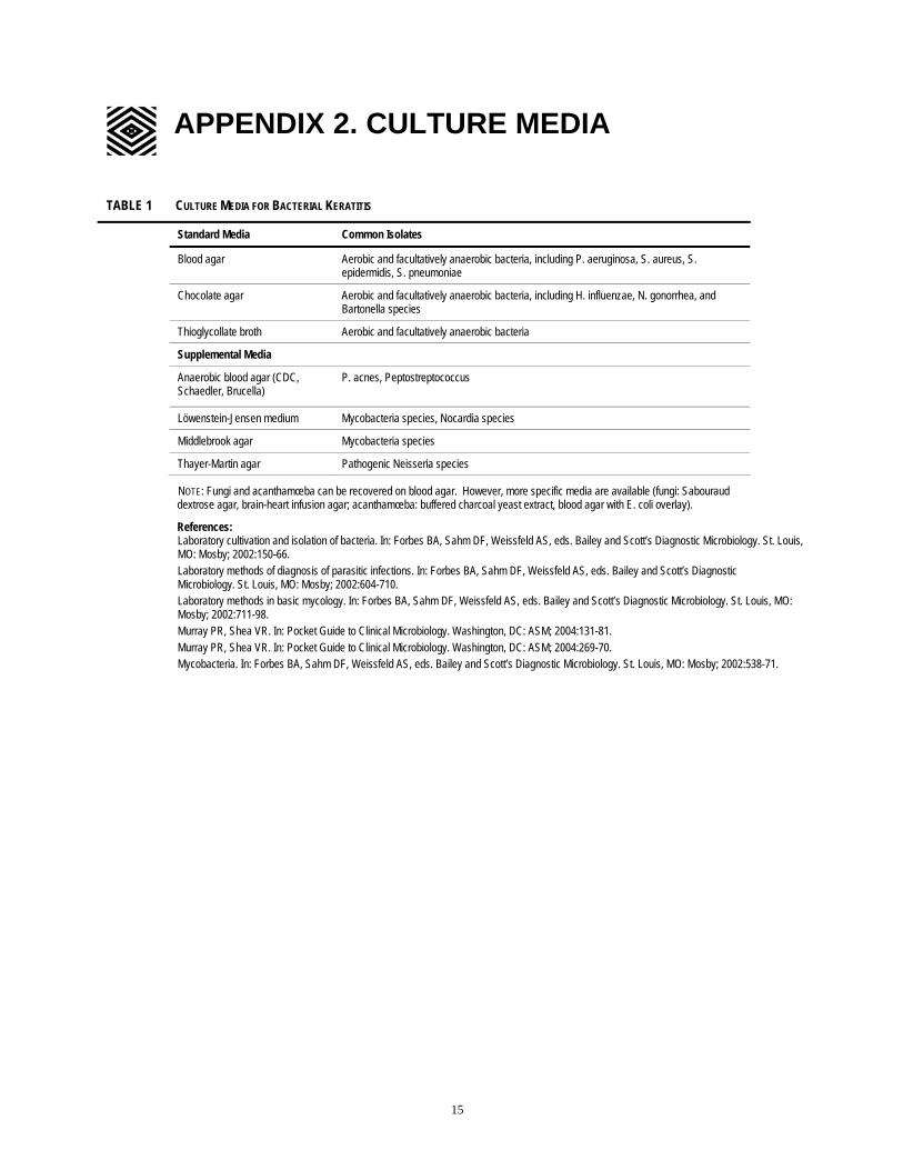
APPENDIX 2. CULTURE MEDIA
TABLE 1 CULTURE MEDIA FOR BACTERIAL KERATITIS
Standard Media Common Isolates
Blood agar Aerobic and facultatively anaerobic bacteria, including P. aeruginosa, S. aureus, S.epidermidis, S. pneumoniae
Chocolate agar Aerobic and facultatively anaerobic bacteria, including H. influenzae, N. gonorrhea, and Bartonella species
Thioglycollate broth Aerobic and facultatively anaerobic bacteria
Supplemental Media
Anaerobic blood agar (CDC, Schaedler, Brucella)
P. acnes, Peptostreptococcus
Löwenstein-Jensen medium Mycobacteria species, Nocardia species
Middlebrook agar Mycobacteria species
Thayer-Martin agar Pathogenic Neisseria species
NOTE: Fungi and acanthamœba can be recovered on blood agar. However, more specific media are available (fungi: Sabouraud dextrose agar, brain-heart infusion agar; acanthamœba: buffered charcoal yeast extract, blood agar with E. coli overlay).
References: Laboratory cultivation and isolation of bacteria. In: Forbes BA, Sahm DF, Weissfeld AS, eds. Bailey and Scott’s Diagnostic Microbiology. St. Louis, MO: Mosby; 2002:150-66. Laboratory methods of diagnosis of parasitic infections. In: Forbes BA, Sahm DF, Weissfeld AS, eds. Bailey and Scott’s Diagnostic Microbiology. St. Louis, MO: Mosby; 2002:604-710. Laboratory methods in basic mycology. In: Forbes BA, Sahm DF, Weissfeld AS, eds. Bailey and Scott’s Diagnostic Microbiology. St. Louis, MO: Mosby; 2002:711-98. Murray PR, Shea VR. In: Pocket Guide to Clinical Microbiology. Washington, DC: ASM; 2004:131-81. Murray PR, Shea VR. In: Pocket Guide to Clinical Microbiology. Washington, DC: ASM; 2004:269-70. Mycobacteria. In: Forbes BA, Sahm DF, Weissfeld AS, eds. Bailey and Scott’s Diagnostic Microbiology. St. Louis, MO: Mosby; 2002:538-71.
15

=
APPENDIX 3. DIAGNOSTIC STAINS
==TABLE 1 DIAGNOSTIC STAINS FOR BACTERIAL KERATITIS
Type of Stain Organisms Visualized Comments
Gram stain* Best for bacteria; can also visualize fungi,† amœbae
Distinguishes gram-positive from gram-negative organisms; widely available; rapid (5 minutes)
Giemsa stain* Bacteria, fungi,† chlamydia Acanthamœba
Basis for Aema-color and Diff-Quik tests; widely available; rapid (2 minutes)
Acid fast Mycobacteria, Nocardia Widely available; takes 1 hour; reliable stain for Mycobacteria
Acridine orange* Bacteria, fungi,† acanthamœba‡ Requires use of epifluorescent microscope; rapid (2 minutes)
Calcoflur white Fungi,† acanthamœba‡ Requires use of epifluorescent microscope; rapid (2 minutes)
* Most useful stains for screening purposes. † PAS (periodic acid-Schiff) and GMS (Gomori methenamine silver) also can be used to identify fungi. ‡ H&E (hematoxylin and eosin) and PAS also can be used to identify acanthamœba.
References: Infections of the eyes, ears, and sinuses. In: Forbes BA, Sahm DF, Weissfeld AS, eds. Bailey and Scott’s Diagnostic Microbiology. St. Louis, MO: Mosby; 2002:917-26. Laboratory methods in basic mycology . In: Forbes BA, Sahm DF, Weissfeld AS, eds. Bailey and Scott’s Diagnostic Microbiology. St. Louis, MO: Mosby; 2002:711-98. Laboratory methods of diagnosis of parasitic infections. In: Forbes BA, Sahm DF, Weissfeld AS, eds. Bailey and Scott’s Diagnostic Microbiology. St. Louis, MO: Mosby; 2002:604-710.
Murray PR, Shea VR. In: Pocket Guide to Clinical Microbiology. Washington, DC: ASM; 2004:131-81.
=
16=

==
RELATED ACADEMY MATERIALS
Basic and Clinical Science Course External Disease and Cornea (Section 8, 2005-2006) Eye Fact Sheets Herpes Simplex Eye Disease (2004) Herpes Zoster (2004) Information Statement Extended Wear of Contact Lenses (2004) LEO Clinical Update Course CD-ROM Cornea & External Disease (2003) Patient Education Brochure Contact Lenses (2005) Preferred Practice Patterns Comprehensive Adult Medical Eye Evaluation (2005) Pediatric Eye Evaluations (2002)
To order any of these materials, please call the Academy’s Customer Service number, 415.561.8540 or visit www.aao.org/store.
17

=
=
REFERENCES
1. Pepose JS, Wilhelmus KR. Divergent approaches to the management of corneal ulcers. Am J Ophthalmol 1992;114:630-2.
2. Wilhelmus KR. Review of clinical experience with microbial keratitis associated with contact lenses. Clao J 1987;13:211-4.
3. Forster RK. Conrad Berens Lecture. The management of infectious keratitis as we approach the 21st century. Clao J 1998;24:175-80.
4. Goldstein MH, Kowalski RP, Gordon YJ. Emerging fluoroquinolone resistance in bacterial keratitis: a 5-year review. Ophthalmology 1999;106:1313-8.
5. Tuft SJ, Matheson M. In vitro antibiotic resistance in bacterial keratitis in London. Br J Ophthalmol 2000;84:687-91.
6. Galentine PG, Cohen EJ, Laibson PR, et al. Corneal ulcers associated with contact lens wear. Arch Ophthalmol 1984;102:891-4.
7. Alfonso E, Mandelbaum S, Fox MJ, Forster RK. Ulcerative keratitis associated with contact lens wear. Am J Ophthalmol 1986;101:429-33.
8. Donnenfeld ED, Cohen EJ, Arentsen JJ, et al. Changing trends in contact lens associated corneal ulcers: an overview of 116 cases. CLAO J 1986;12:145-9.
9. Cohen EJ, Laibson PR, Arentsen JJ, Clemons CS. Corneal ulcers associated with cosmetic extended wear soft contact lenses. Ophthalmology 1987;94:109-14.
10. Alexandrakis G, Alfonso EC, Miller D. Shifting trends in bacterial keratitis in south Florida and emerging resistance to fluoroquinolones. Ophthalmology 2000;107:1497-502.
11. Burns RP, Rhodes DH, Jr. Pseudomonas eye infection as a cause of death in premature infants. Arch Ophthalmol 1961;65:517-25.
12. Poggio EC, Glynn RJ, Schein OD, et al. The incidence of ulcerative keratitis among users of daily-wear and extended-wear soft contact lenses. N Engl J Med 1989;321:779-83.
13. Dart JK. Predisposing factors in microbial keratitis: the significance of contact lens wear. Br J Ophthalmol 1988;72:926-30.
14. Schein OD, Glynn RJ, Poggio EC, et al. The relative risk of ulcerative keratitis among users of daily-wear and extended-wear soft contact lenses. A case-control study. Microbial Keratitis Study Group. N Engl J Med 1989;321:773-8.
15. Matthews TD, Frazer DG, Minassian DC, et al. Risks of keratitis and patterns of use with disposable contact lenses. Arch Ophthalmol 1992;110:1559-62.
16. Buehler PO, Schein OD, Stamler JF, et al. The increased risk of ulcerative keratitis among disposable soft contact lens users. Arch Ophthalmol 1992;110:1555-8.
17. Schein OD, Buehler PO, Stamler JF, et al. The impact of overnight wear on the risk of contact lens-associated ulcerative keratitis. Arch Ophthalmol 1994;112:186-90.
18. Najjar DM, Aktan SG, Rapuano CJ, et al. Contact lens-related corneal ulcers in compliant patients. Am J Ophthalmol 2004;137:170-2.
19. Siganos CS, Solomon A, Frucht-Pery J. Microbial findings in suture erosion after penetrating keratoplasty. Ophthalmology 1997;104:513-6.
20. Schein OD, Poggio EC. Ulcerative keratitis in contact lens wearers. Incidence and risk factors. Cornea 1990;9 Suppl 1:S55-8; discussion S62-3.
18=

=
21. American Academy of Ophthalmology. Policy Statement: Protective Eyewear for Young Athletes, 2003; (Joint statement with the American Academy of Pediatrics and the American Academy of Ophthalmology). Available at: www.aao.org/member/policy/index.cfm.
22. American Academy of Ophthalmology. Comprehensive Adult Medical Eye Evaluation, Preferred Practice Pattern. San Francisco: American Academy of Ophthalmology, 2005. Available at: www.aao.org/ppp.
23. American Academy of Ophthalmology. Pediatric Eye Evaluations, Preferred Practice Pattern. San Francisco: American Academy of Ophthalmology, 2002. Available at: www.aao.org/ppp.
24. Stein RM, Clinch TE, Cohen EJ, et al. Infected vs sterile corneal infiltrates in contact lens wearers. Am J Ophthalmol 1988;105:632-6.
25. McLeod SD, Kolahdouz-Isfahani A, Rostamian K, et al. The role of smears, cultures, and antibiotic sensitivity testing in the management of suspected infectious keratitis. Ophthalmology 1996;103:23-8.
26. Rodman RC, Spisak S, Sugar A, et al. The utility of culturing corneal ulcers in a tertiary referral center versus a general ophthalmology clinic. Ophthalmology 1997;104:1897-901.
27. Wilhelmus K, Liesegang TJ, Osato MS, Jones DB. Laboratory diagnosis of ocular infections. Washington DC: American Society for Microbiology, 1994; Cumitech Series #13A.
28. McDonnell PJ, Nobe J, Gauderman WJ, et al. Community care of corneal ulcers. Am J Ophthalmol 1992;114:531-8.
29. Marangon FB, Miller D, Alfonso EC. Impact of prior therapy on the recovery and frequency of corneal pathogens. Cornea 2004;23:158-64.
30. Rudolph T, Welinder-Olsson C, Lind-Brandberg L, Stenevi U. 16S rDNA PCR analysis of infectious keratitis: a case series. Acta Ophthalmol Scand 2004;82:463-7.
31. Waxman E, Chechelnitsky M, Mannis MJ, Schwab IR. Single culture media in infectious keratitis. Cornea 1999;18:257-61.
32. Kaye SB, Rao PG, Smith G, et al. Simplifying collection of corneal specimens in cases of suspected bacterial keratitis. J Clin Microbiol 2003;41:3192-7.
33. Newton C, Moore MB, Kaufman HE. Corneal biopsy in chronic keratitis. Arch Ophthalmol 1987;105:577-8.
34. Alexandrakis G, Haimovici R, Miller D, Alfonso EC. Corneal biopsy in the management of progressive microbial keratitis. Am J Ophthalmol 2000;129:571-6.
35. Hwang DG. Lamellar flap corneal biopsy. Ophthalmic Surg 1993;24:512-5.
36. Phinney RB, Schwartz SD, Lee DA, Mondino BJ. Collagen-shield delivery of gentamicin and vancomycin. Arch Ophthalmol 1988;106:1599-604.
37. Mondino BJ. Collagen shields. Am J Ophthalmol 1991;112:587-90.
38. Lee BL, Matoba AY, Osato MS, Robinson NM. The solubility of antibiotic and corticosteroid combinations. Am J Ophthalmol 1992;114:212-5.
39. Hyndiuk RA, Eiferman RA, Caldwell DR, et al. Comparison of ciprofloxacin ophthalmic solution 0.3% to fortified tobramycin-cefazolin in treating bacterial corneal ulcers. Ciprofloxacin Bacterial Keratitis Study Group. Ophthalmology 1996;103:1854-62; discussion 62-3.
40. Ofloxacin monotherapy for the primary treatment of microbial keratitis: a double-masked, randomized, controlled trial with conventional dual therapy. The Ofloxacin Study Group. Ophthalmology 1997;104:1902-9.
19

=
41. O'Brien TP, Maguire MG, Fink NE, et al. Efficacy of ofloxacin vs cefazolin and tobramycin in the therapy for bacterial keratitis. Report from the Bacterial Keratitis Study Research Group. Arch Ophthalmol 1995;113:1257-65.
42. Cokingtin CD, Hyndiuk RA. Insights from experimental data on ciprofloxacin in the treatment of bacterial keratitis and ocular infections. Am J Ophthalmol 1991;112:25S-8S.
43. Knauf HP, Silvany R, Southern PM, Jr., et al. Susceptibility of corneal and conjunctival pathogens to ciprofloxacin. Cornea 1996;15:66-71.
44. Cutarelli PE, Lass JH, Lazarus HM, et al. Topical fluoroquinolones: antimicrobial activity and in vitro corneal epithelial toxicity. Curr Eye Res 1991;10:557-63.
45. Osato MS, Jensen HG, Trousdale MD, et al. The comparative in vitro activity of ofloxacin and selected ophthalmic antimicrobial agents against ocular bacterial isolates. Am J Ophthalmol 1989;108:380-6.
46. Wilhelmus KR, Abshire RL, Schlech BA. Influence of fluoroquinolone susceptibility on the therapeutic response of fluoroquinolone-treated bacterial keratitis. Arch Ophthalmol 2003;121:1229-33.
47. Khokhar S, Sindhu N, Mirdha BR. Comparison of topical 0.3% ofloxacin to fortified tobramycin-cefazolin in the therapy of bacterial keratitis. Infection 2000;28:149-52.
48. Garg P, Sharma S, Rao GN. Ciprofloxacin-resistant Pseudomonas keratitis. Ophthalmology 1999;106:1319-23.
49. Kowalski RP, Dhaliwal DK, Karenchak LM, et al. Gatifloxacin and moxifloxacin: an in vitro susceptibility comparison to levofloxacin, ciprofloxacin, and ofloxacin using bacterial keratitis isolates. Am J Ophthalmol 2003;136:500-5.
50. Gangopadhyay N, Daniell M, Weih L, Taylor HR. Fluoroquinolone and fortified antibiotics for treating bacterial corneal ulcers. Br J Ophthalmol 2000;84:378-84.
51. Mallari PL, McCarty DJ, Daniell M, Taylor H. Increased incidence of corneal perforation after topical fluoroquinolone treatment for microbial keratitis. Am J Ophthalmol 2001;131:131-3.
52. Bower KS, Kowalski RP, Gordon YJ. Fluoroquinolones in the treatment of bacterial keratitis. Am J Ophthalmol 1996;121:712-5.
53. John T, Velotta E. Nontuberculous (atypical) mycobacterial keratitis after LASIK: current status and clinical implications. Cornea 2005;24:245-55.
54. Sexually transmitted diseases treatment guidelines 2002. Centers for Disease Control and Prevention. MMWR Recomm Rep 2002;51:1-78.
55. American Academy of Ophthalmology. Basic Clinical and Science Course, 2005-2006; Section 8, External Disease and Cornea, pg 172.
56. Wilhelmus KR. Indecision about corticosteroids for bacterial keratitis: an evidence-based update. Ophthalmology 2002;109:835-42; quiz 43.
57. Leibowitz HM, Kupferman A. Topically administered corticosteroids: effect on antibiotic-treated bacterial keratitis. Arch Ophthalmol 1980;98:1287-90.
58. Larkin DF, Kilvington S, Easty DL. Contamination of contact lens storage cases by Acanthamoeba and bacteria. Br J Ophthalmol 1990;74:133-5.
59. Stern GA. Contact lens associated bacterial keratitis: past, present, and future. CLAO J 1998;24:52-6.
60. American Academy of Ophthalmology. Vision Rehabilitation for Adults, Preferred Practice Pattern. San Francisco: American Academy of Ophthalmology, 2001. Available at: www.aao.org/ppp.
20=

=
P.O. Box 7424 San Francisco, California 94120-7424 415.561.8500
Bacterial Keratitis
2005
=