bacterial-fungal interactions: a...
TRANSCRIPT

!!
President: Pascale Frey-Klett UMR1136 Interactions arbres-microorganismes,!Centre INRA de Nancy, 54280 Champenoux, France
Vice!President: Deborah Hogan Department of Microbiology and Immunology, Dartmouth Medical School, Hanover, NH 03755, USA
Roscoff (Brittany), France December 7-11, 2013 - 7 -11 décembre 2013
BACTERIAL-FUNGAL INTERACTIONS: A FEDERATIVE FIELD FOR FUNDAMENTAL AND APPLIED MICROBIOLOGY
INTERACTIONS BACTERIES-CHAMPIGNONS : UN DOMAINE DE
RECHERCHE FEDERATEUR POUR LA MICROBIOLOGIE FONDAMENTALE ET APPLIQUEE

Rationale
o Classical separation of microbiological researches between bacteriologists and mycologists
o In many environments bacteria and fungi co-exist and interact
o Multiple practical relevancies of these interactions

A French network: 10 research groups
2005
2008
2007
A review promoting inter-field interactions
2011
An European initiative: 40 research groups
2002
2005
1st International Conference on Bacterial-Fungal Interactions
2013
there is often substantial linkage disequilibriumbetween adjacent blocks (data not shown), al-lowing fewer markers to be used without loss ofpower. It will likely be productive to performinitial haplotype mapping in populations whosehistory contains one or more bottlenecks, be-cause longer-range LD may make initial local-ization more efficient and favorable. Converse-ly, populations with shorter-range LD and great-er haplotype diversity may offer advantages forfine mapping. In suggesting that block bound-aries and common haplotypes are largely sharedacross populations, our data suggest that manycommon disease alleles can be studied—andlikely will be broadly relevant—across humanpopulations. In the future, comprehensive anal-ysis of human haplotype structure promises in-sights into the origin of human populations, theforces that shape genetic diversity, and the pop-ulation basis of disease.
References and Notes1. E. S. Lander, Science 274, 536 (1996).2. F. S. Collins, M. S. Guyer, A. Chakravarti, Science 278,1580 (1997).
3. N. Risch, K. Merikangas, Science 273, 1516 (1996).4. R. Sachidanandam et al., Nature 409, 928 (2001).5. J. C. Venter et al., Science 291, 1304 (2001).6. L. Kruglyak, D. A. Nickerson, Nature Genet. 27, 234(2001).
7. E. G. Puffenberger et al., Cell 79, 1257 (1994).8. B. Kerem et al., Science 245, 1073 (1989).9. J. Hastbacka et al., Nature Genet. 2, 204 (1992).10. J. D. Rioux et al., Nature Genet. 29, 223 (2001).11. J. P. Hugot et al., Nature 411, 599 (2001).12. Y. Ogura et al., Nature 411, 603 (2001).13. J. K. Pritchard, M. Przeworski, Am. J. Hum. Genet. 69,1 (2001).
14. L. B. Jorde, Genome Res. 10, 1435 (2000).15. M. Boehnke, Nature Genet. 25, 246 (2000).16. M. J. Daly, J. D. Rioux, S. F. Schaffner, T. J. Hudson, E. S.Lander, Nature Genet. 29, 229 (2001).
17. A. J. Jeffreys, L. Kauppi, R. Neumann, Nature Genet.29, 217 (2001).
18. N. Patil et al., Science 294, 1719 (2001).19. G. C. Johnson et al., Nature Genet. 29, 233 (2001).20. Materials and methods are available as supportingmaterial on Science Online.
21. K. Tang et al., Proc. Natl. Acad. Sci. U.S.A. 96, 10016(1999).
22. Though 82% of assays were successful in at least onepopulation, genotyping success rates in each popula-tion range from 72 to 79%. The difference betweenthese numbers is due to a low rate of laboratoryfailure in each attempt.
23. L. L. Cavalli-Sforza, P. Menozzi, A. Piazza, The Historyand Geography of Human Genes (Princeton Universi-ty Press, Princeton, NJ, 1994).
24. R. C. Lewontin, Genetics 49, 49 (1964).25. An upper confidence bound of 0.98 was used insteadof 1.0 because even a single observation of a fourthhaplotype makes it mathematically impossible for D!to be consistent with a value of 1.0, though theconfidence interval could be arbitrarily close to 1.0.
26. D. E. Reich et al., Nature 411, 199 (2001).27. As a further test of the model, we simulated theproportion of pairs at a fixed distance (5 kb) thatshould show evidence of crossing block boundaries(that is, show strong evidence of historical recom-bination). The model predicts these proportions tobe 47% (Yoruban and African-American samples)and 27% (European and Asian samples). In theempirical data, we observe 42 and 23%, similar tothese predictions.
28. A low rate of genotyping error is critical to obtainingan accurate measure of haplotype diversity and theproportion in common haplotypes. Even a modest (1to 2%) genotyping error will create a substantial
number of false rare haplotypes; for example, with a10-marker haplotype and a 2% error rate, 18% ofchromosomes will contain at least one error and,thus, not match the few common haplotypes.
29. Within blocks, the common haplotypes showed littleevidence for historical recombination. For example,we performed the four gamete tests using SNPsdrawn only from haplotypes with frequency 5% orhigher in each block. One or more violations of thefour gamete tests were observed in only 5% of theblocks.
30. To maximize power, these comparisons were madeonly for SNP pairs spaced 5 to 10 kb apart. At shorterdistances, nearly all SNP pairs are in a single block; atgreater distances, most SNP pairs are in differentblocks.
31. Blocks and haplotypes were identified separately ineach population sample, and the results were com-pared for those blocks that were physically overlap-ping in all three samples.
32. R. L. Cann, W. M. Brown, A. C. Wilson, Genetics 106,479 (1984).
33. C. B. Stringer, P. Andrews, Science 239, 1263 (1988).34. D. E. Reich, D. B. Goldstein, Proc. Natl. Acad. Sci.U.S.A. 95, 8119 (1998).
35. M. Ingman, H. Kaessmann, S. Paabo, U. Gyllensten,Nature 408, 708 (2000).
36. S. A. Tishkoff et al., Science 271, 1380 (1996).37. S. A. Tishkoff et al., Am. J. Hum. Genet. 67, 901(2000).
38. We examined SNP pairs that were in different blocksin the Yoruban samples but in a single block in theEuropean sample. Such pairs had higher D! values inthe Yoruban sample (D! " 0.46) than pairs found indifferent blocks in both population samples (D! "0.28). The average frequency of all haplotypes in the
Yoruban population was 0.21, whereas those thatwere found only in the Yoruban sample (but not inthe European and Asian samples) had a mean fre-quency of 0.16.
39. A. G. Clark et al., Am. J. Hum. Genet. 63, 595 (1998).40. A. R. Templeton et al., Am. J. Hum. Genet. 66, 69 (2000).41. S. M. Fullerton et al., Am. J. Hum. Genet. 67, 881(2000).
42. The small fraction of SNPs that show r2 values #0.5could be attributable to a range of causes: branches ofthe gene tree not defined with the number of markersused, gene conversion events, or recurrent mutations.We note that errors in genotyping or map positiondecrease (but cannot increase) the value of r2.
43. This work was supported by a grant to D.A. from TheSNP Consortium. We thank members of the Programin Medical and Population Genetics at the White-head/MIT Center for Genome Research for helpfuldiscussion, particularly J. Hirschhorn, D. Reich, and N.Patterson. The authors also thank the following col-leagues for sharing data and analyses before publica-tion: D. Bentley (Sanger Institute); L. Cardon (Oxford);and D. Cutler, M. Zwick, and A. Chakravarti (JohnsHopkins). D.A. is a Charles E. Culpeper Scholar of theRockefeller Brothers Fund and a Burroughs WelcomeFund Clinical Scholar in Translational Research.
Supporting Online Materialwww.sciencemag.org/cgi/content/full/1069424/DC1Materials and MethodsFigs. S1 to S3Table S1
28 December 2001; accepted 13 May 2002Published online 23 May 2002;10.1126/science.1069424Include this information when citing this paper.
Pseudomonas-CandidaInteractions: An Ecological Role
for Virulence FactorsDeborah A. Hogan and Roberto Kolter*
Bacterial-fungal interactions have great environmental, medical, and economicimportance, yet few have been well characterized at the molecular level. Here,we describe a pathogenic interaction between Pseudomonas aeruginosa andCandida albicans, two opportunistic pathogens. P. aeruginosa forms a densebiofilm on C. albicans filaments and kills the fungus. In contrast, P. aeruginosaneither binds to nor kills yeast-form C. albicans. Several P. aeruginosa virulencefactors that are important in disease are involved in the killing of C. albicansfilaments. We propose that many virulence factors studied in the context ofhuman infection may also have a role in bacterial-fungal interactions.
Interactions between prokaryotes and eu-karyotes are ubiquitous. Although the patho-genic and symbiotic relationships bacteriahave with plants and animals have garneredthe most attention, the prokaryote-eukaryoteencounters that occur among microbes arelikely far more common. Many of the viru-lence factors that we study in the context ofhuman disease may also have an ecologicalrole within microbial communities.
Bacteria and unicellular eukaryotes, such asyeasts and filamentous fungi, are found togetherin a myriad of environments and exhibit bothsynergistic and antagonistic interactions (1, 2).Here, we describe a pathogenic relationship be-tween a fungus, Candida albicans, and a bacte-rium, Pseudomonas aeruginosa, that involvesgenes important for bacterial virulence in mam-mals. P. aeruginosa is prevalent in soils and isoften found on the skin and mucosa of healthyindividuals (3). In compromised hosts, however,P. aeruginosa uses an arsenal of virulence fac-tors to cause serious infections associated withburns, catheters, and implants. C. albicans isalso a benign member of the skin and mucosalflora. When host defenses falter, however, C.
Department of Microbiology and Molecular Genetics,Harvard Medical School, 200 Longwood Avenue, Bos-ton, MA 02115, USA.
*To whom correspondence should be addressed. E-mail: [email protected]
R E P O R T S
www.sciencemag.org SCIENCE VOL 296 21 JUNE 2002 2229
regulation of biofilm development on inert sur-faces. P. aeruginosa PA14 mutants lackingpole-localized type IV pili (pilB and pilC) alsodo not form mature biofilms on abiotic sub-strates (17, 18), yet these mutants still formedrobust biofilms on filamentous C. albicans (Fig.3C). Because some P. aeruginosa strains usetype IV pili to attach to epithelial cells (17), weassayed their initial attachment to C. albicansfilaments and found a decreased rate of adher-ence (Table 1). These data indicate that pilisomehow participate in the initial attachment toC. albicans filaments, but they are not requiredfor biofilm formation at later time points (com-pare Fig. 1D with Fig. 3C). The rpoN mutantforms extremely poor biofilms on C. albicansfilaments (Fig. 3D), likely owing to multiplefactors, including the lack of a flagellum and adecreased growth rate in minimal media (19).With the exception of the rpoN mutant, all P.aeruginosa mutants had the same planktonicgrowth rate as the wild type (20).
Pseudomonas aeruginosa mutants were as-sayed for their virulence toward C. albicans.First, we analyzed P. aeruginosa mutants lack-ing type IV pili and the polar flagellum. Al-though the type IV pili mutants made robustbiofilms surrounding the filament (Fig. 3C),they did not kill C. albicans tup1 until after 48hours (Fig. 3E). The mechanisms by which typeIV pili influence fungal killing remains un-known, but may involve pilus retraction to bringthe bacterium in close contact with the fungal
cell or the use of pili as sensors that signalattachment to the fungal surface (17, 21). Theflagellar mutant kills C. albicans filaments withkinetics similar to those of the wild type (Fig.3E), even though it forms slightly smaller bio-films on fungal filaments (Fig. 3A) (22). Thesecond class of P. aeruginosa mutants was de-fective in the production of broad-spectrum se-creted factors that contribute to virulence towarddiverse organisms including mammals, plants,and insects (23, 24). Several of these mutantswere also attenuated for their virulence towardC. albicans filaments. P. aeruginosa mutantsunable to produce the hemolytic phospholipaseC (because of disruption of either plcS, thestructural gene, or plcR, which is required forphospholipase C secretion) were significantlyattenuated in their ability to kill C. albicansfilaments (Fig. 3F) (25, 26). The same was truefor the phnAB mutant, which is unable to syn-thesize phenazine antibiotics (Fig. 3F) (27). AP. aeruginosa mutant lacking exotoxin A, a typeII–secreted toxin that targets translation in eu-karyotic cells, was not significantly affected inits virulence toward C. albicans (Fig. 3F) (23).Last, inactivation of several virulence-factorregulators including GacA, LasR, RhlR, andRpoN, resulted in either delayed or attenuatedvirulence toward C. albicans filaments (Fig.3G). The P. aeruginosa rpoN mutant, whichwas not capable of biofilm formation on fungalfilaments, was also unable to kill C. albicans. P.aeruginosa mutants defective in gacA, lasR, and
rpoN are also significantly attenuated in othervirulence models (19, 23, 28). The less virulentphenotypes of pleiotropic regulatory mutants(gacA, lasR, rhlR, and rpoN) likely resultedfrom the decreased expression of multiplegenes. In addition, some genes, such as thoseinvolved in phenazine production, are controlledby multiple regulators (19, 27, 29). However,the attenuated virulence of multiple mutants de-fective in single traits (plcR, plcS, pilB, pilC, orphnAB) more clearly shows that multiple mech-anisms act in concert to kill C. albicans fila-ments. This explains the eventual decrease in C.albicans CFU/ml in almost all cultures after 60hours (Fig. 3, E to G).
Our data suggest a link between biofilmformation and the activity of some eukaryotic-specific virulence factors toward fungal cells.Both clinical and environmental isolates of P.aeruginosa produce a similar spectrum of viru-lence factors including type IV pili, phospho-lipase C, and phenazines (30, 31). Thus, wespeculate that antagonism between bacteria andmicroscopic fungi has contributed to the evolu-tion and maintenance of many pathogenesis-related genes. Furthermore, we propose that theinteractions between P. aeruginosa and C. albi-cans reflect the relationships of bacterial andfungal species that coexist in other environ-ments. A deeper understanding of bacterial-fun-gal interactions may provide a new perspectiveon the role of known virulence determinants andmay lead to the discovery of new factors in-volved in pathogenicity in multiple hosts.
References and Notes1. J. Garbaye, New Phytol. 128, 197 (1994).2. I. Chet, J. Inbar, Appl. Biochem. Biotechnol. 48, 37(1994).
3. N. Palleroni, in The Prokaryotes, M. Dworkin, Ed.(Springer-Verlag, New York, ed. 1, published online,2000). Available at http://link.springer-ny.com/link/service/books/10125/.
4. F. C. Odds, Candida and Candidosis (Bailliere Tindall,London, ed. 2, 1988).
5. H. Lo et al., Cell 90, 939 (1997).
Table 1. Initial attachment of P. aeruginosa to C.albicans filaments. See Science Online for experi-mental details (12). P. aeruginosa wild type (WT ),pilB, and pilC were taken from early stationary-phase cultures (OD600 of 1.2). WT-EP cells weretaken from exponential phase cultures (OD600 of0.4). The percentage of filaments colonized wasdetermined by microscopic examination of 150filaments per sample after a 20-min incubation.Each value represents the average of triplicatesamples. In the last column, number representsthe average number of bacteria attached to 300filaments.
P. aeruginosastrain
Percentfilamentscolonized
Bacterial cellsper filament
WT 80.9! 3.1 4.4WT-EP 29.1! 0.4 1.2pilB 39.1! 8.2 1.6pilC 37.8! 8.2 1.5
Fig. 3. (A to D) Representative phase-contrast images show P. aeruginosa mutant biofilms on C.albicans tup1 after 72 hours of coincubation. (E to G) C. albicans tup1 survival in the presence ofisogenic P. aeruginosamutants. C. albicans tup1 viability (measured as CFU/ml) was followed in thepresence of wild-type cells (black) and conditioned medium without cells (black dashed). Sameresults plotted in (E), (F), and (G). (E) C. albicans viability was monitored in the presence of mutantslacking type IV pili, pilB (blue), pilC (yellow), or lacking the polar flagellum, flgK (red). (F) C. albicansviability was monitored in the presence of mutants defective in the production of virulence factorsincluding phospholipase, plcS (yellow) and plcR (blue); phenazines, phnAB (green); and exotoxin A,toxA (red). (G) C. albicans viability was measured in cultures with P. aeruginosa mutants defectivein regulators that control the production of virulence factors including rpoN (green), gacA (blue),rhlR (yellow), and lasR (red). The values plotted represent the averages of four replicate cultures,and the experiment was performed multiple times with similar results. At 42 hours, the differencesbetween the wild type and all mutants except flgK and toxA were statistically significant at P "0.05 as determined by a t test analysis.
R E P O R T S
www.sciencemag.org SCIENCE VOL 296 21 JUNE 2002 2231
A reference paper from the USA
2002
Mycorrhiza Helper Bacteria a new
dimension to the mycorrhizal symbiosis
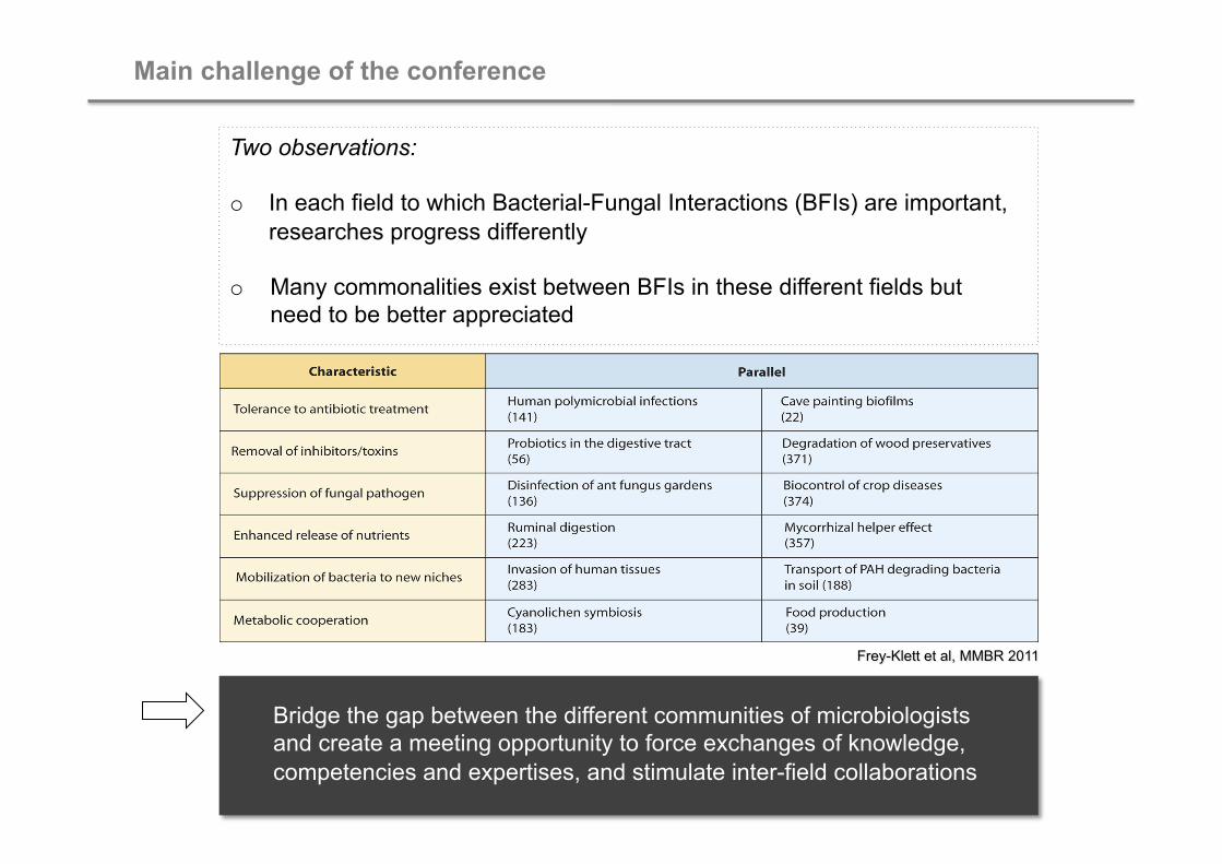
Main challenge of the conference
Two observations:
o In each field to which Bacterial-Fungal Interactions (BFIs) are important, researches progress differently
o Many commonalities exist between BFIs in these different fields but need to be better appreciated
Bridge the gap between the different communities of microbiologists and create a meeting opportunity to force exchanges of knowledge, competencies and expertises, and stimulate inter-field collaborations

26 1
1 3
6 8 13
5 2 3 4
2 2
1
1
Conference participants: geographical origins
77 participants, including 17 PhD students
15 nationalities : 26 participants from France, 43 participants from other European countries, 8 non-European participants
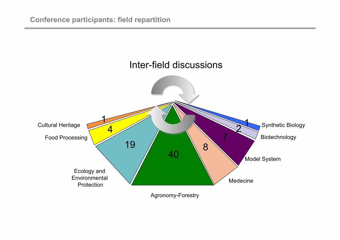
Agronomy-Forestry
Ecology and Environmental
Protection Medecine
Model System
Biotechnology
Synthetic Biology
Food Processing
Cultural Heritage
Conference participants: field repartition
40 19 8
4 1
7 2 1
Inter-field discussions