bacterial diversity in human subgingival plaquejb.asm.org/content/183/12/3770.full.pdf ·...
TRANSCRIPT
JOURNAL OF BACTERIOLOGY,0021-9193/01/$04.0010 DOI: 10.1128/JB.183.12.3770–3783.2001
June 2001, p. 3770–3783 Vol. 183, No. 12
Copyright © 2001, American Society for Microbiology. All Rights Reserved.
Bacterial Diversity in Human Subgingival PlaqueBRUCE J. PASTER,1,2* SUSAN K. BOCHES,1 JAMIE L. GALVIN,1 REBECCA E. ERICSON,1 CAROL N. LAU,1
VALERIE A. LEVANOS,1 ASHISH SAHASRABUDHE,1 AND FLOYD E. DEWHIRST1,2
Department of Molecular Genetics, The Forsyth Institute,1 and Department of Oral Biology,Harvard School of Dental Medicine,2 Boston, Massachusetts
Received 8 November 2000/Accepted 28 March 2001
The purpose of this study was to determine the bacterial diversity in the human subgingival plaque by usingculture-independent molecular methods as part of an ongoing effort to obtain full 16S rRNA sequences for allcultivable and not-yet-cultivated species of human oral bacteria. Subgingival plaque was analyzed from healthysubjects and subjects with refractory periodontitis, adult periodontitis, human immunodeficiency virus peri-odontitis, and acute necrotizing ulcerative gingivitis. 16S ribosomal DNA (rDNA) bacterial genes from DNAisolated from subgingival plaque samples were PCR amplified with all-bacterial or selective primers andcloned into Escherichia coli. The sequences of cloned 16S rDNA inserts were used to determine species identityor closest relatives by comparison with sequences of known species. A total of 2,522 clones were analyzed.Nearly complete sequences of approximately 1,500 bases were obtained for putative new species. About 60% ofthe clones fell into 132 known species, 70 of which were identified from multiple subjects. About 40% of theclones were novel phylotypes. Of the 215 novel phylotypes, 75 were identified from multiple subjects. Knownputative periodontal pathogens such as Porphyromonas gingivalis, Bacteroides forsythus, and Treponema denticolawere identified from multiple subjects, but typically as a minor component of the plaque as seen in cultivablestudies. Several phylotypes fell into two recently described phyla previously associated with extreme naturalenvironments, for which there are no cultivable species. A number of species or phylotypes were found only insubjects with disease, and a few were found only in healthy subjects. The organisms identified only fromdiseased sites deserve further study as potential pathogens. Based on the sequence data in this study, thepredominant subgingival microbial community consisted of 347 species or phylotypes that fall into 9 bacterialphyla. Based on the 347 species seen in our sample of 2,522 clones, we estimate that there are 68 additionalunseen species, for a total estimate of 415 species in the subgingival plaque. When organisms found on otheroral surfaces such as the cheek, tongue, and teeth are added to this number, the best estimate of the totalspecies diversity in the oral cavity is approximately 500 species, as previously proposed.
It has been previously estimated that about 500 species ofbacteria inhabit the human oral cavity (32, 43, 52). While themajority of these organisms are commensals, a subset of themare likely to be opportunistic pathogens that can cause systemicdisease. For example, oral bacteria have been implicated inbacterial endocarditis (3), aspiration pneumonia (42), osteo-myelitis in children (10), preterm low birth weight (33), andcoronary heart disease and cerebral infarction (or stroke) (2,53). Consequently, it is important to know what microorgan-isms are present in the oral cavity for the diagnosis and rationaltreatment of systemic as well as oral diseases.
Studies with molecular techniques have shown that the bac-terial diversity in most environments is severely underesti-mated in surveys with cultivation-based techniques (1, 19). Inmany natural environments, less than 1% of the organisms arecultivable (34). Because of the significant effort extended tocultivate oral bacteria, it is thought that about 50% of oralbacteria have been cultivated. However, any understanding ofthe oral environment requires knowledge of the entire bacte-rial community. There is no reason to expect that fewer patho-gens exist among the uncultivated segment of the communitythan among the cultivated segment. Highly host-adapted or-ganisms such as Treponema pallidum and Mycoplasma pneu-
moniae cannot yet be grown or are extremely difficult to growin culture because they have lost the ability to synthesize manyessential molecules that they normally obtain from their host.In previous work, we have found that 75% of oral species ofTreponema have not been cultivated (6, 9). The current bestmodel for exploring microbial diversity is based on isolatingDNA from the target environment, PCR amplifying the ribo-somal DNA (rDNA), cloning the amplicons into Escherichiacoli, and sequencing the cloned 16S rDNA inserts (18, 34).These culture-independent molecular phylogenetic methodshave been used to deduce the identity of novel phylotypes fromperiodontitis subjects (6, 45), from dentoalveolar abscesses(11, 50), and from a single subject with mild gingivitis (21).More recently, Sakamoto et al. (41) used these methods tocompare the bacterial species and phylotypes in saliva from ahealthy subject and two periodontitis subjects. In preliminarystudies with similar methods, we identified known species andnovel phylotypes in subgingival plaque from subjects with re-fractory periodontitis (6, 22), adult periodontitis (6, 24), andacute necrotizing ulcerative gingivitis (ANUG) (6, 8); in ad-vanced lesions of noma or facial gangrene (B. J. Paster, W. A.Falkler, Jr., C. O. Enwonwu, E. O. Idigbe, K. O. Savage, V. A.Levanos, M. A. Tamer, R. L. Ericson, C. N. Lau, and F. E.Dewhirst, Abstr. 98th Gen. Meet. Am. Soc. Microbiol., p. 480,1998); on or in epithelial cells of the tongue dorsum of healthysubjects and subjects with halitosis (4); on or in crevicularepithelial cells from healthy and diseased subjects (25); and in
* Corresponding author. Mailing address: The Forsyth Institute, 140Fenway, Boston, MA 02115. Phone: (617) 262-5200, ext. 288. Fax:(617) 262-4021. E-mail: [email protected].
3770
on July 19, 2018 by guesthttp://jb.asm
.org/D
ownloaded from
dental plaque from children with early childhood caries (M. R.Becker, A. L. Griffen, E. J. Leys, S. G. Kenyon, S. K. Boches,J. L. Galvin, F. E. Dewhirst, and B. J. Paster, Abstr. 100th Gen.Meet. Am. Soc. Microbiol., p. 244, 2000).
The primary purpose of this study was to determine thediversity of bacteria in human subgingival plaque by usingculture-independent methods. The secondary purpose was toobtain qualitative data on the diversity of bacteria in differentperiodontal disease states and periodontal health. This studyreports on the analysis of 2,522 16S rRNA sequences, thusmaking it an order of magnitude larger than previous 16SrRNA clonal analyses of oral bacteria (6, 11, 21, 41).
MATERIALS AND METHODS
Subject populations. (i) Refractory periodontitis. Refractory periodontitis isdefined as failure to respond to at least three forms of therapy, including scalingand root planing, periodontal surgery, and systemically administered tetracyclineantibiotic. A poor treatment response was defined as a mean periodontal attach-ment loss computed on a whole-mouth basis or attachment loss of .2.5 mm at.3 periodontal sites occurring within 1 year posttherapy. Attachment refers tothe periodontal connective tissue surrounding the root of a tooth that separatesit from and attaches it to the alveolar bone. All subjects had at least 20 teeth.
(ii) Periodontally healthy. Periodontally healthy subjects had no pockets .3mm and no attachment loss .2 mm at any site in the mouth. The subjects hadless than 15% of sites with bleeding on probing or with redness.
(iii) Periodontitis. All periodontitis subjects had at least 20 teeth, at least eightsites with pocket depths of .4 mm, and six sites with attachment level of .3 mm.Subjects had received no systemic antibiotics in the previous 6 months and noprior periodontal therapy. Sites may or may not have shown signs of gingivalinflammation or suppuration.
(iv) HIV periodontitis. The human immunodeficiency virus (HIV) periodon-titis patients were classified according to the criteria used above, except thatsubjects tested positive for HIV. Stanley C. Holt (presently at The ForsythInstitute) kindly provided these samples.
(v) ANUG. ANUG subjects had no known systemic abnormality. The sitessampled were necrotic and ulcerative at the tips of the interdental papillae.
Microbiological sampling. After removal of supragingival plaque with a sterileGracey curette, a subgingival plaque sample was removed from the four deepestor most diseased sites with individual sterile Gracey curettes.
Sample lysis. Samples were directly suspended in 50 ml of a mixture of 50 mMTris buffer (pH 7.6), 1 mM EDTA (pH 8), 0.5% Tween 20, and 200 mg ofproteinase K per ml. The samples were heated at 55°C for 2 h. Proteinase K wasthen inactivated by heating at 95°C for 5 min.
Amplification of 16S rRNA cistrons by PCR and purification of PCR products.The 16S rRNA genes were amplified under standard conditions with threedifferent primer sets—all-bacterial selective, Spirochaetes selective, and Bacte-roidetes selective. The sequences of the primers are shown in Table 1. PCR wasperformed in thin-walled tubes with a Perkin-Elmer 9700 Thermocycler. One
microliter of the DNA template was added to a reaction mixture (50-ml finalvolume) containing 20 pmol of each primer, 40 nmol of deoxynucleoside triphos-phates (dNTPs), and 1 U of Taq 2000 polymerase (Stratagene, La Jolla, Calif.)in buffer containing Taqstart antibody (Sigma Chemical Co.). In a hot startprotocol, samples were preheated at 95°C for 8 min followed by amplificationunder the following conditions: denaturation at 95°C for 45 s, annealing at 60°Cfor 45 s, and elongation for 1.5 min with an additional 5 s for each cycle. A totalof 30 cycles were performed, which was followed by a final elongation step at72°C for 10 min. The results of PCR amplification were examined by electro-phoresis in a 1% agarose gel. DNA was stained with ethidium bromide andvisualized under short-wavelength UV light.
Cloning procedures. Cloning of PCR-amplified DNA was performed with aZero Blunt Cloning kit, TOPO TA Cloning kit (Invitrogen, San Diego, Calif.), orPrime PCR Cloner cloning system (59-39, Inc., Boulder, Colo.) according to themanufacturers’ instructions. Transformation was done with competent E. coliTOP10 cells provided by the manufacturer. The transformed cells were thenplated onto Luria-Bertani agar plates supplemented with kanamycin and incu-bated overnight at 37°C. Colonies were then placed into 40 ml of 10 mM Tris.One microliter was used as the template to determine the correct sizes of insertsin PCR with an M13 (240) forward primer and an M13 reverse primer. The sizeof inserts (approximately 1,500 bp) was determined by PCR with flanking vectorprimers followed by electrophoresis on a 1% agarose gel.
16S rRNA sequencing. Purified DNA from PCR was sequenced with an ABIPrism cycle-sequencing kit (BigDye Terminator Cycle Sequencing kit with Am-pliTaq DNA polymerase FS; Perkin-Elmer). The primers in Table 1 were usedfor sequencing. Quarter dye chemistry was performed with 80 mM primers and1.5 ml of PCR product in a final volume of 20 ml. Cycle sequencing was per-formed with an ABI 9700 sequencer with 25 cycles of denaturation at 96°C for10 s and annealing and extension at 60°C for 4 min. Sequencing reactions wererun on an ABI 377 DNA sequencer.
16S rRNA sequencing and data analysis of unrecognized inserts. A total of2,522 clones with the correct size insert of approximately 1,500 bases wereanalyzed—approximately 50 to 100 per subject. In these studies, approximately500 bases were obtained first to determine the identity or approximate phyloge-netic position. Full sequences (about 1,500 bases with five to six additionalsequencing primers) (Table 1) were obtained for most of the novel species. Foridentification of the closest relatives, the sequences of the unrecognized insertswere compared to the 16S rRNA gene sequences of over 4,000 microorganismsin our database and the 16,000 sequences in the Ribosomal Database Project(RDP) (28) and GenBank. Programs for data entry, editing, sequence alignment,secondary structure comparison, similarity matrix generation, and phylogenetictree construction were written by F. E. Dewhirst (36). The similarity matriceswere corrected for multiple base changes at single positions by the method ofJukes and Cantor (20). Similarity matrices were constructed from the alignedsequences by using only those sequence positions for which 90% of the strainshad data. Phylogenetic trees were constructed by the neighbor-joining method ofSaitou and Nei (40). TREECON, a software package for the Microsoft Windowsenvironment, was used for the construction and drawing of evolutionary trees(47). Two hundred bootstrap trees were generated, and bootstrap confidencelevels were determined with the TREECON program.
We are aware of the potential creation of 16S rDNA chimera molecules
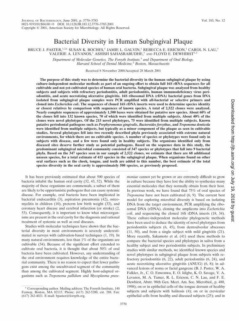
TABLE 1. PCR and sequencing primers used in this study
Primer Function Position Orientation Bacterial specificity Sequence
D88 PCR 7–27 Forward Most GAGAGTTTGATYMTGGCTCAGE94 PCR 1525–1541 Reverse Most GAAGGAGGTGWTCCARCCGCAC90 PCR 1483–1503 Reverse Spirochaetes GTTACGACTTCACCCTCCTF01 PCR 1487–1508 Reverse Bacteroidetes CCTTGTTACGACTTAGCCCC75 Sequencing 7–27 Forward Most GAGAGTTTGATYCTGGCTCAGB34 Sequencing 344–358 Forward Most ACGGGAGGCAGCAGYF16 Sequencing 789–806 Forward Most TAGATACCCYGGTAGTCCF18 Sequencing 1099–1113 Forward Most GCAACGAGCGCAACCF19 Sequencing 1099–1114 Forward Bacteroidetes ATAACGAGCGCAACCCE94 Sequencing 1522–1541 Reverse Most GAAGGAGGTGWTCCARCCGCAF20 Sequencing 1226–1242 Reverse Most CCATTGTARCACGTGTGF21 Sequencing 1226–1242 Reverse b-Proteobacteria CCATTGTATGACGTGTGF17 Sequencing 907–926 Reverse Most CCGTCWATTCMTTTGAGTTTF15 Sequencing 519–533 Reverse Most TTACCGCGGCTGCTGF22 Sequencing 344–358 Reverse Most RCTGCTGCCTCCCGT
VOL. 183, 2001 BACTERIAL MICROFLORA OF SUBGINGIVAL PLAQUE 3771
on July 19, 2018 by guesthttp://jb.asm
.org/D
ownloaded from

assembled during the PCR (26). The percentage of chimeric inserts in 16S rRNAlibraries ranged from 1 to 15%. Chimeric sequences were identified by using theChimera Check program in RDP, by treeing analysis, or by base signatureanalysis. Species identification of chimeras was obtained, but the sequences werenot examined for phylogenetic analysis.
Estimation of unseen species. In 1943, Fisher et al. (13) described a model forestimating the number of unseen species in a population. In addition to ecolog-ical studies of biological diversity, similar methods have been used to estimatethe number of words known but not used by Shakespeare and other authors (12).In this study, the number of unseen species that were missed was calculated withan improved estimator as proposed by Boneh et al. (5). The estimator is basedon a continuous time model of parallel independent Poisson processes. Theestimator is:
C~t! 5 Ok51
kmax
Nke2k 2 Ok51
kmax
Nke2k~1 1 t!
where C(t) is the number of new species expected to be observed over time zeroto t when the original observations were over time 21 to 0. k is the number oftimes a species is seen. Nk is the number of species seen k times. Bias correctionand other details of the estimator are beyond the scope of the current discussion,and the reader is referred to the publication of Boneh et al. (5).
Nucleotide sequence accession number. The complete 16S rRNA gene se-quences of clones representing novel phylotypes defined in this study, sequencesof known species not previously reported, and published sequences are availablefor electronic retrieval from the EMBL, GenBank, and DDBJ nucleotide se-quence databases under the accession numbers shown in Fig. 1 through 7.
RESULTS
Partial sequences of about 500 bp were obtained for 2,52216S rRNA clones in order to identify the predominant bacte-rial species present in the subgingival plaque of healthy anddiseased subjects. Approximately 60% of the 2,522 clones hadgreater than 99% sequence similarity to one of 132 knownspecies. The remaining 40% of the clones fell into 215 previ-ously unrecognized clusters, termed phylotypes. Full 1,500-base sequences were determined for representatives of eachnovel phylotype. The term “phylotype” is used for clusters ofclone sequences that differed from known species by approxi-mately 30 bases (or 2%) and were at least 99% similar tomembers of their cluster. The diversity of known species andnovel phylotypes with respect to periodontal health status isshown in Table 2. Approximately 40 to 60% of clones analyzedin each category of healthy or diseased subjects were novelphylotypes. Overall, we detected 347 species or phylotypes insubgingival plaque.
The intent of using different PCR primer sets was to obtainthe widest spectrum of phylogenetic groups. In previous stud-ies, results obtained with the Spirochaetes-selective primersindicate that 85% of the clones have spirochetal inserts (6).
Our results using the Bacteroidetes-selective primers were notas selective but yielded a wide range of bacterial types—in-cluding novel taxa at the phylum level. In designing the “uni-versal” bacterial primer set, a “G” was added to the 59 end ofthe reverse primer (E94) to enhance cloning of amplicons withthe TOPO-TA cloning kit. Despite presenting a mismatch atthis position to most 16S rRNA sequences in the RDP, acontinuous stretch of 20 matching bases were available foroptimal PCR. Consequently, a wide range of phylogenetictypes was obtained by using this “universal” primer set.
Estimation of unseen species. By using the estimator ofBoneh et al. (5) and applying the suggested bias reductionprocedure, we calculated that there are 68 unseen species inaddition to the 347 found, for a total estimate of 415 subgin-gival species. Table 3 shows the number of additional speciesthat one would expect to identify by examining various num-bers of additional clones. As shown in Table 3, fewer newspecies were predicted to be found for each thousand addi-tional clones analyzed. The model suggests that by examining10,000 additional clones, we would find all but one of theestimated 415 species.
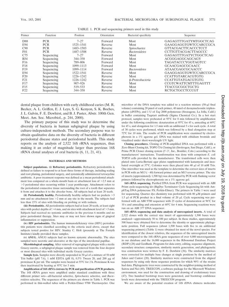
As shown in Fig. 1, the oral bacteria identified in this studyfell into nine bacterial groups or phyla as follows: ObsidianPool OP11 (19), TM7 (38); Deferribacteres, Spirochaetes, Fuso-bacteria, Actinobacteria, Firmicutes, Proteobacteria, and Bacte-roidetes (the latter seven phyla are listed in the current taxo-nomic outline for Bergey’s Manual of Systematic Bacteriology,2nd ed., vol. 2, at http://www.cme.msu.edu/bergeys/). The dis-tribution of known species and novel phylotypes within each ofthese phyla is shown in Table 4. In Fig. 1 through 7, thephylogenetic diversity within each phylum is shown and is dis-cussed in detail below. The information presented includesbacterial species or phylotype; strain or clone identification,sequence accession number, total number of retrieved clones,and number of subjects in each health status category in whicheach species was identified (color- and shape-coordinated sym-bols). Prevalent species or phylotypes are defined as thoseidentified from four or more subjects and are noted in bold andunderlined in each dendrogram.
Obsidian Pool OB11. As shown in Fig. 1, oral clone X112represents the sole oral member of this phylum. Although onlyone phylotype was identified, it was found in multiple subjects.Distant relatives include phylotypes identified from sludge and
TABLE 2. Diversity of species and phylotypes in healthy anddiseased sites
Health status (n)No. ofclones
analyzed
No. (%)of known
species
No. (%)of novel
phylotypes
Total no.of species/phylotypes
Healthy (5) 268 42 (58) 30 (42) 72Periodontitis (9) 417 46 (51) 45 (49) 91Refractory
periodontitis (11)1,386 112 (53) 101 (47) 213
ANUG (4) 417 36 (38) 59 (62) 96HIV (2) 34 6 (55) 5 (45) 11
Total 2,522 132 (38) 215 (62) 347
TABLE 3. Number of unseen species
No. of additionalclones analyzed
No. of expectednew species
Increment innew speciesa
1,000 26.86 26.862,000 42.36 15.503,000 51.72 9.364,000 57.55 5.835,000 61.27 3.726,000 63.68 2.417,000 65.26 1.588,000 66.31 1.049,000 67.00 0.69
10,000 67.46 0.46` 68.41 0.95
a Number of species expected minus that in the row above.
3772 PASTER ET AL. J. BACTERIOL.
on July 19, 2018 by guesthttp://jb.asm
.org/D
ownloaded from
deep-sea sediments. There are no cultivable representatives ofthis phylum.
TM7. Five oral phylotypes of the TM7 phylum were identi-fied as shown in Fig. 1. Distant relatives of this phylum havebeen identified in 16S rRNA clones derived from DNA iso-lated from soil and deep-sea sediments. The oral phylotypeswere initially identified with Bacteroidetes-selective primers,but later identified with all-bacterial-selective primers, whichindicates that these phylotypes are relatively common in sub-
gingival plaque. There are no cultivable representatives of thisphylum.
Deferribacteres. In the Deferribacteres phylum, the oral spe-cies formed a coherent cluster of seven separate phylotypes,four of which were prevalent in diseased subjects, especially inthe ANUG and refractory periodontitis subjects. Conse-quently, the latter phylotypes may be good candidates as pu-tative pathogens. Members of this oral cluster are distantlyrelated to Synergistes jonesii, a cultivable, bovine ruminal bac-
FIG. 1. Bacterial phyla identified from subgingival plaque. Nine bacterial phyla were represented from clonal analysis. As shown, the first twophyla, Obsidian Pool OP11 and TM7, have no cultivable representatives. Oral members of the phylum Deferribacteres form a cluster consisting ofonly not-yet-cultivated phylotypes. The information presented in Fig. 1 to 7 includes bacterial species or phylotype, strain or clone identification,sequence accession number, total number of retrieved clones, and number of subjects in each health status category in which each species wasidentified (as indicated by color-coordinated symbols). Novel phylotypes are defined as those taxa that are ,98.5 to 99% similar in sequencecomparisons to the phylotype’s closest relative. Prevalent species are defined as those phylotypes or species identified in four or more subjects andare noted in boldface and underlined in each dendrogram. Two hundred bootstrap trees were generated, and bootstrap confidence levels aspercentages (only values over 40%) are shown at tree nodes.
VOL. 183, 2001 BACTERIAL MICROFLORA OF SUBGINGIVAL PLAQUE 3773
on July 19, 2018 by guesthttp://jb.asm
.org/D
ownloaded from
terium able to degrade the pyridinediol toxin in the plantLeucaena leucocephala (27, 30). One oral clone, BA121, fellwithin the S. jonesii cluster rather than the oral cluster.
Spirochaetes. The human periodontal pocket harbors ahighly diverse and numerous spirochetal community (6, 9). Allspecies identified thus far fall in the genus Treponema, andpresently there are about 60 oral treponemal species or phy-lotypes (6) (Fig. 2). As shown in Fig. 2, several prevalentspecies or phylotypes were identified. The predominant culti-vable known Treponema species included T. medium (but notthe closely related “T. vincentii”), T. denticola (which was oneof the most commonly identified species in both disease andhealth), T. maltophilum, and two subspecies of T. socranskii.There were at least four predominant novel phylotypes, one ofwhich, Treponema sp. 1:G:T21, was found only in diseasedsites. The latter phylotype appears to be a good candidate as aputative pathogen.
It should be noted that the treponemal clones had beenspecifically sought by constructing 16S rRNA libraries withspirochete-selective primers in the initial PCR amplification.Microscopic examination of plaque samples from diseasedsites, especially those from ANUG subjects, revealed that 20 to50% of the members of the bacterial community were spiro-chetes (6). Subsequently, spirochetes were also detected in 16SrRNA libraries when all-bacterial primers were used, albeit ata lower frequency.
Fusobacteria. As shown in Fig. 3, the members of the oralFusobacteria identified include species of the genera Fusobac-terium and Leptotrichia. Most noteworthy was that Fusobacte-rium naviforme (earlier referred to as Fusobacterium nucleatumsubsp. vincentii) was often identified, especially in periodontitissubjects (eight of nine subjects). In six of these eight periodon-titis subjects, Fusobacterium naviforme represented from 50 to80% of the clones. Other species and phylotypes of Fusobac-terium and Leptotrichia buccalis from healthy and diseasedsubjects were also commonly identified. Fusobacterium anima-lis was detected only in diseased subjects but not in healthysubjects.
Actinobacteria. The Actinobacteria phylum was previously re-ferred to as the high-G1C-content, gram-positive phylum. Asshown in Fig. 3, oral genera of the phylum Actinobacteria thatwere detected include Actinomyces, Atopobium, Bifidobacte-rium, Corynebacterium, Propionibacterium, and Rothia. Within
these genera, Actinomyces naeslundii II, Corynebacteriummatruchotii, and Rothia dentocariosa were commonly detectedin health and disease. Corynebacterium matruchotii appearedto be more associated with “healthy” subgingival plaque. Twospecies of Atopobium were prevalent in diseased but nothealthy subjects.
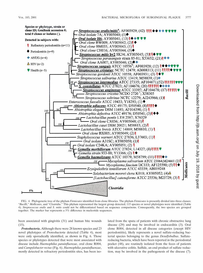
Firmicutes. Most of the members of the Firmicutes phylumwere originally classified as belonging to the low-G1C-con-tent, gram-positive phylum. The Firmicutes are presently di-vided into three classes—namely the proposed “Bacilli,” theMollicutes, and the proposed “Clostridia.” The phylogeneticposition of oral members of the “Bacilli ” and Mollicutes isshown in Fig. 4. In the class “Bacilli,” the predominant speciesincluded the streptococci Streptococcus oralis or S. mitis, S.mitis biovar 2, S. sanguis, S. intermedius, S. constellatus, and S.anginosus; Abiotrophia adiacens; and two species of Gemella.Since it was difficult to differentiate S. oralis and S. mitis by 16SrRNA comparisons, they were considered as one species in thisstudy. Of the predominant species, S. constellatus and Gemellahaemolysans were most associated with diseased sites, as theywere not detected in subgingival plaque from any of thehealthy subjects. Although most of the other predominant spe-cies were detected in both subjects with disease and those withhealth, many of the Streptococcus spp. were commonly identi-fied from the healthy subjects.
Not many members of the class Mollicutes were detected:two species of Mycoplasma; Solobacterium moorei, a microor-ganism isolated from feces that is related to species of Erysip-elothrix; and a misclassified Lactobacillus species, [L.] cat-enaforme. Infections by species of Erysipelothrix have beenlinked to endocarditis (16). It was encouraging to detect thepurported hard-to-lyse mycoplasmas (23), since the resultsdemonstrated that our procedures do indeed recover a broadspectrum of bacterial species.
Members of the class “Clostridia” represented one of thelargest groups detected, as shown in Fig. 5. Oral membersincluded species within the genera Catonella, Dialister, Eubac-terium, Megasphaera, Peptostreptococcus, Selenomonas, andVeillonella. There were several good candidates as additionalputative pathogens, i.e., those species or phylotypes that werecommonly detected in disease, but not (or rarely) in health—namely Eubacterium saphenum, clone PUS9.170, Filifactor alo-cis (previously Fusobacterium alocis), Catonella morbi, Megas-phaera sp. oral clone BB166, Dialister sp. strain GBA27, andSelenomonas sputigena.
Catonella morbi has been associated with periodontitis (31),clone PUS9.170 and Filifactor alocis have been associated withdentoalveolar abscesses (11, 50), and Selenomonas sputigenahas been associated with periodontitis and, in rare cases, hasbeen involved in fatal septicemias (51). Eubacterium saphenumand species of Megasphaera have not been considered as pu-tative periodontal pathogens (44).
Oral isolate GBA27 is a slow-growing Dialister species thatwas commonly detected in clones from all disease categoriesand from one healthy subject (Fig. 6). Since strain GBA27 cellsform only pinpoint colonies on agar medium (A. Tanner, TheForsyth Institute, personal communication), it is likely that thisspecies has eluded detection in predominant cultivable studiesover the years. The closely related Dialister pneumosintes has
TABLE 4. Known and novel species or phylotypesidentified from clonal analysis
Phylum
Known species Novel phylotypes
No. of clones(% of total)
No. ofspecies
No. of clones(% of total)
No. ofphylotypes
Obsidian Pool OB11 0 0 6 (100) 1TM7 0 0 34 (100) 5Deferribacteres 0 0 86 (100) 8Spirochaetes 199 (37) 9 338 (63) 49Fusobacteria 257 (73) 7 96 (27) 12Actinobacteria 229 (83) 14 46 (17) 18Firmicutes 454 (69) 53 205 (31) 62Proteobacteria 244 (72) 28 94 (28) 23Bacteroidetes 148 (63) 21 86 (37) 37
Total 1,531 (60.7) 132 991 (39.3) 215
3774 PASTER ET AL. J. BACTERIOL.
on July 19, 2018 by guesthttp://jb.asm
.org/D
ownloaded from

FIG. 2. Phylogenetic tree of oral treponemes identified from clone libraries. All species identified thus far fall in the genus Treponema, andpresently there are about 60 oral treponemal species or phylotypes (6). The marker bar represents a 10% difference in nucleotide sequences.
VOL. 183, 2001 BACTERIAL MICROFLORA OF SUBGINGIVAL PLAQUE 3775
on July 19, 2018 by guesthttp://jb.asm
.org/D
ownloaded from
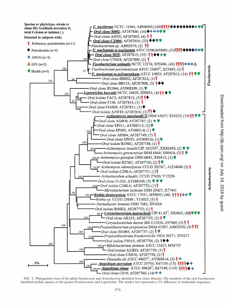
FIG. 3. Phylogenetic trees of the phyla Fusobacteria and Actinobacteria identified from clone libraries. The members of the oral Fusobacteriaidentified include species of the genera Fusobacterium and Leptotrichia. The marker bar represents a 5% difference in nucleotide sequences.
3776
on July 19, 2018 by guesthttp://jb.asm
.org/D
ownloaded from
been associated with gingivitis (31) and human bite wounds(15).
Proteobacteria. Although there were 28 known species and 23novel phylotypes of Proteobacteria detected (Table 4), mostwere only sporadically identified, as shown in Fig. 6. Thosespecies or phylotypes detected that were most associated withdisease include Haemophilus parainfluenzae, oral clone R004,and Campylobacter rectus (Fig. 6). Haemophilus parainfluenzae,mostly detected in refractory periodontitis sites, has been iso-
lated from the sputa of patients with chronic obstructive lungdisease (29) and may be involved in endocarditis (3). Oralclone R004, detected in all disease categories (except HIVperiodontitis), likely represents a novel sulfate-reducing bac-terial species belonging to the genus Desulfobulbus. Sulfate-reducing bacteria, which have been reported in the periodontalpocket (48), are routinely isolated from the feces of patientswith ulcerative colitis. Sulfide, an end product of sulfate reduc-tion, may be involved in the pathogenesis of the disease (7).
FIG. 4. Phylogenetic tree of the phylum Firmicutes identified from clone libraries. The phylum Firmicutes is presently divided into three classes:“Bacilli,” Mollicutes, and “Clostridia.” This phylum represented the largest group detected; 115 species or novel phylotypes were identified (Table4). Streptococcus oralis and S. mitis could not be differentiated based on sequence comparisons. Consequently, the two species are groupedtogether. The marker bar represents a 5% difference in nucleotide sequences.
VOL. 183, 2001 BACTERIAL MICROFLORA OF SUBGINGIVAL PLAQUE 3777
on July 19, 2018 by guesthttp://jb.asm
.org/D
ownloaded from
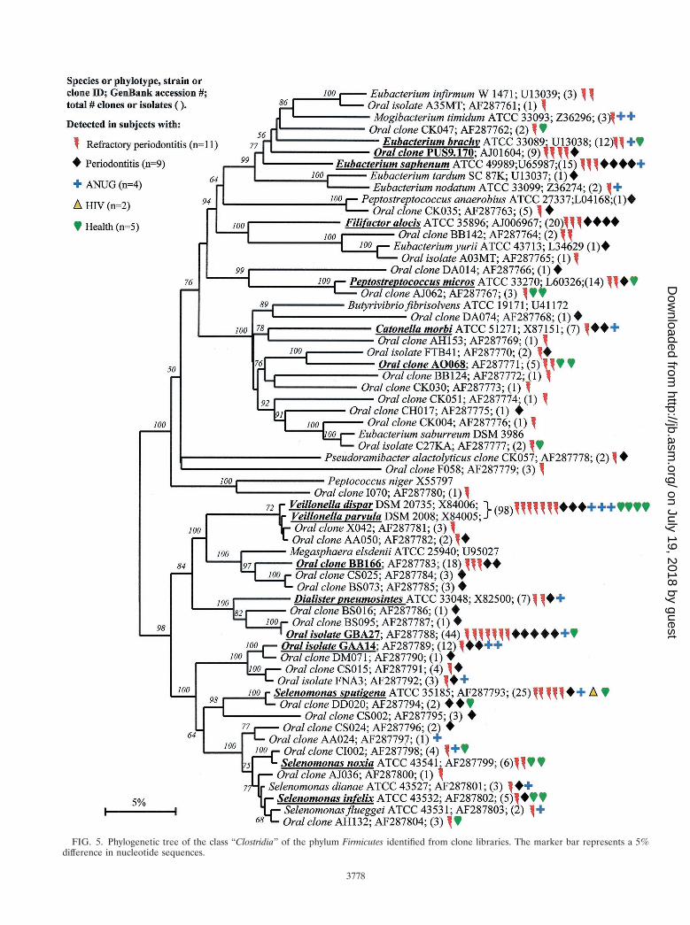
FIG. 5. Phylogenetic tree of the class “Clostridia” of the phylum Firmicutes identified from clone libraries. The marker bar represents a 5%difference in nucleotide sequences.
3778
on July 19, 2018 by guesthttp://jb.asm
.org/D
ownloaded from

FIG. 6. Phylogenetic tree of the phylum Proteobacteria identified from clone libraries. The marker bar represents a 5% difference in nucleotidesequences.
VOL. 183, 2001 BACTERIAL MICROFLORA OF SUBGINGIVAL PLAQUE 3779
on July 19, 2018 by guesthttp://jb.asm
.org/D
ownloaded from

Campylobacter rectus has long been associated with periodontaldisease (44). Species or phylotypes commonly detected in bothdiseased and healthy subjects include Neisseria mucosa and twoadditional species of Campylobacter, C. gracilis and C. concisus.
Bacteroidetes. There were 21 known species and 37 novelphylotypes of Bacteroidetes (previously referred to as the Cap-nocytophaga/Flavobacterium/Bacteroides phylum) detected in16S rRNA libraries (Table 4). As anticipated, P. gingivalis andB. forsythus, two species typically associated with periodontitis,were detected, although not as frequently as some other spe-cies or phylotypes (Fig. 7). Other species or phylotypes thatwere commonly detected include Prevotella denticola, Pre-votella oris, Prevotella tannerae, Porphyromonas endodontalis,oral clone AU126, Capnocytophaga ochracea, and Capnocyto-phaga gingivalis (Fig. 7). Porphyromonas endodontalis has beenassociated with endodontal infections (49), but it was oftendetected in diseased subjects and not in any of the healthysubjects (Fig. 7). Prevotella oris has been associated with den-toalveolar abscesses (11). The remaining cultivable species ofthis phylum have not been typically considered periodontalpathogens (44).
DISCUSSION
Human subgingival plaque harbors a highly diverse bacterialcommunity, as demonstrated by our detection of 347 species orphylotypes by clonal analysis. Using the estimator of Boneh etal. (5), we predicted that approximately 68 additional unseenspecies were present and could be detected by examining10,000 additional clones. This estimate is a rough approxima-tion because the clones came from libraries constructed fromsubjects with different health and disease categories, violatingthe assumption of sampling with replacement from a homoge-neous population. However, the model suggests that we havealready identified 85% of the species and makes testable pre-dictions as to how many unseen species should be found byexamining additional clones (Table 3). Examination of 179additional clones, not included in this data set, from subgingi-val plaque from sites of necrotizing ulcerative periodontitis(29) yielded six novel species where the model would havepredicted five. Thus, the model and predictions appear quiteaccurate. When organisms found in other studies (11, 21, 41,45, 50) and on cheek, tongue, and other oral tissues are addedto our estimate of 415 subgingival species, the best estimate ofthe total species diversity in the oral cavity is between 500 and600 species, as has been previously proposed. A list of knownhuman oral species and phylotypes can be obtained from thecorresponding author.
Of the 215 phylotypes identified in this study, 33 were cul-tivable strains that have not yet been characterized. The re-maining 182 were represented only by clones. It is likely thatthe vast majority of these phylotypes cannot be cultivated withstandard anaerobic media and techniques. It is also likely thatmany of these phylotypes are “hidden” within culture collec-tions, since the strains could not be differentiated from closelyrelated known species. In this regard, we are currently exam-ining isolates of over 100 unnamed taxa from the oral anaer-obic collection of W. E. C. Moore and L. V. Moore (VirginiaPolytechnic Institute, Blacksburg, Va.). It will be interesting tosee how many of Moore’s groups represent cultivable examples
of groups previously represented only by clone phylotypes.Treponema amylovorum and Treponema lecithinolyticum wereidentified first by clone data before the cultivable strains werecharacterized (6; this study). The phylogenetic position of not-yet-cultivated novel phylotypes, especially if they are closelyrelated to cultivable species, may provide insight into theircultivation. For example, physiological properties or antibioticresistance can be inferred to some degree. DNA probes spe-cific for the phylotype can be used to monitor enrichmentefforts and to positively identify an isolate.
The definition of a species is controversial, particularly whenonly molecular sequence data exist (14, 46). Therefore, wehave used the term “phylotype” in place of species for referringto novel clusters of clone sequences. In most cases, a 2%difference in 16S rRNA sequences does indicate separate spe-cies status, but there are exceptions. Formal naming of a spe-cies also requires a full description of the phenotypic charac-teristics of an organism. It is probable that the majority of thephylotypes identified in this report will eventually be validatedas species. In the meantime, DNA probes can be designed toidentify phylotypes and to assess their roles in disease orhealth. If a phylotype proves to be associated with disease, thenefforts can be made to isolate and characterize the new species.
In general, the first 500 bases of the 16S rRNA gene are themost informative for identifying an organism, because thereare several variable regions. However, for constructing phylo-genetic trees, it is essential that nearly complete sequences of1,500 bases be used. This is a deficiency of some previousstudies. We have also obtained full sequences so that the dataare most useful to databases such as GenBank or the RDP(28).
A total of 72 species or novel phylotypes were identified inthe analysis of 268 clones from healthy subjects (Table 2).Based on the overall total of 347, there are 275 species or novelphylotypes that were identified in diseased subjects. Part of theexplanation for the discrepancy between the number of speciesfound in healthy subjects versus that found in diseased subjectsis that only 10.6% of the clones analyzed were from healthysubjects. However, even with this bias, a number of species andphylotypes were found only in multiple ($4) diseased subjectsand not in any healthy subjects. These often-identified disease-associated species and phylotypes are shown in Table 5 and areclearly candidates for further study. Eighteen of the 29 speciesare named species, including Porphyromonas gingivalis and B.forsythus. One represents a cultivable, but unnamed species.Ten of the phylotypes are represented only by clones, and thusmay represent currently not-yet-cultivated or unrecognized po-tential pathogens. Additional putative pathogens are likely,since there were several species or phylotypes that were oftenidentified from diseased sites, but only rarely from health: e.g.,Dialister sp. strain GBA27 (Fig. 5), other species or phylotypesof Fusobacterium (Fig. 3), or oral clone AU126 (Fig. 7). Therewere also several species that were more associated withhealth, namely Actinobaculum sp. clone EL030 and Corynebac-terium matruchotii (Fig. 3). However, to rigorously assess theassociation of specific species or phylotypes with certain peri-odontal diseases or periodontal health, it will be necessary toanalyze large numbers of clinical samples for the levels of alloral bacteria in well-controlled clinical studies. The definitionof five subgingival plaque bacterial complexes by Socransky et
3780 PASTER ET AL. J. BACTERIOL.
on July 19, 2018 by guesthttp://jb.asm
.org/D
ownloaded from
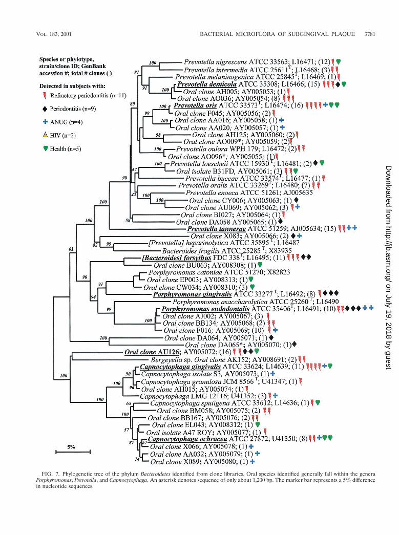
FIG. 7. Phylogenetic tree of the phylum Bacteroidetes identified from clone libraries. Oral species identified generally fall within the generaPorphyromonas, Prevotella, and Capnocytophaga. An asterisk denotes sequence of only about 1,200 bp. The marker bar represents a 5% differencein nucleotide sequences.
VOL. 183, 2001 BACTERIAL MICROFLORA OF SUBGINGIVAL PLAQUE 3781
on July 19, 2018 by guesthttp://jb.asm
.org/D
ownloaded from

al. (44) was based on analysis of the microbial community ofover 13,000 plaque samples from 185 subjects by using DNAprobes in checkerboard hybridization assays. We are currentlydeveloping DNA probes for approximately all 500 known spe-cies and novel phylotypes for use in large clinical studies. TheseDNA probes are being developed for use in the checkerboardhybridization assay (35) and DNA microarray formats. Basedupon knowledge of the full bacterial diversity, and using mo-lecular techniques to identify these bacteria, future studies willbe able to associate a number of additional oral bacteria withvarious oral and systemic diseases.
ACKNOWLEDGMENTS
This study was supported by NIH grants DE-11443 and DE-10374from the National Institute of Dental and Craniofacial Research.
REFERENCES
1. Amman, R., W. Ludwig, and K.-H. Schleifer. 1995. Phylogenetic identifica-tion and in situ detection of individual microbial cells without cultivation.Microbiol. Rev. 59:143–169.
2. Beck, J., R. Garcia, G. Heiss, P. S. Vokonas, and S. Offenbacher. 1996.Periodontal disease and cardiovascular disease. J. Periodontol. 67:1123–1137.
3. Berbari, E. F., F. R. Cockerill III, and J. M. Steckelberg. 1997. Infective
endocarditis due to unusual or fastidious microorganisms. Mayo Clin. Proc.72:532–542.
4. Boches, S. K., P. M. Mitchell, J. L. Galvin, W. J. Loesche, C. E. Kazor, F. E.Dewhirst, and B. J. Paster. 2000. Cultivable and uncultivable bacteria on thehealthy tongue dorsum. J. Dent. Res. 79:396.
5. Boneh, S., A. Boneh, and R. J. Caron. 1998. Estimating the predictionfunction and the number of unseen species in sampling with replacement.J. Am. Statist. Assoc. 93:372–379.
6. Choi, B. K., B. J. Paster, F. E. Dewhirst, and U. B. Gobel. 1994. Diversity ofcultivable and uncultivable oral spirochetes from a patient with severe de-structive periodontitis. Infect. Immun. 62:1889–1895.
7. Christl, S. U., H. D. Eisner, G. Dusel, H. Kasper, and W. Scheppach. 1996.Antagonistic effects of sulfide and butyrate on proliferation of colonic mu-cosa: a potential role for these agents in the pathogenesis of ulcerative colitis.Dig. Dis. Sci. 41:2477–2481.
8. Coleman, B. E., N. Tzellas, B. J. Paster, and F. E. Dewhirst. 1996. Identifi-cation of 16S rRNA clones from a subject with ANUG as Atopobium species.J. Dent. Res. 75:207.
9. Dewhirst, F. E., M. A. Tamer, R. E. Ericson, C. N. Lau, V. A. Levanos, S. K.Boches, J. L. Galvin, and B. J. Paster. 2000. The diversity of periodontalspirochetes by 16S rRNA analysis. Oral Microbiol. Immunol. 15:196–202.
10. Dodman, T., J. Robson, and D. Pincus. 2000. Kingella kingae infections inchildren. J. Paediatr. Child Health 36:87–90.
11. Dymock, D., A. J. Weightman, C. Scully, and W. G. Wade. 1996. Molecularanalysis of microflora associated with dentoalveolar abscesses. J. Clin. Mi-crobiol. 34:537–542.
12. Efron, B., and R. Thisted. 1976. Estimating the number of unseen species.How many words did Shakespeare know? Biometrika 63:435–447.
13. Fisher, R. A., A. S. Corbet, and C. B. Williams. 1943. The relation betweenthe number of species and the number of individuals in a random sample ofan animal population. J. Anim. Ecol. 12:42–58.
14. Fox, G. E., J. D. Wisotzkey, and P. Jurtshuk, Jr. 1992. How close is close: 16SrRNA sequence identity may not be sufficient to guarantee species identity.Int. J. Syst. Bacteriol. 42:166–170.
15. Goldstein, E. J., D. M. Citron, and S. M. Finegold. 1984. Role of anaerobicbacteria in bite-wound infections. Rev. Infect. Dis. 6:S177–S183.
16. Gorby, G. L., and J. E. Peacock, Jr. 1988. Erysipelothrix rhusiopathiae endo-carditis: microbiologic, epidemiologic, and clinical features of an occupa-tional disease. Rev. Infect. Dis. 10:317–325.
17. Harper-Owen, R., D. Dymock, B. Booth, A. J. Weightman, and W. G. Wade.1999. Detection of unculturable bacteria in periodontal health and disease byPCR. J. Clin. Microbiol. 37:1469–1473.
18. Hugenholtz, P., and N. R. Pace. 1996. Identifying microbial diversity in thenatural environment: a molecular phylogenetic approach. Trends Biotech-nol. 14:190–197.
19. Hugenholtz, P., C. Pitulle, K. L. Hershberger, and N. R. Pace. 1998. Noveldivision level bacterial diversity in a Yellowstone hot spring. J. Bacteriol.180:366–376.
20. Jukes, T. H., and C. R. Cantor. 1969. Evolution of protein molecules, p.21–132. In H. N. Munro (ed.), Mammalian protein metabolism, vol. 3.Academic Press, Inc., New York, N.Y.
21. Kroes, I., P. W. Lepp, and D. A. Relman. 1999. Bacterial diversity within thehuman subgingival crevice. Proc. Natl. Acad. Sci. USA 96:14547–14552.
22. Lau, C. N., B. J. Paster, V. Levanos, R. L. Ericson, and F. E. Dewhirst. 1998.Cultivable and uncultivable predominant bacterial species in refractory pe-riodontitis. J. Dent. Res. 77:139.
23. Leng, Z., G. E. Kenny, and M. C. Roberts. 1994. Evaluation of the detectionlimits of PCR for identification of Mycoplasma pneumoniae in clinical sam-ples. Mol. Cell. Probes 8:125–130.
24. Levanos, V. A., J. L. Galvin, S. K. Boches, F. E. Dewhirst, and B. J. Paster.2000. Predominant cultivable and uncultivable bacterial species in adultperiodontitis. J. Dent. Res. 79:234.
25. Levin, I. M., C. N. Lau, S. S. Socransky, A. D. Haffajee, L. Martin, J. L.Galvin, S. K. Boches, B. J. Paster, and F. E. Dewhirst. 1999. Cultivable anduncultivable species on or in gingival epithelial cells. J. Dent. Res. 78:453.
26. Liesack, W., H. Weyland, and E. Stackebrandt. 1991. Potential risk of geneamplification by PCR as determined by 16S rDNA analysis of a mixed-culture of strict barophilic bacteria. Microbiol. Ecol. 21:191–198.
27. Ludwig, W., G. Wallner, A. Tesch, and F. Klink. 1991. A novel eubacterialphylum: comparative nucleotide sequence analysis of a tuf-gene of Flexistipessinusarabici. FEMS Microbiol. Lett. 62:139–143.
28. Maidak, B. L., J. R. Cole, T. G. Lilburn, C. T. Parker, Jr., P. R. Saxman,J. M. Stredwick, G. M. Garrity, B. Li, G. J. Olsen, S. Pramanik, T. M.Schmidt, and J. M. Tiedje. 2000. The RDP (Ribosomal Database Project)continues. Nucleic Acids Res. 28:173–174.
29. Mitchell, J. L., and S. L. Hill. 2000. Immune response to Haemophilusparainfluenzae in patients with chronic obstructive lung disease. Clin. Diagn.Lab. Immunol. 7:25–30.
30. McSweeny, C. S., M. J. Allison, and R. I. Mackie. 1993. Amino acid utiliza-tion by the ruminal bacterium Synergistes jonesii strain 78–1. Arch. Microbiol.159:131–135.
31. Moore, L. V., and W. E. C. Moore. 1994. Oribaculum catoniae gen. nov., sp.
TABLE 5. Putative pathogens in subgingival plaquea
Phylotype or species by phylum No. of clonesidentified
TM7Clone I025................................................................................... 6
DeferribacteresClone BH017 .............................................................................. 43Clone D084................................................................................. 20Clone W028 ................................................................................ 6Clone W090 ................................................................................ 18
SpirochaetesClone Treponema sp. 1:G:T21.................................................. 27Clone Treponema sp. 2:10:D12................................................. 9Treponema socranskii sub sp. buccale ...................................... 17
FusobacteriaFusobacterium animalis.............................................................. 43
ActinobacteriaAtopobium parvulum .................................................................. 15Atopobium rimae ........................................................................114
FirmicutesClone Eubacterium sp. PUS9.170............................................. 9Clone Megasphaera sp. BB166.................................................. 18Abiotrophia adiacens .................................................................. 8Catonella morbi........................................................................... 7Dialister pneumonsintes .............................................................. 7Eubacterium saphenum .............................................................. 15Filifactor alocis ............................................................................ 20Gemella haemolysans ................................................................. 11Selenomonas sp. strain GAA14 ................................................ 12Streptococcus constellatus........................................................... 20
ProteobacteriaClone Desulfobulbus sp. R004 .................................................. 23Campylobacter gracilis ................................................................ 11Campylobacter rectus .................................................................. 8Haemophilus parainfluenzae ...................................................... 11
BacteroidetesBacteroides forsythus ................................................................... 11Prevotella tannerae ...................................................................... 15Porphyromonas endodontalis ..................................................... 10Porphyromonas gingivalis ........................................................... 8
a Detected in at least four diseased subjects, but not in healthy subjects. Datawere derived from Fig. 1 through 7.
3782 PASTER ET AL. J. BACTERIOL.
on July 19, 2018 by guesthttp://jb.asm
.org/D
ownloaded from
nov.; Catonella morbi gen. nov., sp. nov.; Hallella seregens gen. nov., sp. nov.;Johnsonella ignava gen. nov., sp. nov.; and Dialister pneumosintes gen. nov.,comb. nov., nom. rev., anaerobic gram-negative bacilli from the humangingival crevice. Int. J. Syst. Bacteriol. 44:187–192.
32. Moore, W. E. C., and L. V. H Moore. 1994. The bacteria of periodontaldiseases. Periodontol. 2000 5:66–77.
33. Offenbacher, S., H. L. Jared, P. G. O’Reilly, S. R. Wells, G. E. Salvi, H. P.Lawrence, S. S. Socransky, and J. D. Beck. 1998. Potential pathogenicmechanisms of periodontitis associated pregnancy complications. Ann. Peri-odontol. 3:233–250.
34. Pace, N. R., D. A. Stahl, D. J. Lane, and G. J. Olsen. 1986. The analysis ofnatural microbial populations by ribosomal RNA sequences. Adv. Microb.Ecol. 9:1–55.
35. Paster, B. J., I. M. Bartoszyk, and F. E. Dewhirst. 1998. Identification of oralstreptococci using PCR-based, reverse-capture, checkerboard hybridization.Methods Cell Sci. 20:223–231.
36. Paster, B. J., and F. E. Dewhirst. 1988. Phylogeny of campylobacters,wolinellas, Bacteroides gracilis, and Bacteroides ureolyticus by 16S rRNAsequencing. Int. J. Syst. Bacteriol. 38:56–62.
37. Paster, B. J., F. E. Dewhirst, W. G. Weisburg, L. A. Tordoff, G. J. Fraser,R. B. Hespell, T. B. Stanton, L. Zablen, L. Mandelco, and C. R. Woese. 1991.Phylogenetic analysis of the spirochetes. J. Bacteriol. 173:6101–6109.
38. Rheims, H., F. A. Rainey, and E. Stackebrandt. 1996. A molecular approachto search for diversity among bacteria in the environment. J. Ind. Microbiol.17:159–169.
39. Russell, M. K., T. Alpagot, S. K. Boches, J. L. Galvin, F. E. Dewhirst, andB. J. Paster. 2001. Bacterial species and phylotypes in necrotizing ulcerativeperiodontitis. J. Dent. Res. 80:167.
40. Saitou, N., and M. Nei. 1987. The neighbor-joining method: a new methodfor reconstructing phylogenetic trees. Mol. Biol. Evol. 4:406–425.
41. Sakamoto, M., M. Umeda, I. Ishikawa, and Y. Benno. 2000. Comparison ofthe oral bacterial flora in saliva from a healthy subject and two periodontitispatients by sequence analysis of 16S rDNA libraries. Microbiol. Immunol.44:643–652.
42. Scannapieco, F. A. 1999. Role of oral bacteria in respiratory infection. J.Periodontol. 70:793–802.
43. Socransky, S. S., and A. D. Haffajee. 1994. Evidence of bacterial etiology: ahistorical perspective. Periodontol. 2000 5:7–25.
44. Socransky, S. S., A. D. Haffajee, M. A. Cugini, C. Smith, and R. L. Kent, Jr.1998. Microbial complexes in subgingival plaque. J. Clin. Periodontol. 25:134–144.
45. Spratt, D. A., A. J. Weightman, and W. G. Wade. 1999. Diversity of oralasaccharolytic Eubacterium species in periodontitis: identification of novelphylotypes representing uncultivated taxa. Oral Microbiol. Immunol. 14:56–59.
46. Stackebrandt, E., and B. M. Goebel. 1994. Taxonomic note: a place forDNA-DNA reassociation and 16S rRNA sequence analysis in the presentspecies definition in bacteriology. Int. J. Syst. Bacteriol. 44:846–849.
47. Van de Peer, Y., and R. De Wachter. 1994. TREECON for Windows: asoftware package for the construction and drawing of evolutionary trees forthe Microsoft Windows environment. Comput. Appl. Biosci. 10:569–570.
48. van der Hoeven, J. S., C. W. van den Kieboom, and M. J. Schaeken. 1995.Sulfate-reducing bacteria in the periodontal pocket. Oral Microbiol. Immu-nol. 10:288–290.
49. van Winkelhoff, A. J., T. J. van Steenbergen, and J. de Graaff. 1992. Porphy-romonas (Bacteroides) endodontalis: its role in endodontal infections. J.Endod. 18:431–434.
50. Wade, W. G., D. A. Spratt, D. Dymock, and A. J. Weightman. 1997. Molec-ular detection of novel anaerobic species in dentoalveolar abscesses. Clin.Infect. Dis. 25:S235–S236.
51. Westh, H., J. J. Christensen, J. Blom, and W. Frederiksen. 1991. Fatalsepticaemia with Selenomonas sputigena and Acinetobacter calcoaceticus. Acase report. APMIS 99:75–77.
52. Wilson, M. J., A. J. Weightman, and W. G. Wade. 1997. Applications ofmolecular ecology in the characterisation of uncultured microorganismsassociated with human disease. Rev. Med. Microbiol. 8:91–101.
53. Wu, T., M. Trevisan, R. J. Genco, J. P. Dorn, K. L. Falkner, and C. T.Sempos. 2000. Periodontal disease and risk of cerebrovascular disease: thefirst national health and nutrition examination survey and its follow-up study.Arch. Intern. Med. 160:2749–2755.
VOL. 183, 2001 BACTERIAL MICROFLORA OF SUBGINGIVAL PLAQUE 3783
on July 19, 2018 by guesthttp://jb.asm
.org/D
ownloaded from