background paper on toxicology of melamine and its … · background paper on toxicology of...
TRANSCRIPT

Background Paper on Toxicology of Melamine and Its Analogues*
Prepared for the WHO Expert Meeting on Toxicological and Health Aspects of Melamine and Cyanuric Acid
In collaboration with FAO Supported by Health Canada
Health Canada, Ottawa, Canada
1–4 December 2008
Prepared by R. Reimschuessel, Center for Veterinary Medicine, United States Food and
Drug Administration, Laurel, MD, USA D.G. Hattan and Y. Gu, Center for Food Safety and Applied Nutrition, United
States Food and Drug Administration, College Park, MD, USA * The opinions and information in this paper are those of the authors and do not
represent the views and/or policies of the United States Food and Drug Administration.
World Health Organization
Geneva, 2009

iii
CONTENTS 1. MELAMINE..........................................................................................................................1
1.1 Absorption, distribution, metabolism and excretion of melamine (MEL).....................1 1.1.1 Rats ........................................................................................................................1 1.1.2 Pigs.........................................................................................................................1 1.1.3 Other species..........................................................................................................1
1.2 Short-term studies of toxicity.........................................................................................1 1.2.1 Rodents ..................................................................................................................1 1.2.2 Dogs .......................................................................................................................3 1.2.3 Sheep......................................................................................................................3 1.2.4 Humans ..................................................................................................................4
1.3 Long-term studies of toxicity and carcinogenicity ........................................................5 1.3.1 NTP 2-year carcinogenicity study .........................................................................5 1.3.2 Other rodent studies ...............................................................................................5
1.4 Other end-points.............................................................................................................6 1.4.1 Genotoxicity...........................................................................................................6 1.4.2 Reproductive and developmental toxicity .............................................................6 1.4.3 Other toxicity .........................................................................................................6
2. CYANURIC ACID................................................................................................................7
2.1 Absorption, distribution and excretion of cyanuric acid (CYA)....................................7 2.1.1 Rats ........................................................................................................................7 2.1.2 Dogs .......................................................................................................................7 2.1.3 Humans ..................................................................................................................7
2.2 Short-term studies of toxicity.........................................................................................7 2.2.1 Rodents ..................................................................................................................7 2.2.2 Sheep......................................................................................................................8
2.3 Long-term studies of toxicity and carcinogenicity ........................................................9 2.3.1 Mice .......................................................................................................................9 2.3.2 Rats ........................................................................................................................9 2.3.3 Dogs .....................................................................................................................10
2.4 Other end-points...........................................................................................................10 2.4.1 Developmental toxicity........................................................................................10 2.4.2 Reproductive toxicity...........................................................................................10 2.4.3 Mutagenicity ........................................................................................................11
3. COMBINED MELAMINE (MEL) AND CYANURIC ACID (CYA) ...............................11
3.1 Case reports..................................................................................................................11 3.2 Short-term experimental data.......................................................................................11
3.2.1 Rats ......................................................................................................................12 3.2.2 Cats ......................................................................................................................12 3.2.3 Fish.......................................................................................................................13 3.2.4 Pigs.......................................................................................................................13 3.2.5 Chickens...............................................................................................................13
3.3 Toxicity of the melamine–cyanurate complex.............................................................13 3.4 Crystal characteristics ..................................................................................................13 3.5 Clinical effects .............................................................................................................14 3.6 Residues .......................................................................................................................14

Toxicological and Health Aspects of Melamine and Cyanuric Acid
iv
4. COMMENTS.......................................................................................................................16
4.1 Mechanism of renal failure: melamine–cyanurate crystalline nephropathy compared with uric acid nephropathy...........................................................................................16
4.2 Special considerations of infant physiology ................................................................17 4.3 Comparative aspects of uric acid metabolism .............................................................16 4.4 Mechanism of action: Melamine alone........................................................................18 4.5 Comment on uric acid solubility..................................................................................18 4.6 Knowledge gaps...........................................................................................................18 4.7 Recommendations and future work .............................................................................19
5. REFERENCES ....................................................................................................................19

1
1. MELAMINE 1.1 Absorption, distribution, metabolism and excretion of melamine (MEL) 1.1.1 Rats MEL is rapidly absorbed and attains maximal plasma concentrations in 60 min following administration of a single oral dose to male Fischer 344 rats. It appears to distribute to the body water, as concentrations in blood, plasma and liver are similar. The plasma half-life is approximately 2.7 h (Mast et al., 1983). Radiolabelled MEL measurements support the conclusion that urinary elimination is virtually the only route of excretion (93% ± 4% of dose). Excretion of MEL by respiration and faeces were each less than 1% of the administered dose. The urinary half-life is 3 h, and urinary clearance 2.5 mg/min (Mast et al., 1983). MEL is eliminated essentially unchanged by the kidney. The percentage of urinary 14C present as MEL was the same as the radiochemical purity of the dosed material. Almost all (97–100%) 14C in the liver, blood and plasma was present as MEL. The authors concluded that the male Fischer 344 rat does not metabolize MEL, because all radioactivity in plasma and urine was in the same form as that administered in the dosing solution (Worzalla et al., 1974; Mast et al., 1983). 1.1.2 Pigs MEL administered intravenously to five weanling pigs at a dose of 6.13 mg/kg body weight (bw) had a half-life of 4.04 ± 0.37 h, clearance of 0.11 ± 0.01 l/h per kilogram and volume of distribution of 0.61 ± 0.04 l/kg (Baynes et al., 2008). To establish an appropriate withdrawal interval, a physiologically based pharmacokinetic model for MEL was developed for rats and extrapolated to pigs. The rat model underpredicted plasma concentrations, but better predicted tissue residues. Correlation values for plasma, kidney and liver were 0.59, 0.76 and 0.73, respectively. The pig model underpredicted early plasma time points but had greater accuracy at later time points, which is relevant to withdrawal times (Buur, Baynes & Riviere, 2008). 1.1.3 Other species Limited information is available for other species. MEL has been detected in urine of dogs (Lipschitz & Stokey, 1945) and in urine of cats sickened during the pet food episode of 2007 (Brown et al., 2007). In a case report, MEL has been found to be excreted in the milk of cows exposed to MEL-contaminated high-protein concentrate (Reyers, 2008). 1.2 Short-term studies of toxicity 1.2.1 Rodents 1.2.1.1 Acute
MEL has low acute toxicity. F344/N rats orally dosed by gavage had a median lethal dose (LD50) of 3161 (1344–4722) mg/kg bw for males and 3828 (2787–5255) mg/kg bw for females (NTP, 1983). The oral LD50 for mice was in the same range as that for rats.

Toxicological and Health Aspects of Melamine and Cyanuric Acid
2
1.2.1.2 Two-week study
Male and female F344 rats were fed MEL at concentrations ranging from 5000 to 30 000 mg/kg in the diet (approximately 250–1500 mg/kg bwa).1 All animals survived to the end of study. Both male and female rats given middle to high doses (15 000 mg/kg diet and higher) showed reduced body weight and body weight gain. Hard crystalline solid was observed in the urinary bladder in males and females at high doses (20 000 and 30 000 mg/kg diet). Pale and pitted kidneys were also reported in some highest-dose males. The same doses were given to male and female B6C3F1 mice, and hard crystalline materials were found in the urinary bladder of all treated males and of some females at the highest dose (NTP, 1983). 1.2.1.3 Thirteen-week study
The cumulative toxicity of MEL was evaluated in a series of United States National Toxicology Program (NTP) subchronic oral studies. Male and female F344/N rats (3–4 weeks of age) were fed MEL at dietary concentrations of 750–18 000 mg/kg (approximately 38–900 mg/kg bwa). Reported systemic toxicity included reduced body weight gain and decreased body weight in males receiving 6000 mg/kg diet and in both sexes receiving 12 000 mg/kg diet or more. Stones were found in the urinary bladders of most treated males, even in the lowest dose group. The incidence of stone formation was dose related. Hyperplasia of the transitional epithelium of the bladder was present in 1/10 male rats receiving MEL at 3000 mg/kg diet, in 3/10 receiving 6000 mg/kg diet and in 9/9 receiving 12 000 mg/kg diet. The hyperplastic epithelial changes, which were found only in male rats that had bladder stones, were accompanied by prominent capillaries and occasional oedema and scattered mast cells in the submucosa. The same lesion was observed in females receiving the highest dose only. Again, the incidence of hyperplasia appeared to be dose related. Dose-related calcareous deposits were observed in the straight segments of the proximal tubules in female rats (3/10 receiving MEL at 750 mg/kg diet, 4/10 receiving 1500 mg/kg diet, 10/10 receiving 3000 mg/kg diet, 8/10 receiving 6000 mg/kg diet and 10/10 receiving 12 000 mg/kg diet). Urinalysis was conducted at only the 750 mg/kg diet dosage and appeared not significantly different from that of controls (NTP, 1983). In a concurrent mouse study, male and female B6C3F1 mice (12 per sex per dose) were fed MEL at dietary concentrations ranging from 6000 to 18 000 mg/kg (approximately 857–2571 mg/kg bwb). Body weight gain was reduced in all treated groups. The incidence of bladder stones was dose related in mice also, and the incidence was greater in males than in females as well. Ulceration of bladder epithelium was also observed and seemed dose related. Sixty per cent of animals with ulcers also exhibited kidney stones. Epithelial hyperplasia was observed only in male mice treated at the highest dose (NTP, 1983).
Both ammonium and MEL treatments were given to the rats to evaluate whether combined MEL/ammonium treatment could reduce the kidney stone formation. Male and female rats were fed MEL at 0 or 18 000 mg/kg in diet plus 1% ammonium chloride in drinking-water. The results showed that ammonium chloride did not have any observed effect on the bladder stone formation (NTP, 1983). 1 The following conversion factors have been used in this report:
a 1 mg/kg bw per day = 20 mg/kg diet (adult rats) (IPCS, 1987). b 1 mg/kg bw per day = 7 mg/kg diet (mice) (IPCS, 1987). c 1 mg/kg bw per day = 10 mg/kg diet (young rats) (IPCS, 1987). d 1 mg/kg bw per day = 25 mg/kg diet (sheep) (IPCS, 1987). e 1 mg/kg bw per day = 33 mg/kg diet (rabbits) (IPCS, 1987). f 1 mg/kg bw per day = 33 mg/kg diet (guinea-pig) (IPCS, 1987). g 1 mg/kg bw per day ≈ 15.6 mg/kg diet (rats and mice) (WHO, 2004).

Background Paper: Toxicology of Melamine and Analogues
3
In a study examining the MEL-related formation of stones (0.2, 0.4, 0.7, 1.0, 1.3, 1.6 and 1.9% in diet, comparable to 200, 400, 700, 1000, 1300, 1600 and 1900 mg/kg in the diet; approximately 2000, 4000, 7000, 10 000, 13 000, 16 000 and 19 000 mg/kg bwc) in weanling rats, Heck & Tyl (1985) found that there was a definite dose–response curve (Figure 1). Calculi were detected at dietary concentrations ranging from 0.4% to 1.9%. In the 1.6% dosage group, uroliths were found in 90% (36/40) of rats after only 4 weeks. Food consumption was significantly reduced in the 1.6% and 1.9% dosage groups. The concentration of MEL in the urine ranged from 1 mg/ml in the 0.2% group to 2 mg/ml in the two highest dose groups. The calculi were composed primarily of MEL and protein, with traces of phosphate, oxalate and uric acid, but the authors could not quantify the stone components. Histopathology revealed hyperplasia in 94 bladders (total number of animals examined was not given), of which 93 contained calculi. The authors noted that the incidence of calculi by histopathology was much decreased compared with that determined by gross examination. They stated that “these results indicated that conventional methods used to prepare bladder tissue for microscopic examination often result in the loss of stones” and that this could account for the observation that “lesions have been detected in bladders that did not appear to contain calculi”. Figure 1. Incidence of urolithiasis in weanling male F344 rats ingesting dietary melamine for 28–29 days (Heck & Tyl, 1985)
[Reprinted from Regulatory Toxicology and Pharmacology, Volume 5, Heck, H.D.’A. & Tyl, R.W. The induction of bladder stones by terephthalic acid, dimethyl terephthalate, and melamine (2,4,6-triamino-s-triazine) and its relevance to risk assessment, Pages 294–313, Copyright (1985), with permission from Elsevier.] 1.2.2 Dogs
There is only one dog study publicly available, and the information is very brief. Dogs (Beagles, three per dose) were fed MEL at 1200 mg/kg bw per day for 1 year. During the study, crystalluria was reported after 60–90 days of dosing and persisted throughout the entire study. No other effect was observed or reported (IUCLID, 2000; Bingham, Cohrssen & Powell, 2001). One dog in a study looking at the utility of MEL as a diuretic developed a large bladder stone and had evidence of chronic cystitis after repeated intermittent dosing with various concentrations of MEL (Lipschitz & Stokey, 1945). 1.2.3 Sheep MEL-induced crystalluria and mortality have also been reported in sheep in an investigation of the compound’s use as a possible alternative nitrogen source (Clark, 1966). A

Toxicological and Health Aspects of Melamine and Cyanuric Acid
4
sheep weighing 46 kg was given a single dose of 100 g (approximately 2500 mg/kg bw or 50 000 mg/kg in the dietd). Blood urea nitrogen (BUN) rose progressively, and the animal displayed signs of distress, anorexia and anuria on the 10th day. Necropsy examination showed nephrosis, and the kidney tubules were packed with crystals. Erosive abomasitis was also noted. A second sheep weighing 37 kg was dosed with 50 g MEL daily for 6 days (approximately 1000 mg/kg bw or 25 000 mg/kg in the dietd). On day 5, it was anorexic and had a BUN of 340 mg/dl. A small amount of urine passed that contained blood. The animal was found dead on the 7th day. Necropsy showed crystals in renal tubules, nephrosis, haemorrhagic cystitis, abomasal (fourth ruminant stomach) ulcers and acute typhlitis (appendicitis). A third sheep weighing 49 kg was dosed with 25 g MEL daily for 18 days (approximately 500 mg/kg bw or 12 500 mg/kg in the dietd). On day 15, the animal became anorexic and had a BUN of 70 mg/dl. On the 18th day, the BUN was 260 mg/dl, and the animal died on day 19. Necropsy was similar to that of previous animals. Three additional sheep were dosed with 10 g MEL/day (approximately 250 mg/kg bw or 6250 mg/kg in the dietd); one died on day 16, one died on day 31 and the third remained unaffected until day 39. Both fatal cases had shown anorexia, anuria and elevations in BUN and creatinine 3 days prior to death. Necropsy revealed crystals in the kidneys. MacKenzie (1966) also reported weight loss and mortalities in sheep fed MEL, although the cause of death was not determined. The animals’ average weight was 38.7 kg, and they received either 9.8 g or 19.6 g MEL (approximately 250 or 500 mg/kg bw, comparable to 6250 or 12 500 mg/kg in the dietd) per day. These animals lost weight and became anorexic, and several were in “critical condition” after 28 days. No postmortem examination was conducted. Two sheep fed 50 g or 70 g MEL (approximately 1300 and 1800 mg/kg bw or 32 500 and 45 000 mg/kg in the dietd) daily died within 6 days (Van der Merwe, 1966). Sheep fed the MEL analogues ammeline (pure) or ammelide (pure) at dosages between approximately 500 and 2300 mg/kg bw (comparable to 12 500–57 500 mg/kg in dietd) had no obvious adverse effects. However, sheep given ammeline or ammelide as mixtures in different proportions with various concentrations of MEL developed crystalluria and renal failure and died after 4 weeks (MacKenzie & van Rensburg, 1968). In addition, neither the ammeline nor the ammelide used was in pure form, according to the authors. No significant lesions were found in other organs. The kidneys were grossly enlarged and contained streaks caused by crystals. Histopathology revealed casts, tubular dilatation and numerous crystals in the lumens of the tubules in all parts of the kidney. They were best seen with polarized light and had a laminated appearance with radiating streaks from the centre. Dosages were reported as a percentage of total nitrogen; however, the authors noted that daily intake of 3.7 g ammeline (comprising 28% of the mixture, with 63% being ammeline and 3.5% being MEL) caused the deaths of 5/10 sheep. 1.2.4 Humans Crystalluria has been reported in a human treated with a MEL analogue, triethylenemelamine, at 5 mg/day for 4 days (Kravitz, Diamond & Craver, 1951). She developed dysuria. When examined by a cystoscope, “sand” was seen exuding from the right ureteral orifice. Uric acid and BUN were elevated (26 mg and 140 mg/dl, respectively). In this case, the authors stated that the patient had uric acid crystalluria and assumed that she had developed uric acid nephropathy. However, there is no mention of analysis of the crystals. The nephropathy did, however, arise more quickly than the authors had expected for such a complication to occur following the chemotherapy. It is possible that this patient had developed a melamine–uric acid crystalluria. It is important to note that this information is

Background Paper: Toxicology of Melamine and Analogues
5
from treatment with a chemotherapeutic analogue of MEL, and the purine metabolism of this patient was most likely disrupted. 1.3 Long-term studies of toxicity and carcinogenicity 1.3.1 NTP 2-year carcinogenicity study
In an NTP carcinogenicity study, F344/N rats were fed MEL at 0, 4500 or 9000 mg/kg diet (approximately 0, 225 and 450 mg/kg bwa) for females and 0, 2250 or 4500 mg/kg diet (approximately 0, 112 and 225 mg/kg bwa) for males for 2 years. At the end of the study, there was no significant survival difference between all groups. Transitional cell carcinomas in the urinary bladder of treated males showed a significant positive trend. Chronic inflammation was observed in significantly increased incidence in dosed female rats. The dose relationship and intensity of the increased interstitial lymphoplasmocytic infiltrates and cortical fibrosis “clearly set these changes apart from the minor inflammatory component that may accompany the progressive nephropathy normally encountered in aging F344/N rats. The changes in the high-dose females were often observed grossly as pitted or roughened renal cortical surfaces.” C-cell carcinoma of the thyroid was observed in treated females, with a statistically significant positive trend. On the other hand, pancreatic islet cell carcinomas in males and endometrial stromal polyps in females were observed with a statistically significant negative trend (NTP, 1983). In a concurrent mouse study, B6C3F1 mice (50 per sex per dose) were fed MEL at 0, 2250 or 4500 mg/kg diet (approximately 0, 338 and 675 mg/kg bwb) in the diet. Lowered survival rate was observed in the high-dose males. Low- and high-dose males had stones (calculi) (85%, 93%) and acute/chronic inflammatory (53%, 55%) and hyperplastic changes (23%, 30%) in the urinary bladder. Those changes were also found in the high-dose females, but with a much lower incidence rate. No neoplasia was detected in the female bladders (NTP, 1983). 1.3.2 Other rodent studies
Male F344 rats were fed 1% or 3% thymine or 0.3%, 1% or 3% MEL (equal to 3000, 10 000 or 30 000 mg/kg in feed, or approximately 150, 500 and 1500 mg/kg bwa) in the diet for 36 weeks followed by a 4-week recovery period. In addition to calculi, both carcinoma and papilloma were evidenced in urinary bladders and ureters. The results showed that MEL induces calculus formation and the incidence is dose related: 100% in the 3% group and 70% in the 0.3% group. It also showed that the incidences of carcinomas and papillomas of bladder and ureter are MEL treatment and dose related (Okumura et al., 1992).
The effects of sodium chloride on MEL-induced calculi and proliferation of lesions in the kidney were evaluated in male F344/DuCrj rats. Animals were fed 1% or 3% MEL (approximately 500 and 1500 mg/kg bwa) only in the diet or combined with either 5% or 10% sodium chloride or fed with 10% sodium chloride alone for 36 weeks followed by a 4-week recovery period. Clinical signs noted in the 3% dosed animals included decreased food consumption with slight weight loss and an increase in urine volume and decreased osmolality. At the end of the study, calculi were observed, and analysis indicated that the composition was MEL and uric acid in an equimolar ratio. The results showed a suppressive effect of sodium chloride on MEL-induced calculus formation and hyperplasia of the papilla in the kidney. Microcrystals were observed in urine sediments. Histopathology noted ischaemic changes, with focal fibrosis, inflammation and renal tubule regeneration (Ogasawara et al., 1995).

Toxicological and Health Aspects of Melamine and Cyanuric Acid
6
The consistent hyperplastic response of the rodent bladder to MEL has been used as a model for studying the effects of potential therapeutic agents (Cremonezzi et al., 2001, 2004). Mice fed 1.2% (12 000 mg/kg) MEL in the diet (approximately 600 mg/kg bwa) beginning 10 days after weaning for 18–22 weeks developed stones ranging in size from 1 to 5 mm (60–85% of all groups). Several degrees of uretero-hydronephrosis were observed in ureters and pelvises of treated mice. Post-weanling rats (Wistar, both sexes) fed 1.5% (15 000 mg/kg in the diet, or approximately 750 mg/kg bwa) MEL for 22–25 or 36–40 weeks, however, did not develop stones. Nonspecific kidney lesions including coarse scarring, acute and chronic inflammation, and dilatation with scattered eosinophilic casts in collecting tubules. Proliferative lesions were primarily noted in the papillae and renal pelvises, with no hydroureter or hydronephrosis. The authors noted that “Even though calculi or hydroureters were not observed during autopsy, the presence of minute areas of calcification in the papilla may suggest crystal depots which spontaneously dissolved thereafter.” As lesions were more frequently seen in the renal papillae, they speculated that those lesions were “perhaps linked to the presence of crystal casts which may contain depots of melamine”. 1.4 Other end-points 1.4.1 Genotoxicity
It has been reported in several abstracts that MEL was not mutagenic in the Salmonella typhimurium mutagenicity test, in sister chromatid exchange in Chinese hamster ovary cells in vitro or in the in vivo micronucleus assay (Mast et al., 1982, 1983; OECD, 1998; IARC, 1999). 1.4.2 Reproductive and developmental toxicity
There was no evidence of adverse effects on reproductive organs—mammary glands, ovaries, prostate, seminal vesicles, testes and uterus (macroscopic and microscopic exam)—from 13-week or carcinogenicity studies (Melnick et al., 1984). MEL is not teratogenic in the rat. The no-observed-adverse-effect level (NOAEL) is about 1060 mg/kg bw per day for fetuses and about 400 mg/kg bw per day for maternal toxicity (decreased body weight and feed consumption and haematuria) (Helwig, Gembrandt & Hildebrandt, 1996). Maternal toxicity could have caused the fetal effects. 1.4.3 Other toxicity
No published immunotoxicity or other special toxicity study was available. Some gene mutations have also been shown to occur with related compounds such as triethylenemelamine (Sheu et al., 1978, 1982; Rutledge et al., 1986; Dellarco, 1993). Such translocations and chromosome imbalances can induce fetal malformations. Hendry, Rose & Walpole (1951) noted that MEL administered intraperitoneally at 1750 mg/kg bw in aqueous suspension for 14 days appeared to promote growth of implanted Walker tumour in albino rats. The neoplasms were 20% heavier in MEL-treated rats than in the untreated controls.
Background Paper: Toxicology of Melamine and Analogues
7
2. CYANURIC ACID 2.1 Absorption, distribution and excretion of cyanuric acid (CYA) 2.1.1 Rats
[14C]CYA given to rats at 5 mg/kg bw (orally or intravenously) and 500 mg/kg bw (orally) is rapidly absorbed. The lower dose was completely absorbed and eliminated mainly in the urine (half-life = 30–60 min). The higher dose was not completely absorbed, and a larger fraction was eliminated in the faeces (half-life = 2.5 h). No residual radioactivity was detected 7 days after dosing. Radiolabelled CYA was given after several doses of unlabelled CYA. There was no evidence of bioaccumulation or modifications of disposition or metabolism compared with the single dosage, and only unchanged CYA was found in the excreta (Chadwick et al., 1982; Barbee et al., 1983, 1984; Hammond et al., 1986). 2.1.2 Dogs
Studies were performed on dogs using the same experimental design as for rats above. The lower dose (5 mg/kg bw) of CYA was completely absorbed, and the bolus dose of 500 mg/kg bw was only partially absorbed. The apparent volume of distribution (0.7 l/kg) indicates distribution to slightly more than the body water. The half-life was 1.5–2 h, and elimination occurred readily through the kidneys. The residual level of radioactivity was below the level of detection at all times with both single and repeated doses. This finding indicates no bioaccumulation in the tissues. No non-CYA metabolites were detected in the excretion products (Barbee et al., 1984; Hammond et al., 1986). 2.1.3 Humans
These characteristics of absorption, distribution and excretion appear to be comparable for human subjects. Absorption and excretion in long-distance swimmers exposed by swimming in pools disinfected with chlorinated isocyanurates and in two volunteers given an unspecified solution of CYA orally demonstrated that more than 98% of the administered dose was recovered unchanged in urine after 24 h. The half-life of excretion was about 3 h (Allen, Briggle & Pfaffenberger, 1982). 2.2 Short-term studies of toxicity 2.2.1 Rodents 2.2.1.1 Acute
CYA has low acute toxicity. The lowest oral LD50s reported were 7700 mg/kg bw in rats and 3400 mg/kg bw in mice (OECD, 1999). 2.2.1.2 Repeated dose
In a short-term study of Sprague-Dawley (Crj.CD) rats, cyanuric acid was administered by gavage at doses of 10, 40, 150 or 600 mg/kg bw (equivalent to approximately 200, 800, 3000 and 12 000 mg/kg in the dieta) in males and from day 14 before mating to day 3 of lactation in females. In both sexes, the 600 mg/kg bw dose caused
Toxicological and Health Aspects of Melamine and Cyanuric Acid
8
toxicity and reddish urine. Crystals were present in the urine of males, considered by the authors to be chemical precipitate, along with erythrocytes and leukocytes. Males also had increased BUN and creatinine. Body weight gain was decreased in males. Histopathology revealed dilated renal tubules, necrosis or hyperplasia of the tubular epithelium, increased basophilic tubules, inflammation, mineralization and fibrosis. The urinary bladder also had mucosal epithelial hyperplasia, and adrenals showed zona fasciculata vacuolization (both sexes). Absolute and relative kidney weights and relative adrenal weights were increased in both sexes. There was a tendency for increased incidence of atrophic thymus in females. No toxic signs were observed at 150 mg/kg bw. No reproductive toxicity was noted (Ministry of Health and Welfare, Japan, 1997).
Mortalities occurred in a 20-week study of Wistar rats (Rochester strain) gavaged with sodium cyanurate at 0.8% or 8% (8000 or 80 000 mg/kg in diet, equivalent to approximately 400 or 4000 mg/kg bwa). Mortality was 20% in the low dose group and 70% in the high dose group. No changes were observed in haematological examination or organ weights (except kidney). Histology showed dilated distal collecting tubules and ducts of Bellini with focal areas of epithelial proliferation in high dose groups of both sexes (Hodge et al., 1965; OECD, 1999).
Charles River rats were given drinking-water containing sodium cyanurate at a concentration of 896, 1792 or 5375 mg/l, equivalent to about 72, 145 or 495 mg/kg bw per day (WHO, 2004), for 13 weeks. No treatment-related effects were observed in the kidney or other tissues (Rajasekaran et al., 1981).
Male and female B6C3F1 mice dosed at 0, 896, 1792 or 5375 mg/l in drinking-water for 90 days (equivalent to about 0, 252, 522 and 1500 mg/kg bw per day; WHO, 2004) developed no clinical signs of toxicity, although increased water consumption was noted in the group receiving the highest dose. The only compound-related change reported was the occurrence of bladder calculi in two males in the highest dose group (Serota et al., 1982).
No treatment-related effects were observed when male and female Beagle dogs were dosed at 0.8% (600 mg/kg bw) in diet for 6 months or 8% (6000 mg/kg bw) in diet for 2 years (OECD, 1999).
In a 6-month study of white rats and guinea-pigs given 0.3, 3 or 30 mg/kg bw orally (comparable to approximately 7.5, 75 and 750 mg/kg in dietf) (Mazaev, 1962), histology showed “some dystrophic changes in the parenchyma of the kidneys” in all the test animals that had received CYA at 30 mg/kg bw and “dystrophy of the heart muscle” in some of the animals at that dose. No changes were noted in animals receiving 0.3 or 3 mg/kg bw. 2.2.2 Sheep
Sheep were given 12 g CYA in the rumen via a fistula, followed 5 days later with 24 g, 8 days later with 48 g and 7 days later with 96 g. No adverse effects were noted. In a second trial to assess acute toxicity, four maiden ewes were drenched orally with CYA:water at 30 g:100 ml, 60 g:200 ml, 90 g:300 ml or 120 g:400 ml. No adverse effects were noted. Two other trials evaluating CYA in a molasses lick also showed no adverse effects (Altona & Mackenzie, 1964). Other related high-nitrogen compounds have also been tested in ruminants, with mixed results. The non-protein nitrogen supplement biuret can contain up to 30% CYA (USFDA, 2003). Several studies have reported biuret-induced crystalluria in sheep (Hatfield et al., 1959; Grey & Clark, 1964). In contrast, biuret was reported as non-toxic in a report that compared the effects of urea, biuret, triuret and CYA on nitrogen retention in sheep (Clark, Barratt & Kellerman, 1965). Some of the variations in experimental results could have arisen from incomplete analysis of the test article in these studies. Recent improvements in

Background Paper: Toxicology of Melamine and Analogues
9
analytical methods for the different triazines were not available at the time these studies were being conducted. 2.3 Long-term studies of toxicity and carcinogenicity 2.3.1 Mice
In a study in which B6C3F1 mice were given sodium isocyanurate in drinking-water at concentrations of 100, 400, 1200 or 5375 mg/l (equivalent to 30, 110, 340 or 1523 mg/kg bw per day; WHO, 2004) for 2 years, there was a dose-related increase in water consumption in both sexes. Enlarged abdomens were observed in males in the high and intermediate dose groups beginning at week 15; these were presumed to be related to the increase in water consumption. A similar effect was observed in females, but with a lower frequency. There were no effects on survival, clinical pathology (except for urinary sodium), organ weight, gross pathology or histopathology. Females had lower body weights in the 110, 340 and 1523 mg/kg bw per day groups. There was no evidence of test article–related carcinogenesis (Serota et al., 1986). 2.3.2 Rats
Male and female Charles River CD1 rats (80–100 per sex per dose) were treated with sodium cyanurate at 0, 400, 1200, 2400 or 5375 mg/l (maximum solubility) in drinking-water for 2 years (equivalent to about 0, 26, 77, 154 or 371 mg/kg bw per day; WHO, 2004). Treatment-related mortality was observed in the high-dose males (13/100) in the 1st year of treatment. This was attributed to the development of calculi in the urinary tract. Male rats were anatomically more susceptible to blockage from calculi. In female rats, no bladder calculi were observed in the 1st year. Inflammatory and degenerative lesions of the heart were apparent in some high-dose males that died earlier. Nine of the 11 males with heart lesions receiving a dose of 371 mg/kg bw per day that died or were killed in the 1st year of the study also showed calculi in and distension of the bladder. Although urinary calculi were not found in all animals showing urinary tract lesions and cardiovascular lesions, it was postulated that a number of these calculi had been lost in fixation, and an expert panel observed calculi fragments in a number of histological slides (Cohen et al., 1999). Pathological changes secondary to urinary tract blockage, such as hyperplasia, bleeding, inflammation of the bladder epithelium, dilated and inflamed ureters, and renal tubular nephrosis, were observed in the male rats that died early. It was concluded that the acute myocarditis, necrosis and vascular mineralization were secondary to uraemia caused by the urinary tract lesions. No mortality was observed during the 2nd year. No apparent change in body weight, food consumption or clinical parameters was observed during the entire study. No treatment-related increase in neoplasms was observed, and it was concluded that sodium cyanurate was not carcinogenic in rats (International Research and Development Corporation, 1985).
Another study in which rats were dosed with CYA at 500–900 mg/kg bw per day for 2 years found that CYA “acted via a physical mechanism, i.e., precipitation unchanged in urine, resulting in secondary effects (mechanical irritation/physical obstruction).” Neoplasms were not observed in that study either (Cascieri et al., 1985; Hammond et al., 1986).

Toxicological and Health Aspects of Melamine and Cyanuric Acid
10
2.3.3 Dogs
Three dogs that were started on a 2-year toxicity study were fed 8% CYA (assuming 10 kg bw and food intake at 750 g/day, the dose would be 6000 mg/kg bw per day). One dog died after 16 weeks, and a second dog after 21 months. The authors did not establish a cause of death. The third dog was sacrificed after 24 months and had low red blood cell counts during the terminal weeks. Kidney weights in the two dogs surviving 20 weeks were low. Gross evidence of kidney fibrosis was noted in those dogs: “gray cobbled surfaces contained depressed fibrotic bands which set off smoothly elevated rounded sectors of pale renal cortex up to 0.5 cm in diameter. Sections revealed numerous linear streaks of gray fibrous tissue extending from the papillary tip to the cortical surface.” Microscopically, all three dogs had “linear, chiefly paratubular fibrosis extending from the papillary tip of the cortical surface”. Thus, there was evidence of renal damage in all experimental dogs from this long-term study (Hodge et al., 1965). 2.4 Other end-points 2.4.1 Developmental toxicity
Dutch Belted pregnant rabbits were orally dosed by gavage with sodium cyanurate daily at 0, 50, 200 or 500 mg/kg bw per day from gestational day (GD) 6 to GD 18 (comparable to approximately 0, 1650, 6600 or 16 500 mg/kg in diete). Animals were examined on GD 28. No evidence of treatment-related fetotoxicity or maternal toxicity was apparent. Fetal body weight and crown/rump lengths were reduced slightly in the high dose group compared with controls (Hammond et al., 1986).
Pregnant CD rats were orally dosed by gavage with sodium cyanurate daily at 0, 200, 1000 or 5000 mg/kg bw per day (comparable to 0, 3116, 15 580 and 77 900 mg/kg in dietg) from GD 6 to GD 15. No mortality, maternal toxicity or fetotoxicity was observed during the study (Cascieri et al., 1983; Hammond et al., 1986). 2.4.2 Reproductive toxicity
In a three-generation reproduction study, the long-term effects of sodium cyanurate on reproductive performance were assessed. CD rats were treated with 0, 400, 1200 or 5375 mg/l (maximum solubility) of sodium cyanurate (equivalent to about 0, 26, 77 or 371 mg/kg bw per day; WHO, 2004) in drinking-water from 36 days of age for parents and continued for at least 100 days before mating. F1 and F2 generations were treated 120 days after weaning. No treatment-related mortality or adverse effects were observed during the study. A few high-dose males exhibited calculi in the urinary bladder with microscopic evidence of epithelial hyperplasia or chronic cystitis. No treatment-related macroscopic or microscopic pathological changes were apparent during the study (Aldridge et al., 1985; Wheeler et al., 1985; Hammond et al., 1986).
In another combined repeated-dose and reproductive screening study, Crj:CD (SD) rats were orally dosed by gavage with sodium cyanurate at 0, 10, 40, 150 or 600 mg/kg bw per day (comparable to 0, 156, 623, 2337 and 9348 mg/kg in dietg) from 14 days before mating up to day 3 of lactation for females or a total of 44 days for males. No treatment-related effect was observed on reproductive parameters in either sex. No maternal or offspring toxicity was observed, including external or visceral abnormalities as examined (OECD, 1999).

Background Paper: Toxicology of Melamine and Analogues
11
2.4.3 Mutagenicity
CYA was considered non-genotoxic in several in vitro and in vivo tests. It was reported to be negative in the Ames test (S. typhimurium TA98, TA100, TA1535 and TA1537, up to 10 000 µg/plate) with or without the presence of metabolic activation. It was also reported to be negative in mouse lymphoma assay (L5178/TK), sister chromatid exchanges and in vivo chromosomal aberration tests in rat bone marrow cells (5000 mg/kg bw, sacrificed after 24-h and 48-h dosing) (Lusby, Simmons & McGuire, 1979; Hammond et al., 1985).
Additional information on the toxicity of CYA has been summarized and evaluated by the Joint FAO/WHO Expert Committee on Food Additives (JECFA) at its 61st meeting in the context of evaluation of sodium dichloroisocyanurate for water disinfection. The full monograph is available from the web site (WHO, 2004).
3. COMBINED MELAMINE (MEL) AND CYANURIC ACID (CYA) 3.1 Case reports
During 2007, the effects of exposure to a combination of triazines, especially MEL and CYA, were apparent in the many cat and dog cases that occurred from the eating of contaminated pet food (Brown et al., 2007; Reyers; 2007; Cianciolo et al., 2008; Thompson et al., 2008). The animals developed renal crystals that caused kidney failure. Most of these crystals were composed of MEL and CYA.
A previous (2004) incident in the Republic of Korea (Jeong et al., 2006) reported dogs with stones and renal crystals, but incorrectly concluded that these were due to mycotoxin. This was later corrected in Brown et al. (2007), and MEL and CYA were detected in the tissues of these animals.
Several other case reports have been listed on the Internet, one in pigs in Thailand (Luengyosluechakul, 2007) and one in frogs in Thailand (Wongthai et al., 2008). In the case of the pigs, thousands of nursery pigs died between July and August 2007. Feed was analysed, and MEL was present at 3026 mg/kg, ammeline at 958 mg/kg and CYA as high as 69 031 mg/kg. Crystals were noted in the pig kidneys (Luengyosluechakul, 2007). In a veterinary case report, it was reported that during August and September 2007, six frog farms, using both concrete and earth ponds, experienced a problem of frogs gradually dying off over a period of 2 months. Histopathological examination found degeneration and brown crystals similar to MEL in the renal tubules. The farms used commercial feed (Wongthai et al., 2008). 3.2 Short-term experimental data
Recent laboratory exposure studies have shown that pigs, fish, cats and rats fed MEL and CYA in a 1:1 ratio develop renal crystals composed of melamine–cyanurate (Puschner et al., 2007; Dobson et al., 2008; Reimschuessel et al., 2008; S. Ensley et al., unpublished data, 2008) (see Table 1).

Toxicological and Health Aspects of Melamine and Cyanuric Acid
12
Table 1. Comparative formation of crystals after experimental exposure to melamine, cyanuric acid or both chemicals
Incidence of crystal formation (dose in mg/kg bw) Species MEL CYA MEL + CYA
Reference
Fish (four species)
0/21 (400) 1/21 (400) 25/26 (400 + 400) Reimschuessel et al. (2008)
Pigs 0/1 (400) 0/1 (400) 1/1 (400 + 400) Reimschuessel et al. (2008)
Pigs 0/12 (200, 400, 1000)
0/4 (400) 4/4 (400 + 400) S. Ensley et al. (unpublished data, 2008)
Rats 10/10 (400 + 400) Dobson et al. (2008)1 Cat 0/2 (121, 180) 0/1 (32, 121, 180) 3/3 (32 + 32, 121 +
121, 180 + 180) Puschner et al. (2008)
1 Dobson’s study also evaluated rats exposed to 10:1:1:1 MEL:ammeline:ammelide:CYA; 50% of the rats developed crystals.
3.2.1 Rats
Rats given 400 mg MEL + 400 mg CYA/kg bw (equivalent to about 8000 mg/kg of each in dieta) developed renal crystals (Dobson et al., 2008). The investigators also exposed rats to MEL:ammeline:ammelide:CYA in a 10:1:1:1 ratio. Approximately half of those animals developed crystals. Rats given a single dose of either ammeline or ammelide (0, 10, 30 or 100 mg/kg bw, equivalent to about 0, 200, 600 and 2000 mg/kg in the dieta) alone and evaluated 24 h after dosing had no changes in kidney weight, BUN, serum creatinine or creatinine clearance (Dobson et al., 2008).
Additional data submitted from Kim et al. (2008) indicate that in rats given 5 mg MEL + 5 mg CYA/kg bw for 3 days (equivalent to about 100 mg/kg of each in the dieta), there was no clinical effect, whereas kidneys of rats given 50 mg MEL + 50 mg CYA/kg bw for 3 days (equivalent to about 1000 mg/kg of each in the dieta) weighed more and contained intratubular crystals. 3.2.2 Cats
To examine specific renal toxicity (crystallization and acute renal failure), cats (3.5–4.9 kg bw) were fed MEL (0.5% or 1%, one cat per dose) in the diet alone for 11 days, CYA with increasing concentrations (0.2% for 4 days, 0.5% for 3 days, then 1% for 3 days, one cat) alone over a total of 10 days, or combined MEL + CYA (0.2%, 0.5% or 1%, one cat each dose, comparable to approximately 32, 121 and 181 mg/kg bw for each) in the diet for 2 days. No change in renal functions was observed in cats treated with either MEL or CYA alone in any dose groups. The cat treated with combined MEL + CYA was euthanized at 48 h after dosing due to acute renal failure. Crystals were found in urine and kidney in the MEL + CYA–treated cat. Histopathological findings included crystals in tubules of the distal nephron, severe renal interstitial oedema and haemorrhage at the corticomedullary junction. The concentration of two compounds in kidney tissue was in the range of 496–734 mg/kg wet weight for MEL and 487–690 mg/kg for CYA in the MEL + CYA–treated cat (Puschner et al., 2007). The lowest dose tested, 32 mg/kg bw of each of MEL + CYA, caused clinical renal failure within 1 day.
Background Paper: Toxicology of Melamine and Analogues
13
3.2.3 Fish
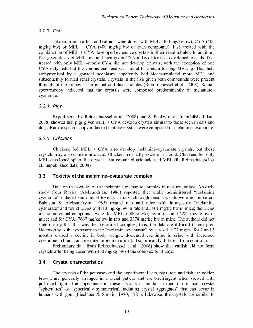
Tilapia, trout, catfish and salmon were dosed with MEL (400 mg/kg bw), CYA (400 mg/kg bw) or MEL + CYA (400 mg/kg bw of each compound). Fish treated with the combination of MEL + CYA developed extensive crystals in their renal tubules. In addition, fish given doses of MEL first and then given CYA 6 days later also developed crystals. Fish treated with only MEL or only CYA did not develop crystals, with the exception of one CYA-only fish, but the commercial feed was found to contain 6.7 mg MEL/kg. That fish, compromised by a gonadal neoplasm, apparently had bioaccumulated more MEL and subsequently formed renal crystals. Crystals in the fish given both compounds were present throughout the kidney, in proximal and distal tubules (Reimschuessel et al., 2008). Raman spectroscopy indicated that the crystals were composed predominantly of melamine–cyanurate. 3.2.4 Pigs
Experiments by Reimschuessel et al. (2008) and S. Ensley et al. (unpublished data, 2008) showed that pigs given MEL + CYA develop crystals similar to those seen in cats and dogs. Raman spectroscopy indicated that the crystals were composed of melamine–cyanurate. 3.2.5 Chickens
Chickens fed MEL + CYA also develop melamine–cyanurate crystals, but those crystals may also contain uric acid. Chickens normally excrete uric acid. Chickens fed only MEL developed spherulite crystals that contained uric acid and MEL (R. Reimschuessel et al., unpublished data, 2008). 3.3 Toxicity of the melamine–cyanurate complex
Data on the toxicity of the melamine–cyanurate complex in rats are limited. An early study from Russia (Aleksandrian, 1986) reported that orally administered “melamine cyanurate” induced some renal toxicity in rats, although renal crystals were not reported. Babayan & Aleksandryan (1985) treated rats and mice with intragastric “melamine cyanurate” and found LD50s of 4110 mg/kg bw in rats and 3461 mg/kg bw in mice; the LD50s of the individual compounds were, for MEL, 6000 mg/kg bw in rats and 4282 mg/kg bw in mice; and for CYA, 7667 mg/kg bw in rats and 3378 mg/kg bw in mice. The authors did not state clearly that this was the preformed complex; thus, the data are difficult to interpret. Noteworthy is that exposure to the “melamine cyanurate” by aerosol at 27 mg/m3 for 2 and 3 months caused a decline in body weight, decreased creatinine in urine with increased creatinine in blood, and elevated protein in urine (all significantly different from controls).
Preliminary data from Reimschuessel et al. (2008) show that catfish did not form crystals after being dosed with 400 mg/kg bw of the complex for 3 days.
3.4 Crystal characteristics
The crystals of the pet cases and the experimental cats, pigs, rats and fish are golden brown, are generally arranged in a radial pattern and are birefringent when viewed with polarized light. The appearance of these crystals is similar to that of uric acid crystal “spherulites” or “spherically symmetrical, radiating crystal aggregates” that can occur in humans with gout (Fiechtner & Simkin, 1980, 1981). Likewise, the crystals are similar to

Toxicological and Health Aspects of Melamine and Cyanuric Acid
14
spherulites of uric acid found in nephrocytes of the ascidian Corella inflata (Lambert et al., 1998). As uric acid crystals are soluble in formalin, several investigators (Dobson et al., 2008; Reimschuessel et al., 2008) examined wet tissue sections at necropsy prior to preserving kidney in formalin for routine histopathology. Reimschuessel et al. (2008) showed that melamine–cyanurate crystals dissolve in formalin over time. This may explain why crystals have not been found in the long-term studies evaluating MEL and CYA, where animals formed calculi and crystalluria was observed grossly, but no crystals were seen by histopathology.
The crystals in mammalian kidneys were reported primarily in distal tubules, although some were noted in proximal tubules. Crystals in fish kidneys were distributed throughout the renal tubules and collecting duct system. Tubular dilatation and epithelial necrosis occurred in close association with the crystals of all species. Chronic changes in animals that survive for several weeks following exposure show regenerating tubular epithelia, as well as glomerular fibrosis, interstitial fibrosis and chronic inflammation. Fish kidneys also began nephroneogenesis.
In preliminary data submitted to WHO (R. Reimschuessel et al., unpublished data, 2008), catfish were given daily doses of MEL and CYA to determine the NOEL dosage for crystal formation, which was found to be 20 mg/kg bw (comparable to 500 mg/kg in diet if fed at 4% body weight). The NOEL for a single dose was 10 mg MEL/kg bw and 10 mg CYA/kg bw. The NOEL for four daily doses was 2.5 mg/kg bw; however, 11 of 12 fish fed 2.5 mg/kg bw (comparable to approximately 63 mg/kg in feed) developed crystals. 3.5 Clinical effects
BUN and creatinine were elevated in rats, pigs and cats exposed to both MEL and CYA. The pigs developed bloody diarrhoea (Reimschuessel et al., 2008; S. Ensley et al., personal communication, 2008), and the cats became anorexic after the 1st day (Puschner et al., 2008). This is similar to the clinical reports of pets from 2007 and Jeong et al.’s (2006) report of dogs with gastrointestinal haemorrhages noted during necropsy. Dobson et al. (2008) reported an initial diuresis followed by anuria. 3.6 Residues
Edible tissue residues have been reported in four species of fish exposed to MEL and CYA (Andersen et al., 2008; Karbiwnyk et al., 2009) (Table 2). Preliminary data on depletion of MEL and CYA when fish are dosed at 20 mg/kg bw showed that peak MEL concentrations were 12.7 mg/kg in catfish and 12.2 mg/kg in trout on day 1 following administration. Within 7 days (catfish) or 14 days (trout), residues were <2.5 mg/kg. Peak CYA residues also occurred on day 1 following administration: 0.68 mg/kg in catfish and 2.6 mg/kg in trout. CYA muscle residues were <2.5 mg/kg by day 3.

15
Table 2. Residues in fish exposed to 400 mg/kg in feed for 3 days (note: tilapia did not get the full dose at all time periods)

Toxicological and Health Aspects of Melamine and Cyanuric Acid
16
4. COMMENTS 4.1 Mechanism of renal failure: melamine–cyanurate crystalline
nephropathy compared with uric acid nephropathy
The mechanism of kidney failure in animals developing melamine–cyanurate crystals is thought to be similar to acute uric acid nephropathy in humans, in that it is a mechanical obstruction that results in renal damage due to the uric acid crystal “spherulites” or “spherically symmetrical, radiating crystal aggregates” that can occur in humans with gout (Fiechtner & Simkin, 1980, 1981).
In humans, increased purine catabolism can also result in the deposition of uric acid crystals within the kidney, resulting in acute urate nephropathy (Conger, 1990). This precipitation is thought to occur due to the increased uric acid concentration in the filtered plasma and the increased acidity in the tubular lumen (urinary pH <5.5) (Conger et al., 1976; Conger & Falk, 1977). In humans, urate nephropathy can be caused by rapid tumour lysis, resulting in elevated uric acid levels (Casdorph, 1964; Davidson et al., 2004; Tiu et al., 2007), or by rhabdomyolysis (Moreau, 2005) and cardiovascular surgery (Chesney et al., 1975; Ellis, Brouhard & Conti, 1983). Davidson et al. (2004) stated that with certain diseases “the threshold at which uric acid precipitates into crystals may be reached. These crystals obstruct urine flow in the tubules, leading to uric acid nephropathy.” The authors, however, did not give a threshold level at which to expect this precipitation. Cases of lethal uric acid nephropathy have reported serum uric acid values of 80 and 185 mg/dl (Casdorph, 1964); however, uric acid levels of 12–20 mg/dl have also been reported in cases of acute uric acid nephropathy in children following cardiovascular surgery, during which uric acid levels rise (Ejaz et al., 2007). Patients develop oliguria or anuria with elevated serum BUN and creatinine.
Renal failure may result from both intrarenal crystal-associated obstruction and an elevation in renal pressure that reduces renal blood flow. Other mechanisms, such as renal vasoconstriction and inflammatory mediators, may also contribute to the pathogenesis of the acute renal failure associated with increased uric acid (Herz, Sauter & Bircher, 1972; Ejaz et al., 2007).
Uric acid nephropathy has been studied to some degree in experimental animal models. Humans and most primates do not have the enzyme, urate oxidase (uricase), that in most other mammals is responsible for the conversion of uric acid to allantoin. Therefore, most experimental mammalian models of uric acid nephropathy use inhibitors of urate oxidase (Stavric, Johnson & Grice, 1969; Wu et al., 1994). Stavric, Johnson & Grice (1969) dosed rats with uric acid in combination with oxonic acid, an s-triazine related to CYA. Frozen sections of kidneys showed many granular birefringent crystals, primarily in the medullary tubules, but also in the inner and outer cortices. Interstitial inflammatory infiltrates, necrotic cellular debris and regenerative basophilic epithelial cells were also noted. Similar crystals were also induced in urate oxidase–deficient mice (Wu et al., 1994).
Fridovich (1965) discovered the inhibitory function of oxalate on urate oxidase in an experiment that had used an old solution of urate to evaluate urate oxidase enzyme kinetics. Over time, some of the urate in the old solution had oxidized to oxonate, which acted as a strong competitive inhibitor. Other s-triazines, including CYA (but not MEL), also inhibited uric acid oxidase.
The effect of the melamine–cyanurate crystals on urate oxidase remains to be explored as a possible co-factor in the pathogenesis of renal damage following the ingestion of both MEL and CYA.

Background Paper: Toxicology of Melamine and Analogues
17
4.2 Special considerations of infant physiology
It is important to examine aspects of infant physiology that could make them more susceptible to MEL-associated renal damage. The renal stones formed in infants ingesting MEL-contaminated formula were composed primarily of uric acid and MEL (2:1). Normal uric acid concentrations in infants are higher than those of older children or adults (Table 3) (Stapleton, 1983; Fathallah-Shaykh & Neiberger, 2008). Table 3. Concentrations of uric acid in serum and urine of neonates, children and adults
Uric acid concentration (mg/dl) Serum Urine
Neonates (29–33 weeks) 7.7 4.8 Neonates (34–40 weeks) 5.2–6 1.7–2.8 Children (3–9 years) 3.4–3.6 0.34 Children (10–14 years) 4 0.34 Adults 4.3 (females), 5.1 (males) 0.4
Children (all pre-pubertal ages) have higher urinary uric acid clearance as well, with a
fractional excretion rate of 38–61% versus the adult rate of approximately 10%. These factors make children more susceptible to developing hyperuricosuria, which
may make them more likely to develop urinary uric acid precipitates. The neonatal kidney is, however, less efficient at concentrating and acidifying urine during the first weeks of life, which may help protect the kidney from potentially harmful effects of excreting the relatively large quantities of uric acid. However, increased uric acid load from surgery, drugs or asphyxia could overwhelm the neonatal kidney and pose a potential risk for developing acute urate nephropathy (Stapleton, 1983).
Elevated uric acid levels can also result in nephrolith formation. In fact, uric acid stones are the most common radiolucent kidney stones of children (Fathallah-Shaykh & Neiberger, 2008). Uric acid stone formation has also been associated with hypertension and low urinary pH (Negri et al., 2007; Losito et al., 2009). Factors that lower urinary pH, such as catabolism and acidosis, may promote stone formation as well.
In the case of the 2008 incident, milk was diluted and therefore contained less protein. Not only were infants drinking adulterated formula exposed to high levels of MEL, but they may have been receiving inadequate protein in their diet. This, along with the fact that infant basal serum uric acid levels and urine filtered levels are higher than those of adults, would increase the likelihood of uric acid stone formation.
Infants, and neonates of most species, have much higher growth rates and consume a larger percentage of food per unit of body weight than do older individuals (Mifflin et al., 1990). In addition, neonates feed much more frequently, sometimes every 2 h. Considering the half-lives of MEL and other triazines, it is conceivable that the frequency of feeding could increase plasma peak levels due to a repeated-dosing effect.
It is important to note that the crystals—either from uric acid nephropathy or the crystals of melamine–cyanurate—would not show up on ultrasound. These are microscopic changes. Only gross structural changes in the kidney or stones larger than 2 mm would be seen with this technique. Additional methods to visualize early changes in kidneys, including diffuse calcareous deposits such as those seen in female rat kidneys in the NTP (1983) study, are needed.

Toxicological and Health Aspects of Melamine and Cyanuric Acid
18
4.3 Comparative aspects of uric acid metabolism Humans and most primates do not have the enzyme, urate oxidase (uricase), that in
most other mammals is responsible for the conversion of uric acid to allantoin (Wu et al., 1989; Watanabe et al., 2002). Consequently, humans have serum uric acid levels that are 10–20 times higher than those of other mammals. Dogs, cats, rats and other primates have serum uric acid concentrations in the range 0.1–1.18 mg/dl compared with human values of 4.3–5.1 mg/dl (Fanelli & Beyer, 1974; ISIS, 1999). It is therefore important to consider the normal endogenous uric acid concentrations when using animal models for risk assessment. MEL dosages needed to produce melamine–uric acid stones in normal rats with normal uric acid oxidase function may underpredict dosages needed in primates. Further, rats with impaired uric acid oxidase are more susceptible to crystal formation in uric acid nephropathy (Johnson, Stavric & Chartrand, 1969; Stavric et al., 1969) and also to uric acid stone formation (Lauterburg et al., 1977; Schramek, Heidenreich & Engelmann, 1993). Thus, the lack of uric acid oxidase may be one added risk factor for the human infant. 4.4 Mechanism of action: Melamine alone
The primary effect of consuming high doses of MEL has repeatedly been seen to be stone formation. This is true for both animal experiments (Ogasawara et al., 1995; Cremonezzi et al., 2001) and the infant formula incident (Chinese Center for Disease Control and Prevention, unpublished data, 2008)). In both animals and humans, the stones are composed primarily of uric acid and MEL. As MEL’s structure allows for hydrogen bonding with uric acid, co-precipitation is not unexpected. In the case of rats, the ratio is 1:1, whereas in the case of humans, it is closer to 2:1. Perhaps the higher concentrations of uric acid found in humans, and specifically neonates, contribute to the higher ratio of uric acid in the stones. 4.5 Comment on uric acid solubility
As uric acid crystals are exquisitely soluble in formalin, it is paramount that in cases of suspected uric acid nephropathy or in the case of stone formation, a subset of tissue samples should be preserved in ethanol or by freezing (Vernon, 2006). 4.6 Knowledge gaps
The United States Food and Drug Administration’s (FDA) Science Board, in response to the 2007 FDA safety/risk assessment of MEL and its analogues, identified the following knowledge gaps as deserving additional investigational effort in order to provide the basis for an even more sound safety/risk assessment: • Determine the concentration and crystallization of MEL-type compounds in urine of
different species, including possible co-crystallization (e.g. MEL and CYA) at lower concentrations, since hydrogen bonding enhances crystallization between the MEL-type compounds, and the effect of liquid and salt ingestion on the concentration of MEL-type compounds in urine.
• Toxicological studies with different species, including the examination of co-toxicity from exposures to multiple MEL-type compounds to determine whether there are other additive or synergistic effects.
• Studies to better understand MEL pharmacokinetics, including the effects of dehydration, common medications (diuretics) and other factors that alter renal excretion.
Background Paper: Toxicology of Melamine and Analogues
19
• Determine whether heating MEL-type compounds forms new compounds in foods with greater toxicity than that of the parent compounds.
• Conduct longer-term studies to determine potential toxic effects, including kidney, liver, reproductive or endocrine dysfunction.
• Determine whether biomarkers can be identified for predicting renal failure secondary to exposure to MEL-type compounds.
4.7 Recommendations and future work
During 2008, it became apparent that chronic ingestion of MEL-adulterated infant formula caused the formation of urinary system calculi in humans. These stones were recently reported to be composed of MEL and uric acid (Reimschuessel, 2008). This finding is similar to the findings in rats by Ogasawara et al. (1995). It would be advisable that studies be undertaken to try to evaluate potential treatments for MEL only–induced renal and bladder stones. Understanding the solubility of these stones in urine with different pH values and responses to chemicals used to treat uric acid nephropathy could help physicians develop treatment strategies.
5. REFERENCES Aldridge D et al. (1985). Three generation reproductive study in the rat with sodium salt of cyanuric acid (s-triazinetriol). Unpublished report from the International Research and Development Corporation. Submitted to WHO by the Industry Ad Hoc Committee on Isocyanurates (Report No. 497-001) [cited in WHO, 2004]. Aleksandrian AV (1986). Toxicity and sanitary standardization of melamine cyanurate (experimental data). Gigiena truda i professional’nye zabolevaniia, 1: 44–45 [translated from Russian by Language Innovations, LLC, Washington, DC, USA]. Allen LM, Briggle TV, Pfaffenberger CD (1982). Absorption and excretion of cyanuric acid in long-distance swimmers. Drug Metabolism Reviews, 13: 499–516. Altona RE, Mackenzie HI (1964). Observations on cyanuric acid as a source of non-protein nitrogen for sheep. Journal of the South African Veterinary Medical Association, 35: 203–205. Andersen WC et al. (2008). Determination and confirmation of melamine residues in catfish, trout, tilapia, salmon, and shrimp by liquid chromatography with tandem mass spectrometry. Journal of Agricultural and Food Chemistry, 56: 4340–4347. Babayan EA, Aleksandryan AV (1985). Toxicological characteristics of melamine cyanurate, melamine and cyanuric acid. Zhurnal Eksperimental’noi i Klinicheskoi Meditsiny, 25(4): 345–349. Barbee SJ et al. (1983). Metabolism and disposition of sodium cyanurate. Toxicologist, 3: 80. Barbee SJ et al. (1984). Metabolism and disposition of sodium cyanurate in the dog. Toxicologist, 4: 92. Baynes RE et al. (2008). Pharmacokinetics of melamine in pigs following intravenous administration. Food and Chemical Toxicology, 46(3): 1196–1200. Bingham E, Cohrssen B, Powell CH, eds. (2001). Patty’s toxicology, 5th ed. Vol. IV. Hydrocarbons/organic nitrogen compounds. New York, NY, John Wiley & Sons. Brown CA et al. (2007). Outbreaks of renal failure associated with melamine and cyanuric acid in dogs and cats in 2004 and 2007. Journal of Veterinary Diagnostic Investigation, 19: 525–531.

Toxicological and Health Aspects of Melamine and Cyanuric Acid
20
Buur JL, Baynes RE, Riviere JE (2008). Estimating meat withdrawal times in pigs exposed to melamine contaminated feed using a physiologically based pharmacokinetic model. Regulatory Toxicology and Pharmacology, 51(3): 324–331. Cascieri T et al. (1983). Absence of teratogenic response in rats with monosodium cyanurate. Toxicologist, 3: 65. Cascieri T et al. (1985). Comprehensive evaluation of the urinary tract after chronic exposure to cyanurate in drinking water. Toxicologist, 5: 58. Casdorph HR (1964). Acute uric acid nephropathy in leukemia. California Medicine, 101: 481–484. Chadwick M et al. (1982) Disposition and metabolism of 14C-labelled sodium cyanurate in dog. Unpublished report from Arthur D. Little, Inc. Submitted to WHO by the Industry Ad Hoc Committee on Isocyanurates (Report No. C-86329) [cited in WHO, 2004]. Chesney RW et al. (1975). Acute renal failure: an important complication of cardiac surgery in infants. Journal of Pediatrics, 87: 381–388. Cianciolo RE et al. (2008). Clinicopathologic, histologic, and toxicologic findings in 70 cats inadvertently exposed to pet food contaminated with melamine and cyanuric acid. Journal of the American Veterinary Medical Association, 233(5): 729–737. Clark R (1966). Melamine crystalluria in sheep. Journal of the South African Veterinary Medical Association, 37: 349–351. Clark R, Barratt EL, Kellerman JH (1965). A comparison between nitrogen retention from urea, biuret, triuret and cyanuric acid by sheep on low protein roughage diet. Journal of the South African Veterinary Medical Association, 36: 79–80. Cohen SM et al. (1999) An evaluation of the long-term toxicity/carcinogenicity of sodium isocyanurate. Unpublished report to Occidental Chemical Corporation, 25 February 1999, provided to United States Environmental Protection Agency and assigned MRID No. 44834403. Submitted to WHO by the Industry Ad Hoc Committee on Isocyanurates [cited in WHO, 2004]. Conger JD (1990). Acute uric acid nephropathy. Medical Clinics of North America, 74(4): 859–871. Conger JD, Falk SA (1977). Intrarenal dynamics in the pathogenesis and prevention of acute urate nephropathy. Journal of Clinical Investigation, 59: 786–793. Conger JD et al. (1976). A micropuncture study of the early phase of acute urate nephropathy. Journal of Clinical Investigation, 58: 681–689. Cremonezzi DC et al. (2001). Dietary polyunsaturated fatty acids (PUFA) differentially modulate melamine-induced preneoplastic urothelial proliferation and apoptosis in mice. Prostaglandins, Leukotrienes and Essential Fatty Acids, 64(3): 151–159. Cremonezzi DC et al. (2004). Neoplastic and preneoplastic lesions induced by melamine in rat urothelium are modulated by dietary polyunsaturated fatty acids. Food and Chemical Toxicology, 42(12): 1999–2007. Davidson MB et al. (2004). Pathophysiology, clinical consequences, and treatment of tumor lysis syndrome. American Journal of Medicine, 116: 546–554. Dellarco VL (1993). Genetic anomalies in mammalian germ cells and their significance for human reproductive and developmental risk. Environmental Health Perspectives, 101(Suppl. 2): 5–11. Dobson RL et al. (2008). Identification and characterization of toxicity of contaminants in pet food leading to an outbreak of renal toxicity in cats and dogs. Toxicological Sciences, 106: 251–262.

Background Paper: Toxicology of Melamine and Analogues
21
Ejaz AA et al. (2007). Could uric acid have a role in acute renal failure? Clinical Journal of the American Society of Nephrology (CJASN), 2: 16–21. Ellis EN, Brouhard BH, Conti VR (1983). Renal function in children undergoing cardiac operations. Annals of Thoracic Surgery, 36: 167–172. Fanelli GM, Beyer KH (1974). Uric acid in non-human primates with special reference to its renal transport. Annual Reviews in Pharmacology, 14: 355–364. Fathallah-Shaykh F, Neiberger R (2008). Uric acid stones. E-medicine (http://emedicine.medscape.com/article/983759-overview; updated 8 July 2008, accessed 2 December 2008). Fiechtner JJ, Simkin PA (1980). Monosodium urate monohydrate as spherulites. Advances in Experimental Medicine and Biology, 122A: 141–143. Fiechtner JJ, Simkin PA (1981). Urate spherulites in gouty synovia. JAMA: the Journal of the American Medical Association, 245: 1533–1536. Fridovich I (1965). The competitive inhibition of uricase by oxonate and by related derivatives of s-triazines. Journal of Biological Chemistry, 240: 2491–2494. Grey RS, Clark R (1964). The excretion of biuret in the urine of sheep fed biuret. Onderstepoort Journal of Veterinary Research, 31: 91–95. Hammond BG et al. (1985). Absence of mutagenic activity for monosodium cyanurate. Fundamental and Applied Toxicology, 5: 655–664. Hammond BG et al. (1986). A review of toxicology studies on cyanurate and its chlorinated derivatives. Environmental Health Perspectives, 69: 287–292. Hatfield EE et al. (1959). Biuret—a source of NPN for ruminants. Journal of Animal Science, 18: 1208–1219. Heck HD’A, Tyl RW (1985). The induction of bladder stones by terephthalic acid, dimethyl terephthalate, and melamine (2,4,6-triamino-s-triazine) and its relevance to risk assessment. Regulatory Toxicology and Pharmacology, 5: 294–313. Helwig J, Gembrandt C, Hildebrandt B (1996). Melamine—Prenatal toxicity in Wistar rats after oral administration (diet). Ludwigshafen, BASF AG, Department of Toxicology (Project No. 32R0242/94007). Hendry JA, Rose FL, Walpole AL (1951). Cytotoxic agents: 1, methylolamides with tumour inhibitory activity, and related inactive compounds. British Journal of Pharmacology and Chemotherapy, 6: 201–234. Herz R, Sauter V, Bircher J (1972). Fortuitous discovery of urate nephrolithiasis in rats subjected to portacaval anastomosis. Experientia, 28: 27–28. Hodge HC et al. (1965). Toxicity of sodium cyanurate. Toxicology and Applied Pharmacology, 7: 667–674. IARC (1999). Melamine. In: Some chemicals that cause tumours of the kidney or urinary bladder in rodents, and some other substances. Lyon, International Agency for Research on Cancer (IARC Monographs, Vol. 73). International Research and Development Corporation (1985). Chronic toxicity and oncogenicity study in rats: s-triazinetriol. Unpublished report from the International Research and Development Corporation. Submitted to WHO by the Industry Ad Hoc Committee on Isocyanurates (Report No. IR-80-177 (I80-441)) [cited in WHO, 2004]. IPCS (1987). Principles for the safety assessment of food additives and contaminants in food. Geneva, World Health Organization, International Programme on Chemical Safety (Environmental Health Criteria 70). ISIS (1999) Physiological data reference values—1999. International Species Information System (http://www.isis.org/).

Toxicological and Health Aspects of Melamine and Cyanuric Acid
22
IUCLID (2000). International Uniform Chemical Information Database. European Commission (http://iuclid.echa.europa.eu/). Jeong WI et al. (2006). Canine renal failure syndrome in three dogs. Journal of Veterinary Science, 7(3): 299–301. Johnson WJ, Stavric B, Chartrand A (1969). Uricase inhibition in the rat by s-triazines: An animal model for hyperuricemia and hyperuricosuria. Proceedings of the Society for Experimental Biology and Medicine, 131: 8–12. Karbiwnyk CM et al. (2009). Determination of cyanuric acid residues in catfish, trout, tilapia, salmon and shrimp by liquid chromatography–tandem mass spectrometry. Analytica Chimica Acta, 637(1–2): 101–111. Kim YB et al. (2008). Worsening nephrotoxicity by combination of melamine and cyanuric acid in rats. Unpublished report. Cheongju Chungbuk, Republic of Korea, Chungbuk National University, College of Veterinary Medicine. Kravitz SC, Diamond HD, Craver LF (1951). Uremia complicating leukemia chemotherapy; report of a case treated with triethylene melamine. Journal of the American Medical Association, 146(17): 1595–1597. Lambert CC et al. (1998). Uric acid accumulation in the solitary ascidian Corella inflata. Journal of Experimental Zoology, 282: 323–331 (http://faculty.fullerton.edu/kkantardjieff/cvs/j_exper_zool_1998.pdf; accessed 19 September 2007). Lauterburg B et al. (1977). The defect of uric acid metabolism in Eck-fistula rats. Journal of Laboratory and Clinical Medicine, 90: 92–100. Lipschitz WL, Stokey E (1945). The mode of action of three new diuretics: melamine, adenine and formoguanamine. Journal of Pharmacology and Experimental Therapeutics, 83: 235–249. Losito A et al. (2009). Increased acid excretion in kidney stone formers with essential hypertension. Nephrology, Dialysis, Transplantation, 24(1): 137–141. Luengyosluechakul S (2007). Evidence of melamine and related substances contamination to animal feed in Thailand. Bangkok, Thailand, Chulalongkorn University, Faculty of Veterinary Science, Department of Veterinary Medicine (http://www.vet.chula.ac.th/~tjvm/full_text/V37/v374/4_37_04.pdf; accessed 17 February 2009). Lusby AF, Simmons Z, McGuire PM (1979). Variation in mutagenicity of s-triazine compounds tested on four Salmonella strains. Environmental Mutagenesis, 1: 287–290. MacKenzie HI (1966). Melamine for sheep. Journal of the South African Veterinary Medical Association, 37: 153–157. MacKenzie HI, van Rensburg IBJ (1968). Ammelide and ammeline as nonprotein nitrogen supplements for sheep. Journal of the South African Veterinary Medical Association, 39: 41–45. Mast RW, Naismith RW, Friedman MA (1982). Mouse micronucleus assay of melamine. Environmental and Molecular Mutagenesis, 4: 340–341 (Abstract No. Bi-8). Mast RW et al. (1983). Metabolism, disposition and excretion of [14C]melamine in male Fischer 344 rats. Food and Chemical Toxicology, 21: 807–810. Mazaev VT (1962). On the question of the content of cyanuric acid and its monovalent sodium salt in the water of reservoirs. Gigiena i Sanitaria, 27: 13–19. Melnick RL et al. (1984). Urolithiasis and bladder carcinogenicity of melamine in rodents. Toxicology and Applied Pharmacology, 72: 292–303.
Background Paper: Toxicology of Melamine and Analogues
23
Mifflin MD et al. (1990). A new predictive equation for resting energy expenditure in healthy individuals. American Journal of Nutrition, 51: 241–247. Ministry of Health and Welfare, Japan (1997). Toxicity Testing Reports of Environmental Chemicals, 5: 429–442 [cited in OECD, 1999]. Moreau D (2005). Pharmacological treatment of acute renal failure in intensive care unit patients. Contributions to Nephrology, 147: 161–173. Negri AL et al. (2007). Clinical and biochemical profile of patients with “pure” uric acid nephrolithiasis compared with “pure” calcium oxalate stone formers. Urological Research, 35(5): 247–251. NTP (1983). Carcinogenesis bioassay of melamine (CAS No. 108-78-1) in F344/N rats and B6C3F1 mice (feed study). Research Triangle Park, NC, and Bethesda, MD, United States Department of Health and Human Services, Public Health Service, National Institutes of Health, National Toxicology Program (NTP Technical Report Series No. 245; NTP-81-86; NIH Publication No. 83-2501; http://ntp.niehs.nih.gov/ntp/htdocs/LT_rpts/tr245.pdf). OECD (1998). Screening Information Dataset (SIDS) for melamine. CAS No. 108-78-1. Paris, Organisation for Economic Co-operation and Development (http://www.inchem.org/documents/sids/sids/108781.pdf). OECD (1999). Screening Information Dataset (SIDS) for isocyanuric acid. CAS No. 108-80-5. Paris, Organisation for Economic Co-operation and Development (http://www.chem.unep.ch/irptc/sids/OECDSIDS/108805.pdf). Ogasawara H et al. (1995). Urinary bladder carcinogenesis induced by melamine in F344 male rats: Correlation between carcinogenicity and urolith formation. Carcinogenesis, 16: 2773–2777. Okumura M et al. (1992). Relationship between calculus formation and carcinogenesis in the urinary bladder of rats administered the non-genotoxic agents thymine or melamine. Carcinogenesis, 13(6): 1043–1045. Puschner B et al. (2007). Assessment of melamine and cyanuric acid toxicity in cats. Journal of Veterinary Diagnostic Investigation, 19: 616–624. Rajasekaran D et al. (1981). 13-week toxicity study in rats. Unpublished report from the International Research and Development Corporation, San Francisco Medical Center, San Francisco, CA. Submitted to WHO by the Industry Ad Hoc Committee on Isocyanurates (Report No.167-151) [cited in WHO, 2004]. Reimschuessel R (2008). Pathology of combined melamine/cyanuric acid. Presented at the China–US Workshops on Food Safety, Beijing, China, November 2008. Reimschuessel R et al. (2008). Evaluation of the renal effects of experimental feeding of melamine and cyanuric acid to fish and pigs. American Journal of Veterinary Research, 69: 1217–1228. Reyers F (2007). Melamine-contaminated pet food. The South African experience. Veterinary News, May issue, pp. 8–12. Reyers F (2008). Melamine is back. In milk and in South Africa. A veterinary perspective. Veterinary News, December issue. Rutledge JC et al. (1986). A balanced translocation in mice with a neurological defect. Science, 231(4736): 395–397. Schramek P, Heidenreich A, Engelmann UH (1993). Sex-dependent urolithiasis in the portacaval shunt rat. Part I. Urology International, 50(2): 77–81. Serota D et al. (1982). Thirteen-week toxicity study in mice: monosodium isocyanurate: Project No. 2169-100. Final report. Unpublished report from Hazleton Laboratories America. Submitted to WHO by the Industry Ad Hoc Committee on Isocyanurates [cited in WHO, 2004].

Toxicological and Health Aspects of Melamine and Cyanuric Acid
24
Serota D et al. (1986) 104-week oncogenicity study in mice: monosodium cyanurate. Project No. 2169-101. Unpublished report from Hazleton Laboratories. Submitted to WHO by the Industry Ad Hoc Committee on Isocyanurates [cited in WHO, 2004]. Sheu CW et al. (1978). Heritable translocation test on random-bred mice after prolonged triethylenemelamine treatment. Mutation Research, 50(2): 241–250. Sheu CW et al. (1982). Detection of triethylenemelamine-induced translocation heterozygotes in CD-1 mice: fertility and cytological methods. Mutation Research, 97(4): 257–265. Stapleton FB (1983). Renal uric acid clearance in human neonates. Journal of Pediatrics, 103(2): 290–294. Stavric B, Johnson WJ, Grice HC (1969). Uric acid nephropathy: An experimental model. Proceedings of the Society for Experimental Biology and Medicine, 130: 512–516. Thompson ME et al. (2008). Characterization of melamine-containing and calcium oxalate crystals in three dogs with suspected pet food–induced nephrotoxicosis. Veterinary Pathology, 45: 417–426. Tiu RV et al. (2007). Tumor lysis syndrome. Seminars in Thrombosis and Hemostasis, 33: 397–407. USFDA (2003). Code of Federal Regulations Title 21, Chapter I, Part 573, Section 573.220, Feed-grade biuret. Washington, DC, United States Food and Drug Administration (http://cfr.vlex.com/vid/19712997). Van der Merwe CT (1966) M.Sc. Thesis, University of the Free State, Bloemfontein, South Africa [cited in MacKenzie, 1966]. Vernon SE (2006). Preservation of tissue urate crystals with the use of a rapid tissue-processing system. Journal of Histotechnology, 29: 17–19. Watanabe S et al. (2002). Uric acid, hominoid evolution, and the pathogenesis of salt-sensitivity. Hypertension, 40: 355–360. Wheeler AG et al. (1985). Three generation reproduction study in rats administered cyanurate. Toxicologist, 5: 189. WHO (2004). Sodium dichloroisocyanurate. In: Safety evaluation of certain food additives and contaminants. Prepared by the sixty-first meeting of the Joint FAO/WHO Expert Committee on Food Additives. Geneva, World Health Organization (WHO Food Additives Series, No. 52; http://whqlibdoc.who.int/publications/2004/924166052X.pdf). Wongthai P et al. (2008). Effects of melamine on frog farms: A veterinary case report. Department of Farm Animal Resources and Production Medicine, Faculty of Veterinary Medicine, Kasetsart University, Kamphaengsaengsaen Campus, Nakorn Pathom, Thailand (http://elib.fisheries.go.th/LIBCAB/DRAWERS/ARTICLE/DATA0007/00007700.PDF). Worzalla JF et al. (1974). Metabolism of hexamethylmelamine-ring-14C in rats and man. Cancer Research, 34: 2669–2674. Wu X et al. (1989). Urate oxidase: Primary structure and evolutionary implications. Proceedings of the National Academy of Sciences of the United States of America, 86: 9412–9416. Wu X et al. (1994). Hyperuricemia and urate nephropathy in urate oxidase–deficient mice. Proceedings of the National Academy of Sciences of the United States of America, 91: 742–746.