bacillus gene encoding inducible dna glycosylase is ... · 6012 morohoshietal....
TRANSCRIPT

JOURNAL OF BACTERIOLOGY, Sept. 1993, p. 6010-60170021-9193/93/186010-08$02.00/0Copyright © 1993, American Society for Microbiology
Vol. 175, No. 18
Bacillus subtilis alkA Gene Encoding Inducible 3-MethyladenineDNA Glycosylase Is Adjacent to the ada OperonFUMIKO MOROHOSHI,1* KENSHI HAYASHI,2t AND NOBUO MUNAKATA'
Radiobiology Division' and Oncogene Division,2 National Cancer CenterResearch Institute, Tsukiji 5-1-1, Chuoku, Tokyo 104, Japan
Received 4 May 1993/Accepted 9 July 1993
In Bacilus subtilis, the adaptive response to DNA alkylation depends on the ada operon, which consists of theadaA and adaB genes, which encode methylphosphotriester DNA methyltransferase (AdaA protein) and06-methylguanine DNA methyltransferase (AdaB protein), respectively. A structural gene (aIk4) that encodes3-methyladenine DNA glycosylase was found upstream of the ada operon, but in the opposite orientation. Thiscluster of genes was mapped at about 235 kb from the SfiI recognition site near the origin of replication in thephysical map of the B. subtilis chromosome. Disruption of the alkA gene sensitized cells to N-propyl-N'-nitro-N-nitrosoguanidine, while its overproduction rendered cells highly resistant to N-propyl-N'-nitro-N-ni-trosoguanidine, indicating that lethal DNA damage produced by bulky alkylating agents was effectivelycounteracted by AlkA glycosylase. Transcription of the al4 gene was induced by treating adAA+ cells withmethylating agents concurrent with transcription of the ada operon. This was accomplished by usingmethylated AdaA protein bound to a 30-bp segment in the middle of the 100-bp sequence between thetranscriptional start sites of the aMl4 gene and ada operon. Thus, in this organism, the adaptive response toDNA alkylation is achieved by autologous activation of a divergent regulon composed of the genes for a DNAglycosylase and two species of DNA alkyltransferase.
When cells are treated with simple alkylating agents, suchas methyl methanesulfonate (MMS), N-methyl-N'-nitro-N-nitrosoguanidine (MNNG), or N-methyl-N-nitrosourea(MNU), various alkylation adducts are produced in DNA(38). The most abundant of these adducts, 7-methylguanine,seems innocuous, while the next most abundant, 3-methyl-adenine, is believed to be primarily responsible for celllethality (17).DNA glycosylases, which release 3-methyladenine from
DNA, are found in various organisms, including bacteria,yeasts, nematodes, and mammals (3, 5, 16, 27, 33). InEscherichia coli, two species of 3-methyladenine DNA gly-cosylase have been identified. 3-Methyladenine DNA glyco-sylase I (Tag protein), has a strict specificity for 3-methyl-adenine residues and is synthesized constitutively (34). Incontrast, 3-methyladenine DNA glycosylase II (AlkA pro-tein) acts on 7-methylguanine and 3-methylguanine residues,as well as on 3-methyladenine, and is inducibly synthesizedby adaptive treatment with low concentrations of alkylatingagents (9, 12, 28). Mutants defective in either one of thesespecies show elevated lethal sensitivity to MMS, and doublemutants are extremely sensitive (7, 12, 13, 41).We have studied the adaptive response to DNA alkylation
in Bacillus subtilis and have shown that this response ismediated by the AdaA protein (21, 23). This protein is one ofthe products of the ada operon, which consists of twooverlapping genes, adaA and adaB, that encode methylphos-photriester DNA methyltransferase (AdaA protein) and o6_methylguanine DNA methyltransferase (AdaB protein), re-spectively (21). We recognized the presence of at least twospecies of DNA glycosylase which release 3-methyladenine
* Corresponding author. Electronic mail address: [email protected].
t Present address: Laboratory of Genome Analysis, Institute ofExperimental Gene Informatics, Kyushu University, Maidashi,Higashi-ku, Fukuoka-shi 812, Japan.
in this organism. They seemed to be similar to E. coli Tagand AlkA with regard to the modes of expression andsubstrate specificities (25). However, neither of these spe-cies had been genetically identified.We now report that the gene alkA, for the inducible
glycosylase, is located divergently in the adjacent 5'-flankingregion of the ada operon. Transcription of the genes iscoregulated by methylated AdaA protein bound to thecentral region between the transcriptional start sites of bothgenes. The disruption of alkA sensitizes cells to bulkyalkylating agents, such as N-propyl-N'-nitro-N-nitroso-guanidine (PNNG), but not to MMS. The deduced aminoacid sequence exhibits significant similarities to those of theAlkA protein of E. coli (28) and the MAG protein ofSaccharomyces cerevisiae (2, 6).
MATERIALS AND METHODS
Bacterial stains and plasmids. The following B. subtilisstrains were used: TKJ1922 (thyA thyB hisHlOl metBlOlleuA8 lys-21), TKJ0922 (same as TKJ1922 but adaAl),TKJ2924 (same as TKJ1922 but adaB4) (24, 25), andTKJ1984 (same as TKJ2924 but alkA::cat). A shuttle vector,pHY300PLK (10), was obtained from Toyobo (Tokyo, Ja-pan). An E. coli expression vector, ptac-85 (19), that carriedtac promoter and Shine-Dalgarno sequences was a gift fromP. Marsh. Bluescript SK M13+ and the host strain XL1-blue,were purchased from Stratagene (La Jolla, Calif.). An inte-grable plasmid, pCP12 (32), which carried a selectable chlor-amphenicol acetyltransferase gene, was a gift from F. Kawa-mura. E. coli BK2012 (ada tag-2xth) (13) was obtained fromM. Sekiguchi (Kyushu University). E. coli K802, JM101,and JM109 were used as hosts for the plasmids.DNA sequencing. Unidirectional deletions of Bluescript
SK M13+ carrying a 2.2-kb EcoRI-SalI fragment (pBS2.2)(21) were used as templates for sequencing reactions. DNAsequences were determined by the dideoxy-chain termina-
6010
on April 6, 2020 by guest
http://jb.asm.org/
Dow
nloaded from

B. SUBTILIS alkA GENE 6011
TABLE 1. Oligonucleotide primers
Primer Sequence (nucleotides')I-i AATTCCTAAACOGATTCTAGA + (509-528)I-r TTCOGCTCGTACOGGTGGGAAC - (819-838)II-i TGGTTAGAATGTAATAGCAA + (192-211)II-r GGAATTAACCCTCACTAAAG Bluescript T3
primerIII-lac GCGGATCCGTGACATGGCATGAAGTCAATG + (304-323)III-Ibc GCGGATCCATGAATGCGAACCTTGGCTA + (361-380)III-rd GCGTCGACTTATTGAGTACATTCCGGTT - (1230-1249)IV-i GTGTTCAACCTCCAATGTCA adaB downstream
sequenceIV-r TCTCCAGCTCATGTGATGAT adaB downstream
sequenceV AAGGTTCGCATTCATGTCGA - (356-375)VI-I TTTATCTACTCCCGTTTAGC + (126-145)VI-r ATTGACTTCATGCCATGTCA - (305-324)
' Transcribed and nontranscribed strands are indicated as - and +,respectively.
b Nucleotides are numbered as shown in Fig. 1.c This primer includes a BamHI site at the 5' end.d This primer includes an Sall site at the 5' end.
tion method using Sequenase (U.S. Biochemicals, Cleve-land, Ohio) and analyzed with a MacVector gene analysisprogram (IBI, New Haven, Conn.).
Oligonucleotides and PCR amplification. The oligonucleo-tides listed in Table 1 were used as primers for polymerasechain reaction (PCR) and sequencing reactions. They weresynthesized with a DNA synthesizer (380B; Applied Biosys-tems, Foster City, Calif.) and purified with oligonucleotidepurification cartridges (Applied Biosystems). DNA frag-ments were amplified in a thermal cycler (Cetus, Norwalk,Conn.) with a Gene Amp Kit (Cetus) as described previously(22).Plasmid construction. Plasmid pCP2151, which was used
to disrupt the alkA gene, was constructed by inserting a330-bp fragment at an EcoRV site of pCP12. The 330-bpfragment from the coding region of the alkA gene wasamplified by PCR with phosphorylated primers I-1 and I-r, asshown in Table 1, and template DNA from TKJ1922 cells.The amplified DNA was purified by Centricon 30 (Amicon,Beverly, Mass.), treated with T4 DNA polymerase (Toy-obo), and ligated with EcoRV-treated and dephosphorylatedpCP12 with a ligation kit (Takara, Osaka, Japan). PlasmidpHYALK, used for expression of the alkA gene, was con-structed by inserting a 1,119-bp sequence from alkA gene atan SmaI-EcoRI site of pHY300PLK (Fig. 1). The alkAsequence was amplified by using phosphorylated primer II-1and primer II-r, with plasmid pBS2.2 (21) as the template.The amplified fragment was treated with T4 DNA poly-merase, digested with EcoRI, and ligated with pHY300PLKDNA digested with SmaI and EcoRI. ptacALKG andptacALKA were constructed by introducing 946- and 889-bpfragments from the GTG and ATG codons, respectively(nucleotides 304 and 361 in Fig. 2), into the cloning site ofptac-85, which consists of a BamHI-NcoI-SalI recognitionsequence (Fig. 1). The fragments amplified by PCR withprimers III-la and III-r or III-lb and III-r were digested withBamHI and Sall and ligated with BamHI- and Sall-digestedptac-85.Assay of 3-methyladenine DNA glycosylase. 3-Methylade-
nine DNA glycosylase activity was measured by the methodof Riazuddin and Lindahl (33). Logarithmically growing cells
1) pHYALKSmal +1 +912 EcoRI
NI I
2) ptacALKGlacl ptac SDBmlSall tats
GT (946 bp)
3) ptacALKAlacl ptac SD
Bal Sateto
A (889 bp)FIG. 1. Constructs of alk4-carrying plasmids. Coarsely striped
rectangles represent alkA coding sequences. SD, Shine-Dalgarnosequence.
were subjected to adaptive treatment by culturing in thepresence ofMNNG (1 ,ug/ml) at 37°C for 70 min. Crude sonicextracts of the cells were incubated at 37°C for 20 min withcalf thymus DNA that had been treated with [3H]dimethyl-sulfate (1.2 Ci/mmol; NEN, Wilmington, Del.). After incu-bation, ethanol was added to the mixture. The supernatantfraction was applied to paper chromatography together with7-methylguanine and 3-methyladenine and run in a solventsystem of isopropanol, ammonium hydroxide (26%), andwater (7:1:2).
Southern and Northern RNA blot and primer extensionanalyses. Southern and Northern blot and primer extensionanalyses were performed as previously described (20). Theprobe used for the blots was a 0.89-kb fragment of the alkAgene and a 0.6-kb fragment from the opposite side of thealkA gene across the SfiI site. These fragments were ampli-fied by PCR with primers III-lb and III-r and primers IV-land IV-r (Table 1). Primer V (Table 1), which was comple-mentary to the sequence from +53 to +72 from the transla-tion initiation site of the alkA gene, was used for primerextension analysis.DNase I footprinting assay. Crude sonic extracts were
prepared from control and MNNG-treated cells of strains
= _|zl jAsCsaCccsc=ACSSCassOCTCscACAsTCcsaTCsTATCaTTsscasGAeCCC&ATTsAasATTTca
CCGC.TTTSTSASACTSGOXCCAS-TTT ASCTACTCCCTA,COsATTCTTssCsSACTsaTcACaCT sacscsassasaacssssGTaa
TGTA&?TA6C6"TA&CA6A&LZSIflPAA6&1G&!TYA!O!GAGA3&pTTYCLACCACGTGA6,,TTCTTAThA,TATTAAGAT&&Q9A3,A&
scsaYGacc66TcaacsasssAssaacTh1G2CAACG?aCGOAacG2Gsc 6acsssssascaasaamso,ac,,aoascmaa
V ST1W Z V N D V I V I T L P1 I D Y N a N L C Y L T I Z r. *3
C Y Y 2 X 3 N IfX X X V X a I G Z X R 5 L V Q V 8 V X * N X Q KAI,cscaOasaAsacTCATaaaCsaaTCATAOCoCATaGGSSLCCCGTCCTTAGTOCaGsaaGCGTACOasATaSasAAACa&aTI V Q VL N D QR P v Q * r. I 2X V h 2Y 2 3 2 3 3D L D N D LcassascaATsCCsa&A"?CAccscacTGT"c&sc_soozcGAcassTcGaaRAssaTsascascaxsocTsssCTscssAsacoamS P Ir Y 2 Y a r. A D P L L r. Y P a R Z 7 Y C L R vXGIPDL AACSCA2TAA&GC_scsasAA^AWJsoCsCATTccTaaasssCCoZGCCCaAcscTATG sTcGoTTGAT2sCCCoATTsAsssca
L C 11 C V L C Q Q X N L a F a Y 3 L R r. Q r V Z A F CCTssasGTGsOGwoGosssAsGCACAAccsacscsAAATACTTGACmCSCTAaGAaCaSC&TGAXaTTSWCSaTCTaTsAcGGAG r. r. Y W V 7 P P Y Z R X a I L T P T D L A D I S. Y T V R X XYsocsaaAAc"TAssc.GcscssCCACCAGLCTTCCAcescacosscCCCCsAaCC"CCsGATTAAA"G"LAcLGTsoaassaasccccsATX X G X a I L Y a 3 c Z L 8 R Z r. L N Z N N Z D a * Z N L X Z I RASCASS4GCAATACCCACTASS9GAASTOGCACSOALTCaSSCACAaSSACsGASSaCSCTrSSASC
C X C P W T a * Y V L Y R C L R Jr P T a r P X D D V G L X * 8 X X.
I L R 11 Y N R X P T Z D 3 X L 9 X S V P V I 3 W Q 3 Y A T r Y L WA&TATsACCTahsascaACCCaaaCcCTACCA&"AGLAsCTTscAGAaTaSkCGOCTCC&T"AsaASoAaGchASCTCAcotCCSSTTASSSaSoa v L YccsTsTCCTssACTcasscaTTTCACTACGAAACTGAaTcoGassAALcCCAAsaCCCAacsacsaacrsecCAKuwAAc TcsTAArTZac
1301 SSCSSSAC
FIG. 2. Nucleotide and predicted amino acid sequences of thealk4 gene. Nucleotides from 304 to 1212 presumably code for theAlkA protein. Transcriptions of the alkA gene and ada operon startat nucleotides 255 and 154 (diamonds), respectively, and proceed inopposite directions. The coding sequence of the adaA gene isunderlined. Dashed and double underlines indicate possible Shine-Dalgarno and -35 and -10 promoter sequences, respectively.
VOL. 175, 1993
I,
101
201
301
401
501
601
701
sol
901
1001
2101
1201
on April 6, 2020 by guest
http://jb.asm.org/
Dow
nloaded from

6012 MOROHOSHI ET AL.
TKJ0922(pHY5.2) and TKJ0922(pHY300PLK) as previouslydescribed (23). To methylate the AdaA protein fully, theextract (0.6 mg of protein) was incubated for 20 min at 37°Cin 70 mM N-2-hydroxyethylpiperazine-N'-2-ethanesulfonicacid (HEPES)-KOH (pH 7.8)-i mM EDTA-1 mM dithio-threitol-5% glycerol with calf thymus DNA (40 ,g) that hadbeen treated with MNU. The mixture was dialyzed againstthe same buffer for 2 h at 4°C. To prepare target DNAfragments for the binding reaction, a 199-bp sequence (cor-responding to nucleotides 126 to 324 in Fig. 2) was amplifiedby PCR with primers VI-l and VI-r (Table 1), one of whichhad been 5' end labeled with [y-32P]ATP (7,000 Ci/mmol;ICN, Costa Mesa, Calif.).
Incubation of labeled DNA with the extract and DNase Itreatment were performed as described by Teo et al. (40).The reaction mixture (20 ,ul) contained end-labeled DNAfragments (1 pmol), cell extract (6 or 30 p,g of protein), 50mM HEPES-KOH (pH 7.8), 50 mM KCl, 10 mM MgCl2, 10mM dithiothreitol, 1 mM EDTA, 5% glycerol, 2 pg of bovineserum albumin, 0.05% Nonidet P-40, and poly(dI-dC) (0.1 or0.5 p,g). After incubation at 22°C for 60 min, 2 Il of DNaseI (10 ,ug/ml) and 5 mM CaCl2 were added and the mixturewas incubated for an additional 1 min at 22°C. The reactionwas terminated by adding 20 ,ul of stop solution (0.1 mMEDTA, 600 mM NaCl, 30 Fig of tRNA). The sample wastreated with proteinase K (200 ,ug/ml) for 30 min; this wasfollowed by extraction with phenol-chloroform and precipi-tation with ethanol. The precipitate was dissolved in 95%formamide and applied to a denaturing 6% polyacrylamidegel for electrophoresis.
Survival assay. The survival assay was carried out asdescribed previously (23).
Nucleotide sequence accession number. The nucleotidesequence of the B. subtilis alkA gene has been deposited inthe DDBJ data base under accession number D14465.
RESULTS
Identification of an ORF in the upstream region of the adaoperon. During sequence analysis of the ada operon, werecognized the presence of an open reading frame (ORF) inthe 5'-flanking region of the operon in the opposite direction.Either the GTG codon at nucleotide 304 or the ATG codonlocated 57 bp downstream of the GTG codon (Fig. 2) wasbelieved to be a putative translational start site. As shownbelow, we determined that the coding sequence beginning atthe GTG codon was functional in E. coli cells. A Shine-Dalgarno sequence, AAGGAG, was found immediately up-stream of the GTG codon. From this site, the ORF is capableof encoding a protein of 303 amino acid residues (Fig. 2). Bysearching the National Biomedical Research Foundationprotein data base, we found that the entire amino acidsequence of this ORF showed significant similarity to that of3-methyladenine DNA glycosylase II (AlkA protein) of E.coli (28).
Expression of the ORF in E. coli. To confirm the activity ofthis ORF, plasmid ptacALKG was constructed by insertinga 946-bp DNA segment from the GTG codon into expressionvector ptac-85 (Fig. 1) (18). The plasmid was introduced intoE. coli BK2012 (ada tag-2xth), which was hypersensitive toMMS because of defects in both 3-methyladenine DNAglycosylases I and II. Cells which contained the plasmidwere grown in the presence of isopropyl-3-D-thiogalactopy-ranoside (IPTG) for 1 h and then treated with MMS. Survivalwas determined by plating the cells in agar medium contain-ing IPTG. BK2012(ptacALKG) cells were about five times
E0.
3000
, 2000
c 1000
a',
0.0 0.5 1.0 1.5Protein (mg/ml)
_
2O 4000' 300' 200
a 100I..
2.0 0.0 0.5 1.0 1.5Protein (mg/ml)
2.0
FIG. 3. 3-Methyladenine DNA glycosylase activity of alk,A-dis-rupted cells carrying the cloned alkA gene. Release of 3-methylad-enine (left panel) and 7-methylguanine (right panel) was determined.Closed symbols, extracts from untreated cells; open symbols,extracts from MNNG-treated cells. Symbols: 0 and 0, TKJ2924(adaB4); A and A, TKJ1984(pHY300PLK); * and 0, TKJ1984(pHYALK).
more resistant to MMS than were BK2012(ptac-85) cells(data not shown). Another construct, ptacALKA, whichcarried an 889-bp DNA segment from the ATG codondownstream of the GTG codon (Fig. 1), did not conferresistance in this assay. We concluded that the ORF whichbegins at the GTG codon is active in conferring resistance toMMS and that 19 N-terminal amino acid residues are re-
quired for this activity.Disruption of the ORF. To identify the product, we dis-
rupted the ORF by using integrable plasmid pCP2151, whichcarried a central portion (330 bp) of the ORF. Cells of strainTKJ2924 (adaB4) were transformed with pCP2151, andchloramphenicol-resistant colonies were selected. Only thecells that integrated the plasmid in the ORF in the chromo-some by Campbell-type homologous recombination shouldexhibit resistance to the drug, since the plasmid could notreplicate autonomously in B. subtilis cells. The DNAs fromTKJ2924 cells and the transformants were examined bySouthern blot analysis with a probe from the coding se-
quence of this ORF. Compared with the DNA of the parentalstrain, the DNA from a transformant clone yielded bands ofhybridization at different positions upon digestion withEcoRI, BglII, and PvuII (data not shown). The patterns wereconsistent with those expected from the integration ofpCP2151 in the ORF. We referred to the strain with ORFdisruption as TKJ1984 (alkA::cat adaB4).
3-Methyladenine DNA glycosylase activity was assayedfor the extracts from cells of strains TKJ2924 and TKJ1984.The extracts from the cells of the two strains released equalamounts of 3-methyladenine from methylated DNA but didnot release 7-methylguanine. The extract from MNNG-treated TKJ2924 cells exhibited an increase of the release of3-methyladenine to about 1.3 times that from untreated cells.This extract also liberated significant amounts of 7-methyl-guanine. These results were consistent with our previousfinding that this organism contained both constitutive andinducible 3-methyladenine DNA glycosylases and that onlythe inducible enzyme could release 7-methylguanine (25).The extract from MNNG-treated TKJ1984 cells liberated thesame amounts of 3-methyladenine and 7-methylguanine asthe extract from untreated TKJ1984 cells (Fig. 3), indicatingthat the inducible enzyme was not synthesized.To confirm that the ORF directed the synthesis of induc-
ible 3-methyladenine DNA glycosylase, we introduced plas-mid pHYALK, which carried a 1.1-kb fragment containingthe entire ORF and adjoining sequences (Fig. 1), into strainTKJ1984. The introduction of pHYALK significantly en-
c,.Zzr .I- PI- -olti P..,
I
I
J. BACTERIOL.
Dt
on April 6, 2020 by guest
http://jb.asm.org/
Dow
nloaded from

B. SUBTILIS alkA GENE 6013
hanced enzymatic activity, whereas the introduction ofvector plasmid pHY300PLK did not have a comparableeffect. The extracts from MNNG-treated and controlTKJ1984(pHYALK) cells liberated 2.5 and 1.3 times higheramounts of 3-methyladenine, respectively, than the extractsfrom untreated TKJ1984(pHY300PLK) cells. The extractfrom untreated TKJ1984(pHYALK) cells liberated amountsof 7-methylguanine similar to those liberated by the extractsfrom MNNG-treated TKJ2924 cells, and adaptive treatmentenhanced the release about 2.5-fold (Fig. 3).From these results, we concluded that this ORF was the
structural gene for the inducible 3-methyladenine DNAglycosylase. We referred to this ORF as the alkA gene in B.subtilis, in accordance with E. coli nomenclature.
Effects of alk4 disruption on the lethal sensitivity to alky-lating agents. Survival of cells of strains TKJ2924 (adaA+adaB4) and TKJ1984 (alkA::cat adaB4) was determinedupon the basis of a challenge with MMS. The lethal sensi-tivities of the two strains were almost equal (Fig. 4a).Moreover, introduction of the cloned alkA gene into strainTKJ1984 did not significantly alter the sensitivity to MMS,with or without adaptive treatment (Fig. 4b). These resultssuggest that neither elimination nor enhancement of theamount of the alkA gene product affected cellular sensitivityto MMS.We previously showed that adaptive MNNG treatment of
TKJ1922 (adaA+ adaB+) cells rendered the cells resistantnot only to methylnitroso compounds but also to bulkyalkylnitroso compounds, such as PNNG or butyl-nitroso-urea. The cellular responses to bulky agents were differentfrom those to methylating agents in that resistance to theformer was attained even in strains which were defective inthe adaB gene (25). Therefore, we believed that inducibleproteins other than AdaB methyltransferase were responsi-ble for the lethal adaptation to bulky alkylating agents.To explore the possibility that the alkA gene product is
involved in the cellular defense against bulky alkylators,cells of strains TKJ2924 (adaB4) and TKJ1984 (alkA::catadaB4) were challenged with PNNG. As shown in Fig. 4c,TKJ1984 cells were about 1.8 times more sensitive to PNNGthan were TKJ2924 cells. In the next attempt, cells of strainTKJ1984(pHYALK), which carried the cloned alkA gene,and TKJ1984(pHY300PLK) were subjected to adaptivetreatment and challenged with PNNG. TKJ1984(pHYALK)cells were extremely resistant compared with TKJ1984(pHY300PLK) cells, regardless of the treatment (Fig. 4d).Thus, the present results indicate that the alk4 gene productis responsible for the repair of lethal damage incurred byPNNG and that overproduction of the AlkA protein confershyperresistance to the agent.
Physical mapping of the alk4 gene. We attempted toposition the alkA and adaAB genes in the physical map of B.subtilis 168 (11). We found an SfiI recognition site about 1.8kb downstream from the 3' end of the adaB coding se-quence. Since the adaAB genes were mapped at around 230(of a total of 3600) between the cysA and lin genes by PBS1transduction (25), this restriction site should correspond tothe one 238 kb from the SfiI site near the replication origin inthe physical map (11). According to the map, the distances totwo adjoining NotI sites from the SfiI site were 10 and 68 kbin the counterclockwise and clockwise directions, respec-tively.To identify the restriction fragment which carried the alkA
gene, Southern blot analysis was performed. DNA fromTKJ1922 cells was digested with Sfi1 and NotI. The frag-ments were separated by 0.4% agarose gel electrophoresis
(a)
'a
aD
1
I
0o-3L0
(b)
1 2 3 4PNNG (mM)
'a
1 0.1
10.e2
'a
cn!
10 20 30 40 50 60
10-41 I.
0 1 2 3 4PNNG (mM)
5
FIG. 4. Survival of TKJ1984 cells with alkA disruption and ofthose carrying the cloned alkA gene upon challenge with MMS orPNNG. Panels: a and b, survival of MMS-treated cells; c and d,survival of PNNG-treated cells. Logarithmically growing cells werediluted twice with fresh medium and cultured in the presence (opensymbols) or absence (closed symbols) of 1 ,ug of MNNG per ml for70 min. The cells were challenged with either MMS for 15 min orPNNG for 30 min, and the colony-forming capabilities were deter-mined. Symbols: *, TKJ2924 (adaB4); *, TKJ1984 (alkA::catadaB4); 0 and 0, TKJ1984(pHY300PLK); A and A, TKJ1984(pHYALK).
and hybridized to either 0.89-kb fragments of the alkA geneor 0.6-kb fragments on the side opposite the alkA geneacross the Sfil site. As shown in Fig. 5, a band correspondingto 10 kb was detected when probed with the 0.89-kb frag-ment, while a band larger than 23 kb was detected whenprobed with the 0.6-kb fragment. These results placed the
1 2
231Kb
9_4b
FIG. 5. Physical mapping of the alkA gene by Southern blotting.DNA from TKJ1922 cells was digested with SfiI and NotI, separatedby electrophoresis, and transferred to a nylon membrane. The DNAwas hybridized to 0.89-kb fragments from the alkA coding sequence(lane 1) or with 0.6-kb fragments from the opposite side of the alkAgene across the SfiI site (lane 2).
.1....
la
I.... I.. 1. --. I.....
VOL. 175, 1993
01 0-31
on April 6, 2020 by guest
http://jb.asm.org/
Dow
nloaded from

6014 MOROHOSHI ET AL.
A BAIG
1 2 3 4
A B C
1.5Kb -i alkA
FIG. 6. Northern blot (A) and primer extension (B) analyses ofalkA transcripts. (A) Total RNA (4 ,ug) was separated by electro-phoresis in a 1.2% agarose-formaldehyde gel and transferred to anylon membrane. The RNA was hybridized to 0.89-kb fragments ofthe alkA sequence. Sources of RNA: lane 1, control TKJ1922(adaA' adaB+) cells; lane 2, MNNG-treated TKJ1922 cells; lane 3,control TKJ0922 (adaAl) cells; lane 4, MNNG-treated TKJ0922cells. (B) Total RNA and primer V (corresponding to nucleotides356 to 375 of the transcribed strand shown in Fig. 1) were used forreverse transcription. The DNA sequence of the transcribed strandis shown in lanes T, C, G, and A. Fifty micrograms of RNA fromcontrol (lane 1) or MNNG-treated (lane 2) cells of TKJ1922 and 75pg of RNA from control (lane 3) or MNNG-treated (lane 4) cells ofTKJ2924 (adaB4) were used as templates.
alkA and adaAB genes about 235 kb from the SfiI site nearthe replication origin with a gene order of cysA-al1kA-adaA-adaB-SfiI site-lin. Thus, the direction ofadaAB transcriptionwas the same as that of chromosomal replication but oppo-site to that of alkA transcription.
Transcription of the alkA gene. Total cellular RNAs fromMNNG-treated and control cells of strains TKJ1922 (adaA+)and TKJ0922 (adaAl) were separated by agarose (1.2%)-formaldehyde gel electrophoresis and hybridized to 0.89-kbDNA fragments from the alk4 gene. As shown in Fig. 6A,radioactive bands were observed only in the RNA fromtreated TKJ1922 cells. The major band corresponded to atranscript of about 1.5 kb. Transcripts were not detectable inthe RNAs from untreated TKJ1922 cells or from TKJ0922cells, regardless of the adaptive treatment. This result dem-onstrated that expression of this gene was transcriptionallyregulated and required the presence of AdaA protein.By primer extension analysis of RNAs from MNNG-
treated cells of TKJ1922 (adaA+ adaB+) and TKJ2924(adaA + adaB4), the transcriptional start sites of alkA mRNAwere determined. As shown in Fig. 6B, transcription startedat the adenine located 49 bp upstream of the initiation codonof the alkA gene. Since no significant ORF was observedwithin 500 bp of the downstream sequence of the gene, thetranscript was considered to be monocistronic. The se-quences ATGAGT and TAAACT from -34 and -12 bp ofthe transcriptional start site might correspond to -35 and-10 promoter sequences, respectively. Since transcriptionof the ada operon started 121 bp upstream of the initiationcodon of adaA (21), the distance between the transcriptionalstart sites of the ada operon and the alkA gene was exactly100 bp.DNase I protection assay of the AdaA-binding region. We
analyzed the binding of AdaA protein to the promoterregions of the alkA4 and adaAB genes by DNase I footprint-
Iad.A
-56
-31
alkA
-27
-50
adaA
23 45 6 7 1 '23 445 16 7 1 2 134 5 6 7 a 8 1
_|. N D ADD y N |AD |n N AD DII li+l+L7 1++1+I1J1+II I
FIG. 7. DNase I protection assay of AdaA-binding sequences.Crude extracts were prepared from MNNG-treated and controlTKJ0922(pHY5.2) cells and MNNG-treated TKJ0922(pHY300PLK)cells. The extracts were incubated with MNU-treated or untreatedDNA. After dialysis, the extracts were incubated with 199-bp DNAfragments including ada and alkA promoter sequences. The mixturewas treated with DNase I, and DNA fragments were analyzed byelectrophoresis. Tables at the bottom: AD, pHY5.2; PY;pHY300PLK; N, no extract; + and -, MNNG, with or withoutadaptive treatment of the cells, respectively, or M-DNA with orwithout in vitro treatment with MNU-treated DNA, respectively.The 5' end of the nontranscribed strand was labeled for panel A, andthat of the transcribed strand was labeled for panels B and C. Theamounts of extract used were 30 ,ug of protein for lanes 2, 4, and 6of panels A and B and lanes 3, 4, 7, and 8 of panel C and 5 ,ug ofprotein for the remaining lanes with extracts. The amounts ofpoly(dI-dC) used were 0.5 p1g for all of the lanes in panels A and Band for the even-numbered lanes in panel C and 0.1 ,ug for theodd-numbered lanes in panel C. The lanes marked A/G indicate thepositions of A and G bases in the sequences.
ing. AdaA protein was overproduced by culturing TKJ0922cells which carried recombinant plasmid pHY5.2, containingintact alkA and adaAB genes, in the presence of MNNG(21). Since AdaA protein could be methylated in vivo duringthe adaptive treatment, the extract from MNNG-treatedcells should contain both nonmethylated and methylatedAdaA proteins. The extract was incubated either with MNU-treated DNA, to methylate the AdaA protein further, or withuntreated DNA. As a target sequence for the binding assay,199-bp fragments, including a region between the transcrip-tional start sites of the two sets of genes, were amplified byPCR. A sequence covering about 20 bp of both strands, withprotrusions of 5 or 6 bp to the 3' direction of each strand,was protected from DNase I digestion by the extracts with orwithout prior incubation with methylated DNA in vitro (Fig.7A and B, lanes 2 to 5). This stretch corresponded to thenucleotides from -27 to -50 of the nontranscribed strandand from -31 to -56 of the transcribed strand of the alk,Apromoter. The extract from TKJ0922(pHY5.2) cells withoutMNNG treatment protected the same region to a lesserdegree (Fig. 7C, lanes 7 to 10). From our previous experi-
J. BACTERIOL.
on April 6, 2020 by guest
http://jb.asm.org/
Dow
nloaded from
B. SUBTILIS alkA GENE 6015
Trancrptlon Tnscription4- -
aSA adaA
IIAd.AingUth
adaB
I l
O.lkbp
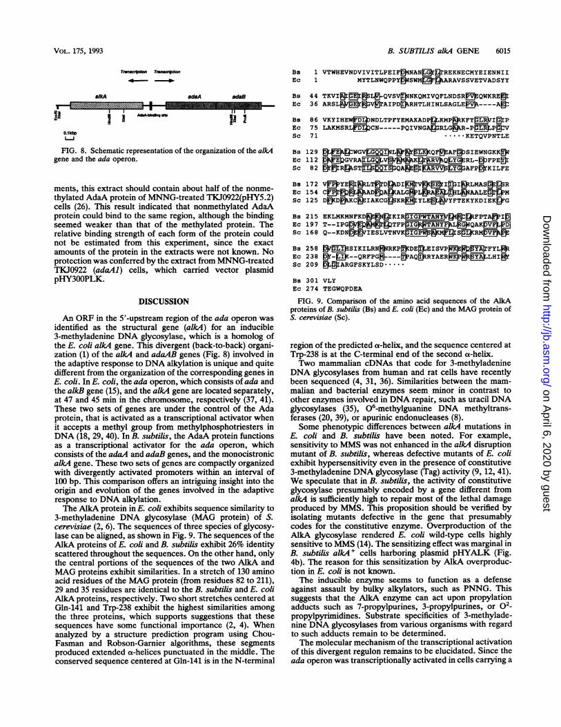
FIG. 8. Schematic representation of the organization of the alkAgene and the ada operon.
ments, this extract should contain about half of the nonme-thylated AdaA protein of MNNG-treated TKJ0922(pHY5.2)cells (26). This result indicated that nonmethylated AdaAprotein could bind to the same region, although the bindingseemed weaker than that of the methylated protein. Therelative binding strength of each form of the protein couldnot be estimated from this experiment, since the exactamounts of the protein in the extracts were not known. Noprotection was conferred by the extract from MNNG-treatedTKJ0922 (adaAl) cells, which carried vector plasmidpHY300PLK.
DISCUSSION
An ORF in the 5'-upstream region of the ada operon wasidentified as the structural gene (alk4) for an inducible3-methyladenine DNA glycosylase, which is a homolog ofthe E. coli alkA4 gene. This divergent (back-to-back) organi-zation (1) of the alkA and adaAB genes (Fig. 8) involved inthe adaptive response to DNA alkylation is unique and quitedifferent from the organization of the corresponding genes inE. coli. In E. coli, the ada operon, which consists of ada andthe alkB gene (15), and the alkA gene are located separately,at 47 and 45 min in the chromosome, respectively (37, 41).These two sets of genes are under the control of the Adaprotein, that is activated as a transcriptional activator whenit accepts a methyl group from methylphosphotriesters inDNA (18, 29, 40). In B. subtilis, the AdaA protein functionsas a transcriptional activator for the ada operon, whichconsists of the adaA and adaB genes, and the monocistronicalkA gene. These two sets of genes are compactly organizedwith divergently activated promoters within an interval of100 bp. This comparison offers an intriguing insight into theorigin and evolution of the genes involved in the adaptiveresponse to DNA alkylation.The AlkA protein in E. coli exhibits sequence similarity to
3-methyladenine DNA glycosylase (MAG protein) of S.cerevisiae (2, 6). The sequences of three species of glycosy-lase can be aligned, as shown in Fig. 9. The sequences of theAlkA proteins of E. coli and B. subtilis exhibit 26% identityscattered throughout the sequences. On the other hand, onlythe central portions of the sequences of the two AlkA andMAG proteins exhibit similarities. In a stretch of 130 aminoacid residues of the MAG protein (from residues 82 to 211),29 and 35 residues are identical to the B. subtilis and E. coliAlkA proteins, respectively. Two short stretches centered atGln-141 and Trp-238 exhibit the highest similarities amongthe three proteins, which supports suggestions that thesesequences have some functional importance (2, 4). Whenanalyzed by a structure prediction program using Chou-Fasman and Robson-Garnier algorithms, these segmentsproduced extended a-helices punctuated in the middle. Theconserved sequence centered at Gln-141 is in the N-terminal
Bs 1 VTWHEVNDVIVITLPEIF 4NAN @ JrREKNECMYEIENNIIEc 1 MYTLNWQPPY ?SWM P PARAVSSVETVADSYY
Bs 44 TKVI& B SL QVSV NKQMIVQFLNDSR BEQWKRE IEc 36 ARSL Sm Sv rAIPD >RHTLHINLSAGLE %----A
Bs 86 VKYIHEW NDLTPFYEMAKADP|LKMPKFYDLIDIPEc 75 LAKMSRLFI 2CN-----PQIVNGALJ3RLG&kR-PLR PmVSc 71 ....KETQVPNTLE
Bs 129 s>LKWGVG QP PQF AF mSIEWNGKK qEc 112 >B GVRA¢LQV>> gr SQ RL-SFPEDSc 82 a!1IRgSTgQIS,YQARBGV AFP&KILFEBs 172 VEDYEEILT0rDI@|rV&CfI0I&LMASgE&REc 154 Cl828gAD&A>ALG?LpA@gDILpAALEMSc 125 DD@kKC& IAKCG3SKREBrYLE Y NYFTEKYKDIEK RG
Bs 215 EKLMKMNFKD& Ig KIRrIGPWTANYM > g >FPTAffI0Ec 197 T--IPGSPBg TFPRTANYFWL G QAKESc 168 Q--KDN& TIESLVTNVKp~&MJIGMS(RM jb
Bs 258 Z YSIKILRNARKP>DE EISVP P &E FYLpIEc 238 Of-KD--QRFPGOY---AEAQ&RYAEREP8EX6kLHIWSc 209 EKgIARGFSKYLSD.Bs 301 VLYEc 274 TEGWQPDEAFIG. 9. Comparison of the amino acid sequences of the AlkA
proteins of B. subtilis (Bs) and E. coli (Ec) and the MAG protein ofS. cerevisiae (Sc).
region of the predicted ax-helix, and the sequence centered atTrp-238 is at the C-terminal end of the second a-helix.Two mammalian cDNAs that code for 3-methyladenine
DNA glycosylases from human and rat cells have recentlybeen sequenced (4, 31, 36). Similarities between the mam-malian and bacterial enzymes seem minor in contrast toother enzymes involved in DNA repair, such as uracil DNAglycosylases (35), 06-methylguanine DNA methyltrans-ferases (20, 39), or apurinic endonucleases (8).Some phenotypic differences between alkA mutations in
E. coli and B. subtilis have been noted. For example,sensitivity to MMS was not enhanced in the alkA disruptionmutant of B. subtilis, whereas defective mutants of E. coliexhibit hypersensitivity even in the presence of constitutive3-methyladenine DNA glycosylase (Tag) activity (9, 12, 41).We speculate that in B. subtilis, the activity of constitutiveglycosylase presumably encoded by a gene different fromalkA is sufficiently high to repair most of the lethal damageproduced by MMS. This proposition should be verified byisolating mutants defective in the gene that presumablycodes for the constitutive enzyme. Overproduction of theAlkA glycosylase rendered E. coli wild-type cells highlysensitive to MMS (14). The sensitizing effect was marginal inB. subtilis alkA+ cells harboring plasmid pHYALK (Fig.4b). The reason for this sensitization by AlkA overproduc-tion in E. coli is not known.The inducible enzyme seems to function as a defense
against assault by bulky alkylators, such as PNNG. Thissuggests that the AlkA enzyme can act upon propylationadducts such as 7-propylpurines, 3-propylpurines, or o2-propylpyrimidines. Substrate specificities of 3-methylade-nine DNA glycosylases from various organisms with regardto such adducts remain to be determined.The molecular mechanism of the transcriptional activation
of this divergent regulon remains to be elucidated. Since theada operon was transcriptionally activated in cells carrying a
WIAIM
VOL. 175, 1993
on April 6, 2020 by guest
http://jb.asm.org/
Dow
nloaded from

6016 MOROHOSHI ET AL.
defect in the AdaB protein, we concluded that the AdaAprotein was primarily responsible for activation (21). Acti-vation of the alkA gene also required the AdaA protein butnot the AdaB protein. The methylated AdaA protein bindsfrom -27 to -56 from the transcriptional start site of thealkA gene. This region corresponds to -45 to -74 from thetranscriptional start site of the ada operon. It is likely thatthis binding facilitates transcriptions in both directions. Thebinding sequence includes AGCAAGA from nucleotides-48 to -42 upstream of the alkA transcript. Similar but notidentical sequences, CGCAAGA and AGCAAAG, are seen
in the Ada-binding sequences of ada and alkA promoters ofE. coli, respectively (18, 29, 30, 40). Notable features of the100-bp segment between the transcriptional start sites of thetwo sets of genes include a high A+T content (75%) andunbalanced distribution of purine and pyrimidine bases inthe two strands, suggesting local distortions of the DNAhelix that might be further enhanced by binding of theprotein. Mutational analyses of the AdaA-binding region are
in progress and may identify the sequence necessary foractivation.
ACKNOWLEDGMENTS
We thank Philip Marsh (King's College, London, England) andFujio Kawamura (Institute of Applied Microbiology, Tokyo Univer-sity, Tokyo, Japan) for providing plasmids and Hiroshi Tanooka ofour laboratory for encouragement.
This work was supported in part by grants-in-aid from the Scienceand Technology Agency of Japan.
REFERENCES1. Beck, C. F., and R. A. J. Warren. 1988. Divergent promoters, a
common form of gene organization. Microbiol. Rev. 52:318-326.
2. Berdal, K. G., M. Bjoras, S. Bjelland, and S. Seeberg. 1990.Cloning and expression in Escherichia coli of a gene for an
alkylbase DNA glycosylase from Saccharomyces cerevisiae; a
homologue to the bacterial alkA gene. EMBO J. 13:4563-4568.3. Brent, T. P. 1979. Partial purification and characterization of a
human 3-methyladenine-DNA glycosylase. Biochemistry 18:911-916.
4. Chakravarti, D., G. C. Ibeanu, K. Tano, and S. Mitra. 1991.Cloning and expression in Escherichia coli of a human cDNAencoding the DNA repair protein N-methylpurine-DNA glyco-sylase. J. Biol. Chem. 24:15710-15715.
5. Chen, J., B. Derfler, A. Maskati, and L. Samson. 1989. Cloningof a eukaryotic DNA glycosylase repair gene by the suppressionof a DNA repair defect in Escherichia coli. Proc. Natl. Acad.Sci. USA 86:7961-7965.
6. Chen, J., B. Derfler, and L. Samson. 1990. Saccharomycescerevisiae 3-methyladenine DNA glycosylase has homology tothe AlkA glycosylase of E. coli and is induced in response toDNA alkylation damage. EMBO J. 9:4569-4575.
7. Clarke, N. D., M. Kvaal, and E. Seeberg. 1984. Cloning ofEscherichia coli genes encoding 3-methyladenine DNA glyco-sylases I and II. Mol. Gen. Genet. 197:368-372.
8. Demple, B., T. Herman, and D. S. Chen. 1991. Cloning andexpression of APE, the cDNA encoding the major humanapurinic endonuclease: definition of a family of DNA repairenzymes. Proc. Natl. Acad. Sci. USA 88:11450-11454.
9. Evensen, G., and E. Seeberg. 1982. Adaptation to alkylationresistance involves the induction of a DNA glycosylase. Nature(London) 296:773-775.
10. Ishiwa, H., and H. Shibahara. 1985. New shuttle vectors forEscherichia coli and Bacillus subtilis. II. Plasmid pHY300PLK,a multipurpose cloning vector with a polylinker, derived frompHY460. Jpn. J. Genet. 60:235-243.
11. Itaya, M., and T. Tanaka. 1991. Complete physical map of theBacillus subtilis 168 chromosome constructed by a gene-di-rected mutagenesis method. J. Mol. Biol. 220:631-648.
12. Karran, P., T. Hjelmgren, and T. Lindahl. 1982. Induction of aDNA glycosylase for N-methylated purines is part of theadaptive response to alkylating agents. Nature (London) 296:770-773.
13. Karran, P., T. lindahl, I. Ofsteng, G. B. Evensen, and E.Seeberg. 1980. Escherichia coli mutants deficient in 3-methylad-enine DNA glycosylase. J. Mol. Biol. 140:101-127.
14. Kassen, I., G. Evensen, and E. Seeberg. 1986. Amplified expres-sion of the tag' and alkA+ genes in Escherichia coli: identifi-cation of gene products and effects on alkylation resistance. J.Bacteriol. 168:642-647.
15. Kondo, H., Y. Nakabeppu, H. Kataoka, S. Kuhara, S. Kawa-bata, and M. Sekiguchi. 1986. Structure and expression of thealkB gene of Escherichia coli K-12 to methylating agents. J.Biol. Chem. 261:15772-15777.
16. Lindahl, T. 1976. New class of enzymes acting on damagedDNA. Nature (London) 259:64-66.
17. Lindahl, T. 1982. DNA repair enzymes. Annu. Rev. Biochem.51:61-87.
18. Lindahl, T., B. Sedgwick, M. Sekiguchi, and Y. Nakabeppu.1988. Regulation and expression of the adaptive response toalkylating. Annu. Rev. Biochem. 57:133-157.
19. Marsh, P. 1986. ptac-85, an E. coli vector for expression ofnon-fusion proteins. Nucleic Acids Res. 14:3603.
20. Morohoshi, F., K. Hayashi, and N. Munakata. 1989. Bacillussubtilis gene coding for constitutive 06-methylguanine-DNAalkyltransferase. Nucleic Acids Res. 17:6531-6543.
21. Morohoshi, F., K. Hayashi, and N. Munakata. 1990. Bacillussubtilis ada operon encodes two DNA alkyltransferases. Nu-cleic Acids Res. 18:5473-5480.
22. Morohoshi, F., K. Hayashi, and N. Munakata. 1991. Molecularanalysis of Bacillus subtilis ada mutants deficient in the adaptiveresponse to simple alkylating agents J. Bacteriol. 173:7834-7840.
23. Morohoshi, F., and N. Munakata. 1983. Adaptive response tosimple alkylating agents in Bacillus subtilis cells. Mutat. Res.110:23-37.
24. Morohoshi, F., and N. Munakata. 1985. Bacillus subtilis mutantsdeficient in the adaptive response to simple alkylating agents. J.Bacteriol. 161:825-830.
25. Morohoshi, F., and N. Munakata. 1986. Two classes of Bacillussubtilis mutants deficient in the adaptive response to simplealkylating agents. Mol. Gen. Genet. 202:200-206.
26. Morohoshi, F., and N. Munakata. 1987. Multiple species ofBacillus subtilis DNA alkyltransferase involved in the adaptiveresponse to simple alkylating agents. J. Bacteriol. 169:587-592.
27. Munakata, N., and F. Morohoshi. 1986. DNA glycosylase activ-ities in the nematode, Caenorhabditis elegans. Mutat. Res.165:101-107.
28. Nakabeppu, Y., T. Miyata, H. Kondo, S. Iwanaga, and M.Sekiguchi. 1984. Structure and expression of the alkA gene ofEscherichia coli involved in the adaptive response to alkylatingagents. J. Biol. Chem. 259:13730-13736.
29. Nakabeppu, Y., and M. Sekiguchi. 1986. Regulatory mecha-nisms for induction of synthesis of repair enzymes in responseto alkylating agents: Ada protein acts as a transcriptionalregulator. Proc. Natl. Acad. Sci. USA 83:6297-6301.
30. Nakamura, T., Y. Tokumoto, K. Sakumi, G. Koike, Y. Naka-beppu, and M. Sekiguchi. 1988. Expression of the ada gene ofEscherichia coli in response to alkylating agents. Identificationof transcriptional regulatory elements. J. Mol. Biol. 202:483-494.
31. O'Connor, T. R., and F. Laval. 1990. Isolation and structure ofa cDNA expressing a mammalian 3-methyladenine-DNA gly-cosylase. EMBO J. 9:3337-3342.
32. Price, C. W., M. A. Gitt, and R. H. Doi. 1983. Isolation andphysical mapping of the gene encoding the major a factor ofBacillus subtilis RNA polymerase. Proc. Natl. Acad. Sci. USA80:4074-4078.
33. Riazuddin, S., and T. Lindahl. 1978. Properties of 3-methylad-enine-DNA glycosylase from Escherichia coli. Biochemistry17:2110-2118.
34. Sakumi, K., Y. Nakabeppu, Y. Yamamoto, S. Kawabata, S.
J. BACTERIOL.
on April 6, 2020 by guest
http://jb.asm.org/
Dow
nloaded from
B. SUBTILIS alkA GENE 6017
Iwanaga, and M. Sekiguchi. 1986. Purification and structure of3-methyladenine-DNA glycosylase I of Escherichia coli. J. Biol.Chem. 261:15761-15766.
35. Sakumi, K., and M. Sekiguchi. 1990. Structures and functions ofDNA glycosylases. Mutat. Res. 236:161-172.
36. Samson, L., B. Derfler, M. Boosalis, and K. Call. 1991. Cloningand characterization of a 3-methyladenine DNA glycosylasecDNA from human cells whose gene maps to chromosome 16.Proc. Natl. Acad. Sci. USA 88:9127-9131.
37. Sedgwick, B. 1982. Genetic mapping of ada and adc mutationsaffecting the adaptive response of Escherichia coli to allylatingagents. J. Bacteriol. 150:984-988.
38. Singer, B. 1975. The chemical effects of nucleic acid alkylation
and their relation to mutagenesis and carcinogenesis. Prog.Nucleic Acids Res. Mol. Biol. 15:219-284.
39. Tano, K., S. Shiota, J. Collier, R. S. Foote, and S. Mitra. 1990.Isolation and structural characterization of a cDNA cloneencoding the human DNA repair protein for 06-alkylguanine.Proc. Natl. Acad. Sci. USA 87:686-690.
40. Teo, I., B. Sedgwick, M. V. Kilpatrick, T. V. McCarthy, and T.Lindahl. 1986. The intracellular signal for induction of resis-tance to alkylating agents in E. coli. Cell 45:315-324.
41. Yamamoto, Y., M. Katsuki, M. Sekiguchi, and N. Otsuji. 1978.Escherichia coli gene that controls sensitivity to alkylatingagents. J. Bacteriol. 135:144-152.
VOL. 175, 1993
on April 6, 2020 by guest
http://jb.asm.org/
Dow
nloaded from