axioskop 2 plus axioskop 2 mot plus upright microscopes ... · scopes combine a variety of...
TRANSCRIPT
Setting New Standards in Biology and Medicine
M i c r o s c o p y f r o m C a r l Z e i s s
Axioskop 2 plusAxioskop 2 mot plusUpright Microscopes for the Life Sciences

Fast and reliable viewing, analyzing and evaluation of an extensive rangeof biological specimens: all thanks to the excellent imaging quality ofthe Axioskop 2 plus and Axioskop 2 mot plus microscopes. All the standard microscopy techniques – from transmitted light bright-field to epi-fluorescence – can be used without any restrictions. Modern image-producing techniques and sophisticated, user-orientedsoftware are also available.
Both microscopes offer unsurpassed versatility and provide numerousfeatures for even greater ease of operation. The motorized Axioskop 2mot plus enables easy reproduction of many functions and is the basisfor software-controlled processes.
The Axioskop 2 plus and Axioskop 2 mot plus turn routine dailymicroscopy into something to look forward to. A choice of manual,coded and motorized components permits user-specific requirements tobe met in every detail.
The Standardin Life Science Microscopy
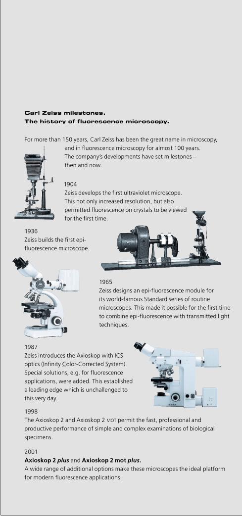
1904 Zeiss develops the first ultraviolet microscope. This not only increased resolution, but also permitted fluorescence on crystals to be viewedfor the first time.
Carl Zeiss milestones.The history of fluorescence microscopy.
For more than 150 years, Carl Zeiss has been the great name in microscopy, and in fluorescence microscopy for almost 100 years. The company’s developments have set milestones – then and now.
1936 Zeiss builds the first epi-fluorescence microscope.
1965Zeiss designs an epi-fluorescence module for its world-famous Standard series of routine microscopes. This made it possible for the first timeto combine epi-fluorescence with transmitted lighttechniques.
1987Zeiss introduces the Axioskop with ICS optics (Infinity Color-Corrected System).Special solutions, e.g. for fluorescence applications, were added. This established a leading edge which is unchallenged to this very day.
1998The Axioskop 2 and Axioskop 2 MOT permit the fast, professional and productive performance of simple and complex examinations of biological specimens.
2001Axioskop 2 plus and Axioskop 2 mot plus.A wide range of additional options make these microscopes the ideal platformfor modern fluorescence applications.

22
For research and routine
The Axioskop 2 plus andAxioskop 2 mot plus micro-scopes combine a variety of excel-lent optical and technologicallyadvanced features.
These high-performance micro-scopes offer maximum economyfor all standard applications in thelife sciences.
The modular design permits per-fectly tailored configurations tomeet any specific requirements.Optimized illumination and con-trasting techniques combined withthe ICS optics guarantee highimage quality. The ergonomic design of themicroscope components and con-trols ensures fatigue-free opera-tion over long periods of time.
Thanks to the coding and motor-ization of the Axioskop 2 motplus, reproducible microscope set-tings are now even more conven-ient and quicker than before.
Axioskop 2 plusAxioskop 2 mot plus

33

4
Pure Operating Convenience
Field of viewThe 23 mm field provides an opti-mum specimen overview withoutany need for tiring eye movement.
ControlsEasily accessible controls and er-gonomically designed microscopedetails permit a relaxed body pos-ture.
FocusingReliable, fast and precise focusingwith the Harmonic Drive™ gearboxensures high focusing speeds andexcellent reproducibility.
ViewingAll binocular tubes permit the pre-cise, reproducible setting of theuser’s specific interpupillary dis-tance. Swinging the eyepieces whileretaining the interpupillary distancepermits the viewing height to bechanged easily by up to 40 mm.Furthermore, the ergonomic tubesprovide different viewing heightsand angles. Thanks to eyepieces with foldingeyecups, the removal of spectaclesduring microscopy has become un-necessary.
Microscope stageThe vertically adjustable stage driveis positioned very close to the focus-ing drive to permit one-handedoperation of the microscope stageand focusing drive.
Properly applied, ergonomic design
relieves the mind as well as the
body. Daily microscope work over
many hours makes a fatigue-free
body posture particularly important.
Combined with simple, precisely
reproducible operations, this
ensures perfect results.
However, only the correct interac-
tion of all the major ergonomic
components increases productivity.
The Axioskop 2 plus and
Axioskop 2 mot plus microscopes
take all these aspects into consider-
ation.
Ergonomic tube

5
Relaxed, fatigue-free and efficient microscopy
6
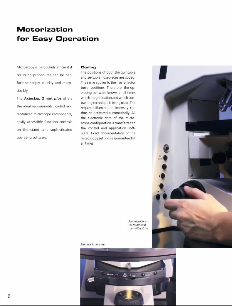
Motorizationfor Easy Operation
CodingThe positions of both the quintupleand sextuple nosepieces are coded.The same applies to the five reflectorturret positions. Therefore, the op-erating software knows at all timeswhich magnification and which con-trasting technique is being used. Therequired illumination intensity canthus be activated automatically. Allthe electronic data of the micro-scope configuration is transferred tothe control and application soft-ware. Exact documentation of themicroscope settings is guaranteed atall times.
Microscopy is particularly efficient if
recurring procedures can be per-
formed simply, quickly and repro-
ducibly.
The Axioskop 2 mot plus offers
the ideal requirements: coded and
motorized microscope components,
easily accessible function controls
on the stand, and sophisticated
operating software.
Motorized condenser
Motorized focus via traditional coarse/fine drive
MotorizationThe Axioskop 2 mot plus featuresone constantly motorized compo-nent: the z-drive. Nevertheless,manual operation is also possible –via a traditionally styled coarse / finedrive on both sides of the stand. In-crements of 50 nm are ideal for a va-riety of applications. Of course, mo-tor control with z-position readout isalso possible via the software.The motorized reflector turret per-mits the change from reflected lightto transmitted light and vice versa atthe push of a button. The requiredlight intensity is adjusted automati-cally.In addition to fluorescence filtersets, the reflector turret also acceptsadditional magnification optics –Optovar modules – for transmittedlight techniques.
The motorized shutter in thereflected-light beam path preventsunnecessary bleaching of the fluo-rescence dyes. The possibility ofintegrating shutter functions in thesoftware is particularly beneficialfor time-lapse experiments.Further motorized componentsinclude the universal condenser, anexternal excitation filter wheel andthe microscope stage. Integration of all the motorizedcomponents in the application soft-ware turns the microscope into ascreening system.
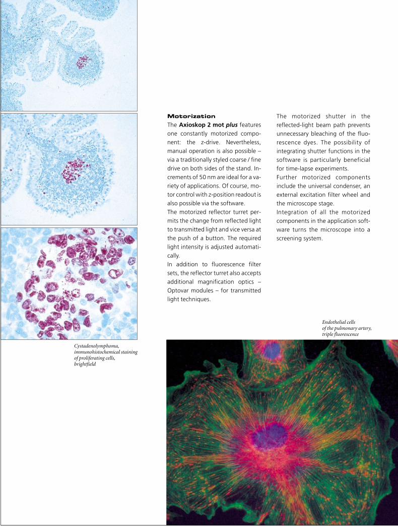
Cystadenolymphoma,immunohistochemical staining of proliferating cells,brightfield
Endothelial cells of the pulmonary artery,triple fluorescence

8
Memory Function
Thanks to the coded and motorized
components, all microscope set-
tings are known at all times. This
permits the operating software to
control all settings in accordance
with default values which can be
defined as required. For example,
the required contrasting technique
is automatically set after an objec-
tive change: brightfield, phase con-
trast or DIC, each with the correct
condenser aperture and the re-
quired light intensity.
Entire microscope configurations
can be easily defined, applied and
modified.
Condenser aperture automatically matched to the objective aperture
Fast change of microscope technique

Adenocarcinoma,immunocytochemical stainingof tumor cells,brightfield
Tongue,hematoxylin and eosin staining,brightfield

Transmitted Light Microscopy
Traditional microscopy techniques
such as brightfield, darkfield, phase
contrast, differential interference
contrast (DIC) and polarization can
be performed without any restric-
tion. The positioning of the DIC
analyzer as a module in the reflector
turret is particularly beneficial, since
turning of the reflector turret per-
mits the fast change from DIC to
brightfield or phase contrast.
Motorized and coded components
make this function extremely con-
venient.
BrightfieldThe traditional microscopy tech-nique for stained histology speci-mens such as tissue sections andsmears.
DarkfieldThe contrasting technique for thedisplay of minute structures at oreven below the resolution limits ofa light microscope.
Phase contrastIdeal for fine tissue and cell struc-tures in very thin, unstained or onlyslightly stained specimens.
PolarizationThe technique for the display ofbirefringence features of crystalsand biological molecules.
Universal condenserArthritis crystal,polarization
Thin section of bone, brightfield,darkfield,phase contrast

11
Cell culture,DIC
Differential InterferenceContrast (DIC)Nomarski interference contrast isideal for thick, unstained specimens. This technique provides optical sec-tions with optimum resolution, thuspermitting the clear identification of fine structures in defined focalplanes.
The contrast is set via objective-spe-cific DIC sliders in the nosepiece, sothe contrast is always maintainedonce it has been set. The DIC prisms in the condenser areavailable with integrated polarizer. When reflector turret and condens-er are motorized, a single push ofthe button is sufficient to changefrom epi-fluorescence to transmit-ted light DIC.
Analyzer module
DIC prism
Polarizer
DIC slider
DIC principle

Illumination and Contrasting in Transmitted Light
The transmission parameters of the
transmitted light beam path permit
all contrasting techniques in the
visible light range. The light intensi-
ty can be set either via modification
of the lamp voltage or by maintain-
ing the color temperature with
neutral-density filters. The switch-
able filter mount also accepts color
filters as required.
The condenser systemThe condenser plays a major part inthe setting of the various contrast-ing techniques and – when properlyadjusted – ensures efficient and ho-mogenous specimen illumination. All condenser systems can be set forKöhler illumination.

13
Beam pathTransmitted Light
Condensers for everytechniqueThe achromatic-aplanatic universalcondenser with aperture 0.9 is suit-able for almost all applications.Whether manual or motorized, itcovers the entire magnificationrange from 1x to 100x. Its seven po-sitions provide ample space for allthe contrasting components of tra-ditional microscopy: phase stops 1to 3, DIC prisms I to III with or with-out polarizer, darkfield stop and po-larizer for transmitted-light polar-ization microscopy.All motorized condenser functionscan be triggered at the push of abutton or via the AxioVision applica-tion software.
The achromatic switching condens-er with aperture 0.9 is suitable forless complex brightfield and phasecontrast applications.
Oil immersion objectives and con-densers with oil immersion opticspermit optimum resolution of themicroscope image. The achromatic-aplanatic condenser with aperture1.4 has been optically corrected forthis purpose.
Whenever specimen vessels requirelong working distances, the achro-
matic condenser 0.8 with 7 mmworking distance is used for bright-field, phase contrast and DIC appli-cations.
Complex darkfield with special dark-field condensers: oil immersion withthe ultra condenser 1.2/1.4, dry ap-plications with the dry darkfield con-denser 0.8/0.95.
Light sourceThe easily adjustable standard 100Watt halogen light source providessufficient light intensity for all thetraditional transmitted light con-trasting techniques.

Epi-FluorescenceMicroscopy
Fluorochromes and special labeling
techniques are being used more fre-
quently where the accurate repro-
duction of defined cell and tissue
structures is required. Today, epi-flu-
orescence is the standard fluores-
cence technique. It is particularly
efficient and permits a compact
design of the filter modules. Fur-
thermore, epi-fluorescence images
and those provided by the transmit-
ted light technique can be com-
bined in a single image.
Today, traditional fluorescence dyessuch as DAPI, FITC, Rhodamine andTexas Red are increasingly beingreplaced by even more efficientfluorochromes, e.g. Alexa Fluor™and Cyanin.Examinations of living specimens intransmitted light are supplementedand extended with fluorescent pro-teins (CFP, GFP, etc.). Experimentspreviously only possible in-vitro cannow be performed directly in theliving cell. The Antiflex epi-polarization systemvisualizes the adhesion structuresof cells on glass substrates. Togeth-er with fluorescence techniques,this permits the analysis of thefunction and structure of cytoskele-tal elements.
Hep-2 epithelial cells,immunofluorescenceof Cyclin I in cell nuclei(FITC)
Hep-2 epithelial cells,immunofluorescence of mitochondria(FITC)
1

15
1–3C. elegans,GFP-marked neurons.H. Hutter,MPI Heidelberg/Germany
2
3
Human endothelial cells,quadruple fluorescence DAPI, Alexa 350, Alexa 488,Phalloidin-Alexa 594.J. Zbaeren, Inselspital,Bern/Switzerland
Human endothelial cells,quadruple fluorescence DAPI, Alexa 350, Alexa 488,Phalloidin-TRITC.J. Zbaeren, Inselspital,Bern/Switzerland
Tongue, rat,double fluorescenceAlexa 594, DAPI.J. Zbaeren, Inselspital,Bern/Switzerland

16
Illumination and Contrastin Fluorescence
Reflector turretThe 5-position reflector turret isalways included in the basic stand.Manual, coded and motorized ver-sions are available. It permits easyswitching between transmitted andreflected light techniques with veryconvenient push-button operationin the motorized version.
P&C modulesThe filters required for fluorescencemicroscopy are integrated into thePush&Click modules which can beattached to the reflector turret andremoved from it without any tools.Therefore, many different fluo-rochromes can be examined quicklyand efficiently. The patented LightTrap principle provides optimumfluorescence signals without anydisturbing background noise.
External filter wheelA further increase in flexibility offluorescence microscopy is provid-ed by the motorized 8-positionfilter wheel (option) for theAxioskop 2 mot plus. Multiplefluorescence applications are easyto perform.
The beam path required for epi-
fluorescence can be integrated –
and retrofitted – into the Axioskop
2 plus and Axioskop 2 mot plus
microscopes. The viewing height
remains unchanged, since the stand
has been prepared for this integra-
tion.
FluorescenceconfigurationsThe beam path is equipped with anadjustable and centerable luminous-field diaphragm and aperture stop.The adjusting aid is available for easy adjustment of the illuminationsource after a lamp change.
The 5-position reflector turret for Push&Click filter cubes
Change of filter cubes – the Push&Click technique
Filter wheel
Adjusting aid

Light sourceThe high-energy HBO 103 mercuryvapor short-arc lamp is used for al-most all fluorescence techniques.Clearly allocated and easy-to-oper-ate setting screws in the lamp hous-ing permit fast and precise lamp ad-justment which can be monitoredthrough the window of the adjustingaid. The lamp is inserted into the pre-centered clamping device withoutany need for a special tool.
Light trapStray radiation passes through abeam splitter (2), leaves the filtercube (1) directly through the rearopening (3) and is then directed outof the beam path through the coni-cal mount (4) of the filter turret (5).The results are self-explanatory:considerable contrast enhancement,increased image definition and bril-liance, higher sensitivity due to animproved signal-to-noise ratio.
Beam path in a traditional reflector module
Beam pathwith light trap
5
4 3 2
1
Beam pathFluorescence

18
Achroplanobjectives
Plan-Neofluarobjectives
Fluarobjectives
Plan-Apochromatobjectives
A-Planobjectives
Objectivesfor Observation and Measurement
Infinity optics were designed and
computed by August Köhler as far
back as the thirties. But it was not
before the invention of ICS (Infinity
Color-corrected System) in 1986
that this optical system became the
centerpiece of modern Light
Microscopy.
Simply said, the function of the ICS
optics is to achieve optimum per-
formance by using the minimum
number of optical elements. Since
every optical element contributes
to the reduction of light transmis-
sion, the minimization of optical
components means the maximiza-
tion of the overall optical perform-
ance.
All in all, this resulted in visible
improvements of image contrast,
brightness and detail resolution.

The photon gatherersThe Fluar line is designed for maximum transmission and photon gath-ering efficiency. Made of special optical glass types, these objectivesfeature high numerical apertures, high contrast and high transmissionfor the entire visible spectrum up to the near UV. If you want to detectthe faintest fluorescence, Fluar objectives from Carl Zeiss are your bestchoice.
The ultimate imaging expertsCombining the highest color correction with highest numerical aper-tures, Plan-Apochromat objectives deliver the ultimate in resolutionand image definition and thus provide the finest details and subtlestcolor nuances. The large apertures enable brilliant brightfield and DICimages – and ensure high performance in fluorescence.
The allroundersWhere flexibility and a wide range of techniques are prime concerns,the universal semi-apochromatic Plan-Neofluar objectives are the bestanswer. With a transmission range up to the near UV, achromatic cor-rection, optimum working distances, low strain and high numericalapertures, they are ideal for brightfield, darkfield, phase contrast, DIC,polarization and fluorescence. High contrast and clear definitionmake them the right choice for image processing and analysis.
A sound basisThese are the basic objectives for your daily transmitted light routineand for epi-fluorescence microscopy with visible-light excitation.Thanks to their high image flatness across field diameters of 23 mm,Achroplan objectives are ideal for image documentation (photomi-crography and digital microscopy).LD-Achroplan objectives are also available for extremely long workingdistances with high apertures. Special Achroplan water objectives areprovided for applications in physiology.
An attractive alternativeAre you looking for a budget-priced alternative to Achroplan objec-tives? Here it is: A-Plan objectives are reliable in daily routine, clinical diagnostics and research. They provide rich contrast, are suitable forfluorescence and can be used with eyepieces with a field of view of upto 23 mm. They are applicable for brightfield and phase contrasttechniques.

20
Microscope Stages to Move the Specimen
Three stage models are available for
the precise movement of the speci-
mens in the x-y direction: manual,
coded and completely motorized.
They are attached to the stage and
condenser carriers of the Axioskop
2 plus and Axioskop 2 mot plus
microscopes without any need for
special tools.
Manual versionMechanical stages, manually movedin x and y, with a travel range of75x50 mm, are the most traditionaltypes of microscope stages. De-pending on requirements, they areavailable with coaxial x-y drive onthe right or left. The centerable ro-tary mechanical stage 75x50/240°Rwith x-y drive on the right provides arotation range of 240°. The controlscan be positioned at any heightrequired and their smoothness set asrequired by the user.
Coded and manual versionAn electrical vernier scale permitsthe reading of x-y data of the me-chanical stage 75x50R. The x-y posi-tions can be displayed either direct-ly or transferred to an applicationsoftware.The mechanical stage 75x50 motfeatures its own control unit. Thespecimen can be moved optionallyvia a 2-axis joystick or a coaxial elec-trical drive.
Fully motorized control The scanning stage DC 4� x 4� hasbeen designed for entirely motor-ized experiments. The connection tothe control computer is made via amotor control unit. However, the x-ymovements of the stage can also beperformed via an optional controlpanel for two axes.
Vertically adjustablecontrol elements on the stage drive

Specimen holdersA wide variety of specimen holders isavailable for microscopy. From thesimple model for manual mechani-cal stages to universal heatingframes for complex experiments andvarious inserts for fully motorizedmicroscope stages.
Mechanical stage withstage drive on the right
Mechanical stage withstage drive on the left
Motorized mechanical stage
DC scanning stage

Keywords: Pol color etched
Comments: Material is color-etched FeC/NiDelivered to MNO LtdDelivery Date: Aug. 31, 1998State: Final approval required
Central Quality Control Laboratory
QC - Image Archive
Amo International Inc.
22
A Single Systemfor Documentationand Evaluation
Digital photographyTraditional, time-consuming photog-raphy is being replaced more andmore by cameras with light-sensitivesensors. The image of the specimen isreadily available on the monitor andcan be processed immediately. Documentation is further facilitatedby the modern, digital AxioCam mi-croscope camera, featuring easy op-eration and optimum resolution.
Digitalimage documentationDocumentation is supported by theapplication-specific AxioVision soft-ware. Image recording, simple im-age processing, annotations and im-age archiving can be performed inno time at all. The hand-written,time-consuming recording of testprocedures is a thing of the past.And if required, the results can beprinted quickly on customized re-port forms.
Image documentation and analysis
are a must in daily routine and
research. No matter whether you
use an SLR, video or digital camera,
the binocular phototubes permit
easy recording and display of the
specimens.
Bild Osteo00012Datum 27.07.98Kommentar Knochenschnitt mit Goldner-Färbung (rot: Osteoid,
grün: Trabekel), WachstumsaktivitätBildaufnahme mit Axioskop, Durchlicht, PlanNeofluar 20 x,SONY 950 3-CCD RGB Kamera.
Stichwort Osteoid Trabekel
Musterstraße 4D-80801 MünchenTelefon: +49 (89) 95 97-0Fax: +49 (89) 9597-100
ZL GmbH
Titel Zahn00014Datum 27.07.98Autor M. MeierKommentar Grenzfläche zwischen Zahnimplantat (schwarz) und
umgebendes Gewebe(rot: Knochen, weiß: Bindegewebe)künstliche Zahnwurzel aus TitanAdaptation an den Knochen. Bildaufnahme mit Axioskop. Durchlicht, PlanNeofluar 5x, SONY MC 3309 RGB Kamera
Stichwort Zahnimplantat
MNO Institut
Titel Fluor00002Datum 22.05.98Autor Dr. Dr. MüllerKommentar Mehrfach-Fluorenszenz:
DAPIFITCTexas Red
Stichwort Fluorescence Multichannel
Prof. Dr. Dr. O. MeierGebäude 08 / 15D-10000 MusterstadtTelefon: +49 (99) 9597-0
AG Zellmorphologie

23

24
AxioVision,Ideal for Documentation
Together with the AxioCam digital
camera, the AxioVision application
software and its additional modules
are ideal for all image-related
documentation tasks using the
Axioskop 2 plus and Axioskop 2
mot plus microscopes. Image
acquisition, processing and archiv-
ing are time-saving and low-cost
procedures.
All the motorized microscope com-
ponents are integrated into the
application workflow and can be
controlled via the software. All the
required documentation parameters
such as magnification factor, con-
trasting technique, exposure times,
etc., are ready and available at the
push of a button. Time-consuming
test records are a thing of the past.
Easy insertion of text and graphics elements
Triple fluorescence

25
DigitalPhotography
AxioVisionsystem packageEven the basic version of AxioVisionpermits complete control of all mo-torized microscope components, inaddition to image documentationand archiving. Suitable softwaremodules are available for complexapplications such as multichannelfluorescence, time-lapse microscopyand z-stacks, or combinations ofthese applications. This permits complex experiments tobe easily configured and triggered atthe push of a button. All system parameters are recordedautomatically and are available at alltimes for recurring experiments,thus ensuring the reproducibility ofrecording conditions.
Magnified section,cat cerebellum, silver staining.J. Zbaeren, Inselspital, Bern/Switzerland

ERRORRUN
POWER
MCU 28
3 4 5 6 7 8
10
20
3040
50
60
70
8090
100
Specimen holder, specialwith particular fixationparticularly for immersion objectives000000-1070-589
System overviewAxioskop 2 plus / Axioskop 2 mot plus
Heatable universal mounting frame A-H000000-1116-055Tempcontrol 37 analog (1-channel)000000-1116-057
Universal mounting frame forPetri dishes and specimen slider000000-1100-843
Mounting frame for heatingand cooling devices000000-1107-544
Specimen holder formechanical stage 75x50000000-1067-330
Eyepiece W-Pl 10x/23 Br.000000-1016-758Eyepiece Pl 16x/16 Br.444053-0000-000
Eyepiece Pl 16x/16 Br. foc.444054-0000-000
Eyepiece W-Pl 10x/23 Br.455043-0000-000(only in combination with the tubes452340-0000-000 and 452344-0000-000)(optional: diaphragm, screwable455043-0206-000)
Format reticuleMC 2.5x/d=26 mm454075-0000-000Format reticuleMC 3.2x/d=26 mm454076-0000-000
according price list)
Eyepiece eyecup444801-0000-000
Eyepiece Pl 10x/23 Br. foc.000000-1026-548
Auxillary microscope d=30000000-1006-362
(additional format reticules
Intermediate tube with light pointer and power unit452380-0000-000
Multi-observation equipment for Axioskop 2
Tube carrier R (connection) for multi-observation equipment452381-0000-000Tube carrier L (connection) for multi-observation equipment452382-0000-000Tube carrier R (panael) for multi-observation equipment452383-0000-000Tube carrier R (panael) for multi-observation equipment452384-0000-000Binocular tube 30°/20 for multi-observation452927-0000-000Eyepiece E-Pl 10x/20 Br. foc.444232-9902-000
Mechanical stage75x50 L000000-1063-836
Scanning stage DC 4"x4"000000-1027-823)(displacement rangemax. 65x50 mm withmotorized condensors)
Mechanical stage75x50 R(with electronic vernier scales)000000-1046-520
413507-9001-000)
Mechanical stage75x50/240° R000000-1067-325
Mechanical stage75x50 R000000-1063-835
DC motor control 3 axes, MCU 28457428-0000-000
Mechanical stage 75x50 motwith display unit000000-1025-145
Coaxial electr. drive000000-1034-960
Joystick for 2 axis000000-1033-996or
Specimen holder473448-0000-000
453538-0000-000453545-0000-000(according price list)
optional
for MOT-standCAN-Bus-cable 2.5 m457411-9011-000
optionalControl panel for 2 axis457433-0000-000(for manual operation)
Stage carrier with condenser carrier452327-0000-000orCondenser carrier for FS000000-1022-999
Axioskop 2 FS plus microscope stand000000-1066-600Axioskop 2 FS MOT microscope stand000000-1066-601
Filter set VIS/IR000000-1031-489
(with display unit SCD
26

3
Video adapter60 ENG 2/3" 1.0x456115-0000-000
Video adapter60 C 1/3" 0.4x456108-0000-000
Video adapter60 C 1/2" 0.5x456106-0000-000(000000-1069-415in preparation)
Video adapter60 C 2/3" 0.63x000000-1069-414
Video adapterC-mount
Video zoom44 C 1/3" (3 CCD)0.33x ... 1.6x452989-0000-000
Video zoom44 ENG 1/2"0.5x ... 2.4x452984-0000-000
Video adapterENG-mount
Video adapter60 C 2/3" 1.0x456105-0000-000
Video adapter44 ENG 1/2" 0.63x452992-0000-000
Adapter from 60 to 44456140-0000-000
Binocular ergonomyphototube6-25°/23 (100:100)452342-0000-000
Binocular tube 30°/23452340-0000-000
Binocular ergonomytube 20°/23,height adjustable000000-1104-293
Binocular ergonomyphototube 20°/23,height adjustable000000-1104-296
Binocular phototube with two TV ports30°/25 (100:0/50:50/0:100)452145-0000-000
Projector tube for image interpolation452181-0000-000
Intermediate zoom tube1.0x ... 2.5x cod.452180-0000-000
Double TV-tube mot. with 2 outputs000000-1054-146Accessoriesacc. price list
Double video adapter000000-1058-640
Optovar intermediate tube1.0x/1.25x/1.6x/2.0x/2.5x cod.452175-0000-000
Binocular phototube with sliding prism30°/25 (100:0/50:50/0:100)452143-0000-000
Adapter452969-0000-000
Senarmont polarizer DIC453622-0000-000
Color filter carrier 3xfor filter d=32 mm452159-0000-000
Polarizer fixed withlambda plate, rotatable445226-0000-000
Polarizer D, rotatable453620-0000-000
Polarizer D, fixed453615-0000-000
Polarizer with filter holder000000-1118-003
Video cameraas desired
Binocular phototube30°/23 (70:30)452344-0000-000
27

POWER LAMP
SAFETY
TEMP
HOURS
230
CURTIS
ttoArc
CarlZeiss
Level1
Level2
Hours
-
+
POWER LAMP
SAFETY
TEMP
HOURS
230
CURTIS
ttoArc
CarlZeiss
Level1
Level2
Hours
-
+
10
20
3040
50
60
70
8090
100
Single-lens reflex camera housingCONTAX-ARIA000000-1057-127
T2-adapter for CONTAX416010-0000-000(additional T2-adapter acc. price list)
Cable release416167-0000-000
Adapter T2 2.5x for SLR456005-0000-000 Attachment for 60-44
456140-0000-000
Compact digital camera,e.g. SONY DSC-S7572 MB, d=52mm (D)000000-0425-186
Microscope camera MC 80See price list for equipmentand accessories
Digital camera adapter44 M52x0.75000000-1108-984
Connector 60 formicroscope camerad=30456006-0000-000
Camera adapterD40 M52x0.75000000-1096-522
Active coobservation device for the Axioskop 2(transmitted light) with light point and power unit452379-0000-000for tubes 452340-0000-000 (only rear);452342-0000-000 (in front and rear)
8-position filter wheel fordia. 25 mm excitation filters(only with MOT stand, PC andmicroscope control software)000000-1008-106
Incident light equipment000000-1150-310
Transformer HBO 103 ebq 100 dc000000-1003-928
Variable intensity lamp control AttoArc 2 N HBOadjustable 100-120-220-240 V 50/60 Hz000000-1007-975
Adjusting aidfor HBO/XBO452369-0000-000
Collector N HBO 103/XBO 75000000-1007-976 orQuartz collector N HBO 103/XBO 75000000-1007-977
Lamp housing N HBO 103000000-1007-980
Mercury vapor short-arc lampHBO 103 W/2380301-9350-000
Achromatic collector000000-1007-978
Transformer mbq 52 ac-z
6-position FL filter slider446377-0000-000
Lamp housing HBO 50 incl.lamp socket447220-0000-000Collektor HBO 50/SF 25447270-0000-000Super-pressure mercury lampHBO 50381619-0000-000
Filter set D000000-1058-230
Notebook000000-1056-852/853Microscope controlsoftware000000-1068-563
Axioskop 2 plus microscope stand000000-1116-5766-position nosepiece H W 0.8with 5-position reflector turret000000-1059-2025-position nosepiece H DIC W 0.8with 5-position reflector turret000000-1056-430
Axioskop 2 mot plus microscope stand000000-1130-6605-position nosepiece H DIC W 0.8 cod.with coded, 5-position reflector turret000000-1058-2556-position nosepiece H W 0.8 cod.with coded, 5-position reflector turret000000-1059-2035-position nosepiece H DIC W 0.8 cod.with motorized, 5-position reflector turret000000-1144-840Shutter for incident light equipment000000-1144-850(only in combination withnosepiece 000000-1144-840)
100 HAL lamp housing with collector,lamp mount447219-0000-000Halogen lamp 12 V 100W380079-9540-000
for HBO 50 220-240 V000000-1113-833
28

for external power supply HALabsoluteley essential for Axioskop 2 FS
Power supply 12 V DC 100 W stabilized000000-1128-574
Achromatic switching condenser 0.9 H D Ph000000-1017-690
Achromatic switching condenser 0.9 H000000-1017-688
Achr.-aplanatic universal condenser 0.9 H D Ph DIC MOT000000-1159-531
Achr.-aplanatic universal condenser 0.9 H D Ph MOT000000-1159-529
Achr.-aplanatic universal condenser 0.9 H MOT000000-1159-528
DIC prism I/0.9 (with polarizer)000000-1005-002DIC prism II/0.9 (with polarizer)000000-1005-003DIC prism III/0.9 (with polarizer)000000-1005-004
DIC prism I/0.9000000-1004-901DIC prism II/0.9000000-1004-902DIC prism III/0.9000000-1004-903
Polarizer for condenser (000000-1159-531)000000-1028-973
Achr.-aplanatic universal condenser 0.9 H D Ph DIC000000-1159-530
Achr.-aplanatic universal condenser 0.9 H D Ph000000-1159-527
Achr.-aplanatic universal condenser 0.9 H000000-1159-526
Polarizer for condenser (000000-1159-530)000000-1028-973
Reflector module H000000-1046-274
Analyzer module fortransmitted light000000-1050-958
Reflector module FL000000-1046-281
Optovar module 1.25x000000-1046-284Optovar module 1.6x000000-1046-283Optovar module 2.5x000000-1046-282
Compensator lambda/4, 6x20473714-0000-000
Compensator lambda, 6x20473704-0000-000
Compensator lambda,rotary, +/-8° 6x20473710-0000-000
Analyzer DIC/FL452374-0000-000(additional to barrier filter on FLor optovar module)
Wedge compensator0-4 lambda, 6x20000000-1140-663
Filter setsaccording price list
Antiglare screen452163-0000-000
DIC slideraccording price list
Objectives(ICS objectivesaccording to price list)
Adapter ringW 0.8 without NG000000-1104-291
Adapter ring W 0.8 NG 0.25000000-1104-289Adapter ring W 0.8 NG 0.10000000-1104-290
Specimen marker000000-1105-072Refill set forspecimen marker000000-0428-327
DIC prism II/1.4445375-0000-000
Ultra condenser1.2/1.4(0.75-1.0)465500-0000-000
Darkfield illuminator445314-9901-000
Dry darkfield condenser0.8/0.95(0.6-0.75)465505-0000-000
Condenser holder Zfor darkfield condenser445323-0000-000
Achr. condenser 0.8 Hd=6.7 mm000000-1087-444
Achr. condenser 0.8 H D Ph DICd=6.7 mm445445-9901-000
DIC prism III/1.4445376-0000-000
DIC prism III /0.8000000-1087-445
DIC prism III/0.8445485-0000-000
Achr. aplanatic condenser1.4 H Ph DIC d=0,5 mm445453-0000-000
29

110
280
Carl Zeiss Light Microscopy
P.O.B. 40 4137030 GöttingenGERMANYPhone: ++49 5 51 50 60 660Telefax: ++49 5 51 50 60 464E-Mail: [email protected]
www.zeiss.de/micro
Axioskop 2 plusAxioskop 2 mot plus
Prin
ted
on e
nviro
nmen
t-fr
iend
ly p
aper
,bl
each
ed w
ithou
t the
use
of c
hlor
ine.
For further details, please contact:
488
447
151
474
213
221
438
260
333
Design and specifications are subject to change in the interest of technical development 40-010 e/03.02

NOTESON THE PDF FILE
VIEW
When paging through the PDF file, as a rule you will seeone page on your screen at a time.
If you wish to see two facing pages of a catalog at a time, please activate the "Continuous - Facing Pages" item in the "View" menu of Acrobat Reader.
PRINTING
The pages of the PDF file are sized 210 x 277 mm.
Please mind that your printer may not print the page area from edge to edge.
To avoid picture cropping, please change the print size. We recommend 95%.
The area to be printed is framed by black lines on every page of this PDF file.