axial skeleton - mt. san antonio collegeinstruction2.mtsac.edu/crexach/anatomy 35/pdf lecture... ·...
TRANSCRIPT
Axial SkeletonAxial SkeletonAxial Skeleton
Dr. Carmen E. RexachAnatomy 35
Mt San Antonio College
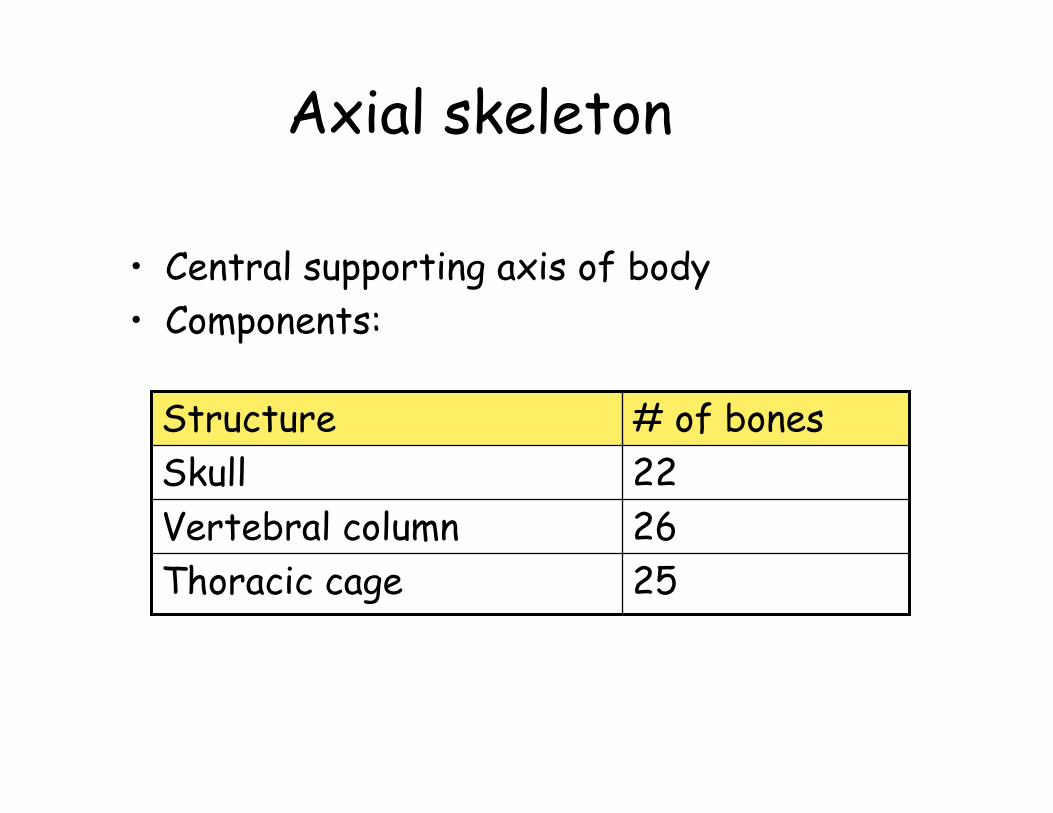
Axial skeleton
• Central supporting axis of body• Components:
25Thoracic cage26Vertebral column22Skull# of bonesStructure

SkullVertebraeThoracic Cage
Axial Skeleton

Skull• Most bones joined by sutures• Cranial bones and facial bones• Prominent cavities• Foramina

Cranium• Encloses cranial cavity• 8 Cranial bones• Meninges• 2 parts
– Calvaria = skull cap– Base = floor
• Cranial fossae = 3 basins– Anterior– Middle– posterior
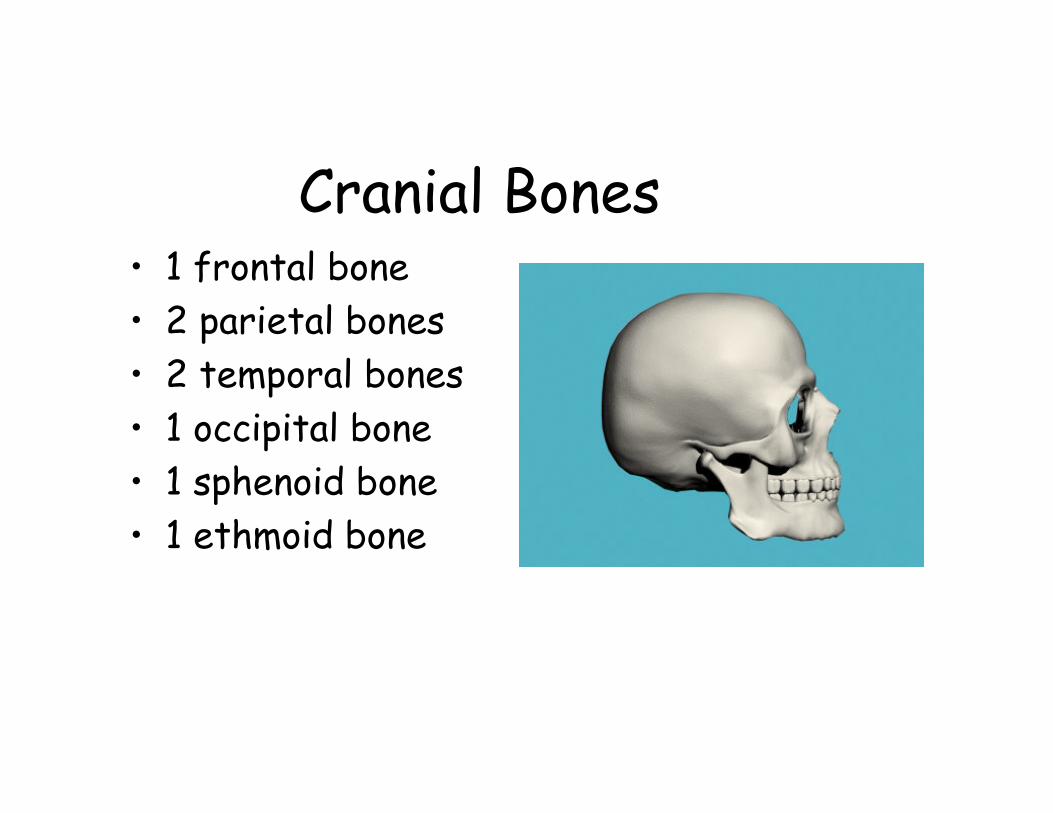
Cranial Bones• 1 frontal bone• 2 parietal bones• 2 temporal bones• 1 occipital bone• 1 sphenoid bone• 1 ethmoid bone
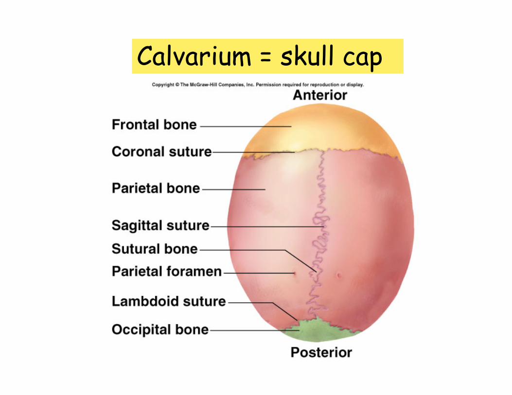
Calvarium = skull cap

Right lateral view

Interior view of right half of skull

Superior View (into cranial fossa)

Cranial Fossae

inferior view

posterior view• Wormian (sutural)
bones
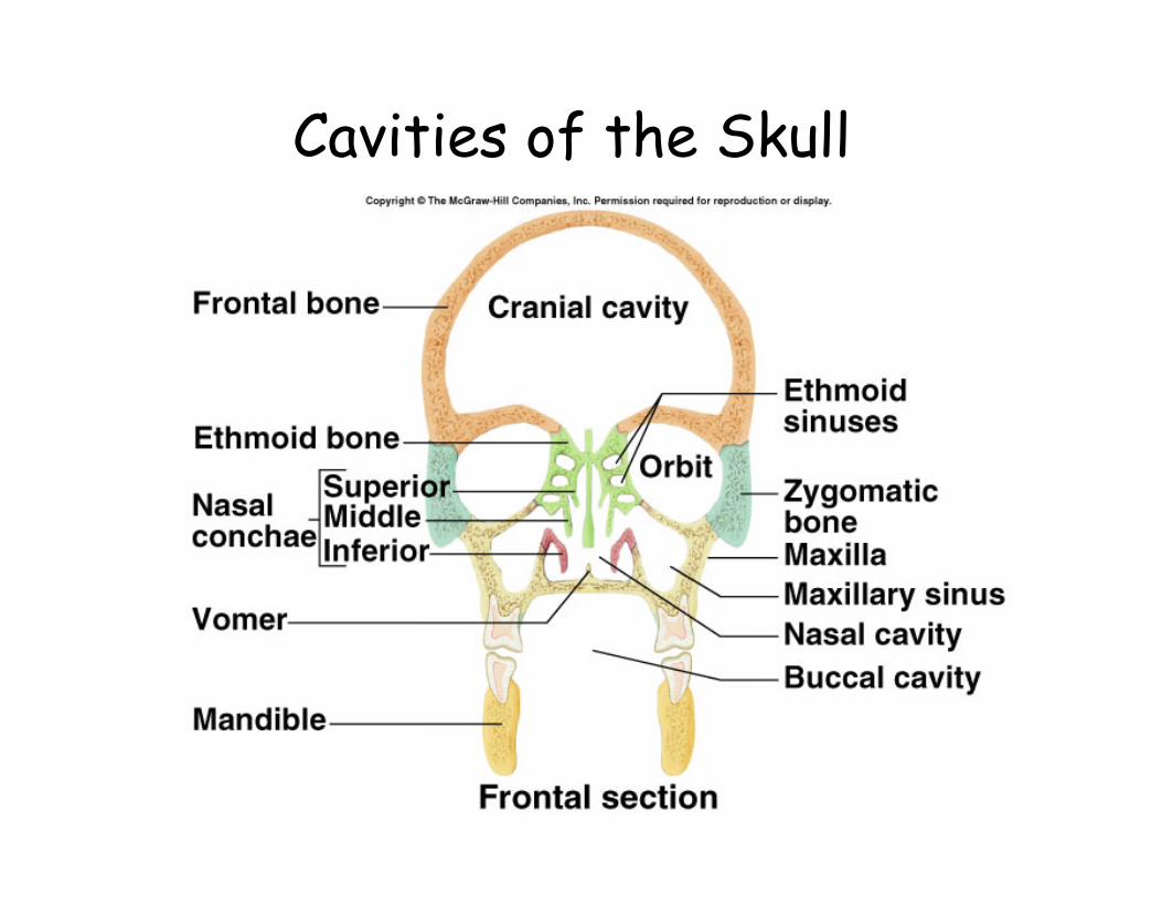
Cavities of the Skull
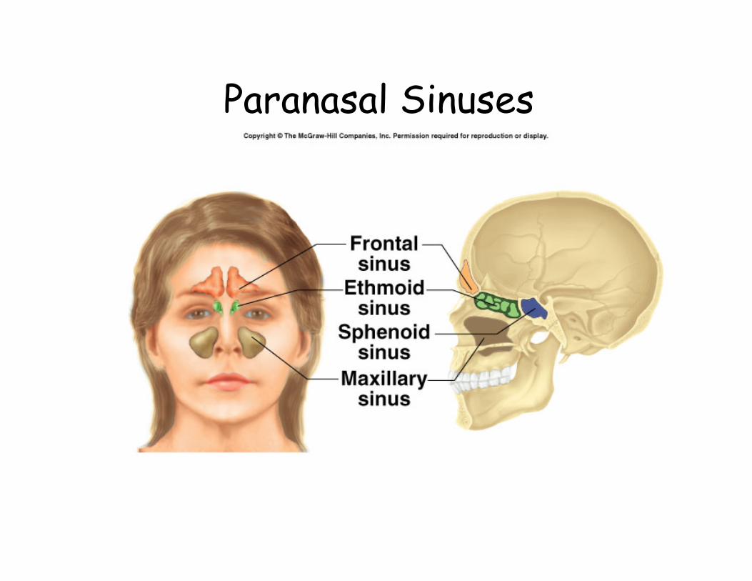
Paranasal Sinuses


Sphenoid Bone

Ethmoid Bone

Shaken baby syndrome

Facial bones
• Do not directly contact brain or meninges
• Functions– Support teeth– Shape and individuality to face– Form part of orbital and nasal cavities– Attachment site for muscles used for
facial expression and for mastication
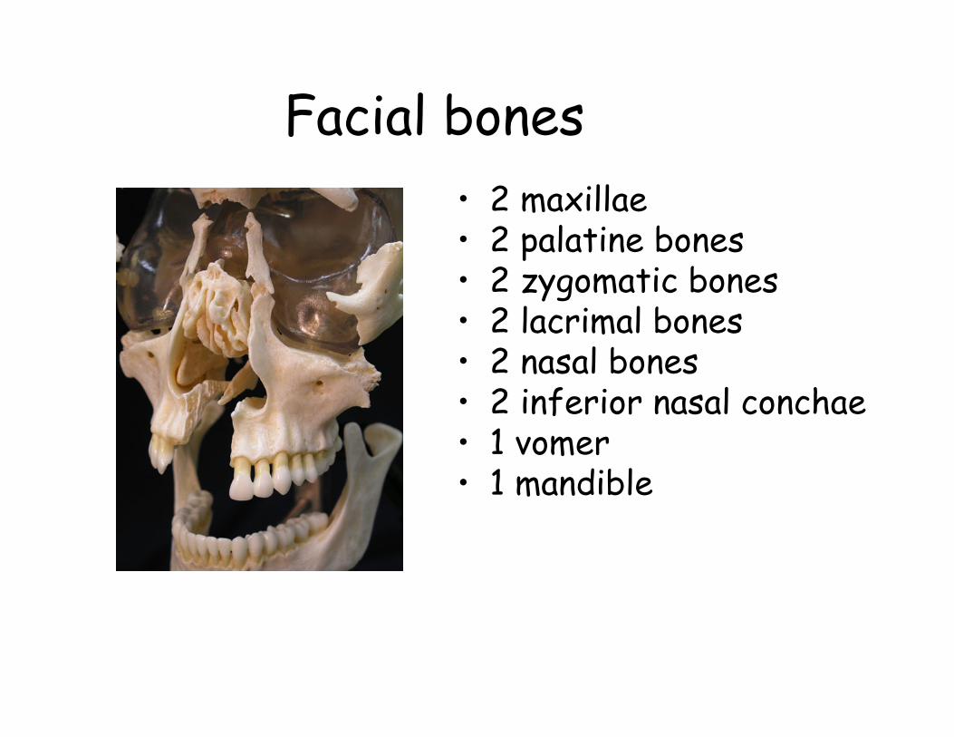
Facial bones• 2 maxillae• 2 palatine bones• 2 zygomatic bones• 2 lacrimal bones• 2 nasal bones• 2 inferior nasal conchae• 1 vomer• 1 mandible

anterior view
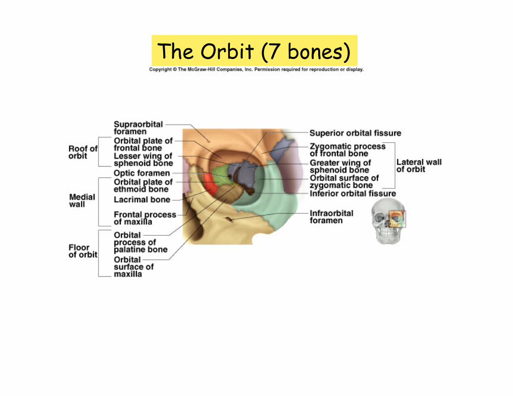
The Orbit (7 bones)
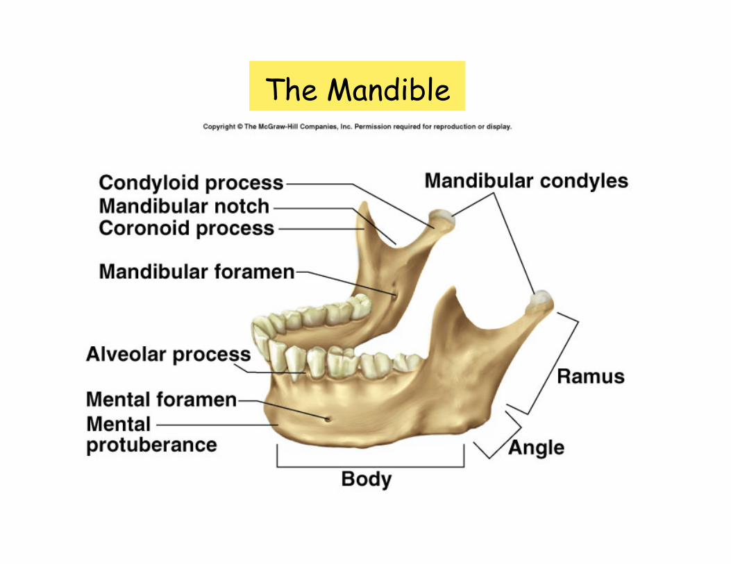
The Mandible

The Nasal Cavity
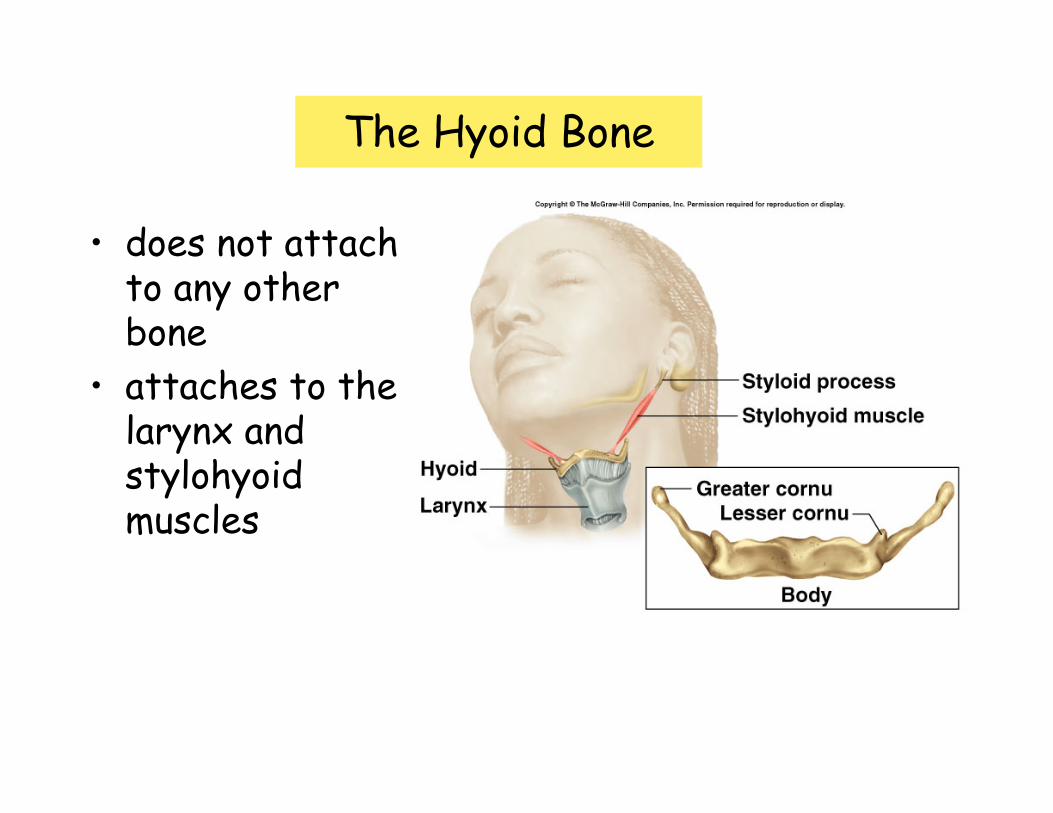
The Hyoid Bone
• does not attach to any other bone
• attaches to the larynx and stylohyoidmuscles

Auditory ossicles

Vertebral column• Physical support for skull and trunk• Protects spinal cord• Absorbs stress from movement• Attachment for postural muscles, limbs,
and thoracic cage• Composition
– 33 vertebrae– Intervertebral discs

Vertebral Column
7 cervical
12 thoracic
5 lumbar
4-5 sacral
3-5 coxygeal
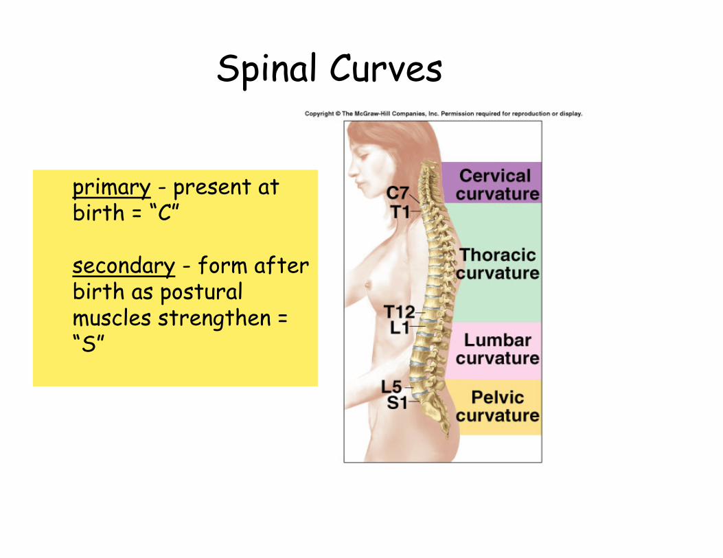
Spinal Curves
primary - present at birth = “C”
secondary - form after birth as postural muscles strengthen = “S”


General vertebral structure

Articulated vertebrae

Cervical vertebrae: Atlas and Axis

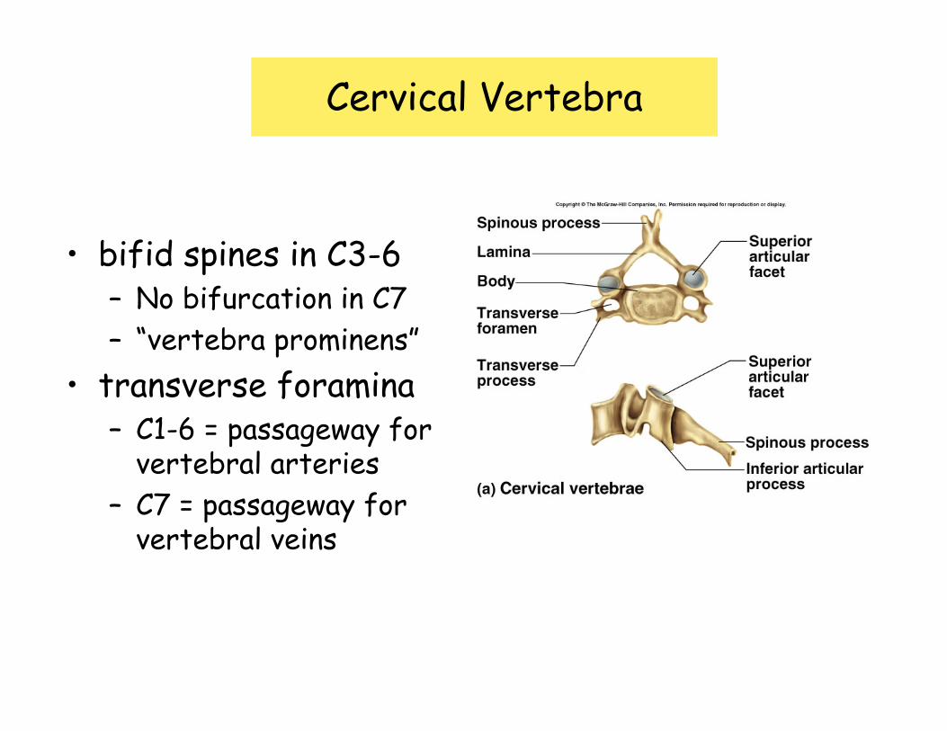
Cervical Vertebra
• bifid spines in C3-6– No bifurcation in C7– “vertebra prominens”
• transverse foramina– C1-6 = passageway for
vertebral arteries– C7 = passageway for
vertebral veins

• General Characteristics:– Pointed spinous process with sharp downward angle– Heart shaped body, more massive than cervicals– Costal facets for attachment of ribs
Thoracic Vertebrae

Lumbar Vertebrae
• Thick, stout body• Blunt, square
process• Orientation of the
articular processes– Superior = medial– Inferior = lateral

Sacrum
• Five separate bones begin to fuse ≈ 16yo– Transverse ridges (8)
• Anterior– Promontory (4)– Sacral foramina (11)– Alae (6)
• Posterior– Median sacral crest (11)– Lateral sacral crest (10)– Posterior sacral foramina (5)– Sacral tuberosity (4)
• Sacral canal (2)• Sacral hiatus (8)• Auricular surface (12)• Superior articular process (1)
anterior
posterior
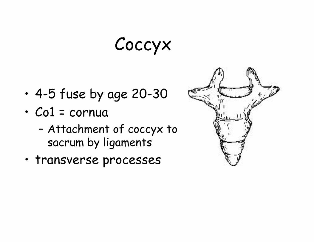
Coccyx
• 4-5 fuse by age 20-30• Co1 = cornua
– Attachment of coccyx to sacrum by ligaments
• transverse processes

Thoracic Cage• Components
– Thoracic vertebrae– Sternum
• Manubrium• Body• Xiphoid
– Ribs• 7 true• 3 false• 2 floating

Thoracic cage• Functions
– Encloses lungs & heart– Attachment for pectoral girdle and
upper limbs– Protect spleen, liver, and help protect
kidneys
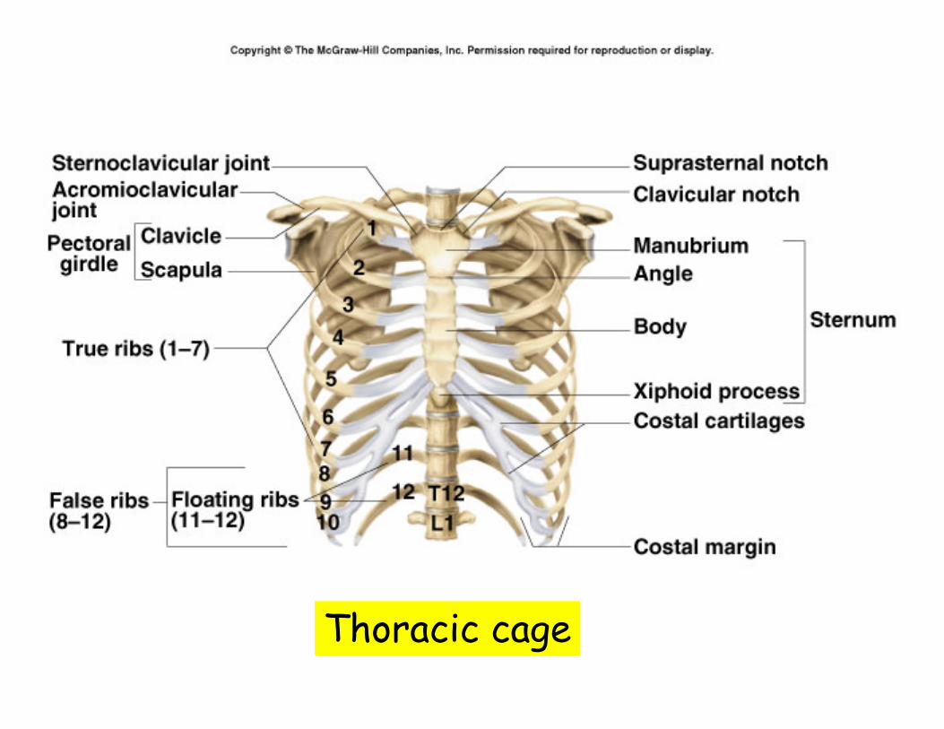
Thoracic cage

• 12 pairs (1-7 true, 8-12 false)• Proximal end attached to vertebral column• Costal cartilage
– Connects ribs 1-7 to sternum at distal ends– Connects ribs 8-10 to rib 7– Not present on ribs 11 & 12 (floating ribs)
Ribs
Rib 1
Ribs 2-10
Ribs 11 & 12