avastin : epar - european medicines agency
TRANSCRIPT
1/61 EMEA 2005
SCIENTIFIC DISCUSSION
1. Introduction Avastin contains bevacizumab, a recombinant humanised monoclonal antibody produced by DNA technology. The applicant sought a marketing authorisation for Avastin in combination with 5-fluorouracil/folinic acid containing regimens for the first-line treatment of patients with metastatic carcinoma of the colon or rectum. The CHMP considered that a positive benefit risk had only been demonstrated in combination with intravenous 5-fluorouracil/folinic acid or intravenous 5-fluorouracil/folinic acid/irinotecan. The scientific discussion in this report focuses on this indication.
Colorectal cancer
Cancer of the colon and rectum (CRC) constitutes a major public health problem and is more prevalent in Western countries where the incidence is nearly double that of developing countries [1]. There, it affects about one of twenty humans and ranks second amongst the most common malignancies in both men and women, with about 334,000 new cases diagnosed every year, distributed almost evenly between the sexes [2]. Deaths from cancers of the colon and rectum rank second (189,000) after lung cancer. Approximately 30% of all patients with CRC have metastatic disease at diagnosis, and 50% of early-stage patients will eventually develop metastatic or advanced disease [3]. The prognosis for this patient population is poor. In Europe, 5-year relative survival for patients diagnosed with cancer of the colon or rectum during 1985–1989 was 48% for patients with colon cancer and 44% for patients with cancer of the rectum. Despite the recent addition of new therapeutic agents, efficacy remains unsatisfactory.
Intravenous 5-fluorouracil/folinic acid
Since its introduction by Heidelberger in 1957, 5-fluorouracil (5-FU) has been the standard agent used in the therapy of CRC, and is the benchmark against which all other therapy is measured. These agents have been developed in many different schedules of administration. Modulation of 5-FU anticancer effects with folinic acid (FA) has become one of the standard treatment regimens for metastatic colon cancer [4]. In a meta-analysis involving 2751 patients with advanced CRC from 18 randomised trials comparing 5-FU/FA with 5-FU alone, the addition of FA conferred a doubling in response rates (23% vs. 12%, p<0.0001) with a small but statistically significant improvement in 1-year survival (48% vs. 43%, p=0.003) [5]. In North America, two FA-modulated 5-FU regimens are most commonly employed, the Mayo regimen (5-FU 425 mg/m2 daily and FA 20 mg/m2 daily administered for five consecutive days, repeated every 4 weeks for two cycles and then every 5 weeks thereafter) and the Roswell Park regimen (5-FU 500 mg/m2 and high-dose FA 500 mg/m2 administered weekly for six consecutive weeks and repeated every 8 weeks). Both regimens are deemed to be of equivalent efficacy [6] and, until recently, were considered to represent the standard of care for the first-line treatment of patients with metastatic CRC. The Roswell Park weekly schedule of 5-FU/FA is better tolerated by the majority of patients [7].
5-FU has a short plasma half-life and its cytotoxicity is S-phase dependent, prompting the evaluation of a prolonged infusion schedule [8]. A pooled analysis of 1219 patients from six randomised trials comparing continuous infusion with bolus administration of 5-FU reported a significantly higher response rate (22% vs. 14%, p=0.0002) and a modest improvement in survival (median 12.1 months vs. 11.3 months, p=0.04) in patients assigned to continuous infusion of 5-FU [9]. The toxicity profile for the infusional 5-FU regimen differed from that of bolus 5-FU, in that there was less haematological toxicity (4% vs. 31%), but more cases of hand-foot-syndrome (34% vs. 13%). The incidence of diarrhoea, mucositis and nausea did not differ. In a subgroup analysis, the benefit of a continuous infusion over bolus 5-FU was not apparent in studies of biomodulation of 5-FU by FA, although the power for such an analysis was limited.
Based on a randomised study that involved 433 assessable patients with advanced colorectal cancer, the bimonthly regimen of infusional 5-FU/FA (de Gramont regimen) was reported in 1997 to be an effective and less toxic alternative to the standard monthly regimen (Mayo Clinic regimen) [10]. The response
Page 2/61 EMEA 2005
rate, determined in 348 patients with measurable disease (448 patients in total were randomised to receive treatment), was 32.6% vs 14.4% and the time to progression was 27.6 vs 22 weeks for the de Gramont and the Mayo regimens, respectively. However, the trial did not meet its primary objective of superiority in survival suggesting that the primary advantage of the infusional 5-FU schedules over the bolus schedules to be an improved tolerability profile.
Irinotecan and oxaliplatin
Irinotecan was approved for use in first-line treatment of metastatic CRC in European countries in 1999. Irinotecan in combination with 5-FU/FA has become a new standard for chemotherapy-naïve patients with metastatic CRC in Europe following the results of two separate randomised trials showing an improved response rate and survival for patients treated with the addition of irinotecan over either bolus [11] or infusional 5-FU [12]. The overall survival with irinotecan plus bolus 5-FU [11] was 15-20% lower as compared to the corresponding arms of the study using irinotecan plus infusional 5-FU reported by Douillard et al. [12]. However, the response rates observed were virtually identical in the two trials: 21% and 23%, respectively, in the control arms and 39% and 35%, respectively, in the experimental arms. Similarly, the time to progression (TTP) was also identical between the respective arms of the two trials. Another factor that may have contributed to this was the availability of oxaliplatin as a second line chemotherapy for patients in the Douillard study but not for patients in the Saltz study [12, 13]. Infusional 5-FU combined with irinotecan is widely used in Europe rather than bolus. The infused schedules of 5-FU have in general a better tolerability than bolus 5-FU, in particular, such regimens are associated with reduced myelosuppression, stomatitis and diarrhoea.
The results of a randomised trial of oxaliplatin added to infusional 5-FU/FA (FOLFOX4) eventually established oxaliplatin as another agent for first-line treatment of CRC. At a median follow-up of 27.7 months, the response rate and progression-free survival favoured the FOLFOX4 combination [14]. Although the median survival was longer for those treated with FOLFOX4 (16.2 vs. 14.7 months), statistical significance was not reached for this endpoint. A survival advantage for FOLFOX4 over IFL has been confirmed in a three arm NCCTG-led trial (N9741) of patients with advanced CRC randomly assigned to IFL, FOLFOX4, or a combination of irinotecan plus oxaliplatin [15]. FOLFOX4 compared with IFL was associated with a better response rate (45% vs. 31%, p=0.002), a similar time to treatment failure (~ 6 months, p=0.80) but an improved survival (19.5 vs. 14.8 months, p=0.0001). Patients in the FOLFOX arm had access to irinotecan in this trial (52% of patients in the FOLFOX4 arm received second-line irinotecan) which has been demonstrated to provide survival benefit as a second-line regimen whereas patients in the IFL arm did not have ready access to oxaliplatin as it was not approved in the US (17% of patients in IFL arm received second-line oxaliplatin). The survival benefit seen might, in part, have reflected the effect of three drugs vs. two drugs. A randomised study in 226 patients reported by Tournigand et al. suggested similar efficacy of the two first-line line regimens FOLFOX4 and FOLFIRI, with a TTP of 8.1 and 8.5 months and an overall survival of 21.5 and 20.4 months, respectively [16]. In summary, FOLFOX4 has been considered as a safe and active first-line line regimen in metastatic CRC; FOLFIRI and FOLFOX4 have generally been considered comparable with respect to efficacy, with different safety profiles.
Oral fluoropyrimidines
Two oral fluoropyrimidines have been approved in Europe: tegafur (in combination with uracil) (UFT) and capecitabine [17, 18]. UFT is usually administered with oral FA. In four large randomised studies, these oral fluoropyrimidines have shown similar efficacy (overall survival), an improved safety profile and convenience advantages compared to intravenous bolus 5-FU [18-20]. Several Phase II trials have explore the use of oral fluoropyrimidines in combination with irinotecan [21-24], or oxaliplatin [25, 26].
About the product
Avastin contains the antineoplastic agent bevacizumab (ATC code: L01XC07), a recombinant humanized IgG1 monoclonal antibody (93% human, 7% murine sequences) that binds with high affinity to human vascular endothelial growth factor (VEGF). Bevacizumab was generated by humanization of the murine parent antibody A4.6.1 [27].

Page 3/61 EMEA 2005
Tumour growth beyond a microscopic size has been shown to depend on angiogenesis [28, 29]. A variety of positive angiogenic factors, including VEGF have been identified. VEGF is a major regulator of angiogenesis during normal and pathological processes, including that associated with tumour growth [30]. There is a very low or undetectable expression of the VEGF receptors in most normal tissues (with exception of renal glomeruli), whereas VEGF is upregulated in most human tumour types [31, 32], including gastrointestinal tumours [33, 34]. VEGF expression is associated with tumour progression or patient survival in a variety of human cancers, including gastrointestinal tumours [35-39].
2. Part II: Chemical, pharmaceutical and biological aspects
Introduction
Bevacizumab, the active ingredient of Avastin, is a recombinant humanised monoclonal immunoglobulin G1 (IgG1) antibody (93% human, 7% murine sequences - molecular weight 149 kDa) that selectively binds with high affinity to all isoforms of human vascular endothelial growth factor (VEGF) and neutralises VEGF’s biologic activity through a steric blocking of the binding of VEGF to its receptors Flt-1 (VEGFR-1) and KDR (VEGFR-2) on the surface of endothelial cells. Receptors activation normally induces their tyrosine phosporylation and the subsequent series of signal transduction events elicit mitogenic and pro-survival activity signals for the vascular endothelial cells. Since there is a very low or undetectable expression of VEGF receptors in most normal tissues (with exception of renal glomeruli) but a significant up-regulation in the vasculature of many tumours (including colorectal cancer), the neutralisation of VEGF by bevacizumab provides the rationale a relative specific inhibition of the tumour angiogenesis and thereby inhibition of tumour growth and metastasising.
Composition
Avastin is provided as a concentrate for solution for infusion in a single-use vial, which contains a nominal amount of either 100 mg of bevacizumab in 4 ml or 400 mg of bevacizumab in 16 ml (concentration of 25 mg/ml). Bevacizumab is formulated with 51 mM sodium phosphate pH 6.2, 60 mg/ml α,α-trehalose dihydrate and 0.04% polysorbate 20. The drug product is a clear to slightly opalescent, colourless to pale brown sterile liquid solution that has to be diluted in 0.9 % sodium chloride solution prior to administration.
Studies were conducted to determine the amount of overfill required to produce the 4 ml and 16 ml nominal fill volumes for drug product vials. For the 100 mg vial, a 4.30 ml fill containing 107.5 mg bevacizumab (7,5% overfill) achieves delivery of 100 mg bevacizumab. For the 400 mg vial, a 16.28 ml fill containing 407 mg bevacizumab (1,75% overfill) achieves a delivery of 400 mg bevacizumab. The overfill requirement is due to the dead volume in the vial and to the syringe used to remove the solution from the vial.
The vial and stopper components comply with Ph. Eur. requirements. The container-closure system consists of a 5 ml or 20 ml Type I borosilicate glass vial, butyl rubber stoppers and an aluminum seal fitted with a plastic flip-off cap.
Drug Substance
Nomenclature INN Name: bevacizumab Compendial name: Not applicable Chemical name: Recombinant humanised monoclonal antibody to VEGF USAN/BAN/JAN Name: Bevacizumab Laboratory Code: RO487-6646 CAS Registry Number: 216974-75-3 Other Names: rhuMAb VEGF, anti-VEGF
Description of the Drug Substance
Page 4/61 EMEA 2005
Bevacizumab is a humanised form of a murine monoclonal antibody containing human constant region sequences and murine light and heavy chain Complementarity Determining Region (CDR) sequences. The human framework contributes to 93% of the overall protein sequence.
Bevacizumab is a full-length IgG1κ isotype antibody composed of two identical light chains (214 amino acid residues) and two heavy chains (453 residues) with a total molecular weight of 149 kDa. The heavy chains demonstrate C-terminal heterogeneity (lysine variants) and also contain one N-linked glycosylation site at asparagine 303. The oligosaccharides are of complex biantennary structures with a core fucose and with the two branches terminating mainly with zero (G0), one (G1) or two (G2) galactose residues. The G0 glycoform predominates at approximately 80 % relative abundance. Each light chain is covalently coupled through a disulfide bond at cysteine 214 to a heavy chain at cysteine 226. The two heavy chains are covalently coupled to each other through two inter-chain disulfide bonds, which is consistent with the structure of a human IgG1.
• Manufacture
Bevacizumab drug substance is manufactured at Genentech South San Francisco (SSF) and Genentech Vacaville (VV), which is an additional site. Bevacizumab manufactured at VV is transported to SSF for filling. These facilities are operated in current GMP compliance, with standard operating procedures in place to describe all procedures and controls. Both sites were last inspected in August 2003 by the German competent authorities and were found to be in compliance with GMP rules.
Development genetics
Bevacizumab was originally derived from a murine monoclonal antibody (muMAb A4.6.1), which was produced at Genentech using hybridomas generated from mice immunised with the 165-residue- form of recombinant human vascular endothelial growth factor (rhuVEGF165) conjugated with keyhole limpet hemocyanin.
The humanisation of the A4.6.1 antibody involved insertion of the six CDRs of A4.6.1, in place of those of a selected human antibody Fab framework (pEMX1), which has a consensus human kappa subgroup I light chain (domains VL-CL) and a truncated human subgroup III immunoglobulin gamma (IgG1) heavy chain (domains VH-CH1). A series of framework residue substitutions were made to produce the final humanised version, Fab-12, which contains eight substitutions of the human framework outside of the CDRs. The VH and VL domains of Fab-12 were combined with human IgG1 constant domains CH1-CH2-CH3 and CL, respectively, to produce bevacizumab.
The expression plasmid pSVID5.ID.LLnspeV.xvegf36HC.LC encoding bevacizumab was introduced into Chinese hamster ovary parental cells CHO DP-12 by lipofection and cells were selected in the presence of increasing concentrations of methotrexate (MTX). Isolates were selected for secretion of active bevacizumab. Isolated subclone 107N was used for the production of phase I and phase II clinical materials and as the starting point for the development of the more highly productive G7 cell line, which was used for the production of phase III clinical materials and which will be used for the production of drug substance intended for marketing.
The generation of A4.6.1 and its subsequent humanisation have been adequately described in the application and in the scientific literature. The construction of the expression plasmid has been sufficiently described and an annotated sequence map has been submitted.
Cell bank system
From the high yield producing G7 clone, a serum-free pre-bank (No. 2036) was established and used to prepare a two-tiered cell bank system of master cell bank (MCB) and working cell banks (WCBs).
The MCB (No. 2055) and WCBs (Nos. 2130, 2206, and 2363) were prepared in accordance with the current Good Manufacturing Practices, using procedures defined in standard operating procedures, and standard methods for preservation of mammalian cells employing carboxymethylcellulose and dimethylsulfoxide as cryopreservatives.
Page 5/61 EMEA 2005
Isoenzyme analysis of MCB No. 2055 and WCB Nos. 2206, 2130 and 2363 confirmed the bevacizumab cell banks as being of Chinese hamster origin, and peptide map analysis confirmed the identity of the product produced by these cells as bevacizumab.
Cell banks were extensively examined for the presence of microbial and viral contaminants and the endogenous retrovirus known to be present in CHO cell line. In addition, bevacizumab pre-harvest cell culture fluids from end of production runs of MCB No. 2055, WCB No. 2130, and WCB No. 2363 were examined for the presence of microbial and viral contaminants. This testing was performed using a battery of biochemical, biological, and immunological assays known to detect contamination by bacterial, fungal, or viral agents associated with mammalian cell culture. All cell banks and end of production cells were shown to be free of detectable microbial contaminants despite the presence of type-A and type-C retroviral particles in the cell line. The retrovirus-like particles present are non-infectious and typical of the parental CHO cell line. Viral testing of the cell banks was in line with the relevant guidelines and was deemed satisfactory.
The applicant does not plan to prepare a new MCB since the current MCB is large enough to prepare more than 100 WCBs.
The applicant does anticipate producing additional WCBs using cells expanded by the spinner process currently in use or by a new cell banking bioreactor process, which enables the accumulation of a larger mass of cells. In the event that new WCBs are prepared using a new procedure, the first new WCB will also have to meet pre-approved acceptance criteria in order to ensure a comparable product quality and cell culture performance with the existing WCBs.
The applicant committed that an application for a variation will be submitted in case of generation of a new WCB by any other protocol than the one approved with the Marketing Authorisation.
Fermentation process
The bevacizumab routine process for cell culture involves three stages: seed train, inoculum train, and the production culture, which is a fed-batch process at a 12000 L scale using CHO cells in suspension.
Raw materials and reagents used during fermentation and purification are not derived from ruminants. All stages use serum-free, low-protein cell culture growth media containing recombinant human insulin. Three ruminant-derived materials are used in the preparation of human recombinant insulin: beef extract, peptone and pepticase (bovine milk). TSE certificates of suitability are provided for peptone and beef extract (RO-CEP 2000-175-Rev 01 and RO-CEP 2000-181-Rev 01, respectively). The bovine milk is sourced from Australia, New Zealand and USA, under the same conditions as milk for human consumption and is prepared without the use of other ruminant materials.
Detailed and sufficient information has been provided on the composition, preparation and testing of the media used for fermentation.
The seed train is a continuous long term, 20 L culture of cells. This culture is used to start up many production cultures. In order to initiate a seed train, cells from WCB No. 2130 or WCB No. 2363 are used. MCB No. 2055 or WCB No. 2206 are also acceptable for manufacturing, but are not expected to be used for routine manufacturing unless the ampoules in the other cell banks are depleted. The seed train is grown in selective medium with MTX. Cells are sub-cultivated (diluted) every 3 to 4 days. After a prescribed maximum period in the seed train culture, the inoculation train is initiated. The non-selective inoculum train (MTX-free medium) is used to expand the cell population for introduction into the production stage and to reduce the carry over of MTX into the production culture. The cell population is expanded by serial sub-cultivation into vessels of increasing volumes (approximately 80 L, 400 L, and 2000 L).
After a prescribed maximum number of days in the inoculum train, the production stage is initiated. Alternatively, an aliquot of cell culture fluid may be left in the original vessel and diluted with fresh medium to initiate another culture. This operation is termed a “solera”. It is performed to generate an additional inoculum train when necessary.
The production culture is performed in a bioreactor of approximate working volume of 12000 L, using an enriched non-selective production medium (MTX-free). The production culture is harvested after a
Page 6/61 EMEA 2005
prescribed number of days after inoculation. The total allowed time in non-selective medium and the total cell age from MCB to harvest have been validated and defined in the application. The production cell culture fluid is separated from the cells by centrifugation and the secreted bevacizumab is recovered from the harvested cell culture fluid (HCCF).
The flow chart and process control parameters for cell culture and harvest have been provided. Alternatives to the routine process have been clearly indicated and justified and sufficient control and monitoring of the process is in place to ensure a defined and consistent quality of bevacizumab.
Purification process
One batch of HCCF is produced from each 12000 L cell culture production run and is used to purify and formulate a single batch of drug substance.
The drug substance purification process consists of four steps: protein A chromatography, anion exchange chromatography (Q sepharose FF), cation exchange chromatography (CM sepharose FF) and ultrafiltration /diafiltration (UF/DF).
Detailed and sufficient information on the bevacizumab purification process has been provided, including information on chromatographic equipments, buffers and solutions and column operation parameters and conditions. The flow chart of the purification process has been submitted.
An affinity column is the first chromatographic step in the bevacizumab purification process. This column utilizes an immobilised protein A resin, which binds to bevacizumab in the HCCF with a high degree of specificity. This affinity step purifies bevacizumab mainly with respect to CHO proteins (CHOP) and DNA. In order to inactivate potential viruses, the affinity pool is subjected to a validated virus inactivation process. The affinity step is followed by an anion exchange chromatographic step which is designed to reduce CHOP, DNA, protein A, and potential viruses. Under the load and wash conditions employed, bevacizumab flows through the column.
The next cation exchange chromatographic step is designed to further reduce the residual amounts of CHOP, gentamicin and bevacizumab aggregates. In order to inactivate potential viruses, the anion exchange chromatographic pool is subjected to a validated vius inactivation process. Bevacizumab binds to the column under the load and wash conditions.
Finally, a cation exchange pool is concentrated on an UF system. The pool is then diafiltered and further concentrated. The UF/DF pool is then removed from the UF/DF system and diluted by diafiltration buffer.
Characterisation
- Comparability from the SSF and VV manufacturing sites:
Samples from three conformance lots from the VV site, three lots from the SSF site and the reference material antivegf801-2 were analysed side-by-side using the release specification methods: peptide map, size-exclusion chromatography (SEC), capillary electrophoresis-SDS (CE-SDS) and ion-exchange chromatography (IEC). The oligosaccharide distribution was evaluated by capillary electrophoresis with laser-induced fluorescence detection (CE-LIF) after release from the protein.
Except for minor differences, good consistency within sites and between the two sites was shown. No differences in the peptide map profiles of the bevacizumab samples were observed. Molecular size distribution by SEC showed that the level of dimers, aggregates and fragments were consistent for all the lots. Glycan occupancy of the N-linked site was shown to be similar in all the lots by CE-SDS analysis. Charge heterogeneity of the bevacizumab lots was examined by cation exchange chromatography, which demonstrated that all lots had consistent charge profiles. Glycan analysis by CE-LIF confirmed that the VV and SSF lots had comparable neutral oligosaccharide profiles.
- 7 lots of bevacizumab were characterised and compared: 1 lot of the reference standard material (antivegf898-1) produced from the 107N cell line and used in phase II clinical studies, 1 lot of the reference standard material (antivegf801-2) used in phase III clinical studies, five qualification lots, all produced at a 12000 L scale from the G7 cell line.
Page 7/61 EMEA 2005
The applicant has performed extensive and in general satisfying characterisation of bevacizumab with regard to physicochemical properties based on molecular weight, size and charge. The results confirm that bevacizumab has the covalent structure, post-translational modifications as well as other characteristics of a human IgG1.
Product-related variants have been characterised to a great extent and their potency have been determined, demonstrating that acidic and basic variants were somewhat reduced in activity. The activity of aggregates and dimers is significantly reduced, while other variants such as oxidised, glycated and deglycosylated materials are fully active.
Minor differences among the materials investigated were identified in C-terminal heterogeneity, glycosylation pattern and charge heterogeneity but studies performed showed that these differences are unlikely to affect the safety and efficacy of the product. The applicant has given several acceptable arguments to support the statement that the measurement of the galactose distribution in the drug substance as an indicator of process consistency is not necessary.
Finally, the biological and immunological properties of bevacizumab have been investigated and the choice of bioassay is addressed.
• Specifications
Batch release results from a total of twenty full-scale batches are presented in the dossier. All results are consistent and within the specifications.
Establishment of specifications is based on data from the twenty full-scale batches (12000 L) and the statistically predicted capability of the manufacturing process with a tolerance interval of 95/99 applied to the manufacturing data.
The potency assay carried out is an anti-proliferation bioassay based upon the ability of bevacizumab to inhibit rhVEGF-induced proliferation of Human Umbilical Vein Endotheliae Cells (HUVEC). It is performed in micro-titre plates and the relative number of viable cells, proportional to inhibition of rhVEGF-induced HUVEC proliferation, is quantified by fluorescence. This assay was chosen as drug substance release test based on its sensitivity (ability to detect significant changes in the activity), robustness, precision (RSD<10%) and accuracy (98-102%).
All methods have been satisfactorily validated with regard to specificity, accuracy, precision, linearity, and robustness. Moreover, the ability of the methods to detect changes in samples of bevacizumab exposed to heat, intense light, oxidation and acidic and basic pH has been evaluated.
The control tests proposed for the drug substance are considered appropriate to ensure sufficient quality with respect to identity, purity, quantity as well as general tests including tests for excipients.
• Stability
The proposed storage time of bevacizumab drug substance is 24 months when stored at − 20 °C ± 5 °C, 45 days at 5 °C ± 3 °C and up to 5 freeze/thaw cycles.
In order to support the proposed storage conditions, the stability of the drug substance was monitored at full-scale with material stored in a 120 L stainless steel tank and at small-scale in 55 ml stainless steel mini-tanks. The stability was assessed using methods addressing the principal degradation pathways of bevacizumab (aggregation and formation of charge-related variants). The stability of drug substance was tested under long-term conditions at − 20 °C ± 5 °C, accelerated conditions at 5 °C ± 3 °C and for stability after freeze/thaw cycles (stress testing). The test procedures to assess the stability of bevacizumab include methods to determine potency, purity and physicochemical changes. The results demonstrated that drug substance stored up to 24 months at − 20 °C ± 5 °C and for 57 days at 5 °C ± 3 °C, including freeze/thaw cycles, showed no changes compared to the initial time point, except for COC (Clarity, Opalescence Coloration) where a slight intensification of colour was measured. The results from the supportive studies showed no changes outside of the variability of the assay. All results were within specifications.
Page 8/61 EMEA 2005
Drug Product
• Pharmaceutical Development
The goal was to develop a stable liquid intravenous formulation. Early pharmacokinetic and toxicological phase I and early phase II clinical studies were conducted with a liquid formulation containing 10mg/ml bevacizumab, 10 mM histidine, 100 mg/ml trehalose dihydrate and 0.02% polysorbate 20. Late phase II and phase III trials used a formulation containing 51 mM sodium phosphate, 60 mg/ml trehalose dihydrate and 0.04% polysorbate 20 (as the formulation to-be-marketed). The bevacizumab concentration was increased from 10 mg/ml to 25 mg/ml in the sodium phosphate formulation for use in the phase III trials.
Selection of excipients was based on stability screening studies using different buffer systems.. A histidine buffer system at pH 5.5 was selected.
Trehalose dihydrate was selected to adjust osmolality.
Due to physical instability of the liquid formulation used in phase I and phase II clinical studies the formulation was changed by increasing the pH to 6.2, changing the histidine buffer for sodium phosphate, increasing the ionic strength by increasing the concentration of the buffering species, decreasing the trehalose concentration to modify the osmolality, and increasing the polysorbate 20 concentration. These changes resulted in a formulation that had acceptable stability at room temperature for shipping and handling of the product. This formulation was used in phase II and phase III clinical trials.
• Manufacture of the product
All manufacturing operations for bevacizumab drug product are performed by Genentech Inc., SSF. Secondary packaging and labelling are performed at Hoffmann-La Roche Ltd, Kaiseraugst, Switzerland. Quality testing and batch release is a responsibility of Hoffmann-La Roche AG, Grenzach-Wyhlen, Germany.
In the production of Avastin, the formulation step is performed as last step in the manufacturing process of the drug substance; no further formulation takes place during the manufacture of the drug product.
A drug product batch is defined as 14 L to 1372 L of bevacizumab drug substance solution. The minimum batch size of 14 L corresponds to 3000 vials (100 mg/vial presentation) or 800 vials (400 mg/vial presentation) and the maximum batch size of 1372 L corresponds to 311,000 vials (100 mg/vial presentation) or 83,000 vials (400 mg/vial presentation). The batch formula for minimal and maximal batch sizes have been provided.
The manufacturing process is adequately described . Briefly, prior to filtration, each 120 L and/or 300 L tank is tested for bioburden. An acceptable action limit for bioburden has been set for the pre-sterile filtration samples. The bulk for storage is prefiltered (0.22 µm) and sterile filtered (0.22 µm) before filling.. The filtration takes place in a closed system with nitrogen. If necessary, the contents of multiple 120 L and/or 300 L freeze/thaw tanks may be pooled during filtration in order for the fill process to yield the required batch size. The resulting sterile-filtered bulk for fill may then be held at 2°C-8°C prior to filling.
The sterile filtered bulk is aseptically transferred to the filling machine using sterile filtered nitrogen and steamed-in-place transfer lines. The product is filled into depyrogenated Type I glass vials, and a steam-sterilised stopper is seated in each vial. The entire filling and stoppering operation is performed within the Class 100 area . The vials are capped with aluminium/plastic flip-off caps. All equipments used during filling are sterilised.
100% of filled vials are inspected.
Reprocessing in the form of refiltration, in order to protect the product, may be necessary. The criteria for reprocessing have been defined and are considered acceptable. Filtration to remove contaminants, such as bioburden, outside of established limits, is not permitted.
The following critical steps have been validated:
Page 9/61 EMEA 2005
- Sterile filtration with respect to microbial retention and product/filter compatibility and leachables.
- Holding steps for bulk for storage (including freeze/thaw cycles) and sterile filtered bulk.
- Steam sterilisation of the major equipment and container closure system.
- Media fills
The process has been adequately validated and stability data have been presented to support the proposed storage conditions and times. For validation, 4 qualification lots (3 lots of 100 mg vials and 1 of 400 mg vials) and 7 clinical (supporting) lots have been produced. All results were in compliance with the specifications and demonstrated quality of the product.
• Product Specifications
17 drug product batches were analysed to develop appropriate specifications. Drug substance and drug product release specifications are identical for molecular size distribution (SEC), bacterial endotoxin, potency assay, protein concentration, extractable volume, pH and osmolality.
The selected parameters have been adequately justified and are considered acceptable. Polysorbate 20 is added as surfactant but no test on the amount present in the drug product was performed. However, this is considered acceptable as a test for polysorbate 20 is performed on the drug substance.
Batch analysis results confirmed the consistency of the drug product and showed that no new impurities are formed during manufacture and that the impurity profile of the drug product is comparable to that of the drug substance.
All methods for release testing of the drug product have been adequately described and are validated according to ICH guidelines.
• Stability of the Product
Stability testing was conducted with 4 qualification lots (three 100 mg and one 400 mg batches) using validated analytical methods. Supporting data is presented for 7 clinical batches (three 100 mg and four 1000 mg batches). The proposed expiry date for the 400 mg intermediate vial size is based on bracketing of stability data obtained with the 100 mg and 1000 mg vials.
Stability testing has been conducted under the following conditions: long-term stability (5 °C ± 3 °C), accelerated stability (30 °C), photostability, stability after reconstitution and during shipping.
The investigated parameters for the shelf-life specifications are considered to be stability-indicating.
The stability data show that no significant changes occur in the investigated parameters when stored at 5°C for up to 24 months, except for the IEC assay where an decrease in main peak was observed. The decrease in main peak correlated with an increase in acidic and an increase in basic variants peaks. No new species were observed in the IEC profile. This decrease in main peak is not correlated to any decrease in potency. All results remained within specifications.
When stored at 30°C for 3 months, changes were observed for SEC, IEC and potency. An decrease in IEC main peak was observed, which was correlated with up to 6% increase in basic variants and an increase in acidic variant peaks. No new species were observed in the IEC chromatographic profile. An decrease in percent monomer was observed with a corresponding increase in percentage of total aggregates. Potency was observed to decrease 20-30%. No other changes in stability were observed compared to the initial time point. All other results remained within specifications.
Similar results were found for 100 mg, 400 mg and 1000 vials.
Photostability data showed that degradation occurs when the drug product is exposed to light. Therefore, the drug product should be protected from light and consequently the vials should be kept in the outer carton.
The submitted stability data support the proposed shelf life of 24 months when stored at 5 °C ± 3 °C.
With respect to the in-use stability of the drug product, chemical and physical stability has been demonstrated for 48 hours at 2°C-30°C in 0.9% sodium chloride solution. From a microbiological point
Page 10/61 EMEA 2005
of view, the product should be used immediately. Otherwise, in-use storage times and conditions are the responsibility of the user and would normally not be longer than 24 hours at 2°C to 8°C, unless dilution has taken place in controlled and validated aseptic conditions.
Discussion on chemical, pharmaceutical and biological aspects
In general, the different aspects of the chemical, pharmaceutical and biological documentation comply with existing guidelines. The information provided in the application demonstrated consistent batch-to-batch production of Avastin, achieving a defined quality for the drug substance and the drug product. The fermentation and purification of the drug substance, bevacizumab, are adequately controlled and validated. Alternatives to the routine process have been clearly indicated and justified and sufficient control and monitoring of the process have been put in place to ensure a defined and consistent quality of bevacizumab. With regard to the manufacturing process validation, data at full-scale presented by the applicant support the initial conclusion that although the increase in cell age has some influence on the cell culture performance (decrease in bevacizumab titre and specific productivity), the quality of the drug substance is not affected. The increase in the percentage of the G0 glycoform observed is minimal and does not affect the quality of the drug substance as well. The proposed cell age limit was therefore considered acceptable.
The drug substance has been well characterised with regard to its physicochemical and biological characteristics, using state-of-the-art methods. The applicant has discussed that the minor differences observed in C-terminal heterogeneity, glycosylation pattern and charge heterogeneity between the materials investigated seem to be without influence on the safety and efficacy of Avastin.
Appropriate drug substance specifications have been set and in general sufficiently justified.
The specifications limits were established mainly on the basis of the manufacturing history, the statistic calculation of data as well as the physicochemical characterisation.
The proposed storage time of the drug substance is 24 months at ≤ -20°C, 45 days at 5°C±3°C. Updated stability data presented by the applicant to support this shelf life were considered satisfactory. Moreover, the applicant provided several reasons why freeze/thaw of the drug substance is needed. They are mainly based on a desire for flexibility during production and are of common practice in the production of monoclonal antibodies. Sufficient justification to support the use of freeze/thaw cycles was provided by the applicant.
The manufacturing process of the drug product has been sufficiently described and validated. The quality of the drug product is controlled by adequate test methods and specifications.
The proposed shelf life of the drug product is 24 months at 2-8oC. With the response to the Day 120 List of Questions, the applicant submitted additional results from an on-going long-term stability study in order to support this shelf life.
The viral safety and safety concerning other adventitious agents (including TSE) have been sufficiently assured.
The overall quality of Avastin has been adequately demonstrated, excepted for a number of points that the applicant has committed to address as part of post-approval follow-up measures. It includes the submission of additional information relating to genetic consistency testing to further support the proposed in vitro cell age limit, information relating to batch-to-batch consistency with regard to charge variants distribution, to the IEC specification that was established for the drug substance and drug product.
3. Part III: Toxico-pharmacological aspects
Introduction
Bevacizumab is a humanised recombinant IgG1 version of the murine anti-human VEGF monoclonal antibody A4.6.1 (muMAb VEGF) [40]. A4.6.1 was humanised by site-directed mutagenesis of a human
Page 11/61 EMEA 2005
IgG1 framework [27]. Bevacizumab consists of about 93% human and 7% murine protein sequence, with the murine sequences comprising the complementarity determining region of the molecule.
All toxicology studies with bevacizumab were conducted according to GLP, with the exception of toxicology studies investigating anti-bevacizumab antibody responses in rats, potential for perturbation of growth plate morphology and ovarian function, and wound-healing, coagulation and renal function studies.
The pharmacology of an anti-VEGF approach was investigated primarily in murine xenograft models using the parent murine antibody A4.6.1. The pharmacokinetics of bevacizumab was investigated in species where the antibody is expected to bind to and neutralise endogenous VEGF, i.e., cynomolgus monkeys [41] and rabbits (albeit with lower affinity than for human VEGF, see toxicology), and in mice and rats, where it is reported not to bind endogenous VEGF [42].
Pharmacology
Native VEGF is a basic, heparin-binding, homodimeric glycoprotein of 45,000 daltons. These properties correspond to those of VEGF165, the predominant VEGF isoform. The human VEGF gene has been located to chromosome 6p21.3 and is organized in eight exons separated by seven introns. Alternative splicing was shown to result in four major VEGF isoforms (VEGF121, VEGF165, VEGF189, and VEGF206) consisting of 121, 165, 189, and 206 amino acids following signal sequence cleavage, respectively [43-45]. Less frequent splice variants also have been reported, including VEGF145 [46], VEGF183 [47], and VEGF165b [48]. An additional level of regulation of VEGF biological activity is provided by the proteolytic cleavage mechanism, including all VEGF isoforms, resulting in the VEGF110 form [49].
VEGF is a major regulator of angiogenesis during normal and pathological processes, including that associated with tumour growth (reviewed in [30, 50]). VEGF has a key regulatory function during developmental angiogenesis [51, 52]. A well-documented in vitro activity of VEGF is the ability to promote growth of vascular endothelial cells (EC) derived from arteries, veins and lymphatics (reviewed in [32]), as well as certain non-endothelial cells (reviewed in [53]). VEGF was shown to be a survival factor for ECs, both in vitro and in vivo [54-57]. VEGF stimulated production of surfactant proteins by cultured type 2 pneumocytes [58]. VEGF induces vasodilatation in vitro [59], and is thought to play a role in inflammation due to its ability to induce vascular leakage [31, 60]. VEGF displays chemotactic effects on endothelial cells and increases expression of proteolytic enzymes in endothelial cells involved in stromal degradation. VEGF has also effects on bone marrow–derived cells, promoting monocyte activation and chemotaxis [61]. VEGF enhanced colony formation by mature subsets of granulocyte-macrophage and erythroid progenitor cells that had been stimulated with a colony stimulating factor [48]. VEGF also displays immune effects via inhibition of maturation of antigen-presenting dendritic cells [62].
VEGF binds two related recptor tyrosine kinases (RTK), named Flt-1 (VEGFR-1) [63, 64], KDR/Flk-1 (VEGFR-2) [65]. The fms-like-tyrosine kinase Flt-4 (VEGFR-3) is a member of the same family of RTKs but is not a receptor for VEGF, binding instead to VEGFC and VEGFD8. In addition to these RTKs, VEGF interacts with a family of coreceptors, the neuropilins. Binding of VEGF to VEGFR-1 and VEGFR-2 induces the homodimerization of two receptor subunits, which in turn triggers autophosphorylation of their tyrosine kinase domains located within the cytoplasm. Autophosphorylation of the tyrosine kinase domains subsequently engages a series of specific signal transduction events, ultimately regulating the various biological activities of VEGF on endothelial cells. Most of VEGF’s mitogenic and survival activity appears to be mediated by VEGFR-2, including the expression of the anti-apoptotic proteins Bcl-2 and A1. Survival signaling by VEGFR-2 is mediated by the PI3 kinase/Akt pathway [56]. VEGFR-2 was also shown to induce other signal transduction pathways including phospholipase C gamma and mitogen-activated kinases MAPK p44/42 [66].
• Primary pharmacodynamics
The generation of muMAb A4.6.1 using the recombinant human VEGF165 as immunogen and its functional characterization have been described [40]. A4.6.1 has potent VEGF neutralizing activities using an in vitro bovine adrenal cortex endothelial cell proliferation assay, in in vivo vascular
Page 12/61 EMEA 2005
permeability assay and an in vivo embryonic chicken angiogenesis assay [40]. The pharmacological activities of bevacizumab were evaluated in a number of in vitro assays using recombinant human VEGF. Bevacizumab and A4.6.1 were compared for their ability to inhibit bovine capillary endothelial cell proliferation in response to VEGF (Presta et al 1997). The two MAbs were essentially equivalent, both in potency and efficacy. The ED50s were, respectively, 50 ± 5 and 48 ± 8 ng/ml. 90 % inhibition was achieved at 500 ng/ml for both antibodies.Bevacizumab and A4.6.1 are pharmacologically equivalent when tested with human cells, human tissues or human VEGF isoforms [27, 40].
Studies of crystal structures and mutational analysis have shown that the VEGF residues critical for antibody-binding are distinct from those important for the high-affinity receptor binding but occupy a common region on VEGF, suggesting that the inhibitory effect of the antibody is by steric blocking of VEGF-receptor interaction [67].
A4.6.1 inhibited the growth of human rhabdomyosarcoma, glioblastoma multiforme or leiomyosarcoma cell lines injected into nude mice, but had no effect on the growth rate of the tumour cells in vitro. The density of vessels was decreased in the antibody-treated tumours [68].
In xenograft models of cancer in nude mice, antibody administration resulted in marked growth inhibition of a variety of tumour types, including rhabdomyosarcoma [68-70], glioblastoma [54, 68-73], leiomyosarcoma [68], ovarian carcinoma [74-76], prostate carcinoma [77-79], colon adenocarcinoma [54, 72, 80], Wilms tumour [81-83], hepatoblastoma [84], neuroblastoma [83, 85], breast carcinoma [86-89], melanoma [54], and pancreatic cancer [90]. Dose-dependent tumour growth inhibition was observed independent of tumour location and route of administration (IV, IP, and intratumoural). Growth inhibition of primary tumours was obtained with early as well as with late treatment initiation. The effects as assessed by tumour size and/or weight ranged from 25% to >95% inhibition relative to control treatment. Immunohistochemistry of tumours revealed moderate to complete inhibition of tumour angiogenesis.
Magnetic Resonance Imaging studies showed that A4.6.1 is able to counteract the permeability-enhancing effects of VEGF [73, 75, 86, 87].
In three different animal models inoculated with human tumour cells (colon carcinoma, prostate and Wilm’s tumour), treatment with A4.6.1 markedly inhibited primary tumour growth and reduced metastasis dissemination to liver and lung [78, 80, 81].
• Secondary pharmacodynamics
No dedicated studies of safety pharmacology were performed. Nonclinical studies of up to 26 weeks duration were performed with bevacizumab in cynomolgus monkeys and rabbits. Drug-related effects were consistent with the inhibition of VEGF-dependent angiogenesis. These studies revealed dose-related effects on sites of active neo-angiogenesis and include an increase in hypertrophied chondrocytes, subchondral bony plate formation, and inhibition of vascular invasion of the growth plate in young adult cynomolgus monkeys [91]. Decreased ovarian and uterine weights and absence of corpora lutea were observed in female cynomolgus monkeys after treatment with bevacizumab [91]. Both the physeal and ovarian changes were reversible with cessation of treatment. These studies are described in further detail in the toxicology section of this report.
• Safety pharmacology
No formal in vivo safety pharmacology studies were conducted with bevacizumab or A4.6.1. Safety pharmacology endpoints were evaluated as part of the toxicology studies (see Toxicology). No treatment-related effects on physical examinations, including respiration rate, blood pressure, ECG, rectal body temperature and urinalysis, were observed in cynomolgus monkeys administered 2 to 50 mg/kg of bevacizumab IV once or twice weekly for up to 26 weeks.
• Pharmacodynamic drug interactions
Several non-clinical studies were performed in order to study the effect of anti-VEGF antibody treatment in combination with other anti-neoplastic treatments in animal models inoculated with cells from human tumour cell lines. No studies including fluoropyrimidine-based therapy have been submitted.
Page 13/61 EMEA 2005
A4.6.1 inhibited tumour growth, reduced microvascular density and increased the tumour pO2 in mice inoculated by human glioblastoma and colon adenocarcinoma cell lines. In addition, tumour growth delay caused by radiation was enhanced by pretreatment with A4.6.1 [72].
Mono-therapy with A4.6.1, bevacizumab, paclitaxel [76, 79], or doxorubicin [88] inhibited tumour growth and angiogenesis in tumours from human breast cancer [88], prostate cancer [79] and ovarian cancer [76]. In these studies, co-administration of either antibody and paclitaxel or antibody and doxorubicin, resulted in additional growth inhibition. Withdrawal of mono- or combined therapies induced recurrent growth, which was smallest in the co-administered group [79].
Co-administration of antibodies and topotecan [85] or docetaxel [89] resulted in additional inhibition of angiogenesis and tumour growth in tumours from human neuroblastoma [85], breast cancer [89] and Wilm’s tumour cell lines [82]. The co-administration of A4.6.1 with topotecan treatment prevented metastasis generation from Wilm’s tumour [82].
Mono-therapy with either A4.6.1 or the synthetic matrix metalloproteinase inhibitor BB-94 resulted in inhibited primary tumour growth and dissemination in two animal models of human pancreatic adenomas (poorly differentiated AsPC-1, moderately differentiated HPAF-2). Significant inhibition of tumour growth after co-administration was only observed in HPAF-2 [90].
Pharmacokinetics
Pharmacokinetic studies following single dose administration were performed in mouse, rat, rabbit and cynomolgus monkey. Examination of pharmacokinetic parameters following multiple doses was primarily conducted in repeat-dose toxicology studies in rabbit and cynomolgus monkey. The pharmacokinetics were in most studies evaluated following IV administration, which is the intended clinical administration route.
Concentrations of bevacizumab in serum from rabbit, rat, cynomolgus monkey, and mouse, as well as rabbit amniotic fluid were measured by enzyme-linked immunosorbent assay (ELISA). The same principle was used for measurement of antibodies to bevacizumab in rabbit serum, cynomolgus monkey serum and amniotic fluid from rabbit.
• Absorption- Bioavailability
Absorption of bevacizumab subsequent to a single intraperitoneal (ip) or subcutaneous (sc) administration has been examined in mouse, rat, and cynomolgus monkey. Absorption subsequent to ip administration was complete in mouse. Sc administration resulted in a slower absorption that was complete in mouse (>100%) and cynomolgus monkey (98%) , but with a bioavailability of 69% in rat [42].
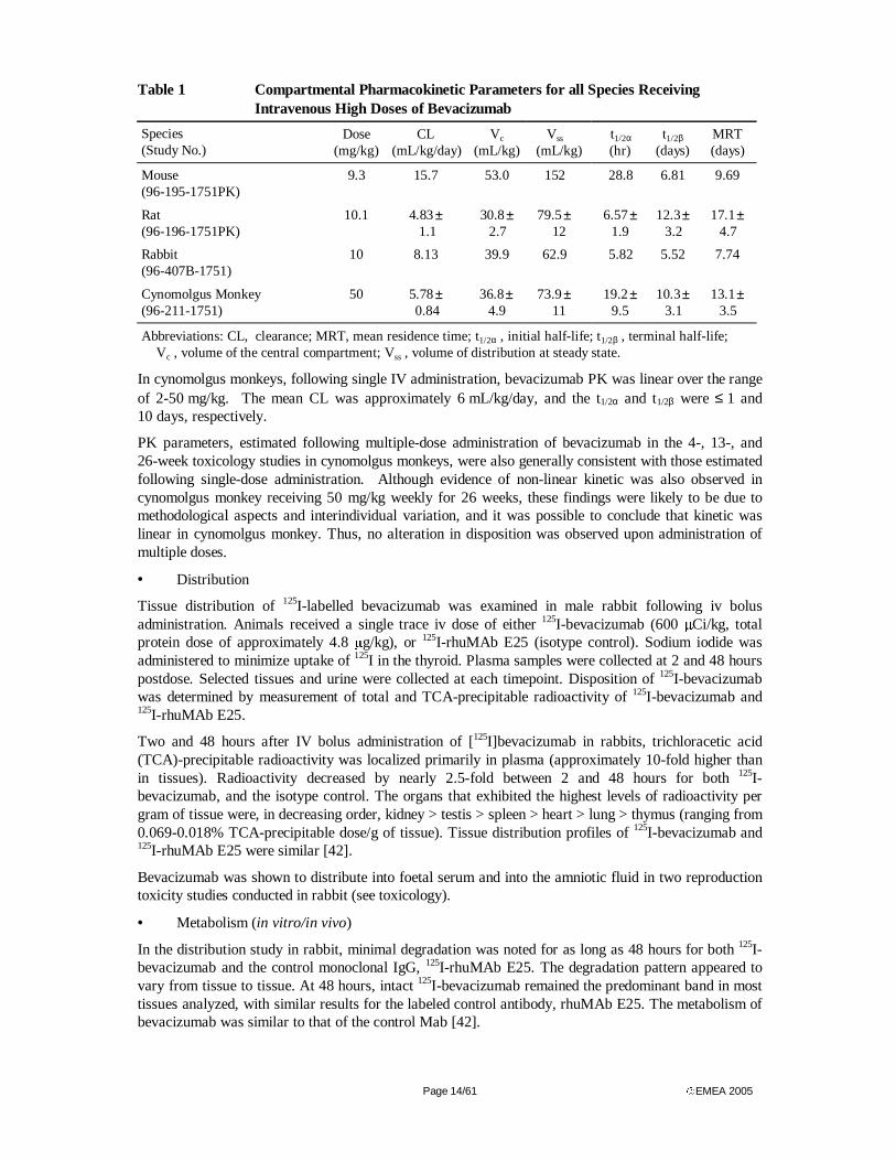
Table 1 summarizes key PK parameters in all species following administration of high doses of bevacizumab. Following IV administration of bevacizumab to mice, rats, rabbits, and cynomolgus monkeys, bevacizumab concentrations decreased with an initial half-life (t1/2α) of approximately 1 day followed by a slower phase, with a terminal half-life (t1/2β) that was between approximately 1 and 2 weeks. Nonlinear PK parameters were observed in mice, rats, and rabbits following administration of doses of < 1 mg/kg.
Bevacizumab clearance (CL) was slower following administration of higher doses. In mice, the CL was approximately two times faster at a dose of 0.8 mg/kg, compared to the CL estimated after administration of 8.5 mg/kg. In rats, CL was 1.7 times faster at a dose of 0.664 mg/kg compared to 10.1 mg/kg. In rabbit CL after a single dose of 0.5 mg/kg was approximately twice as fast as after four repeated doses of 10 mg/kg bevacizumab. This difference was reflected in increased half-time.
Page 14/61 EMEA 2005
Table 1 Compartmental Pharmacokinetic Parameters for all Species Receiving Intravenous High Doses of Bevacizumab
Species (Study No.)
Dose (mg/kg)
CL (mL/kg/day)
Vc (mL/kg)
Vss
(mL/kg) t1/2α
(hr) t1/2β
(days) MRT (days)
Mouse (96-195-1751PK)
9.3 15.7 53.0 152 28.8 6.81 9.69
Rat (96-196-1751PK)
10.1 4.83 ± 1.1
30.8 ± 2.7
79.5 ±
12 6.57 ±
1.9 12.3 ±
3.2 17.1 ±
4.7
Rabbit (96-407B-1751)
10 8.13 39.9 62.9 5.82 5.52 7.74
Cynomolgus Monkey (96-211-1751)
50 5.78 ± 0.84
36.8 ±
4.9 73.9 ±
11 19.2 ±
9.5 10.3 ±
3.1 13.1 ±
3.5
Abbreviations: CL, clearance; MRT, mean residence time; t1/2α , initial half-life; t1/2β , terminal half-life; Vc , volume of the central compartment; Vss , volume of distribution at steady state.
In cynomolgus monkeys, following single IV administration, bevacizumab PK was linear over the range of 2-50 mg/kg. The mean CL was approximately 6 mL/kg/day, and the t1/2α and t1/2β were ≤ 1 and 10 days, respectively.
PK parameters, estimated following multiple-dose administration of bevacizumab in the 4-, 13-, and 26-week toxicology studies in cynomolgus monkeys, were also generally consistent with those estimated following single-dose administration. Although evidence of non-linear kinetic was also observed in cynomolgus monkey receiving 50 mg/kg weekly for 26 weeks, these findings were likely to be due to methodological aspects and interindividual variation, and it was possible to conclude that kinetic was linear in cynomolgus monkey. Thus, no alteration in disposition was observed upon administration of multiple doses.
• Distribution
Tissue distribution of 125I-labelled bevacizumab was examined in male rabbit following iv bolus administration. Animals received a single trace iv dose of either 125I-bevacizumab (600 µCi/kg, total protein dose of approximately 4.8 µg/kg), or 125I-rhuMAb E25 (isotype control). Sodium iodide was administered to minimize uptake of 125I in the thyroid. Plasma samples were collected at 2 and 48 hours postdose. Selected tissues and urine were collected at each timepoint. Disposition of 125I-bevacizumab was determined by measurement of total and TCA-precipitable radioactivity of 125I-bevacizumab and 125I-rhuMAb E25.
Two and 48 hours after IV bolus administration of [125I]bevacizumab in rabbits, trichloracetic acid (TCA)-precipitable radioactivity was localized primarily in plasma (approximately 10-fold higher than in tissues). Radioactivity decreased by nearly 2.5-fold between 2 and 48 hours for both 125I-bevacizumab, and the isotype control. The organs that exhibited the highest levels of radioactivity per gram of tissue were, in decreasing order, kidney > testis > spleen > heart > lung > thymus (ranging from 0.069-0.018% TCA-precipitable dose/g of tissue). Tissue distribution profiles of 125I-bevacizumab and 125I-rhuMAb E25 were similar [42].
Bevacizumab was shown to distribute into foetal serum and into the amniotic fluid in two reproduction toxicity studies conducted in rabbit (see toxicology).
• Metabolism (in vitro/in vivo)
In the distribution study in rabbit, minimal degradation was noted for as long as 48 hours for both 125I-bevacizumab and the control monoclonal IgG, 125I-rhuMAb E25. The degradation pattern appeared to vary from tissue to tissue. At 48 hours, intact 125I-bevacizumab remained the predominant band in most tissues analyzed, with similar results for the labeled control antibody, rhuMAb E25. The metabolism of bevacizumab was similar to that of the control Mab [42].
Page 15/61 EMEA 2005
• Excretion
No specific study has been conducted to evaluate excretion. In the pharmacokinetic study in rabbit, less than 10% of the radioactivity in the urine at 2 and 48 hours post dose was TCA-precipitable [42].
• Pharmacokinetic drug interactions
The safety and PK of the combination of IFL (irinotecan /5-FU/FA), with or without bevacizumab, were investigated in cynomolgus monkeys. All animals received irinotecan at a dose of either 125 or 100 mg/m2, 5-FU at 500 mg/m2, and FA at 20 mg/m2 on Days 1 and 8. Only animals in Group 2 received IFL at doses of 125/500/20 mg/m2 concomitantly with bevacizumab, administered at 10 mg/kg on Days 1 and 8. Because of the limited volume of blood that could be collected from the animals, the PK investigation was focused primarily on irinotecan and 5-FU disposition and was not designed to assess the impact of the IFL regimen on bevacizumab PK. Based on the similarity of the PK parameters between groups, irinotecan and 5-FU disposition were unchanged by the concomitant administration of bevacizumab. SN38 concentrations, the active metabolite of irinotecan, were below the limit of quantification in most samples, and no parameters were estimated.
• Other pharmacokinetic studies
In clinical trials following bevacizumab administration, total VEGF concentrations (free and bound to bevacizumab) were shown to increase over time. This increase in total VEGF concentrations was attributed to a decrease in VEGF CL when bound to bevacizumab. This hypothesis was tested in rats following single-dose administration of the proteins. Animals in Group 1 received recombinant human VEGF165 (rhVEGF) (30.4 µCi/kg of [125I]rhVEGF + rhVEGF, 25.7 µg/kg total). Animals in Group 3 received a single dose of bevacizumab (1 mg/kg), and animals in Group 2 received preformed complexes of bevacizumab and rhVEGF at the same doses in Groups 1 and 3. The doses of rhVEGF and bevacizumab were selected to result in serum concentrations of bevacizumab that were approximately 10 times larger on a molar basis than serum VEGF concentrations. This ratio was selected to approximate the clinical setting where bevacizumab concentrations are in large excess relative to the endogenous concentrations of VEGF. VEGF concentrations were measured by scintillation counting and bevacizumab concentrations by ELISA. Binding and complexation of rhVEGF to bevacizumab decreased rhVEGF CL by approximately 3.4-fold, while complexation of a small portion of bevacizumab to rhVEGF did not change bevacizumab CL. Bevacizumab volume of distribution was, however, increased by approximately 20% when complexed with rhVEGF.
Toxicology
The toxicology program was designed to support IV administration of bevacizumab. Toxicology studies were performed in cynomolgus monkeys and rabbits. Bevacizumab was administered alone for up to 26 weeks or in combination with commonly used chemotherapy regimens in cynomolgus monkeys. Cynomolgus monkey VEGF is predicted to have a protein sequence identical to that of human VEGF [41]. According to the applicant, bevacizumab also binds rabbit VEGF, although with a lower affinity than for human VEGF; the dissociation constant (Kd) is 8.0 nM for rabbit VEGF compared with 1.1 nM for human VEGF. Due to the lower affinity of bevacizumab to rabbit VEGF compared to human VEGF the design of studies in rabbits used higher doses, and were of shorter duration to avoid the development of anti-drug antibodies. Several non-GLP investigative experiments in rabbits were designed to explore findings observed in human clinical trials (see other toxicity studies).
• Single dose toxicity
No single dose toxicity studies were performed.
• Repeat dose toxicity Twice weekly iv administration of rhuMAbVEGF for 4 or 13 weeks were carried out in young adult to adult cynomolgus monkeys [91]. Additionally, a 26-week iv with a 12-week recovery was conducted in adult cynomolgus monkeys.
In the 4-week study, young monkeys were treated with bevacizumab vehicle or bevacizumab 2, 10 or 50 mg/kg twice weekly. There were no overt clinical signs of toxicity, and no treatment-related effects were
Page 16/61 EMEA 2005
noted on body weight, food consumption, blood pressure, rectal body temperature, respiration rates, ECG, ophthalmic or electroretinographic observations or clinical pathology (including clinical chemistry, urinalysis). Enlarged spleens were reported for one male and one female in the 50 mg/kg group. One male given 50 mg/kg had slight, multifocal renal haemorrhage. There were no pathologic changes to explain the finding. In the male 10 and 50 mg/kg group, microscopic findings of physeal dysplasia of the distal femur were noted. The physeal dysplasia was slight to moderate at 10 mg/kg and moderate to severe at 50 mg/kg. This was characterized by thickening of the growth plate, clusters of hyperplastic chondrocytes, and a distinct zone of cessation of bone growth. Physeal dysplasia was present in both males following 4 week recovery, being slight in 1 animal and severe in the other. Minimal diffuse degeneration and necrosis of the metaphyseal bone marrow was present in the recovery animal with severe physeal dysplasia. Antibodies to bevacizumab were not detected in any animal at any timepoint.
The 13 week study used similar dose levels and regimen as the 4 week study. No toxicity on body weight, blood pressure, ECG, ophthalmology or clinical pathology parameters was observed. One 50 mg/kg male had notably decreased serum protein and albumin and increased cholesterol at week 13, coinciding with the histopathologic finding of glomerulonephritis in this animal. The finding was considered idiopathic and unrelated to treatment. Ovarian and uterine weights were reduced in females at 10 or 50 mg/kg. The changes coincided with a reduced number or absence of corpora lutea. Distinct corpora lutea were essentially absent in females at 50 mg/kg and were noted in only two of four females at 10 mg/kg. Following 4 week’s recovery, females at 50 mg/kg had an absence of corpora lutea, but ovarian and uterine weight were no longer decreased, suggesting that the effect on female reproductive function is at least partially reversible upon treatment cessation. A dose-dependent increased incidence and severity of physeal dysplasia (as described for the 4-week study) was noted, and in males it was seen at all dose levels and regarded as moderately severe at 10 and 50 mg/kg. Additionally, linear fissuring of the cartilaginous growth plate was occasionally observed. In females, physeal dysplasia was minimal to slight at 10 or 50 mg/kg. Physeal dysplasia was present but less severe after 4 week’s recovery, suggesting repair of damaged growth plate cartilage. The bias toward males was not considered a direct gender effect, since most of the treated females had closed growth plates at treatment initiation. Antibodies to bevacizumab were not detected in any animal at any timepoint.
In the 26-week study, adult monkeys (4 to 7 years old) were treated once weekly at 2, 10 or 50 mg/kg or twice weekly at 10 mg/kg. Treatment induced no effects on physical examination, ECG, blood pressures, radiograms, ophthalmology, haematology or urinalysis. At 10 mg/kg (twice weekly) and 50 mg/kg, mildly lower albumin and albumin-to-globulin ratio and moderately higher globulin were seen in males. Additionally, body weights, weight gain and food consumption were reduced for males in these groups. The body weight effects were no longer evident following 12-week’s recovery. It is possible that the animals may have grown at a slower rate because of physeal dysplasia. In females, a dose of 10 mg/kg or higher reduced endometrial proliferation, produced lower uterine weights and decreased the incidence of menstrual cycles. At 50 mg/kg and 10 mg/kg (twice weekly), follicular maturation was inhibited at the early Graafian follicle stage in ovaries. These effects were reversed following recovery. Corpora lutea were absent at 10 mg/kg (twice weekly) and at 50 mg/kg. Following recovery, only 1 of two females had absent corpora lutea, suggesting at least partial reversibility. Like in the other studies, a dose-dependent increase in the incidence and severity of physeal dysplasia was noted in both genders. The severity was less pronounced than in the previous studies, probably because an effort was made to select animals with closed growth plates by performing radiography pre-study. Linear fissuring was seen as a severe manifestation of physeal dysplasia in one 10 mg/kg (twice weekly) male and one 50 mg/kg male. Serum from one control animal at study day 15 and serum from one 50 mg/kg animal at day 183 were positive for antibodies to bevacizumab. The responses were very weak (just above the minimal detectable level) and were directed to the Fab portion of bevacizumab. A positive result in the control animal was considered to be due to assay interference, and all other timepoints for this animal were negative.
Given the physeal dysplasia observed in studies of bevacizumab in cynomolgus monkeys following 4 to 26 weeks of treatment, an investigative study in rabbits was conducted to assess the suitability of the rabbit for further study of physeal dysplasia. In contrast to the effect noted in monkeys, bevacizumab
Page 17/61 EMEA 2005
did not inhibit vascular invasion or induce subchondral bony plate formation in rabbits at doses up to 75 mg/kg. However, the duration of dosing in rabbits is limited by development of antibodies to bevacizumab; this short exposure period may possibly be insufficient for physeal dysplasia to develop (see other toxicity studies).
A study in NZW rabbits was performed to further investigate the effect of bevacizumab on luteal function. Rabbits were given four IV injections of bevacizumab at 50 mg/kg or bevacizumab vehicle (n=8 per group) over a 10-day period. Bevacizumab exposure inhibited the function of the corpus luteum at a dose of 50 mg/kg given every 2 days in the rabbit. In a second study, the dose response of the effect of bevacizumab on ovarian function was investigated in female NZW rabbits. Rabbits were given three IV injections of bevacizumab at 2, 10, or 50 mg/kg or bevacizumab vehicle (n=5 per group) over a 9-day period. No significant effects were observed in the 2 or 10 mg/kg dose groups. Rabbits given 50 mg/kg of bevacizumab plus hCG had fewer corpora lutea than vehicle-treated animals.
The toxicity of bevacizumab in combination with chemotherapeutic regimens (IFL, or cisplatin plus paclitaxel) was investigated in the GLP-compliant 2-week monkey studies (see also pharmacokinetic drug interactions). Administration of IFL resulted in diarrhoea, body weight loss, and decreased food consumption, in addition to a decreased white blood cell count and, in some of the animals, a small thymus with corresponding lymphoid depletion. Microscopic effects noted in the sternal bone morrow of most animals consisted mainly of erythroid and/or myeloid hyperplasia. The co-administration of bevacizumab with IFL did not alter the magnitude of the effects related to treatment with the antineoplastic therapy regimen.
• Genotoxicity in vitro and in vivo
No genotoxicity studies were performed.
• Carcinogenicity
No carcinogenicity studies were performed.
• Reproductive and developmental studies
Studies on embryofoetal development were performed in rabbit. In a pilot study, 35 presumed pregnant New Zealand White rabbits were assigned to seven groups (5/group) and administered bevacizumab or placebo iv during organogenesis. Solutions of the test article or placebo were administered once daily on gestation days (GDs) 6, 9, 12, 15 and 18 (group 1), GDs 6, 9 and 12 (groups 2, 3 and 4), or GDs 12, 15 and 18 (groups 5, 6 and 7). Doses of 0 (control)-10-30-100 mg/kg were administered (to maintain an average serum concentration approximately equivalent to the human clinical exposure). Bevacizumab administration induced a decrease in maternal body weight and body weight gain throughout the gestation period and a decrease in food consumption during the post-dose period in high-dose animals (GDs 12, 15 and 18). Food consumption was reduced in the same animals. Average foetal body weights were reduced in the same dose group. At day 29, 47% of animals treated with bevacizumab on GDs 12, 15 and 18 developed antibodies. Antibodies to bevacizumab were detected in foetal serum of 37% (11/30) of the treated does and in the amniotic fluid of 20% (6/30) of the treated does. Bevacizumab was detected in maternal serum, foetal serum and amniotic fluid of most rabbits at GD 29 (11 or 17 days after the last dose). In most cases, foetal serum concentrations were greater than maternal serum concentrations with a median ratio of foetal serum:maternal serum concentrations of 1.87. Amniotic fluid concentrations were generally lower than maternal serum concentrations with a median ratio of 0.197. Examination of soft tissue and skeleton were not performed in this pilot study.
In another study, presumed pregnant New Zealand White rabbits (20/group, with 4 rabbits per group included exclusively for PK samplig) were treated iv with vehicle or bevacizumab at dose levels of 0 (placebo)-10-30-100 mg/kg on GDs 6, 9, 12, 15 and 18. A dose-related significant decrease in maternal body weight gain and significant mean body weight loss were observed in the two higher dose groups. Foetal body weights were significantly reduced in all treatment groups. The number of late resorptions was increased in the 100 mg/kg dose group, resulting in an increase in the total number of resorptions and the per cent dead or resorbed foetuses per litter. There was a dose-related increase in foetal malformations, and in the two higher dose groups the number of litters with malformations was
Page 18/61 EMEA 2005
statistically significantly increased as compared to the control group. The most frequent malformations included meningocele (14 foetuses from six litters in the high-dose group), thin skin over the fontanelle (18 foetuses from seven litters in the high-dose group), abdominal distention (11 foetuses from five litters in the high-dose group), rotated hindlimbs (10 foetuses from five litters in the high-dose group), enlarged fontanelle, hypoplastic ribs, bent ribs, malformed maxilla, irregular tibia, absence of digits, hemivertebrae (cervical, thoracic and lumbar), and several blood vessel variations. The number of litters with any alteration in the control, 10, 30 and 100 mg/kg groups was 8 (42%), 9 (53%), 13 (76.5%) and 19 (95%), respectively. In addition, the number of ossification sites per foetus per litter for metacarpals was reduced or significantly reduced in all treatment groups. Ossification site averages were also significantly reduced in the high-dose group for caudal vertebrae and fore- and hind-limb phalanges. The maternal NOAEL was 10 mg/kg. The foetal NOAEL was less than 10 mg/kg, since all treatment doses reduced foetal weights and number of ossification sites, whereas the two higher treatment doses produced a statistically significant increase in multiple malformations. Antibody titers to bevacizumab were detected in maternal serum of 9/73 (12%) of does treated with bevacizumab plus 1 animal in the control group, in foetal serum of 9/71 (13%) of foetuses from treated does, and in amniotic fluid from 7/73 (10%) of pregnant does treated with bevacizumab on GD 29. Following the administration of a single dose or multiple doses on GDs 6, 9, 12, 15 and 18 of bevacizumab, concentrations in maternal serum and amniotic fluid increased linearly with dose, while concentrations in foetal serum increased less than proportionally. Concentrations were higher in foetal than in maternal serum (ratio>1) at GD 29 following five doses of bevacizumab, while concentrations were lower in foetal then in maternal serum (ratio<1) at GD 21 following a single dose on GD 18.
No prenatal and postnatal development, including maternal function studies or studies in which offspring (juvenile animals) are dosed and/or further evaluated, were performed.
• Local tolerance
No local tolerance study was performed.
• Other toxicity studies
One in vitro study assessed the hemolytic potential and the compatibility of rhuMAb VEGF and rhuMAb VEGF vehicle for cynomolgus monkey and human whole blood and serum and plasma. RhuMAb VEGF (at a final concentration of 5 mg/mL) and rhuMAb VEGF Vehicle (at a dilution equivalent to a final concentration of 5-mg/mL rhuMAb VEGF) did not cause hemolysis of cynomolgus monkey or human erythrocytes and are compatible with cynomolgus monkey and human serum and plasma.
Tissue specificity of bevacizumab against a panel of 9 normal rabbit tissues, 30 normal cynomolgus tissues, and 36 normal human tissues, was determined via an immunohistochemical technique using 10 and 400 µg/mL of bevacizumab conjugated with biotin. No cross-reactivity between bevacizumab and different tissues was observed with an immunohistochemical method.
Bevacizumab antibodies were detected in rabbits after multiple dosing. Rabbits produce antibodies to humanized monoclonal antibodies. The time course of antibody development was thus studied in the rabbit so that further studies conducted could be optimized to avoid anti-bevacizumab antibodies. In one study in rabbits (N=4), bevacizumab vehicle or 10 mg/kg of bevacizumab were administered intravenously on Days 1, 4, 8, and 11. Antibodies developed between 8 and 11 days after the initiation of dosing.
Because of the concern that bevacizumab may delay wound healing in patients undergoing biopsy procedures or surgery, the effect of bevacizumab on wound healing was investigated using a linear-incision model (to mimic a surgical incision) in rabbits. Bevacizumab administration resulted in a dose-dependent and significant decrease in the tensile strength of the wounds in these studies, indicating that bevacizumab interferes with wound healing in the rabbit. A second model of wound healing, the circular wound model (to mimic an ulcerative lesion), was used in rabbits to confirm the results of the studies using the linear incision model. Male NZW rabbits weighing 2.0.2.5 kg were treated with IV injections of bevacizumab (50 mg/kg), bevacizumab vehicle, or methylprednisolone (35 mg/kg) as a positive control. Injections were given every other day for 2 weeks (total of five doses; n = 3 per group). By Day
Page 19/61 EMEA 2005
12, the wounds in the bevacizumab vehicle-treated group were 78% closed, while in the methylprednisolone- and bevacizumab-treated groups the wounds were 33% and 46% closed, respectively. Microscopically, 67% of the wounds had completely healed in the bevacizumab vehicle-treated group, whereas 50% and 17% of the wounds in methylprednisolone- and bevacizumab-treated animals, respectively, had undergone complete re-epithelialization. The dose-response of this effect was explored in a subsequent study. Delays in wound healing were noted at doses as low as 2 mg/kg given twice weekly for 2 weeks in the rabbit, with a trend toward a dose-response relationship, and evidence of reversibility upon cessation of treatment. Similar experiments using the linear incision wound model were also performed in cynomolgus monkeys. One to 3 male monkeys per group were treated with IV doses of bevacizumab (0.5 or 2.0 mg/kg) every other day over 8 days (total of four doses). The effects of bevacizumab on wound healing in the monkey were extremely variable, and no dose-response relationship was evident.
To investigate the potential for bevacizumab treatment to increase the incidence of thrombosis, an acute rabbit model of thrombosis was used. Male NZW rabbits were treated with either bevacizumab (75 mg/kg) or bevacizumab vehicle daily for 8 days. Following the eighth dose, a thrombus was induced. No changes in haematology or coagulation parameters were noted for either treatment group. No differences were observed between groups in time to clot formation, clot weight, or cuticle bleeding time.
A preliminary study was conducted to examine the deposition of bevacizumab in the kidney. Two female NZW rabbits per treatment group were given IV doses of bevacizumab at 2, 10, or 100 mg/kg or bevacizumab vehicle on Days 1 and 3. No changes in clinical signs or body weight as a result of bevacizumab administration were noted on Day 5. Examination of the kidneys by light and electron microscopy indicated no histological differences between control and treated animals and no selective deposition of bevacizumab
An animal model of cisplatin-induced renal dysfunction was used to simulate a subject with pre-existing renal disease. Seven male NZW rabbits were assigned to each of four groups (28 animals total) and treated with IV administration of cisplatin (1 mg/kg) or saline and bevacizumab (50 mg/kg) or bevacizumab vehicle. Cisplatin or saline was given every other day for 2 weeks (six doses total), and bevacizumab or bevacizumab vehicle was given concurrently every other day during the second week of treatment (three doses total). The administration of cisplatin alone or the combination of cisplatin and bevacizumab resulted in significant decreases in body weight compared to the control group. Treatment with cisplatin and cisplatin plus bevacizumab resulted in a significant increase in serum blood urea nitrogen (BUN) and creatinine at the end of the treatment period compared with the levels in the control group and the group treated with bevacizumab alone. Treatment with bevacizumab alone did not affect BUN or creatinine. No effects of bevacizumab treatment were noted on urinary specific gravity. Bevacizumab treatment alone induced no alteration in any of the endpoints studied in this model. Two studies of protein (bovine serum albumin, BSA) overload in the rabbit were used to complement the cisplatin model to further examine the effect of bevacizumab on renal function in the presence of different types of pre-existing renal damage. Rabbits were treated with BSA (5 or 20 mg/kg) for 6 weeks followed by 4 doses of bevacizumab at 50 mg/kg during week 6 or 8. No significant differences in serum BUN or creatinine were observed following bevacizumab administration.
Ecotoxicity/environmental risk assessment
The applicant has performed an environmental risk assessment using formulae for Predicted Environmental Concentration (PEC) modelling according to the draft CHMP/SWP note for guidance on environmental risk assessment of medicinal products for human use. The applicant claims that if used as a pharmaceutical product according to its purpose, Avastin will not enter the environment directly. Only minimal traces originating from manufacturing may be expected to be released into sewage systems and reach an industrial wastewater treatment plant. Domestic disposal will result in rapid and full degradation of Avastin. Avastin is excreted in the form of degradation products that are not recognisable or biologically active. Further, based on the high human specificity, the ecotoxic potential of Avastin is regarded as decidedly low. The PEC has been calculated for the aquatic compartment for the whole of Europe. A provisional threshold limit for aquatic PEC is set at 10 ng/l, below which no
Page 20/61 EMEA 2005
further environmental risk assessment is considered necessary. The crude surface water PEC for the whole of EU extrapolated for Avastin, assuming no metabolism in patients and no degradation in sewage works, as a worst-case estimate, is calculated to 0.48 ng/l.
Discussion on the non-clinical aspects
The nonclinical studies submitted demonstrated interference with VEGF to be an effective anti-angiogenic strategy for a broad range of tumour types. The submitted non-clinical documentation regarding the pharmacological properties of bevacizumab was bibliographical.
Multiple manufacturing changes were implemented over the course of bevacizumab drug substance development. Most changes were minor and did not affect the primary structure of the molecule.
Discussion on non-clinical pharmacodynamics
Bevacizumab and its murine parental homolog A4.6.1 bind with high affinity to human VEGF isotypes, and inhibit the function of VEGF in regulation of angiogenesis. Most submitted pharmacological studies are performed with A4.6.1 but equivalence between bevacizumab and A4.6.1 has been adequately documented with regard to VEGF affinity and anti-proliferative effect. Anti-VEGF antibody treatment inhibits primary tumour growth and metastases in animal models xenografted with a number of human cancer cell lines, including colon carcinoma. The anti-tumourigenic effect is most likely exerted through inhibition of the VEGF-induced angiogenesis and reduction of the VEGF-induced vascular permeability. The degree of growth inhibition varied with the cell lines used, and for colon carcinoma tumours about 80 % reduction was observed following antibody treatment for 2-3 weeks. However, altered invasive properties of G55 glioblastoma tumour cells following anti-VEGF antibody treatment, causing generation of satellite tumours around pre-existing vessels has been shown. Combination therapy with conventional cytotoxic agents increased the anti-tumour effect of anti-VEGF antibodies. However, as no studies with co-administration of anti-VEGF antibodies and fluoropyrimidine based therapy have been submitted, non-clinical assessment of the pharmacological basis for the applied combination is not possible.
Discussion on non-clinical pharmacokinetics
Bevacizumab pharmacokinetics were characterized after IV administration in mice, rats, rabbits, and monkeys to investigate bevacizumab PK in several species and to help predict human exposure and dose selection and to provide estimates of bevacizumab exposure when multiple doses of bevacizumab were to be administered to cynomolgus monkeys in toxicology studies. Bevacizumab PK in all species was characterized by a distribution limited mostly to the vasculature and by slow CL, consistent with the low tissue uptake and low catabolism of IgGs. Bevacizumab CL was faster at doses < 1 mg/kg in rodents and rabbits, while disposition in monkeys was linear at doses ranging from 2.50 mg/kg. Agreement was observed between the predicted and the observed CL of bevacizumab in humans, suggesting that bevacizumab CL mechanism is similar across species (data not shown). Bevacizumab pharmacokinetics were additionally investigated following multiple doses during the 4-, 13-, and 26-week toxicology studies. Based on examination of the mean concentration time profile, the decline in concentrations appears to be the same in all three studies, indicating similar disposition in all studies. However, the mean CL for the 26-week study was approximately 30% lower. These apparent differences are most likely artifacts
There was evidence of non-linear kinetic in mice and rats after iv administration of bevacizumab. Although peak serum concentrations appeared to increase in relation to the dose, increases in AUC levels were more than dose-proportional. Likewise, terminal half-times increased with increasing single iv doses in rats and mice. According to the applicant, this finding in rats may be due to limited sampling duration in the high dose group. However, pharmacokinetic parameters observed in rabbit after single as well as multiple doses of bevacizumab, suggest that non-linear kinetic occurred in this species as well.
The results from distribution studies are consistent with the Vc and Vss estimated in all species, which ranged from 50.150 mL/kg, suggesting limited distribution outside of the serum compartment. Overall, no significant differences in the distribution of bevacizumab and rhuMAb E25 were evident, suggesting that at the doses used in this study, the bevacizumab distribution pattern was representative and consistent with the tissue distribution of a humanized IgG1 monoclonal antibody [92, 93]. The ability of
Page 21/61 EMEA 2005
bevacizumab to bind rabbit VEGF did not significantly alter its disposition, relative to the isotypic control antibody.
Assessment of bevacizumab metabolism in rabbits following a single IV dose of 125I-bevacizumab indicated that its metabolic profile was similar to that expected for a native IgG molecule which does not bind VEGF. There is extensive evidence that the major histocompatibility complex (MHC) class I-related receptor FсRn, also known as the IgG salvage receptor, modulates IgG trafficking across tissues, and is responsible for the long half-life and high concentrations of IgGs in the circulation [94-98]. FсRn is widely expressed in adult tissues and can be found in the endothelium of small arterioles and capillaries [94, 98-101]. Endothelial cells located in the skin, muscle and liver appear to be the major sites of FсRn expression and activity [100] and therefore are likely the target tissues of IgG recycling and catabolism in non-pregnant adults [102]. FсRn expression is mostly intracellular and associated with acidified transport vesicles and endosomes. IgG antibodies are internalised either following binding to cell surface antigens or receptors or by bulk phase non-specific endocytosis. Intracellular FсRn-bound IgG is protected from degradation and instead recycled. Failure of IgG to bind via the Fc domain to FсRn in the endosomes, either because of saturation of the FсRn receptor or lack of expression, results in unbound antibody being directed towards lysosomes and degradation to small peptides and amino acids [97].
Comparability studies in rats indicated that bevacizumab CL was similar between lots (data not shown).
Complexation of bevacizumab with VEGF decreased VEGF CL. This is consistent with a report where administration of the antibody omalizumab resulted in an increase in concentrations of the target antigen (IgE) [103].
No studies have been conducted to investigate excretion in milk of lactating animals but excretion of IgGs is expected to occur in breast milk.
The interaction potential of bevacizumab and chemotherapeutic agents was studied in two studies in cynomolgus monkeys. No pharmacokinetic interactions between bevacizumab and cisplatin, paclitaxel were observed (data not shown). Irinotecan and 5-FU disposition were unchanged by the concomitant administration of bevacizumab.
Discussion on toxicology
Bevacizumab was generally well tolerated at doses up to 50 mg/kg in cynomolgus monkeys. Treatment-related effects were consistent with the pharmacological activity to inhibit VEGF-dependent angiogenesis. The toxicity included a dose-dependent occurrence of physeal dysplasia secondary to the inhibition of blood vessel formation in long bone growth plates. Physeal dysplasia occurred in males at about 2 mg/kg and in females at 10 mg/kg. Based on weekly dose, this effect occurred at dose levels slightly below (>0.8-fold) the proposed clinical dose, and exposure levels were approximately 1.5- to 2.0-fold below the expected human clinical exposure, based on average serum concentrations. Physeal dysplasia occurred only in actively growing animals and tended to be reversible. A treatment-related delay in wound healing was observed in rabbits at doses of 0.5 mg/kg, which is below the proposed human clinical dose.
No specific studies in animals have been conducted to evaluate the effect on fertility. An adverse effect on female fertility can however be expected as repeat dose toxicity studies in animals have shown inhibition of the maturation of ovarian follicles and a decrease/absence of corpora lutea and associated decrease in ovarian and uterus weight as well as a decrease in the number of menstrual cycles. Female reproductive toxicity was observed in the repeat-dose studies in monkeys at doses ≥ 4-fold or ≥ 2-fold above the expected human exposure based on weekly dose or average serum concentration in female monkeys, respectively. The effects observed in the rabbit studies were observed at doses near those proposed for use in human clinical trials. Bevacizumab was also embryotoxic and teratogenic when administered to rabbits. Observed effects included decreases in maternal and foetal body weights, an increased number of foetal resorptions and an increased incidence of specific gross and skeletal foetal malformations. Adverse foetal outcomes were observed at all tested doses, of which the lowest dose resulted in average serum concentrations approximately 3 times larger than in humans receiving 5 mg/kg every 2 weeks. The toxicity of bevacizumab on fertility and pregnancy is expected based on the
Page 22/61 EMEA 2005
critical role of angiogenesis in ovarian function (Fraser and Lunn 2001) and normal foetal development (Carmeliet et al. 1996; Ferrara et al. 1996; Fong et al. 1995; Shalaby et al. 1995). Pharmacological inhibition of angiogenesis by bevacizumab during organogenesis is likely to result in an adverse outcome of pregnancy. In conclusion, bevacizumab exposure during development presents a risk to the fetus. Avastin must not be used during pregnancy (see also discussion on clinical safety, and SPC section 4.6).
No effect of bevacizumab on safety pharmacology parameters like cardiac function, respiration rates, ophthalmic or electroretinography observations and urinalysis parameters were observed in the monkey studies. Co-administration of bevacizumab had no apparent effect on the toxicity of irinotecan, 5-FU/FA. Data on the co-administration of bevacizumab and cisplatin/paclitaxel have also been submitted (data not shown).
VEGF is known to be involved in the process of wound healing [104-106]. The studies submitted confirmed a reversible dose-related delay in wound healing in bevacizumab–treated rabbits from doses as low as 0.5 mg/kg every second day. In cynomolgus monkeys of varying age, the wound healing results were variable. Administration of bevacizumab to normal healthy rabbit did not affect hemostasis or exacerbate thrombosis when a thrombus has been induced by mechanical manipulation.
Bevacizumab did not accumulate in the kidney in rabbits treated acutely with two doses up to 100 mg/kg. The administration of 50 mg/kg of bevacizumab did not exacerbate renal injury induced by cisplatin or protein overload in rabbit models.
Studies to evaluate the mutagenic and carcinogenic potential of bevacizumab have not been performed. The omission of genotoxicity and carcinogenicity studies is acceptable for this type of compound [107]. The omission of prenatal and postnatal development, including maternal function studies can be accepted, because the most important parts of reproductive toxicology were examined in the segment II study. The omission of local tolerance studies can be accepted because the toxicity studies demonstrated that intravenous bolus administrations of bevacizumab twice a week or once a week were well tolerated for up to 26 weeks.
Considering use pattern, dosages and degradation pathways of IgG type antibodies and maximal estimated amounts of Avastin placed on the EU market, no exposure levels of concern to the environment are to be expected.
In summary, based on the panel of non-clinical studies performed, the concerns for use of bevacizumab in humans are the risk of impaired bone growth in growing individuals, the risk of impaired wound healing, and the adverse effects on fertility and foetal development.
4. Part IV: Clinical aspects
Introduction
The clinical program for bevacizumab comprises 12 trials in different tumour populations: colo-rectal cancer, breast cancer, lung (NSCLC) cancer, renal cancer and prostatic cancer. Two phase I studies in solid tumours, 7 phase II studies and 3 phase III studies were performed. Five of the studies were performed in patients with metastatic colo-rectal cancer. Three of these studies were Genentech sponsored and conducted in the US and two were carried out in a NCI/ECOG collaboration. Only two of the studies (AVF2107g and AVF0780g) have been finally analysed and form the basis for this marketing authorisation application. The clinical studies all conformed to the GCP guidelines and the appropriate Ethics Committees and Institutional Review Boards reviewed and approved the studies.
Avastin must be administered under the supervision of a physician experienced in the use of antineoplastic medicinal products. It is recommended that bevacizumab treatment be continued until progression of the underlying disease. The recommended dose of Avastin is 5 mg/kg of body weight given once every 14 days as an intravenous infusion. Dose reduction of Avastin for adverse events is not recommended. If indicated, Avastin should either be discontinued or temporary suspended as described in section 4.4 of the SPC. The initial Avastin dose should be delivered over 90 minutes as an intravenous infusion. If the first infusion is well tolerated, the second infusion may be administered over 60 minutes. If the 60-minute infusion is well tolerated, all subsequent infusions may be administered
Page 23/61 EMEA 2005
over 30-minutes. The initial dose of Avastin should be administered following chemotherapy, all subsequent doses can be given before or after chemotherapy. Avastin should not be administered as an intravenous push or bolus. Avastin infusions should not be administered or mixed with dextrose or glucose solutions (see SPC, sections 4.2 and 6.2). Instructions for use and handling are detailed in the SPC (see section 6.6).
Pharmacokinetics
Serum bevacizumab concentrations were measured in 8 different clinical studies in patients with different solid tumours. In all clinical trials, bevacizumab was administered as an IV infusion. The rate of infusion was based on tolerability, with an initial infusion duration of 90 minutes.
More extensive sampling was made in one Phase I study (AVF0737g), where single and multiple doses from 0.1 mg/kg to 10 mg/kg were evaluated. In the remaining Phase studies mainly peak and trough bevacizumab concentrations were determined, where doses ranged from 3 to 20 mg/kg at intervals of two or three weeks. Four of the studies can be defined as dose-escalation studies (AVF0737g, AVF0776g, AVF0757g, AVF0780g) with bevacizumab as monotherapy or in combination with cytotoxic agents. Only two of the clinical studies have been performed with the target dose of 5 mg/kg/2 weeks (AVF0780g, AVF02107g). No clinical studies were performed to specifically investigate the pharmacokinetics. Rather, pharmacokinetic data were obtained as part of the clinical safety and efficacy studies. A summary of bevacizumab pharmacokinetic parameters by study is shown in Table 2.
For concomitantly administered drugs, 5-FU was analysed by HPLC and GC-MS, irinotecan was analysed by HPLC, capecitabine was analysed by tandem mass LC-MS. All of the above analyses are well validated and the methods seem suitable for their purposes. Pharmacokinetic data was analysed using standard two-compartment and one-compartment methods. Terminal elimination half-life was also addressed using non-compartmental methods. Population pharmacokinetics was performed using a mixed-effects model. The pharmacokinetic data were primarily derived using the original formulation of bevacizumab. The manufacturing process changes were not considered to alter the pharmacokinetics of bevacizumab.
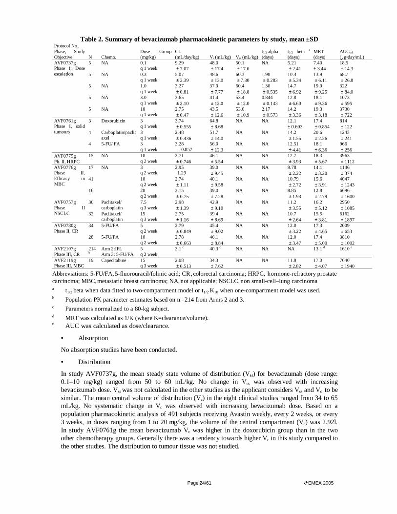
Page 24/61 EMEA 2005
Table 2. Summary of bevacizumab pharmacokinetic parameters by study, mean ±SD Protocol No., Phase, Study Objective N Chemo.
Dose Group (mg/kg)
CL (mL/day/kg) Vc (mL/kg) Vss (mL/kg)
t1/2 alpha (days)
t1/2 beta a
(days) MRT (days)
AUCinf
(µg•day/mL) 5 NA 0.1
q 1 week 9.29 ± 7.07
48.0 ± 17.4
50.1 ± 17.0
NA 5.21 ± 2.41
7.40 ± 3.44
18.5 ± 14.3
5 NA 0.3 q 1 week
5.07 ± 2.39
48.6 ± 13.0
60.3 ± 7.30
1.90 ± 0.283
10.4 ± 5.34
13.9 ± 6.11
68.7 ± 26.8
5 NA 1.0 q 1 week
3.27 ± 0.81
37.9 ± 7.77
60.4 ± 18.8
1.30 ± 0.535
14.7 ± 6.92
19.9 ± 9.25
322 ± 84.0
5 NA 3.0 q 1 week
3.65 ± 2.10
41.4 ± 12.0
53.4 ± 12.0
0.844 ± 0.143
12.8 ± 6.60
18.1 ± 9.36
1073 ± 595
AVF0737g Phase I, Dose escalation
5 NA 10 q 1 week
2.75 ± 0.47
43.5 ± 12.6
53.0 ± 10.9
2.17 ± 0.573
14.2 ± 3.36
19.3 ± 3.18
3730 ± 722
3 Doxorubicin 3 q 1 week
3.74 ± 0.555
64.8 ± 8.68
NA NA 12.1 ± 0.603
17.4 ± 0.854
814 ± 122
4 Carboplatin/paclitaxel
3 q 1 week
2.48 ± 0.436
51.7 ± 14.0
NA NA 14.2 ± 1.55
20.6 ± 2.26
1243 ± 241
AVF0761g Phase I, solid tumours
4 5-FU/ FA 3 q 1 week
3.28 � 0.857
56.0 ± 12.3
NA NA 12.51 ± 4.41
18.1 ± 6.36
966 ± 256
AVF0775g Ph. II, HRPC
15 NA 10 q 2 week
2.71 ± 0.746
46.1 ± 5.54
NA NA 12.7 ± 3.93
18.3 ± 5.67
3963 ± 1112
17 NA 3 q 2 week
2.95 1.29
39.0 ± 9.45
NA NA 9.78 ± 2.22
14.1 ± 3.20
1146 ± 374
41 10 q 2 week
2.74 ± 1.11
40.1 ± 9.58
NA NA 10.79 ± 2.72
15.6 ± 3.91
4047 ± 1243
AVF0776g Phase II, Efficacy in MBC
16 20 q 2 week
3.15 ± 0.75
39.0 ± 7.28
NA NA 8.85 ± 1.93
12.8 ± 2.79
6696 ± 1600
30 Paclitaxel/ carboplatin
7.5 q 3 week
2.98 ± 1.39
42.9 ± 9.10
NA NA 11.2 ± 3.55
16.2 ± 5.12
2950 ± 1085
AVF0757g Phase II NSCLC 32 Paclitaxel/
carboplatin 15 q 3 week
2.75 ± 1.16
39.4 ± 8.69
NA NA 10.7 ± 2.64
15.5 ± 3.81
6162 ± 1897
34 5-FU/FA 5 q 2 week
2.79 ± 0.849
45.4 ± 9.02
NA NA 12.0 ± 3.22
17.3 ± 4.65
2009 ± 653
AVF0780g Phase II, CR
28 5-FU/FA 10 q 2 week
2.78 ± 0.663
46.1 ± 8.84
NA NA 12.0 ± 3.47
17.4 ± 5.00
3810 ± 1002
AVF2107g Phase III, CR
214 b
Arm 2:IFL
Arm 3: 5-FU/FA 5 q 2 week
3.1 c 40.3 c NA NA NA 13.1 d 1610 e
AVF2119g Phase III, MBC
19 Capecitabine 15 q 3 week
2.08 ± 0.513
34.3 ± 7.62
NA NA 11.8 ± 2.82
17.0 ± 4.07
7640 ± 1940
Abbreviations: 5-FU/FA, 5-fluorouracil/folinic acid; CR , colorectal carcinoma; HRPC, hormone-refractory prostate carcinoma; MBC, metastatic breast carcinoma; NA, not applicable; NSCLC, non small-cell–lung carcinoma
a t1/2 beta when data fitted to two-compartment model or t1/2 K10 when one-compartment model was used. b Population PK parameter estimates based on n= 214 from Arms 2 and 3. c Parameters normalized to a 80-kg subject. d MRT was calculated as 1/K (where K=clearance/volume). e AUC was calculated as dose/clearance.
• Absorption
No absorption studies have been conducted.
• Distribution
In study AVF0737g, the mean steady state volume of distribution (Vss) for bevacizumab (dose range: 0.1–10 mg/kg) ranged from 50 to 60 mL/kg. No change in Vss was observed with increasing bevacizumab dose. Vss was not calculated in the other studies as the applicant considers Vss and Vc to be similar. The mean central volume of distribution (Vc) in the eight clinical studies ranged from 34 to 65 mL/kg. No systematic change in Vc was observed with increasing bevacizumab dose. Based on a population pharmacokinetic analysis of 491 subjects receiving Avastin weekly, every 2 weeks, or every 3 weeks, in doses ranging from 1 to 20 mg/kg, the volume of the central compartment (Vc) was 2.92l. In study AVF0761g the mean bevacizumab Vc was higher in the doxorubicin group than in the two other chemotherapy groups. Generally there was a tendency towards higher Vc in this study compared to the other studies. The distribution to tumour tissue was not studied.
Page 25/61 EMEA 2005
• Elimination
No human studies on the metabolism of bevacizumab have been performed. It is expected that the metabolism of bevacizumab follows the general route of metabolism of IgG class antibodies. Bevacizumab clearance was 0.231 L/day. The volume of the central compartment (Vc) and clearance correspond to an initial half-life of 1.4 days and a terminal half-life of about 20 days. This half-life is consistent with the terminal elimination half-life for human endogenous IgG, which is 18 to 23 days. In patients with low albumin (≤ 29g/dL) and high alkaline phosphatase (≥ 484U/L) (both markers of disease severity), bevacizumab clearance was approximately 20% higher than in patients with median laboratory values.
• Dose proportionality and time dependencies
The pharmacokinetics of bevacizumab as monotherapy after single or repeated doses was tested over a dose range of 0.1 to 10 mg/kg in study AVF0737g. It appeared that the PK of bevacizumab is dose independent, except for doses <1 mg/kg. The pharmacokinetics of bevacizumab appears to be linear over the dose range 1 to 10 mg/kg.
Comparing single dose and short-term multiple dosing (four doses) of bevacizumab using all available data in study AVF0737g, the overall CL estimates were 2.9% higher using the single-dose data, and the Vc estimates were 3.2% lower.
AUC0-inf in study AVF0737g increased more than dose-proportionally from 0.1 to 10 mg/kg (Table 2). However, for the dose range >1 mg/kg AUC0-inf increased dose proportionally.
In four Phase II studies, AVF0775g, AVF0776g, AVF0757g and AVF0780g, the serum concentrations of bevacizumab following multiple doses every 2 or 3 weeks were followed for up to 6–12 months in some subjects. In the first study, time to steady state was not estimated even though some concentration data were available, while in study AVF0776 time to steady state was estimated to be ~70 days. In studies AVF0757g and AVF0780g the time to steady state was estimated to be ~ 100 days, which is consistent with the estimated half-life of ~ 20 days based on population PK analysis.
In all phase II multiple-dosing studies, the mean bevacizumab concentrations following the last dose were higher than the concentrations following the first dose. The accumulation index was calculated by comparing average bevacizumab trough concentrations at steady state (Ctrough, ss) with the average trough concentrations following the first dose (Ctrough, first). An accumulation index of 1.4-2.9 was calculated for bevacizumab administered every 2 or 3 weeks. Based on the estimated β half-life of ~20 days (population PK estimate), the expected accumulation index was calculated. Based on this, a 2.6 and 1.9 fold accumulation of bevacizumab is expected at steady state following dosing every 2 or 3 weeks, respectively.
• Special populations
There are no studies specifically performed in special populations. No studies have been conducted to investigate the pharmacokinetics of bevacizumab in paediatric patients. No studies have been conducted to investigate the pharmacokinetics of bevacizumab in patients with renal or hepatic impairment.
A population pharmacokinetic model was made using a non-linear mixed effects-model. Data from all 8 studies that have yielded pharmacokinetic data have been pooled and analysed. The analysis included a total of 4629 bevacizumab concentrations for 491 subjects who received IV infusion doses weekly, every 2 weeks, or every 3 weeks at doses ranging from 1 to 20 mg/kg. Serum bevacizumab concentration–time data were modelled using a population analysis approach to estimate bevacizumab population PK parameters (mean and intersubject variability) as well as relationships between the PK parameters and various covariates. In the final multivariate model, weight, gender, albumin, SGOT, alkaline phosphatase, and chemotherapy were statistically significantly associated with bevacizumab pharmacokinetics. No significant difference in the pharmacokinetics of bevacizumab was observed in relation to age.
The best structural model was a two-compartment model with first order elimination. Population mean parameters was CL = 0.195 L/day; Vc = 2.98 L; and terminal t1/2 = 24.1 days. Intersubject variability
Page 26/61 EMEA 2005
for CL and Vc was 33.6% and 26.7 %, respectively, in the base model. The covariate effects in the final model explained about 40% of intersubject variance for CL and 61% of intersubject variance for Vc. Body weight and sex were the most significant covariates to explain intersubject variability for both CL and Vc. Creatinine clearance did not significantly influence bevacizumab clearance in this analysis. Subjects with low serum albumin levels (< 29 g/L, 5th percentile) had a 19% higher CL, while subjects with higher alkaline phosphatase levels (> 483 IU/L, 95th percentile) had a 23% faster CL, compared with the typical subject with median values of albumin and alkaline phosphatase. In the final model, bevacizumab CL was 26% faster in males compared with females, corresponding to a CL of 0.207 and 0.262 L/day for the typical (subject with median covariates) female and male subjects, respectively. For the final model, after correcting for body weight, male subjects had a larger Vc (+ 22%) than females, with the corresponding Vc of 2.66 and 3.25 L for female and male subjects, respectively.
The patients included in the population PK analysis were Caucasian (416 patients, representing 85%), Black (47 patients, representing 10%), Hispanic (17 patients, representing 3%), and Other (11 patients, representing 2%). An evaluation of impact on race could not be made. The population PK analysis revealed no impact of age on the PK of bevacizumab. Children were not allowed to participate in any of the studies.
• Pharmacokinetic interaction studies
No in vitro studies were performed. Formal in vivo drug-drug interaction studies were not performed. However detailed pharmacokinetic data was obtained from several studies in which bevacizumab was administered concomitantly with various other antineoplastic drugs.
In Phase II Study AVF0780g, patients with metastatic colorectal cancer received 5-FU (500 mg/m2/week)/FA (500 mg/m2/week) with or without bevacizumab. Plasma 5-FU concentrations were determined in 19 persons who received 10 mg/m2/2 weeks bevacizumab. The individual 5-FU concentration-time profiles at day 0 (before bevacizumab) and at day 35 (after three doses of bevacizumab) showed no systematic changes, however, data were variable.
In Study AVF2107g (Phase III), patients with metastatic colorectal cancer received irinotecan (125 mg/m2)/5-FU (500 mg/m2/week)/FA (20 mg/m2/week) without (Arm 1) or with bevacizumab (5 mg/m2/2 weeks, Arm 2), or 5-FU (500 mg/m2/week)/FA (500 mg/m2/week) with bevacizumab (5 mg/ m2/2 weeks, Arm 3). Concentrations of irinotecan and its active metabolite, SN38, were measured in about 50 subjects in each of the arms 1 and 2, 43 days after bevacizumab or placebo administration. The ratios (95% CI) for dose-normalised AUC0-5hr for irinotecan with or without bevacizumab and SN38 were 1.10 (0.96, 1.27) and 1.33 (1.07, 1.65), respectively, indicating increased SN38 exposure in bevacizumab-treated subjects. Bevacizumab disposition was studied in about 100 persons per arm in arms 2 and 3. Estimates for clearance of bevacizumab were similar in the two arms, indicating that the addition of irinotecan to 5-FU/FA/bevacizumab does not affect the pharmacokinetics of bevacizumab.
In Study AVF2119g (Phase III), patients with metastatic breast cancer were treated with capecitabine (1875-2500 mg/m2 daily for 2 weeks every 3rd week) with or without bevacizumab (15 mg/m2/3 weeks). AUC measurements for capecitabine and metabolites (5 -́DFUR and 5-FU) (cycle 1, week 2) were calculated for about 20 subjects in each group. No significant difference in capecitabine and metabolites exposure was found between the two groups.
Discussion on Clinical PK
The pharmacokinetics of bevacizumab have been reasonably well studied in various patient populations, following intravenous infusion. The pharmacokinetics of bevacizumab is best characterized by a two-compartment model. Apparent dose proportionality, as assessed by clearance, was observed for doses above 1 mg/kg/day whereas lower doses were associated with higher levels of clearance. After four multiple IV doses (on Days 0, 28, 35, and 42), the clearance of bevacizumab ranged from 2.75 to 3.65 mL/day/kg. The central volume of distribution ranged from 37.9–48.6 mL/kg at all doses, which is about the human plasma volume of 43 mL/kg. Protein binding has not been studied. The estimated mean terminal half-life of bevacizumab at doses > 1 mg/kg ranged from 13 to 19 days. No studies have been performed in special populations. The knowledge on bevacizumab pharmacokinetics in these populations origin from the population pharmacokinetic model derived. A non-linear mixed-effect model
Page 27/61 EMEA 2005
based on data from all 8 pharmacokinetic studies was developed. The final model explained about 40% the observed variability in CL, with the single most significant variables being body weight and gender. Simulation of plasma concentrations using the model yielded result fairly comparable to those actually observed. A weight-based dosing scheme seems reasonably justified by these data.
The metabolism of bevacizumab has not been specifically studied. Bevacizumab is assumed to be eliminated by the same mechanism as other IgG antibodies.
No formal drug interaction studies with other antineoplastic agents have been conducted. However, the existing data suggest that bevacizumab does not affect the pharmacokinetics of 5-FU, carboplatin, paclitaxel, and doxorubicin to a clinically relevant extent (see SPC section 4.5).
Plasma protein binding studies are lacking. However, this is considered acceptable as bevacizumab is a high molecular weight protein (149 000 Daltons).
Pharmacodynamics
• Mechanism of action
The suggested mechanism of action is supported by results of in vitro studies that are referred in the non-clinical section. No studies investigating the pharmacodynamics and mechanism of action of bevacizumab have been performed in humans.
• Primary and secondary pharmacology
In vitro studies using human umbilical vein endothelial cells (HUVEC) explored the effect of bevacizumab on VEGF-induced effects. In study 1765-582-RPT-1 HUVEC cells were incubated with bevacizumab (3.9–1000 ng/mL) and 10 ng/ml of rhVEGF165, rhVEGF121, or rhVEGF110. Proliferation induced by all splice variants of VEGF was dose-dependently inhibited by bevacizumab, with IC50s in the range of 32-86 ng/mL. In study 1765-582-RPT-2 HUVEC cells were incubated with 50 ng/mL VEGF, with or without bevacizumab (500 ng/mL), and bevacizumab abolished VEGF-induced nitric oxide production. In study 1765-582-RPT-3, bevacizumab at a concentration ratio of 10:1 effectively abolished VEGF-induced permability in HUVEC, and in study 1765-582-RPT-4 bevacizumab (500 mg/mL) abolished the cell survival activity induced by 50 ng/mL rhVEGF. Thus bevacizumab was shown to inhibit VEGF-induced pharmacological activity in human umbilical vein endothelial cells at concentrations 10-fold (3-fold at molar basis) above those of VEGF.
In Phase I (AVF0737g, AVF0761g) and Phase II (AVF0757g, AVF0775g, AVF0776g, AVF0780g) clinical trials total serum or plasma VEGF were investigated as a pharmacodynamic endpoint. AVF0737g was a dose-escalating trial in which bevacizumab was administered at five dose levels ranging from 0.1 to 10 mg/kg. Total serum VEGF concentrations were similar between the 0.1 and 0.3 mg/kg doses and remained stable during the study, while following bevacizumab doses of 1-10 mg/kg total serum VEGF concentrations increased rapidly over 7 days and then declined following bevacizumab elimination. Subsequent weekly doses (at day 28, 35, and 42) further increased VEGF concentrations. Bevacizumab was administered as a multidose regimen (3-20 mg/kg every 1 to 3 weeks) in the subsequent five Phase I/II clinical trials. Total plasma VEGF concentrations were monitored. In all studies, total VEGF concentrations increased with increasing bevacizumab doses and bevacizumab serum concentrations.
Hypertension, mucocutaneous bleeding, thromboembolic events, and proteinuria were identified as bevacizumab-related adverse events (see section IV Clinical safety), all of which may be related to VEGF antagonism through e.g. reduced endothelial nitric oxide synthesis (see study 1765-582-RPT-2) and other VEGF antagonism-related effects.
Discussion on Clinical PD
Bevacizumab binds VEGF, thereby inhibiting binding of VEGF to the VEGF receptors and inhibition of VEGF-induced tumour angiogenesis. No studies investigating the pharmacodynamics and mechanism of action of bevacizumab have been performed in humans.

Page 28/61 EMEA 2005
In human umbilical vein endothelial cells (HUVEC) bevacizumab dose-dependently inhibited VEGF-induced proliferation, and abolished VEGF-induced effects on nitric oxide production, cell permeability, and cell survival.
The total plasma or serum VEGF concentration was used as the sole surrogate pharmacodynamic marker reaction. This may be the best in vivo marker at the present time, but it remains less than satisfactory as the true relation between VEGF and the effect of bevacizumab is virtually unknown. It seems plausible that some association exist as VEGF concentrations increases with increasing plasma concentrations of bevacizumab. However the interpretation of this, especially with respect to optimal dose selection is unclear.
Clinical efficacy
• Dose response study(ies)
Four of the studies can be defined as dose-escalation studies (AVF0737g, AVF0776g, AVF0757g, AVF0780g) with bevacizumab as monotherapy or in combination with cytotoxic agents. Only one of these studies has been performed in the target population (AVF0780g).
Maximum tolerated dose (MTD) was not reached in any of the clinical studies. Based on pre-clinical results (see Non-Clinical Assessment Report) and the pharmacokinetic results in the Phase I trial AVF0737g, doses of 5 mg/kg/2 weeks and 10 mg/kg/2 weeks were chosen for the Phase II colorectal trial AVF0780g. Based on pharmacokinetic modelling these doses were predicted to result in steady state trough concentrations of ~50 µg/mL and ~100 µg/mL, respectively. At the dose of 5 mg/kg/2 weeks the circulating VEGF was, on average, > 98% bound with bevacizumab. Significant inhibition was maintained throughout a dosing interval. Any further increase in the dose of bevacizumab would result in minor additional inhibition of circulating VEGF.
• Main study(ies)
Three study reports of controlled clinical studies pertinent to the claimed indication were submitted. AVF2107g was a phase III randomized, active-controlled clinical trial to evaluate bevacizumab in combination with IFL and Roswell-Park regimens in subjects with metastatic colorectal cancer [108]. AVF2192g was a phase II, double-blind, randomized, active-controlled clinical trial of bevacizumab in combination with 5-FU/FA chemotherapy in subjects with metastatic colorectal cancer who are not optimal candidates for first-line irinotecan [109]. AVF0780g was a phase II, multidose, randomized clinical trial to evaluate the efficacy, safety, and pharmacokinetics of bevacizumab in combination with 5-FU/FA as compared to 5-FU/FA alone in subjects with metastatic colorectal cancer [110].
Study AVF 2107g
METHODS
Study Participants
This was a multicentre, randomized, active-controlled phase III clinical trial. The main inclusion criteria were histologically confirmed colorectal carcinoma with evidence of metastases (i.e. by radiographic imaging or biopsy), ECOG performance status of 0 or 1, life expectancy of > 3 months, bidimensionally measurable disease (minimum of two lesions). The main exclusion criteria were: prior chemotherapy other than adjuvant fluoropyrimidines in combination with FA and/or levamisole, administration of adjuvant fluoropyrimidines in combination with FA and/or levamisole completed ≤12 months prior to Day 0; prior radiotherapy to a measurable, metastatic lesion(s) to be used to measure response; radiation therapy within 14 days prior to Day 0; proteinuria at baseline or clinically significant impairment of renal function; clinically significant cardiovascular disease; clinical laboratory values: neutrophil count <1500/µL, platelet count <75,000/µL, INR ≥1.5, total bilirubin >1.6 mg/dL, AST or ALT ≥5 times upper limit of normal for subjects with documented liver metastases; >2.5 times the upper limit of normal for subjects without evidence of liver metastases, serum creatinine >2.0 mg/dL, hemoglobin <9 g/dL; major surgery procedure within 28 days prior to Day 0.
Treatments
Page 29/61 EMEA 2005
The patients were randomized to one of three regimens. Arm 1: placebo plus bolus- IFL (‘Saltz’) regimen consisting of 125 mg/m2 irinotecan, 500 mg/m2 5-FU by IV bolus injection, 20 mg/m2 FA by IV bolus, administered in repeating 6-week cycles consisting of weekly treatments for 4 weeks followed by 2 weeks of rest. Arm 2: bevacizumab 5 mg/kg IV infusion (90 →30 min) once every 2 weeks (regardless of possible chemotherapy delays) plus bolus-IFL regimen as in arm 1. Arm 3: bevacizumab as in arm 2 plus bolus 5-FU/FA (‘Roswell Park’) regimen (5-FU 500 mg/m2+FA 20 mg/m2) weekly for 6 weeks of every 8- week cycle. Arm 3 was added since the safety of the IFL combination was not sufficiently known. An interim safety analysis was planned to be performed by an independent data monitoring committee (DMC) when 100 patients had been entered per arm in order to determine whether it was feasible to continue the trial with arm 1 and 2 only. The DMC concluded, following their review, that the safety profile of the IFL plus bevacizumab regimen (arm 2) was acceptable. Enrolment in arm 3 was then closed while it continued into arm 1 and 2 until 400 patients per arm had been included.
Patients were treated until progression or for a maximum of 16 treatment cycles (96 weeks). Second-line treatment could be given within or outside the protocol. Patients in the bevacizumab treatment arms were eligible to discontinue chemotherapy and continue bevacizumab alone if they had a confirmed complete response or unacceptable chemotherapy-related toxicity. Patients in the control arm were not allowed to cross over to bevacizumab.
Objectives
The primary objective was to evaluate the efficacy (survival) and safety of the ad-on treatment with multiple doses of bevacizumab (arm 1 vs. arm 2). Secondary objectives were to evaluate the efficacy by response rate (CR or PR), time to progression , duration of response and quality of life. Further to evaluate PK and disposition of bevacizumab. Other objectives: comparison of arm 3 vs. arm 2 results, evaluation of efficacy and safety in the second-line therapy.
Outcomes/endpoints
The primary outcome, duration of survival, was defined as the time from randomization to death from any cause. All reported deaths were included, whether the death occurred during first- or second-line therapy or following treatment discontinuation.
The secondary outcome measures included progression free survival (PFS) during first-line therapy assessed by the investigator according to the Response Evaluation Criteria in Solid Tumours (RECIST). Progression-free survival during first line therapy was defined as the time from randomisation to disease progression or death during first-line therapy. Data for subjects who received second-line therapy without having documented disease progression and who did not die within 30 days of last dose of first-line therapy were censored at the time of the last tumour assessment. This was done to avoid bias since all patients receiving bevacizumab second-line therapy had to remain in the study and were followed for progression, whereas patients receiving other types of second-line therapy were only followed for survival.
Responses were assessed only by the investigators. No independent review of the data was deemed necessary, as this was a double-blind study with overall survival as the primary endpoint. Assessments were done every six weeks for the first 24 weeks and every 12 weeks thereafter. PFS was defined as the time from randomisation to disease progression or death due to any cause during first-line therapy. Objective response rate during first-line therapy was defined as a complete response or partial response according to RECIST determined on two consecutive investigator assessments ≥ 4 weeks apart during first-line therapy. Duration of objective response was determined for the subset of subjects who achieved objective response during first-line therapy. Duration of objective response was defined as the time from the first tumour assessment that supported the subject’s objective response to the time of disease progression or death due to any cause during first-line therapy. Time to deterioration in QoL was measured by FACT-C.
Sample size
The sample size estimate was based on 80% power to detect a hazard ratio of 0.75 at the 5% significance level (two-sided), which corresponds approximately to a 33% improvement in median time
Page 30/61 EMEA 2005
to death from 15 months in the control group to 20 months in the bevacizumab group. Based on two planned interim analyses, the required number of deaths in the two principal treatment arms (1 and 2) at the final analysis was 385. It was estimated that a total of 800 patients (60 per month for 13 months) and a follow-up period of 10 months would provide 385 deaths. Four hundred patients in the bevacizumab group would provide 87% power to detect an adverse event that occurs at a rate of 0.5%. The FACT-C QoL assessment was to be collected in 100 patients in each treatment group. This would provide 80% power to detect an effect size (mean/SD) of 0.4.
Randomization
Randomization was based on a minimization algorithm with stratification for centre, number of organ sites with disease (1 versus >1), ECOG performance status (0 versus 1), and site of original disease (colon vs rectal).
Blinding (masking)
Patients in arm 1 and 2 received the study drug in a double-blind fashion during the treatment period.
A complete evaluation of safety information took place when 300 subjects had been randomised (100 subjects per treatment arm). At that time, an interim analysis of safety was conducted by an unblinded, independent Data Monitoring Committee (DMC) evaluating all adverse events as well as subjects status, study drug administration, laboratory studies, and vital signs.
Subjects in Arms 1 and 2 were unblinded for the following reasons: completion of the study, complete response, disease progression, and toxicity.
Statistical methods
Formal hypothesis testing was only performed for the treatment comparison between the two principal treatment arms (arms 1 and 2). Patients still alive were censored as of the last date the patient was known to be alive. Overall survival was compared by a stratified logrank test. The stratification factors were ECOG performance status, number of organ sites with disease, and site of primary tumour (all dichotomous). In secondary analyses, the hazard ratio was estimated in a stratified Cox proportional hazards model. Results from unstratified analyses were also presented. A Lan-DeMets implementation of the O’Brien-Fleming α-spending function was used to control the significance level for two interim analyses.
Objective response was compared between treatment arms using the chi-square test. Patients without post baseline tumour assessment were counted as non-responders.
Time to deterioration of QoL (TDQ) was assessed for patients with a baseline and a post baseline measurement and compared between groups by the logrank test. Deterioration was defined as a decline of ≥3 for colorectal cancer specific questions (CCS), ≥7 for TOI-C (the sum of physical and functional well being and CCS), and ≥9 for the FACT-C. Patients progressing or dying during first-line therapy were treated as having deterioration in QoL. The Wilcoxon rank sum test was used to compare the change from baseline to the last available assessment.
RESULTS
Participant flow
At least one dose of the assigned study treatment was received by 897/923 (97.2%) patients (396 in Arm 1, 392 in Arm 2 and 109 in Arm 3). The mean duration of treatment was 31.1 weeks in the control arm (Arm 1) and 40.4 weeks in the bevacizumab treatment arm (Arm 2). The main reason for treatment discontinuation was disease progression (64.5% in Arm 1 and 50% in Arm 2). Discontinuation due to adverse events was similar in both treatment arms (6.6% in Arm 1 and 7.7% in Arm 2). More patients in Arm 2 than in Arm 1 had dose reductions for 5-FU (80.6% versus 68.4%) and irinotecan (79.8% versus 69.1%). Dose intensity percentages for study drug and chemotherapy were slightly lower for patients in Arm 2 compared with those in Arm 1.
Second-line treatment was well balanced between the two arms. Fifty-six percent of patients in Arm 1 and 55% of patients in Arm 2 received second-line chemotherapy on or off study. Forty-seven patients

Page 31/61 EMEA 2005
(11.9%) in the Arm 1 and 110 patients (28.1%) in Arm 2 received second-line treatment while on study. Use of oxaliplatin (27% versus 24%), irinotecan (10% in both arms) and capecitabine (23% in both arms) was similar in both arms. As per protocol none of the patients in the control arm received second-line bevacizumab, while 107/392 and 55/109 in arms 2 and 3, respectively, did.
The minimal follow-up time for survival was 11 months for the last patient randomized.
Conduct of the study
Three amendments of the protocol were made during the study. The first dealt with numbers of patients for the first interim analysis (100 instead of 50 per arm) and other details. The second reported the decision to stop entrance of patients into arm 3. The third dealt with irinotecan treatment and other details. Eligibility exceptions were noted for 10% of the patients and major protocol deviations occurred in 3.5%. They were evenly distributed and did not seem to affect the validity of the comparison of arm 1 and 2.
Baseline data
Demographic and disease baseline characteristics are shown in Table 3 and 4. The median age was 60 years, ECOG performance status was 0 in 57% of the patients, 78% of tumours were located in the colon and almost all (>99%) were adenocarcinomas. More patients in the discontinued bevacizumab plus 5-FU/FA arm (Arm 3) had previous radiotherapy (22% versus 15%) and fewer patients had surgery (81% versus 87%). Twenty-six percent of the patients had received adjuvant chemotherapy. The most frequent metastatic sites were liver (78%), lung (48%) and lymph nodes (25%). Fewer patients in the discontinued arm had lung lesions (39% in Arm 3 versus 48% in Arm 1 and 49% in Arm 2).
Table 3. Demographic and Baseline Characteristics in Study AVF2107g
Arm 1 b-IFL Placebo
(n = 411)
Arm 2 b-IFL BV (n = 402)
Arm 3 5-FU/FA BV
(n = 110) Total
(n = 923)
Age (yr)
Mean (SD) 59.2 (11.47) 59.5 (11.29) 59.7 (12.08) 59.4 (11.45)
Median 60.0 60.0 61.5 60.0
Range 21.0–83.0 23.0–86.0 29.0–88.0 21.0–88.0
Sex
Female 163 (39.7%) 165 (41.0%) 41 (37.3%) 369 (40.0%)
Male 248 (60.3%) 237 (59.0%) 69 (62.7%) 554 (60.0%)
Race/ethnicity
American Indian or Alaskan Native
0 (0.0%) 2 (0.5%) 0 (0.0%) 2 (0.2%)
Asian or Pacific Islander 14 (3.4%) 12 (3.0%) 4 (3.6%) 30 (3.3%)
Black 46 (11.2%) 49 (12.2%) 14 (12.7%) 109 (11.8%)
Hispanic 23 (5.6%) 18 (4.5%) 2 (1.8%) 43 (4.7%)
White 328 (79.8%) 317 (78.9%) 90 (81.8%) 735 (79.6%)
Other 0 (0.0%) 4 (1.0%) 0 (0.0%) 4 (0.4%)
ECOG performance status
0 227 (55.2%) 234 (58.4%) 61 (55.5%) 522 (56.6%)
1 182 (44.3%) 166 (41.4%) 48 (43.6%) 396 (43.0%)
2 2 (0.5%) 1 (0.2%) 1 (0.9%) 4 (0.4%)
Abbreviations: BV, bevacizumab; b-IFL , bolus irinotecan/5-fluorouracil/folinic acid; 5-FU/FA, 5-fluorouracil/folinic acid.
Page 32/61 EMEA 2005
Table 4. Disease baseline characteristics in Study AVF2107g in Colorectal Cancer
Arm 1 b-IFL Placebo
(n = 411)
Arm 2 b-IFL BV (n = 402)
Arm 3 5-FU/FA BV
(n = 110) Total
(n = 923) Duration of disease (months)
Mean (SD) 16 (22.0) 15 (23.2) 16 (22.3) 16 (22.6) Median 3 3 3 3 Range 1–142 1–170 1–107 1–170
Duration of metastatic disease (months) Mean (SD) 4 (9.2) 4 (9.2) 4 (8.8) 4 (9.1) Median 2 2 2 2 Range 1–125 1–91 1–70 1–125
Location of primary tumour Colon 334 (81.3%) 310 (77.1%) 77 (70.0%) 721 (78.1%) Rectum 77 (18.7%) 92 (22.9%) 33 (30.0%) 202 (21.9%)
Histologic classification Adenocarcinoma 384 (93.4%) 373 (92.8%) 104 (94.5%) 861 (93.3%) Mucinous adenocarcinoma 21 (5.1%) 26 (6.5%) 6 (5.5%) 53 (5.7%) All Other 6 (1.5%) 3 (0.7%) 0 (0.0%) 9 (1.0%)
Number of organ sites with metastases 1 159 (38.9%) 147 (36.6%) 48 (43.6%) 354 (38.4%) > 1 252 (61.6%) 255 (63.4%) 62 (56.4%) 569 (61.8%)
Prior cancer treatment Surgery 360 (87.6%) 350 (87.1%) 89 (80.9%) 799 (86.6%) Radiotherapy 59 (14.4%) 60 (14.9%) 24 (21.8%) 143 (15.5%) Systemic chemotherapy 119 (29.0%) 108 (26.9%) 35 (31.8%) 262 (28.4%)
Neo-adjuvant 10 (2.4%) 11 (2.7%) 7 (6.4%) 28 (3.0%) Adjuvant 113 (27.5%) 96 (23.9%) 29 (26.4%) 238 (25.8%) Metastatic 4 (1.0%) 7 (1.7%) 0 (0.0%) 11 (1.2%) Other 0 (0.0%) 1 (0.2%) 2 (1.8%) 3 (0.3%)
Treated with first-line therapy 396 (96.4%) 392 (97.5%) 109 (99.1%) 897 (97.2%) Completed study 4 (1.0%) 8 (2.0%) 8 (7.3%) 20 (2.2%) Discontinued first-line therapy 359 (87.3%) 313 (77.9%) 98 (89.1%) 770 (83.4%)
Death 13 (3.2%) 14 (3.5%) 7 (6.4%) 34 (3.7%) Disease progression 265 (64.5%) 201 (50.0%) 71 (64.5%) 537 (58.2%) Adverse event 27 (6.6%) 31 (7.7%) 11 (10.0%) 69 (7.5%) Lost to follow-up 2 (0.5%) 1 (0.2%) 0 (0.0%) 3 (0.3%) Patient’s decision 25 (6.1%) 39 (9.7%) 4 (3.6%) 68 (7.4%) Physician’s decision 27 (6.6%) 27 (6.7%) 5 (4.5%) 59 (6.4%)
Not treated with study drug 15 (3.6%) 10 (2.5%) 1 (0.9%) 26 (2.8%) Discontinued 15 (3.6%) 10 (2.5%) 1 (0.9%) 26 (2.8%)
Disease progression 2 (0.5%) 2 (0.5%) 0 (0.0%) 4 (0.4%) Adverse event 1 (0.2%) 2 (0.5%) 0 (0.0%) 3 (0.3%) Physician’s decision 3 (0.7%) 3 (0.7%) 0 (0.0%) 6 (0.7%) Patient’s decision 9 (2.2%) 3 (0.7%) 1 (0.9%) 13 (1.4%)
Treated with second-line therapy on study
47 (11.4%) 110 (27.4%) 55 (50.0%) 212 (23.0%)
Completed study 0 (0.0%) 2 (0.5%) 8 (7.3%) 10 (1.1%) Abbreviations: BV, bevacizumab; b-IFL, bolus irinotecan/5-fluorouracil/folinic acid; 5-FU/FA, 5-fluorouracil/folinic acid.
Outcomes and estimation
The response rate was significantly higher in the bevacizumab arm (p=0.0036). The majority of responses were partial responses (table 5).
The higher number of responses seen in the bevacizumab treatment arm was accompanied by a longer duration of response (10.4 months in Arm 2 versus 7.1 months in the Arm 1) (table III.7)

Page 33/61 EMEA 2005
Table 5 Objective Response, and duration, in the Phase III Metastatic CRC Study AVF2107g
Arm 1 IFL Placebo
(N = 411)
Arm 2 IFL Bevacizumab
(N = 402) Objective response No. (%) 143 (34.8%) 180 (44.8%)
95% CI (30.2%, 39.6%) (39.9%, 49.8%) p-value (χ2 ) 0.0036 Between-arm difference 10.0% 95% CI (3.3%, 16.7%) Best response CR 9 (2.2%) 15 (3.7%) PR 134 (32.6%) 165 (41.0%)
Duration of objective response (months)
Median 7.06 10.35
95% CI (5.95, 9.07) (9.30, 11.66)
Range 1.31+ to 20.93+ 1.08+ to 20.80+
25–75 percentile (months) 4.7 – 11.8 6.7 – 15.0
Censored observations 56 (39.2%) 87 (48.3%)
Abbreviations: + indicates censored observation; IFL, irinotecan/5-fluorouracil/folinic acid; CI, confidence interval.
Survival
The addition of bevacizumab to IFL led to a statistically significant prolongation of survival with an increase in median duration of survival from 15.6 months in Arm 1 to 20.3 months in Arm 2 (p<0.0001) (Fig. 1 and Table 6). Median follow-up time for survival was 21 months. A similar proportion of patients in arms 1 and 2 received second-line treatment and the chemotherapy agents used were also similar.
Figure 1. Duration of Survival in the Phase III Metastatic CRC Study AVF2107g
Table 6. Duration of Survival in the Phase III Metastatic CRC Study AVF2107g Endpoint Arm 1
IFL + Placebo (N = 411)
Arm 2 IFL + Bevacizumab
(N = 402)
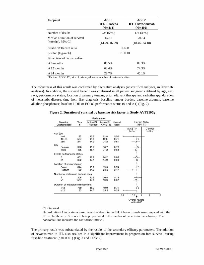
Page 34/61 EMEA 2005
Endpoint Arm 1 IFL + Placebo
(N = 411)
Arm 2 IFL + Bevacizumab
(N = 402)
Number of deaths 225 (55%) 174 (43%)
Median Duration of survival (months), 95% CI
15.61
(14.29, 16.99)
20.34
(18.46, 24.18)
Stratifieda Hazard ratio 0.660
p-value (log-rank) <0.0001
Percentage of patients alive
at 6 months 85.5% 89.3%
at 12 months 63.4% 74.3%
at 24 months 29.7% 45.1% a Factors: ECOG PS, site of primary disease, number of metastatic sites.
The robustness of this result was confirmed by alternative analyses (unstratified analyses, multivariate analyses). In addition, the survival benefit was confirmed in all patient subgroups defined by age, sex, race, performance status, location of primary tumour, prior adjuvant therapy and radiotherapy, duration of metastatic disease, time from first diagnosis, baseline tumour burden, baseline albumin, baseline alkaline phosphatase, baseline LDH or ECOG performance status (0 and ≥ 1) (Fig. 2).
Figure 2. Duration of survival by baseline risk factor in Study AVF2107g
CI = interval Hazard ratio < 1 indicates a lower hazard of death in the IFL + bevacizumab arm compared with the IFL + placebo arm. Size of circle is proportional to the number of patients in the subgroup. The horizontal line indicates the confidence interval.
The primary result was substantiated by the results of the secondary efficacy parameters. The addition of bevacizumab to IFL also resulted in a significant improvement in progression free survival during first-line treatment (p<0.0001) (Fig. 3 and Table 7).
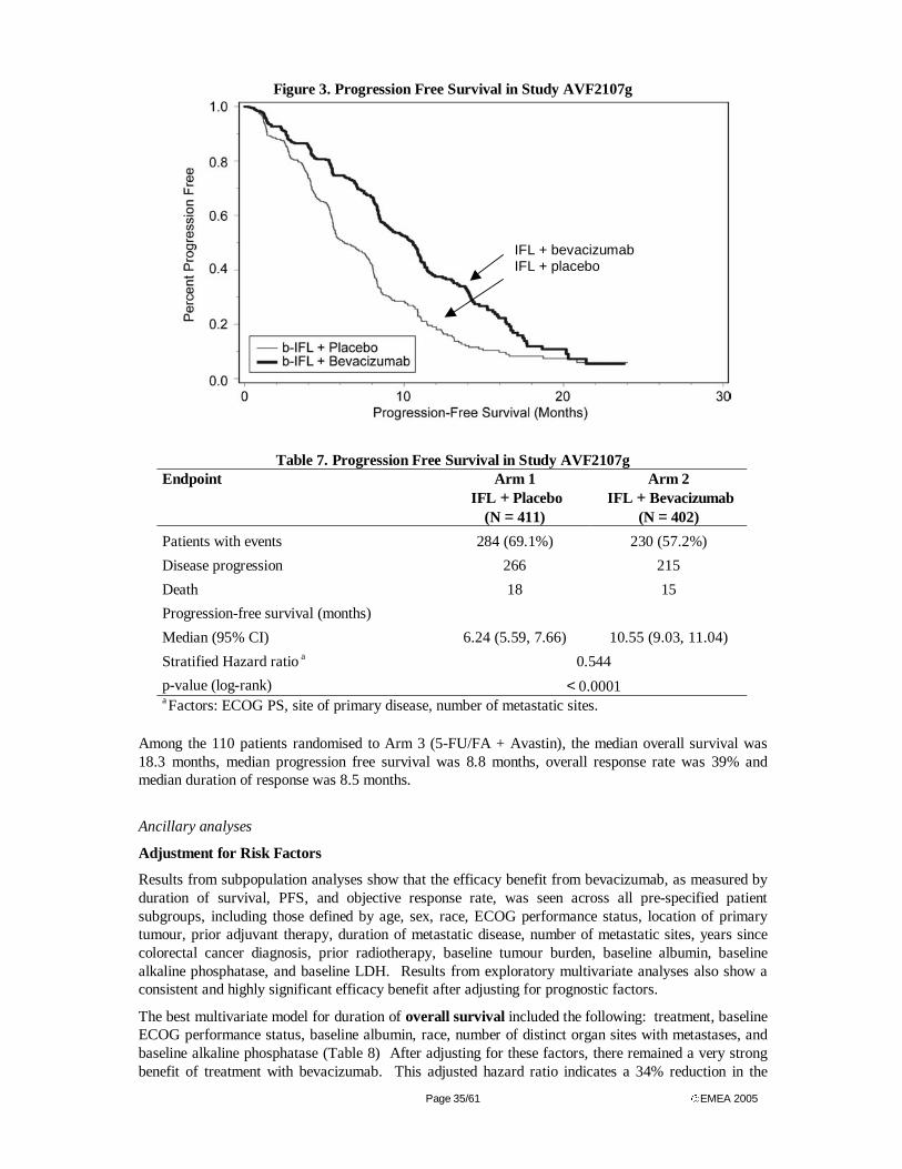
Page 35/61 EMEA 2005
Figure 3. Progression Free Survival in Study AVF2107g
Table 7. Progression Free Survival in Study AVF2107g
Endpoint Arm 1 IFL + Placebo
(N = 411)
Arm 2 IFL + Bevacizumab
(N = 402)
Patients with events 284 (69.1%) 230 (57.2%)
Disease progression 266 215
Death 18 15
Progression-free survival (months)
Median (95% CI) 6.24 (5.59, 7.66) 10.55 (9.03, 11.04)
Stratified Hazard ratio a 0.544
p-value (log-rank) < 0.0001 a Factors: ECOG PS, site of primary disease, number of metastatic sites.
Among the 110 patients randomised to Arm 3 (5-FU/FA + Avastin), the median overall survival was 18.3 months, median progression free survival was 8.8 months, overall response rate was 39% and median duration of response was 8.5 months.
Ancillary analyses
Adjustment for Risk Factors
Results from subpopulation analyses show that the efficacy benefit from bevacizumab, as measured by duration of survival, PFS, and objective response rate, was seen across all pre-specified patient subgroups, including those defined by age, sex, race, ECOG performance status, location of primary tumour, prior adjuvant therapy, duration of metastatic disease, number of metastatic sites, years since colorectal cancer diagnosis, prior radiotherapy, baseline tumour burden, baseline albumin, baseline alkaline phosphatase, and baseline LDH. Results from exploratory multivariate analyses also show a consistent and highly significant efficacy benefit after adjusting for prognostic factors.
The best multivariate model for duration of overall survival included the following: treatment, baseline ECOG performance status, baseline albumin, race, number of distinct organ sites with metastases, and baseline alkaline phosphatase (Table 8) After adjusting for these factors, there remained a very strong benefit of treatment with bevacizumab. This adjusted hazard ratio indicates a 34% reduction in the
IFL + bevacizumab IFL + placebo
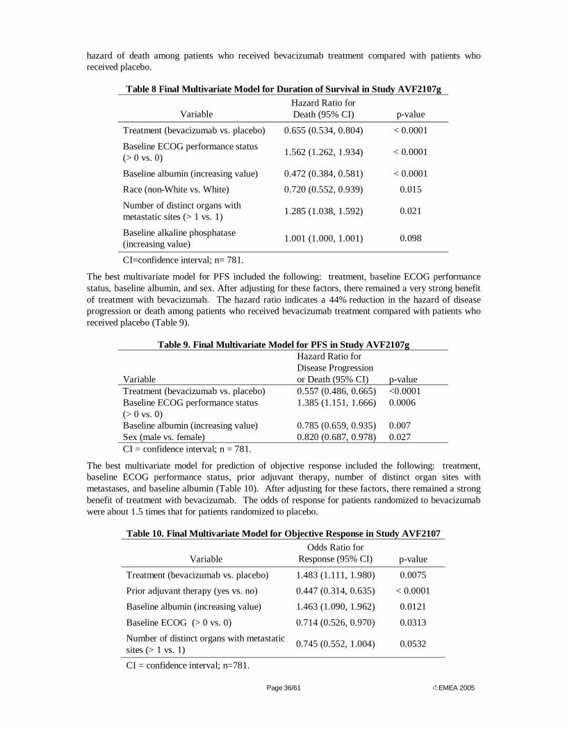
Page 36/61 EMEA 2005
hazard of death among patients who received bevacizumab treatment compared with patients who received placebo.
Table 8 Final Multivariate Model for Duration of Survival in Study AVF2107g
Variable Hazard Ratio for Death (95% CI) p-value
Treatment (bevacizumab vs. placebo) 0.655 (0.534, 0.804) < 0.0001
Baseline ECOG performance status (> 0 vs. 0)
1.562 (1.262, 1.934) < 0.0001
Baseline albumin (increasing value) 0.472 (0.384, 0.581) < 0.0001
Race (non-White vs. White) 0.720 (0.552, 0.939) 0.015
Number of distinct organs with metastatic sites (> 1 vs. 1)
1.285 (1.038, 1.592) 0.021
Baseline alkaline phosphatase (increasing value)
1.001 (1.000, 1.001) 0.098
CI=confidence interval; n= 781.
The best multivariate model for PFS included the following: treatment, baseline ECOG performance status, baseline albumin, and sex. After adjusting for these factors, there remained a very strong benefit of treatment with bevacizumab. The hazard ratio indicates a 44% reduction in the hazard of disease progression or death among patients who received bevacizumab treatment compared with patients who received placebo (Table 9).
Table 9. Final Multivariate Model for PFS in Study AVF2107g
Variable
Hazard Ratio for Disease Progression or Death (95% CI) p-value
Treatment (bevacizumab vs. placebo) 0.557 (0.486, 0.665) <0.0001 Baseline ECOG performance status (> 0 vs. 0)
1.385 (1.151, 1.666) 0.0006
Baseline albumin (increasing value) 0.785 (0.659, 0.935) 0.007 Sex (male vs. female) 0.820 (0.687, 0.978) 0.027 CI = confidence interval; n = 781.
The best multivariate model for prediction of objective response included the following: treatment, baseline ECOG performance status, prior adjuvant therapy, number of distinct organ sites with metastases, and baseline albumin (Table 10). After adjusting for these factors, there remained a strong benefit of treatment with bevacizumab. The odds of response for patients randomized to bevacizumab were about 1.5 times that for patients randomized to placebo.
Table 10. Final Multivariate Model for Objective Response in Study AVF2107
Variable Odds Ratio for
Response (95% CI) p-value
Treatment (bevacizumab vs. placebo) 1.483 (1.111, 1.980) 0.0075
Prior adjuvant therapy (yes vs. no) 0.447 (0.314, 0.635) < 0.0001
Baseline albumin (increasing value) 1.463 (1.090, 1.962) 0.0121
Baseline ECOG (> 0 vs. 0) 0.714 (0.526, 0.970) 0.0313
Number of distinct organs with metastatic sites (> 1 vs. 1)
0.745 (0.552, 1.004) 0.0532
CI = confidence interval; n=781.
Page 37/61 EMEA 2005
Comparison of arm 1&2 vs arm 3 results
Results for the 110 patients in Arm 3 were compared for efficacy with those of the first 100 patients in Arm 1 enrolled before the cut-off point of the interim safety analysis. Median duration of survival was 18.27 months v. 15.08 months in the bevacizumab plus 5-FU/FA arm as compared to the placebo plus IFL arm in this subset of the patient population (log-rank p-value = 0.2521). Median progression-free survival was 8.77 months v. 6.83 months (log-rank p-value = 0.4192, stratified hazard ratio = 0.862, CI: 0.60, 1.24), and response rate was 40.0% (95% CI: 30.9%, 49.8%, based on the normal approximation to the binomial distribution) v. 37.0% (95% CI = 27.7%, 47.3%; χ2 p-value = 0.6556), respectively.
Quality of life
The addition of bevacizumab did not extend the time to deterioration in patients’ quality of life, nor did it contribute to a more rapid worsening of quality of life compared with standard first-line chemotherapy alone. There was no statistically significant difference in the time to deterioration in colorectal cancer–specific (CCS) score between treatment arms (d = 3; p = 0.5807). Median time to deterioration in quality of life, as measured by CCS, was 2.73 months in Arm 1 and 2.89 months in Arm 2. Similarly, there were no significant differences observed in time to deterioration in quality of life, as measured by Trial Outcome Index, total Functional Assessment of Cancer Therapy–Colorectal score, or change from baseline to last available quality-of-life score.
Analysis performed across trials (pooled analyses and meta-analysis)
An analysis across trials of the single agent activity of bevacizumab was performed to describe the activity of single-agent bevacizumab in Genentech-sponsored Phase I and Phase II clinical trials in metastatic NSCLC, colorectal cancer, prostate cancer, breast cancer, and mixed solid tumours (data not shown). In the NCI-sponsored Phase III trial of the FOLFOX4 regimen ± bevacizumab in second-line metastatic colorectal cancer (Study E3200) the DMC discontinued enrollment in the third treatment arm of 5 mg/kg/wk bevacizumab (10 mg/kg/2wk) alone because of lack of efficacy compared with the FOLFOX-containing regimens. The NCI-sponsored trial Study AVF0890s, a randomized, double-blind, placebo-controlled trial of single-agent bevacizumab at two dose strengths in patients with metastatic renal cell cancer who had progressed on IL-2 therapy, demonstrated that single-agent bevacizumab could prolong PFS in this disease. Although both doses prolonged PFS compared with placebo, the 5 mg/kg/wk dose (10 mg/kg/2wk) showed a significantly stronger treatment effect than the 1.5 mg/kg/wk dose (3 mg/kg/2wk)
The estimated treatment effect of bevacizumab combined with 5-FU based regimens 5-FU/FA and 5-FU/FA+Irinotecan is illustrated in Fig. 4 and 5.
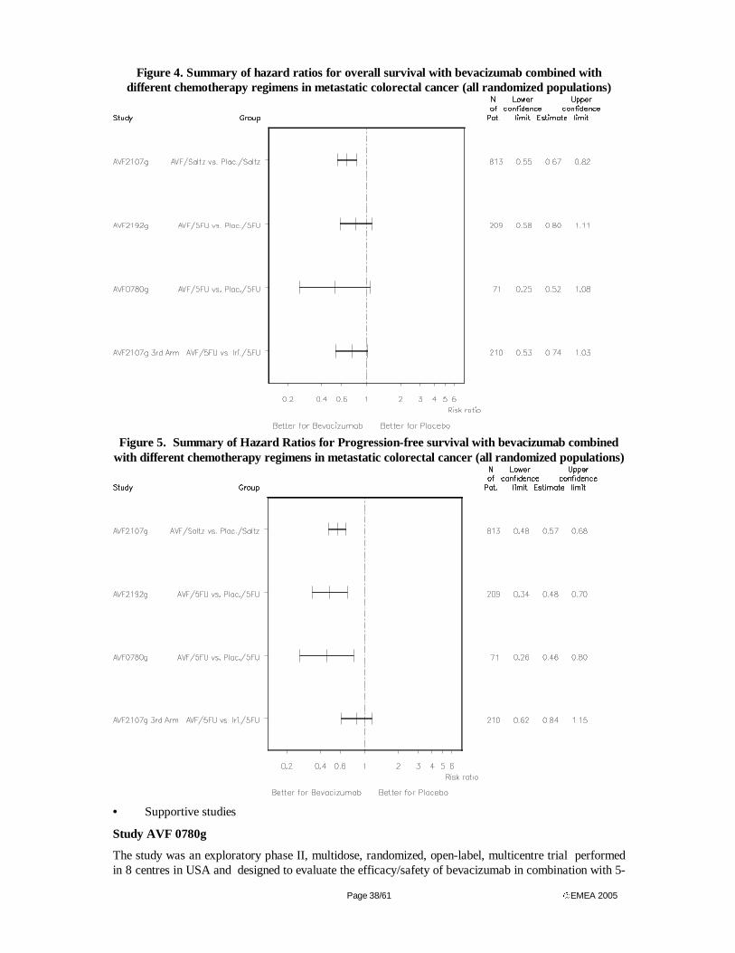
Page 38/61 EMEA 2005
Figure 4. Summary of hazard ratios for overall survival with bevacizumab combined with different chemotherapy regimens in metastatic colorectal cancer (all randomized populations)
Figure 5. Summary of Hazard Ratios for Progression-free survival with bevacizumab combined
with different chemotherapy regimens in metastatic colorectal cancer (all randomized populations)
• Supportive studies
Study AVF 0780g
The study was an exploratory phase II, multidose, randomized, open-label, multicentre trial performed in 8 centres in USA and designed to evaluate the efficacy/safety of bevacizumab in combination with 5-
Page 39/61 EMEA 2005
FU/FA as compared to 5-FU/FA alone. The study preceded the pivotal study discussed above. The patients should have measurable, metastatic, colorectal cancer previously untreated, except for surgery, radiotherapy and adjuvant chemotherapy. Randomization was stratified by centre, prior 5-FU/FA treatment and prior pelvic irradiation.
Treatment. FA 500 mg/m2 and bolus 5-FU 500 mg/m2 were given weekly for six weeks of every 8-week cycle (“Roswell Park regimen”). (This regimen was the same as that used in Arm 3 in the pivotal trial). Chemotherapy was given for up to six cycles unless earlier progression occurred. By randomization either placebo or 5 mg/kg or 10 mg/kg bevacizumab was administered every two weeks for a maximum of 24 doses (48 weeks). Patients randomized to the two bevacizumab arms who had CR, PR or SD at the end of their treatment period were eligible for additional bevacizumab treatment in extension study AVF0778g if they progressed within six months. Patients in the control arm could cross over to the 10 mg/kg dose of bevacizumab in the extension study after progression. Treatment was given for the remainder of the 48 weeks treatment period or until disease progression following a minimum of four bevacizumab doses. The primary efficacy analysis was based on the investigator’s assessment and a second assessment was performed by an independent response evaluation facility blinded to the type of treatment. The primary objectives were time to progression (patients without documented progression were censored at the time of last available tumour assessment), response and safety. Secondary objectives were survival, duration of response, quality of life and PK analyses. The bevacizumab arms were compared to the control arm but not to each other.
A summary of efficacy results for studies AVF0780g is shown in Table 10b. A total of 104 patients were randomized into this study between June 20, 1998 and November 3, 1998. The median age was 64 years. 19 % of the patients had received prior chemotherapy and 14 % prior radiotherapy. 98% of tumours were adenocarcinomas. By chance, more women were randomized into the two bevacizumab arms (51% and 55% versus 25% in the control arm). Baseline characteristics of the two bevacizumab arms were similar, but patients in the placebo arm had better prognostic factors compared to the test arms. Fewer patients in the placebo arm had low albumin levels (17% and 15% versus 6% in the control arm), liver metastases (83% and 82% versus 69% in the control arm) and lung metastases (40% and 36% versus 22% in the control arm). This imbalance could have biased the study results towards a prolonged time to progression and survival in the placebo arm. Male/female ratio was higher in the control arm (75/25) than in the 5 mg bevacizumab arm (49/51), which could have favoured the experimental arm. Slightly fewer patients in the bevacizumab arms had received prior treatment of any kind (83% and 91% versus 97% in the control arm) and fewer patients in the bevacizumab 5 mg/kg arm had received prior chemotherapy (14% versus 22% in the control arm and 21% in the 10 mg arm). One hundred two patients received at least one dose of the assigned study drug: 35 patients received 5-FU/FA alone, 35 patients received bevacizumab 5 mg/kg plus 5-FU/FA and 32 patients bevacizumab 10 mg/kg plus 5-FU/FA. The exposure to study treatment was consistent with time to progression and was highest for the treatment arm with the longest time to progression (bevacizumab 5 mg/kg). The main reason for treatment discontinuation was in all groups disease progression. Twenty of 35 control patients received bevacizumab after disease progression. Two additional patients crossed over early before disease progression. The minimal follow up in the study was 12 months, and median follow up was 15 months in all three treatment arms.
Progression free survival, based on the investigator assessment as well as the blinded IRF assessment, was longer in patients receiving bevacizumab 5 mg/kg. In both assessments the difference reached statistical significance (p=0.043 for investigator assessment and p=0.005 for the IRF assessment). For the 10 mg/kg treatment arm, significance was reached for the investigators ( p=0.027) but not the IRF assessment (p= 0.217). Adjusting progression-free survival for baseline albumin and liver/lung metastases increased the statistical significance of the treatment effect (p=0.003 for the 5 mg/kg treatment arm and p=0.095 in the 10 mg/kg treatment arm). The response rate was higher in both bevacizumab arms, but statistically significant only in the 5 mg/kg group (40% vs. 17%, p=0.029). Survival was longer, especially in the 5 mg/kg treatment arm (fig. III.5), but statistical significance was not reached (p=0.07). The short follow-up in this study did not allow a comprehensive analysis of survival. No differences in Quality of Life scores between the treatment groups were observed from the
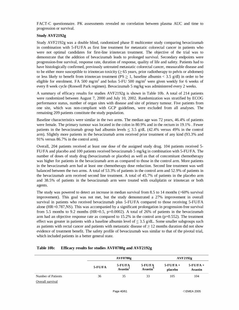
Page 40/61 EMEA 2005
FACT-C questionnaire. PK assessments revealed no correlation between plasma AUC and time to progression or survival.
Study AVF2192g
Study AVF2192g was a double blind, randomized phase II multicenter study comparing bevacizumab in combination with 5-FU/FA as first line treatment for metastatic colorectal cancer in patients who were not optimal candidates for first-line irinotecan treatment. The objective of the trial was to demonstrate that the addition of bevacizumab leads to prolonged survival. Secondary endpoints were progression-free survival, response rate, duration of response, quality of life and safety. Patients had to have histologically confirmed, previously untreated metastatic colorectal cancer, measurable disease and to be either more susceptible to irinotecan toxicity (≥ 65 years, prior radiotherapy to pelvis or abdomen) or less likely to benefit from irinotecan treatment (PS ≥ 1, baseline albumin < 3.5 g/dl) in order to be eligible for enrolment. FA 500 mg/m2 and bolus 5-FU 500 mg/m2 were given weekly for 6 weeks of every 8 week cycle (Roswell Park regimen). Bevacizumab 5 mg/kg was administered every 2 weeks.
A summary of efficacy results for studies AVF2192g is shown in Table 10b. A total of 214 patients were randomized between August 7, 2000 and July 10, 2002. Randomization was stratified by ECOG performance status, number of organ sites with disease and site of primary tumour. Five patients from one site, which was non-compliant with GCP guidelines, were excluded from all analyses. The remaining 209 patients constitute the study population.
Baseline characteristics were similar in the two arms. The median age was 72 years, 46.4% of patients were female. The primary tumour was located in the colon in 80.9% and in the rectum in 19.1% . Fewer patients in the bevacizumab group had albumin levels ≤ 3.5 g/dL (42.4% versus 49% in the control arm). Slightly more patients in the bevacizumab arms received prior treatment of any kind (93.3% and 91% versus 86.7% in the control arm).
Overall, 204 patients received at least one dose of the assigned study drug. 104 patients received 5-FU/FA and placebo and 100 patients received bevacizumab 5 mg/kg in combination with 5-FU/FA. The number of doses of study drug (bevacizumab or placebo) as well as that of concomitant chemotherapy was higher for patients in the bevacizumab arm as compared to those in the control arm. More patients in the bevacizumab arm had at least one chemotherapy dose reduction. Second line treatment was well balanced between the two arms. A total of 53.3% of patients in the control arm and 52.9% of patients in the bevacizumab arm received second line treatment. A total of 45.7% of patients in the placebo arm and 38.5% of patients in the bevacizumab arm were treated with oxaliplatin or irinotecan or both agents.
The study was powered to detect an increase in median survival from 8.5 to 14 months (>60% survival improvement). This goal was not met, but the study demonstrated a 27% improvement in overall survival in patients who received bevacizumab plus 5-FU/FA compared to those receiving 5-FU/FA alone (HR=0.787,NS). This was accompanied by a significant prolongation in progression-free survival from 5.5 months to 9.2 months (HR=0.5, p=0.0002). A total of 26% of patients in the bevacizumab arm had an objective response rate as compared to 15.2% in the control arm (p=0.552). The treatment effect was greater in patients with a baseline albumin level of ≤ 3.5 g/dL. Some smaller subgroups such as patients with rectal cancer and patients with metastatic disease of ≥ 12 months duration did not show evidence of treatment benefit. The safety profile of bevacizumab was similar to that of the pivotal trial, which included patients in a better general state.
Table 10b: Efficacy results for studies AVF0780g and AVF2192g
AVF0780g AVF2192g
5-FU/FA 5-FU/FA Avastina
5-FU/FA Avastinb
5-FU/FA + placebo
5-FU/FA + Avastin
Number of Patients 36 35 33 105 104
Overall survival
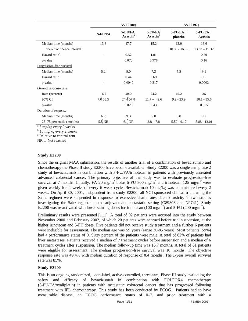
Page 41/61 EMEA 2005
AVF0780g AVF2192g
5-FU/FA 5-FU/FA Avastina
5-FU/FA Avastinb
5-FU/FA + placebo
5-FU/FA + Avastin
Median time (months) 13.6 17.7 15.2 12.9 16.6
95% Confidence Interval 10.35 - 16.95 13.63 – 19.32
Hazard ratioc - 0.52 1.01 0.79
p-value 0.073 0.978 0.16
Progression-free survival
Median time (months) 5.2 9.0 7.2 5.5 9.2
Hazard ratio 0.44 0.69 0.5
p-value - 0.0049 0.217 0.0002
Overall response rate
Rate (percent) 16.7 40.0 24.2 15.2 26
95% CI 7.0 33.5 24.4 57.8 11.7 – 42.6 9.2 - 23.9 18.1 - 35.6
p-value 0.029 0.43 0.055
Duration of response
Median time (months) NR 9.3 5.0 6.8 9.2
25–75 percentile (months) 5.5 NR 6.1 NR 3.8 – 7.8 5.59 - 9.17 5.88 - 13.01 a 5 mg/kg every 2 weeks b 10 mg/kg every 2 weeks c Relative to control arm NR � Not reached
Study E2200
Since the original MAA submission, the results of another trial of a combination of bevacizumab and chemotherapy the Phase II study E2200 have become available. Study E2200 was a single arm phase 2 study of bevacizumab in combination with 5-FU/FA/irinotecan in patients with previously untreated advanced colorectal cancer. The primary objective of the study was to evaluate progression-free survival at 7 months. Initially, FA 20 mg/m2 bolus 5-FU 500 mg/m2 and irinotecan 125 mg/m2 were given weekly for 4 weeks of every 6 week cycle. Bevacizumab 10 mg/kg was administered every 2 weeks. On April 30, 2001, independent from study E2200, all NCI-sponsored clinical trials using the Saltz regimen were suspended in response to excessive death rates due to toxicity in two studies investigating the Saltz regimen in the adjuvant and metastatic setting (C89803 and N9741). Study E2200 was re-activated with lower starting doses for irinotecan (100 mg/m2) and 5-FU (400 mg/m2).
Preliminary results were presented [111]. A total of 92 patients were accrued into the study between November 2000 and February 2002, of which 20 patients were accrued before trial suspension, at the higher irinotecan and 5-FU doses. Five patients did not receive study treatment and a further 6 patients were ineligible for assessment. The median age was 59 years (range 30-85 years). Most patients (59%) had a performance status of 0. Sixty percent of the patients were male. A total of 82% of patients had liver metastases. Patients received a median of 7 treatment cycles before suspension and a median of 6 treatment cycles after suspension. The median follow-up time was 16.7 months. A total of 81 patients were eligible for assessment. The median progression-free survival was 10 months. The objective response rate was 49.4% with median duration of response of 8.4 months. The 1-year overall survival rate was 85%.
Study E3200
This is an ongoing randomized, open-label, active-controlled, three-arm, Phase III study evaluating the safety and efficacy of bevacizumab in combination with FOLFOX4 chemotherapy (5-FU/FA/oxaliplatin) in patients with metastatic colorectal cancer that has progressed following treatment with IFL chemotherapy. This study has been conducted by ECOG. Patients had to have measurable disease, an ECOG performance status of 0–2, and prior treatment with a

Page 42/61 EMEA 2005
fluoropyrimidine-based regimen and an irinotecan-based regimen (alone or in combination) for advanced disease. Eligible patients were randomized into one of three treatment arms: 5-FU/FA/oxaliplatin (A), 5-FU/FA/oxaliplatin+ bevacizumab (B), or bevacizumab alone (C). The bevacizumab dose is 5 mg/kg/wk (delivered as 10 mg/kg every 2 weeks).
The FOLFOX4 regimen (85 mg/m2 oxaliplatin, 200 mg/m2 FA, and 400 mg/m2 5-FU bolus + 600 mg/m2 IV infusion) is administered every 2 weeks. The study has been closed to enrollment with 835 patients. This was increased from the original protocol-specified enrollment of 693 patients because of a faster-than-expected enrollment rate. The primary efficacy endpoint is overall survival; secondary efficacy endpoints are response rate and time to progression.
The final analysis will occur ~31 months after the start of accrual (22 months of accrual and 9 months of follow-up); interim analyses of efficacy were to be conducted at 50% and 75% information time (~16 and ~21 months after the start of accrual, respectively. Interim safety data from 757 patients were presented [112]. A total of 495 patients received bevacizumab, 265 in combination with FOLFOX4. Bevacizumab added to FOLFOX4 did not substantially alter the regimen’s toxicity profile in this already pre-treated patient population. The single agent bevacizumab arm of this study was closed to accrual on March 11, 2003. The study is ongoing and is planned to be completed by September 30, 2005.
Discussion on clinical efficacy
The definition of an optimal dose of bevacizumab, an anti-angiogenic agent exhibiting predominantly features of a cytostatic, has been challenging because no established surrogate for a disease progression endpoint exists and a multi-dose approach is not practical. Features used in the establishment of an optimal dose for cytotoxic agents, such as a steep dose-response curve or a typical toxicity profile, cannot be applied under such circumstances. As a consequence, the Applicant decided to conduct a randomized, placebo controlled Phase II dose-finding study (AVF0780g) of two doses of bevacizumab which were previously shown to be associated with bevacizumab serum levels of above the target level of 30 µg/mL. In this trial the 5 mg/kg every 2 weeks dose was shown to maintain >98% of VEGF in the circulation bound to bevacizumab, and this was identified as being effective and well tolerated. The results of both pharmacokinetic parameters and clinical efficacy and safety from trials AVF0780g, AVF2107g and AVF2192g all suggest that the 5 mg/kg every 2 weeks dose of bevacizumab is safe and effective. Limited clinical experience using a higher dose of bevacizumab (10 mg/kg every 2 weeks or 5.0 mg/kg/week) from trials AVF0780g and E2200 does not suggest any improvement in efficacy, but there were indications of higher toxicity across all dose ranging studies in all indications. The Applicant has initiated a large global development program in major cancer indications, e.g. in metastatic renal cell, non-small cell lung, and pancreatic cancer. The investigation of bevacizumab will be expanded in metastatic colorectal cancer and into the adjuvant treatment of patients with colon cancer. A variety of standard backbone chemotherapy regimens will be combined with bevacizumab in these trials and further PK/PD data will be generated in association with this development program. The applicant has provided a thorough discussion on the dose-response issue and the lack of true MTD finding studies. The arguments for choosing the 5mg/kg q2wk are reasonable.
The application is based on one phase III and one phase II study conducted to demonstrate efficacy of bevacizumab in combination with 5-FU based chemotherapy. The design of the pivotal trial, the randomization methods and the stratification are appropriate. The patients had overall good performance status, were relatively young (mean age 59 years) and close to 38 % had only one metastatic site. The population included may thus have had a more favourable prognosis than the general population with metastatic CRC.
The pivotal trial showed that bevacizumab in combination with IFL gives a significant prolongation of survival of 4.7 months compared to IFL alone (20.3 vs. 15.6 months), the hazard ratio in favour of bevacizumab + IFL was 0.66. The survival benefit from bevacizumab was seen in all pre-specified patient subgroups. Since a similar percentage of patients in Arms 1 and 2 received second-line treatment and the chemotherapy agents used were also similar in both arms, it is unlikely that second-line treatment introduced a bias on the assessment of overall survival. The addition of bevacizumab to IFL
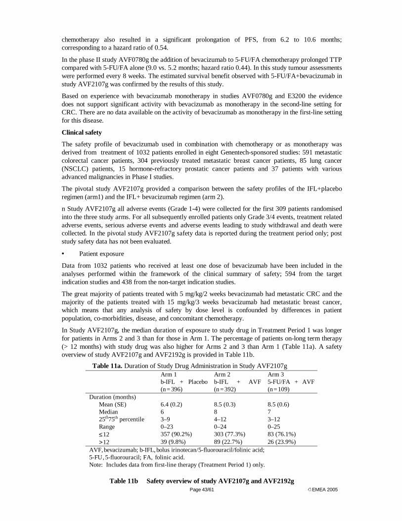
Page 43/61 EMEA 2005
chemotherapy also resulted in a significant prolongation of PFS, from 6.2 to 10.6 months; corresponding to a hazard ratio of 0.54.
In the phase II study AVF0780g the addition of bevacizumab to 5-FU/FA chemotherapy prolonged TTP compared with 5-FU/FA alone (9.0 vs. 5.2 months; hazard ratio 0.44). In this study tumour assessments were performed every 8 weeks. The estimated survival benefit observed with 5-FU/FA+bevacizumab in study AVF2107g was confirmed by the results of this study.
Based on experience with bevacizumab monotherapy in studies AVF0780g and E3200 the evidence does not support significant activity with bevacizumab as monotherapy in the second-line setting for CRC. There are no data available on the activity of bevacizumab as monotherapy in the first-line setting for this disease.
Clinical safety
The safety profile of bevacizumab used in combination with chemotherapy or as monotherapy was derived from treatment of 1032 patients enrolled in eight Genentech-sponsored studies: 591 metastatic colorectal cancer patients, 304 previously treated metastatic breast cancer patients, 85 lung cancer (NSCLC) patients, 15 hormone-refractory prostatic cancer patients and 37 patients with various advanced malignancies in Phase I studies.
The pivotal study AVF2107g provided a comparison between the safety profiles of the IFL+placebo regimen (arm1) and the IFL+ bevacizumab regimen (arm 2).
n Study AVF2107g all adverse events (Grade 1-4) were collected for the first 309 patients randomised into the three study arms. For all subsequently enrolled patients only Grade 3/4 events, treatment related adverse events, serious adverse events and adverse events leading to study withdrawal and death were collected. In the pivotal study AVF2107g safety data is reported during the treatment period only; post study safety data has not been evaluated.
• Patient exposure
Data from 1032 patients who received at least one dose of bevacizumab have been included in the analyses performed within the framework of the clinical summary of safety; 594 from the target indication studies and 438 from the non-target indication studies.
The great majority of patients treated with 5 mg/kg/2 weeks bevacizumab had metastatic CRC and the majority of the patients treated with 15 mg/kg/3 weeks bevacizumab had metastatic breast cancer, which means that any analysis of safety by dose level is confounded by differences in patient population, co-morbidities, disease, and concomitant chemotherapy.
In Study AVF2107g, the median duration of exposure to study drug in Treatment Period 1 was longer for patients in Arms 2 and 3 than for those in Arm 1. The percentage of patients on-long term therapy (> 12 months) with study drug was also higher for Arms 2 and 3 than Arm 1 (Table 11a). A safety overview of study AVF2107g and AVF2192g is provided in Table 11b.
Table 11a. Duration of Study Drug Administration in Study AVF2107g
Arm 1 b-IFL + Placebo (n = 396)
Arm 2 b-IFL + AVF (n = 392)
Arm 3 5-FU/FA + AVF (n = 109)
Duration (months) Mean (SE) 6.4 (0.2) 8.5 (0.3) 8.5 (0.6) Median 6 8 7 25th 75th percentile 3–9 4–12 3–12 Range 0–23 0–24 0–25 ≤ 12 357 (90.2%) 303 (77.3%) 83 (76.1%) > 12 39 (9.8%) 89 (22.7%) 26 (23.9%)
AVF, bevacizumab; b-IFL, bolus irinotecan/5-fluorouracil/folinic acid; 5-FU , 5-fluorouracil; FA, folinic acid. Note: Includes data from first-line therapy (Treatment Period 1) only.
Table 11b Safety overview of study AVF2107g and AVF2192g
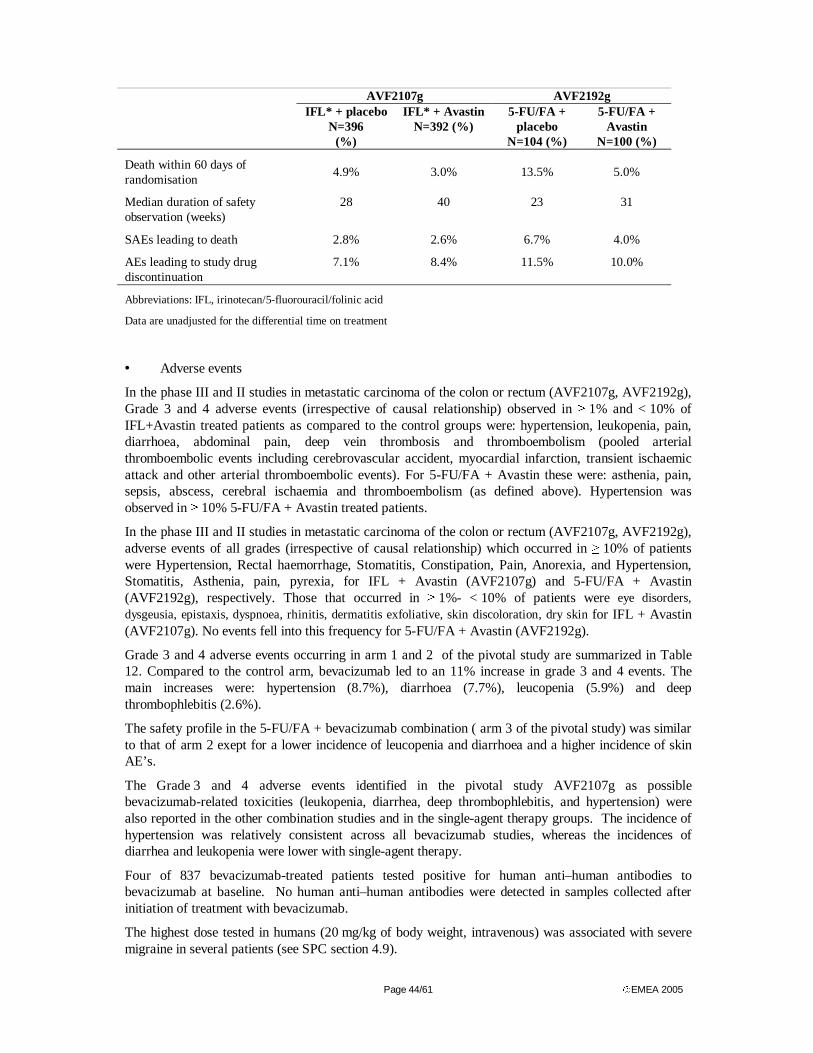
Page 44/61 EMEA 2005
AVF2107g AVF2192g
IFL* + placebo N=396
(%)
IFL* + Avastin N=392 (%)
5-FU/FA + placebo
N=104 (%)
5-FU/FA + Avastin
N=100 (%)
Death within 60 days of randomisation 4.9% 3.0% 13.5% 5.0%
Median duration of safety observation (weeks)
28 40 23 31
SAEs leading to death 2.8% 2.6% 6.7% 4.0%
AEs leading to study drug discontinuation
7.1% 8.4% 11.5% 10.0%
Abbreviations: IFL, irinotecan/5-fluorouracil/folinic acid
Data are unadjusted for the differential time on treatment
• Adverse events
In the phase III and II studies in metastatic carcinoma of the colon or rectum (AVF2107g, AVF2192g), Grade 3 and 4 adverse events (irrespective of causal relationship) observed in ≥ 1% and < 10% of IFL+Avastin treated patients as compared to the control groups were: hypertension, leukopenia, pain, diarrhoea, abdominal pain, deep vein thrombosis and thromboembolism (pooled arterial thromboembolic events including cerebrovascular accident, myocardial infarction, transient ischaemic attack and other arterial thromboembolic events). For 5-FU/FA + Avastin these were: asthenia, pain, sepsis, abscess, cerebral ischaemia and thromboembolism (as defined above). Hypertension was observed in ≥ 10% 5-FU/FA + Avastin treated patients.
In the phase III and II studies in metastatic carcinoma of the colon or rectum (AVF2107g, AVF2192g), adverse events of all grades (irrespective of causal relationship) which occurred in ≥ 10% of patients were Hypertension, Rectal haemorrhage, Stomatitis, Constipation, Pain, Anorexia, and Hypertension, Stomatitis, Asthenia, pain, pyrexia, for IFL + Avastin (AVF2107g) and 5-FU/FA + Avastin (AVF2192g), respectively. Those that occurred in ≥ 1%- < 10% of patients were eye disorders, dysgeusia, epistaxis, dyspnoea, rhinitis, dermatitis exfoliative, skin discoloration, dry skin for IFL + Avastin (AVF2107g). No events fell into this frequency for 5-FU/FA + Avastin (AVF2192g).
Grade 3 and 4 adverse events occurring in arm 1 and 2 of the pivotal study are summarized in Table 12. Compared to the control arm, bevacizumab led to an 11% increase in grade 3 and 4 events. The main increases were: hypertension (8.7%), diarrhoea (7.7%), leucopenia (5.9%) and deep thrombophlebitis (2.6%).
The safety profile in the 5-FU/FA + bevacizumab combination ( arm 3 of the pivotal study) was similar to that of arm 2 exept for a lower incidence of leucopenia and diarrhoea and a higher incidence of skin AE’s.
The Grade 3 and 4 adverse events identified in the pivotal study AVF2107g as possible bevacizumab-related toxicities (leukopenia, diarrhea, deep thrombophlebitis, and hypertension) were also reported in the other combination studies and in the single-agent therapy groups. The incidence of hypertension was relatively consistent across all bevacizumab studies, whereas the incidences of diarrhea and leukopenia were lower with single-agent therapy.
Four of 837 bevacizumab-treated patients tested positive for human anti–human antibodies to bevacizumab at baseline. No human anti–human antibodies were detected in samples collected after initiation of treatment with bevacizumab.
The highest dose tested in humans (20 mg/kg of body weight, intravenous) was associated with severe migraine in several patients (see SPC section 4.9).
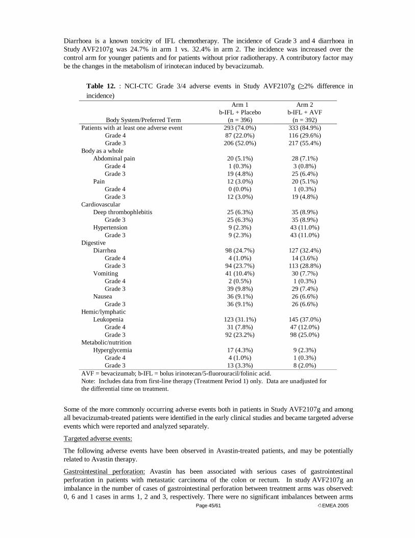
Page 45/61 EMEA 2005
Diarrhoea is a known toxicity of IFL chemotherapy. The incidence of Grade 3 and 4 diarrhoea in Study AVF2107g was 24.7% in arm 1 vs. 32.4% in arm 2. The incidence was increased over the control arm for younger patients and for patients without prior radiotherapy. A contributory factor may be the changes in the metabolism of irinotecan induced by bevacizumab.
Table 12. : NCI-CTC Grade 3/4 adverse events in Study AVF2107g (≥2% difference in incidence)
Body System/Preferred Term
Arm 1 b-IFL + Placebo
(n = 396)
Arm 2 b-IFL + AVF
(n = 392) Patients with at least one adverse event 293 (74.0%) 333 (84.9%)
Grade 4 87 (22.0%) 116 (29.6%) Grade 3 206 (52.0%) 217 (55.4%)
Body as a whole Abdominal pain 20 (5.1%) 28 (7.1%)
Grade 4 1 (0.3%) 3 (0.8%) Grade 3 19 (4.8%) 25 (6.4%)
Pain 12 (3.0%) 20 (5.1%) Grade 4 0 (0.0%) 1 (0.3%) Grade 3 12 (3.0%) 19 (4.8%)
Cardiovascular Deep thrombophlebitis 25 (6.3%) 35 (8.9%)
Grade 3 25 (6.3%) 35 (8.9%) Hypertension 9 (2.3%) 43 (11.0%)
Grade 3 9 (2.3%) 43 (11.0%) Digestive
Diarrhea 98 (24.7%) 127 (32.4%) Grade 4 4 (1.0%) 14 (3.6%) Grade 3 94 (23.7%) 113 (28.8%)
Vomiting 41 (10.4%) 30 (7.7%) Grade 4 2 (0.5%) 1 (0.3%) Grade 3 39 (9.8%) 29 (7.4%)
Nausea 36 (9.1%) 26 (6.6%) Grade 3 36 (9.1%) 26 (6.6%)
Hemic/lymphatic Leukopenia 123 (31.1%) 145 (37.0%)
Grade 4 31 (7.8%) 47 (12.0%) Grade 3 92 (23.2%) 98 (25.0%)
Metabolic/nutrition Hyperglycemia 17 (4.3%) 9 (2.3%)
Grade 4 4 (1.0%) 1 (0.3%) Grade 3 13 (3.3%) 8 (2.0%)
AVF = bevacizumab; b-IFL = bolus irinotecan/5-fluorouracil/folinic acid. Note: Includes data from first-line therapy (Treatment Period 1) only. Data are unadjusted for the differential time on treatment.
Some of the more commonly occurring adverse events both in patients in Study AVF2107g and among all bevacizumab-treated patients were identified in the early clinical studies and became targeted adverse events which were reported and analyzed separately.
Targeted adverse events:
The following adverse events have been observed in Avastin-treated patients, and may be potentially related to Avastin therapy.
Gastrointestinal perforation: Avastin has been associated with serious cases of gastrointestinal perforation in patients with metastatic carcinoma of the colon or rectum. In study AVF2107g an imbalance in the number of cases of gastrointestinal perforation between treatment arms was observed: 0, 6 and 1 cases in arms 1, 2 and 3, respectively. There were no significant imbalances between arms
Page 46/61 EMEA 2005
for other possibly perforation-related events including abdominal wound dehiscence, abdominal wound fistula, and abscess. The applicant sees intraabdominal inflammation as a common feature of these cases and recommends caution to be exercised when treating such colorectal patients with bevacizumab. In clinical trials in metastatic carcinoma of the colon or rectum, gastrointestinal perforation was observed in 1.4% - 2.0% of the Avastin-treated patients. Of these, 0.4% - 1% had fatal outcome. The presentation of these events varied in type and severity, ranging from free air seen on the plain abdominal X-ray, which resolved without treatment, to a colonic perforation with abdominal abscess and fatal outcome. The common feature among these cases was intra-abdominal inflammation, either from gastric ulcer disease, tumour necrosis, diverticulitis, or chemotherapy-associated colitis. There were no cases of gastrointestinal perforation in any other Genentech-sponsored clinical trial of bevacizumab.
Wound healing: As Avastin may adversely impact wound healing, patients who had major surgery within the last 28 days were excluded from participation in clinical trials for metastatic cancer of the colon or rectum. In clinical trials of metastatic carcinoma of the colon or rectum, patients who underwent cancer-related surgery between 28 and 60 days prior to starting therapy did not have increased risk of post-operative bleeding or wound healing complications during treatment compared to the controlled groups. Adverse events consistent with post-operative bleeding or wound healing complication were observed in 10% - 20% of Avastin-treated patients who underwent major surgery while receiving treatment.
Hypertension: An increased incidence of hypertension has been observed in Avastin-treated patients. Hypertension was generally treated with oral anti-hypertensives such as angiotensin-converting enzyme inhibitors, diuretics and calcium-channel blockers. It rarely resulted in discontinuation (0.7% of all Avastin-treated patients) or hospitalisation, and resulted in hypertensive encephalopathy in one case (0.1%). The risk of Avastin-associated hypertension did not correlate with the patients’ baseline characteristics, underlying disease or concomitant therapy. No association was observed between the risk of Avastin-associated hypertension and patients’ baseline characteristics, underlying disease or concomitant therapy. In clinical trials of metastatic carcinoma of the colon or rectum, hypertension of any grade occurred in 22.4% - 32.0% of Avastin-treated patients. Grade 3 hypertension (requiring oral anti-hypertensive medication) was reported in 11.0% - 16.0% of Avastin-treated patients. No hypertensive crisis (Grade 4) was reported. At week 24 of treatment, the mean change of blood pressure (BP) from baseline was diastolic BP +4.1 to +5.4 mmHg and systolic BP + 5.5 to +8.4 mmHg in the treated patients.
Proteinuria: Proteinuria, reported as adverse event, was observed in 23.3% of all Avastin-treated patients. It ranged in severity from clinically asymptomatic, transient, trace proteinuria to nephrotic syndrome, with the great majority as Grade 1 proteinuria. The proteinuria seen in clinical trials was not associated with renal dysfunction and rarely required permanent discontinuation of therapy. In clinical trials of metastatic carcinoma of the colon or rectum, proteinuria was reported as an adverse event in 21.7% - 38.0% of Avastin-treated patients. No Grade 4 proteinuria was reported.
Haemorrhage: Overall, 4.0% of NCI-CTC Grade 3 and 4 bleeding events were observed in all Avastin treated patients. In clinical trials of metastatic carcinoma of the colon or rectum, there was no significant difference in the incidence of grade 3 and 4 bleeding events observed between Avastin-treated patients (3.1% - 5.1%) compared to that observed in the controls (2.5% - 2.9%). The haemorrhagic events that have been observed in clinical studies were predominantly tumour-associated haemorrhage and minor mucocutaneous haemorrhage. Tumour-associated haemorrhage was observed in phase I and phase II studies. In patients with non-small cell lung cancer receiving Avastin, serious haemorrhage was observed in 9% (6% fatal) of treated patients. These events occurred suddenly and presented as major or massive haemoptysis in patients with either squamous cell histology and/or tumours located in the centre of the chest in close proximity to major blood vessels. In some cases, these haemorrhages were preceded by cavitation and/or necrosis of the tumour. Tumour-associated haemorrhage was also seen rarely in other tumour types and locations, including central nervous system (CNS) bleeding in a patient with hepatoma with occult CNS metastases and continuous oozing of blood from a thigh sarcoma with necrosis. In clinical trials of metastatic carcinoma of the colon or rectum, tumour-associated haemorrhagic events were observed in 1% - 3% of the Avastin-treated patients. The
Page 47/61 EMEA 2005
addition of Avastin did not result in significant increase in the incidence or severity of Grade 3 or 4 haemorrhagic events. Across all clinical trials, mucocutaneous haemorrhage has been seen in 20% - 40% of Avastin-treated patients. These were most commonly NCI-CTC Grade 1 epistaxis that lasted less than 5 minutes, resolved without medical intervention and did not require any changes in treatment regimen. In clinical trials of metastatic carcinoma of the colon or rectum, epistaxis was reported in 22.0% - 34.3% of Avastin-treated patients. There have also been less common events of minor mucocutaneous haemorrhage in other locations, such as gingival bleeding and vaginal bleeding.
Thromboembolism: In clinical trials of metastatic carcinoma of the colon or rectum, the overall incidence of thromboembolic events was similar between Avastin-treated patients (18.0% - 19.4%) and the controls (16.2% - 18.3%). In clinical trials of metastatic carcinoma of the colon or rectum, the incidence of arterial thromboembolic events including CVAs, MIs, TIAs, and other arterial thromboembolic events was higher in Avastin-treated patients (3.3% - 10.0%) compared to the controls (1.3% - 4.8%). In five randomised trials including metastatic carcinoma of the colon or rectum trials (N=1745), arterial thromboembolic events including CVAs, MIs, TIAs, and other thromboembolic events occurred in 4.5% (45/1004) of patients treated with Avastin in combination with chemotherapy compared to 2.0% (15/741) of patients treated with chemotherapy alone. In patients treated with Avastin plus chemotherapy, arterial thromboembolic events led to a fatal outcome in 0.8% (8/1004). In patients treated with chemotherapy alone, a fatal outcome from arterial thromboembolic events was reported in 0.4% (3/741). CVAs (including TIAs) occurred in 2.2% of patients treated with Avastin in combination with chemotherapy and 0.5% of patients treated with chemotherapy alone. MI occurred in 1.9% of patients treated with Avastin in combination with chemotherapy compared to 1.1% of patients treated with chemotherapy alone. In clinical trials of metastatic carcinoma of the colon or rectum, venous thromboembolic events, including deep venous thrombosis, pulmonary embolism and thrombophlebitis occurred in 9.0% - 16.6% of Avastin-treated patients compared to that of 13.5% - 15.2% in the controls. It could not be determined if these events were due to the patients’ underlying cancer, their cytotoxic chemotherapy, Avastin or other risk factors.
Congestive Heart Failure (CHF)/Cardiomyopathy: In the phase III controlled clinical trial of metastatic breast cancer, CHF/cardiomyopathy was reported in 3% of the Avastin-treated patients compared with 1% in the controlled group. These events varied in severity from asymptomatic declines in left ventricular ejection fraction to symptomatic CHF requiring hospitalisation and treatment. All the Avastin-treated patients were previously treated with anthracyclines (doxorubicin cumulative dose range 240–360 mg/m2). Many of these patients also had prior radiotherapy to the left chest wall. Most of these patients showed improved symptoms and/or left ventricular function following appropriate medical therapy. There was no information on patients with pre-existing CHF (NYHA II-IV) at the time of initiating the therapy, as these patients were excluded from studies. In patients with metastatic cancer of the colon or rectum, there was no increased incidence of CHF in Avastin-treated patients.
• Serious adverse event/deaths/other significant events
In Study AVF2107g, serious adverse events were reported in 43.2% of patients in Arm 1 and 51.0% of patients in Arm 2. Deep thrombophlebitis was the only serious adverse event occurring with a ≥ 2% difference in incidence between Arms 1 and 2 during first-line therapy. More than 90% of the deaths in study AVF2107g were due to disease progression. The incidences of SAE’s that resulted in deaths in study AVF2107g were similar in arm 1 and 2 (2.8% and 2.6%, respectively).
Among all bevacizumab-treated patients, serious adverse events were reported in 41.6% of the patients. The most frequently occurring serious adverse events were diarrhoea (7.3%) and deep thrombophlebitis (5.4%).
The incidence of adverse events leading to study discontinuation in Study AVF2107g was similar for Arms 1 and 2 (7.1% in Arm 1 and 8.4% in Arm 2). The most common adverse events leading to study discontinuation were diarrhea (1.5% in Arm 1 and 1.5% in Arm 2), asthenia (1.3% in Arm 1 and 0.8% in Arm 2), and pulmonary embolus (1.8% in Arm 1 and 0.3% in Arm 2). Two patients (0.5%) in Arm 2 experienced a subarachnoid hemorrhage that led to study discontinuation.
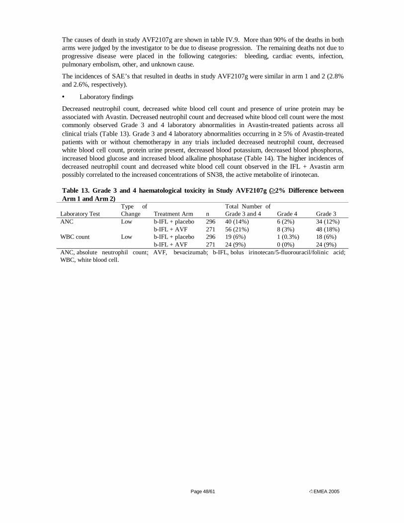
Page 48/61 EMEA 2005
The causes of death in study AVF2107g are shown in table IV.9. More than 90% of the deaths in both arms were judged by the investigator to be due to disease progression. The remaining deaths not due to progressive disease were placed in the following categories: bleeding, cardiac events, infection, pulmonary embolism, other, and unknown cause.
The incidences of SAE’s that resulted in deaths in study AVF2107g were similar in arm 1 and 2 (2.8% and 2.6%, respectively).
• Laboratory findings
Decreased neutrophil count, decreased white blood cell count and presence of urine protein may be associated with Avastin. Decreased neutrophil count and decreased white blood cell count were the most commonly observed Grade 3 and 4 laboratory abnormalities in Avastin-treated patients across all clinical trials (Table 13). Grade 3 and 4 laboratory abnormalities occurring in ≥ 5% of Avastin-treated patients with or without chemotherapy in any trials included decreased neutrophil count, decreased white blood cell count, protein urine present, decreased blood potassium, decreased blood phosphorus, increased blood glucose and increased blood alkaline phosphatase (Table 14). The higher incidences of decreased neutrophil count and decreased white blood cell count observed in the IFL + Avastin arm possibly correlated to the increased concentrations of SN38, the active metabolite of irinotecan.
Table 13. Grade 3 and 4 haematological toxicity in Study AVF2107g (≥2% Difference between Arm 1 and Arm 2)
Laboratory Test Type of Change Treatment Arm n
Total Number of Grade 3 and 4 Grade 4 Grade 3
Low b-IFL + placebo 296 40 (14%) 6 (2%) 34 (12%) ANC b-IFL + AVF 271 56 (21%) 8 (3%) 48 (18%) Low b-IFL + placebo 296 19 (6%) 1 (0.3%) 18 (6%) WBC count b-IFL + AVF 271 24 (9%) 0 (0%) 24 (9%)
ANC, absolute neutrophil count; AVF, bevacizumab; b-IFL, bolus irinotecan/5-fluorouracil/folinic acid; WBC, white blood cell.
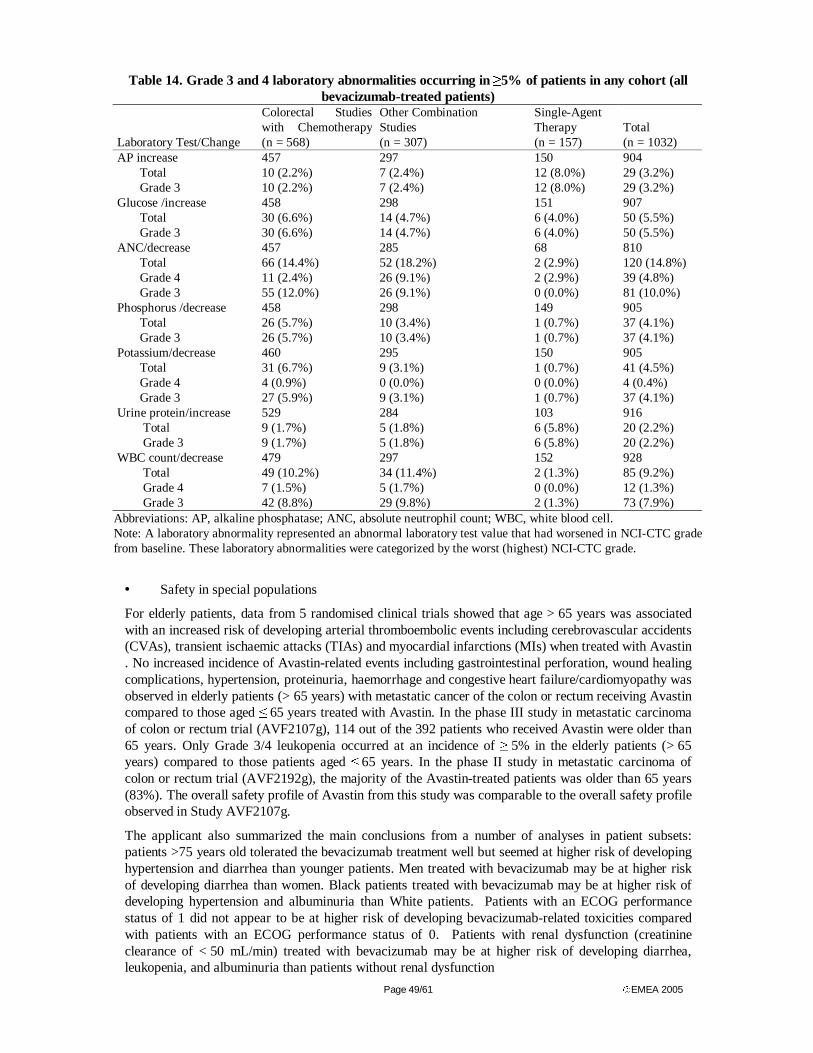
Page 49/61 EMEA 2005
Table 14. Grade 3 and 4 laboratory abnormalities occurring in ≥5% of patients in any cohort (all bevacizumab-treated patients)
Laboratory Test/Change
Colorectal Studies with Chemotherapy (n = 568)
Other Combination Studies (n = 307)
Single-Agent Therapy (n = 157)
Total (n = 1032)
AP increase 457 297 150 904 Total 10 (2.2%) 7 (2.4%) 12 (8.0%) 29 (3.2%) Grade 3 10 (2.2%) 7 (2.4%) 12 (8.0%) 29 (3.2%)
Glucose /increase 458 298 151 907 Total 30 (6.6%) 14 (4.7%) 6 (4.0%) 50 (5.5%) Grade 3 30 (6.6%) 14 (4.7%) 6 (4.0%) 50 (5.5%)
ANC/decrease 457 285 68 810 Total 66 (14.4%) 52 (18.2%) 2 (2.9%) 120 (14.8%) Grade 4 11 (2.4%) 26 (9.1%) 2 (2.9%) 39 (4.8%) Grade 3 55 (12.0%) 26 (9.1%) 0 (0.0%) 81 (10.0%)
Phosphorus /decrease 458 298 149 905 Total 26 (5.7%) 10 (3.4%) 1 (0.7%) 37 (4.1%) Grade 3 26 (5.7%) 10 (3.4%) 1 (0.7%) 37 (4.1%)
Potassium/decrease 460 295 150 905 Total 31 (6.7%) 9 (3.1%) 1 (0.7%) 41 (4.5%) Grade 4 4 (0.9%) 0 (0.0%) 0 (0.0%) 4 (0.4%) Grade 3 27 (5.9%) 9 (3.1%) 1 (0.7%) 37 (4.1%)
Urine protein/increase 529 284 103 916 Total 9 (1.7%) 5 (1.8%) 6 (5.8%) 20 (2.2%) Grade 3 9 (1.7%) 5 (1.8%) 6 (5.8%) 20 (2.2%)
WBC count/decrease 479 297 152 928 Total 49 (10.2%) 34 (11.4%) 2 (1.3%) 85 (9.2%) Grade 4 7 (1.5%) 5 (1.7%) 0 (0.0%) 12 (1.3%) Grade 3 42 (8.8%) 29 (9.8%) 2 (1.3%) 73 (7.9%)
Abbreviations: AP, alkaline phosphatase; ANC, absolute neutrophil count; WBC, white blood cell. Note: A laboratory abnormality represented an abnormal laboratory test value that had worsened in NCI-CTC grade from baseline. These laboratory abnormalities were categorized by the worst (highest) NCI-CTC grade.
• Safety in special populations
For elderly patients, data from 5 randomised clinical trials showed that age > 65 years was associated with an increased risk of developing arterial thromboembolic events including cerebrovascular accidents (CVAs), transient ischaemic attacks (TIAs) and myocardial infarctions (MIs) when treated with Avastin . No increased incidence of Avastin-related events including gastrointestinal perforation, wound healing complications, hypertension, proteinuria, haemorrhage and congestive heart failure/cardiomyopathy was observed in elderly patients (> 65 years) with metastatic cancer of the colon or rectum receiving Avastin compared to those aged ≤ 65 years treated with Avastin. In the phase III study in metastatic carcinoma of colon or rectum trial (AVF2107g), 114 out of the 392 patients who received Avastin were older than 65 years. Only Grade 3/4 leukopenia occurred at an incidence of ≥ 5% in the elderly patients (> 65 years) compared to those patients aged ≤ 65 years. In the phase II study in metastatic carcinoma of colon or rectum trial (AVF2192g), the majority of the Avastin-treated patients was older than 65 years (83%). The overall safety profile of Avastin from this study was comparable to the overall safety profile observed in Study AVF2107g.
The applicant also summarized the main conclusions from a number of analyses in patient subsets: patients >75 years old tolerated the bevacizumab treatment well but seemed at higher risk of developing hypertension and diarrhea than younger patients. Men treated with bevacizumab may be at higher risk of developing diarrhea than women. Black patients treated with bevacizumab may be at higher risk of developing hypertension and albuminuria than White patients. Patients with an ECOG performance status of 1 did not appear to be at higher risk of developing bevacizumab-related toxicities compared with patients with an ECOG performance status of 0. Patients with renal dysfunction (creatinine clearance of < 50 mL/min) treated with bevacizumab may be at higher risk of developing diarrhea, leukopenia, and albuminuria than patients without renal dysfunction
Page 50/61 EMEA 2005
• Safety related to drug-drug interactions and other interactions
The pharmacokinetics of bevacizumab did not appear to be affected by dosing of concomitant chemotherapies, including doxorubicin, carboplatin/paclitaxel, 5-FU/FA, capecitabine, and IFL. With the exception of irinotecan, the pharmacokinetics of these chemotherapeutic drugs were not affected by bevacizumab. In Study AVF2107g, irinotecan concentrations were not affected when dosed in combination with bevacizumab; however, there was an estimated 33% increase in exposure to SN38, the active metabolite of irinotecan, which could be a cause of the increased incidence of diarrhoea.
• Discontinuation due to adverse events
The most frequently occurring adverse events among all bevacizumab–treated patients leading to discontinuation were asthenia, deep thrombophlebitis, diarrhoea, hypertension, pulmonary embolus, and albuminuria. Thirteen percent of all bevacizumab–treated patients discontinued treatment due to adverse events.
The incidence of adverse events leading to study discontinuation in Study AVF2107g was similar for Arms 1 and 2 and relatively low (7.1% in Arm 1 and 8.4% in Arm 2). The most common adverse events leading to study discontinuation were diarrhoea, asthenia, and pulmonary embolus. Two patients in Arm 2 experienced a subarachnoid haemorrhage that led to study discontinuation.
• Post marketing experience
No post-marketing experience was available at the time of submission.
• Discussion on clinical safety
The applicant has summarized safety data from 1032 bevacizumab-treated patients from nine Genentech-sponsored studies, including 594 patients with metastatic colorectal cancer and 310 patients with metastatic breast cancer. However, the most informative and relevant study was the pivotal study AVF2107, in which the IFL+ placebo regimen was directly comparable to the IFL+ bevacizumab regimen with almost 400 patients in each arm.
Against the background of a relatively toxic IFL regimen, treatment with bevacizumab in general did add toxicity, which however was a moderate 11% considering that patients in the IFL + bevacizumab arm – due to an improved efficacy- received more doses of chemotherapy than those in the IFL + placebo arm. The toxicity in general was easily manageable except for a few rare cases of gastrointestinal perforation (only seen in colorectal cancer studies). Patients with metastatic carcinoma of the colon or rectum and an intra-abdominal inflammatory process may be at increased risk for the development of gastrointestinal perforation when treated with Avastin and chemotherapy. Therefore, caution should be exercised when treating these patients. Therapy should be permanently discontinued in patients who develop gastrointestinal perforation (see also SPC, sections 4.4 and 4.8).
No increase was seen in adverse events leading to death or study discontinuation or in 60-day mortality. The safety pattern was duplicated in other studies where bevacizumab was added to other chemotherapy regimens or in the collection of patients treated by single agent bevacizumab. There were no consistent trends suggesting that the incidence of any adverse events increased after prolonged treatment with bevacizumab. Most deaths occurred because of disease progression.
Avastin may adversely affect the wound healing process. Avastin therapy should not be initiated for at least 28 days following major surgery or until the surgical wound is fully healed. In patients who experienced wound healing complications during Avastin treatment, Avastin should be withheld until the wound is fully healed. Avastin therapy should be withheld for elective surgery (see also SPC, sections 4.4 and 4.8).
An increased incidence of hypertension was observed in Avastin-treated patients. Clinical safety data suggest that the incidence of hypertension is likely to be dose-dependent. There is no information on the effect of Avastin in patients with uncontrolled hypertension at the time of initiating Avastin therapy. Therefore, caution should be exercised before initiating Avastin therapy in these patients. Monitoring of blood pressure is generally recommended during Avastin therapy. In patients with severe hypertension requiring medical therapy temporary interruption of Avastin is recommended until adequate control is
Page 51/61 EMEA 2005
achieved. If hypertension cannot be controlled with medical therapy, Avastin treatment should be permanently discontinued. Avastin should be permanently discontinued in patients who develop hypertensive crisis (see also SPC, sections 4.4 and 4.8).
Patients with a history of hypertension may be at increased risk for the development of proteinuria when treated with Avastin. There is evidence suggesting that Grade 1 proteinuria, based on the U.S. National Cancer Institute-Common Toxicity Criteria (NCI-CTC) version 2.0, may be related to Avastin dose. Monitoring of proteinuria by dipstick urinalysis is recommended prior to starting and during Avastin therapy. Avastin should be discontinued in patients who develop Grade 4 proteinuria (nephrotic syndrome) (see also SPC, sections 4.4 and 4.8).
In five randomised clinical trials, the incidence of arterial thromboembolic events including cerebrovascular accidents (CVAs), transient ischaemic attacks (TIAs) and myocardial infarctions (MIs) was higher in patients receiving Avastin in combination with chemotherapy compared to those who received chemotherapy alone. A history of arterial thromboembolic events or age greater than 65 years was associated with an increased risk of developing arterial thromboembolic events during Avastin therapy. Caution should be taken when treating these patients with Avastin. Therapy should be permanently discontinued in patients who develop arterial thromboembolic events (see also SPC, sections 4.4 and 4.8)..
The risk of CNS haemorrhage in patients with CNS metastases receiving Avastin could not be fully evaluated, as these patients were excluded from clinical trials. Thus, Avastin should not be used in these patients. Avastin is contraindicated in patients with untreated CNS metastases (see also SPC, sections 4.3, 4.4 and 4.8).
Patients with metastatic cancer of the colon or rectum might have an increased risk of developing tumour-associated haemorrhage. Avastin should be discontinued permanently in patients who experience Grade 3 or 4 bleeding during Avastin therapy (see also SPC, sections 4.4 and 4.8).
There is no information on the safety profile of Avastin in patients with congenital bleeding diathesis, acquired coagulopathy or in patients receiving full dose of anticoagulants for the treatment of thromboembolism prior to starting Avastin treatment, as such patients were excluded from clinical trials. Therefore, caution should be exercised before initiating Avastin therapy in these patients. However, patients who developed venous thrombosis while receiving Avastin therapy did not appear to have increased rate of serious bleeding when treated with full dose of warfarin and Avastin concomitantly (see also SPC, sections 4.4 and 4.8).
Prior anthracycline exposure and/or prior radiation to the chest wall may be possible risk factors for the development of CHF. Caution should be exercised before initiating Avastin therapy in patients with these risk factors (see also SPC, sections 4.4 and 4.8).
Bevacizumab was shown to be teratogenic when administered to rabbits. Observed effects included decreases in maternal and fetal body weights, an increased number of fetal resorptions, and an increased incidence of specific gross and skeletal fetal alterations. Adverse fetal outcomes were observed at all doses tested; the lowest dose level in this study was 8-fold higher than the human clinical weekly dose. Avastin must not be used during pregnancy. Women must not breast-feed during Avastin treatment and for at least six months after the last dose of Avastin (see also SPC, sections 4.3 and 4.6).
Avastin is contraindicated in patients with hypersensitivity to the active substance or any of the excipients, or hypersensitivity to Chinese hamster ovary (CHO) cell products or other recombinant human or humanised antibodies (see also SPC, sections 4.3).
Bevacizumab must not be used during pregnancy. There are no data on the use of bevacizumab in pregnant women. Studies in animals have shown reproductive toxicity including malformations (see section 5.3). IgGs are known to cross the placenta, and bevacizumab is anticipated to inhibit angiogenesis in the foetus. Thus, Avastin is contaidicated in pregnancy. In women of childbearing potential, appropriate contraceptive measures must be used during bevacizumab therapy, and for at least 6 months following the last dose of bevacizumab (see SPC, section 4.6).
Page 52/61 EMEA 2005
It is not known whether bevacizumab is excreted in human milk. As maternal IgG is excreted in milk and bevacizumab could harm infant growth and development, women must discontinue breast-feeding during bevacizumab therapy and not breast feed for at least six months following the last dose of bevacizumab (see SPC, section 4.6).
No studies on the effects on the ability to drive and use machines have been performed. However, there is no evidence that Avastin treatment results in an increase in adverse events that might lead to impairment of the ability to drive or operate machinery or impairment of mental ability.
Concerning special populations, Avastin was associated with physeal dysplasia in young cynomolgus monkeys with open growth plates, at average serum concentrations below the level expected at the recommended doses used in humans (see SPC sections 4.2 and 5.3). The safety and efficacy of Avastin in children and adolescents have not been studied. Avastin should not be used in the paediatric group until further data become available (see SPC, section 5.3). No dose adjustment for Avastin is required in the elderly. The safety and efficacy of Avastin have not been studied in patients with renal impairment or in patients with hepatic impairment (see SPC, section 4.2).
In one study, irinotecan concentrations were similar in patients receiving Irinotecan/5-FU/FA alone (IFL) and in combination with bevacizumab. Concentrations of SN38, the active metabolite of irinotecan, were analysed in a subset of patients (approximately 30 per treatment arm). Concentrations of SN38 were on average 33% higher in patients receiving IFL in combination with bevacizumab compared with IFL alone. Due to high inter-patient variability and limited sampling, it is uncertain if the increase in SN38 levels observed was due to bevacizumab. There was a small increase in diarrhoea and leukopenia adverse events (known adverse drug reactions of irinotecan), and also more dose reductions of irinotecan were reported in the IFL + bevacizumab-treated patients (see SPC, section 4.5). Patients who develop severe diarrhoea, leukopenia or neutropenia with Avastin and irinotecan combination should have irinotecan dose modifications as specified in the irinotecan SPC.
5. Overall conclusions and benefit/risk assessment
Quality
In general, the different aspects of the chemical, pharmaceutical and biological documentation comply with existing guidelines. The information provided in the application demonstrated consistent batch-to-batch production of Avastin. The fermentation and purification of the drug substance, have been adequately described, controlled and validated. Appropriate drug substance specifications have been set. The drug substance has been well characterised with regard to its physicochemical and biological characteristics using state-of the-art methods. The manufacturing process of the drug product has been satisfactorily described and validated. The quality of the drug product is controlled by adequate test methods and specifications. The viral safety and the safety concerning other adventitious agents (including TSE) have been sufficiently assured. Except for a number of quality points, which will be addressed as part of post-approval commitments, the overall quality of Avastin is considered acceptable.
Non-clinical pharmacology and toxicology
Bevacizumab binds to VEGF and thereby inhibits the binding of VEGF to its receptors, Flt-1 (VEGFR-1) and KDR (VEGFR-2), on the surface of endothelial cells. Neutralising the biologic activity of VEGF reduces the vascularisation of tumours, thereby inhibiting tumour growth.
Administration of bevacizumab or its parental murine antibody to xenotransplant models of cancer in nude mice resulted in extensive anti-tumour activity in human cancers, including colon, breast, pancreas and prostate. Metastatic disease progression was inhibited and microvascular permeability was reduced (see SPC, section 5.1).
In studies of up to 26 weeks duration in cynomolgus monkeys, physial dysplasia was observed in young animals with open growth plates, at bevacizumab average serum concentrations below the expected human therapeutic average serum concentrations. In rabbit, bevacizumab was shown to inhibit wound healing at doses below the proposed clinical dose. Effects on wound healing were shown to be fully
Page 53/61 EMEA 2005
reversible. Studies to evaluate the mutagenic and carcinogenic potential of bevacizumab have not been performed.
No specific studies in animals have been conducted to evaluate the effect of bevacizumab on fertility. An adverse effect on female fertility can however be expected as repeat dose toxicity studies in animals have shown inhibition of the maturation of ovarian follicles and a decrease/absence of corpora lutea and associated decrease in ovarian and uterus weight as well as a decrease in the number of menstrual cycles.
Bevacizumab has been shown to be embryotoxic and teratogenic when administered to rabbits. Observed effects included decreases in maternal and foetal body weights, an increased number of foetal resorptions and an increased incidence of specific gross and skeletal foetal malformations. Adverse foetal outcomes were observed at all tested doses, of which the lowest dose resulted in average serum concentrations approximately 3 times larger than in humans receiving 5 mg/kg every 2 weeks (see SPC, section 5.3).
Efficacy
In a phase III trial (AVF2107g) in first line treatment of metastatic colorectal cancer, bevacizumab in combination with IFL has been demonstrated to give a statistically significant prolongation of survival of 4.7 months compared to IFL alone (20.3 vs. 15.6 months) and a statistically significant prolongation of progression free survival (PFS). The survival benefit was seen in all pre-specified patient subgroups. Consistent efficacy results were observed in studies AVF2192g and AVF0780g. No significant increase in time to deterioration of quality of life was found. There was a trend toward prolonged survival in the bevacizumab + 5-FU/FA arm as compared to the placebo + IFL arm. There is no evidence of significant activity of bevacizumab as monotherapy in the second line setting.
Safety
The overall safety profile of Avastin is based on 1132 patients with metastatic carcinoma of colon or rectum, locally advanced or metastatic non-small cell lung, metastatic breast and hormone-resistant prostate cancer, who received Avastin either as a single agent or in combination with chemotherapy in clinical trials. The most serious adverse events were gastrointestinal perforations, haemorrhage, and arterial thromboembolism (see also SPC, section 4.4 and 4.8). Gastrointestinal perforations were seen in seven patients in the pivotal trial, of which two were fatal, and in two patients in one of the other trials. Haemorrhages were predominantly tumour associated and mainly seen in patients with NSCLC. The most frequently observed adverse events across all clinical trials in patients receiving Avastin with or without chemotherapy were asthenia, diarrhoea, nausea and pain (not otherwise specified). Analyses of the clinical safety data suggest that the occurrence of hypertension and proteinuria with Avastin therapy are likely to be dose-dependent. The hypertension seemed to be reversible. Patients with prior hypertension seem to be predisposed for proteinuria. The proteinuria was not associated with known renal dysfunction. Clinically unimportant, but frequently occurring were mucocutaneous bleedings (epistaxis). Other safety signals observed in different trials were thromboembolic events, CHF/cardiomyopathy and impaired wound healing. In the trials bevacizumab treatment was not initiated until 28 days after major surgery.
An increased incidence of diarrhoea is seen when adding bevacizumab to IFL. This is probably due to increased exposure to the active irinotecan metabolite, SN38. The mechanism of this interaction has not been clarified, and formal drug-drug interaction studies with bevacizumab are lacking.
Benefit/risk assessment
Study AVF2107g is the first Phase III trial of an angiogenesis inhibitor that demonstrates a survival advantage in a randomized active controlled trial. The anti-VEGF antibody bevacizumab, which as single agent has shown very little clinical antineoplastic activity in early trials, when added to the irinotecan + 5-FU/FA (IFL) regimen resulted in a statistically and clinically significant prolongation of all the main efficacy endpoints such as survival, progression-free survival, response rate and duration. The results were robust, internally consistent and clinically meaningful. The improvement in overall survival of 5 months when bevacizumab was added to IFL is substantially larger than that seen with the addition of other anti-neoplastic agents and was achieved with acceptable toxicity.
Page 54/61 EMEA 2005
The application submitted by the applicant, included randomized controlled for Avastin only in combination with the (bolus) IFL regimen. However, current state-of-the-art therapies for metastatic CRC which are practiced in Europe include a variety of standard first-line regimens including bolus or infusional 5-FU/FA, often in combination with irinotecan or oxaliplatin, as well as the use of oral fluoropyrimidines. FOLFIRI is a standard infusional 5-FU/FA irinotecan containing regimen, which is often considered to be a better regimen than IFL in first line with regard to both efficacy and safety. This raises the question of whether the treatment effect observed for bevacizumab in combination with the (bolus) IFL regimen could be extrapolated to bevacizumab in combination with the (infusional) FOLFIRI regimen. Concerning the efficacy of the two regimens, in a historical comparison the overall survival using irinotecan plus bolus 5-FU [11] (IFL) was lower as compared to the corresponding arms of the study using irinotecan plus infusional 5-FU (FOLFIRI) [12], but some have put forward the hypothesis that different availability of further treatments after progression may partly account for the apparent difference [12] [13]. Indeed, the response rates observed and time to progression were similar. Although differences between the two regimens might exist, important pharmacodynamic differences that might interfere significantly with bevacizumab activity are considered unlikely.
The same reasoning can be applied to different standard intravenous regimens of 5-FU/FA (without irinotecan). With regard to bevacizumab in combination with 5-FU/FA alone, there is limited documentation. The regimen studied (Roswell-Park) is not considered to be the best regimen available. Although significant improvements were observed in terms of PFS only, the results presented are in support of a beneficial effect of bevacizumab when added to the 5-FU/FA bolus regimen. Even if minor differences existed between intravenous 5-FU/FA containing regimens, or intravenous 5-FU/FA and irinotecan containing regimens, the improvement in overall survival of 5-months when bevacizumab was added to IFL was large and was achieved with acceptable toxicity. Thus, given the data presented and based on pharmacodynamic grounds, it seems reasonable to assume that the treatment effect observed for bevacizumab in the study comparing IFL+bevacizumab v. IFL, and the data presented for bevacizumab when added to the 5-FU/FA bolus regimen, can be expected to apply also to other standard intravenous 5-FU/FA regimens, with, or without the addition of irinotecan.
To what extent the treatment effect observed for bevacizumab in this combination might also apply to other fluoropyrimidine-based chemotherapy has been discussed by the CHMP, which sought advice from the CHMP scientific advisory group (SAG) for oncology on this issue. The advisory group suggested that given the data presented for bevacizumab, particularly the compelling efficacy results for an intravenous combination treatment with irinotecan and bolus 5-FU/FA observed in the pivotal trial, there is a strong scientific rationale for further studies of bevacizumab in combination with other standard anticancer agents for the treatment of colorectal cancer. However, from a clinical perspective, strong assumptions would be needed in order to conclude that the efficacy and safety profile observed in the pivotal study would apply to combination treatment with other anticancer agents, such as oxaliplatin and oral fluoropyrimidines. This is particularly true since several key pharmacological aspects of bevacizumab and its use with other agents are not known. Even in combination with intravenous 5-FU/FA (with or without irinotecan) the clinical data currently available with bevacizumab are rather limited for certain relevant subgroups, and this adds to the uncertainty of any extrapolation. As there is little experience with infusional 5-FU/FA irinotecan containing regimens and bevacizumab, a close follow-up of the toxicity observed in this group of patients is recommended. Finally, concerning oral fluoropyrimidines, there is a lack of informative comparative data for capecitabine, and no data exist for combinations with UFT. Although the scientific rationale for further studies may be well-founded, to extrapolate the findings of bevacizumab in combination with intravenous 5-FU/FA to oral fluoropyrimidines would also require strong assumptions.
Following the discussion at the SAG, and further discussions with the rapporteurs, the applicant revised the claimed indication for Avastin limiting it to use in combination with i.v. 5-FU/FA with or without irinotecan. The CHMP agreed that the data supported an indication for Avastin for use in combination with intravenous 5-FU/FA, or intravenous 5-FU/FA/irinotecan, for first-line treatment of patients with metastatic carcinoma of the colon or rectum.
Page 55/61 EMEA 2005
Recommendation
”Based on the CHMP review of data on quality, safety and efficacy, the CHMP considered by consensus that the benefit/risk ratio of Avastin (bevacizumab) in combination with intravenous 5-fluorouracil/folinic acid or intravenous 5-fluorouracil/folinic acid/irinotecan for first-line treatment of patients with metastatic carcinoma of the colon or rectum was favourable and therefore recommended the granting of the marketing authorisation.
Page 56/61 EMEA 2005
References
1. Parkin, D.M., P. Pisani, and J. Ferlay, Global cancer statistics. CA Cancer J Clin, 1999. 49(1): p. 33-64, 1.
2. Rougier, P. and E. Mitry, Epidemiology, treatment and chemoprevention in colorectal cancer. Ann Oncol, 2003. 14 Suppl 2: p. ii3-5.
3. Midgley, R. and D. Kerr, Colorectal cancer. Lancet, 1999. 353(9150): p. 391-9. 4. Moertel, C.G., Chemotherapy for colorectal cancer. N Engl J Med, 1994. 330(16): p. 1136-
42. 5. Piedbois, P., S. Michiels, and the Meta-Analysis Group in Cancer, Survival benefit of 5FU/LV
over 5FU bolus in patients with advanced colorectal cancer: an updated meta-analysis based on 2,571 patients. Proc Am Soc Clin Oncol 2003;22:293 (Abstract 1180), 2003.
6. Buroker, T.R., et al., Randomized comparison of two schedules of fluorouracil and leucovorin in the treatment of advanced colorectal cancer. J Clin Oncol, 1994. 12(1): p. 14-20.
7. Benson, A.B., 3rd and R.M. Goldberg, Optimal use of the combination of irinotecan and 5-fluorouracil. Semin Oncol, 2003. 30(3 Suppl 6): p. 68-77.
8. Myers, C.E., et al., Pharmacokinetics of the fluoropyrimidines: implications for their clinical use. Cancer Treat Rev, 1976. 3(3): p. 175-83.
9. Efficacy of intravenous continuous infusion of fluorouracil compared with bolus administration in advanced colorectal cancer. Meta-analysis Group In Cancer. J Clin Oncol, 1998. 16(1): p. 301-8.
10. de Gramont, A., et al., Randomized trial comparing monthly low-dose leucovorin and fluorouracil bolus with bimonthly high-dose leucovorin and fluorouracil bolus plus continuous infusion for advanced colorectal cancer: a French intergroup study. J Clin Oncol, 1997. 15(2): p. 808-15.
11. Saltz, L.B., et al., Irinotecan plus fluorouracil and leucovorin for metastatic colorectal cancer. Irinotecan Study Group. N Engl J Med, 2000. 343(13): p. 905-14.
12. Douillard, J.Y., et al., Irinotecan combined with fluorouracil compared with fluorouracil alone as first-line treatment for metastatic colorectal cancer: a multicentre randomised trial. Lancet, 2000. 355(9209): p. 1041-7.
13. Pharmacia, ODAC Brochure, Pharmacia & Upjohn, NDA #20-571. 2001. 14. de Gramont, A., et al., Leucovorin and fluorouracil with or without oxaliplatin as first-line
treatment in advanced colorectal cancer. J Clin Oncol, 2000. 18(16): p. 2938-47. 15. Goldberg, R.M., et al., N9741: oxaliplatin or CPT-11 + 5-FU/LV or oxaliplatin + CPT-11 in
advanced colorectal cancer (CRC). Updated efficacy and quality of life data from an Intergroup study. Proc Am Soc Clin Oncol 2003;22:Abstract 1009., 2003.
16. Tournigand C, A.E., Liedo G, et al., FOLFIRI followed by FOLFOX or FOLFOX followed by FOLFIRI in metastatic colorectal cancer (MCRC). Preliminarty results of a randomized phase III study of the GERCOR. Ann Oncol 2000:11(4);43-44 (Abstract 1870), 2000.
17. Hoff, P.M., et al., Comparison of oral capecitabine versus intravenous fluorouracil plus leucovorin as first-line treatment in 605 patients with metastatic colorectal cancer: results of a randomized phase III study. J Clin Oncol, 2001. 19(8): p. 2282-92.
18. Carmichael J et al., Randomised comparative study of ORZEL (UFT) plus LV in patients with metastatic colorectal cancer. Proc Am Soc Clin Oncol 1999;18:264a (abstr 1015). 1999.
19. Pazdur R et al, Multicentre phase III study of 5-FU and UFT in combination with LV in patients with metastatic colorectal cancer. Proc Am Soc Clin Oncol 1999;18:263a (abstr 1009), 1999.
20. Mayer, R.J., Oral versus intravenous fluoropyrimidines for advanced colorectal cancer: by either route, it's all the same. J Clin Oncol, 2001. 19(21): p. 4093-6.
21. Malet-Martino, M. and R. Martino, Clinical studies of three oral prodrugs of 5-fluorouracil (capecitabine, UFT, S-1): a review. Oncologist, 2002. 7(4): p. 288-323.
22. Punt CJA et al, Combination of irinotecan and capecitabine as first line treatment in advanced colorectal cancer (ACC): results of a phase II multicenter trial. Eur J Cancer 2003;1(5);S85, Abstract, 2003.
Page 57/61 EMEA 2005
23. Grothey A et al, Capecitabine plus irinotecan (CAPIRI) vs capecitabine plus oxaliplatin (CAPOX) as first-line therapy of advanced colorectal cancer (ACC): updated results of a randomized phase II trial. Eur J Cancer 2003;1(5);S90, Abstract, 2003.
24. Patt YZ et al, Capecitabine plus irinotecan (XELIRI) in first line metastatic colorectal cancer (MCRC): upgate of a phase II trial. Eur J Cancer 2003;1(5);S93, Abstract, 2003.
25. Tabernero, J., et al., Capecitabine and oxaliplatin in combination with oxaliplatin (Xelox) as first line therapy for patients with metastatic colorectal cancer (MCRC); Results of an international multicentre phase II study. Proc Am Soc Clin Oncol 2002;21:133a (abstr 531), 2002.
26. Grothey A et al, Randomized phase II trial of capecitabine plus irinotecan (CapIri) versus capecitabine plus oxaliplatin (CapOx) as first-line therapy of advanced colorectal cancer (ACRC). Proc Am Soc Clin Oncol 2003;22:255 (abstr 1022), 2003.
27. Presta, L.G., et al., Humanization of an anti-vascular endothelial growth factor monoclonal antibody for the therapy of solid tumors and other disorders. Cancer Res, 1997. 57(20): p. 4593-9.
28. Folkman, J., Angiogenesis in cancer, vascular, rheumatoid and other disease. Nat Med, 1995. 1(1): p. 27-31.
29. Folkman, J., Endothelial cells and angiogenic growth factors in cancer growth and metastasis. Introduction. Cancer Metastasis Rev, 1990. 9(3): p. 171-4.
30. Ferrara, N. and H.P. Gerber, The role of vascular endothelial growth factor in angiogenesis. Acta Haematol, 2001. 106(4): p. 148-56.
31. Dvorak, H.F., et al., Vascular permeability factor/vascular endothelial growth factor, microvascular hyperpermeability, and angiogenesis. Am J Pathol, 1995. 146(5): p. 1029-39.
32. Ferrara, N. and T. Davis-Smyth, The biology of vascular endothelial growth factor. Endocr Rev, 1997. 18(1): p. 4-25.
33. Brown, L.F., et al., Expression of vascular permeability factor (vascular endothelial growth factor) and its receptors in adenocarcinomas of the gastrointestinal tract. Cancer Res, 1993. 53(19): p. 4727-35.
34. Uchida, S., et al., In oesophageal squamous cell carcinoma vascular endothelial growth factor is associated with p53 mutation, advanced stage and poor prognosis. Br J Cancer, 1998. 77(10): p. 1704-9.
35. Maeda, K., et al., Prognostic value of vascular endothelial growth factor expression in gastric carcinoma. Cancer, 1996. 77(5): p. 858-63.
36. Ichikura, T., et al., Prognostic significance of the expression of vascular endothelial growth factor (VEGF) and VEGF-C in gastric carcinoma. J Surg Oncol, 2001. 78(2): p. 132-7.
37. Kimura, H., et al., Prognostic significance of expression of thymidine phosphorylase and vascular endothelial growth factor in human gastric carcinoma. J Surg Oncol, 2001. 76(1): p. 31-6.
38. Karayiannakis, A.J., et al., Circulating VEGF levels in the serum of gastric cancer patients: correlation with pathological variables, patient survival, and tumor surgery. Ann Surg, 2002. 236(1): p. 37-42.
39. Shimada, H., et al., Clinical significance of serum vascular endothelial growth factor in esophageal squamous cell carcinoma. Cancer, 2001. 92(3): p. 663-9.
40. Kim, K.J., et al., The vascular endothelial growth factor proteins: identification of biologically relevant regions by neutralizing monoclonal antibodies. Growth Factors, 1992. 7(1): p. 53-64.
41. Shima, D.T., et al., Cloning and mRNA expression of vascular endothelial growth factor in ischemic retinas of Macaca fascicularis. Invest Ophthalmol Vis Sci, 1996. 37(7): p. 1334-40.
42. Lin, Y.S., et al., Preclinical pharmacokinetics, interspecies scaling, and tissue distribution of a humanized monoclonal antibody against vascular endothelial growth factor. J Pharmacol Exp Ther, 1999. 288(1): p. 371-8.
43. Ferrara, N. and W.J. Henzel, Pituitary follicular cells secrete a novel heparin-binding growth factor specific for vascular endothelial cells. Biochem Biophys Res Commun, 1989. 161(2): p. 851-8.
Page 58/61 EMEA 2005
44. Houck, K.A., et al., The vascular endothelial growth factor family: identification of a fourth molecular species and characterization of alternative splicing of RNA. Mol Endocrinol, 1991. 5(12): p. 1806-14.
45. Leung, D.W., et al., Vascular endothelial growth factor is a secreted angiogenic mitogen. Science, 1989. 246(4935): p. 1306-9.
46. Poltorak, Z., et al., VEGF145, a secreted vascular endothelial growth factor isoform that binds to extracellular matrix. J Biol Chem, 1997. 272(11): p. 7151-8.
47. Jingjing, L., et al., Human Muller cells express VEGF183, a novel spliced variant of vascular endothelial growth factor. Invest Ophthalmol Vis Sci, 1999. 40(3): p. 752-9.
48. Bates, D.O., et al., VEGF165b, an inhibitory splice variant of vascular endothelial growth factor, is down-regulated in renal cell carcinoma. Cancer Res, 2002. 62(14): p. 4123-31.
49. Houck, K.A., et al., Dual regulation of vascular endothelial growth factor bioavailability by genetic and proteolytic mechanisms. J Biol Chem, 1992. 267(36): p. 26031-7.
50. Ferrara, N., H.P. Gerber, and J. LeCouter, The biology of VEGF and its receptors. Nat Med, 2003. 9(6): p. 669-76.
51. Ferrara, N., et al., Heterozygous embryonic lethality induced by targeted inactivation of the VEGF gene. Nature, 1996. 380(6573): p. 439-42.
52. Carmeliet, P., et al., Abnormal blood vessel development and lethality in embryos lacking a single VEGF allele. Nature, 1996. 380(6573): p. 435-9.
53. Matsumoto, T. and L. Claesson-Welsh, VEGF receptor signal transduction. Sci STKE, 2001. 2001(112): p. RE21.
54. Yuan, F., et al., Time-dependent vascular regression and permeability changes in established human tumor xenografts induced by an anti-vascular endothelial growth factor/vascular permeability factor antibody. Proc Natl Acad Sci U S A, 1996. 93(25): p. 14765-70.
55. Gerber, H.P., V. Dixit, and N. Ferrara, Vascular endothelial growth factor induces expression of the antiapoptotic proteins Bcl-2 and A1 in vascular endothelial cells. J Biol Chem, 1998. 273(21): p. 13313-6.
56. Gerber, H.P., et al., Vascular endothelial growth factor regulates endothelial cell survival through the phosphatidylinositol 3'-kinase/Akt signal transduction pathway. Requirement for Flk-1/KDR activation. J Biol Chem, 1998. 273(46): p. 30336-43.
57. Benjamin, L.E., et al., Selective ablation of immature blood vessels in established human tumors follows vascular endothelial growth factor withdrawal. J Clin Invest, 1999. 103(2): p. 159-65.
58. Compernolle, V., et al., Loss of HIF-2alpha and inhibition of VEGF impair fetal lung maturation, whereas treatment with VEGF prevents fatal respiratory distress in premature mice. Nat Med, 2002. 8(7): p. 702-10.
59. Ku, D.D., et al., Vascular endothelial growth factor induces EDRF-dependent relaxation in coronary arteries. Am J Physiol, 1993. 265(2 Pt 2): p. H586-92.
60. Senger, D.R., et al., Tumor cells secrete a vascular permeability factor that promotes accumulation of ascites fluid. Science, 1983. 219(4587): p. 983-5.
61. Clauss, M., et al., Vascular permeability factor: a tumor-derived polypeptide that induces endothelial cell and monocyte procoagulant activity, and promotes monocyte migration. J Exp Med, 1990. 172(6): p. 1535-45.
62. Gabrilovich, D.I., et al., Production of vascular endothelial growth factor by human tumors inhibits the functional maturation of dendritic cells. Nat Med, 1996. 2(10): p. 1096-103.
63. de Vries, C., et al., The fms-like tyrosine kinase, a receptor for vascular endothelial growth factor. Science, 1992. 255(5047): p. 989-91.
64. Shibuya, M., et al., Nucleotide sequence and expression of a novel human receptor-type tyrosine kinase gene (flt) closely related to the fms family. Oncogene, 1990. 5(4): p. 519-24.
65. Terman, B.I., et al., Identification of the KDR tyrosine kinase as a receptor for vascular endothelial cell growth factor. Biochem Biophys Res Commun, 1992. 187(3): p. 1579-86.
66. Gille, H., et al., Analysis of biological effects and signaling properties of Flt-1 (VEGFR-1) and KDR (VEGFR-2). A reassessment using novel receptor-specific vascular endothelial growth factor mutants. J Biol Chem, 2001. 276(5): p. 3222-30.
Page 59/61 EMEA 2005
67. Muller, Y.A., et al., VEGF and the Fab fragment of a humanized neutralizing antibody: crystal structure of the complex at 2.4 A resolution and mutational analysis of the interface. Structure, 1998. 6(9): p. 1153-67.
68. Kim, K.J., et al., Inhibition of vascular endothelial growth factor-induced angiogenesis suppresses tumour growth in vivo. Nature, 1993. 362(6423): p. 841-4.
69. Mordenti, J., et al., Efficacy and concentration-response of murine anti-VEGF monoclonal antibody in tumor-bearing mice and extrapolation to humans. Toxicol Pathol, 1999. 27(1): p. 14-21.
70. Borgstrom, P., et al., Complete inhibition of angiogenesis and growth of microtumors by anti-vascular endothelial growth factor neutralizing antibody: novel concepts of angiostatic therapy from intravital videomicroscopy. Cancer Res, 1996. 56(17): p. 4032-9.
71. Rubenstein, J.L., et al., Anti-VEGF antibody treatment of glioblastoma prolongs survival but results in increased vascular cooption. Neoplasia, 2000. 2(4): p. 306-14.
72. Lee, C.G., et al., Anti-Vascular endothelial growth factor treatment augments tumor radiation response under normoxic or hypoxic conditions. Cancer Res, 2000. 60(19): p. 5565-70.
73. Gossmann, A., et al., Dynamic contrast-enhanced magnetic resonance imaging as a surrogate marker of tumor response to anti-angiogenic therapy in a xenograft model of glioblastoma multiforme. J Magn Reson Imaging, 2002. 15(3): p. 233-40.
74. Mesiano, S., N. Ferrara, and R.B. Jaffe, Role of vascular endothelial growth factor in ovarian cancer: inhibition of ascites formation by immunoneutralization. Am J Pathol, 1998. 153(4): p. 1249-56.
75. Gossmann, A., et al., Magnetic resonance imaging in an experimental model of human ovarian cancer demonstrating altered microvascular permeability after inhibition of vascular endothelial growth factor. Am J Obstet Gynecol, 2000. 183(4): p. 956-63.
76. Hu, L., et al., Vascular endothelial growth factor immunoneutralization plus Paclitaxel markedly reduces tumor burden and ascites in athymic mouse model of ovarian cancer. Am J Pathol, 2002. 161(5): p. 1917-24.
77. Borgstrom, P., et al., Neutralizing anti-vascular endothelial growth factor antibody completely inhibits angiogenesis and growth of human prostate carcinoma micro tumors in vivo. Prostate, 1998. 35(1): p. 1-10.
78. Melnyk, O., et al., Neutralizing anti-vascular endothelial growth factor antibody inhibits further growth of established prostate cancer and metastases in a pre-clinical model. J Urol, 1999. 161(3): p. 960-3.
79. Fox, W.D., et al., Antibody to vascular endothelial growth factor slows growth of an androgen-independent xenograft model of prostate cancer. Clin Cancer Res, 2002. 8(10): p. 3226-31.
80. Warren, R.S., et al., Regulation by vascular endothelial growth factor of human colon cancer tumorigenesis in a mouse model of experimental liver metastasis. J Clin Invest, 1995. 95(4): p. 1789-97.
81. Rowe, D.H., et al., Anti-VEGF antibody suppresses primary tumor growth and metastasis in an experimental model of Wilms' tumor. J Pediatr Surg, 2000. 35(1): p. 30-2; discussion 32-3.
82. Soffer, S.Z., et al., Combination antiangiogenic therapy: increased efficacy in a murine model of Wilms tumor. J Pediatr Surg, 2001. 36(8): p. 1177-81.
83. Kim, E., et al., All angiogenesis is not the same: Distinct patterns of response to antiangiogenic therapy in experimental neuroblastoma and Wilms tumor. J Pediatr Surg, 2001. 36(2): p. 287-90.
84. McCrudden, K.W., et al., Anti-VEGF antibody in experimental hepatoblastoma: suppression of tumor growth and altered angiogenesis. J Pediatr Surg, 2003. 38(3): p. 308-14; discussion 308-14.
85. Kim, E.S., et al., Distinct response of experimental neuroblastoma to combination antiangiogenic strategies. J Pediatr Surg, 2002. 37(3): p. 518-22.
86. Pham, C.D., et al., Magnetic resonance imaging detects suppression of tumor vascular permeability after administration of antibody to vascular endothelial growth factor. Cancer Invest, 1998. 16(4): p. 225-30.

Page 60/61 EMEA 2005
87. Brasch, R., et al., Assessing tumor angiogenesis using macromolecular MR imaging contrast media. J Magn Reson Imaging, 1997. 7(1): p. 68-74.
88. Borgstrom, P., et al., Importance of VEGF for breast cancer angiogenesis in vivo: implications from intravital microscopy of combination treatments with an anti-VEGF neutralizing monoclonal antibody and doxorubicin. Anticancer Res, 1999. 19(5B): p. 4203-14.
89. Sweeney, C.J., et al., The antiangiogenic property of docetaxel is synergistic with a recombinant humanized monoclonal antibody against vascular endothelial growth factor or 2-methoxyestradiol but antagonized by endothelial growth factors. Cancer Res, 2001. 61(8): p. 3369-72.
90. Hotz, H.G., et al., Evaluation of vascular endothelial growth factor blockade and matrix metalloproteinase inhibition as a combination therapy for experimental human pancreatic cancer. J Gastrointest Surg, 2003. 7(2): p. 220-7; discussion 227-8.
91. Ryan, A.M., et al., Preclinical safety evaluation of rhuMAbVEGF, an antiangiogenic humanized monoclonal antibody. Toxicol Pathol, 1999. 27(1): p. 78-86.
92. Fox, J.A., et al., Tissue distribution and complex formation with IgE of an anti-IgE antibody after intravenous administration in cynomolgus monkeys. J Pharmacol Exp Ther, 1996. 279(2): p. 1000-8.
93. Arizono, H., et al., Pharmacokinetics of a new human monoclonal antibody against cytomegalovirus. First communication: plasma concentration, distribution, metabolism and excretion of the new monoclonal antibody, regavirumab after intravenous administration in rats and rabbits. Arzneimittelforschung, 1994. 44(7): p. 890-8.
94. Ghetie, V., et al., Abnormally short serum half-lives of IgG in beta 2-microglobulin-deficient mice. Eur J Immunol, 1996. 26(3): p. 690-6.
95. Junghans, R.P. and C.L. Anderson, The protection receptor for IgG catabolism is the beta2-microglobulin-containing neonatal intestinal transport receptor. Proc Natl Acad Sci U S A, 1996. 93(11): p. 5512-6.
96. Israel, E.J., et al., Increased clearance of IgG in mice that lack beta 2-microglobulin: possible protective role of FcRn. Immunology, 1996. 89(4): p. 573-8.
97. Ghetie, V. and E.S. Ward, Multiple roles for the major histocompatibility complex class I- related receptor FcRn. Annu Rev Immunol, 2000. 18: p. 739-66.
98. Simister, N.E. and K.E. Mostov, Cloning and expression of the neonatal rat intestinal Fc receptor, a major histocompatibility complex class I antigen homolog. Cold Spring Harb Symp Quant Biol, 1989. 54 Pt 1: p. 571-80.
99. Story, C.M., J.E. Mikulska, and N.E. Simister, A major histocompatibility complex class I-like Fc receptor cloned from human placenta: possible role in transfer of immunoglobulin G from mother to fetus. J Exp Med, 1994. 180(6): p. 2377-81.
100. Borvak, J., et al., Functional expression of the MHC class I-related receptor, FcRn, in endothelial cells of mice. Int Immunol, 1998. 10(9): p. 1289-98.
101. Junghans, R.P., Finally! The Brambell receptor (FcRB). Mediator of transmission of immunity and protection from catabolism for IgG. Immunol Res, 1997. 16(1): p. 29-57.
102. Henderson, L.A., J.W. Baynes, and S.R. Thorpe, Identification of the sites of IgG catabolism in the rat. Arch Biochem Biophys, 1982. 215(1): p. 1-11.
103. Schoenhoff M, et al., Pharmacokinetics/dynamics following administration of a recombinant humanized monoclonal anti IgE antibody in the cynomolgus monkey [abstract]. J Allergy Clin Immunol, 1995. 95(1 Pt 2): p. 356.
104. Howdieshell, T.R., et al., Normoxic wound fluid contains high levels of vascular endothelial growth factor. Ann Surg, 1998. 228(5): p. 707-15.
105. Nissen, N.N., et al., Vascular endothelial growth factor mediates angiogenic activity during the proliferative phase of wound healing. Am J Pathol, 1998. 152(6): p. 1445-52.
106. Tonnesen, M.G., X. Feng, and R.A. Clark, Angiogenesis in wound healing. J Investig Dermatol Symp Proc, 2000. 5(1): p. 40-6.
Page 61/61 EMEA 2005
107. ICH S6, Preclinical safety evaluation of biotechnology-derived pharmaceuticals, International Conference on Harmonisation of Technical Requirements for Registration of Pharmaceuticals for Human Use, Editor. 1997.
108. Hurwitz, H., et al., Bevacizumab plus irinotecan, fluorouracil, and leucovorin for metastatic colorectal cancer. N Engl J Med, 2004. 350(23): p. 2335-42.
109. Kabbinavar, F.F., et al., Bevacizumab (a monoclonal antibody to vascular endothelial growth factor) to prolong progression-free survival in first-line colorectal cancer (CRC) in subjects who are not suitable candidates for first-line CPT-11 (Abstract No: 3516 ). Journal of Clinical Oncology 2004 ASCO Annual Meeting Proceedings (Post-Meeting Edition), 2004. 22(14S July 15 Supp.): p. 2004: 3516.
110. Kabbinavar, F., et al., Phase II, randomized trial comparing bevacizumab plus fluorouracil (FU)/leucovorin (LV) with FU/LV alone in patients with metastatic colorectal cancer. J Clin Oncol, 2003. 21(1): p. 60-5.
111. Giantonio, B.J., et al., Bevacizumab (anti-VEGF) plus IFL (irinotecan, fluorouracil, leucovorin) as front-line therapy for advanced colorectal cancer (advCRC): Updated results from the Eastern Cooperative Oncology Group (ECOG) Study E2200. Proc ASCO GI 2004 (abstr 289). 2004.
112. Giantonio, B.J., et al., The addition of bevacizumab (anti-VEGF) to FOLFOX4 in previously treated advanced colorectal cancer (advCRC): An updated interim toxicity analysis of the Eastern Cooperative Oncology Group (ECOG) study E3200. Proc ASCO GI 2004 (abstr 241), 2004.