autonomic nervous system regulation -...
TRANSCRIPT
The Brain in Space: A Teacher’s Guide With Activities for Neuroscience,EG-1998-03-118-HQ, Education Standards, Grades 5–8, 9–12 101
Autonomic Nervous System Regulation
Lessons and Activities
GRADES 5–12
Section IV

102The Brain in Space: A Teacher’s Guide With Activities for Neuroscience,
EG-1998-03-118-HQ, Education Standards, Grades 5–8, 9–12
Autonomic Nervous System Regulation Introduction
Autonomic Nervous System RegulationTOPIC How did the bodies of Neurolab astronauts function in space?
The autonomic nervous system is the control center of automatic body functions.
INTRODUCTION In the course of daily living, we seldom think about the many processesthat are occurring automatically within our bodies. Fortunately, we don’thave to remember to direct our process of digestion; to maintain ourbody temperature at 37˚celsius; or to control blood pressure and the flowof blood within our circulatory system. All of these processes (and manymore) are governed by a component of the nervous system known as theautonomic nervous system—the body’s automatic control center.
One of the many questions that the Neurolab team asked concerns whathappens to blood pressure regulation in a weightless environment.Thecirculatory system functions to distribute blood throughout the bodyunder conditions created by Earth’s gravity. For example, when you stand,the pull of gravity draws blood away from your head and toward yourfeet. However, you do not faint from lack of oxygen-carrying blood to thebrain. Why not? The answer lies in the body’s ability to regulate bloodpressure. In spite of standing rapidly, the pressure is adjusted in order tomaintain a constant flow of blood to the brain.The level of blood pres-sure—the pressure within your arteries—is determined by the pumping ofblood into the vessels by the heart and the resistance to blood flow bythose blood vessels.Your blood flow to the brain does not change becauseyour autonomic nervous system responds to the challenge of gravity onblood flow by modulating the blood pressure.
This section contains three activities that will help you and your students explore the autonomic nervous system’s regulation of blood pressure.
Things to Know
THE HEART The heart and blood vessels called arteries are responsible for carrying AND BLOOD FLOW oxygen, nutrients, and other substances to all parts of the body. Metabolic
waste products are removed from the tissues by the blood and carried tospecific organs designed to remove them from the body by capillaries andveins. Blood moves through the heart and body because of a very simpleprinciple: blood (or for that matter, any fluid) flows according to pressuredifference—from where pressure is higher to where pressure is lower. The

The Brain in Space: A Teacher’s Guide With Activities for Neuroscience,EG-1998-03-118-HQ, Education Standards, Grades 5–8, 9–12 103
heart, which acts as a pump, is responsible for creating pressure within theclosed confines of the circulatory system.
Blood pressure within a blood vessel is defined as the force exerted by theblood against the walls of the vessels. Blood pressure within the arteriesrises and falls with the cycles of the heart, or stages by which blood ispumped through the four chambers of the heart and out to the circulation.Blood passes through the heart in a fixed sequence.The heart has fourvalves—the tricuspid, pulmonary, mitral, and aortic—which regulate bloodflow between chambers and between the heart, the pulmonary vessels, andthe systemic vessels (Figure 72). First, blood enters the right atrium fromvery large veins (the superior and inferior vena cavae).The right atriumsqueezes, the tricuspid valve opens, and blood flows to the right ventricle.Next, the right ventricle squeezes, the tricuspid valve closes, the pulmonaryvalve opens, and blood is injected into the pulmonary artery. Blood thentravels through the lungs, where it takes on oxygen and discharges carbondioxide, and enters the left atrium through the pulmonary veins.The leftatrium squeezes, the mitral valve opens, and blood flows into the left ven-tricle. Finally, the very muscular left ventricle squeezes, the mitral valve clos-es, the aortic valve opens, and blood is ejected into the aorta, the largestartery of the body. From there, the oxygen-rich blood is carried to otherparts of the body by a branching system of vessels.
The walls of the blood vessels contain concentric layers of smooth muscle.These muscles and the muscles of the heart are controlled by autonomicnervous system nerves that regulate blood vessel size. A decrease in the
diameters of the blood vessels makes itmore difficult for blood to flow throughthe vessels.When this decrease in sizeoccurs, greater pressure is required toforce the blood through the vessels.Thisincreased pressure is exerted by the heartas it forces blood into the aorta. In con-trast, when the smooth muscles of theblood vessels relax, increasing the diam-eters of the vessels, resistance to bloodflow is reduced and less blood pressureis required to maintain a given flow rate.Not only does the autonomic nervoussystem control the diameter of bloodvessels, it also modulates the rate atwhich the heart pumps—either byspeeding it up, or slowing it down.Through these functions, the autonomicnervous system regulates blood pressure.Figure 72 Diagram of the heart and course of Adapted by permission–Ron White
blood flow through the heart chambers.
Autonomic Nervous System Regulation Introduction

104The Brain in Space: A Teacher’s Guide With Activities for Neuroscience,
EG-1998-03-118-HQ, Education Standards, Grades 5–8, 9–12
Autonomic Nervous System Regulation Introduction
REGULATION OF The challenge of maintaining constant blood flow to the brain and other BLOOD PRESSURE tissues while subjected to the effects of Earth’s gravity is met by theBY THE NERVOUS regulation of blood pressure by the autonomic nervous system.The brainSYSTEM receives continuous information regarding the pressure exerted by the
blood on the wall of the larger arteries. This is accomplished by pressuresensors (baroreceptors) strategically located in the wall of these vessels,especially one of the major arteries to the brain (internal carotid artery).These sensors transmit pressure information through nerves to the brain-stem where the autonomic nervous system (Figure 73) monitors thisinformation and reflexively makes the appropriate adjustment of theblood pressure. For example, if the blood pressure momentarily fallsbelow a given set point, the autonomic nervous system increases theresistance to blood flow by reducing the diameter of various arterieswithin the body and increases the rate and force of contraction of theheart, which together result in an appropriate increase in blood pressure.
Figure 73 Diagram of the Autonomic Nervous System.
PARASYMPATHETIC DIVISIONSYMPATHETIC DIVISION
Slows heartbeatStimulatesdigestion
Constrictsairways
Stimulatessalivation
Stimulates pupilDilatespupil
Inhibitssalivation
Constrictsblood vessels
Relaxesairways
Acceleratesheartbeat
Stimulates secretionby sweat glands
Inhibitsdigestion
Stomach
Gall bladder
Pancreas
Liver
Stimulates gallbladder torelease bile
Dilates blood vesselsin intestines and rectum
Stimulates urinarybladder tocontract
Stimulates penile erectionStimulates ejaculation
Inferiormesentericganglion
Stimulatessecretion of epinephrine andnorepinephrine
Relaxes urinarybladder
Stimulatesglucoseproductionand release
Cranial
Cervical
Thoracic
Lumbar
Sacral
Cranial
Cervical
Thoracic
Lumbar
Sacral
Celiacganglion
The Brain in Space: A Teacher’s Guide With Activities for Neuroscience,EG-1998-03-118-HQ, Education Standards, Grades 5–8, 9–12 105
Autonomic Nervous System Regulation Learning Activity I
The Neurolab Autonomic Team investigated the adaptive changes in theregulation of blood pressure in the microgravity environment of space.They were particularly interested in understanding more about why someastronauts faint from low blood pressure when they try to stand upontheir return to Earth from space missions. Focus has been directed on pos-sible changes in the sensitivity of the pressure sensors and possible changesin the autonomic nervous system in its ability to effectively coordinatechanges in arterial resistance and heart rate in the appropriate modulationof blood pressure, after exposure to microgravity for extended periods.
LEARNING ACTIVITY I:Measuring Blood Pressure in Space
OVERVIEW Students will learn how to measure heart rate and blood pressure accurately and to obtainconsistent measurements during repeated tests.
SCIENCE & Observing, communicating, collecting MATHEMATICS quantitative data, creating charts and graphs,SKILLS drawing conclusions
PREPARATION 10 minutesTIME
CLASS TIME 25 minutes
MATERIALS Each group of students will need:
• Watch or access to clock with a second hand• Stethoscope*• Sphygmomanometer*• Alcohol wipes to clean stethoscope ear plugs
after each use• Copy of student activity sheets: “Determining
Pulse Rate Manually,” “Determining Heart Rate with a Stethoscope,” and “Measuring Blood Pressure with a Sphygmomanometer.”
*These may be obtained from the school nurse.
MAJOR CONCEPTS
• The heart suppliesthe energy to moveblood through the system.
• Blood moves fromwhere pressure ishigher to wherepressure is lower.
• Blood pressure isrecorded as sys-tolic (highestpressure in thepulse) and dias-tolic (lowest pres-sure in the pulse).

106The Brain in Space: A Teacher’s Guide With Activities for Neuroscience,
EG-1998-03-118-HQ, Education Standards, Grades 5–8, 9–12
Autonomic Nervous System Regulation Learning Activity I
BACKGROUND This lesson introduces students to two tools—the sphygmomanometerand the stethoscope—that are used to measure blood pressure and heartrate and engages them in measuring blood pressure and heart ratechanges that occur when the body assumes different postures—lying,sitting, and standing.The Neurolab Autonomic Team was interested inobtaining information on the astronauts’ blood pressures in the micro-gravity environment.They used more sophisticated instruments to contin-uously measure blood pressure and heart rate. Blood pressure is generallyobtained by using a sphygmomanometer and a stethoscope. Heart ratecan be obtained by using the stethoscope (Figure 74) to listen to the rateat which the heart beats or by palpating arteries that are close to the sur-face of the body, and counting the pulse beats.
During this activity, students will hear the sounds ofthe blood as it is pumped into the aorta to identifysounds caused by blood turbulence as a result of twosets of valves within the heart that close at slightly dif-ferent times.The first and second heart sounds, whichoccur in close sequence, represent the closing of dif-ferent valves. When using a stethoscope, the doctorwill hear these valves’ closure as “lup dup, lup dup,lup dup.” Heart rate is determined by counting eitherthe “lups” or “dups.” Usually it is easier to count thelatter during the relatively long pause. (This is thediastole or resting period that occurs between the sec-ond and first heart sounds.)
Students also will learn to find their pulse points andto take (or count) their pulse beats. Palpating pulse
occurs as a result of the expansion of an artery from the blood pushedout by the contraction of the left ventricle. Expansion of an artery, orpulse, can be felt in the neck, at the front of the elbow, behind the knee,and in the foot. Most physicians and nurses palpate the radial artery pulse.It can be felt in the forearm, on the thumb side, where the forearm meetsthe palm.
Heart rate, or the speed at which the heart is beating, can be measured inseveral ways.The simplest method is to record the electrical activity of theheart with an electrocardiogram. However, counting heartbeats heardthrough a stethoscope or pulse beats felt in the wrist or neck for a periodof time are other ways to determine heart rate.
Figure 74 Diagram of student measuringheart rate with stethoscope.

The Brain in Space: A Teacher’s Guide With Activities for Neuroscience,EG-1998-03-118-HQ, Education Standards, Grades 5–8, 9–12 107
Determining Pulse Rate Manually
PROCEDURE Note to Teacher:You may want to invite a nurse to assist younger students in identifyingheart rate by counting the number of pulse beats. Divide the class into teams of three. One stu-dent will be the subject, one will count the pulse beats, and one will record the results.
1. Have the student counting the pulse beats place the tip of his/her indexand middle fingers on the radius (the bone on the thumb side of the human forearm), and then gradually move the fingertips toward the center of the subject’s wrist (Figure 75).
2. Tell the student not to begin counting the pulse beats until the subject is stationary.The student can begin counting the subject’s pulse beats when the second hand of the watch is on 12. (Have the student do thisfor 30 seconds and he/she should record his/her answers.)
3. Have the student count the number of pulsebeats felt during 30 seconds and record onchart (Figure 76).
4. Each pair of students should take at least three measurements of each other’s pulse rate. Have students record the results.You also may want each student to calculate his/her average pulse rate. Record the average pulse rate of all the students on a class chart on the board.
5. Have the student repeat procedures three andfour with the same subject and record the number of pulse beats in 30 seconds.
6. Ask the students whether, based on the results shown on their individual charts, the subjects of each of the tests were in a steady state.Why or why not?
Autonomic Nervous System Regulation Learning Activity I
Figure 75 Photograph of a manual pulse rate count.
Student Name Pulse Rate
Figure 76 Chart for recording pulse rate.
Pulse Rate Data

108The Brain in Space: A Teacher’s Guide With Activities for Neuroscience,
EG-1998-03-118-HQ, Education Standards, Grades 5–8, 9–12
STUDENT ACTIVITY SHEETDetermining Pulse Rate Manually
Name ________________________________________________ Date __________________
OBJECTIVE To learn how to measure heart rate accurately by counting pulses and toobtain consistent measurements during repeated tests.
MATERIALS • Watch or access to clock with second hand.• Note pad• Pen or pencil
DIRECTIONS This activity requires a team of three. One student will be the subject. Onestudent will count pulses and one will record the results.
PROCEDURE 1. The student being measured should sit comfortably in a chair with feetflat on the floor.
2. The student selected to take the pulse should place the tip of your index and middle fingerson the radius (the bone on the thumb side of the subject’s forearm) and then gradually move the fingertips toward the center of the sub-ject’s wrist (Figure 77).
3. Do not begin counting the pulse beats until the subject is stationary.You can begin count-ing the subject’s pulse beats when the second hand of the watch is on 12. (Do this for 30 seconds.)
4. Count the number of pulses felt during the 30seconds and multiply the result by two to express the pulse rate as heart beats/minute.Record your answers (Figure 78).
5. Take at least three measurements of each other’s pulses. Record the results.
6. Repeat procedures three and four with thesame subject and record the number of beats.
Autonomic Nervous System Regulation Student Activity Sheet: Learning Activity I
Figure 77 Photograph of a manual pulse rate count.
Student Name Pulse Rate
Figure 78 Chart for recording pulse rate.
Pulse Rate Data
The Brain in Space: A Teacher’s Guide With Activities for Neuroscience,EG-1998-03-118-HQ, Education Standards, Grades 5–8, 9–12 109
Autonomic Nervous System Regulation Learning Activity I
Listening to Heart Sounds with a Stethoscope
PROCEDURE Note to Teacher: This experiment should be done in pairs of students of the same gender.
1. The first task is for students to learn how to use a stethoscope.They should take care to make accurate measurements. Students may work inpairs or groups of four, depending on materials available.
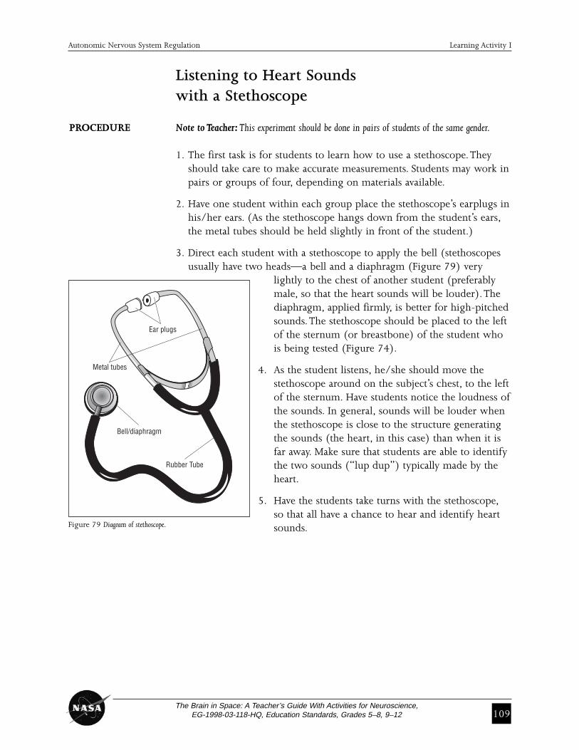
2. Have one student within each group place the stethoscope’s earplugs inhis/her ears. (As the stethoscope hangs down from the student’s ears,the metal tubes should be held slightly in front of the student.)
3. Direct each student with a stethoscope to apply the bell (stethoscopes usually have two heads—a bell and a diaphragm (Figure 79) very
lightly to the chest of another student (preferably male, so that the heart sounds will be louder).The diaphragm, applied firmly, is better for high-pitchedsounds.The stethoscope should be placed to the left of the sternum (or breastbone) of the student who is being tested (Figure 74).
4. As the student listens, he/she should move thestethoscope around on the subject’s chest, to the leftof the sternum. Have students notice the loudness ofthe sounds. In general, sounds will be louder whenthe stethoscope is close to the structure generatingthe sounds (the heart, in this case) than when it isfar away. Make sure that students are able to identifythe two sounds (“lup dup”) typically made by theheart.
5. Have the students take turns with the stethoscope,so that all have a chance to hear and identify heartsounds.
Ear plugs
Bell/diaphragm
Rubber Tube
Metal tubes
Figure 79 Diagram of stethoscope.

110The Brain in Space: A Teacher’s Guide With Activities for Neuroscience,
EG-1998-03-118-HQ, Education Standards, Grades 5–8, 9–12
Student Name Heart RateBeats Per 30 Secs.
Figure 80 Chart for recording heart rate.
Individual Heart Rate Data
Student 1.
Student 2.
Autonomic Nervous System Regulation Learning Activity I
Determining Heart Rate with a Stethoscope
PROCEDURE Note to Teacher: This experiment requires at least three students.The experiment should bedone by two students of the same gender.Another student should be the recorder.
1. Have the student being measured sit comfortably in a chair with feet flat on the floor.
2. Have one student place the stethoscope’s earplugs in his/her ears. (As the stethoscope hangs down, the metal tube parts should be directed slightly ahead).
3. Instruct the student to apply the bell of the stethoscope very lightly to the subject’s chest on the left side of the breastbone.
4. Tell the student to listen to the subject’s heart rates by moving the stethoscope around the subject’s chest. Inform the student that sounds will be louder as you get closer to the heart valves. Have one student record the number of beats in 30 seconds on the chart on the Student Activity Sheet (Figure 80).
5. The student should take at least three measurements.
6. Have the students select another subject of the same gender and repeat procedures two and three.
7. To convert heart rate to beats/minute, students should multiply valuesin Figure 78 by two.
The Brain in Space: A Teacher’s Guide With Activities for Neuroscience,EG-1998-03-118-HQ, Education Standards, Grades 5–8, 9–12 111
Autonomic Nervous System Regulation Student Activity Sheet: Learning Activity I
STUDENT ACTIVITY SHEETDetermining Heart Rate with a Stethoscope
Name ________________________________________________ Date __________________
OBJECTIVE To learn how to measure heart rate with a stethoscope.
MATERIALS • Watch or access to a clock with a second hand• Stethoscope• Pen or pencil• Note pad
DIRECTIONS Do the following procedures and record your data on the chart.
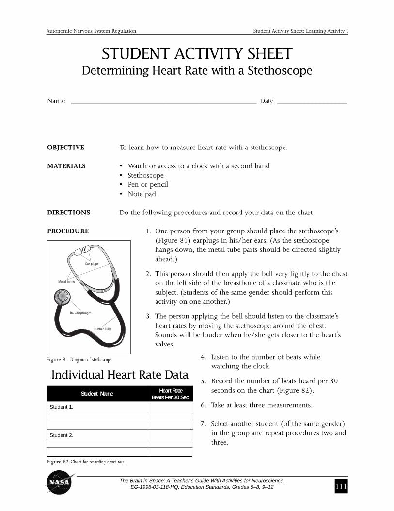
PROCEDURE 1. One person from your group should place the stethoscope’s (Figure 81) earplugs in his/her ears. (As the stethoscope hangs down, the metal tube parts should be directed slightly ahead.)
2. This person should then apply the bell very lightly to the cheston the left side of the breastbone of a classmate who is the subject. (Students of the same gender should perform this activity on one another.)
3. The person applying the bell should listen to the classmate’s heart rates by moving the stethoscope around the chest.Sounds will be louder when he/she gets closer to the heart’s valves.
4. Listen to the number of beats while watching the clock.
5. Record the number of beats heard per 30seconds on the chart (Figure 82).
6. Take at least three measurements.
7. Select another student (of the same gender)in the group and repeat procedures two andthree.
Ear plugs
Bell/diaphragm
Rubber Tube
Metal tubes
Figure 81 Diagram of stethoscope.
Student Name Heart Rate Beats Per 30 Sec.
Figure 82 Chart for recording heart rate.
Individual Heart Rate Data
Student 1.
Student 2.
112The Brain in Space: A Teacher’s Guide With Activities for Neuroscience,
EG-1998-03-118-HQ, Education Standards, Grades 5–8, 9–12
Autonomic Nervous System Regulation Learning Activity I
Measuring Blood Pressure with a Sphygmomanometer
PROCEDURE 1. Introduce the students to the sphygmomanometer. Most of them will have had their blood pressure taken at some time at a doctor’s office. Askfor a show of hands of students who have had their blood pressure measured.
2. With a student, demonstrate how to use the stethoscope and sphygmo-manometer together to measure blood pressure.
3. Have the person being measured sit comfortably in a chair with feetflat on the floor.
4. Be sure that the person has not over-exerted himself/herself during thelast thirty minutes, e.g., exercising.
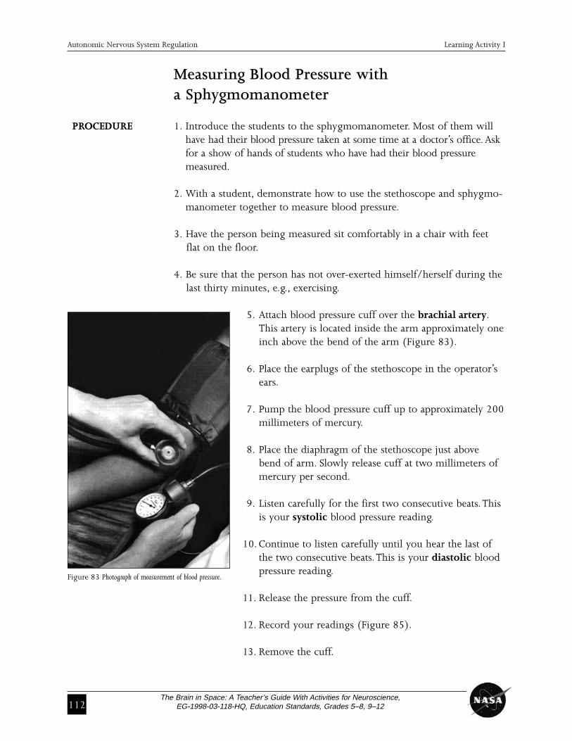
5. Attach blood pressure cuff over the brachial artery.This artery is located inside the arm approximately oneinch above the bend of the arm (Figure 83).
6. Place the earplugs of the stethoscope in the operator’sears.
7. Pump the blood pressure cuff up to approximately 200millimeters of mercury.
8. Place the diaphragm of the stethoscope just abovebend of arm. Slowly release cuff at two millimeters ofmercury per second.
9. Listen carefully for the first two consecutive beats. Thisis your systolic blood pressure reading.
10. Continue to listen carefully until you hear the last ofthe two consecutive beats. This is your diastolic bloodpressure reading.
11. Release the pressure from the cuff.
12. Record your readings (Figure 85).
13. Remove the cuff.
Figure 83 Photograph of measurement of blood pressure.
The Brain in Space: A Teacher’s Guide With Activities for Neuroscience,EG-1998-03-118-HQ, Education Standards, Grades 5–8, 9–12 113
Autonomic Nervous System Regulation Student Activity Sheet: Learning Activity I
STUDENT ACTIVITY SHEETMeasuring Blood Pressure with a Sphygmomanometer
Name ________________________________________________ Date __________________
OBJECTIVE To learn how to measure blood pressure with a sphygmomanometer (aninstrument for measuring arterial blood pressure consisting of an inflat-able blood pressure cuff, inflating bulb, a gauge showing the blood pressure, and a stethoscope).
MATERIALS • Watch or access to clock with a second hand• Stethoscope• Sphygmomanometer • Alcohol wipes to clean stethoscope ear plugs
after each use• Pen or pencil• Note pad
DIRECTIONS Do the following procedures and record your data in the table.
PROCEDURE 1. Have the person being measured sit comfortably in a chair with feet flat on the floor.
2. Be sure that the person has not over-exerted himself/her-self during the last thirty minutes, such as by exercising.
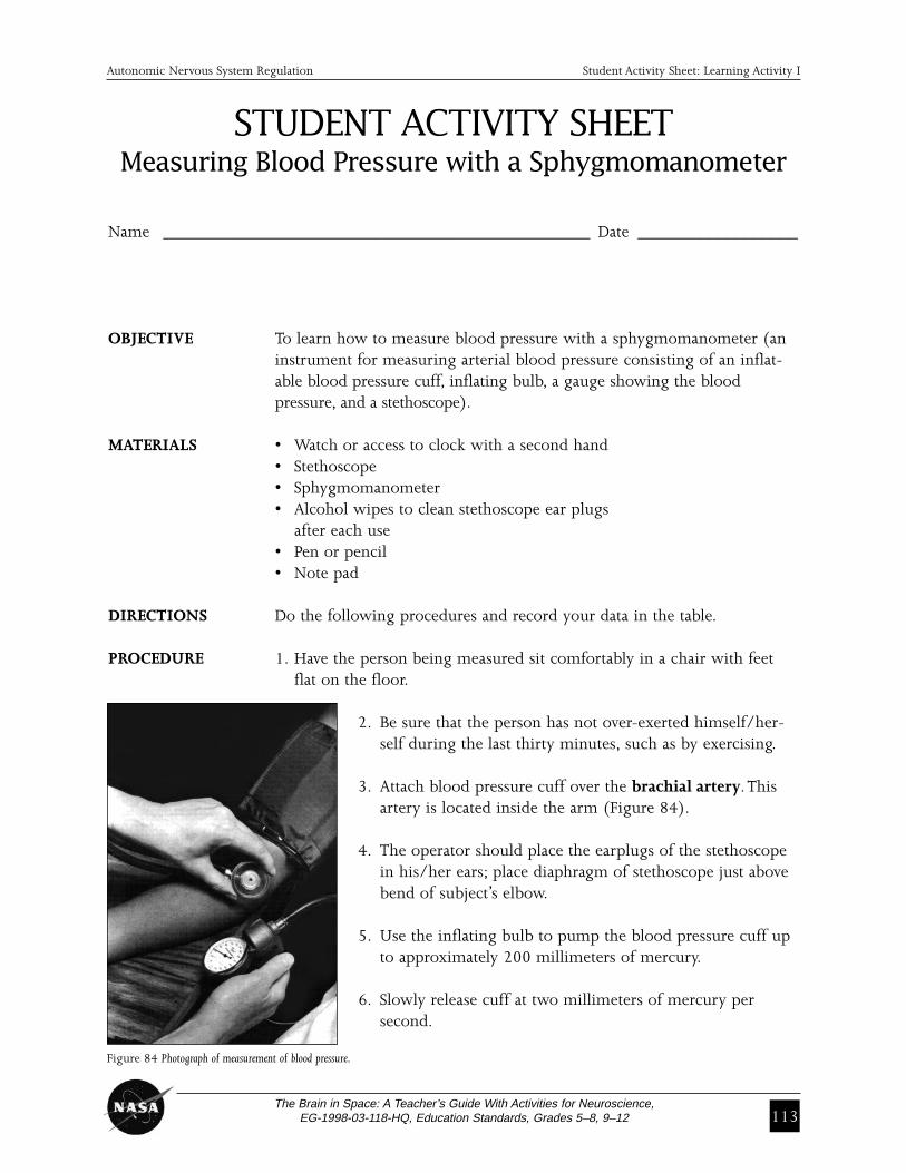
3. Attach blood pressure cuff over the brachial artery. Thisartery is located inside the arm (Figure 84).
4. The operator should place the earplugs of the stethoscopein his/her ears; place diaphragm of stethoscope just abovebend of subject’s elbow.
5. Use the inflating bulb to pump the blood pressure cuff upto approximately 200 millimeters of mercury.
6. Slowly release cuff at two millimeters of mercury per second.
Figure 84 Photograph of measurement of blood pressure.
114The Brain in Space: A Teacher’s Guide With Activities for Neuroscience,
EG-1998-03-118-HQ, Education Standards, Grades 5–8, 9–12
Autonomic Nervous System Regulation Student Activity Sheet: Learning Activity I
7. Listen carefully for the first two consecutive beats. This is your systolicblood pressure reading.
8. Continue to listen carefully until you hear the last of the two consecu-tive beats. This is your diastolic blood pressure reading.
9. Release the pressure from the cuff.
10. Remove the cuff.
11. Record your readings (Figure 85).
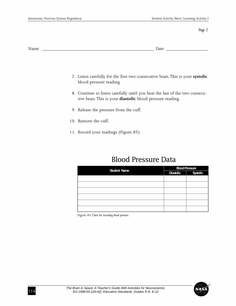
Blood Pressure DataStudent Name
Blood PressureDiastolic Systolic
Figure 85 Chart for recording blood pressure.
Name ________________________________________________ Date __________________
Page 2

The Brain in Space: A Teacher’s Guide With Activities for Neuroscience,EG-1998-03-118-HQ, Education Standards, Grades 5–8, 9–12 115
Autonomic Nervous System Regulation Learning Activity I
SIGNIFICANCE • Blood pressure measures utilizing the stethoscope and sphygmo-OF THE TWO manometer cuff will yield the systolic and diastolic pressures.MEASURESTAKEN • Heart rate can be monitored with a stethoscope placed near the valves
of the heart.
• Taking pulse palpitations by placing the tip of the index and middlefingers on the radius will also yield heart rate.
Pulse = heart rate
Sphygmomanometer and stethoscope = diastolic and systolic pressures
EvaluationAt this point, your students should understand two basic tools necessaryfor studying the heart and circulatory system, the stethoscope and sphyg-momanometer.They should also know how to measure and record indi-vidual heart rates.
REVIEWQUESTIONS 1. What different methods can you use to measure heart rate?
The simplest way to measure heart rate is to count the pulse beats.Pulses can be found wherever an artery is accessible. Most people use theradial pulse, located at the wrist. Heart rate can also be measured from beat-by-beat blood pressure recordings or from the electrocardiogram.
2. What is responsible for the movement we call pulse?The pulse occurs when a sudden rush of blood stretches the artery.Arteries stretch as the left ventricle ejects part (over 55%) of its volumeinto the aorta—the largest artery.
3. Why do we take measurements of both systolic and diastolic to determine blood pressure?To determine the maximum and minimum pressures in the circulatory system.
THINKING 1. Using the class data, have students determine if the heart rate and blood CRITICALLY pressure averages they observed from their classmates are high, low or
average for their age group. Ask students to identify and find resources necessary to make this determination.
SKILL BUILDING 1. Have students use resources at the library or on the Internet to investigate lifestyle habits that may cause high blood pressure(hypertension).
2. Based on their research, have students design a lifestyle for someone who may suffer from hypertension.

116The Brain in Space: A Teacher’s Guide With Activities for Neuroscience,
EG-1998-03-118-HQ, Education Standards, Grades 5–8, 9–12
Autonomic Nervous System Regulation Learning Activity I
CONCLUSION Explain that the astronauts on the Neurolab mission used different meth-ods to study blood pressure.They used a small cuff device on the finger(photoplethysmograph) that actually measures the amount of light pass-ing through the tissues of the fingers at different times (Figure 86). As theheart beats, light transmission through the finger varies with the amountof blood in the finger.The pressure in the cuff is regulated continuouslyto maintain light transmission in the finger at a constant level. Pressure inthe cuff is measured by an electronic pressure gauge, and this measure-ment is used as an accurate index of actual blood pressure.You may wantto make an overhead transparency of the photograph below to show tostudents.
When you believe your students are comfortable with, and have masteredmeasuring blood pressure, the next activity can be assigned as an exten-sion to the previous one. It will allow students to see how the body adaptsto changes of posture and the regulation of blood pressure.
AIR
Figure 86 Diagram of a Photoplethysmograph around a finger.
Bladder
Arteries
Air
Bone Detector
LightEmittingDiode
The Brain in Space: A Teacher’s Guide With Activities for Neuroscience,EG-1998-03-118-HQ, Education Standards, Grades 5–8, 9–12 117
Autonomic Nervous System Regulation Learning Activity II
LEARNING ACTIVITY II:Changing Body Positions:
How Does the Circulatory System Adjust?
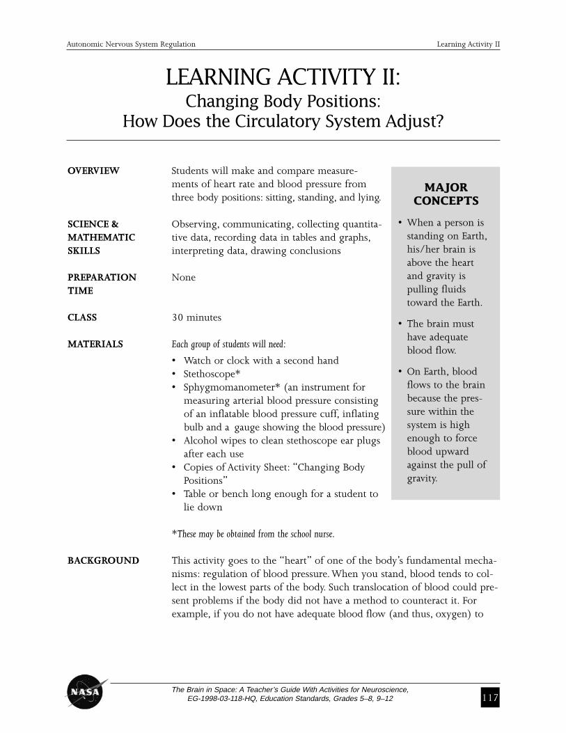
OVERVIEW Students will make and compare measure-ments of heart rate and blood pressure fromthree body positions: sitting, standing, and lying.
SCIENCE & Observing, communicating, collecting quantita-MATHEMATIC tive data, recording data in tables and graphs,SKILLS interpreting data, drawing conclusions
PREPARATION NoneTIME
CLASS 30 minutes
MATERIALS Each group of students will need:
• Watch or clock with a second hand• Stethoscope*• Sphygmomanometer* (an instrument for
measuring arterial blood pressure consisting of an inflatable blood pressure cuff, inflating bulb and a gauge showing the blood pressure)
• Alcohol wipes to clean stethoscope ear plugs after each use
• Copies of Activity Sheet: “Changing Body Positions”
• Table or bench long enough for a student to lie down
*These may be obtained from the school nurse.
BACKGROUND This activity goes to the “heart” of one of the body’s fundamental mecha-nisms: regulation of blood pressure. When you stand, blood tends to col-lect in the lowest parts of the body. Such translocation of blood could pre-sent problems if the body did not have a method to counteract it. Forexample, if you do not have adequate blood flow (and thus, oxygen) to
MAJOR CONCEPTS
• When a person isstanding on Earth,his/her brain isabove the heartand gravity ispulling fluidstoward the Earth.
• The brain musthave adequateblood flow.
• On Earth, bloodflows to the brainbecause the pres-sure within thesystem is highenough to forceblood upwardagainst the pull ofgravity.

118The Brain in Space: A Teacher’s Guide With Activities for Neuroscience,
EG-1998-03-118-HQ, Education Standards, Grades 5–8, 9–12
the brain, you will faint. Fortunately, blood within the circulatory systemflows under pressure that is high enough to counteract the force of gravi-ty and allows blood to reach your brain even when you are standing.
This activity will demonstrate one way in which the body responds to changes in posture.
While conducting the activity, students will measure heart rate and bloodpressure in three different body positions—(1) sitting, (2) standing, and(3) lying (Figure 87). Since students will be spending a short period oftime in each position, they probably will find that heart rate and blood
pressure have not reached a steady statewhen they make their measurements.Students should document thesechanges during the time spent in eachof the positions. Measurements madeevery two minutes will allow studentsto observe some of the effects ofchanges of body positions.
As discussed in the previous lessonsand activities, scientists on theAutonomic Team used tools that allowfor much more precise measurementsof blood pressure and heart rate. Afterthis activity, students should have a
very good idea about how the body adapts to changes of posture, basedon their own experimental data.
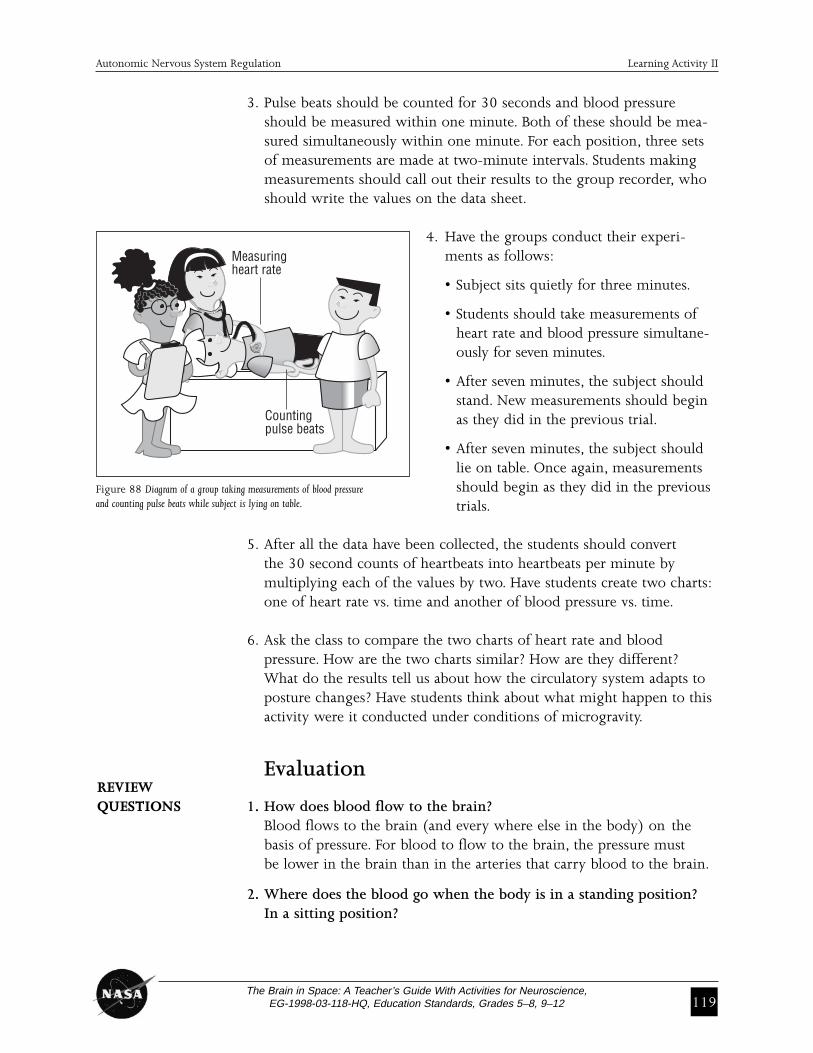
PROCEDURE 1. Have students conduct this activity in groups of four. One student in each group should serve as the subject, one should measure heart rate (measured as number of pulse beats felt in one minute), one should measure the blood pressure from the other arm, and the other should record measurements as they are called out (Figure 88).
2. Explain to the groups that they will be investigating changes in heart rate and blood pressure that occur when the body moves from sitting to standing to lying. Ask the students to think about and predict changes that might happen. Explain that measurements for each position will takeseven minutes with a maximum of 30 seconds allowed for subject to change positions.
3 1 2
Figure 87 Diagram of three different body positions.
Autonomic Nervous System Regulation Learning Activity II
The Brain in Space: A Teacher’s Guide With Activities for Neuroscience,EG-1998-03-118-HQ, Education Standards, Grades 5–8, 9–12 119
3. Pulse beats should be counted for 30 seconds and blood pressureshould be measured within one minute. Both of these should be mea-sured simultaneously within one minute. For each position, three setsof measurements are made at two-minute intervals. Students makingmeasurements should call out their results to the group recorder, whoshould write the values on the data sheet.
4. Have the groups conduct their experi-ments as follows:
• Subject sits quietly for three minutes.
• Students should take measurements of heart rate and blood pressure simultane-ously for seven minutes.
• After seven minutes, the subject should stand. New measurements should begin as they did in the previous trial.
• After seven minutes, the subject should lie on table. Once again, measurements should begin as they did in the previous trials.
5. After all the data have been collected, the students should convert the 30 second counts of heartbeats into heartbeats per minute by multiplying each of the values by two. Have students create two charts:one of heart rate vs. time and another of blood pressure vs. time.
6. Ask the class to compare the two charts of heart rate and blood pressure. How are the two charts similar? How are they different? What do the results tell us about how the circulatory system adapts to posture changes? Have students think about what might happen to this activity were it conducted under conditions of microgravity.
EvaluationREVIEWQUESTIONS 1. How does blood flow to the brain?
Blood flows to the brain (and every where else in the body) on the basis of pressure. For blood to flow to the brain, the pressure must be lower in the brain than in the arteries that carry blood to the brain.
2. Where does the blood go when the body is in a standing position? In a sitting position?
Measuringheart rate
Countingpulse beats
Figure 88 Diagram of a group taking measurements of blood pressureand counting pulse beats while subject is lying on table.
Autonomic Nervous System Regulation Learning Activity II
120The Brain in Space: A Teacher’s Guide With Activities for Neuroscience,
EG-1998-03-118-HQ, Education Standards, Grades 5–8, 9–12
Autonomic Nervous System Regulation Learning Activity II
Blood goes to the lowest part of the body, according to the degree of gravitational pull. Tall people, who have much higher columns of bloodthan short people, are more prone to experience reductions of blood pressure when they stand. Less blood is pooled in the sitting position because the height of the column of blood is less than in the standing position.
3. What happens to blood flow, blood pressure, and heart rate when the body moves from lying to sitting?The heart rate begins to speed; this occurs very rapidly, often within one second. Speeding of the heart helps to prevent a major fall in blood pressure. Also, the vessels constrict to increase resistance to flow and keep blood pressure up.
CRITICAL 1. Challenge your students to think about how the circulatory systemmight respond to changes in a microgravity environment.Which aspectsof the system might stay the same? Which might be different?Absence of gravity causes major changes in the circulation. The mostimportant of these is that there is no gravitational pull of blood towardthe feet when a person stands. Pressure in the feet is the same in lyingand standing positions. Therefore, bloodredistributes from the lower body to theupper body. The autonomic nervous systemdoes not have to adjust to body positions ason Earth. This is not to say that there is noautonomic nervous system control of the cir-culation in space.
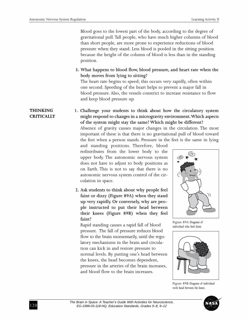
2. Ask students to think about why people feelfaint or dizzy (Figure 89A) when they standup very rapidly. Or conversely, why are peo-ple instructed to put their head betweentheir knees (Figure 89B) when they feelfaint?Rapid standing causes a rapid fall of bloodpressure. The fall of pressure reduces bloodflow to the brain momentarily, until the regu-latory mechanisms in the brain and circula-tion can kick in and restore pressure to normal levels. By putting one’s head betweenthe knees, the head becomes dependent,pressure in the arteries of the brain increases,and blood flow to the brain increases.
THINKINGCRITICALLY
Figure 89A Diagram of individual who feels faint.
Figure 89B Diagram of individualwith head between the knees.
The Brain in Space: A Teacher’s Guide With Activities for Neuroscience,EG-1998-03-118-HQ, Education Standards, Grades 5–8, 9–12 121
Autonomic Nervous System Regulation Learning Activity II
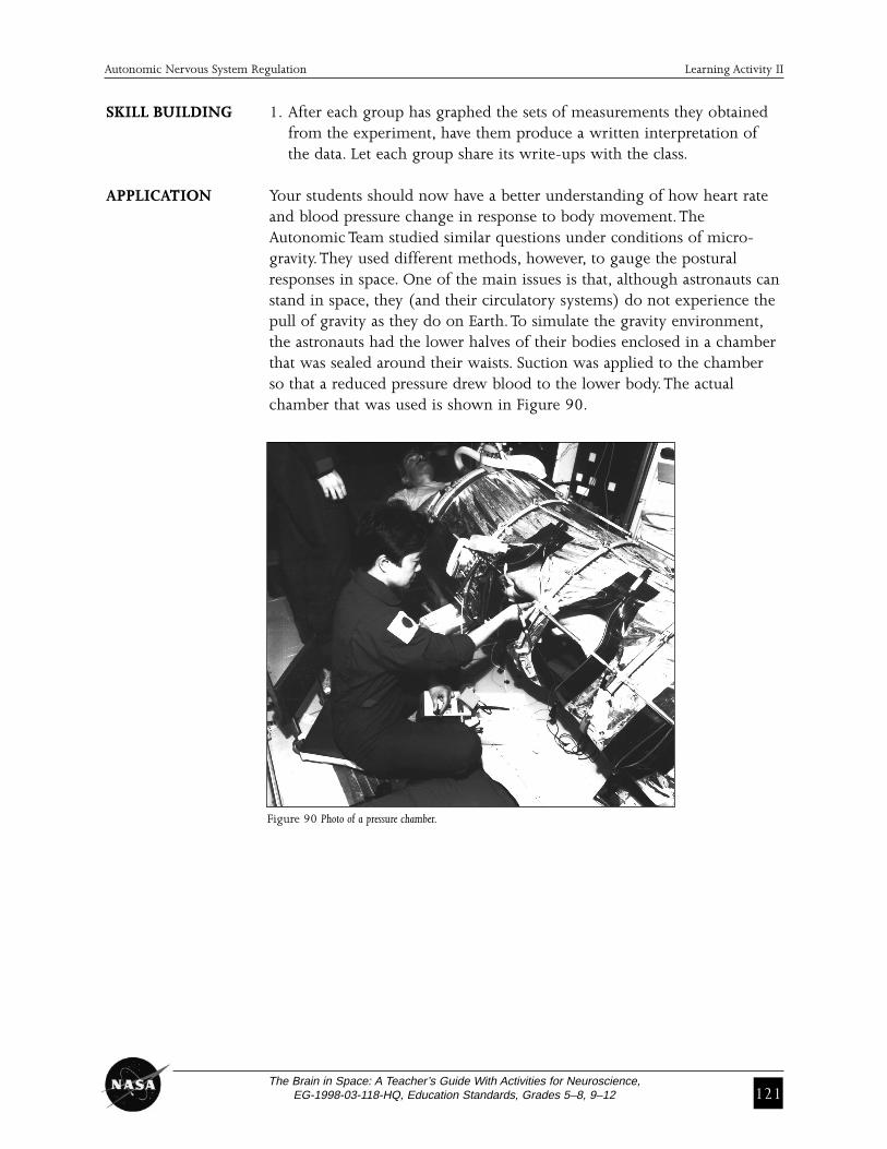
Figure 90 Photo of a pressure chamber.
SKILL BUILDING 1. After each group has graphed the sets of measurements they obtained from the experiment, have them produce a written interpretation of the data. Let each group share its write-ups with the class.
APPLICATION Your students should now have a better understanding of how heart rateand blood pressure change in response to body movement.TheAutonomic Team studied similar questions under conditions of micro-gravity.They used different methods, however, to gauge the posturalresponses in space. One of the main issues is that, although astronauts canstand in space, they (and their circulatory systems) do not experience thepull of gravity as they do on Earth.To simulate the gravity environment,the astronauts had the lower halves of their bodies enclosed in a chamberthat was sealed around their waists. Suction was applied to the chamberso that a reduced pressure drew blood to the lower body.The actualchamber that was used is shown in Figure 90.
122The Brain in Space: A Teacher’s Guide With Activities for Neuroscience,
EG-1998-03-118-HQ, Education Standards, Grades 5–8, 9–12
STUDENT ACTIVITY SHEETChanging Body Positions
Name ________________________________________________ Date __________________
OBJECTIVE To measure the changes of heart rate and blood pressure with changingbody positions.
MATERIALS • A watch or clock • Stethoscope • Alcohol wipeswith a second hand • Pen or pencil • Note pad
• Sphygmomanometer
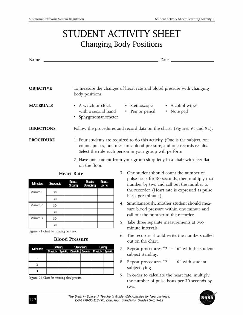
DIRECTIONS Follow the procedures and record data on the charts (Figures 91 and 92).
PROCEDURE 1. Four students are required to do this activity. (One is the subject, one counts pulses, one measures blood pressure, and one records results.Select the role each person in your group will perform.
2. Have one student from your group sit quietly in a chair with feet flat on the floor.
3. One student should count the number ofpulse beats for 30 seconds, then multiply thatnumber by two and call out the number tothe recorder. (Heart rate is expressed as pulsebeats per minute.)
4. Simultaneously, another student should mea-sure blood pressure within one minute andcall out the number to the recorder.
5. Take three separate measurements at twominute intervals.
6. The recorder should write the numbers calledout on the chart.
7. Repeat procedures “2” – “6” with the studentsubject standing.
8. Repeat procedures “2” – “6” with student subject lying.
9. In order to calculate the heart rate, multiplythe number of pulse beats per 30 seconds bytwo.
Sitting Standing LyingMinutesDiastolic Systolic Diastolic Systolic Diastolic Systolic
Autonomic Nervous System Regulation Student Activity Sheet: Learning Activity II
30
30
30
30
30
30
Beats Beats BeatsMinutes Seconds Sitting Standing Lying
1
2
3
Heart Rate
Blood Pressure
Figure 92 Chart for recording blood pressure.
Minute 1
Minute 2
Minute 3
Figure 91 Chart for recording heart rate.
The Brain in Space: A Teacher’s Guide With Activities for Neuroscience,EG-1998-03-118-HQ, Education Standards, Grades 5–8, 9–12 123
Autonomic Nervous System Regulation Learning Activity III
LEARNING ACTIVITY III:Baroreceptor Reflex Role Play
OVERVIEW In this activity, the students will learn theimportance of maintaining adequate arterialblood pressure through a role playing exercisewhich demonstrates the baroreceptor reflex(BR) arc—a human model of the BR loop.TheBR arc will demonstrate how the brain process-es information and sends out signals to theheart and arteries.
SCIENCE & Prediction, observing, collecting quantitative MATHEMATICS data and interpreting dataSKILLS
PREPARATION 10 minutesTIME
CLASS TIME 45 minutes
MATERIALS • Index cards to be used as cue cards indicating students’ roles (nine students per group)
BACKGROUND Baroreceptors are specialized neural receptors that sense changes in bloodpressure.The baroreceptor reflex relies on sensing pressure changes in theaorta and carotid artery. If stimulation of the baroreceptors is altered,such as in a microgravity environment, a change would occur in the fir-ing frequency of the nerves connected to the baroreceptors that send sig-nals to the brain.
The baroreceptor reflex circuit includes both sympathetic and parasym-pathetic nerves to the arteries and heart in order to maintain proper arterial blood pressure.The baroreceptor reflex can either increase ordecrease arterial blood pressure in order to return it to normal levels. Ifthe arterial blood pressure increases, the firing rate of the baroreceptorsincreases, sending a greater frequency of impulses through the afferent(inflowing, i.e., towards the brain) baroreceptor nerves.The reflex in thisinstance will cause activation of the vagus nerve, decreasing the rate ofheart contraction to reduce arterial pressure. If the pressure falls, decreased
MAJOR CONCEPTS
• If development inmicrogravity altersthe developmentof the baroreceptorafferent neurons,the circuit may notfunction normallyon Earth.
• The functions ofeach part of thebaroreceptor reflexare required toproperly maintainarterial blood pressure.

124The Brain in Space: A Teacher’s Guide With Activities for Neuroscience,
EG-1998-03-118-HQ, Education Standards, Grades 5–8, 9–12
Autonomic Nervous System Regulation Learning Activity III
firing and frequency of impulse result. In turn, the sympathetic nerves areactivated, thereby causing the heart to contract more rapidly and of bloodvessels to constrict, which will increase arterial blood pressure.
Through this reflex circuit, arterial blood pressure is maintained. It iscrucial to maintain blood pressure within this specified range because theorgan functions of the body depend upon an adequate supply of blood ata relatively constant pressure.
PROCEDURE This activity requires nine students per group.
1. Students should be arranged in groups of nine to represent the parts of the baroreceptor reflex loop according to the diagram in Figure 94.
2. Explain the role of each student so that the students will have a clear understanding of their roles before the activity begins (Figure 93).
3. Tell the student playing the arterial pressure (student #1) to squeeze the arm of the baroreceptor (student #2) firmly to represent increased pressure. In turn, instruct the baroreceptor (student #2) to use a num-ber system to signal increased or decreased pressure to the baroreceptorafferent (student #3).The number system could be one to signal low pressure, two to signal normal pressure and three to signal increased orhigh pressure.
Position Role Task
1 Arterial pressure Squeeze the arm of student #2
2 Baroreceptor Assign the numeric value according to pressure to student #3
3 Baroreceptor afferent Relay the numeric value to student #4 or #5
4 Vasomotor center If the number is low (low pressure), student #4 becomes activated
5 Vagal center If the number is high (high pressure), student #5 becomes activated
6 Vagus nerve Receive information from student #5 and tell student #8 (heart) to decrease rate of pumping
7 Sympathetic nerves Receive information from student #4 and tell student #8 (heart) to increase rate of pumping and tell student #9 (arteries) to constrict to increase pressure
8 Heart Tell student #1 what to do
9 Arteries Tell student #1 what to do, then student #1 tell student #2 what to do
Figure 93 Chart of students’ positions, roles and tasks representing the baroreceptor reflex loop.
The Brain in Space: A Teacher’s Guide With Activities for Neuroscience,EG-1998-03-118-HQ, Education Standards, Grades 5–8, 9–12 125
Autonomic Nervous System Regulation Learning Activity III
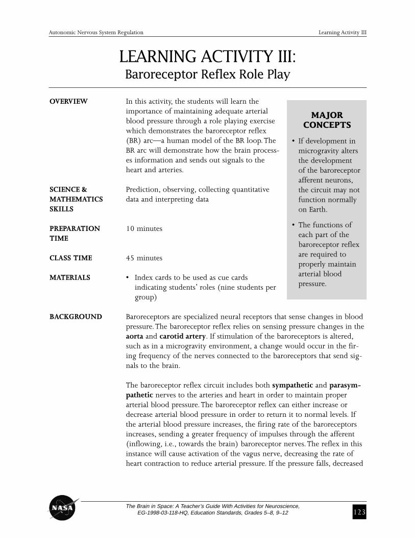
Figure 94 Diagram of a baroreceptor reflex loop involved in maintaining proper blood pressure.
4. Direct the baroreceptor (student #2) to report these numbers to the two areas of the medulla—the vasomotor center (student #4) and vagal center (student #5).
EXPLANATION Note to the teacher: At this point, use procedures 5 through 8 to help the students under-stand how the baroreceptor reflex loop works.
5. Once the baroreceptor signals high pressure, the vagal center will tell the vagus nerve to go into action.
6. The vagus nerve goes to the heart and tells it to stop pumping quite so hard and fast. The heart will tell arterial pressure to relax the grip on the baroreceptor’s arm to reduce the number reported by the barore-ceptor to two, normal pressure.
7. If the baroreceptor signals number one (low pressure), the vasomotor center tells the sympathetic nerves to go into action.
8. The sympathetic nerves tell the heart to increase its pumping rate and the arteries to constrict. The arteries and the heart tell the arterial pressure to increase the grip to two, normal pressure. In this case,the baroreceptor would signal normal pressure.
Once pressure is back to normal, you can introduce certain things thatwould change the pressure. For instance, you could say that a lion wasjust spotted and the heart should automatically tell pressure that itincreased its pumping, the vessels should tell pressure that they constrict-ed and the pressure should go to number three (high pressure).
Number correlates to the position (see procedure).
126The Brain in Space: A Teacher’s Guide With Activities for Neuroscience,
EG-1998-03-118-HQ, Education Standards, Grades 5–8, 9–12
Autonomic Nervous System Regulation Learning Activity III
Evaluation
REVIEW 1. What two things are primarily responsible for making arterial bloodQUESTIONS pressure increase or decrease?
The rate at which the blood is pumped into the arteries by the heart and the resistance to blood flow within the arteries are primarily responsible for changes in arterial blood pressure.
2. What areas of the brain are involved with the baroreceptor reflex?The areas of the brain that are involved in the baroreceptor reflex is thevasomotor center and vagal center.
3. What part of the loop might microgravity influence through its presence during human development?Any part of the baroreceptor reflex loop could be influenced by lack ofgravity during human development. The baroreceptor nerves were studied on Neurolab.
4. Why is blood pressure so important?An appropriate blood pressure is important to maintain blood flow through the blood vessels to the brain and other vital organs.
THINKING Have your students write a brief paragraph and explain why they do CRITICALLY not faint when they stand up. You also can have them explain why they
feel light-headed for a few moments when they stand up quickly.