author's personal copy - uhasselt · author's personal copy current concepts current...
TRANSCRIPT

This article appeared in a journal published by Elsevier. The attachedcopy is furnished to the author for internal non-commercial researchand education use, including for instruction at the authors institution
and sharing with colleagues.
Other uses, including reproduction and distribution, or selling orlicensing copies, or posting to personal, institutional or third party
websites are prohibited.
In most cases authors are permitted to post their version of thearticle (e.g. in Word or Tex form) to their personal website orinstitutional repository. Authors requiring further information
regarding Elsevier’s archiving and manuscript policies areencouraged to visit:
http://www.elsevier.com/copyright

Author's personal copy
Current Concepts
Current Perspectives on Rotator Cuff Anatomy
Michael J. DeFranco, M.D., and Brian J. Cole, M.D.
Abstract: Understanding the anatomy of the rotator cuff and the surrounding structures thatinfluence its function is essential to treating rotator cuff disease. During the past decade,advances in basic science and surgical technology have improved our knowledge of thisanatomy. This review article presents the current concepts on rotator cuff anatomy and how theyshould be used in the surgical management of rotator cuff tears. Key Words: Rotator cuff—Anatomy—Coracoacromial arch—Acromioplasty—Bursectomy—Vascularity.
Although many factors influence the treatment ofrotator cuff tears, understanding the anatomy and
how it relates to function is the most important one.Indeed, fundamental to rotator cuff surgery is knowl-edge of the normal anatomic relations. Both the osse-ous and soft-tissue structures have a significant impacton rotator cuff function. Recent research has expandedour knowledge specifically with regard to the rotatorcuff as well as the coracoacromial (CA) arch, bursae,and neurovascular structures. On the basis of thesedata, there are several controversial issues that con-tinue to be debated. Some of these issues include theinfluence of the morphology of the acromion, CAligament, and coracoid process on the development ofrotator cuff tears; the role of the subacromial bursa asa source of pain or as an essential contributor to afibrovascular response that may help rotator cuff re-pairs heal; the relation between greater tuberosity os-teopenia and rotator cuff disease; the anatomic defi-nition of the rotator cuff footprint; and the anatomiclocation of the neurovascular structures surrounding
the rotator cuff. The purpose of this article is to reviewthe current literature on rotator cuff anatomy and howit influences decision making in the surgical care ofpatients with rotator cuff tears.
CORACOACROMIAL ARCH
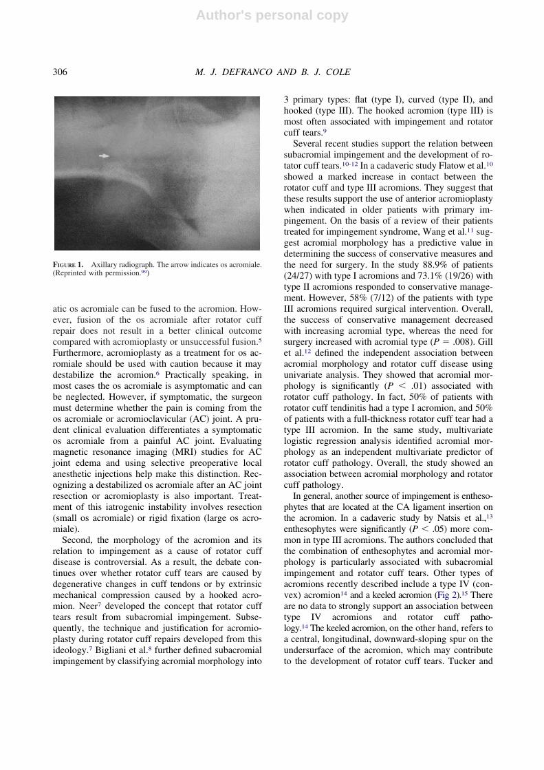
The CA arch is defined as a confluence of theacromion, the CA ligament, and the coracoid process.The morphology of the acromion is relevant to thesurgical management of rotator cuff disease for sev-eral reasons. First, abnormalities in the developmentof the acromion may lead to the formation of an osacromiale (Fig 1). Approximately 8% of patients havean os acromiale. In 33% of patients this developmentabnormality occurs bilaterally.1 Recent studies sug-gest an association between os acromiale and rotatorcuff tears, but this relation is not well defined.2-4 Infact, on the basis of the data in the literature, it isunlikely that the os acromiale has a pathologic effecton the rotator cuff.5 The presence of an os acromialealso does not influence the number of tendons in-volved in the rotator cuff tear.5 These findings areimportant considerations in the preoperative planningfor rotator cuff repairs. Boehm et al.5 retrospectivelyreviewed the surgical management of 33 patients whoreceived treatment for a rotator cuff tear and an osacromiale. They concluded that at the time of rotatorcuff repair, resection is an appropriate treatment for asmall, symptomatic os acromiale. A large, symptom-
From Midwest Orthopaedics, Rush University Medical Center,Chicago, Illinois, U.S.A.
The authors report no conflict of interest.Address correspondence and reprint requests to Brian J. Cole,
M.D., 1725 W Harrison Ave, Suite 1063, Chicago, IL 60612,U.S.A. E-mail: [email protected]
© 2009 by the Arthroscopy Association of North America0749-8063/09/2503-8217$36.00/0doi:10.1016/j.arthro.2008.07.023
305Arthroscopy: The Journal of Arthroscopic and Related Surgery, Vol 25, No 3 (March), 2009: pp 305-320
Author's personal copy
atic os acromiale can be fused to the acromion. How-ever, fusion of the os acromiale after rotator cuffrepair does not result in a better clinical outcomecompared with acromioplasty or unsuccessful fusion.5
Furthermore, acromioplasty as a treatment for os ac-romiale should be used with caution because it maydestabilize the acromion.6 Practically speaking, inmost cases the os acromiale is asymptomatic and canbe neglected. However, if symptomatic, the surgeonmust determine whether the pain is coming from theos acromiale or acromioclavicular (AC) joint. A pru-dent clinical evaluation differentiates a symptomaticos acromiale from a painful AC joint. Evaluatingmagnetic resonance imaging (MRI) studies for ACjoint edema and using selective preoperative localanesthetic injections help make this distinction. Rec-ognizing a destabilized os acromiale after an AC jointresection or acromioplasty is also important. Treat-ment of this iatrogenic instability involves resection(small os acromiale) or rigid fixation (large os acro-miale).
Second, the morphology of the acromion and itsrelation to impingement as a cause of rotator cuffdisease is controversial. As a result, the debate con-tinues over whether rotator cuff tears are caused bydegenerative changes in cuff tendons or by extrinsicmechanical compression caused by a hooked acro-mion. Neer7 developed the concept that rotator cufftears result from subacromial impingement. Subse-quently, the technique and justification for acromio-plasty during rotator cuff repairs developed from thisideology.7 Bigliani et al.8 further defined subacromialimpingement by classifying acromial morphology into
3 primary types: flat (type I), curved (type II), andhooked (type III). The hooked acromion (type III) ismost often associated with impingement and rotatorcuff tears.9
Several recent studies support the relation betweensubacromial impingement and the development of ro-tator cuff tears.10-12 In a cadaveric study Flatow et al.10
showed a marked increase in contact between therotator cuff and type III acromions. They suggest thatthese results support the use of anterior acromioplastywhen indicated in older patients with primary im-pingement. On the basis of a review of their patientstreated for impingement syndrome, Wang et al.11 sug-gest acromial morphology has a predictive value indetermining the success of conservative measures andthe need for surgery. In the study 88.9% of patients(24/27) with type I acromions and 73.1% (19/26) withtype II acromions responded to conservative manage-ment. However, 58% (7/12) of the patients with typeIII acromions required surgical intervention. Overall,the success of conservative management decreasedwith increasing acromial type, whereas the need forsurgery increased with acromial type (P � .008). Gillet al.12 defined the independent association betweenacromial morphology and rotator cuff disease usingunivariate analysis. They showed that acromial mor-phology is significantly (P � .01) associated withrotator cuff pathology. In fact, 50% of patients withrotator cuff tendinitis had a type I acromion, and 50%of patients with a full-thickness rotator cuff tear had atype III acromion. In the same study, multivariatelogistic regression analysis identified acromial mor-phology as an independent multivariate predictor ofrotator cuff pathology. Overall, the study showed anassociation between acromial morphology and rotatorcuff pathology.
In general, another source of impingement is entheso-phytes that are located at the CA ligament insertion onthe acromion. In a cadaveric study by Natsis et al.,13
enthesophytes were significantly (P � .05) more com-mon in type III acromions. The authors concluded thatthe combination of enthesophytes and acromial mor-phology is particularly associated with subacromialimpingement and rotator cuff tears. Other types ofacromions recently described include a type IV (con-vex) acromion14 and a keeled acromion (Fig 2).15 Thereare no data to strongly support an association betweentype IV acromions and rotator cuff patho-logy.14 The keeled acromion, on the other hand, refers toa central, longitudinal, downward-sloping spur on theundersurface of the acromion, which may contributeto the development of rotator cuff tears. Tucker and
FIGURE 1. Axillary radiograph. The arrow indicates os acromiale.(Reprinted with permission.99)
306 M. J. DEFRANCO AND B. J. COLE

Author's personal copy
Snyder15 retrospectively reported on 20 patients withthis type of acromion. Of these patients, 100% (20/20)had significant bursal-sided tears and 60% (12/20) hadfull-thickness rotator cuff tears associated with akeeled acromion. Although additional studies need toconfirm and further define these findings, the authorsrecommend early surgery for a keeled acromion tominimize the risk of rotator cuff tear progression.
In an MRI study of rotator cuff disease, Baechlerand Kim16 reported that the percentage of the humeralhead not covered superiorly by the anterolateral acro-mion may be a factor in the pathogenesis of full-thickness rotator cuff tears. Greater “uncoverage” mayallow hinging of the humeral head on the anterolateraledge of the acromion during early shoulder abduction,causing impingement of the supraspinatus tendon be-tween these 2 structures.16 Conversely, on the basis ofa radiographic study of patients with rotator cuff dis-ease, Nyffeler et al.17 reported a statistically signifi-cant (P � .001) association between a large lateralextension of the acromion and full-thickness degener-ative rotator cuff tears. In another radiographic studyTorrens et al.18 studied acromial coverage of the hu-meral head as a factor in the development of rotatorcuff tears. They suggest that patients with rotator cufftears have statistically significantly (P � .001) morecoverage of the humeral head by the acromion com-pared with the control group without tears. Overall,additional studies need to clarify the degree of acro-mial coverage that contributes to the development ofrotator cuff tears.
Even though research studies support the associ-ation between type III acromions and rotator cufftears, there is an equivalent amount of evidence
disputing it. Zuckerman et al.19 were unable toidentify the 3 acromial types in a cadaveric study.They concluded that the acromial classification de-scribed by Bigliani et al.8 does not accurately de-scribe anatomic findings, and the relation to rotatorcuff tears remains unclear and requires furtherstudy. Chang et al.20 used MRI to perform 3-dimen-sional analysis of the acromion. They concludedthat osseous impingement by the acromion is not aprimary cause of shoulder impingement syndromeor rotator cuff tears. In another MRI study Hiranoet al.21 determined that with type III acromions,rotator cuff tears were significantly larger than intypes I and II. The study suggests that acromionmorphology influences rotator cuff tear size. Interest-ingly, comparison of age-matched patients with andwithout rotator cuff tears showed that the occurrencerate of type III acromial shape in the rotator cuff teargroup was not significantly higher. These results sug-gest that a type III acromion does not always correlatewith the development of rotator cuff tears. Schip-pinger et al.22 reported that no type III acromions wereidentified in their study. Their findings suggest thathooked acromions (type III) are not present in thenormal population and are likely to be an acquiredabnormality. Several recent clinical studies also sug-gest that avoiding acromioplasty at the time of rotatorcuff repair does not change the clinical or anatomicoutcome.23-27
Given the results of these studies, the associationbetween acromial morphology and rotator cuff tearsmay not be as strong as described in the literature.
FIGURE 2. Anteroposterior radiograph with outline of keeled ac-romion. (Reprinted with permission.15)
FIGURE 3. Inadequate acromioplasty in a right shoulder. A resid-ual anterolateral spur is shown in the photograph. The radiofre-quency (RF) probe has been inserted through the lateral portal.
307ROTATOR CUFF ANATOMY
Author's personal copy
Nevertheless, acromioplasty continues to be usedby most shoulder surgeons, except in cases wherethe CA arch provides superior stability to preventescape of the humeral head, as in massive rotatorcuff tears. During acromioplasty, preservation ofthe normal anteroposterior dimension of the acro-mion is essential. Large acquired osteophytes foundwithin the CA ligament should be removed withoutdestroying the acromial architecture. According toFlatow et al.,28 smoothing the anterior third of theacromial undersurface removes all focused contacton the supraspinatus insertion (Figs 3 and 4). To-tally flattening the acromion is unnecessary to re-lieve impingement. Excessive release of the deltoidorigin should also be avoided during acromioplasty.According to a study by Green et al.,29 4 mm ofacromial bone resection results in release of 56% �11% of the deltoid origin. Increasing the amount ofresection to 5.5 mm leads to release of 77% � 15%of the deltoid origin. They conclude that the amountof deltoid released correlates statistically with boththe thickness of the acromion and acromial angle(P � .0001 and P � .04, respectively). These fac-tors should be considered during preoperative plan-ning to decrease the risk of inadvertent deltoiddetachment. Overall, although a casual relation be-tween impingement syndrome, rotator cuff pathol-ogy, and acromial morphology is strongly sug-gested by published scientific data, the exactsequence of cause and effect between these entitiesis not well defined in the orthopaedic literature.
CA LIGAMENT
The CA ligament originates along the distal twothirds of the lateral aspect of the coracoid process as abroad ligament. It passes posteriorly to insert onto theanteromedial and anteroinferior surfaces of the acro-mion.30,31 A recent cadaveric study showed variationin the morphology of the CA ligament. The mostcommon configuration of the CA ligament is 2 distinctligamentous bands: anterolateral and posteromedial.32
Spur formation occurs preferentially in the anterolat-eral band. As a result, the anterolateral band is com-monly involved in impingement syndrome. If the pos-teromedial bundle is mistaken to be the entireligament, then the surgeon may fail to visualize theanterolateral corner of the acromion and perform anincomplete subacromial decompression.
Despite the conclusions by Fealy et al.32 regardingthe role of the CA ligament in impingement syn-drome, Pieper et al.33 found no significant relationbetween the morphology of the CA ligament and theincidence of rotator cuff degenerative changes or spurformation. Similarly, Kesmezacar et al.34 reported on5 anatomic variations of the CA ligament. On thebasis of the results of this study, the Y-shaped liga-ment is the most common type. There was no statis-tical significance (P � .05) between rotator cuff de-generation and the type of geometric measurement ofthe ligament.34 However, the CA ligaments with morethan 1 bundle showed a significant (P � .05) associ-ation with rotator cuff degeneration. These CA liga-ments were unique in having a longer lateral borderand larger coracoid insertion than other ligaments.
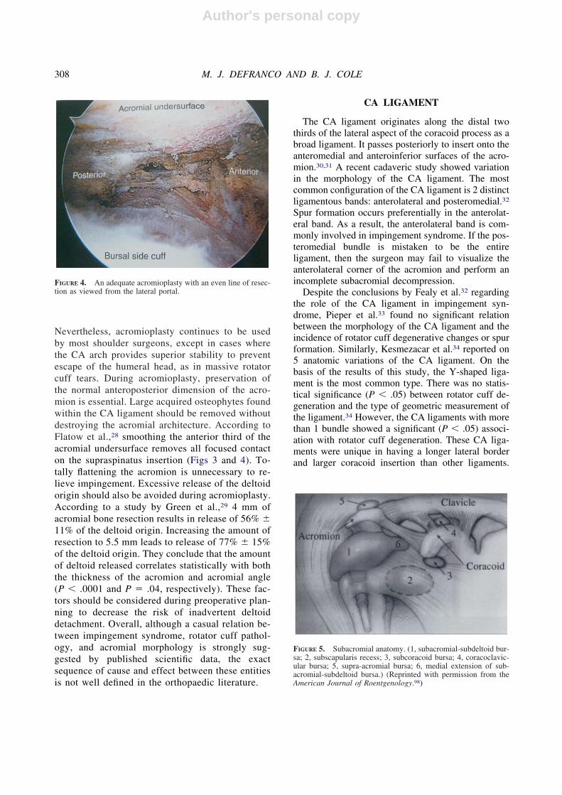
FIGURE 4. An adequate acromioplasty with an even line of resec-tion as viewed from the lateral portal.
FIGURE 5. Subacromial anatomy. (1, subacromial-subdeltoid bur-sa; 2, subscapularis recess; 3, subcoracoid bursa; 4, coracoclavic-ular bursa; 5, supra-acromial bursa; 6, medial extension of sub-acromial-subdeltoid bursa.) (Reprinted with permission from theAmerican Journal of Roentgenology.98)
308 M. J. DEFRANCO AND B. J. COLE

Author's personal copy
Fremerey et al.35 also used a cadaveric study to defineanatomic and biomechanical properties of the CAligament in shoulders with either intact rotator cuffs orrotator cuff disease. Specimens from cadavers withrotator cuff disease had a shorter medial and lateralband of the CA ligament than specimens taken fromshoulders with intact rotator cuffs. The cross-sectionalarea of the lateral band was enlarged in older speci-mens with rotator cuff degeneration. These enlargedlateral bands had a decreased failure stress in shoul-ders with rotator cuff degeneration, but this was foundonly in the older cadaveric groups. On the basis ofthese findings, the authors suggest that substantialalterations in morphologic and biomechanical proper-ties occur in the CA ligament during aging. However,more studies are needed to determine whether varia-tion in the CA ligament morphology causes impinge-ment or is a function of it.
CORACOID PROCESS
Many anatomic studies define the morphology ofthe coracoid process.36-40 In a recent study Bhatiaet al.41 used quantitative and statistical analysis oflinear and angular dimensions to define the individualpillars of the coracoid process. The pillar anatomy ofthe coracoid and its effect on subcoracoid space areessential to understanding the concept of coracoidimpingement. According to Bhatia et al., impingementof the rotator cuff occurs between the posterolateralcoracoid and humeral head. Clinically, this conditionmanifests itself as anterior shoulder pain with forwardflexion, internal rotation, and horizontal adduction ofthe humerus.42 Subcoracoid pain is the result of im-pingement of the subscapularis tendon between thelesser tuberosity and coracoid process.42-46 Changesassociated with this impingement include supraspi-natus tendon injury, subscapularis tendon injury,changes to the rotator interval, and thickening of theCA ligament.
Theoretically, an increase in axial angulation ofeither pillar, a decrease in interpillar angulation, or adecrease in the length of either pillar may predisposean individual to coracoid impingement and place thesupraspinatus or subscapularis at risk for tearing.41 Ina cadaveric study, Ferreira Neto et al.47 showed thatwomen have a smaller distance between the apex ofthe coracoid process and the lesser tuberosity of thehumerus with the arm in internal rotation. This findingsuggests that impingement may be more likely be-tween these 2 bony structures in female patients.47
Previous studies have also suggested that mechanicalbony irritation caused in part by pathologic coracoidmorphology is an important etiologic factor in thedevelopment of coracoid impingement.48
Schulz et al.49 correlated coracoid tip position withrotator cuff tears. In their radiographic study they useda true anteroposterior view of the shoulder to classifythe coracoid into 1 of 2 types. Type I coracoids (inwhich the tip of the coracoid process projects onto theinferior half of the glenoid surface) are associated withsupraspinatus tears (P � .0002). Type II coracoids (inwhich the tip of the coracoid process projects onto thesuperior half of the glenoid surface) are associatedwith subscapularis tears (P � .0001). Overall, theauthors suggest that identification of the coracoid typein shoulders suspected of having rotator cuff pathol-ogy shows an 86% sensitivity and a 71% specificityfor supraspinatus tears (type I coracoid). In subscap-ularis tears (type II coracoids) the sensitivity andspecificity are 71% and 86%, respectively.
Richards et al.50 used a retrospective cohort to showa significant relation between a narrowed coracohu-meral distance and subscapularis pathology. In thisstudy the coracohumeral distance was measured onaxial magnetic resonance images. The mean coraco-humeral distance in the group with no subscapularispathology was 10 � 1.3 mm. In the group with sub-scapularis tears, the distance was 5 � 1.7 mm, whichwas a statistically significant difference (P � .0001).This information is helpful as an adjunct in the clinicalevaluation of anterior shoulder pain and in the preop-erative planning in patients undergoing rotator cuffrepair who may also need a coracoplasty.
When coracoid impingement is refractory to non-operative management for at least 6 months, the pa-tient may be a candidate for coracoplasty. In a pro-spective study by Kragh et al.,51 coracoplasty resultedin statistically significant relief of pain (P � .0001)and improved function (P � .006) in patients withprimary coracoid impingement in whom nonoperativemanagement failed. Coracoplasty may be indicated inpatients with rotator cuff repairs when it is obviousthat abrasion from the coracoid has contributed totearing of the tendon. Subcoracoid impingement canalso be a problem during the postoperative period.Suenaya et al.52 studied postoperative subcoracoidimpingement syndrome in 11 of 216 patients whounderwent an acromioplasty and rotator cuff repair. Inthese 11 patients, the authors identified subcoracoidimpingement as the cause of ongoing pain and unsat-isfactory clinical outcome. If a patient has a rotatorcuff tear, symptomatic coracoid impingement, and a
309ROTATOR CUFF ANATOMY
Author's personal copy
narrow coracohumeral space, then all of these prob-lems should be managed during the same surgery byperforming a rotator cuff repair, anterior acromio-plasty, and coracoplasty.52 Even though these studiessuggest a relation between subcoracoid stenosis andthe development of rotator cuff tendon tears,50,53,54
recent studies are not in agreement with this hy-pothesis.55,56
Tan et al.55 used MRI of the coracoid and subcor-acoid space to study the association between thesestructures and rotator cuff tears. They reported nosignificant differences in coracoid morphology be-tween patients with normal findings and patients withvarying degrees of rotator cuff disease involving thesupraspinatus tendon. On the basis of their results,they were unable to define the role of coracoid anat-omy in the development of pathology of the subscap-ularis tendon and long head of the biceps.
In a cadaveric study Radas and Pieper56 evaluatedcoracoid impingement of the subscapularis. The dis-tance between the lesser tuberosity and the coracoidwas measured at different degrees of humeral rotation.The lesser tuberosity approaches and, in some cases,touches the coracoid process at early stages of internalrotation. In most cases contact between the 2 bonesoccurs at 50° to 60° of internal rotation.56 With regardto the measurements, no significant differences (Pvalue not reported) were found between the shoulderswith and without rupture of the subscapularis tendon.On the basis of the findings of this study, coracoid
impingement does not seem to be caused by anatomicvariations of the coracoid. Instead, it results from afunctional problem, such as anterior instability of theshoulder joint, which leads to a functional narrowingof the coracohumeral distance.56
BURSAE
There are 3 bursae relevant to the development ofshoulder pain and rotator cuff disease: subacromial, sub-deltoid, and subcoracoid (Fig 5). The subacromial bursaoccupies a space above the rotator cuff and under theacromion. It is a synovium-lined cavity that acts as agliding surface in 2 locations: (1) between the rotatorcuff tendons and the CA arch and (2) between the deltoidmuscle and the cuff tendon. The subdeltoid bursa is anindependent structure located on the deep surface of thedeltoid muscle.57 In some cases, this bursa is referred toas the subdeltoid extension of the subacromial bursa. Thesubacromial and subdeltoid bursae act together and ex-tend as far medially as the coracoid process. The sub-coracoid bursa is located inferior to the coracoid processbetween the subscapularis tendon and the conjoined ten-don of the coracobrachialis muscle and short head of thebiceps muscle. There may be a connection between thesubcoracoid and subacromial bursae.58 Voloshin et al.59
reported that high levels of inflammatory cytokines andenzymes produce a catabolic environment in the bursaeof patients with rotator cuff tears. This emphasizes theimportance of bursectomy to reduce pain and inflamma-tion associated with rotator cuff disease.
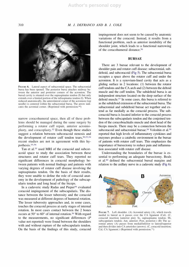
Understanding the boundaries of the bursae is es-sential to performing an adequate bursectomy. Bealset al.60 defined the subacromial bursal margins andrelation to the axillary nerve in a cadaveric study (Fig 6).
FIGURE 6. Lateral aspect of subacromial space. The subacromialbursa has been opened. The posterior bursa attaches midway be-tween the anterior and posterior corners of the acromion. Thebursal cavity is situated over the supraspinatus tendon (S) but onlyextends over a limited portion of the infraspinatus tendon (I). Whenreduced anatomically, the anterolateral corner of the acromion (topneedle) is centered within the subacromial bursa. The arrow indi-cates the acromial corner. (Reprinted with permission.60)
FIGURE 7. Left shoulder. (A) Acromial artery (A), which travelsmedial to lateral as it passes over the CA ligament (Cal). (C,coracoid insertion [anterior pin]; Ss, supraspinatus tendon; Sb,subscapularis tendon; Ant, anterior; Post, posterior.) (B) The ac-romial artery (A) passes from medial/inferior to lateral/superiorand then divides into CA arterioles (arrows). (C, coracoid insertion;Cal, CA ligament.) (Reprinted with permission.71)
310 M. J. DEFRANCO AND B. J. COLE

Author's personal copy
In general, the margins of the bursa are 2 cm ormore from the anterolateral corner of the undersurfaceof the acromion. More specifically, the mean distancefrom the anterolateral corner of the acromion to theposterior bursal cavity is 2.8 � 0.6 cm (55% of the an-teroposterior acromial length). The mean distancefrom the midpoint of the acromion to the subdeltoidbursal reflection of the subacromial bursa is 4.0 � 1.0cm. The distance between the AC joint and the medialextent of the subacromial bursa is variable. Somebursae do not cross medial to the plane of the ACjoint. However, others cross at a maximum of 2.3 mmmedial to the AC joint. The distance from the medialAC joint to the medial extent of the bursa is 0.7 � 0.7mm. Only the anterior half of the distance betweenthe anterolateral and posterolateral corners of theacromion is contained within the subacromial bursacavity. The anterolateral corner, therefore, is centralto the boundary reflections of the subacromialbursa. For this reason, the anterolateral corner is auseful landmark for placement of an arthroscopicportal into the bursa.
In the same study by Beals et al.,60 the mean dis-tance from all points of the acromion to the axillarynerve was 5 cm. The mean minimum distance fromthe subdeltoid bursal reflection to the axillary nervewas 0.8 � 0.5 cm, with a range of 0.0 to 1.4 cm. In theunelevated extremity the inferior bursal reflection wasalways cephalad to the axillary nerve. Given thesedata, surgeons should exercise caution at the inferiorboundary of the subdeltoid bursal reflection becauseof the proximity of the axillary nerve.60
Duranthon and Gagey57 performed a cadavericstudy to define the anatomy and function of the sub-deltoid bursa. An increase in thickness of the subdel-toid bursa can contribute to subacromial impingement.They identified 2 attachments for the subdeltoid bursa.The first is proximal and superficial along the entirefree border of the CA ligament and along the deepsurface of the deltoid. There are no insertions anteri-orly, posteriorly, or directly to the acromion. Thedistal attachment is to the greater tuberosity of thehumerus, lateral to the origin of the supraspinatus andinfraspinatus. The distal pouch passes just beyond thedistal deltoid attachment to the humerus. The distalend of the pouch is located at a mean of 10 mm (range,5 to 12 mm) proximal to the axillary nerve. The studyalso found anatomic continuity between the CA liga-ment and the subdeltoid bursa, which can mask theouter edge of the CA ligament. Recognition of theseanatomic findings is helpful in performing a thoroughsubacromial decompression.
In an MRI study White et al.61 analyzed the sub-acromial-subdeltoid fluid in relation to rotator cufftears. The normal subacromial-subdeltoid bursa fluidis rarely thicker than 2 mm and tends to be locatedposteriorly. An abnormal amount of fluid is suggestiveof a rotator cuff tear. More specifically, there shouldbe a high index of suspicion for a rotator cuff tearwhen the subacromial-subdeltoid thickness exceeds 3mm, when bursal fluid is present medial to the ACjoint, and when fluid is seen in the part of the bursaanterior to the humerus.61
Subacromial disease encompasses a spectrum ofdisease ranging from bursitis to adhesion formation.Rotator cuff tears are often associated with subacro-mial bursitis, and this bursitis leads to the formation ofadhesions, which contributes to impingement.62-65
Machida et al.66 studied 18 patients with shoulderpain. They found that adhesions of the subacromialbursa increase impingement between the acromionand the insertion of the rotator cuff. The adhesionsneed to be released completely during surgery of therotator cuff as part of the subacromial decompression.When an adequate release is performed, the meansubacromial pressure and mean subacromial contactarea decrease significantly.66
Funk et al.67 studied patients who were diagnosedwith a subacromial plica during arthroscopic subacro-mial decompression. The odds of impingement were
FIGURE 8. Lateral view of shoulder with deltoid removed. Dottedlines represent insertion points for the CA ligament, which hasbeen removed. The anterior capsular artery (Ca) passes through thetriangle formed by the base of the coracoid (C), anterior border ofthe supraspinatus (Ss), and coracohumeral ligament (Chl). Theanterior capsular artery is at risk for getting cut when an anteriorinterval slide (solid line) is completed by the surgeon to mobilizethe retracted tendon. (TAa, thoracoacromial artery; Aa, acromialartery; A, acromion.) (Reprinted with permission.71)
311ROTATOR CUFF ANATOMY
Author's personal copy
3.41 times higher in shoulders with a plica comparedwith shoulders without a plica. Overall, the prevalenceof subacromial plica is 6% in shoulders presentingwith subacromial impingement. The impingementchanges caused by the plica are not degenerative.They are mechanical abrasions due to rubbing be-tween the rotator cuff and the undersurface of theacromion and the CA ligament. In younger patientsthe diagnosis of plica as a reason for impingementshould be considered only after secondary impinge-ment due to instability has been ruled out.67
Overall, the bursae are essential to normal rotatorcuff function. Knowing the boundaries of the bursaeguides the ability to perform an adequate decompres-sion and to avoid injury to neurovascular structures.Inflammation within the bursae can lead to pain andshoulder dysfunction. Bursectomy and plica removalas part of a thorough subacromial decompression arerequired to help alleviate pain and to make an accurateassessment of other structures, such as the acromion,CA ligament, and rotator cuff. This opinion, however,is not without controversy. There are investigatorswho recommend bursal preservation at the time ofrotator cuff repair because of the theoretic contribu-tion of blood supply to healing at the tendo-osseousjunction.65,68-70 For example, on the basis of biopsyspecimens from 115 patients with complete rotatorcuff tears, Uhthoff and Sarkar65 suggest that exten-sive subacromial debridement including bursec-tomy should be avoided. According to them, the mainsource of fibrovascular response after a rotator cufftear is the wall of the subacromial bursa. Therefore, ifthis tissue is preserved during rotator cuff repair, itcould hypothetically contribute to tendon reconstitu-tion and remodeling.65
SUBACROMIAL VASCULATURE
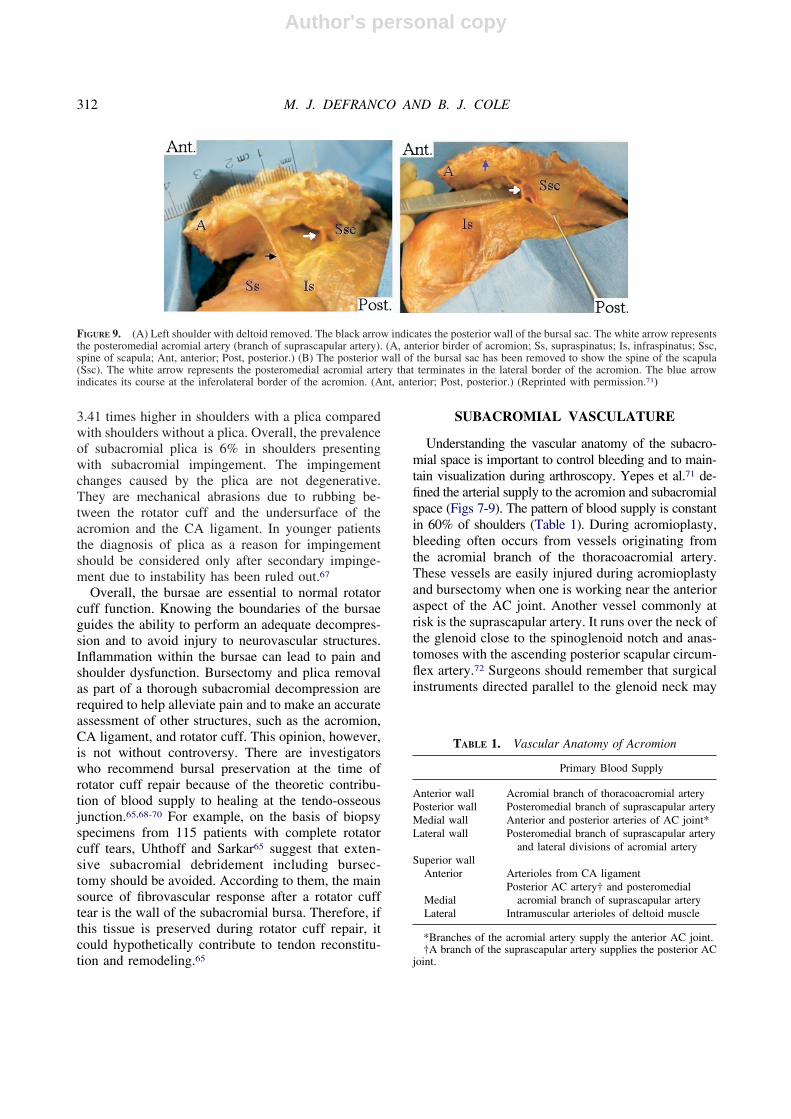
Understanding the vascular anatomy of the subacro-mial space is important to control bleeding and to main-tain visualization during arthroscopy. Yepes et al.71 de-fined the arterial supply to the acromion and subacromialspace (Figs 7-9). The pattern of blood supply is constantin 60% of shoulders (Table 1). During acromioplasty,bleeding often occurs from vessels originating fromthe acromial branch of the thoracoacromial artery.These vessels are easily injured during acromioplastyand bursectomy when one is working near the anterioraspect of the AC joint. Another vessel commonly atrisk is the suprascapular artery. It runs over the neck ofthe glenoid close to the spinoglenoid notch and anas-tomoses with the ascending posterior scapular circum-flex artery.72 Surgeons should remember that surgicalinstruments directed parallel to the glenoid neck may
FIGURE 9. (A) Left shoulder with deltoid removed. The black arrow indicates the posterior wall of the bursal sac. The white arrow representsthe posteromedial acromial artery (branch of suprascapular artery). (A, anterior birder of acromion; Ss, supraspinatus; Is, infraspinatus; Ssc,spine of scapula; Ant, anterior; Post, posterior.) (B) The posterior wall of the bursal sac has been removed to show the spine of the scapula(Ssc). The white arrow represents the posteromedial acromial artery that terminates in the lateral border of the acromion. The blue arrowindicates its course at the inferolateral border of the acromion. (Ant, anterior; Post, posterior.) (Reprinted with permission.71)
TABLE 1. Vascular Anatomy of Acromion
Primary Blood Supply
Anterior wall Acromial branch of thoracoacromial arteryPosterior wall Posteromedial branch of suprascapular arteryMedial wall Anterior and posterior arteries of AC joint*Lateral wall Posteromedial branch of suprascapular artery
and lateral divisions of acromial arterySuperior wall
Anterior Arterioles from CA ligament
MedialPosterior AC artery† and posteromedial
acromial branch of suprascapular arteryLateral Intramuscular arterioles of deltoid muscle
*Branches of the acromial artery supply the anterior AC joint.†A branch of the suprascapular artery supplies the posterior AC
joint.
312 M. J. DEFRANCO AND B. J. COLE

Author's personal copy
injure these arteries if they are advanced beyond 20mm from the glenoid rim.72
GREATER TUBEROSITY
The greater tuberosity has 3 facets (superior, mid-dle, and inferior) where rotator cuff tendons (supraspi-natus, infraspinatus, and teres minor) insert (Fig 10).Bone mineral density in this area is an importantfactor in the surgical treatment of rotator cuff tears.Osteopenia at the greater tuberosity can complicatesurgical repair and healing of the rotator cuff tendons(Fig 11). In a retrospective review of 27 patients byCadet et al.,73 there were significantly greater os-teopenic changes in the greater tuberosity in patientswith chronic retracted rotator cuff tears. According toWolff’s law,74 bone remodels in response to mechan-ical stress and this process helps determine bone massdensity. After a rotator cuff tear, the forces normallytransmitted to the greater tuberosity through the rota-tor cuff tendons are no longer present. As a result,osteopenia develops and can decrease the pulloutstrength of anchors and sutures used for rotator cuff
repair.73,75,76 Jiang et al.77 suggested that the degree ofbone loss was dependent on the nature of the rotatorcuff tear. In other words, full-thickness tears result inmore bone loss than partial-thickness tears.77,78 Previ-ous studies have defined intraoperative strategies forimproving fixation in osteopenic bone.75 Overall,proximal humerus bone mineral density should beevaluated preoperatively as a factor relevant to theclinical outcome of patients undergoing rotator cuffrepair.
ROTATOR CUFF FOOTPRINT
Understanding the insertional anatomy of the rota-tor cuff tendons is important not only in diagnosingthe extent of rotator cuff tears but also in repairingthem correctly. Table 2 outlines the recent studies thatdefine the dimensions of the rotator cuff insertionsonto the proximal humerus. This area is known as therotator cuff footprint.79,84 It is the basis for anatomicrepair of the rotator cuff (Figs 12-14).
Using cadaveric shoulders, Minagawa et al.80 de-scribed the insertional anatomy (width only) of thesupraspinatus and infraspinatus tendons in referenceto the greater tuberosity. According to their measure-ments, the supraspinatus tendon attaches to the supe-rior facet and the superior half of the middle facet ofthe greater tuberosity. The infraspinatus attaches to
FIGURE 11. Anteroposterior radiograph of a tight shoulder show-ing osteopenia of humeral head (arrows) in a patient with rotatorcuff tear. (Reprinted with permission.73)
FIGURE 10. Proximal humerus, showing 3 facets of greater tuber-osity (superior [S], middle [M], and inferior [I]). (Reprinted withpermission.80)
313ROTATOR CUFF ANATOMY

Author's personal copy
the entire middle facet and covers part of the supraspi-natus tendon. More specifically, Dugas et al.79 definedthe mean anteroposterior distance across the rotatorcuff insertion onto the greater tuberosity as 37.8 mm.The mean minimum medial-to-lateral distance acrossthe rotator cuff insertion onto the greater tuberosity is14.7 mm. The mean area of insertion of the supraspi-natus, infraspinatus, and teres minor onto the greatertuberosity is 6.2 cm2. In conclusion, they suggest thatrecreating this normal anatomic area at the time ofsurgery increases the likelihood of normal healing andsubsequent normal function.79
Volk and Vangsness85 used cadaveric shoulders todefine the insertional anatomy of the supraspinatus.The primary measurement was the length of the ante-rior and posterior tendinous and muscular portionsfrom the lateral insertion. The anterior lateral portionof the supraspinatus had more tendon, which in one
third of the specimens was associated with separatemuscle fibers. Posteriorly, the tendinous portion of thelateral supraspinatus muscle did not extend as farmedially from its insertion at the greater tuberosity.The authors suggest that the consistent anterior tendi-nous portion of the supraspinatus (5.4 cm in length)may provide a firm area for rotator cuff repair orrotator interval closure.
In a cadaveric study, Roh et al.86 reported that theanterior muscle belly of the supraspinatus is largerthan the posterior one. The larger anterior portion ofthe supraspinatus is primarily responsible for armabduction and humeral head depression. Therefore, arotator cuff tear in this area results in a loss of func-tional tendon length as well as an inability to transmitcontractile loads to the humerus to perform thesefunctions. In addition, they propose that the largeranterior muscle pulls through a smaller cross-sectionalarea of tendon. As a result, the anterior portion of thetendon experiences more stress. Therefore, during ro-tator cuff surgery, this area should be incorporatedinto the repair because it transmits most of the con-tractile load and will allow for the best functionaloutcome.86
Boon et al.83 performed a cadaveric study to look atthe extension of the supraspinatus tendon into thesubscapularis. The subscapularis extends over the bi-cipital groove interdigitating with the supraspinatusover the greater tuberosity of the humerus. The direc-tion of the subscapularis over the lesser tuberosity andthe direction of the tendon of the supraspinatus towardthe bicipital groove facilitate their biomechanicalfunction of stabilizing the shoulder joint. The area ofinterdigitation between the subscapularis and the su-praspinatus may become disrupted as part of a rotatorcuff tear. In such cases the tendons should be re-aligned and sutured to provide additional strength tothe rotator cuff repair.83
TABLE 2. Dimensions of Rotator Cuff
Author
Rotator Cuff Dimensions (mm) (Mean Length � Width)
Subscapularis Supraspinatus Infraspinatus Teres Minor
Minagawa et al.80 NA NA � 22.5 � 3.1 NA � 14.1 � 3.9 NAVolk and Vangsness85 NA 27.9 � NA NA NARoh et al.86 NA NA � 21.2 NA NADugas et al.79 24 � 18 16 � 12 16 � 13 20 � 11Ruotolo et al.82 NA NA � 25 NA NACurtis et al.84 40 � 20 23 � 16 29 � 19 29 � 21
Abbreviation: NA, not available.
FIGURE 12. Insertion points of rotator cuff tendons. The supraspi-natus (SSP) inserts onto the superior facet of the greater tuberosityand the superior half of the middle facet. The infraspinatus tendon(ISP) is attached to the entire length of the middle facet coveringthe posterior half of the supraspinatus. (TM, teres minor; SSC,subscapularis; S, superior; M, middle; I, inferior.) (Reprinted withpermission.80)
314 M. J. DEFRANCO AND B. J. COLE
Author's personal copy
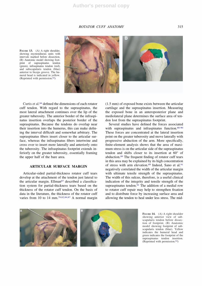
Curtis et al.84 defined the dimensions of each rotatorcuff tendon. With regard to the supraspinatus, themost lateral attachment continues over the lip of thegreater tuberosity. The anterior border of the infraspi-natus insertion overlaps the posterior border of thesupraspinatus. Because the tendons do overlap neartheir insertion into the humerus, this can make defin-ing the interval difficult and somewhat arbitrary. Thesupraspinatus fibers insert closer to the articular sur-face, whereas the infraspinatus fibers intertwine andcross over to insert more laterally and anteriorly ontothe tuberosity. The infraspinatus footprint extends in-feriorly on the greater tuberosity, essentially framingthe upper half of the bare area.
ARTICULAR SURFACE MARGIN
Articular-sided partial-thickness rotator cuff tearsdevelop at the attachment of the tendon just lateral tothe articular margin. Ellman87 described a classifica-tion system for partial-thickness tears based on thethickness of the rotator cuff tendon. On the basis ofdata in the literature, the thickness of the rotator cuffvaries from 10 to 14 mm.79,82,84,87 A normal margin
(1.5 mm) of exposed bone exists between the articularcartilage and the supraspinatus insertion. Measuringthe exposed bone in an anteroposterior plane andmediolateral plane determines the surface area of ten-don lost from the supraspinatus footprint.
Several studies have defined the forces associatedwith supraspinatus and infraspinatus function.88-90
These forces are concentrated at the lateral insertionpoint on the greater tuberosity and move laterally withprogressive abduction of the arm. More specifically,finite-element analysis shows that the area of maxi-mum stress is on the articular side of the supraspinatustendon and shifts closer to its insertion at 60° ofabduction.89 The frequent finding of rotator cuff tearsin this area may be explained by its high concentrationof stress with arm elevation.89 Indeed, Sano et al.91
negatively correlated the width of the articular marginwith ultimate tensile strength of the supraspinatus.The width of this sulcus, therefore, is a useful clinicalindication of the integrity and tensile strength of thesupraspinatus tendon.91 The addition of a medial rowto rotator cuff repair may help to strengthen fixationand to distribute force by increasing surface area andallowing the tendon to heal under less stress. The mid-
FIGURE 13. (A) A right shoulder,showing myotendinous units withintervals marked before dissection.(B) Anatomic model showing foot-print of supraspinatus tendon(green), infraspinatus tendon (red),and subscapularis tendon (blue)anterior to biceps groove. The hu-meral head is indicated in yellow.(Reprinted with permission.84)
FIGURE 14. (A) A right shouldershowing anterior view of sub-scapularis tendon before dissec-tion of footprint. (B) Anatomicmodel showing footprint of sub-scapularis tendon (blue). Yellowindicates the humeral head andgreen indicates the footprint of thesupraspinatus tendon insertion.(Reprinted with permission.84)
315ROTATOR CUFF ANATOMY

Author's personal copy
point of tendon insertion can be moved up to 10 mmmedially with no resultant negative biomechanical con-sequences.92 Overall, the greater extent to which a givenrepair covers the healing zone (footprint), the greater thechance for tendon-bone healing.
The relation between the insertion of the rotator cuffand the articular surface varies in the literature. Ac-cording to Dugas et al.,79 the distance from the artic-ular margin to the most medial rotator cuff fibers wasless than 1 mm along the anterior-most 2.1 cm of therotator cuff insertion onto the greater tuberosity. Cur-tis et al.84 reported that the subscapularis inserts ontothe lesser tuberosity adjacent to the biceps groove atthe edge of the articular surface. It tapers from 0 mmsuperiorly to 18 mm at its inferior border. The su-praspinatus inserts at the articular surface along itsentire insertion from the bicipital groove to the top ofthe bare area. The insertion appears at a mean of 0.9mm (range, 0 to 4 mm) from the edge of the articularsurface. In most cases the supraspinatus inserts di-rectly onto the articular surface through the entirelength of the tendon. The infraspinatus wraps theposterior border of the supraspinatus superiorly at thearticular surface. It tapers from the articular margin 0mm superiorly to 16 mm inferiorly. The gap betweenthe articular surface and inferior insertion forms the“bare area.”84
Ruotolo et al.82 defined the mean distance from thearticular cartilage to the supraspinatus footprint (ten-dinous attachment) as 1.7 mm (1.9 mm at the rotatorinterval, 1.5 mm at the midtendon, and 1.8 mm at theposterior edge). They define all tears with more than 7mm of exposed bone lateral to the articular cartilageedge as significant (approximately 50% of the tendonsubstance) and suggest that they be repaired.
LANDMARKS
Reliable landmarks are helpful in evaluating normalanatomy as well as defining the characteristics (ten-don, location, dimensions) of rotator cuff tears. Oneuseful landmark is the anterior margin of the greatertuberosity. Measuring from the most anterior aspect ofthe greater tuberosity, the initial 12.6 � 1.1 mm ofrotator cuff is supraspinatus. The next 9.8 � 3.2 mmconsists of supraspinatus and infraspinatus ten-don.80,82 The most posterior portion (12.9 � 3.2 mm)consists only of the infraspinatus tendon. More spe-cifically, the superior facet of the greater tuberosity isa useful landmark because only the supraspinatus ten-don attaches to it. However, in cases of degenerativejoint disease, the superior facet may not be readily
identifiable. Therefore the distance between the ante-rior margin of the greater tuberosity and the anteriormargin of the infraspinatus tendon (12.6 � 1.1 mm)can be used to determine whether the infraspinatus isinvolved in the tear.80,82 Likewise, the supraspinatusand infraspinatus overlap at the superior aspect of themiddle facet. When a full-thickness tear occurs in thisarea or more than 12.6 mm from the anterior margin ofthe greater tuberosity, it means that the tear involvesboth the supraspinatus and infraspinatus tendons.80,82
Curtis et al.84 used 3 easily identifiable landmarks toassess the rotator cuff. The landmarks are the bicepsgroove, the articular surface, and the bare area. Thebare area is created as the infraspinatus and teresminor taper laterally away from the articular margin.More specifically, there is a 5- to 10-mm area wherethe infraspinatus fibers overlap the supraspinatus fi-bers and insert more laterally and anteriorly onto thegreater tuberosity. This area of overlap is just anteriorto the tip of the bare area, which is an arthroscopiclandmark for the interval between the supraspinatusand infraspinatus. The infraspinatus extends inferiorlyon the greater tuberosity, framing the upper half of thebare area. Minagawa et al.80 also described the sulcusbetween these 2 tendons as a useful landmark. Theydescribed its location as slightly posterior (4.3 � 2.4mm) to the posterior margin of the supraspinatustendon.
SUPRASCAPULAR AND AXILLARYNERVES
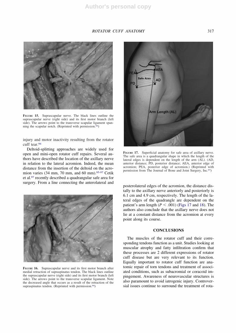
Damage to the suprascapular nerve during lateralmobilization (�3 cm) for repair of a rotator cufftendon places the suprascapular nerve at risk for in-jury. It may also explain the inability to regainstrength in the supraspinatus and infraspinatus mus-cles postoperatively.93 A recent study suggests thatmedial retraction of a torn rotator cuff may also injurethe suprascapular nerve (Figs 15 and 16).94 Retractionof a large or massive rotator cuff tear may change thecourse of the suprascapular nerve through the supra-scapular notch. This change may lead to increasedtension and cause a traction injury to the suprascapularnerve. In a cadaveric study Albritton et al.94 showedthat medial retraction of a rotator cuff tear (supraspi-natus and infraspinatus) decreases the angle betweenthe main trunk of the suprascapular nerve and its firstmotor branch. The nerve tension that develops maycontribute to the development of atrophy in the musclebellies of the supraspinatus and infraspinatus. Thisatrophy is most likely due to the combination of nerve
316 M. J. DEFRANCO AND B. J. COLE
Author's personal copy
injury and motor inactivity resulting from the rotatorcuff tear.94
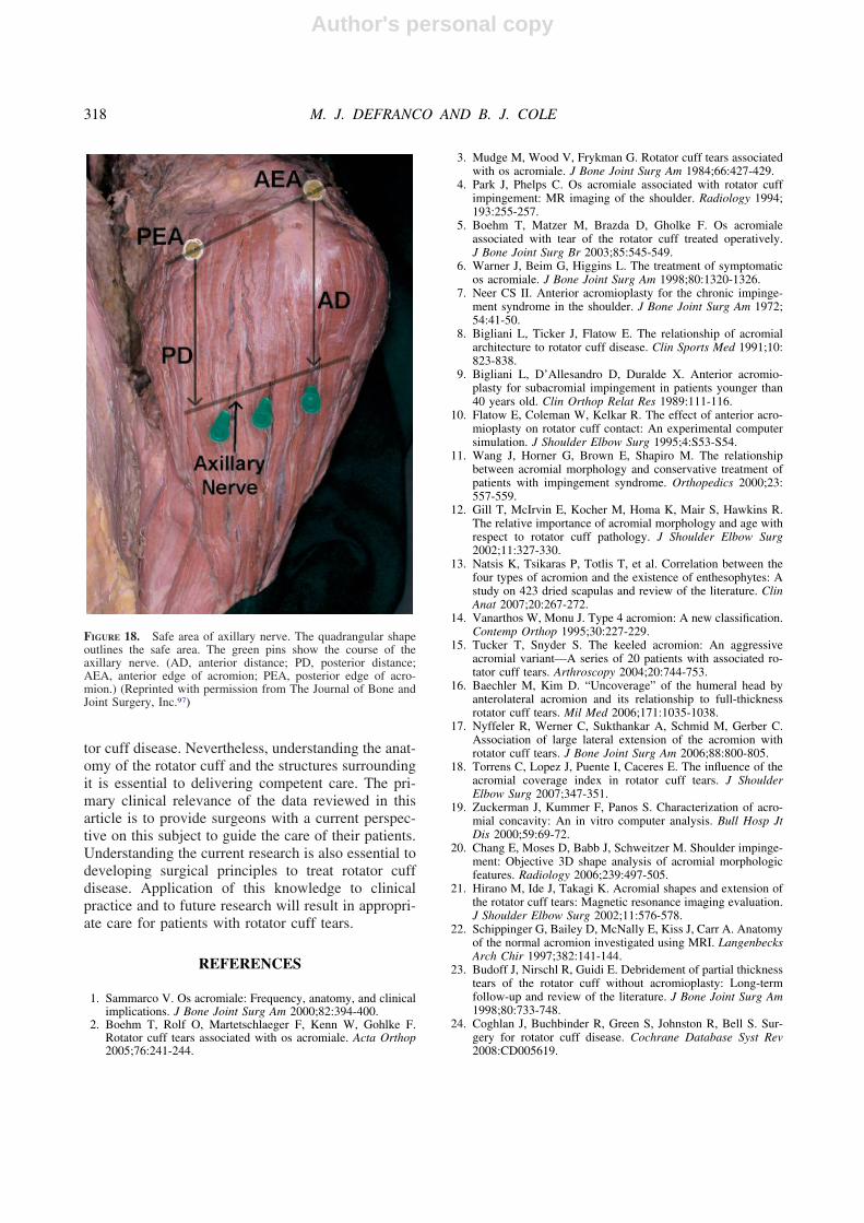
Deltoid-splitting approaches are widely used foropen and mini-open rotator cuff repairs. Several au-thors have described the location of the axillary nervein relation to the lateral acromion. Indeed, the meandistance from the insertion of the deltoid on the acro-mion varies (34 mm, 70 mm, and 60 mm).95-97 Cetiket al.97 recently described a quadrangular safe area forsurgery. From a line connecting the anterolateral and
posterolateral edges of the acromion, the distance dis-tally to the axillary nerve anteriorly and posteriorly is6.1 cm and 4.9 cm, respectively. The length of the la-teral edges of the quadrangle are dependent on thepatient’s arm length (P � .001) (Figs 17 and 18). Theauthors also conclude that the axillary nerve does notlie at a constant distance from the acromion at everypoint along its course.
CONCLUSIONS
The muscles of the rotator cuff and their corre-sponding tendons function as a unit. Studies looking atmuscular atrophy and fatty infiltration confirm thatthese processes are 2 different expressions of rotatorcuff disease but are very relevant to its function.Equally important to rotator cuff function are ana-tomic repair of torn tendons and treatment of associ-ated conditions, such as subacromial or coracoid im-pingement. Awareness of neurovascular structures isalso paramount to avoid iatrogenic injury. Controver-sial issues continue to surround the treatment of rota-
FIGURE 15. Suprascapular nerve. The black lines outline thesuprascapular nerve (right side) and its first motor branch (leftside). The arrows point to the transverse scapular ligament span-ning the scapular notch. (Reprinted with permission.94)
FIGURE 16. Suprascapular nerve and its first motor branch aftermedial retraction of supraspinatus tendon. The black lines outlinethe suprascapular nerve (right side) and its first motor branch (leftside). The arrows point to the transverse scapular ligament. Notethe decreased angle that occurs as a result of the retraction of thesupraspinatus tendon. (Reprinted with permission.94)
FIGURE 17. Superficial anatomy for safe area of axillary nerve.The safe area is a quadrangular shape in which the length of thelateral edges is dependent on the length of the arm (AL). (AD,anterior distance; PD, posterior distance; AEA, anterior edge ofacromion; PEA, posterior edge of acromion.) (Reprinted withpermission from The Journal of Bone and Joint Surgery, Inc.97)
317ROTATOR CUFF ANATOMY
Author's personal copy
tor cuff disease. Nevertheless, understanding the anat-omy of the rotator cuff and the structures surroundingit is essential to delivering competent care. The pri-mary clinical relevance of the data reviewed in thisarticle is to provide surgeons with a current perspec-tive on this subject to guide the care of their patients.Understanding the current research is also essential todeveloping surgical principles to treat rotator cuffdisease. Application of this knowledge to clinicalpractice and to future research will result in appropri-ate care for patients with rotator cuff tears.
REFERENCES
1. Sammarco V. Os acromiale: Frequency, anatomy, and clinicalimplications. J Bone Joint Surg Am 2000;82:394-400.
2. Boehm T, Rolf O, Martetschlaeger F, Kenn W, Gohlke F.Rotator cuff tears associated with os acromiale. Acta Orthop2005;76:241-244.
3. Mudge M, Wood V, Frykman G. Rotator cuff tears associatedwith os acromiale. J Bone Joint Surg Am 1984;66:427-429.
4. Park J, Phelps C. Os acromiale associated with rotator cuffimpingement: MR imaging of the shoulder. Radiology 1994;193:255-257.
5. Boehm T, Matzer M, Brazda D, Gholke F. Os acromialeassociated with tear of the rotator cuff treated operatively.J Bone Joint Surg Br 2003;85:545-549.
6. Warner J, Beim G, Higgins L. The treatment of symptomaticos acromiale. J Bone Joint Surg Am 1998;80:1320-1326.
7. Neer CS II. Anterior acromioplasty for the chronic impinge-ment syndrome in the shoulder. J Bone Joint Surg Am 1972;54:41-50.
8. Bigliani L, Ticker J, Flatow E. The relationship of acromialarchitecture to rotator cuff disease. Clin Sports Med 1991;10:823-838.
9. Bigliani L, D’Allesandro D, Duralde X. Anterior acromio-plasty for subacromial impingement in patients younger than40 years old. Clin Orthop Relat Res 1989:111-116.
10. Flatow E, Coleman W, Kelkar R. The effect of anterior acro-mioplasty on rotator cuff contact: An experimental computersimulation. J Shoulder Elbow Surg 1995;4:S53-S54.
11. Wang J, Horner G, Brown E, Shapiro M. The relationshipbetween acromial morphology and conservative treatment ofpatients with impingement syndrome. Orthopedics 2000;23:557-559.
12. Gill T, McIrvin E, Kocher M, Homa K, Mair S, Hawkins R.The relative importance of acromial morphology and age withrespect to rotator cuff pathology. J Shoulder Elbow Surg2002;11:327-330.
13. Natsis K, Tsikaras P, Totlis T, et al. Correlation between thefour types of acromion and the existence of enthesophytes: Astudy on 423 dried scapulas and review of the literature. ClinAnat 2007;20:267-272.
14. Vanarthos W, Monu J. Type 4 acromion: A new classification.Contemp Orthop 1995;30:227-229.
15. Tucker T, Snyder S. The keeled acromion: An aggressiveacromial variant—A series of 20 patients with associated ro-tator cuff tears. Arthroscopy 2004;20:744-753.
16. Baechler M, Kim D. “Uncoverage” of the humeral head byanterolateral acromion and its relationship to full-thicknessrotator cuff tears. Mil Med 2006;171:1035-1038.
17. Nyffeler R, Werner C, Sukthankar A, Schmid M, Gerber C.Association of large lateral extension of the acromion withrotator cuff tears. J Bone Joint Surg Am 2006;88:800-805.
18. Torrens C, Lopez J, Puente I, Caceres E. The influence of theacromial coverage index in rotator cuff tears. J ShoulderElbow Surg 2007;347-351.
19. Zuckerman J, Kummer F, Panos S. Characterization of acro-mial concavity: An in vitro computer analysis. Bull Hosp JtDis 2000;59:69-72.
20. Chang E, Moses D, Babb J, Schweitzer M. Shoulder impinge-ment: Objective 3D shape analysis of acromial morphologicfeatures. Radiology 2006;239:497-505.
21. Hirano M, Ide J, Takagi K. Acromial shapes and extension ofthe rotator cuff tears: Magnetic resonance imaging evaluation.J Shoulder Elbow Surg 2002;11:576-578.
22. Schippinger G, Bailey D, McNally E, Kiss J, Carr A. Anatomyof the normal acromion investigated using MRI. LangenbecksArch Chir 1997;382:141-144.
23. Budoff J, Nirschl R, Guidi E. Debridement of partial thicknesstears of the rotator cuff without acromioplasty: Long-termfollow-up and review of the literature. J Bone Joint Surg Am1998;80:733-748.
24. Coghlan J, Buchbinder R, Green S, Johnston R, Bell S. Sur-gery for rotator cuff disease. Cochrane Database Syst Rev2008:CD005619.
FIGURE 18. Safe area of axillary nerve. The quadrangular shapeoutlines the safe area. The green pins show the course of theaxillary nerve. (AD, anterior distance; PD, posterior distance;AEA, anterior edge of acromion; PEA, posterior edge of acro-mion.) (Reprinted with permission from The Journal of Bone andJoint Surgery, Inc.97)
318 M. J. DEFRANCO AND B. J. COLE

Author's personal copy
25. McCallister W, Parsons I, Titelman R, Matsen F. Open rotatorcuff repair without acromioplasty. J Bone Joint Surg Am2005;87:1278-1283.
26. Hyvonen P, Lohi S, Javovaara P. Open acromioplasty does notprevent the progression of an impingement syndrome to a tear.Nine year follow-up of 96 cases. J Bone Joint Surg Br 1998;80:813-816.
27. Weber S. Arthroscopic debridement and acromioplasty versusmini-open repair in the treatment of significant partial thick-ness rotator cuff tears. Arthroscopy 1999;15:126-131.
28. Flatow E, Soslowsky L, Ticker J, et al. Excursion of the rotatorcuff under the acromion. Patterns of subacromial contact. Am JSports Med 1994;22:779-788.
29. Green A, Griggs S, Labrador D. Anterior acromial anatomy:Relevance to arthroscopic acromioplasty. Arthroscopy 2004;20:1050-1054.
30. Petersilge C, Witte D, Sewell B, Bosch E, Resnick D. Normalregional anatomy of the shoulder. Magn Reson Imaging Clin NAm 1997;5:667-681.
31. Matthews L, Fadale P. Subacromial anatomy for the arthro-scopist. Arthroscopy 1989;5:36-40.
32. Fealy S, April E, Khazzam M, Armengol-Barallat J, BiglianiL. The coracoacromial ligament: Morphology and study ofacromial enthesopathy. J Shoulder Elbow Surg 2005;14:542-548.
33. Pieper H, Radas C, Krahl H, Blank M. Anatomic variation ofthe coracoacromial ligament: A macroscopic and microscopiccadaveric study. J Shoulder Elbow Surg 1997;6:291-296.
34. Kesmezacar H, Akgun I, Ogut T, Gokay S, Uzun I. Thecoracoacromial ligament: The morphology and relation to ro-tator cuff pathology. J Shoulder Elbow Surg 2008;17:182-188.
35. Fremerey R, Bastian L, Siebert W. The coracoacromial liga-ment: Anatomical and biomechanical properties with respectto age and rotator cuff disease. Knee Surg Sports TraumatolArthrosc 2000;8:309-313.
36. Dines D, Warren R, Inglis A, Pavlov H. The coracoid impinge-ment syndrome. J Bone Joint Surg Br 1990;72:314-316.
37. Edelson J. The hooked acromion. J Bone Joint Surg Br 1995;77:284-287.
38. Mallon W, Brown H, Vogler J, Martinez S. Radiographic andgeometric anatomy of the scapula. Clin Orthop Relat Res1992:142-145.
39. Anetzberger H, Putz R. Morphometry of the subacromialspace and its clinical relevance. Unfallchirurg 1995;98:407-414 (in German).
40. Gumina S, Postacchini F, Orsina L, Cinotti G. The morphom-etry of the coracoid process—Its aetiologic role in subcoracoidimpingement syndrome. Int Orthop 1999;23:198-201.
41. Bhatia D, deBeer J, du Toit D. Coracoid process anatomy:Implications in radiographic imaging and surgery. Clin Anat2007;20:774-784.
42. Kleist K, Freehill M, Hamilton L, Buss D, Fritts H. Computedtomography analysis of the coracoid process and anatomicstructures of the shoulder after arthroscopic coracoid decom-pression: A cadaveric study. J Shoulder Elbow Surg 2007;16:245-250.
43. Dumontier C, Sautet A, Gagey O, Apoil A. Rotator intervallesions and their relation to coracoid impingement syndrome.J Shoulder Elbow Surg 1999;8:130-135.
44. Ferrick M. Coracoid impingement. A case report and review ofthe literature. Am J Sports Med 2000;28:117-119.
45. Gerber C, Terrier F, Ganz R. The role of the coracoid processin the chronic impingement syndrome. J Bone Joint Surg Br1985;67:703-708.
46. Paulson M, Watnik N, Dines D. Coracoid impingement syn-drome, rotator interval reconstruction, and biceps tenodesis inthe overhead athlete. Orthop Clin North Am 2001;32:485-493.
47. Ferreira Neto A, Almeida A, Maiorino R, Zoppi Filho A,Benegas E. An anatomical study of the subcoracoid space.Clinics 2006;61:467-472.
48. Friedman R, Bonutti P, Genez B. Cine magnetic resonanceimaging of the subcoracoid region. Orthopedics 1998;21:545-548.
49. Schulz C, Anetzberger H, Glaser C. Coracoid tip position onfrontal radiographs of the shoulder: A predictor of commonshoulder pathologies. Br J Radiol 2005;78:1005-1008.
50. Richards D, Burkhart S, Campbell S. Relation between narrowedcoracohumeral distance and subscapularis tears. Arthroscopy2005;21:1223-1228.
51. Kragh J Jr, Doukas W, Basamania C. Primary coracoid im-pingement syndrome. Am J Orthop 2004;5:229-232.
52. Suenaya N, Minami A, Kaneda K. Postoperative subcoracoidimpingement syndrome in patients with rotator cuff repair. JShoulder Elbow Surg 2000;9:275-278.
53. Colas F, Nevoux J, Gagey O. The subscapular and subcoracoidbursae: Descriptive and functional anatomy. J Shoulder ElbowSurg 2004;13:454-458.
54. Lo I, Parten P, Burkhart S. Combined subcoracoid and sub-acromial impingement in association with anterosuperior ro-tator cuff tears: An arthroscopic approach. Arthroscopy 2003;19:1068-1078.
55. Tan V, Moore R Jr, Omarini L, Kneeland B, Williams G Jr,Iannotti J. Magnetic resonance imaging analysis of coracoidmorphology and its relation to rotator cuff tears. Am J Orthop2002;31:329-333.
56. Radas C, Pieper H. The coracoid impingement of the subscap-ularis tendon: A cadaveric study. J Shoulder Elbow Surg2004;13:154-159.
57. Duranthon L, Gagey O. Anatomy and function of the subdel-toid bursa. Surg Radiol Anat 2001;23:23-25.
58. Horwitz M, Tocantins L. An anatomical study of the role ofthe long thoracic nerve and the related scapular bursae in thepathogenesis of local paralysis of the serratus anterior muscle.Anat Rec 1938;71:375-385.
59. Voloshin I, Gelinas J, Maloney M, O’Keefe R, Bigliani L,Blaine T. Proinflammatory cytokines and metalloproteases areexpressed in the subacromial bursa in the patients with rotatorcuff disease. Arthroscopy 2005;21:1076.e1-1076.e9. Availableonline at www.arthroscopyjournal.org.
60. Beals T, Harryman D, Lazarus M. Useful boundaries of thesubacromial bursa. Arthroscopy 1998;14:465-470.
61. White E, Schweitzer M, Haims A. Range of normal andabnormal subacromial/subdeltoid bursa fluid. J Comput AssistTomogr 2006;30:316-320.
62. Fukuda H, Hamada K, Nakajima T, Tomonaga A. Pathologyand pathogenesis of the intratendinous tearing of the rotatorcuff viewed from en bloc histologic sections. Clin OrthopRelat Res 1994:60-67.
63. Fukuda H. The management of partial-thickness tears of therotator cuff. J Bone Joint Surg Br 2003;85:3-11.
64. Payne L, Deng X, Craig E, Torzilli P, Warren R. The com-bined dynamic and static contributions to subacromial im-pingement. Am J Sports Med 1997;25:801-808.
65. Uhthoff H, Sarkar K. Surgical repair of rotator cuff tendons:The importance of the subacromial bursa. J Bone Joint Surg Br1991;73:399-401.
66. Machida A, Sugamoto K, Miyamoto T, Inui H, Watanabe T,Yoshikawa H. Adhesion of the subacromial bursa may causesubacromial impingement in patients with rotator cuff tears:Pressure measurements in 18 patients. Acta Orthop Scand2004;75:109-113.
67. Funk L, Levy O, Even T, Copeland S. Subacromial plica as acause of impingement in the shoulder. J Shoulder Elbow Surg2006;15:697-700.
68. Bateman J. The shoulder and neck. Ed 2. Philadelphia: WBSaunders, 1978;286.
319ROTATOR CUFF ANATOMY
Author's personal copy
69. Hanamura H, Yahuhara N, Maeda H. Surgical approach torotator cuff tears: Posterior displacement acromioplasty. In:Takagishi N, ed. The shoulder. Tokyo: Professional Postgrad-uate Services, 1987;231-240.
70. Watson M. Practical shoulder surgery. London: Grune &Stratton, 1985;199-224.
71. Yepes H, Al-Hibshi A, Tang M, Morris S, Stanish W. Vascularanatomy of the subacromial space: A map of bleeding pointsfor the arthroscopic surgeon. Arthroscopy 2007;23:978-984.
72. Andary J, Peterson S. The vascular anatomy of the glenohu-meral capsule and ligaments: An anatomic study. J Bone JointSurg Am 2002;84:2258-2265.
73. Cadet E, Hsu J, Levine W, Bigliani L, Ahmad C. The relationshipbetween greater tuberosity osteopenia and the chronicity of rota-tor cuff tears. J Shoulder Elbow Surg 2008;17:73-77.
74. Wolff J. Das Gesetz der Transformation der Knochen. Berlin:Hirschwald Verlag, 1892.
75. Tingart M, Apreleva M, Lehtinen J, Zurakowski D, Warner J.Anchor design and bone mineral density affect the pull-outstrength of suture anchors in rotator cuff repair: Which anchorsare best to use in patients with low bone quality? Am J SportsMed 2004;32:1466-1473.
76. Tingart M, Apreleva M, Zurakowski D, Warner JJ. Pulloutstrength of suture anchors used in rotator cuff repair. J BoneJoint Surg Am 2003;85:2190-2198.
77. Jiang Y, Zhao J, van Holsbeeck M. Trabecular microstructureand surface changes in the greater tuberosity in rotator cufftears. Skeletal Radiol 2002;31:252-258.
78. Meyer D, Fucentese S, Koller B, Gerber C. Association ofosteopenia of the humeral head with full-thickness rotator cufftears. J Shoulder Elbow Surg 2004;13:333-337.
79. Dugas J, Campbell D, Warren R, Robie B, Millet P. Anatomyand dimensions of the rotator cuff. J Shoulder Elbow Surg2002;11:498-503.
80. Minagawa H, Itoi E, Konno N, et al. Humeral attachment ofthe supraspinatus and infraspinatus tendons: An anatomicstudy. Arthroscopy 1998;14:302-306.
81. Tierney J, Curtis A, Kowalik D, Scheller A. The footprint ofthe rotator cuff. Arthroscopy 1999;15:556-557.
82. Ruotolo C, Fow J, Nottage W. The supraspinatus footprint: Ananatomic study of the supraspinatus insertion. Arthroscopy2004;20:246-249.
83. Boon J, de Beer M, Botha D, Maritz N, Fouche A. Theanatomy of the subscapularis tendon insertion as applied torotator cuff repair. J Shoulder Elbow Surg 2004;13:165-169.
84. Curtis A, Burbank K, Tierney J, Scheller A, Curran A. The
insertional footprint of the rotator cuff: An anatomic study.Arthroscopy 2006;22:603-609.
85. Volk A, Vangsness C. An anatomic study of the supraspinatusmuscle and tendon. Clin Orthop Relat Res 2001:280-285.
86. Roh M, Wang V, April E, Pollock R, Bigliani L, Flatow E.Anterior and posterior musculotendinous anatomy of the su-praspinatus. J Shoulder Elbow Surg 2000;9:436-440.
87. Ellman H. Diagnosis and treatment of incomplete rotator cufftears. Clin Orthop Relat Res 1990:64-74.
88. Hughes R, An K. Force analysis of rotator cuff muscles. ClinOrthop Relat Res 1996:75-83.
89. Wakabayashi I, Itoi E, Sano H, et al. Mechanical environmentof the supraspinatus tendon: A two-dimensional finite elementmodel analysis. J Shoulder Elbow Surg 2003;12:612-617.
90. Juul-Kristensen B, Bojsen-Moller F, Finsen L, et al. Musclesizes and moment arms of rotator cuff muscles determined bymagnetic resonance imaging. Cell Tissues Organs 2000;167:214-222.
91. Sano H, Uhthoff H, Backman D. Structural disorders at theinsertion of the supraspinatus tendon and relation to tensilestrength. J Bone Joint Surg Br 1998;80:720-725.
92. Liu S. Arthroscopically assisted rotator cuff repair. J BoneJoint Surg Br 1994;76:592-595.
93. Warner J, Krushell R, Masquelet A, Gerber C. Anatomy andrelationships of the suprascapular nerve: Anatomical constraintsto mobilization of the supraspinatus and infraspinatus muscles inthe management of massive rotator cuff tears. J Bone Joint SurgAm 1992;74:36-45.
94. Albritton M, Graham R, Richards R, Basamania C. An ana-tomic study of the effects on the suprascapular nerve due toretraction of the supraspinatus muscle after a rotator cuff tear.J Shoulder Elbow Surg 2003;12:497-500.
95. Bigliani L, Flatow E, Pollock R. Fractures of the proximalhumerus. In: Rockwood CA Jr, Green D, Bucholz R, HeckmanJ, eds. Rockwood and Green’s fractures in adults. Ed 4, Vol 1.Philadelphia: Lippincott-Raven, 1996;1055-1107.
96. Riemer B, D’Ambrosia R. The risk of injury to the axillarynerve, artery and vein from the proximal locking screws ofhumeral intramedullary nails. Orthopedics 1992;15:697-699.
97. Cetik O, Uslu M, Acar H, Comert A, Tekdemir I, Cift H. Isthere a safe area for the axillary nerve in the deltoid muscle?J Bone Joint Surg Am 2006;88:2395-2399.
98. Schraner AB, Major NM. MR imaging of the subcoracoidbursa. AJR Am J Roentgenol 1999;172:1567-1571.
99. Wright R, Heller M, Quick D, Buss D. Arthroscopic decom-pression for impingement syndrome secondary to unstable osacromiale. Arthroscopy 2000;16:595-599.
320 M. J. DEFRANCO AND B. J. COLE