author's personal copy - serveur bictel/e...
TRANSCRIPT

This article appeared in a journal published by Elsevier. The attachedcopy is furnished to the author for internal non-commercial researchand education use, including for instruction at the authors institution
and sharing with colleagues.
Other uses, including reproduction and distribution, or selling orlicensing copies, or posting to personal, institutional or third party
websites are prohibited.
In most cases authors are permitted to post their version of thearticle (e.g. in Word or Tex form) to their personal website orinstitutional repository. Authors requiring further information
regarding Elsevier’s archiving and manuscript policies areencouraged to visit:
http://www.elsevier.com/copyright

Author's personal copy
Toxicology Letters 193 (2010) 41–49
Contents lists available at ScienceDirect
Toxicology Letters
journa l homepage: www.e lsev ier .com/ locate / tox le t
Ototoxic drugs: Difference in sensitivity between mice and guinea pigs
A.L. Poirriera,b,∗, P. Van den Ackervekenb, T.S. Kimc, R. Vandenboschb, L. Nguyenb,P.P. Lefebvrea, B. Malgrangeb
a Department of Otolaryngology, University Hospital of Liège, Av. de l’Hopital 1 (B35), 4000 Liège, Belgiumb GIGA-Neurosciences, Developmental Neurobiology Unit, University of Liège, Av. de l’Hopital 1 (B36), 4000 Liège, Belgiumc Department of Otolaryngology-Head and Neck Surgery, Graduate School of Medicine, Kyoto University, Kyoto, Japan
a r t i c l e i n f o
Article history:Received 8 September 2009Received in revised form 2 December 2009Accepted 6 December 2009Available online 14 December 2009
Keywords:OtotoxicityAminoglycosideCisplatinMouseGuinea pig
a b s t r a c t
The development of experimental animal models has played an invaluable role in understanding themechanisms of neurosensory deafness and in devising effective treatments. The purpose of this studywas to develop an adult mouse model of ototoxic drug-induced hearing loss and to compare the oto-toxicity in the adult mouse to that in the well-described guinea pig model. Mice are a powerful modelorganism, especially due to the large availability of antibodies, probes and genetic mutants. In this study,mice (n = 114) and guinea pigs (n = 35) underwent systemic treatment with either kanamycin or cisplatin.Auditory brainstem responses showed a significant threshold shift in guinea pigs 2 weeks after the begin-ning of the ototoxic treatment, while there was no significant hearing impairment recorded in mice. Haircells and neuronal loss were correlated with hearing function in both guinea pigs and mice. These resultsindicate that the mouse is not a good model for ototoxicity, which should be taken into consideration inall further investigations concerning ototoxicity-induced hearing loss.
© 2009 Elsevier Ireland Ltd. All rights reserved.
1. Introduction
In mammals, hearing largely depends on the presence of outerand inner sensory hair cells in the organ of Corti and of spiral gan-glion neurons. There are multiple aetiologies that are responsiblefor neurosensory hearing loss (Billings and Kenna, 1999; Morzariaet al., 2004), which has led to numerous attempts to developappropriate animal models aimed at understanding the pathophys-iology and developing protective strategies. Experimentally, theloss of hair cells and/or spiral ganglion neurons can be inducedeither by pharmacological treatments or repetitive exposure tonoise trauma. The drugs that most commonly affect the cochleaare aminoglycosides and cisplatin.
Aminoglycoside antibiotics are largely used in clinical practice,especially in multiresistant Gram-negative infections and tubercu-losis. In the organ of Corti, aminoglycosides, such as kanamycin,interact with membranous phosphatidyl inositol biphosphate,which could be the first possible step in hair cell damage. Sub-sequent caspase-mediated apoptosis and deafness are induced byfree-radical generation and iron mobilisation (Forge and Schacht,2000; Schacht, 1999). Cisplatin is a platinum compound that is
∗ Corresponding author at: GIGA-Neurosciences, Developmental NeurobiologyUnit, University of Liège, Av. de l’Hopital (B36), 4000 Liège, Belgium.Tel.: +32 04 366 59 33; fax: +32 04 366 59 53.
E-mail address: [email protected] (A.L. Poirrier).
responsible for poisoning hair cells, auditory neurons and stria vas-cularis (Rybak et al., 2007; van Ruijven et al., 2004). Reactive oxygenspecies are known to play a pivotal role in cisplatin-induced oto-toxicity (Rybak, 2007; Rybak and Whitworth, 2005).
The apoptotic death of outer hair cells has emerged as a finalcommon pathway in response to these “ototoxicants”. The initialloss of outer hair cells in the cochlear basal turn is followed by a pro-gressive loss of inner hair cells in a baso-apical direction. This haircell loss causes secondary degeneration of auditory neurons as aresult of the elimination of trophic support (Humes, 1999; Rizzi andHirose, 2007; Rybak and Whitworth, 2005; Rybak et al., 2007). Evi-dence for direct cisplatin-induced neuronal toxicity has also beenreported (Banfi et al., 2004; van Ruijven et al., 2004).
Animal models have played a crucial role in the developmentof otoprotective drugs. In vivo studies allow the observation ofintricate interactions between specialized cells of the inner ear,which are required for hearing and balance functions. Typically,the cochlear effects of ototoxic drugs are studied in the guinea pig,which is the most common animal model used in hearing research(Aran et al., 1999; Rybak and Whitworth, 2005). Studying ototox-icity in mice is of the utmost interest because it allows for moreflexibility and offers the potential for transgenic studies (Brownet al., 2008). In an attempt to establish a reliable mouse model ofdeafness, we examined the effects of kanamycin and cisplatin onauditory function in young adult (4-week-old) and adult (9-week-old) mice in comparison to the well-established guinea pig model.The primary endpoint of this study was hearing function, which was
0378-4274/$ – see front matter © 2009 Elsevier Ireland Ltd. All rights reserved.doi:10.1016/j.toxlet.2009.12.003
Author's personal copy
42 A.L. Poirrier et al. / Toxicology Letters 193 (2010) 41–49
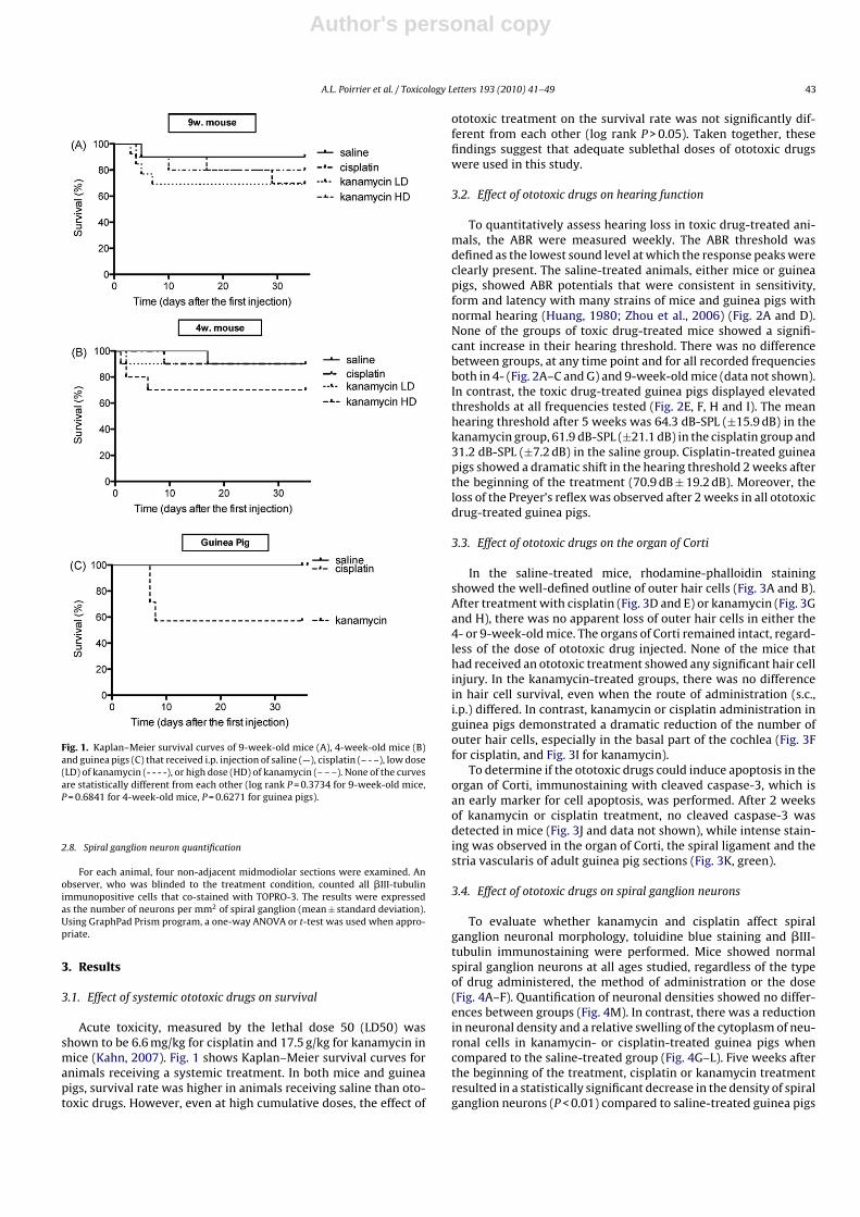
Table 1Experimental groups.
Specie Age Treatment Way of administration Cumulative dose n
Mouse 9 weeks Saline during 14 days Intraperitoneal 10
Kanamycin low dose (LD) during 14 days Subcutaneous 19.6 g/kg 5Intraperitoneal 12
Kanamycin high dose (HD) during 14 days Intraperitoneal 28 g/kg 12
Cisplatin during 10 days Subcutaneous 20 mg/kg 5Intraperitoneal 10
4 weeks Saline during 14 days Intraperitoneal 10Kanamycin low dose (LD) during 14 days Intraperitoneal 19.6 g/kg 10Kanamycin high dose (HD) during 14 days Intraperitoneal 28 g/kg 20Cisplatin during 10 days Intraperitoneal 20 mg/kg 20
Guinea pig 9 weeks Saline during 7 days Intraperitoneal 4Kanamycin during 7 days Intraperitoneal 2.8 g/kg 15Cisplatin during 10 days Intraperitoneal 20 mg/kg 16
A summary of all experimental groups (species, age at the beginning of the treatment, drug administration and number of animals in each group). Control groups receivedsaline.
assessed by recording auditory brainstem responses. Histopatho-logical phenotypes were analysed as secondary endpoints.
2. Materials and methods
2.1. Drugs
Kanamycin was obtained from Sigma–Aldrich (St. Louis, MO, USA, Kanamycinsulphate, reference # K1377). According to the manufacturer’s specification, thepreparation of kanamycin contains more than 95% kanamycin A. Cisplatin wasobtained from Sigma–Aldrich (Cis-Platinum (II) Diamine Dichloride, reference #P4394). Control animals were injected with the same volume of vehicle.
2.2. Animals
BALB/c male mice (purchased from the animal facility of the University of Liège)and Dunkin Hartley guinea pigs (Harlan, Horst, The Netherlands, reference Hsd-Poc:DH) were maintained on a 12-h light–dark cycle and had unrestricted access towater and food. All experimental protocols were performed in accordance with theAnimal Welfare Committee of the University of Liège.
2.3. Treatments
Animals were randomly divided into 11 groups, as summarized in Table 1.Animal body weights were recorded daily at the time of dosing. At either 4- or 9-week-old, mice were treated as follows: (1) saline for 14 days (n = 10 at both ages),(2) a “low dose” of kanamycin, 700 mg/kg intraperitoneal (i.p.) or subcutaneous (s.c.)injection twice daily for 14 days, (3) a “high dose” of kanamycin, 1.4 g/kg i.p. injec-tion twice daily for 14 days, (4) a daily dose of cisplatin (2 mg/kg i.p. or s.c.) duringtwo sessions on 5 consecutive days, separated by 2 days of rest. Mice were sacrificed35 days after the first injection, except for a cohort of 4-week-old mice belonging tothe “high-dose” kanamycin regimen (n = 10) and to the i.p. cisplatin group (n = 10),which were sacrificed after 15 days to assess apoptosis. As aluminum can inactivatecisplatin (Prestayko et al., 1979), we used aluminum-free material.
Guinea pigs were divided into three groups, as follows: (1) “control” (n = 4),which received saline i.p. for 7 consecutive days; (2) “kanamycin” (n = 15), whichreceived 400 mg/kg/day kanamycin i.p. for 7 consecutive days; and (3) “cisplatin”(n = 16), which received 2 mg/kg/day cisplatin i.p. for two sessions on 5 consecutivedays. They were sacrificed 35 days after the beginning of the treatment, with theexception of eight kanamycin-treated and eight cisplatin-treated guinea pigs, whichwere sacrificed after 15 days to assess apoptosis. In each group, the survival rateswere calculated by the Kaplan–Meier method and differences evaluated with logrank test (GraphPad Prism 4.0 software, San Diego, CA, USA).
2.4. Auditory assessment
Auditory brainstem responses (ABR) were recorded prior to any treatment inorder to confirm normal hearing. ABR were then recorded weekly until sacrifice.For sedation, animals spontaneously breathed isoflurane (1.5%) in 1 L/min oxygen.Body temperature was maintained with an electric heating pad. Subcutaneous nee-dle electrodes were inserted to the vertex (active), the mastoid (reference) and thehind limb (ground). Ear probes were gently introduced into the external canal. UsingIntelligent Hearing System (IHS, Miami, FL, USA) hardware and software, click stim-uli and pure tones of 16 kHz were delivered monaurally with calibrated ER2 InsertEarphones and High Frequency Transducers (IHS). The frequency most sensitive to
ototoxic injury is 16 kHz (Church et al., 2004; Dickey et al., 2005; Li et al., 2001;Minami et al., 2004). Stimuli for each condition were presented 1024 times at therate of 19.3 s−1. Stimulation levels were decreased with 10 dB steps until no responsepeaks were detectable and were subsequently increased with 5 dB steps until theresponse reappeared. Hearing thresholds were determined by the minimal soundpressure level that was able to evoke a reproducible electrophysiological response.The thresholds were verified at least three times for each stimulus condition. Anincrease in the hearing threshold was the primary endpoint of this study. Normalityof the data distribution was assessed using the Kolmogorov–Smirnov test. Data wereanalysed using a two-way analysis of variance (ANOVA). The between-group vari-able was the ototoxic treatment while the time was the within-group variable (i.e., arepeated measure). Bonferroni method was used for post hoc analyses. Results wereconsidered statistically significant if P < 0.05 for both the ANOVA and the post-test(*P < 0.05; **P < 0.01; ***P < 0.001). GraphPad prism software (Graph Pad, San Diego,CA, USA) was used to compute the analyses.
2.5. Tissue preparation
Mice were sacrificed by neck dislocation, and guinea pigs by CO2 overdose.After sacrifice, the temporal bones were collected and stored overnight in 4%paraformaldehyde before decalcification in EDTA (4% in phosphate-buffered saline(PBS), pH = 6.4) for a total of 72 h for mice or 2 weeks for guinea pigs. The left ear wasthen cryoprotected in 20% sucrose for 24 h before being frozen for cryostat section-ing into 14-�m thin sections. The right otic capsule was removed, and the cochleawas carefully isolated from the surrounding bony tissue, taking care to preserve theorgan of Corti. Basal turn of the organ of Corti was separated, delicately unrolled andimmediately processed for immunofluorescent staining.
2.6. Toluidine blue staining
Sections were stained with a solution of toluidine blue (2 g/L in Walpole’s buffer[27 g/L natrium acetate, 1.2% acetic acid in distilled water, pH 7.4]) for 80 s and thenwashed two times for 5 min in Walpole’s buffer. The coloration was fixed in 50 g/Lammonium molybdate. Sections were washed in distilled water and then dried for15 min before mounting with coverslips (Safe MountTM, reference # 006 47520,Labonord, Templemars, France).
2.7. Immunofluorescence
Tissue sections or basal turns of the organ of Corti were incubated with PBS + 0.1%Triton X-100 for 5 min, followed by incubation with PBS + 0.3% Triton X-100 + 0.25%gelatine for 30 min at room temperature. Samples were then incubated overnightwith a specific neuronal antibody directed against �III-tubulin (1/1000, cloneTUJ1, Covance, Princeton, NJ, USA) or with an antibody directed against cleavedcaspase-3 (1/1000, Promega, Madison, WI, USA). After three rinses in PBS, a sec-ondary antibody conjugated to fluorescein isothiocyanate (the F(ab’)2 fragment ofthe affinity-purified antibody, 1/500, Jackson ImmunoResearch, West Grove, PA,USA) and phalloidin conjugated to tetramethylrhodamine B isothiocyanate (1/1000,Sigma–Aldrich) were added for 1 h at 37 ◦C. After three rinses in PBS, the sampleswere incubated in the presence of the DNA dye TOPRO-3 (1/2000, Invitrogen, Carls-bad, CA, USA) and then embedded in Vectashield hardset (Vector Labs, Burlingame,CA, USA). Samples were observed with a FV1000 Olympus confocal microscope. Forthe whole-mount preparations (i.e. basal turns of the organ of Corti), each cochleawas evaluated qualitatively for the presence or absence of outer and inner hair cells,with a particular attention to the basal turn of the cochlea.
Author's personal copy
A.L. Poirrier et al. / Toxicology Letters 193 (2010) 41–49 43
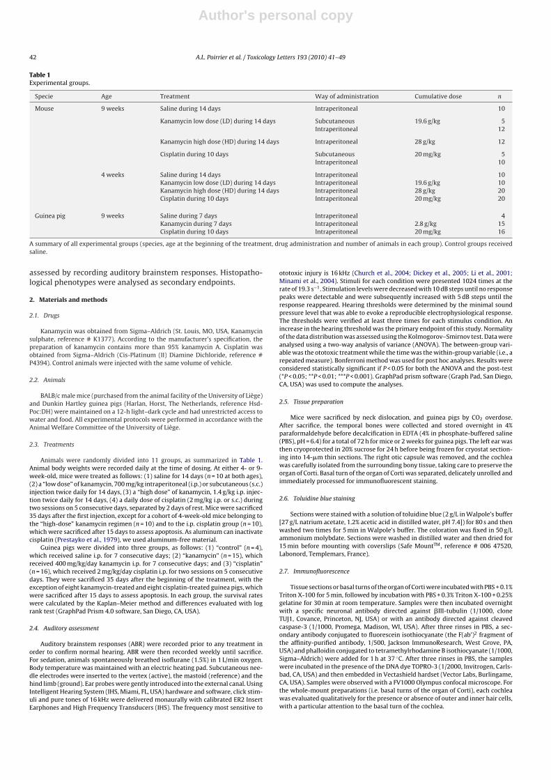
Fig. 1. Kaplan–Meier survival curves of 9-week-old mice (A), 4-week-old mice (B)and guinea pigs (C) that received i.p. injection of saline (—), cisplatin (– - –), low dose(LD) of kanamycin (- - - -), or high dose (HD) of kanamycin (– – –). None of the curvesare statistically different from each other (log rank P = 0.3734 for 9-week-old mice,P = 0.6841 for 4-week-old mice, P = 0.6271 for guinea pigs).
2.8. Spiral ganglion neuron quantification
For each animal, four non-adjacent midmodiolar sections were examined. Anobserver, who was blinded to the treatment condition, counted all �III-tubulinimmunopositive cells that co-stained with TOPRO-3. The results were expressedas the number of neurons per mm2 of spiral ganglion (mean ± standard deviation).Using GraphPad Prism program, a one-way ANOVA or t-test was used when appro-priate.
3. Results
3.1. Effect of systemic ototoxic drugs on survival
Acute toxicity, measured by the lethal dose 50 (LD50) wasshown to be 6.6 mg/kg for cisplatin and 17.5 g/kg for kanamycin inmice (Kahn, 2007). Fig. 1 shows Kaplan–Meier survival curves foranimals receiving a systemic treatment. In both mice and guineapigs, survival rate was higher in animals receiving saline than oto-toxic drugs. However, even at high cumulative doses, the effect of
ototoxic treatment on the survival rate was not significantly dif-ferent from each other (log rank P > 0.05). Taken together, thesefindings suggest that adequate sublethal doses of ototoxic drugswere used in this study.
3.2. Effect of ototoxic drugs on hearing function
To quantitatively assess hearing loss in toxic drug-treated ani-mals, the ABR were measured weekly. The ABR threshold wasdefined as the lowest sound level at which the response peaks wereclearly present. The saline-treated animals, either mice or guineapigs, showed ABR potentials that were consistent in sensitivity,form and latency with many strains of mice and guinea pigs withnormal hearing (Huang, 1980; Zhou et al., 2006) (Fig. 2A and D).None of the groups of toxic drug-treated mice showed a signifi-cant increase in their hearing threshold. There was no differencebetween groups, at any time point and for all recorded frequenciesboth in 4- (Fig. 2A–C and G) and 9-week-old mice (data not shown).In contrast, the toxic drug-treated guinea pigs displayed elevatedthresholds at all frequencies tested (Fig. 2E, F, H and I). The meanhearing threshold after 5 weeks was 64.3 dB-SPL (±15.9 dB) in thekanamycin group, 61.9 dB-SPL (±21.1 dB) in the cisplatin group and31.2 dB-SPL (±7.2 dB) in the saline group. Cisplatin-treated guineapigs showed a dramatic shift in the hearing threshold 2 weeks afterthe beginning of the treatment (70.9 dB ± 19.2 dB). Moreover, theloss of the Preyer’s reflex was observed after 2 weeks in all ototoxicdrug-treated guinea pigs.
3.3. Effect of ototoxic drugs on the organ of Corti
In the saline-treated mice, rhodamine-phalloidin stainingshowed the well-defined outline of outer hair cells (Fig. 3A and B).After treatment with cisplatin (Fig. 3D and E) or kanamycin (Fig. 3Gand H), there was no apparent loss of outer hair cells in either the4- or 9-week-old mice. The organs of Corti remained intact, regard-less of the dose of ototoxic drug injected. None of the mice thathad received an ototoxic treatment showed any significant hair cellinjury. In the kanamycin-treated groups, there was no differencein hair cell survival, even when the route of administration (s.c.,i.p.) differed. In contrast, kanamycin or cisplatin administration inguinea pigs demonstrated a dramatic reduction of the number ofouter hair cells, especially in the basal part of the cochlea (Fig. 3Ffor cisplatin, and Fig. 3I for kanamycin).
To determine if the ototoxic drugs could induce apoptosis in theorgan of Corti, immunostaining with cleaved caspase-3, which isan early marker for cell apoptosis, was performed. After 2 weeksof kanamycin or cisplatin treatment, no cleaved caspase-3 wasdetected in mice (Fig. 3J and data not shown), while intense stain-ing was observed in the organ of Corti, the spiral ligament and thestria vascularis of adult guinea pig sections (Fig. 3K, green).
3.4. Effect of ototoxic drugs on spiral ganglion neurons
To evaluate whether kanamycin and cisplatin affect spiralganglion neuronal morphology, toluidine blue staining and �III-tubulin immunostaining were performed. Mice showed normalspiral ganglion neurons at all ages studied, regardless of the typeof drug administered, the method of administration or the dose(Fig. 4A–F). Quantification of neuronal densities showed no differ-ences between groups (Fig. 4M). In contrast, there was a reductionin neuronal density and a relative swelling of the cytoplasm of neu-ronal cells in kanamycin- or cisplatin-treated guinea pigs whencompared to the saline-treated group (Fig. 4G–L). Five weeks afterthe beginning of the treatment, cisplatin or kanamycin treatmentresulted in a statistically significant decrease in the density of spiralganglion neurons (P < 0.01) compared to saline-treated guinea pigs

Author's personal copy
44 A.L. Poirrier et al. / Toxicology Letters 193 (2010) 41–49
Fig. 2. A–F. Examples of 16 kHz tone burst-evoked ABR obtained from 4-week-old mice (A–C) or from guinea pigs (D–E) 5 weeks after the beginning of the treatment.Representative data from six different animals for each group are shown. There was no difference in the ABR pattern in mice receiving saline (A), cisplatin (B) or high-dosekanamycin (C). In guinea pigs, the lowest sound level to evoke a response increased after cisplatin (E) or kanamycin (F) when compared to saline (D) treatment. (G–I) Theevolution of the ABR threshold over time in 4-week-old mice (G) and in guinea pigs treated by cisplatin (H) or kanamycin (I). Values are expressed as the mean ± S.D. (G)ABR thresholds in mice treated with high-dose kanamycin (circle), cisplatin (black triangle) and saline (white reversed triangle) remained stable over a period of weeks. ABRthresholds in guinea pigs treated with cisplatin (black triangle, H) or kanamycin (circle, I) showed a significant difference compared to the saline group beginning in thesecond week after the start of treatment.

Author's personal copy
A.L. Poirrier et al. / Toxicology Letters 193 (2010) 41–49 45
Fig. 3. (A–I) Outer hair cells in the basal turn of the organ of Corti in a 9-week-old mouse (A), a 4-week-old mouse (B), or a guinea pig (C) 35 days after saline treatment.Outer hair cells in the organ of Corti were preserved after cisplatin or kanamycin treatment both in 9-week-old mice (D and G, respectively) and in 4-week-old mice (E andH, respectively). In contrast, outer hair cell loss was observed after both cisplatin (F) and kanamycin (I) treatment in guinea pigs, mainly in the basal turn of the cochlea.(J–K) Basal turns of organ of Corti stained with antibodies against active caspase-3 (green), �III-tubulin (blue) and phalloidin (red), along with Dapi (gray), at the end of thekanamycin treatment in mice (J) and guinea pigs (K). Cleaved caspase-3 was present in the organ of Corti, the spiral ligament and the stria vascularis of guinea pigs. Scale barshown in A = 10 �m for A–I and 50 �m for J–K. (For interpretation of the references to color in this figure legend, the reader is referred to the web version of the article.)
(Fig. 4N–O). Afferent fibres extended normally to the organ of Cortiunder all conditions (data not shown).
4. Discussion
This investigation demonstrated significant ototoxic differencesbetween mice and guinea pigs after treatment with an aminoglyco-side or cisplatin. While kanamycin or cisplatin is clearly ototoxic inthe guinea pig, the BALB/c mouse is resistant to much higher dosesof kanamycin or cisplatin.
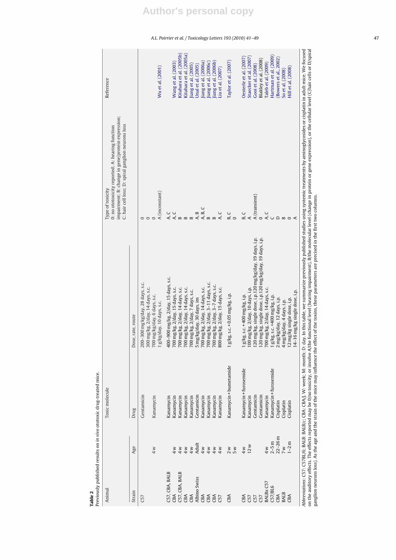
Our results in mice are in line with some previously publisheddata but discordant with others (Table 2). This discrepancy could beexplained by genetic or environmental differences between Amer-ican and European mouse strains. Genetic differences betweenmouse strains influence responses to various insults or toxins,including “ototoxicants” (Hamre et al., 1999; Peng et al., 1997;Perera et al., 2008; Schauwecker and Steward, 1997). In fact, theBALB/c strain was recently shown to be more sensitive to amino-glycoside toxicity than other strains, such as C57bl or CBA (Wu etal., 2001). The BALB/c mice in our study, however, did not display
any ototoxicity. Severinsen et al. (2006) did not show any perma-nent loss of hair cells in the maculae of European BALB/c micetreated by kanamycin. A potential explanation could be the geneticdifferences between American and European BALB/c strains. Theparental BALB/c mice were derived from their original stock aroundthe year 1913. This strain is now widely distributed and has severalsubstrains with potential genetic diversity.
Dietary habits could also play a role in drug-induced ototoxicity.Food deprivation or treatment with glutathione-depleting drugshas been shown to exacerbate the ototoxicity of aminoglycosides(Lautermann et al., 1995a) or cisplatin (Lautermann et al., 1995b).Conversely, there could be a protective effect against drug-inducedototoxicity provided by antioxidants in the animal diet, such asalpha-tocopherol (vitamin E) (Fetoni et al., 2003, 2004a; Kalkanis etal., 2004; Sergi et al., 2004), d-methionine (Sha and Schacht, 2000),and l-acetylcysteine (Bock et al., 1983; Dickey et al., 2005; Feldmanet al., 2007; Tepel, 2007). On the other hand, iron supplementa-tion increases sensitivity to aminoglycosides (Conlon and Smith,1998). In this study, we used a standard diet provided by SAFE(Scientific Animal Food & Engineering, France). According to the

Author's personal copy
46 A.L. Poirrier et al. / Toxicology Letters 193 (2010) 41–49
Fig. 4. Toluidine blue staining and �III-tubulin immunofluorescence (green) with TOPRO-3 nuclear counterstaining (blue) in the first coil of the cochlea on a midmodiolarsection, 5 weeks after the beginning of the treatment, in a representative 4-week-old mouse (A–F) or a representative guinea pig (G–L) of each group. No difference wasobserved in mice receiving saline (A and D), cisplatin (B and E) or a high dose of kanamycin (C and F). In guinea pigs, toluidine blue nuclei and �III-tubulin immunoreactiveprofiles were more numerous in saline-treated animals (G and J) than in those treated with cisplatin (H and K) or kanamycin (I and L). A reduction in both the neuronaldensity and cytoplasmic swelling was observed in neurons from ototoxic drug-treated animals. (M–O) Quantification of �III-tubulin immunoreactive profiles in 4-week-oldmice and in guinea pigs treated with cisplatin or kanamycin. In each animal, four non-contiguous midmodiolar sections were selected, and each coil of the cochlea wasconsidered separately. Each cell containing �III-tubulin-positive cytoplasm and a TOPRO-3-positive nucleus was considered as a spiral ganglion neuron (SGN). Results areexpressed as the number of �III-tubulin immunoreactive profiles per mm2 and are presented as the mean ± S.D. No significant differences were observed between groupsof mice (M). In guinea pigs, a significant decrease in the number of neurons per mm2 was observed 5 weeks after the beginning of cisplatin (black column, N) or kanamycin(black column, O) administration. Scale bar shown in A = 50 �m for A–L. (For interpretation of the references to color in this figure legend, the reader is referred to the webversion of the article.)
Author's personal copy
A.L. Poirrier et al. / Toxicology Letters 193 (2010) 41–49 47
Tab
le2
Prev
iou
sly
pu
blis
hed
resu
lts
onin
vivo
otot
oxic
dru
g-tr
eate
dm
ice.
An
imal
Toxi
cm
olec
ule
Typ
eof
toxi
city
0:n
oot
otox
icit
yre
por
ted
;A
:h
eari
ng
fun
ctio
nim
pai
rmen
t;B
:ch
ange
inge
ne/
pro
tein
exp
ress
ion
;C
:h
air
cell
loss
;D
:sp
iral
gan
glio
nn
euro
ns
loss
Ref
eren
ce
Stra
inA
geD
rug
Dos
e,ra
te,r
oute
C57
Gen
tam
icin
200–
300
mg
kg/d
ay,2
8d
ays,
s.c.
030
0m
g/kg
,2/d
ay,1
4d
ays,
s.c.
04
wK
anam
ycin
700
mg/
kg/d
ay,6
day
s,s.
c.0
1g/
kg/d
ay,1
0d
ays,
s.c.
A(i
nco
nst
ant)
Wu
etal
.(20
01)
C57
,CB
A,B
ALB
Kan
amyc
in40
0–90
0m
g/kg
,2/d
ay,1
5d
ays,
s.c.
A,C
CB
A4
wK
anam
ycin
700
mg/
kg,2
/day
,15
day
s,s.
c.A
,CW
ang
etal
.(20
03)
C57
,CB
A,B
ALB
4w
Kan
amyc
in70
0m
g/kg
,2/d
ay,1
4d
ays,
s.c.
BK
itah
ara
etal
.(20
05b)
CB
A4
wK
anam
ycin
700
mg/
kg,2
/day
,14
day
s,s.
c.B
Kit
ahar
aet
al.(
2005
a)C
BA
4w
Kan
amyc
in70
0m
g/kg
,2/d
ay,7
day
s,s.
c.B
Jian
get
al.(
2005
)A
lbin
oSw
iss
Ad
ult
Gen
tam
icin
5m
g/kg
/day
,30
day
s,im
A,B
Un
alet
al.(
2005
)C
BA
4w
Kan
amyc
in70
0m
g/kg
,2/d
ay,1
4d
ays,
s.c.
A,B
,CJi
ang
etal
.(20
06a)
CB
A4
wK
anam
ycin
700
mg/
kg,2
/day
,3–1
1d
ays,
s.c.
BJi
ang
etal
.(20
06c)
CB
A4
wK
anam
ycin
700
mg/
kg,2
/day
,3–7
day
s,s.
c.B
Jian
get
al.(
2006
b)C
574
wK
anam
ycin
800
mg/
kg,2
/day
,15
day
s,s.
c.A
,CLi
uet
al.(
2007
)
CB
A2
wK
anam
ycin
+bu
met
amid
e1
g/kg
,s.c
.+0.
05m
g/kg
,i.p
.B
,CTa
ylor
etal
.(20
07)
5w
0
CB
A4
wK
anam
ycin
+fu
rose
mid
e1
g/kg
,s.c
.+40
0m
g/kg
,i.p
.B
,CO
este
rle
etal
.(20
07)
C57
12w
Kan
amyc
in10
0m
g/kg
,2/d
ay,1
0d
ays,
i.p.
ASt
aeck
eret
al.(
2007
)C
57G
enta
mic
in12
0m
g/kg
,sin
gle
dos
e,i.p
.120
mg/
kg/d
ay,1
9d
ays,
i.p.
A(t
ran
sien
t)G
ooie
tal
.(20
08)
C57
Gen
tam
icin
120
mg/
kg,s
ingl
ed
ose,
i.p.1
20m
g/kg
/day
,19
day
s,i.p
.0
Bla
kley
etal
.(20
08)
BA
LBx
C57
4w
Kan
amyc
in70
0m
g/kg
,2/d
ay,1
4d
ays,
s.c.
A,C
Tale
bet
al.(
2009
)C
57/B
L62–
5m
Kan
amyc
in+
furo
sem
ide
1g/
kg,s
.c.+
400
mg/
kg,i
.p.
CH
artm
anet
al.(
2009
)C
BA
22–2
6m
Cis
pla
tin
2m
g/kg
/day
,12
day
s,i.p
.D
(Bow
ers
etal
.,20
02)
BA
LB7
wC
isp
lati
n4
mg/
kg/d
ay,4
day
s,i.p
.B
Soet
al.(
2008
)C
BA
1–2
mC
isp
lati
n12
mg/
kgsi
ngl
ed
ose,
i.p.
0H
ille
tal
.(20
08)
14–1
6m
g/kg
sin
gle
dos
e,i.p
.A
Abb
revi
atio
ns:C
57:C
57B
L/6;
BA
LB:B
ALB
/c;C
BA
:CB
A/J
.W:w
eek;
M:m
onth
;D:d
ay.I
nth
ista
ble,
we
sum
mar
ize
pre
viou
sly
pu
blis
hed
stu
die
su
sin
gsy
stem
ictr
eatm
ents
byam
inog
lyco
sid
esor
cisp
lati
nin
adu
ltm
ice.
We
focu
sed
onth
eau
dit
ory
effe
cts.
The
effe
cts
rep
orte
dm
aybe
0/n
oto
xici
ty,o
rin
volv
eA
/th
efu
nct
ion
alle
vel(
hea
rin
gim
pai
rmen
t),B
/th
em
olec
ula
rle
vel(
chan
gein
pro
tein
orge
ne
exp
ress
ion
),or
the
cell
ula
rle
vel(
C/h
air
cell
sor
D/s
pir
alga
ngl
ion
neu
ron
slo
ss).
As
the
age
and
the
stra
inof
the
mic
em
ayin
flu
ence
the
effe
ctof
the
toxi
ns,
thes
ep
aram
eter
sar
ep
reci
sed
inth
efi
rst
two
colu
mn
s.

Author's personal copy
48 A.L. Poirrier et al. / Toxicology Letters 193 (2010) 41–49
manufacturer’s specification, these mouse chows contain 30 UI/kgalpha-tocopherol which may partly explain the absence of ototox-icity in this study. However, 30 UI/kg is 3–100 times lower thanthe dose used to reach otoprotection (Fetoni et al., 2003, 2004a,b;Kalkanis et al., 2004; Teranishi et al., 2001).
There was a dramatic difference in ototoxicity between guineapigs and mice. A potential explanation for the lack of cochlear injuryin mice could be the mouse pharmacokinetic profile at both thelevel of the organ of Corti and the whole organism. First, mice havea high metabolic rate and a high renal clearance, which keep serumlevels of drugs very low (Walton et al., 2004; Yang and Bankir,2005). All interspecies studies point out the importance of phar-macokinetic in the differential effect observed between speciesafter aminoglycoside (Meza and Aguilar-Maldonado, 2007), cis-platin (Blakley et al., 2008) or other ototoxic agents (Lataye et al.,2003). Urinary excretion is the major route of platinum and amino-glycoside elimination in all species (Farris et al., 1988; King et al.,1986; Tran Ba Huy et al., 1986). In mice, the high plasma clearanceallows an early urinary excretion (Walton et al., 2004; Yang andBankir, 2005), and thus a shorter serum half-life of ototoxic drugs(Wu et al., 2001).
Second, the organ of Corti is hidden in the deep compart-ment of the inner ear and is protected by a blood–labyrinthbarrier in a manner analogous to the way that the central ner-vous system is protected by the blood–brain barrier. This barriercan be roughly divided into the blood–perilymph barrier and theperilymph–endolymph barrier, although the functional divisionbetween these compartments is not fully understood. A small por-tion of the inner ear, the stria vascularis, is readily accessible fromthe systemic circulation. It is surrounded by cells that are sealedby tight junctions, which contribute to the blood–perilymph bar-rier (Swan et al., 2008). This barrier is most likely the major obstaclethat restricts the entry of drugs into the inner ear and would therebyserve to dampen the effects of these drugs after systemic admin-istration. Ototoxic drugs primarily affect the organisation of celljunctions (Anniko, 1985; Leonova and Raphael, 1997). A temptingspeculation is that the sensitivity of guinea pigs to ototoxic drugsmay be due to an essential tight junction protein with a quantitativeor functional defect that is not present in mice.
Cisplatin- and aminoglycoside-induced ototoxicity is known tobe mediated via injury to several terminally differentiated cellulartargets in the cochlea (Rybak and Whitworth, 2005; van Ruijvenet al., 2005), including the marginal and intermediate cells of thestria vascularis (Dai and Steyger, 2008; Laurell et al., 2007; Wangand Steyger, 2009). These cells are a major component of theperilymph–endolymph barrier. Whether these cells have the sameproperties in mice and guinea pigs remains to be determined andcould provide another explanation for the difference in ototoxicdrug sensitivity. Penetration of aminoglycosides into the cochleacould be less important in mice compared to guinea pigs (Dai etal., 2006). Differences in cochlear uptake are sufficient to explaindifferent ototoxic profiles (Hellberg et al., 2009).
Several reports have concluded that the generation of reactiveoxygen species (ROS) is linked both to aminoglycoside and cisplatinototoxicity (Rizzi and Hirose, 2007; Rybak and Whitworth, 2005;Rybak et al., 2007). BALB/c mice have high levels of superoxidedismutase (SOD), which functions to block the oxidative stress gen-erated by ROSs (Misra et al., 1991). Thus, the presence of high SODlevels in BALB/c mouse cochlear cells could reduce or abolish theototoxicity of these drugs. This hypothesis could also be appliedto other antioxidant defense and repair systems. In fact, ototoxicmechanisms are not sufficiently understood to identify reasons forspecies-specific differentiations.
In conclusion, these data support a marked difference betweenthe inner ear responses of guinea pig and mouse to pharmacolog-ically induced ototoxicity. Clearly, the intact hearing function of
the mouse is not attributable to technical errors, since the samemolecules administered to the guinea pig induced cochlear injury,as has been previously described in the literature. The presenteddata suggest that the adult guinea pig model of cochlear toxicityremains the most reliable model for neurosensory deafness. Mousemodel for neurosensory deafness must be considered with caution.
Conflict of interest
The authors report no conflict of interest.
Acknowledgements
This study was supported by grants from the National Fund forScientific Research (Belgium) and the Fonds Leon Fredericq. Theauthors are grateful to Mrs. Arlette Brose for valuable technicalsupport.
References
Anniko, M., 1985. Principles in cochlear toxicity. Arch. Toxicol. Suppl. 8, 221–239.Aran, J.M., Erre, J.P., Lima da, C.D., Debbarh, I., Dulon, D., 1999. Acute and chronic
effects of aminoglycosides on cochlear hair cells. Ann. N.Y. Acad. Sci. 884, 60–68.Banfi, B., Malgrange, B., Knisz, J., Steger, K., Dubois-Dauphin, M., Krause, K.H., 2004.
NOX3, a superoxide-generating NADPH oxidase of the inner ear. J. Biol. Chem.279, 46065–46072.
Billings, K.R., Kenna, M.A., 1999. Causes of pediatric sensorineural hearing loss:yesterday and today. Arch. Otolaryngol. Head Neck Surg. 125, 517–521.
Blakley, B.W., Hochman, J., Wellman, M., Gooi, A., Hussain, A.E., 2008. Differences inototoxicity across species. J. Otolaryngol. Head Neck Surg. 37, 700–703.
Bock, G.R., Yates, G.K., Miller, J.J., Moorjani, P., 1983. Effects of N-acetylcysteine onkanamycin ototoxicity in the guinea pig. Hear. Res. 9, 255–262.
Bowers, W.J., Chen, X., Guo, H., Frisina, D.R., Federoff, H.J., Frisina, R.D., 2002.Neurotrophin-3 transduction attenuates cisplatin spiral ganglion neuron oto-toxicity in the cochlea. Mol. Ther. 6, 12–18.
Brown, S.D., Hardisty-Hughes, R.E., Mburu, P., 2008. Quiet as a mouse: dissecting themolecular and genetic basis of hearing. Nat. Rev. Genet. 9, 277–290.
Church, M.W., Blakley, B.W., Burgio, D.L., Gupta, A.K., 2004. WR-2721 (Amifostine)ameliorates cisplatin-induced hearing loss but causes neurotoxicity in ham-sters: dose-dependent effects. J. Assoc. Res. Otolaryngol. 5, 227–237.
Conlon, B.J., Smith, D.W., 1998. Supplemental iron exacerbates aminoglycoside oto-toxicity in vivo. Hear. Res. 115, 1–5.
Dai, C.F., Mangiardi, D., Cotanche, D.A., Steyger, P.S., 2006. Uptake of fluorescentgentamicin by vertebrate sensory cells in vivo. Hear. Res. 213, 64–78.
Dai, C.F., Steyger, P.S., 2008. A systemic gentamicin pathway across the stria vascu-laris. Hear. Res. 235, 114–124.
Dickey, D.T., Wu, Y.J., Muldoon, L.L., Neuwelt, E.A., 2005. Protection against cisplatin-induced toxicities by N-acetylcysteine and sodium thiosulfate as assessed at themolecular, cellular, and in vivo levels. J. Pharmacol. Exp. Ther. 314, 1052–1058.
Farris, F.F., Dedrick, R.L., King, F.G., 1988. Cisplatin pharmacokinetics: applicationsof a physiological model. Toxicol. Lett. 43, 117–137.
Feldman, L., Efrati, S., Eviatar, E., Abramsohn, R., Yarovoy, I., Gersch, E., Averbukh, Z.,Weissgarten, J., 2007. Gentamicin-induced ototoxicity in hemodialysis patientsis ameliorated by N-acetylcysteine. Kidney Int. 72, 359–363.
Fetoni, A.R., Sergi, B., Ferraresi, A., Paludetti, G., Troiani, D., 2004a. alpha-Tocopherolprotective effects on gentamicin ototoxicity: an experimental study. Int. J.Audiol. 43, 166–171.
Fetoni, A.R., Sergi, B., Ferraresi, A., Paludetti, G., Troiani, D., 2004b. Protective effectsof alpha-tocopherol and tiopronin against cisplatin-induced ototoxicity. ActaOtolaryngol. 124, 421–426.
Fetoni, A.R., Sergi, B., Scarano, E., Paludetti, G., Ferraresi, A., Troiani, D., 2003. Pro-tective effects of alpha-tocopherol against gentamicin-induced oto-vestibulotoxicity: an experimental study. Acta Otolaryngol. 123, 192–197.
Forge, A., Schacht, J., 2000. Aminoglycoside antibiotics. Audiol. Neurootol. 5, 3–22.Gooi, A., Hochman, J., Wellman, M., Blakley, L., Blakley, B.W., 2008. Ototoxic effects
of single-dose versus 19-day daily-dose gentamicin. J. Otolaryngol Head NeckSurg. 37, 664–667.
Hamre, K., Tharp, R., Poon, K., Xiong, X., Smeyne, R.J., 1999. Differential strainsusceptibility following 1-methyl-4-phenyl-1,2,3,6-tetrahydropyridine (MPTP)administration acts in an autosomal dominant fashion: quantitative analysis inseven strains of Mus musculus. Brain Res. 828, 91–103.
Hartman, B.H., Basak, O., Nelson, B.R., Taylor, V., Bermingham-McDonogh, O., Reh,T.A., 2009. Hes5 Expression in the Postnatal and Adult Mouse Inner Ear and theDrug-Damaged Cochlea. J. Assoc. Res. Otolaryngol.
Hellberg, V., Wallin, I., Eriksson, S., Hernlund, E., Jerremalm, E., Berndtsson, M., Eks-borg, S., Arner, E.S., Shoshan, M., Ehrsson, H., Laurell, G., 2009. Cisplatin andoxaliplatin toxicity: importance of cochlear kinetics as a determinant for oto-toxicity. J. Natl. Cancer Inst. 101, 37–47.
Hill, G.W., Morest, D.K., Parham, K., 2008. Cisplatin-induced ototoxicity: effect ofintratympanic dexamethasone injections. Otol. Neurotol. 29, 1005–1011.
Author's personal copy
A.L. Poirrier et al. / Toxicology Letters 193 (2010) 41–49 49
Huang, C.M., 1980. A comparative study of the brain stem auditory response inmammals. Brain Res. 184, 215–219.
Humes, H.D., 1999. Insights into ototoxicity. Analogies to nephrotoxicity. Ann. N. Y.Acad. Sci. 884, 15–18.
Jiang, H., Sha, S.H., Forge, A., Schacht, J., 2006a. Caspase-independent pathways ofhair cell death induced by kanamycin in vivo. Cell. Death Differ. 13, 20–30.
Jiang, H., Sha, S.H., Schacht, J., 2005. NF-kappaB pathway protects cochlear hair cellsfrom aminoglycoside-induced ototoxicity. J. Neurosci. Res. 79, 644–651.
Jiang, H., Sha, S.H., Schacht, J., 2006b. Kanamycin alters cytoplasmic and nuclearphosphoinositide signaling in the organ of Corti in vivo. J. Neurochem. 99,269–276.
Jiang, H., Sha, S.H., Schacht, J., 2006c. Rac/Rho pathway regulates actin depolymer-ization induced by aminoglycoside antibiotics. J. Neurosci. Res. 83, 1544–1551.
Kahn, C.M., 2007. The Merck Veterinary Manual.Kalkanis, J.G., Whitworth, C., Rybak, L.P., 2004. Vitamin E reduces cisplatin ototoxi-
city. Laryngoscope 114, 538–542.King, F.G., Dedrick, R.L., Farris, F.F., 1986. Physiological pharmacokinetic modeling
of cis-dichlorodiammineplatinum(II) (DDP) in several species. J. Pharmacokinet.Biopharm. 14, 131–155.
Kitahara, T., Li, H.S., Balaban, C.D., 2005a. Changes in transient receptor potentialcation channel superfamily V (TRPV) mRNA expression in the mouse inner earganglia after kanamycin challenge. Hear. Res. 201, 132–144.
Kitahara, T., Li-Korotky, H.S., Balaban, C.D., 2005b. Regulation of mitochondrialuncoupling proteins in mouse inner ear ganglion cells in response to systemickanamycin challenge. Neuroscience. 135, 639–653.
Lataye, R., Campo, P., Pouyatos, B., Cossec, B., Blachere, V., Morel, G., 2003. Solventototoxicity in the rat and guinea pig. Neurotoxicol. Teratol. 25, 39–50.
Laurell, G., Ekborn, A., Viberg, A., Canlon, B., 2007. Effects of a single high dose ofcisplatin on the melanocytes of the stria vascularis in the guinea pig. Audiol.Neurootol. 12, 170–178.
Lautermann, J., McLaren, J., Schacht, J., 1995a. Glutathione protection against gen-tamicin ototoxicity depends on nutritional status. Hear. Res. 86, 15–24.
Lautermann, J., Song, B., McLaren, J., Schacht, J., 1995b. Diet is a risk factor in cisplatinototoxicity. Hear. Res. 88, 47–53.
Leonova, E.V., Raphael, Y., 1997. Organization of cell junctions and cytoskeleton inthe reticular lamina in normal and ototoxically damaged organ of Corti. Hear.Res. 113, 14–28.
Li, G., Frenz, D.A., Brahmblatt, S., Feghali, J.G., Ruben, R.J., Berggren, D.,Arezzo, J., Van De Water, T.R., 2001. Round window membrane delivery ofl-methionine provides protection from cisplatin ototoxicity without compro-mising chemotherapeutic efficacy. Neurotoxicology 22, 163–176.
Liu, Y.H., Ke, X.M., Qin, Y., Gu, Z.P., Xiao, S.F., 2007. Adeno-associated virus-mediatedBcl-xL prevents aminoglycoside-induced hearing loss in mice. Chin. Med. J.(Engl) 120, 1236–1240.
Meza, G., Aguilar-Maldonado, B., 2007. Streptomycin action to the mammalian innerear vestibular organs: comparison between pigmented guinea pigs and rats.Comp. Biochem. Physiol. C: Toxicol. Pharmacol. 146, 203–206.
Minami, S.B., Sha, S.H., Schacht, J., 2004. Antioxidant protection in a new animalmodel of cisplatin-induced ototoxicity. Hear. Res. 198, 137–143.
Misra, M., Rodriguez, R.E., North, S.L., Kasprzak, K.S., 1991. Nickel-induced renallipid peroxidation in different strains of mice: concurrence with nickel effecton antioxidant defense systems. Toxicol. Lett. 58, 121–133.
Morzaria, S., Westerberg, B.D., Kozak, F.K., 2004. Systematic review of the etiology ofbilateral sensorineural hearing loss in children. Int. J. Pediatr. Otorhinolaryngol.68, 1193–1198.
Oesterle, E.C., Campbell, S., Taylor, R.R., Forge, A., Hume, C.R., 2007. Sox2 and Jagged1Expression in Normal and Drug-Damaged Adult Mouse Inner Ear. J. Assoc. Res.Otolaryngol..
Peng, Y.G., Clayton, E.C., Means, L.W., Ramsdell, J.S., 1997. Repeated independentexposures to domoic acid do not enhance symptomatic toxicity in outbred orseizure-sensitive inbred mice. Fundam. Appl. Toxicol. 40, 63–67.
Perera, P.Y., Lichy, J.H., Mastropaolo, J., Rosse, R.B., Deutsch, S.I., 2008. Expression ofNR1, NR2A and NR2B NMDA receptor subunits is not altered in the genetically-inbred Balb/c mouse strain with heightened behavioral sensitivity to MK-801,a noncompetitive NMDA receptor antagonist. Eur. Neuropsychopharmacol. 18,814–819.
Prestayko, A.W., Cadiz, M., Crooke, S.T., 1979. Incompatibility of aluminum-containing iv administration equipment with cis-dichlorodiammine-platinum(II) administration. Cancer Treat. Rep. 63, 2118–2119.
Rizzi, M.D., Hirose, K., 2007. Aminoglycoside ototoxicity. Curr. Opin. Otolaryngol.Head Neck Surg. 15, 352–357.
Rybak, L.P., 2007. Mechanisms of cisplatin ototoxicity and progress in otoprotection.Curr. Opin. Otolaryngol. Head Neck Surg. 15, 364–369.
Rybak, L.P., Whitworth, C.A., 2005. Ototoxicity: therapeutic opportunities. Drug Dis-cov. Today 10, 1313–1321.
Rybak, L.P., Whitworth, C.A., Mukherjea, D., Ramkumar, V., 2007. Mechanisms ofcisplatin-induced ototoxicity and prevention. Hear. Res. 226, 157–167.
Schacht, J., 1999. Biochemistry and pharmacology of aminoglycoside-induced hear-ing loss. Acta Physiol. Pharmacol. Ther. Latinoam 49, 251–256.
Schauwecker, P.E., Steward, O., 1997. Genetic determinants of susceptibility to exci-totoxic cell death: implications for gene targeting approaches. Proc. Natl. Acad.Sci. U.S.A. 94, 4103–4108.
Sergi, B., Fetoni, A.R., Ferraresi, A., Troiani, D., Azzena, G.B., Paludetti, G., Maur-izi, M., 2004. The role of antioxidants in protection from ototoxic drugs. ActaOtolaryngol. Suppl., 42–45.
Severinsen, S.A., Kirkegaard, M., Nyengaard, J.R., 2006. 2,3-Dihydroxybenzoic acidattenuates kanamycin-induced volume reduction in mouse utricular type I haircells. Hear. Res. 212, 99–108.
Sha, S.H., Schacht, J., 2000. Antioxidants attenuate gentamicin-induced free radicalformation in vitro and ototoxicity in vivo:d-methionine is a potential protectant.Hear. Res. 142, 34–40.
So, H., Kim, H., Kim, Y., Kim, E., Pae, H.O., Chung, H.T., Kim, H.J., Kwon, K.B., Lee, K.M.,Lee, H.Y., Moon, S.K., Park, R., 2008. Evidence that Cisplatin-induced AuditoryDamage is Attenuated by Downregulation of Pro-inflammatory Cytokines ViaNrf2/HO-1. J. Assoc. Res. Otolaryngol..
Staecker, H., Praetorius, M., Baker, K., Brough, D.E., 2007. Vestibular hair cell regener-ation and restoration of balance function induced by math1 gene transfer. Otol.Neurotol. 28, 223–231.
Swan, E.E., Mescher, M.J., Sewell, W.F., Tao, S.L., Borenstein, J.T., 2008. Inner ear drugdelivery for auditory applications. Adv. Drug Deliv. Rev. 60, 1583–1599.
Taleb, M., Brandon, C.S., Lee, F.S., Harris, K.C., Dillmann, W.H., Cunningham, L.L.,2009. Hsp70 inhibits aminoglycoside-induced hearing loss and cochlear haircell death. Cell. Stress Chaperones..
Taylor, R.R., Nevill, G., Forge, A., 2007. Rapid Hair Cell Loss: A Mouse Model forCochlear Lesions. J. Assoc. Res. Otolaryngol..
Tepel, M., 2007. N-acetylcysteine in the prevention of ototoxicity. Kidney Int. 72,231–232.
Teranishi, M., Nakashima, T., Wakabayashi, T., 2001. Effects of alpha-tocopherol oncisplatin-induced ototoxicity in guinea pigs. Hear. Res. 151, 61–70.
Tran Ba Huy, P., Bernard, P., Schacht, J., 1986. Kinetics of gentamicin uptake andrelease in the rat. Comparison of inner ear tissues and fluids with other organs.J. Clin. Invest. 77, 1492–1500.
Unal, O.F., Ghoreishi, S.M., Atas, A., Akyurek, N., Akyol, G., Gursel, B., 2005. Preven-tion of gentamicin induced ototoxicity by trimetazidine in animal model. Int. J.Pediatr. Otorhinolaryngol. 69, 193–199.
van Ruijven, M.W., de Groot, J.C., Klis, S.F., Smoorenburg, G.F., 2005. The cochleartargets of cisplatin: an electrophysiological and morphological time-sequencestudy. Hear. Res. 205, 241–248.
van Ruijven, M.W., de Groot, J.C., Smoorenburg, G.F., 2004. Time sequence of degen-eration pattern in the guinea pig cochlea during cisplatin administration. Aquantitative histological study. Hear. Res. 197, 44–54.
Walton, K., Dorne, J.L., Renwick, A.G., 2004. Species-specific uncertainty factors forcompounds eliminated principally by renal excretion in humans. Food Chem.Toxicol. 42, 261–274.
Wang, A.M., Sha, S.H., Lesniak, W., Schacht, J., 2003. Tanshinone (Salviae milti-orrhizae extract) preparations attenuate aminoglycoside-induced free radicalformation in vitro and ototoxicity in vivo. Antimicrob. Agents Chemother. 47,1836–1841.
Wang, Q., Steyger, P.S., 2009. Trafficking of systemic fluorescent gentamicin into thecochlea and hair cells. J. Assoc. Res. Otolaryngol..
Wu, W.J., Sha, S.H., McLaren, J.D., Kawamoto, K., Raphael, Y., Schacht, J., 2001.Aminoglycoside ototoxicity in adult CBA, C57BL and BALB mice and theSprague–Dawley rat. Hear. Res. 158, 165–178.
Yang, B., Bankir, L., 2005. Urea and urine concentrating ability: new insights fromstudies in mice. Am. J. Physiol. Renal Physiol. 288, F881–F896.
Zhou, X., Jen, P.H., Seburn, K.L., Frankel, W.N., Zheng, Q.Y., 2006. Auditory brainstemresponses in 10 inbred strains of mice. Brain Res..