author's personal copy - cairo...
TRANSCRIPT

This article appeared in a journal published by Elsevier. The attachedcopy is furnished to the author for internal non-commercial researchand education use, including for instruction at the authors institution
and sharing with colleagues.
Other uses, including reproduction and distribution, or selling orlicensing copies, or posting to personal, institutional or third party
websites are prohibited.
In most cases authors are permitted to post their version of thearticle (e.g. in Word or Tex form) to their personal website orinstitutional repository. Authors requiring further information
regarding Elsevier’s archiving and manuscript policies areencouraged to visit:
http://www.elsevier.com/authorsrights

Author's personal copy
Original article
Botanical and genetic characterization of Mentha suaveolens Ehrh.cultivated in Egypt
El-Sayeda A. El-Kashoury, Hesham I. El-Askary, Zeinab A. Kandil*, Mohamed A. SalemDepartment of Pharmacognosy, Faculty of Pharmacy, Cairo University, Cairo 11562, Egypt
a r t i c l e i n f o
Article history:Received 29 June 2013Accepted 12 October 2013Available online 12 November 2013
Keywords:DNA fingerprintingMacromorphologicalMicromorphologicalMint
a b s t r a c t
This paper deals with the detailed botanical study of the stems and leaves of Mentha suaveolens Ehrh.(Labiatae), a highly valued medicinal and fragrant plant. Mentha suaveolens Ehrh. is useful as a carmi-native, antispasmodic, diaphoretic, analgesic, anti-inflammatory and antimicrobial drug. It is also usefulin case of nausea, bronchitis and anorexia. Botanical investigation of Mentha suaveolens Ehrh has notbeen reported so far. Therefore, The macro- and micromorphological characteristics of the stems andleaves of Mentha suaveolens Ehrh. cultivated in Egypt are presented, with the aim of finding out thediagnostic characters for identification and differentiation of this species either in the entire or powderedforms. The major microscopic features of the stems and leaves are the presence of both glandular andnon-glandular trichomes. Labiaceous glandular trichomes, a significant feature responsible for thesecretion of volatile oil, are numerous on both stem and leaf surfaces. Also, the presence of diacyticstomata is another important diagnostic feature of the plant. Furthermore, the DNA of the plant wasextracted and amplified using 10 decamer primers to reveal RAPD fragments. Each of the ten primerssuccessfully directed the amplification of a genome-specific fingerprint of DNA fragment. A total of 65different fragments have been generated by the two primers: OPC-07 and OPA-11 respectively, which canbe used for the selective discrimination ofM. suaveolens Ehrh. The highest numbers of fragments were 15and 9 produced by the primers OPC-07 and OPA-11 respectively. The analysis of RAPD data, under theexperimental conditions, can thus be used to distinguish the plant from other related species.
Copyright � 2013, Phcog.Net, Published by Reed Elsevier India Pvt. Ltd. All rights reserved.
1. Introduction
Family Labiatae (also known as Lamiaceae or Mint family)consists of about 280 genera and 6700 species of cosmopolitandistribution. They are mostly found in the Mediterranean region tocentral Asia.1e3 Plants of the family are usually aromatic. A verycharacteristic feature of the family is the possession of the essentialoils which are excreted mainly beneath the cuticle of various kindsof hairs.3,4 The genusMentha (Mint), one of the importantmembersof the Lamiaceae family, is represented by about 25e30 species ofaromatic, perennial herbs. All are native to the north temperatezone, about half of them being native or naturalized in NorthAmerica.5e8 The genus is widely used in flavor, fragrance, medicinaland pharmaceutical applications. The medicinal uses of mints,which date back to ancient times, include carminative, antispas-modic, antiemetic, diaphoretic, analgesic, anti-inflammatory and
emmenagogue. They are also used against nausea, bronchitis,anorexia, ulcerative colitis and liver complains. Mint essential oilsare generally used for antipruritis, astringent, rubefacient, anti-septic and antimicrobial purposes and for treating neuralgia,myalgia, headache and migraine.9 Mentha suaveolens Ehrh. (knownas apple mint or woolly mint) is native to Africa, Temperate Asiaand Europe.10 The leaves of this plant can be used to make a minttea, as a garnish, or in salads. It is used as antifungal, antibacterial,antihypertensive, antiemetic, antidiarrheal, antihaemorrhoidal,analgesic, anti-inflammatory and antioxidant.11
Regarding the botanical or DNA characteristics of M. suaveolensEhrh. cultivated in Egypt, no informationwas traced in the availableliterature. Therefore, the macro- and micromorphological charac-teristics of the stem and leaves of the plant cultivated in Egypt arepresented, with the aim of finding out the diagnostic characters foridentification and differentiation of this species either in the entireor powdered forms. Moreover, (DNA) fingerprinting, is now themost popular method for identifying and quality control of herbalmaterial.12 DNA fingerprints, resembling barcodes, are unique tothe individual and hence can be used in the same way as
* Corresponding author. Tel.: þ20 1006886793; fax: þ20 2 25320005.E-mail address: [email protected] (Z.A. Kandil).
Contents lists available at ScienceDirect
Pharmacognosy Journal
journal homepage: www.elsevier .com/locate/phcgj
0975-3575/$ e see front matter Copyright � 2013, Phcog.Net, Published by Reed Elsevier India Pvt. Ltd. All rights reserved.http://dx.doi.org/10.1016/j.phcgj.2013.10.002
Pharmacognosy Journal 5 (2013) 228e237

Author's personal copy
conventional fingerprints to identify individuals with absolutecertainty.13 Therefore, DNA fingerprinting of this species is alsodone as a contribution to the macro- and micromorphologicalcharacterization of the plant.
2. Materials and methods
2.1. Plant material
The samples of M. suaveolens Ehrh. cultivated in Egypt werecollected from plants cultivated in The Experimental Station ofMedicinal and Aromatic Plants, Pharmacognosy Department, Fac-ulty of Pharmacy, Cairo University, Giza, Egypt. The plant was kindly
Table 1Components required for PCR reaction in DNA fingerprinting of M. suaveolens Ehrh.
Component Amount of one PCR reaction
10X buffer 2.5 mldNTPs mix (2 mM) 2.5 mlMgCl2 (25 mM) 2 mlPrimer 3 mlTaq (5 u/ml) 0.3 mlTemplate DNA (10 ng/ml) 3 mlDistilled water 11.7 mlTotal volume 25 ml
Fig. 1. Photographs of Mentha suaveolens Ehrh. A e The whole plant (X ¼ 0.2). B e A flowering branch (X ¼ 0.4). C e The creeping sucker (X ¼ 1). D e The stem (X ¼ 1). E e The leaf(X ¼ 1). cr.su., creeping sucker; inf., inflorescence; l., lower surface of the leaf; lv., leaves; rt., root; st., stem; u., upper surface of the leaf.
E.-S.A. El-Kashoury et al. / Pharmacognosy Journal 5 (2013) 228e237 229

Author's personal copy
authenticated by Dr. Mohamed Gebaly, Botany Specialist. Theidentity of the plant was kindly confirmed by Dr Gemma L.C.Bramley, Curator of the Lamiaceae collections, and Dr GovaertsRafaël H. A., Senior Checklist Complier, Herbarium Department,Library, Art & Archives, Royal Botanic Gardens, Kew, Richmond,Surrey, United Kingdom. Specimens were dried according to thestandard herbarium techniques and voucher samples (M-20/313)were kept at the Herbarium of the Department of Pharmacognosy,Faculty of Pharmacy, Cairo University. Anatomical investigationswere performed on crossesections of the fresh stem and leaveswhich were preserved in 70% ethyl alcohol containing 5% glycerinas well as air-dried finely powdered samples.
For DNA investigation, a sample of the leaves (80mg) was freezedried and ground under liquid nitrogen to a fine powder.
2.2. Chemicals for DNA fingerprinting
2.2.1. Buffers, solvents for extraction and enzymesNaCl (0.7 M), 0.2 M TriseHCI (Hydroxyl methyl amino methane
with HCl to pH 7.5), 0.01 M EDTA, 1% (w/v) N-cetyl-N,N,N-trime-thylammonium bromide (CTAB), 1% (v/v) b-mercaptoethanol(added immediately before use), 76% ethanol, 0.2 M sodium ace-tate, 76% ethanol, 10 mM ammonium acetate, 10 mM TriseHCl(pH ¼ 8.0), 1 mM EDTA, 100 mM TriseHCl (pH 8.3), 500 mM KCl,
Fig. 2. Transverse section of the young stem of Mentha suaveolens Ehrh. A Low power view (X ¼ 12.5). B High power view (X ¼ 750). C High power view from the corners (X ¼ 450).c.par., cortical parenchyma; col., collenchyma; end., endodermis; ep., epidermis; m.r., medullary ray; n.gl.t., non-glandular trichomes; per., pericycle; ph., phloem; pi., pith; xy.,xylem.
E.-S.A. El-Kashoury et al. / Pharmacognosy Journal 5 (2013) 228e237230

Author's personal copy
0.01% (w/v) gelatin, chloroform/isoamyl alcohol (24:1 v/v) andIsopropanol, Taq DNA polymerase (PerkineElmer/Cetus, USA;Advanced Biotechnologies, UK).
RNAse: Boehringer Mannheim and dNTP’s mix: Pharmacia,Sweden.
Molecular size marker: 100 bp ladder (Promega Corporation,Madison, USA).
2.2.2. PrimersPrimers from commercially available kits (OPA, OPB, OPC, OPE,
OPG, OPH and OPZ), purchased from Operon Technologies Inc.(Alameda, California, USA), were used for Random AmplifiedPolymorphic DNA (RAPD) analysis.
2.2.3. Stationary phase for electrophoresisAgarose gel (1.5%) with running Tris-borate-EDTA (TBE) buffer
(Sigma Co.)
2.3. Equipments
1 For botanical study: Casio digital camera for morphologicalinvestigation and Leica DFC500 digital camera for anatomicalinvestigation were used.
2 For DNA profiling: a Perkin Elmer Cetus 480 DNA Thermal Cycler(Perkin Elmer, Warrington, UK) for amplification of DNA, A GibcoBRL Life Technologies (Paisley, UK) agarose gel electrophoresistool for separation of RAPD fragments according to size, A UVPolaroid camera type 57 (ASA 3000) for visualization of RAPDfragments anddata analysis software:Gel-ProAnalyser v.3.1, USA.
2.4. Genetic profiling
2.4.1. DNA extractionDNA was extracted adopting the method of Doyle and Doyle,
1987.14 Sample of frozen leaves (50 mg) were powdered in liquid
Fig. 3. Transverse section of the old stem of Mentha suaveolens Ehrh. A e Low power view (X ¼ 12.5). B e High power view (X ¼ 750). C e High power view from thecorners (X ¼ 450). c.par., cortical parenchyma; col., collenchyma; end., endodermis; ep., epidermis; m.r., medullary ray; n.gl.t., non-glandular trichomes; per., pericycle; ph., phloem;pi., pith; xy., xylem.
E.-S.A. El-Kashoury et al. / Pharmacognosy Journal 5 (2013) 228e237 231

Author's personal copy
nitrogen, extracted with 0.8 ml N-cetyl-N,N,N-trimethylammo-nium bromide CTAB, precipitated with isopropanol, washed in 70%ethanol and dissolved in deionized water.
2.4.2. RAPD reactionPolymerase chain reaction (PCR) amplifications were tested
with RAPD primers (OPA, OPB, OPC, OPE, OPG, OPH and OPZ).Ten oligonucleotide primers of arbitrary sequences were
screened in this study for the presence of distinct and consistentbands. The polymerase chain reactions were carried out with100 ng of genomic DNA template following a thermal cyclicprogram.
2.5. Steps for RAPD reaction
2.5.1. Preparation of PCR reactionA master mix was prepared in a 1.5 ml microfuge tube, ac-
cording to the number of PCR reactions to be performed, with anextra reaction included to compensate for the loss of a part of thesolution due to frequent pipetting. Each reaction contained therequired components listed in Table 1.
2.5.2. PCR program and temperature profileAmplification of DNA was carried out in a thermal cycler, pro-
grammed as one cycle at 94 �C for 5 min (initial standard
Fig. 4. Powdered stem of Mentha suaveolens Ehrh. AeD: Fragments of the epidermis showing striated cuticle, striated cuticle, diacytic stomata, glandular and non-glandular tri-chomes. .st., diacytic stomata; ep., epidermis (X ¼ 650); gl.t., glandular trichomes (X ¼ 450); lab.h., labiaceous hair; m.r., medullary ray (X ¼ 375); n.gl.t., non-glandular trichomes(X ¼ 350); st.cu., striated cuticle; t.v., tracheidial vessel (X ¼ 500); w.f., wood fibers (X ¼ 367); w.p., wood parenchyma (X ¼ 375); x.v., xylem vessel (X ¼ 500).
E.-S.A. El-Kashoury et al. / Pharmacognosy Journal 5 (2013) 228e237232

Author's personal copy
separation), forty cycles at 94 �C each for 40 s (denaturation), fortycycles at 36 �C each for 1 min (annealing), forty cycles at 72 �C eachfor 1 min (elongation), one cycle at 72 �C for 7 min (final extension)and one cycle at 4 �C for 7 min (infinitive).
2.5.3. Electrophoresis of PCR productsPCR Amplified DNA fragments were separated on 1.5% agarose
gel plate. 10 ml of each PCR product were mixed with 3 ml loadingbuffer and loaded on the gel which run at 95 V for about 3 h.
2.5.4. Visualization, scoring and photographyAfter electrophoresis, the RAPD pattern was visualized by
staining the gel with 0.5 mg/ml ethidium bromide solution andphotographed under UV light using a Polaroid camera. RAPDmarkers were scored as DNA fragments present in some lanes andabsent in others.
3. Results
3.1. Macromorphology
M. suaveolens Ehrh. (Fig. 1) is an herbaceous, upright perennialplant. It typically grows to 40e60 cm tall and spreads by creepingsuckers that measure 0.3e0.6 cm in diameter and reaches 10e40 cm in length. Stems are erect, quadrangular and tomentose.Leaves are green and nearly ovate.
3.1.1. The stemThe stem (Fig. 1A,B and D) is herbaceous, quadrangular, solid,
with tomentose and green to purplish green surface. It is mono-podially branched, with short internodes, carrying simple greenleaves. It measures 0.2e0.6 cm in diameter and reaches 40e60 cm
Fig. 5. Transverse section of the leaf of Mentha suaveolens Ehrh. A. Low power view. (X ¼ 50). B. High power view of the midrib. (X ¼ 150). C. High power view of the lamina.(X ¼ 215). c.par., cortical parenchyma; ca., cambium; cap.gl.t., capitate glandular trichomes; l.ep., lower epidermis; lab.gl.t., labiaceous glandular trichomes; n.gl.t., non-glandulartrichomes; pal.t., palisade tissue; per., pericycle; ph., phloem; sp.t., spongy tissue; u.ep., upper epidermis; xy., xylem.
E.-S.A. El-Kashoury et al. / Pharmacognosy Journal 5 (2013) 228e237 233

Author's personal copy
in length. It has aromatic odor and taste. The fresh stem is flexible,but the dried one breaks with a fibrous fracture. A prostrate branch(creeping sucker) arising in the axil of the leaves at the base of theflowering stem and grows below the level of the ground then givesroot and turning upwards to give a new shoot. The creeping sucker(Fig. 1) is quadrangular, solid, with tomentose, purplish to greenishbrown surface. It measures 0.3e0.6 cm in diameter and reaches10e40 cm in length. It has aromatic odor and taste.
3.1.2. The leafThe leaves (Fig. 1B and E) are simple, green and pubescent,
opposite decussate, exstipulate and sessile. The midrib is moreprominent on the lower surface. The lamina is dark green at theupper surface and light green at the lower one. It is nearly ovatewith obtuse apex, serrate margin and symmetric base. The textureis rugose with pinnate reticulate venation and the areas between
veinlets are strongly depressed. The lamina is 2.5e4.5 cm in lengthand 1.5e3.5 cm in width. It has aromatic odor and taste.
3.2. Micromorphology
3.2.1. The young stemA transverse section in the young stem (Fig. 2) is more or less
quadrangular in outline. It shows an outer epidermis surrounding arelatively narrow parenchymatous cortex and lined with almostdifferentiated endodermis enclosing the pericycle and vasculartissue. The ring of the vascular tissue surrounds a wide parenchy-matous pith.
- The epidermal cells (Figs. 2 and 4) are polygonal axially elon-gated with straight anticlinal walls, covered with thin striatedcuticle and showing few diacytic stomata.
Fig. 6. Powdered leaf of Mentha suaveolens Ehrh. A Upper epidermal cells. (X ¼ 550). B Lower epidermal cells (X ¼ 550). C Upper neural cells (X ¼ 550). D Lower neuralcells (X ¼ 475). cap.gl.t., capitate glandular trichomes; d.st., diacytic stomata; gl.t., glandular trichomes (X ¼ 350); lab.gl.t., labiaceous glandular trichomes; n.gl.t., non-glandulartrichomes (X ¼ 350); pal., palisade cells (X ¼ 500); str.cu., striated cuticle; xylem vessels (X ¼ 200).
E.-S.A. El-Kashoury et al. / Pharmacognosy Journal 5 (2013) 228e237234

Author's personal copy
Trichomes (Figs. 2 and 4) are numerous. They are of glandularand non-glandular types. The glandular trichomes are of two types:
1. Trichomes with unicellular stalk and unicellular head, coveredwith thin cuticle (capitate type).
2. Trichomes with unicellular stalk and multicellular head of 8radiating cells (labiaceous type).
The non-glandular trichomes are abundant and arises from oneor more epidermal cells. They are either simple unicellular ormulticellular (5e10), uniseriate, conical, with acute or blunt apicesand covered with warty cuticle. The apical cells are sometimescurved and rarely collapsed.
The cortex (Fig. 2) is moderately narrow and consists of 10e18rows of parenchymatous cells, while at the corners, it is formed of6e10 rows of thick cellulosic walled-collenchymatous cells fol-lowed by 3e5 rows of larger thin-walled parenchymatous cells. Theendodermis is almost differentiated by casparian thickenings(stained redwith Sudan III on the radial walls). The pericycle (Fig. 2)is formed of a continuous ring of collapsed parenchymatous cellssurrounding a relatively narrow vascular tissue which is concen-trated at the four corners. The vascular tissue consists of thephloem (Fig. 2), formed of thin-walled cellulosic phloem elements;sieve tubes, companion cells and phloem parenchyma with no fi-bers and traversed by uni- to biseriate medullary rays, formed ofradially elongated cells with thin cellulosic and non-lignified walls(Figs. 2 and 4). The cambium is almost indistinguishable. The xylem(Figs. 4 and 6) is a continuous ring consisting of fibers, vessels,tracheidial vessels and wood parenchyma. The wood fibers (Fig. 4)have slightly thin lignified walls, wide lumina and acute apices. Thevessels (Figs. 2 and 4) are lignified, diffused either isolated or inradial rows with spiral thickenings. The tracheidial vessels (Fig. 4)are few, elongated, mostly pitted and showing moderately thickwalls. Wood parenchyma (Figs. 2 and 4) are diffused and formed ofslightly thick-walled, pitted and lignified polygonal elongated cells.The pith (Fig. 2) is wide and formed of large polygonal parenchy-matous cells with thin cellulosic walls and narrow intercellularspaces.
3.2.2. The old stemA transverse section in the old stem (Fig. 3) is nearly similar to
that of the young stem but characterized by a wider xylem andnarrower pith. The transverse section of the creeping suckersshows the same micromorphological characters of the old stemwith no differences.
The microscopical measurements of the different elements ofthe stem are listed in Table 2.
3.2.3. Powdered stemPowdered stem (Fig. 4) is green in color with aromatic odor and
taste. It is characterized microscopically by the following features:
Table 2Microscopical measurements of the stem and leaf of Mentha suaveolens Ehrh.
Element La Wa Ha Da
The stemEpidermal cells 7e15e31 3e7e16 4e8e12 e
Stomata 10e15e22 7e8e10 e e
Glandular trichomes:1-Capitate type:➢ The stalk 2e4e9 1e2e4 e e
➢ The head 6e8e13 4e6e9 e e
2-Labiaceous type:➢ The head e e e 16e26e36Non-glandular trichomes 22e80e97 5e8e15 e e
Medullary rays 20e22e25 12e15e18 e e
Wood fibers 196e217e300 5e8e11 e e
Wood parenchyma 20e26e30 10e15e18 e e
Xylem vessels e e e 3e7e12Tracheids 145e170e180 14e16e19Volatile oil droplets e e e 3e5e7The leafUpper epidermal cells 4e11e23 3e7e10 21e23e25 e
Lower epidermal cells 5e10e17 3e6e11 19e21e23 e
Upper neural cells 10e14e21 6e10e17 e e
Lower neural cells 11e22e27 11e13e16 e e
Stomata 3e4e8 1e3e7 e e
Glandular trichomes:1-Capitate type:➢ The stalk 9e15e30 6e9e14 e e
➢ The head 9e15e22 9e11e15 e e
2-Labiaceous type:➢ The head e e e 30e40e47Non-glandular trichomes 100e107e150 6e9e15 e e
Palisade cells 15e18e25 5e8e11 e e
Medullary rays 10e14e37 9e11e13 e e
Wood fibers 260e270e300 23e27e30 e e
Wood parenchyma 107e112e115 50e54e59 e e
Xylem vessels e e e 13e20e27
Significance of the underlined numbers means that most cell measurements.a L, W, H, D are length, width, height and diameter scaled in micrometer.
Fig. 7. The RAPD electrophoretic profile of M. suaveolens Ehrh. generated by the ten decamer primers.
E.-S.A. El-Kashoury et al. / Pharmacognosy Journal 5 (2013) 228e237 235
Author's personal copy
Fragments of epidermis with polygonal, more or less axially elon-gated cells with straight anticlinal walls, covered with striatedcuticle and showing few diacytic stomata. Numerous glandulartrichomes of capitate and labiaceous types. Numerous non-glandular trichomes, unicellular and multicellular (5e10). Frag-ments of lignified spiral xylem vessels and few fragments of ligni-fied pitted-walled tracheidial vessels. Fragments of pitted lignified-walled wood parenchyma. Fragments of medullary rays showingradially elongated cells with thin cellulosic non-lignified walls. The
wood fibers have moderately thin-lignified walls, wide lumina andacute apices. Absence of calcium oxalate crystals and starchgranules.
3.2.4. The leafA transverse section in the leaf (Fig. 5) shows a planoconvex
midrib, the lamina is dorsiventral with one row of palisade cellsbeing interrupted in the midrib region with parenchymatous cells.The midrib is strongly prominent on the lower surface. It shows a
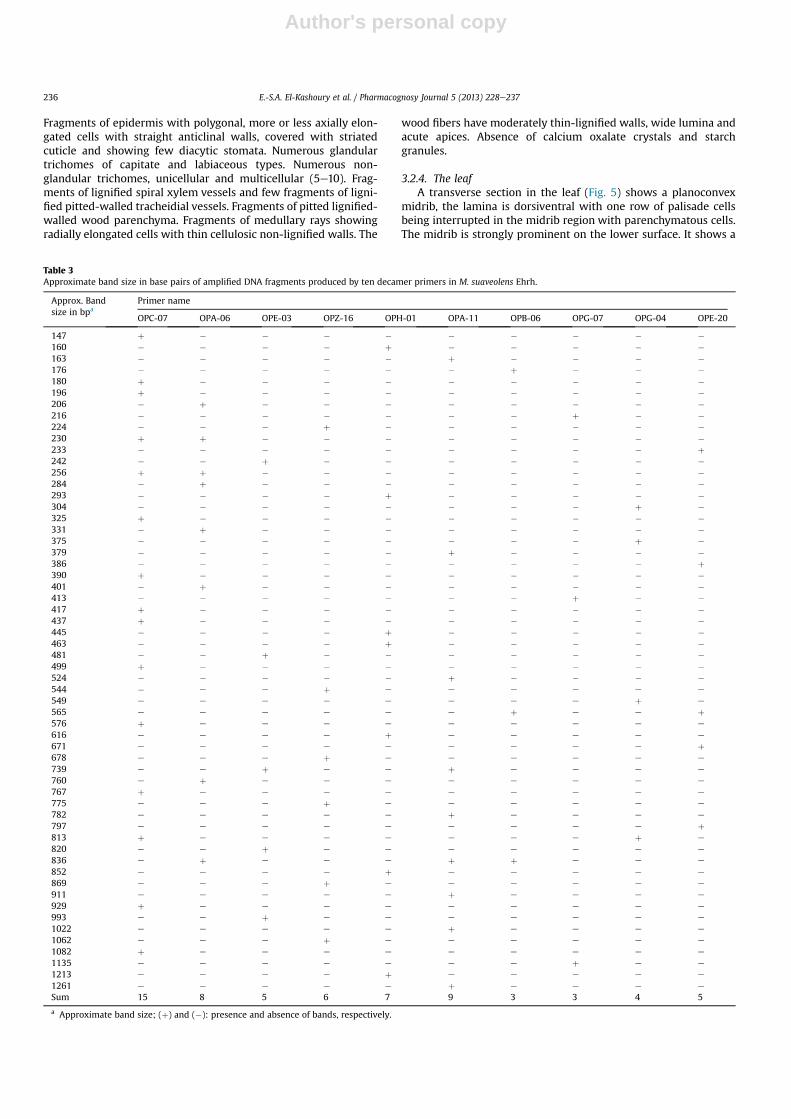
Table 3Approximate band size in base pairs of amplified DNA fragments produced by ten decamer primers in M. suaveolens Ehrh.
Approx. Bandsize in bpa
Primer name
OPC-07 OPA-06 OPE-03 OPZ-16 OPH-01 OPA-11 OPB-06 OPG-07 OPG-04 OPE-20
147 þ � � � � � � � � �160 � � � � þ � � � � �163 � � � � � þ � � � �176 � � � � � � þ � � �180 þ � � � � � � � � �196 þ � � � � � � � � �206 � þ � � � � � � � �216 � � � � � � � þ � �224 � � � þ � � � � � �230 þ þ � � � � � � � �233 � � � � � � � � � þ242 � � þ � � � � � � �256 þ þ � � � � � � � �284 � þ � � � � � � � �293 � � � � þ � � � � �304 � � � � � � � � þ �325 þ � � � � � � � � �331 � þ � � � � � � � �375 � � � � � � � � þ �379 � � � � � þ � � � �386 � � � � � � � � � þ390 þ � � � � � � � � �401 � þ � � � � � � � �413 � � � � � � � þ � �417 þ � � � � � � � � �437 þ � � � � � � � � �445 � � � � þ � � � � �463 � � � � þ � � � � �481 � � þ � � � � � � �499 þ � � � � � � � � �524 � � � � � þ � � � �544 � e e þ e e e e e e
549 e e e e e e e e þ e
565 e e e e e e þ e e þ576 þ e e e e e e e e e
616 e e e e þ e e e e e
671 e e e e e e e e e þ678 e e e þ e e e e e e
739 e e þ e e þ e e e e
760 e þ e e e e e e e e
767 þ e e e e e e e e e
775 e e e þ e e e e e e
782 e e e e e þ e e e e
797 e e e e e e e e e þ813 þ e e e e e e e þ e
820 e e þ e e e e e e e
836 e þ e e e þ þ e e e
852 e e e e þ e e e e e
869 e e e þ e e e e e e
911 e e e e e þ e e e e
929 þ e e e e e e e e e
993 e e þ e e e e e e e
1022 e e e e e þ e e e e
1062 e e e þ e e e e e e
1082 þ e e e e e e e e e
1135 e e e e e e e þ e e
1213 e e e e þ e e e e e
1261 e e e e e þ e e e e
Sum 15 8 5 6 7 9 3 3 4 5
a Approximate band size; (þ) and (�): presence and absence of bands, respectively.
E.-S.A. El-Kashoury et al. / Pharmacognosy Journal 5 (2013) 228e237236

Author's personal copy
single crescent-shaped collateral vascular bundle, accompaniedwith a parenchymatous pericycle. The upper epidermal cells of theleaf (Figs. 5 and 6A) are polygonal nearly isodiametric havingslightly wavy anticlinal walls, covered with thin smooth cuticle andshowing diacytic stomata while the lower epidermis is similar tothe upper but the anticlinal walls are more wavy (Figs. 5 and 6B).The upper and lower neural epidermal cells (Fig. 6C and D) areaxially elongated cells with straight anticlinal walls and coveredwith striated cuticle. Trichomes (Figs. 5 and 6) are very numerouson both surfaces. They are of glandular and non-glandular types.The glandular trichomes are more frequent on the lower epidermis.They are similar to those present on the stem and differ only in size.The mesophyll (Fig. 5AeC) is dorsiventral with an upper palisadeformed of one row of radially elongated columnar cells withstraight anticlinal walls and containing green chloroplasts. It isinterrupted by polygonal parenchymatous cells in the midrib re-gion. The spongy tissue is formed of irregular shaped parenchy-matous cells with wide intercellular spaces. The cortex (Fig. 5A andB) consists of upper and lower cortical tissues. The upper corticaltissue of the midrib consists of 3e5 rows of more or less roundedparenchymatous cells with thin cellulosic walls followed by 3e4rows of irregular thin-walled parenchyma. The lower cortical tissueof the midrib consists of 2e3 rows of collenchymas followed by 6e8 rows of more or less rounded parenchymatous cells with thincellulosic walls. The endodermis is indistinct. The pericycle (Fig. 5Aand B) consists of parenchymatous cells below the vascular bundle,they are small, polygonal to rounded cells with thick cellulosicwalls. The vascular tissue (Fig. 5A and B) shows a single crescent-shaped collateral vascular bundle formed of: The phloem (Fig. 5Aand B) is comparatively narrow, consists of thin-walled cellulosicphloem elements; sieve tubes, companion cells and phloem pa-renchyma, traversed by uni- to biseriate medullary rays, formed ofradially elongated cells with thin cellulosic non-lignified walls(Fig. 5A and B). The cambium (Fig. 5A and B) is almost distin-guishable, formed of about 2e3 rows of thin-walled tangentiallyelongated, radially arranged cells. The xylem (Fig. 5) consists of fi-bers, vessels and wood parenchyma. The vessels (Figs. 5A, B and 6)are lignified, diffused in radial rows with spiral and pitted thick-enings. Fibers and wood parenchyma are similar to those of thestem.
The microscopical measurements of the different elements ofthe leaf are listed in Table 2.
3.2.5. Powdered leafPowdered leaf (Fig. 6) is dark green in color with aromatic odor
and taste. The powder is characterized microscopically by thefollowing features: Fragments of the upper and lower epidermisesshowing polygonal nearly isodiametric cells having wavy anticlinalwalls, covered with thin smooth cuticle and showing numerousdiacytic stomata. Fragments of the upper and lower neural epi-dermises showing axially elongated cells with straight anticlinalwalls and covered with striated cuticle. Numerous glandular tri-chomes of capitate and labiaceous types. Numerous non-glandulartrichomes, unicellular and multicellular (5e10). Fragments oflignified spiral and pitted xylem vessels. Fragments of columnar,thin-walled palisade cells containing green chloroplasts. Absence ofcalcium oxalate crystals and starch granules.
3.3. Genetic profiling
The banding profile produced by the ten decamer primers usedin RAPD analysis of M. suaveolens Ehrh. cultivated in Egypt isillustrated in (Fig. 7). The RAPD electrophoretic profile of DNAsample amplified with the ten decamer primers showed distin-guishable bands and generated 65 fragment patterns. RAPD frag-ments showed 15 bands by OPC-07, 9 bands by OPA-11, 8 bands byOPA-06, 7 bands by OPH-01, 6 bands by OPZ-16, 5 bands by OPE-03and OPE-20, 4 bands by OPG-04, 3 bands by both OPG-07 and OPB-06. The analysis of RAPD data can thus select the use of primersOPC-07 (15 bands ranging from 0.15 to 1.08 Kbp) and OPA-11 (9bands ranging from 0.16 to 1.26 Kbp) for the selective discrimina-tion of M. suaveolens Ehrh.
4. Conclusion
Themacro- andmicromorphological characteristics of the stemsand leaves of M. suaveolens Ehrh. cultivated in Egypt are presented,with the aim of finding out the diagnostic characters for identifi-cation and differentiation of this species in both entire or powderedforms. The DNA of the plant was amplified using ten decamer toreveal RAPD fragments. Each of the ten primers successfullydirected the amplification of a genome-specific fingerprint of DNAfragment. A total of 65 different fragments have been producedmainly by these two primers: OPC-07 and OPA-11. The analysis ofRAPD data can thus select the use of these two primers for theselective discrimination ofM. suaveolens Ehrh. The analysis of RAPDdata, under the experimental conditions, can thus be used todistinguish the plant from other related species Table 3.
Conflicts of interest
All authors have none to declare.
References
1. Bailey LH. The Standard Cyclopedia of Horticulture. vol. 3. New York: The Mac-Millan Company; 1953:2034e2036.
2. Youngken HW. Pharmaceutical Botany. 17th ed. Philadelphia: The BlakistanCompany; 1954, 601e303.
3. Boulos L. Flora of Egypt. vol. 3. Cairo, Egypt: Al-Hadara Publishing; 2002:11e13.4. Lawrence HM. Taxonomy of Vascular Plants. New York: The MacMillan Com-
pany; 1958:688e691.5. Täckholm V. Students’ Flora of Egypt. Cairo, Egypt: Anglo-Egyptian Bookshop;
1956, 19 and 139e143.6. Quattrocchi U. CRC World Dictionary of Plant Names. vol. 3. New York: London:
CRC Press; 1999:1658e1659.7. Brown D. Encyclopedia of Herbs and Their Uses. London: Dorling Kindersley
Limited and New York: Munich, Melbourne, Delhi; 2001:290e300.8. Cheers G. Botanica. Australia: Random House Australian Pty. Ltd and North
Sedney: Pacific Highway; 2004:571e573.9. Iscan G, Kirimer N, KurkcuogluM, Baser KHC, Demirci F. Antimicrobial screening
ofMentha piperita essential oils. J Agric Food Chem. 2002;50:3943e3946.10. Abbaszadeh B, Valadabadi S, Farahani H, Darvishi H. Studying of essential oil
variations in leaves of Mentha species. Afr J Plant Sci. 2009;3:217e221.11. Oumzil H, Ghoulami S, Rhajaoui M, et al. Antibacterial and antifungal activity of
essential oils of Mentha suaveolens. Phytother Res. 2002;16:727e731.12. Shinde VM, Dhalwal K. DNA fingerprinting of Tinospora cordifolia using RAPD
analysis. J Global Pharm Technol. 2010;2:38e42.13. Jaffreys AJ, Wilson V, Thein SL. Individual specific fingerprints of human DNA.
Nature. 1985;316:76e79.14. Doyle J, Doyle JL. A rapid DNA isolation procedure for small quantities of fresh
leaf tissue. Phytochem Bull. 1987;19:11e15.
E.-S.A. El-Kashoury et al. / Pharmacognosy Journal 5 (2013) 228e237 237