atypical pet prof.salah 1
TRANSCRIPT

Atypical PET&Eclapmsia
DR. SALAH ROSHDY,MD
PROFESSOR.OF
OBSTETRICS/GYNECOLOGY QASSIM COLLEGE OF MEDICINE
Sohag University,Egypt

PRE: History
2200 BC Egypt: pregnant women with fits
Eclampsia: Greek word: suddenly, flash
1619: Varardus: first use of word eclampsia
1843: Lever. Proteinuria. Swelling and convulsions: Nephritic toxemia
1897: Vaquez. Hypertension
1899: Strogonov: treatment, sedation
1900s: prenatal care, preeclampsia
New concept in the 20th century
1902: Ballantyne. Pro-maternity clinic.
1910: USA. Nursing visits at home.
1920: Prenatal visits: check for hypertension, swelling, proteinuria to detect : Preeclampsia
Eclampsia - Preeclampsia Prenatal Care

3
Cur
rent
App
roa
ch
to
Re
duc
tion
of
Mat
ern
al
Mor
talit
y
Maternal Mortality: A Global Tragedy
Annually, 585,000 women die of pregnancy related complications
99% in developing world
~ 1% in developed countries

4
Every Minute...
Maternal Death Watch 380 women become
pregnant
190 women face unplanned or unwanted pregnancy
110 women experience a pregnancy related complication
40 women have an unsafe abortion
1 woman dies from a pregnancy-related complication

Global Causes of Maternal Mortality
24.8
14.9
12.96.9
12.9
7.9
19.8
Hemorrhage 24.8%
Infection 14.9%
Eclampsia 12.9%
Obstructed Labor6.9%Unsafe Abortion12.9%Other Direct Causes7.9%Indirect Causes19.8%

Maternal Mortality
USA: 15/100,000 live births
Mali: 800/100,000 live births
Hemorrhage
Embolism
Preeclampsia
Infection

Hypertensive Disorders of Pregnancy
Pregnancy Induced
Hypertension
PIH
(no
proteinuria)
HELLP
Syndrome
Chronic Hypertension
(pre-existing or
undiagnosed prior to
pregnancy))
6-8% of
all gestations
Preeclampsia
(PIH with
proteinuria)
Eclampsia Severe
Preeclampsia AFLP

National High Blood Pressure Education Program Classification ( NHEP) 2000
1-Gestational hypertension.
2-Preeclampsia (mild, severe)
3-Eclampsia. 4-Superimposed preeclampsia upon chronic
hypertension.
5-Chronic hypertension with pregnancy.
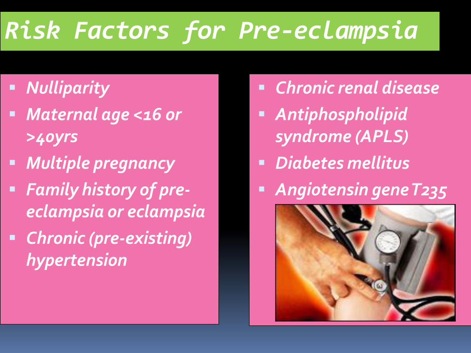
Risk Factors for Pre-eclampsia
Nulliparity
Maternal age <16 or >40yrs
Multiple pregnancy
Family history of pre-eclampsia or eclampsia
Chronic (pre-existing) hypertension
Chronic renal disease
Antiphospholipid syndrome (APLS)
Diabetes mellitus
Angiotensin gene T235
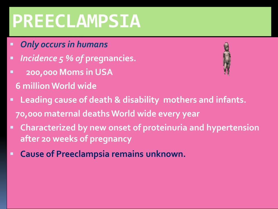
PREECLAMPSIA Only occurs in humans
Incidence 5 % of pregnancies.
200,000 Moms in USA
6 million World wide
Leading cause of death & disability mothers and infants.
70,000 maternal deaths World wide every year
Characterized by new onset of proteinuria and hypertension after 20 weeks of pregnancy
Cause of Preeclampsia remains unknown.
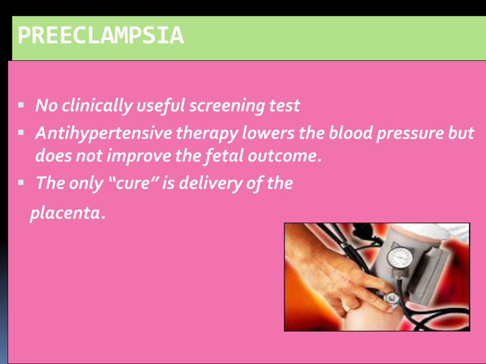
PREECLAMPSIA
No clinically useful screening test
Antihypertensive therapy lowers the blood pressure but does not improve the fetal outcome.
The only “cure” is delivery of the
placenta.


The placenta – perhaps as
as a result of ischemia –
secretes a factor into the
maternal circulation
which
produces systemic
endothelial dysfunction

ENDOTHELIAL DYSFUNCTION LEADS TO :
Hypertension - disturbed endothelial control of vascular tone
Proteinuria - increased glomerular vascular permeability
Coagulopathy – abnormal expression of pro & anti coagulants
Liver Dysfunction – ischemia & vasoconstriction


BIOMARKERS OF PLACENTAL DYSFUNCTION
sVEGF R1 Soluble Vascular Endothelial Growth Factor Receptor 1
Also known as soluble fms-like tyrosine kinase 1 (sFlt-1)
Anti-angiogenic protein
Elevated in preeclampsia

BIOMARKERS OF PLACENTAL
DYSFUNCTION
PlGF Placental Growth Factor
Angiogenic protein, promotes angiogenesis
Binds to VEGF Receptor (VEGF R1 and sVEGF R1)
Free PlGF is reduced in preeclampsia

sVEGF R1 PlGF
PlGF
sVEGF R1
Normal Pregnancy Preeclampsia
Healthy endothelial cell
•Maintains vascular tone
•Maintains glomerular filtration
•Maintains blood-brain barrier
•Maintains anti-coagulant state
Endothelial cell injury
•Hypertension
•Proteinuria
•Cerebral edema
•Coagulation/liver function
abnormalities
Anti-angiogenic state: anti↑ / pro↓

The Constant Pathophysiological Changes
Is Vascular endothelial: Damage +Dysfunction
+ Spasm

Possible mechanisms in Preeclampsia
Friedman and Lindheimer,1999


Multisystem Features Of Preeclampsia
Hypertension Proteinuria
Eclampsia HELLP syndrome
Intra-uterine growth
restriction
Multi-organ
disease Cerebral vessels
Fetus
Liver
Systemic blood vessels Kidneys

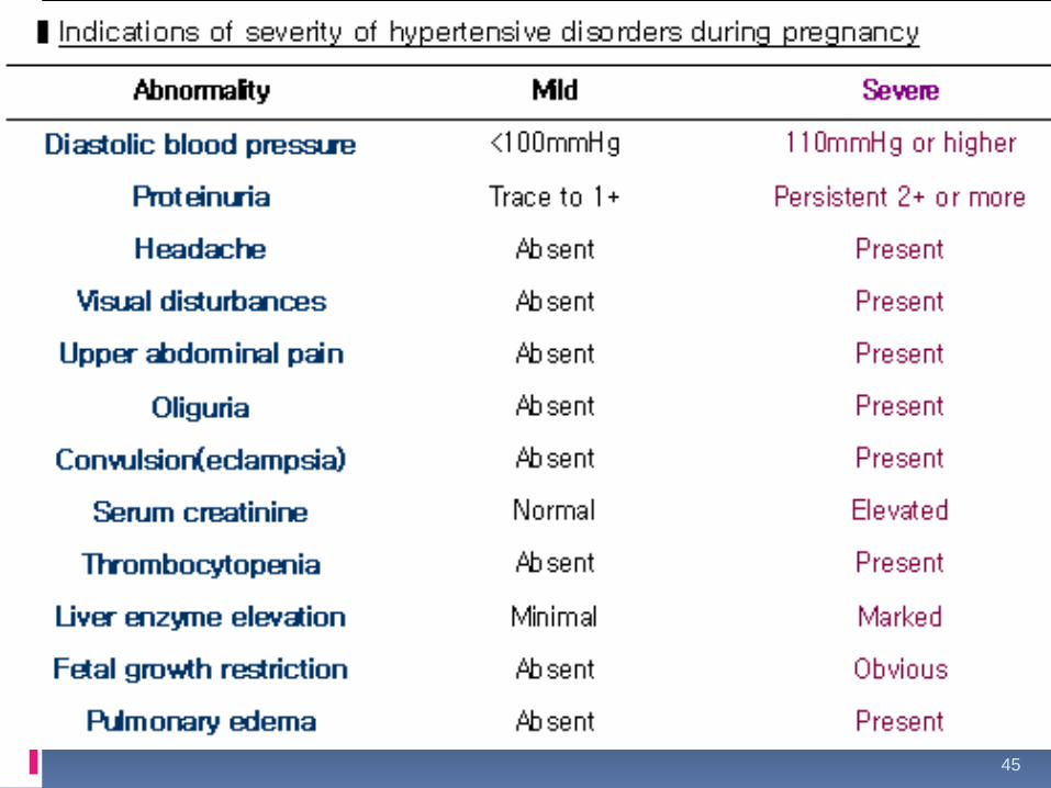
Severe pre-eclampsia: symptoms, signs & diagnostic
criteria
Headaches
Visual Disturbances
Pulmonary Oedema
Hepatic Dysfunction
RUQ or Epigastric Pain
Oliguria
Elevated Creatinine
Thrombocytopaenia or haemolysis
Proteinuria of 5 g or more in 24 hrs
Systolic BP > 160 to 180 mm Hg
Diastolic BP > 110 mm Hg
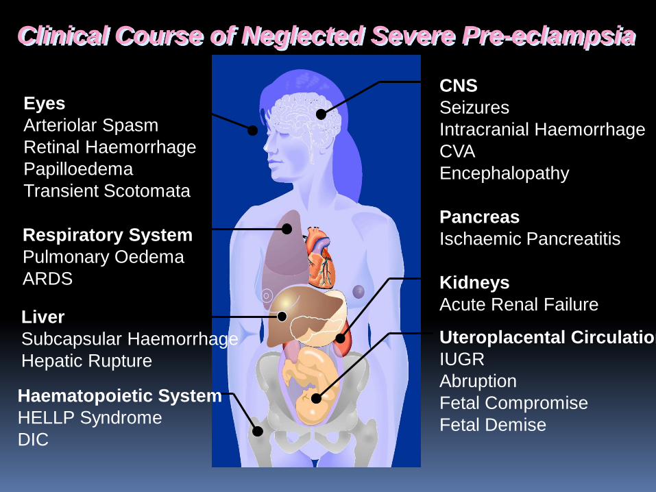
Clinical Course of Neglected Severe Pre-eclampsia
Eyes
Arteriolar Spasm
Retinal Haemorrhage
Papilloedema
Transient Scotomata
Respiratory System
Pulmonary Oedema
ARDS
Liver
Subcapsular Haemorrhage
Hepatic Rupture
Haematopoietic System
HELLP Syndrome
DIC
CNS
Seizures
Intracranial Haemorrhage
CVA
Encephalopathy
Pancreas
Ischaemic Pancreatitis
Kidneys
Acute Renal Failure
Uteroplacental Circulation
IUGR
Abruption
Fetal Compromise
Fetal Demise


Case 1
A 20-year-old primigravida was hospitalized at 37 weeks with regular contractions. She had had irregular antenatal visits, which revealed no abnormality.
She had a blood pressure (BP) between 130/80 and 100/60 mmHg on admission.
Laboratory findings were unremarkable , with a trace of proteinuria in the urinalysis.
She had no prodromes suggestive of hypertensive disease.
She delivered a healthy female baby vaginally a few hours later, uneventfully.

Case 1
At 4 h after delivery, she developed a generalized
convulsion, lasting 23 min, despite being normotensive,and soon after this she had two other seizures.
MgSO4 was given .
Following the seizures, her BP ranged between 140/90 and 100/60 mmHg .
Slight increases in the liver function tests and LDH values and slight decreases in hemoglobin and platelets were detected .

Case 1
hemoglobin 10.3 mg/dL,
hematocrit 31.5%,
platelets 91,000/mm3,
alanine aminotransferase (ALT) 35 U/L,
aspartate transaminase (AST) 66 U/L,
lactate dehydrogenase (LDH) 932 U/L).

Case 1
Computed tomography (CT) was completely normal. Subsequently, she had three more seizures and another
2-g bolus of MgSO4 was infused over 35 min and continued for the following 24 h, during which she suffered no further convulsion.
The next day, a 24 h urine sample revealed 330 mg proteinuria;
cranial magnetic resonance imaging (MRI) showed no abnormality.


Case 2
A 20-year-old nulligravida was admitted with regular contractions at 37 weeks gestation.
All her prenatal visits had been normal, including BP, which was recorded as 120/80 to 110/70 mmHg.
There was no prodrome of hypertensive disease and no laboratory abnormality, including platelet count, liver enzymes, LDH, electrolytes, and glucose, although proteinuria (3+) on dipstick was noticed on admission.

Case 2
She delivered a 2800-g male fetus vaginally, uneventfully.
Following the delivery, her BP increased suddenly to 150/100 to 140/100 mmHg.
Then, she had a generalized seizure lasting 5-10 s. Then, 2 h later, she developed sudden blindness, an occipital headache, and myoclonic seizures, particularly involving the right upper extremity.

Case 2
The postictal BP was around 160/120 mmHg.
MgSO4 was given for 24 h as the patient seemed to have atypical eclampsia.
She had no seizure subsequently. Her BP remained high for a few days, ranging between 150/100 and 140/90 mmHg and normalized on postpartum day 3, with 930 mg/dL proteinuria in the 24 h urine collected postpartum.
Cranial MRI was unremarkable.


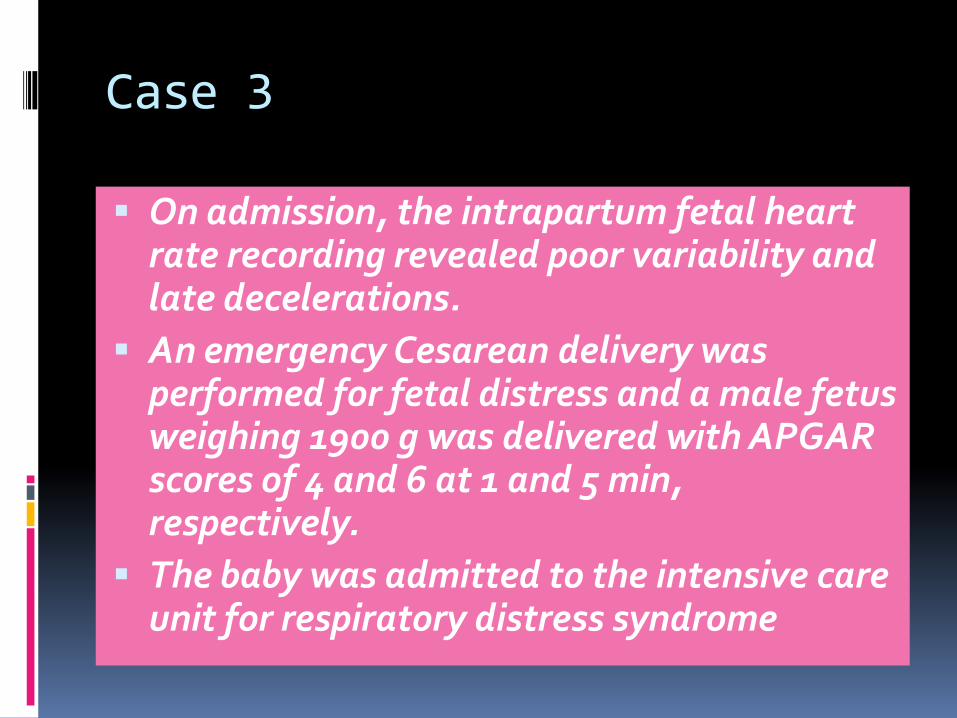
Case 3
A 31-year-old multipara presented with contractions at 33 weeks gestation with the cervix 2 cm dilated and 40% effaced.
Her BP was 110/70 to 110/60 mmHg on admission.
Her CBC,routine biochemical tests, and coagulation studies were normal, but she had a dipstick proteinuria of 3+.
Case 3
On admission, the intrapartum fetal heart rate recording revealed poor variability and late decelerations.
An emergency Cesarean delivery was performed for fetal distress and a male fetus weighing 1900 g was delivered with APGAR scores of 4 and 6 at 1 and 5 min, respectively.
The baby was admitted to the intensive care unit for respiratory distress syndrome

Case 3
The placenta was atrophic, but not abrupted.
Then, 2 h postoperatively, the mother became hypertensive, with a BP of 160/100 to 150/100 mmHg, a severe headache and visual blurring.
A MgSO4 infusion was started with a loading dose over 20 minutes, followed by a maintenance dose of 2 g/h as a continuous intravenous infusion for 24 h.
Three days later, she became normotensive and her complaints resolved.


Atypical preeclampsia
Gestational hypertension plus 1 of the following items:
Symptoms of preeclampsia
Hemolysis
Thrombocytopenia ( 100,000/mm3)
Elevated liver enzymes (2 times the upper limit of the normal value for
aspartate aminotransferase or
alanine aminotransferase)
Gestational proteinuria plus 1 of the following items:
Symptoms of preeclampsia
Hemolysis
Thrombocytopenia
Elevated liver enzymes
Early signs and symptoms of preeclampsia-eclampsia at 20 weeks of gestation
Late postpartum preeclampsia-eclampsia ( 48 hours after delivery)

Signs and symptoms
Signs and symptoms results consistent with preeclampsia
Right upper quadrant pain Epigastric pain Retrosternal chest pain Nausea and vomiting Shortness of breath/congestive heart failure Headaches (not responsive to analgesics) Visual changes Altered mental status Bleeding from mucosal membranes Jaundice
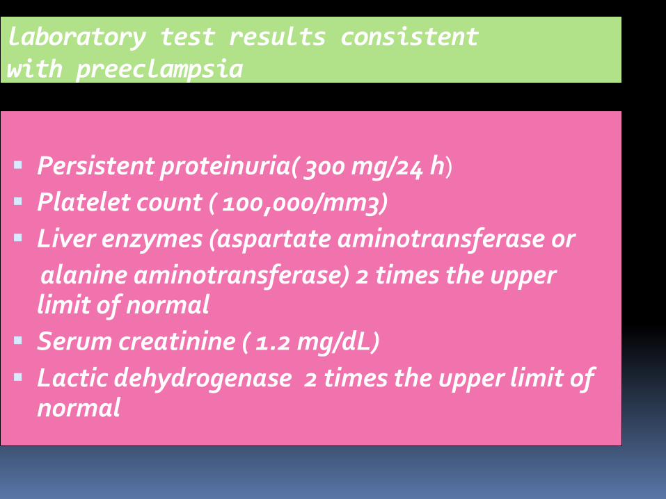
laboratory test results consistent with preeclampsia
Persistent proteinuria( 300 mg/24 h)
Platelet count ( 100,000/mm3)
Liver enzymes (aspartate aminotransferase or
alanine aminotransferase) 2 times the upper limit of normal
Serum creatinine ( 1.2 mg/dL)
Lactic dehydrogenase 2 times the upper limit of normal

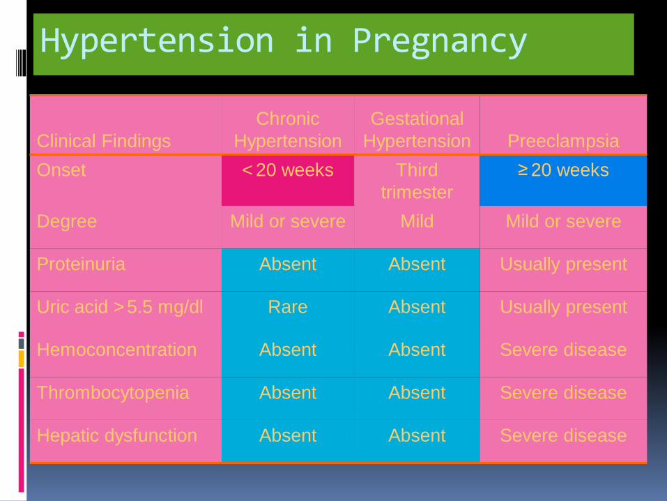
Hypertension in Pregnancy
Clinical Findings
Chronic
Hypertension
Gestational
Hypertension Preeclampsia
Onset < 20 weeks Third
trimester
≥ 20 weeks
Degree Mild or severe Mild Mild or severe
Proteinuria Absent Absent Usually present
Uric acid > 5.5 mg/dl Rare Absent Usually present
Hemoconcentration Absent Absent Severe disease
Thrombocytopenia Absent Absent Severe disease
Hepatic dysfunction Absent Absent Severe disease
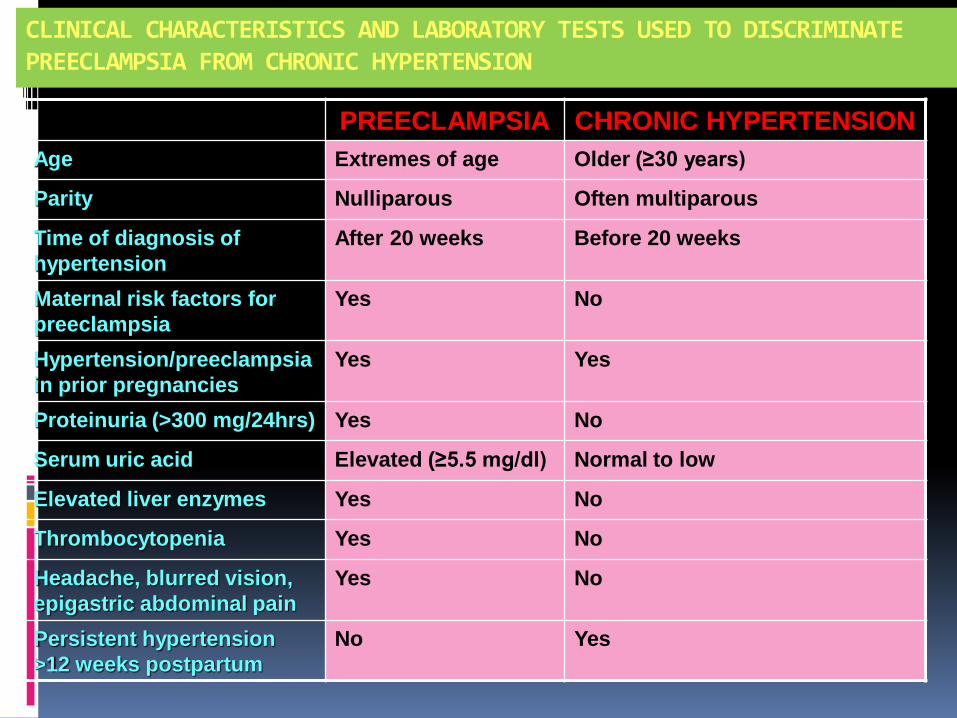
CLINICAL CHARACTERISTICS AND LABORATORY TESTS USED TO DISCRIMINATE PREECLAMPSIA FROM CHRONIC HYPERTENSION
PREECLAMPSIA CHRONIC HYPERTENSION
Age Extremes of age Older (≥30 years)
Parity Nulliparous Often multiparous
Time of diagnosis of
hypertension
After 20 weeks Before 20 weeks
Maternal risk factors for
preeclampsia
Yes No
Hypertension/preeclampsia
in prior pregnancies
Yes Yes
Proteinuria (>300 mg/24hrs) Yes No
Serum uric acid Elevated (≥5.5 mg/dl) Normal to low
Elevated liver enzymes Yes No
Thrombocytopenia Yes No
Headache, blurred vision,
epigastric abdominal pain
Yes No
Persistent hypertension
>12 weeks postpartum
No Yes
45


Frequency of Various Signs and Symptoms Among Imitators of Pre-eclampsia–Eclampsia S&S HELLP
Syndrome AFLP TTP HUS Exce.SLE
Hypertension
85% 50 % 20-75 % 80-90% 80 %
Protenuria 90-95% 30-50% +haemturia 80-90 % 100 %/nephritis
Fever Absent 25-30 % 20-50 % ? Common/flare
Jaundice 5-10 % 40-90 % Rare Rare
Absent
N & Vomiting
40 % 50-80 % Common Common Only/APA
Abd/Pain 60-80 % 35-50 % Common Common Only/APA
CNS 40-60 % 30-40 % 60-70 % ? 50%/APA

Frequency of Various Signs and Symptoms Among Imitators of Pre-eclampsia–Eclampsia
Lab HELLP
AFLP TTP HUS Exce.SLE
Platelet >20,000 >50,000 <20,000 >20,000 >20,000
Haemolysis 50-100% 15-20 % 100 % 100 % 14-23% w/APA
Anemia <50% Absent 100 % 100 % 14-23% w/APA
DIC <20% 73 % Rare Rare
Rare
Hypoglycemia Absent 61% Absent Absent Absent
VW factor multimers
Absent Absent 80-90% 80% <10%
ADAMTS 13% < 5% Absent Absent 33-100% Rare Rare
Impaired renal f. 50% 90-100% 30% 100% 40-80%
LDH (IU/L) >600 Variable >1000 >1000 with APA
Elevated ammonia Rare 50% Absent
Absent
Absent
Elevated bilirubin 50-60% 100% 100% <10%
Elevated transaminases
100%
100%
Usually mild† Usually mild† with APA

Differential diagnosis of eclampsia
Cerebrovascular accidents
Hemorrhage
Ruptured aneurysm
Arterial embolism or thrombosis
Cerebral venous thrombosis
Hypoxic ischemic encephalopathy
Angiomas
Hypertensive encephalopathy

Differential diagnosis of eclampsia
Seizure disorders
Previously undiagnosed brain tumors
Metastatic gestational trophoblastic disease
Metabolic diseases
Reversible posterior leukoencephalopathy syndrome
Thrombophilia
Thrombotic thrombocytopenic purpura
Postdural puncture syndrome
Cerebral vasculitis

Management
Objective
termination of pregnancy with the least possible trauma to mother and fetus
birth of an infant who subsequently thrives
complete restoration of health to the mother

Termination of pregnancy
Delivery is the cure for preeclampsia
The prime objectives
To forestall convulsion
To prevent intracranial hemorrhage
To prevent serious damage to vital organs
To deliver a healthy infant

Management of pre-eclampsia
Sibai et al. Lancet 365:785-99, 2005.

Elective cesarean delivery
Labor induction to effect vaginal delivery has traditionally been considered to be in the best interest of the mother
Several concerns have led some practitioners to advocate cesarean delivery
Unfavorable cervix precluding successful induction of labor
Perceived sense of urgency because of the severity of preeclampsia
The need to coordinate neonatal intensive care

LONG-TERM IMPLICATIONS FOR WOMEN WITH PREECLAMPSIA
Increased incidence of salt-sensitive HTN (Sibai et al, AJOG 1991, Wilson et al, BMJ 2003)
Increased risk for cardiovascular mortality (Irgens et al, BMJ 2001, Funai E et al, Epidemiology 2005,
Arnadottir et al, BJOG 2005)
Increased incidence of chronic renal disease reports of focal sclerosis, increased incidence of renal biopsies, ESRD- Norwegian study – Vikse et al, JASN
2006, NEJM 2008
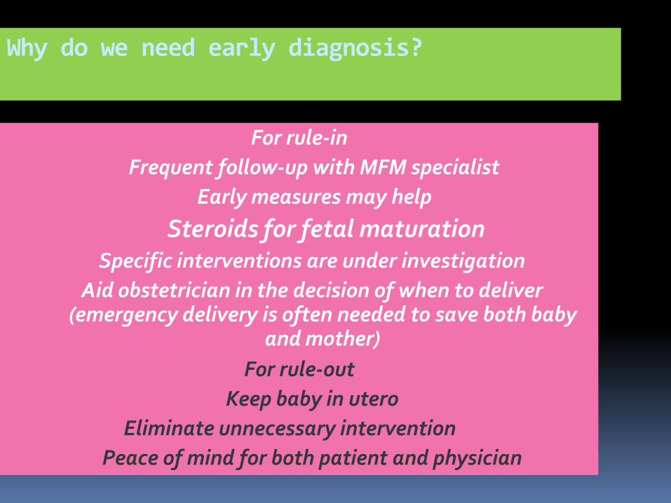
Why do we need early diagnosis?
For rule-in
Frequent follow-up with MFM specialist
Early measures may help
Steroids for fetal maturation Specific interventions are under investigation
Aid obstetrician in the decision of when to deliver (emergency delivery is often needed to save both baby
and mother)
For rule-out
Keep baby in utero
Eliminate unnecessary intervention
Peace of mind for both patient and physician


In conclusion
The absence of hypertension or proteinuria should not preclude diagnosing preeclampsia/eclampsia.
Eclampsia or fetal distress may be an unusual presenting scenario in atypical cases before the detection of overt hypertension or proteinuria.

In conclusion
Even minor clues in diagnoses, such as a marginally elevated BP or trace proteinuria, may be critical for appropriate, timely management.
Obstetricians should be aware of atypical presentations,maintain a high level of suspicion, and be ready to take immediate steps.

Conclusion
Moreover, valuable time should not be spent conducting detailed investigations.
The most common cause of convulsions in association with hypertension or proteinuria during pregnancy or immediately postpartum is eclampsia.
However, late postpartum eclampsia is defined as eclampsia that occurs more than 48 h, but less than four weeks, after delivery

Conclusion
All patients with atypical-onset eclampsia should undergo a neurological evaluation to rule out the presence of neurologic causes of seizures .
Cerebral imaging is indicated for patients with focal neurologic signs, such as hemiparesis, an unconscious state, and prolonged coma.

Conclusion
Additionally, cerebral imaging may be helpful in patients who have an atypical presentation of eclampsia (onset before 20 weeks or more than 48 h after delivery, refractory to magnesium sulfate therapy, and recurrent seizures).


Thank You