atypical aggressive periapical granuloma: a case report · differential diagnosis of apical...
TRANSCRIPT

Article ID: WMC002874 ISSN 2046-1690
Atypical Aggressive Periapical Granuloma: A CaseReportCorresponding Author:Prof. Sergio E Cury,DDS PhD, Oral Pathology - UniFOA - University of Volta Redonda, 27.310-060 - Brazil
Submitting Author:Prof. Sergio E Cury,DDS PhD, Oral Pathology - UniFOA - University of Volta Redonda, 27.310-060 - Brazil
Article ID: WMC002874
Article Type: Case Report
Submitted on:11-Jan-2012, 01:32:26 AM GMT Published on: 11-Jan-2012, 07:54:20 AM GMT
Article URL: http://www.webmedcentral.com/article_view/2874
Subject Categories:ORAL MEDICINE
Keywords:Periapical Granuloma, Periapical disease, Oral surgery, Computer Tomography, Chronic Inflamation.
How to cite the article:Souto M F, Felipe Silva M B, Cury S E, Cury M P, Junqueira J C, Manhaes Jr L C.Atypical Aggressive Periapical Granuloma: A Case Report . WebmedCentral ORAL MEDICINE2012;3(1):WMC002874
Copyright: This is an open-access article distributed under the terms of the Creative Commons AttributionLicense, which permits unrestricted use, distribution, and reproduction in any medium, provided the originalauthor and source are credited.
Source(s) of Funding:
None
Competing Interests:
None
Additional Files:
References
Webmedcentral > Case Report Page 1 of 11

WMC002874 Downloaded from http://www.webmedcentral.com on 07-Mar-2012, 03:00:01 AM
Atypical Aggressive Periapical Granuloma: A CaseReportAuthor(s): Souto M F, Felipe Silva M B, Cury S E, Cury M P, Junqueira J C, Manhaes Jr L C
Abstract
We reported a atypical and aggressive case of aperiapical granuloma, located in the mandibular rightfirst and second molars region of the caucasian 32year-old woman.
Introduction
The jaws are host to several cysts and neoplasms,mainly due to the tissues involved in tooth formation(Regezi, 2002). Periapical lesions resulting from pulpnecrosis are among the most frequent pathologies ofthe alveolar bone (García et al, 2007). Nevertheless, anumber of benign jaw tumors and some cysts (someof them recently described) of both odontogenic andnon-odontogenic origin can exhibit a biologicallyaggressive course and may be difficult to diagnose.The traditional histopathology is still the main basis forthe diagnosis of these lesions, since the impact ofmolecular and immunohistochemical techniques hasbeen scant in this field so far (Regezi, 2002).Apical periodontitis is a disorganization of theperiradicular tissue caused by etiologic agents ofendodontic origin characterized by a chronicinflammatory infiltrate, which can result in formation ofthe lesion with concomitant resorption of hard tissuesand destruction of the periodontal ligament (Fusaka etal, 2009). Thus, it is consequence of an infection in theroot canal that can result in progressive stages ofinflammation and periradicular bone destruction. Alveolar bone resorption around the tooth apexinvolves the production of direct regulators ofosteoclastic activity and chemotactic osteoclasticfactors and receptors (Fusaka et al, 2009).Many cases of periapical granuloma are completelyasymptomatic. There is no perforation of the bone andoverlying mucosa with the formation of a fistulous path,unless the lesion undergoes acute exacerbation. Withthe proliferation of granulation tissue and concomitantbone resorption, the periapical granuloma appears asa radiolucent area of variable size abutting the rootapex (Shafer, 1987). Six possible biological factorshave been described as causes of asymptomaticapical periodontitis followed by root canal treatment:persistent intraradicular infection, extraradicular
infection (mainly actinomycosis), foreign body reactionrelated to obturating material, accumulation ofendogenous cholesterol crystals that irritate theperiapical tissue, true cystic lesions, and cicatricialtissue (García et al, 2007). The interaction ofmicrobiological factors and the host’s defensemechanism, destroying a large quantity of periapicaltissue, originates the different types of periapicallesion (García et al, 2007).When the dental pulp is invaded by bacteria, the rootcanal provides the habitat for a mixed microbiota thatleads to an inflammatory response in the periapicalregion (Fusaka et al, 2009); this is the most commonetiologic factor responsible for apical periodontitis suchas those that manifest as dental granulomas, radicularcysts, and periapical abscesses (Estrela et al, 2009).The latter develops in response to the intracanalantigenic content, mediated by immunopathologicalmechanisms (Soares & Queiroz, 2001; Soares et al,2006). Typically, periapical inflammatory lesions ofendodontic origin are 5 to 8 mm in diameter.Traditionally, lesions larger than 10 mm areconsidered granulomas, and the largest areconsidered apical cysts (Soares et al, 2006). Thedifferential diagnosis of apical periodontitis can includesome lesions of non-endodontic origin (Rodrigues et al,2008).The aim of this article is to report a radiographicfinding of an expansive and osteolytic lesion in theposterior region of the mandible suggestive of anodontogenic tumor or apical cyst , whosehistopathological examination revealed to be an apicalgranuloma. The radiographic, clinical, and histologicalaspects of the lesion, as well as the postsurgicalfollow-up after 5 and 8 months are described.
Case Report(s)
A 32 year-old caucasian woman sought a routineclinical examination at the dentist complaining of painand discomfort on the mandibular left first molar; theclinician requested a panoramic radiograph andperiapical radiographs to complement the examination.A radiolucent lesion circumscribed by a radiopaquehalo, expansive and causing thinning of the rightmandible cortical bone in the region of mandibularright first and second molars was observed in the
Webmedcentral > Case Report Page 2 of 11

WMC002874 Downloaded from http://www.webmedcentral.com on 07-Mar-2012, 03:00:01 AM
panoramic radiograph (Illustration 1A). The mandibularright first molar was endo-perio impaired, and the rootcanals were filled. The last visit to the dentist had beenaround 5 years earlier and no record of the lesion wasnoted.The patient did not report any symptomatology in theregion, since the complaint of discomfort concernedthe opposite side, where there was an apical lesion onthe mandibular left first molar. However, shementioned sporadic abscesses in the buccal apicalregion of the mandibular right first molar.The physical extraoral examination revealed aninconspicuous facial asymmetry in the basal and angleregion of the mandible, as shown in Figure 2. Theintraoral examination revealed an increase of thebuccal volume and an altered (reddish) coloration ofthe gingiva (Figure 3).The radiologist requested a cone beam computedtomography to better evaluate the size and extent ofthe lesion and tooth impairment. The examinationrevealed an osteolyt ic, hypodense lesion,circumscribed by a hyperdense halo, impairing theright mandibular canal, expansive to the lingualcortical and the mandibular base, involving the apex ofmandibular right second molar and communicatingwith the endo-perio lesion of the mandibular right firstmolar (Illustration 1B, C, D, E, F, G and H). Themandibular right third molar was not affected. Thediagnostic hypotheses considered were unicysticameloblastoma, keratocystic odontogenic tumor, andapical cyst.The patient was referred to the head and necksurgeon, who decided to perform an excisional biopsy.Transoperatively the access was first intraoral, withremoval of a fragment of the buccal periodontal lesionof the mandibular right first molar. Because the lesionwas lingually located, the surgeon decided to access itextraorally, via the neck, until the concerned regionwas reached. During removal of the lesion, it wasfound to be encapsulated and enclosing a certainquantity of granulomatous material (Illustration 2A).Upon removal, curettage was performed in thesurgical site (Illustration 2B); the teeth involved werenot removed considering the risk of pathologic fracture.The pieces were then sent to histopathologicalexamination.The histological sections were stained withhematoxylin-eosin and examined by light microscopy.They revealed fragments of connective tissue ofvariable density, the outer portion presenting moredensely packed and randomly distributed collagenfibers, interspersed with fusiform fibroblasts, somemore basophilic, some bulky in focal area; the innerportion presented an intense, mixed inflammatory
infiltrate composed by lymphocytes, plasma cells,neutrophils, and epithelioid and foamy macrophages.Additionally, numerous blood vessels, some of whichengorged, were found. Areas of erythrocyteextravasation bacteria colonies completed the panel(Illustration 2 C, D, E and F). The diagnosis was achronic nonspecific inflammatory process, and hencea dental granuloma. The pathologist further added that,since the histologic panel was strongly associated withthe endo-periodontal impairment of the mandibularright first molar, elimination of the infection focus in theregion and radiographic follow-up was recommended.After four months, the involved mandibular right firstmolar was removed and a new computed tomographywas taken to monitor the involution of the lesion. Theexamination revealed that the lesion had regressedand the expansion had reduced, nevertheless it wasnot possible to assess if the bone had been repaired.Another computed tomography was taken after eightmonths and allowed observation of bone formationand repair.
Discussion
Clinically and radiographically, the extent, enucleationand bone destruction gave the lesion an atypicalpattern of granuloma. One of the diagnostic hypotheses was a keratocysticodontogenic tumor (KOT). According to Regezi (2002)and Neville (2004), the KOT is commonly classified asa developmental cyst of considerable importance dueto its potential for aggressive and recurrent clinicalbehavior. Radiographically, they describe it as awell-defined and generally multilocular radiolucency.KOTs represent 5-15% of all odontogenic cysts.Another diagnostic hypothesis was ameloblastoma,since the unicystic subtype pattern is entirely cystic,generally consists of a single space, occurs in thesecond and third decades of life and in the mandibularmolar region (Regezi, 2002), corroborating thereported clinical case. Furthermore, cysticameloblastomas can expand and perforate themaxillary bone cortex (Regezi, 2002). The apical cysthypothesis was not discarded when the clinical,radiographic and surgical panels were evaluatedtogether, considering, on the other hand, that this isthe most frequent maxillary cystic lesion (Romero et al,2002). Notwithstanding, the hypothesis of apicalgranuloma was not precisely considered because ofthe atypical behavior of the lesion.The healing of lesions associated with apicalperiodontitis, such as granulomas, abscesses, andcysts, fails after root canal therapy due to persistent
Webmedcentral > Case Report Page 3 of 11

WMC002874 Downloaded from http://www.webmedcentral.com on 07-Mar-2012, 03:00:01 AM
intra and extraradicular infection (Lin et al, 2009). Theroot canal treatment performed in the mandibular rightfirst molar in the present case was not enough tocease the infection in the region and probably wouldbe the etiologic factor for the growth and persistenceof the lesion. Soares et al (2006) contended that,among the surgical therapies of extensive osteolyticlesions, marsupialization should be the less invasive,although some discomfort is associated with theprolonged use of drains, hygiene maintenance can bedifficult, healing can be prolonged, and anothersurgery may be necessary. Since the most likelydiagnostic hypotheses involved odontogenic tumors,the choice fell onto excisional biopsy. Soares et al(2006) added that, in the case of enucleation, largervessels and nerves should be protected andreconstructive grafts may be necessary, requiringspecial hospital care. In this case, reconstructive graftswere not necessary, since bone destruction waslargely restricted to the lingual bone plate and did notimpair the patient’s function or esthetics.As to the preoperative evaluation, attempts todiagnose periapical lesions before surgery withmedium contrast, Papanicolau smear, albumin testsand electrophoresis proved to be inaccurate (Simon etal, 2006; Trope et al, 1998). Traditionally, it isinstituted that periapical cysts and granulomas can bedifferentiated in periapical radiographs. Theradiographic characteristics ascribed to a cyst are:circumscribed, well delineated, with sclerotic border,and larger than 10 mm in diameter. Conversely,supposedly a granuloma would have a diffuse,ill-defined appearance, any shape, and generally adiameter smaller than 10mm (Trope et al, 1989). Yet,Lin et al (2009) contended that extensive periapicallesions and radicular cysts cannot be diagnosedbefore treatment, and the diagnosis can only beconfirmed upon biopsy. Shafer (1987) mentioned thatdifferent aspects in conventional radiography cannotbe used to distinguish the different forms of periapicalillness. Recently, with the advent of other types of imagingexaminations, such as computed tomography,magnetic resonance imaging, and cone beamcomputed tomography (CBCT), density differenceshave improved the accuracy of preoperative diagnosis(Simon et al, 2006). The clinical application of conebeam technology can favor the differential diagnosis ofperiapical lesions. According to Rodrigues & Estrela(2008), cone beam tomography and magneticresonance can supplement, and in some casespossibly avoid, the need for aspiration. Trope et al(1989) proposed that a cyst could be differentiatedfrom a granuloma in computed tomography by a
marked difference in density between the cystic cavitycontent and the granulomatous tissue. Granulomashave a narrower extension and a lower gray scalevalue than apical cysts (Simon et al, 2006). Periapicallesions generally consist of soft solid tissue(granulomas) or include a semi-solid and liquefiedarea (bay or true cyst) (Simon et al, 2006). Themeasurement of gray values allows the differentiationof soft or fluid tissue or empty areas. A radicular cystcan be differentiated from a periapical granuloma withcomputed tomography by a markedly lower density ofthe cystic cavity than granulomatous tissue (Trope etal, 1989). Simon et al (2006) concluded that CBCTprovides a better, more accurate, and quicker methodto differentiate the diagnosis of a solid lesion fromanother filled with fluid or cavity. If a positive grayvalue is obtained, then the lesion is a granuloma. Thisallows the clinician to decide whether or not thesurgery is necessary without waiting for the returnperiod to assess if healing occurred. In the presentclinical case, the gray scale value obtained waspositive, thus corroborating the aforementionedstudies. However, Estrela et al (2009) do not agree,since they argue that it is impossible to clinically andradiographically differentiate granulomas from cysts,as well as true apical cysts from bay cysts, or whenthe epithelial tissue is inert or proliferative. Thus, thehistopathological exam would be mandatory for aconclusive diagnosis, but the importance of studyingthe image to obtain a more adequate treatment plancannot be ruled out or minimized. Camps et al (2004)further assert that variation in the gray scale value of aperiapical lesion is associated with histologicalchanges, with a direct correlation between the valueand the type of material that fills the lesion.Rodrigues & Estrela (2008) concluded that thechallenge in endodontic diagnosis involves themanagement of information obtained in the anamnesis(present condition’s history), clinical examination, pulpvitality test, and analysis of the radiographic aspect.Therefore, care during diagnosis is mandatory todecide the best therapeutic option, because periapicalillnesses of non-endodontic origin cannot emulate themost common infections of endodontic origin, andvice-versa. The behavior of endodontic lesions shouldnot be underrated, as demonstrated in the reportedclinical case, where an apparently nonaggressivepathologic disorder ultimately caused extensiveosseous and dental impairment.
References
1. Camps, J; Pommel, L; Bukiet, F. Evaluation of
Webmedcentral > Case Report Page 4 of 11

WMC002874 Downloaded from http://www.webmedcentral.com on 07-Mar-2012, 03:00:01 AM
periapical lesion healing by correction of gray values. J Endod 30, 2004, 11: 762-766.2. Estrela, C; Decurcio, DA; Silva, JA; Mendonça, EF;Estrela, CRA. Persistent apical periodontitisassociated with a calcifying odontogenic cyst. International Endodontic Journal, 2009, 42: 539-545.3. Fusaka, SY; Silva, TA; Garlet, GP; Rosa, AL; daSilva, JS; Cunha, FQ. Factors involved in the T helpertype 1 and type 2 cell commitment and osteoclastregulation in inflammatory apical diseases. OralMicrobiology Immunology, 2009, 24: 25-31.4. García, CC; Sempere, FV; Diago, MP; Bowen, EM. The post-endodontic periapical lesion: histologic andetiopathogenic aspects. Med Oral Patol Oral Cir Bucal,Dec 2007, 1, 12 (8): E585-90.5. Lin, LM; Ricucci, D; Lin, J; Rosenberg, PA. Nonsurgical root canal therapy of large cyst-likeinflammatory periapical lesions and inflammatoryapical cysts. J Endod, 2009, 35: 607-615.6. Neville, BW; Dann, DD; Allen, CM; Bouquot, JE. Patologia Oral e Maxilofacial. Editora GuanabaraKoogan – RJ; 2ª Edição; 2004.7. Regezi, JA. Odontogenic cysts, odontogenic tumors,fibroosseous, and giant cell lesions of the jaws. ModPathol. 2002 Mar, 15 (3):331-41.8. Rodrigues, CD; Estrela, C. Traumatic Bone CystSuggestive of Large Apical Periodontis. JOE, april2008, vol. 34, n. 4: 484-489.9. Romero, DG; Lagares, DT; Calderón, MG; Ruiz,MMR; Cossio, PI; Pérez, JLG. Diagnóstico diferencialy enfoque terapêutico de los quistes radiculares en lapráctica odontológica cotidiana. Medicina Oral, 2002,7: 54-62.10. Shafer, WG; Hine, MK; Levy, BM. Tratado dePatologia Bucal, 4th edition, Guanabara Koogan, 1987.11. Simon, JHS; Enciso, R; Malfaz, JM; Roges, R;Bailey-Perry, M; Patel, A. Differential Diagnosis ofLarge Periapical Lesions using Cone-Beam ComputedTomography Measurements and Biopsy. JOE, sept2006, vol 32, n 9, 833-837.12. Soares, J; Santos, S; Silveira, F; Nunes, E.Nonsurgical treatment of extensive cyst-like periapicallesion of endodontic origin. International EndodonticJournal, 2006, 39: 566-575.13. Trope, M; Pettigrew, J; Petras, J; Barnett, F;Tronstad, L. Differentiation of radicular cyst andgranulomas using computerized tomography. EndodDent Traumatol.
Webmedcentral > Case Report Page 5 of 11

WMC002874 Downloaded from http://www.webmedcentral.com on 07-Mar-2012, 03:00:01 AM
Illustrations
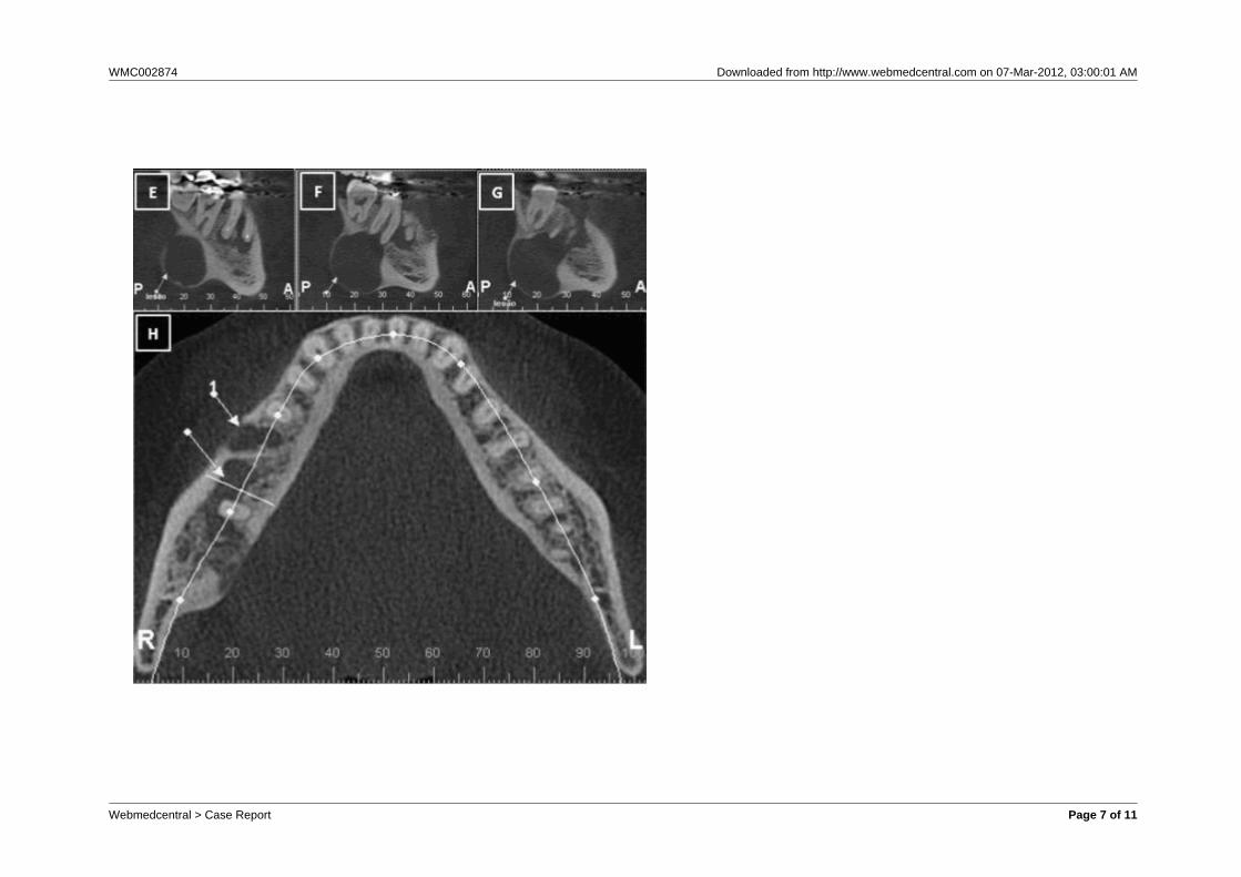
Illustration 1
3D reconstruction (A); Coronal view of the tooth 46 (B); Coronal view of the tooth 47 (C); Coronal view of the tooth 48 (D); Sagittalbuccal view of the lesion (E); Sagittal view of the lesion envolving tooth 47 (F); Sagittal lingual view of the lesion (G); Axial view ofthe top of the lesion (H)
Webmedcentral > Case Report Page 6 of 11
WMC002874 Downloaded from http://www.webmedcentral.com on 07-Mar-2012, 03:00:01 AM
Webmedcentral > Case Report Page 7 of 11
WMC002874 Downloaded from http://www.webmedcentral.com on 07-Mar-2012, 03:00:01 AM
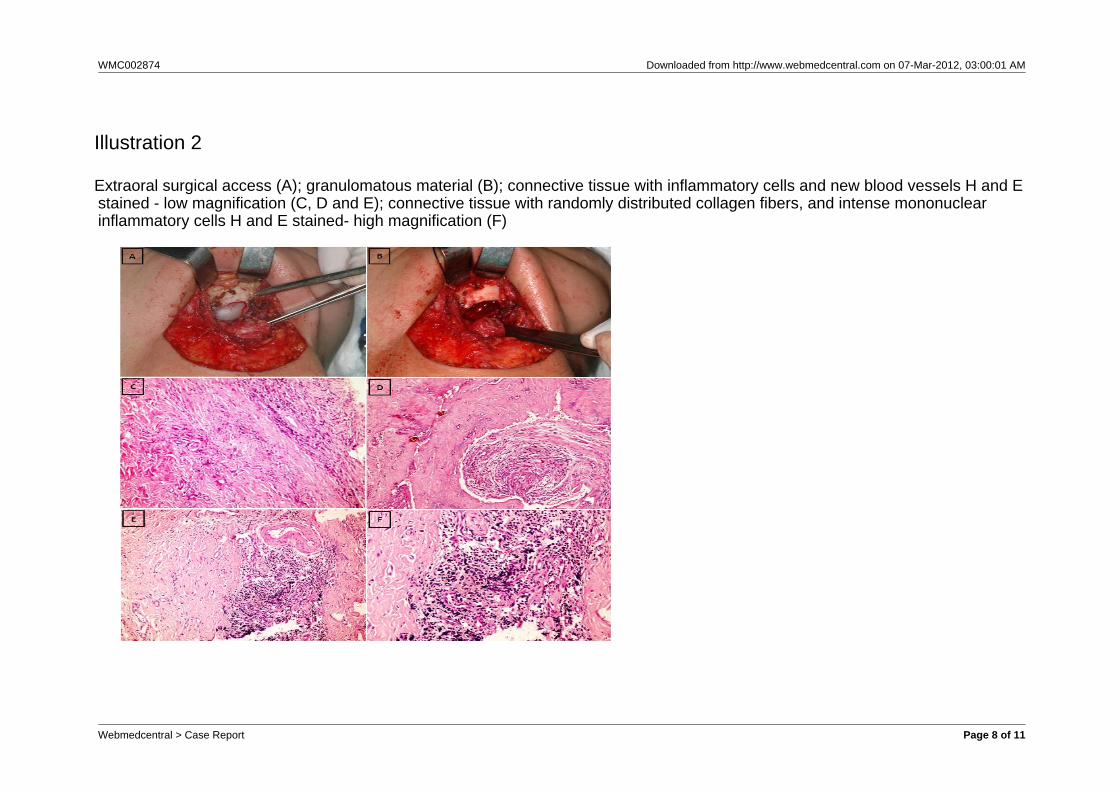
Illustration 2
Extraoral surgical access (A); granulomatous material (B); connective tissue with inflammatory cells and new blood vessels H and Estained - low magnification (C, D and E); connective tissue with randomly distributed collagen fibers, and intense mononuclearinflammatory cells H and E stained- high magnification (F)
Webmedcentral > Case Report Page 8 of 11

WMC002874 Downloaded from http://www.webmedcentral.com on 07-Mar-2012, 03:00:01 AM
Reviews
Review 1
Review Title: WMC002874: Atypical Aggressive Periapical Granuloma: A CaseReportPosted by Dr. Tomofumi Hamada on 07 Mar 2012 02:06:03 AM GMT
1 Is the subject of the article within the scope of the subject category? Yes
2 Are the interpretations / conclusions sound and justified by the data? Yes
3 Is this a new and original contribution? Yes
4 Does this paper exemplify an awareness of other research on the topic? Yes
5 Are structure and length satisfactory? Yes
6 Can you suggest brief additions or amendments or an introductory statement that will increasethe value of this paper for an international audience?
Yes
7 Can you suggest any reductions in the paper, or deletions of parts? No
8 Is the quality of the diction satisfactory? Yes
9 Are the illustrations and tables necessary and acceptable? Yes
10 Are the references adequate and are they all necessary? Yes
11 Are the keywords and abstract or summary informative? Yes
Rating: 6
Comment: The authors described a case report of atypical and aggressive periapical granuloma, located in the rightmandibular. This is a well-written, interesting case report. It will be helpful to make differential diagnosis (e.g.spindle cell tumors, inflammatory reactive conditions) if the authors provide the information ofimmunohistochemical findings.
Competing interests: No
Invited by the author to make a review on this article? : No
Experience and credentials in the specific area of science: DDS and PhD in oral oncology.
Publications in the same or a related area of science: No
How to cite: Hamada T.WMC002874: Atypical Aggressive Periapical Granuloma: A Case Report[Review of thearticle 'Atypical Aggressive Periapical Granuloma: A Case Report ' by ].WebmedCentral1970;3(3):WMCRW001561
Webmedcentral > Case Report Page 9 of 11
WMC002874 Downloaded from http://www.webmedcentral.com on 07-Mar-2012, 03:00:01 AM
Review 2
Review Title: Atypical Agressive Periapical Granuloma: A case reportPosted by Prof. Brunno S Freitas on 12 Jan 2012 11:26:44 AM GMT
1 Is the subject of the article within the scope of the subject category? Yes
2 Are the interpretations / conclusions sound and justified by the data? Yes
3 Is this a new and original contribution? No
4 Does this paper exemplify an awareness of other research on the topic? Yes
5 Are structure and length satisfactory? Yes
6 Can you suggest brief additions or amendments or an introductory statement that will increasethe value of this paper for an international audience?
Yes
7 Can you suggest any reductions in the paper, or deletions of parts? No
8 Is the quality of the diction satisfactory? Yes
9 Are the illustrations and tables necessary and acceptable? Yes
10 Are the references adequate and are they all necessary? Yes
11 Are the keywords and abstract or summary informative? Yes
Rating: 7
Comment: This is a well written case report.
There is some controversy in the definition of KOT in the discussion. I suggest the 2005 World HealthOrganization definition.
Invited by the author to make a review on this article? : Yes
Experience and credentials in the specific area of science: PhD and MSc in oral pathology.
Publications in the same or a related area of science: No
How to cite: Freitas B.Atypical Agressive Periapical Granuloma: A case report[Review of the article 'AtypicalAggressive Periapical Granuloma: A Case Report ' by ].WebmedCentral 1970;3(1):WMCRW001365
Webmedcentral > Case Report Page 10 of 11

WMC002874 Downloaded from http://www.webmedcentral.com on 07-Mar-2012, 03:00:01 AM
DisclaimerThis article has been downloaded from WebmedCentral. With our unique author driven post publication peerreview, contents posted on this web portal do not undergo any prepublication peer or editorial review. It iscompletely the responsibility of the authors to ensure not only scientific and ethical standards of the manuscriptbut also its grammatical accuracy. Authors must ensure that they obtain all the necessary permissions beforesubmitting any information that requires obtaining a consent or approval from a third party. Authors should alsoensure not to submit any information which they do not have the copyright of or of which they have transferredthe copyrights to a third party.
Contents on WebmedCentral are purely for biomedical researchers and scientists. They are not meant to cater tothe needs of an individual patient. The web portal or any content(s) therein is neither designed to support, norreplace, the relationship that exists between a patient/site visitor and his/her physician. Your use of theWebmedCentral site and its contents is entirely at your own risk. We do not take any responsibility for any harmthat you may suffer or inflict on a third person by following the contents of this website.
Webmedcentral > Case Report Page 11 of 11