attentional effects of gaze shifts are influenced by emotion
TRANSCRIPT

Copyright @ 2008 American Academy of Child and Adolescent Psychiatry. Unauthorized reproduction of this article is prohibited.
Attentional Effects of Gaze Shifts Are Influenced byEmotion and Spatial Frequency, but Not in Autism
MAARTJE CATHELIJNE DE JONG, B.SC., HERMAN VAN ENGELAND, M.D., PH.D.,AND CHANTAL KEMNER, PH.D.
ABSTRACT
Objective: Impaired gaze following is an important hallmark of autism spectrum disorders (ASDs) in clinical settings. Yet,
ASD subjects perform normally on laboratory tasks involving gaze shifts. We investigated this contradiction, hypothesizing
that impaired gaze following in ASDs is not related to basic impairments in attention orienting but to impaired emotion
perception and abnormal processing of spatial frequencies (i.e., local and global information).Method:We tested 30 high-
functioning, school-age children with ASDs and 30 age- and IQ-matched controls on a task involving gaze shifts that cue
the location of targets. The cueing faces differed in emotionality and were filtered for different spatial frequencies. We
recorded behavioral responses (reaction times) and brain responses (event-related potentials). Results: ASD subjects
performed normally when neutral faces were used. However, emotional faces elicited modified face and gaze cue
processing in control subjects, but not in the ASD subjects. Furthermore, the control group was biased toward the use of
low spatial frequencies (global information) to process gaze cues, whereas the ASD group was biased toward the use of
high spatial frequencies (local information). Conclusions:We conclude that impaired gaze following in ASDs is related to
impaired emotion processing. Moreover, ASD subjects show an abnormal reliance on local information to process gaze
cues. J. Am. Acad. Child Adolesc. Psychiatry, 2008;47(4):443Y454.KeyWords: autism, gaze, emotion, spatial frequency,
event-related potentials.
An intriguing contradiction exists between clinicalreports of impaired gaze following in autism spectrumdisorders (ASDs), on the one hand, and studies failing toreproduce this effect in the laboratory on the other. Gazefollowing is a social skill by which individuals directtheir attention to the same location by observing eachother`s gaze direction. In clinical settings, impaired gazefollowing is recognized as an important hallmark of
ASDs, both in young children and adolescents.1,2 Infact, it is one of the earliest detectable symptoms of thesedisorders and is probably directly related to problems insocial interactions typical for ASDs.3,4 Nonetheless,several recent studies showed irreproducibility of thisattribute of ASD in the laboratory.5Y10 The presentstudy aimed to investigate this conundrum, hypothesiz-ing that the negative results obtained in laboratorysettings are an effect of the low ecological validity of theexperimental stimuli used.11 To date, face motion andemotion have not been included in experimental setups.Instead, all previous studies relied on static neutral faces.Therefore, we designed an experimental task involvingdynamic emotional faces.Laboratory tasks used to study gaze following
typically comprise a centrally presented, nonemotionalface that cues the location of a peripheral target througha gaze shift. The rest of the face remains static. The gazecue leads to faster detection times for correctly cuedtargets.12,13 To display naturally looking emotionalexpressions that accompany the gaze shift, we designed agaze cue task with dynamic faces. We hypothesized that
Accepted September 25, 2007.The authors are with the Rudolf Magnus Institute of Neuroscience,
Department of Child and Adolescent Psychiatry, University Medical CenterUtrecht, the Netherlands.
The work described was supported by an Innovational Research Incentivesgrant (VIDI-scheme, 402-01-094) of the Netherlands Organisation for ScientificResearch (NWO) to C. Kemner. The authors thank G. Camfferman for technicalassistance.
This article is the subject of an editorial by Dr. Ami Klin in this issue.Correspondence to Maartje C. de Jong, P.O. Box 85500, 3508 GA Utrecht,
the Netherlands; e-mail: [email protected]/08/4704-0443�2008 by the American Academy of Child
and Adolescent Psychiatry.DOI: 10.1097/CHI.0b013e31816429a6
WWW.JAACAP.COM 443J . AM. ACAD. CHILD ADOLESC. PSYCHIATRY, 47:4, APRIL 2008
Copyright @ 2008 American Academy of Child and Adolescent Psychiatry. Unauthorized reproduction of this article is prohibited.
attention orienting in typically developing children isaffected by emotion because emotional expressions areknown to influence spatial attention. Fearful expressionsin particular have the capacity to modify attentionorienting in a fast and involuntary manner.14,15 Forchildren with ASDs, we assumed that emotion wouldnot affect attention orienting because these childrenhave abnormalities in the processing of emotion.16Y20
Our hypothesis is supported by a deficit in theprocessing of facial information in ASD,16 such asimpaired recognition of emotional expressions.17 Inaddition, brain regions involved in the perception offacial emotions are hypoactive in individuals withASD.18Y20 These brain regions include the fusiformgyrus, often referred to as the fusiform face area, theamygdala, known for its role in the appraisal ofemotional stimuli, and the superior temporal sulcus, aregion mediating biological motion perception in, forexample, expression changes and gaze shifts.18Y20 Ittherefore is likely that not impairments in gaze cueprocessing per se but rather abnormalities in emotionprocessing lead to the impaired gaze following seen inASDs in clinical settings and daily life.Some authors have proposed that the deficits in
emotion processing in ASD are the result of a decreasedsocial motivation that corrupts the development of face-processing skills.16 However, there is increasing evi-dence that the cause of the abnormal emotionprocessing in ASDs is not social in nature but is relatedto perceptual abnormalities.21,22 Various previousstudies indicate that ASD subjects focus on local ratherthan global aspects of stimuli,22,23 both for social17,24
and nonsocial25,26 information. In other words, localaspects (i.e., individual face features, such as expression-related wrinkles) are processed more thoroughly in ASD
than global aspects (coarse information about theconfiguration of features).The processing of local and global information may
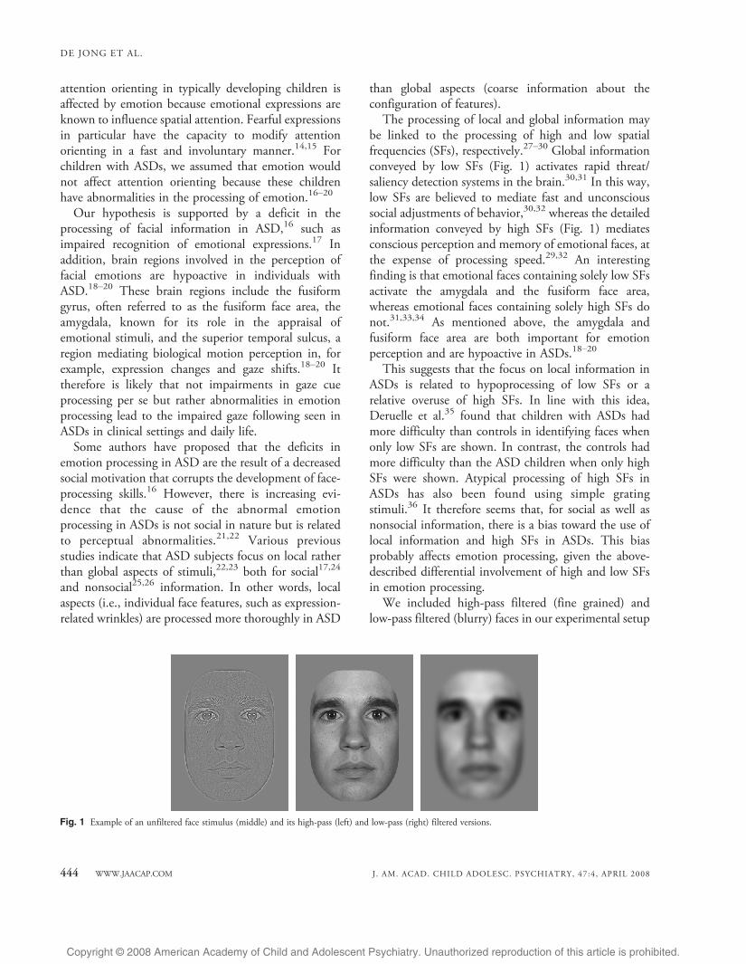
be linked to the processing of high and low spatialfrequencies (SFs), respectively.27Y30 Global informationconveyed by low SFs (Fig. 1) activates rapid threat/saliency detection systems in the brain.30,31 In this way,low SFs are believed to mediate fast and unconscioussocial adjustments of behavior,30,32 whereas the detailedinformation conveyed by high SFs (Fig. 1) mediatesconscious perception and memory of emotional faces, atthe expense of processing speed.29,32 An interestingfinding is that emotional faces containing solely low SFsactivate the amygdala and the fusiform face area,whereas emotional faces containing solely high SFs donot.31,33,34 As mentioned above, the amygdala andfusiform face area are both important for emotionperception and are hypoactive in ASDs.18Y20
This suggests that the focus on local information inASDs is related to hypoprocessing of low SFs or arelative overuse of high SFs. In line with this idea,Deruelle et al.35 found that children with ASDs hadmore difficulty than controls in identifying faces whenonly low SFs are shown. In contrast, the controls hadmore difficulty than the ASD children when only highSFs were shown. Atypical processing of high SFs inASDs has also been found using simple gratingstimuli.36 It therefore seems that, for social as well asnonsocial information, there is a bias toward the use oflocal information and high SFs in ASDs. This biasprobably affects emotion processing, given the above-described differential involvement of high and low SFsin emotion processing.We included high-pass filtered (fine grained) and
low-pass filtered (blurry) faces in our experimental setup
Fig. 1 Example of an unfiltered face stimulus (middle) and its high-pass (left) and low-pass (right) filtered versions.
DE JONG ET AL.
444 WWW.JAACAP.COM J. AM. ACAD. CHILD ADOLESC. PSYCHIATRY, 47:4, APRIL 2008

Copyright @ 2008 American Academy of Child and Adolescent Psychiatry. Unauthorized reproduction of this article is prohibited.
to investigate whether a bias toward the processing oflocal information and high SFs indeed influencesemotion processing and, possibly, gaze cue processingin ASDs. For the control group, we hypothesized thatlow SFs are of particular importance in the processing ofemotional gaze cues because the present task does notemphasize conscious emotion processing. Also, theeffect of gaze cues is believed to depend on the contrastbetween the iris and sclera,37 which is a low SF feature innormal viewing conditions as well as in our experimentalsetup. In contrast, we expected the ASD group to show arelative overreliance on high SF information.
While subjects performed the task, we recordedreaction times (RTs) and brain activity (event relatedpotentials [ERPs]) related to face processing (N170peak) and attention orienting (P100 and N200 peaks).It is well documented that the N170 peak in response tofaces reflects face-specific brain activity originating mostlikely from the fusiform gyrus or superior temporalsulcus.38,39 Previous studies indicated that emotion andSF filtering affect the N17040,41 (see also a study doneby Holmes et al.42). The occipitotemporal P100 andN200 peaks in response to the peripheral targets aretypically earlier and larger for correctly cued targets,which probably reflects increased neural activity in theextrastriatal visual cortex to facilitate the processing ofattended stimuli.43 Kemner et al.5 found the N170,P100, and N200 to be normal in ASD patients during agaze cue task with neutral expressions. In our emotionalgaze cue task, we expect abnormal brain activity in theASD group because of the effects of emotion and SFfiltering. A group of school-age children with a diagnosisof either autistic disorder or Asperger syndrome and anevenly numbered group of age- and IQ-matchedtypically developing controls participated in this study.
METHOD
Subjects
The ASD and control groups both consisted of 30 high-functioning (IQ >80) children, matched for sex, age (school age,7Y13 years old), and IQ (Table 1). IQ was obtained using the revisedDutch edition of the WISC. All of the subjects had normal orcorrected to normal vision and no neurological history. Controlchildren were screened for psychopathology by means of the ChildBehavior Checklist44 and excluded if scores were within the clinicalrange. Sixty-seven percent of subjects were also screened by means ofthe Teacher`s Report Form.45 The ASD subjects were diagnosedwith either autistic disorder or Asperger syndrome by a child andadolescent psychiatrist using DSM-IV criteria.4 In addition, the
Autism Diagnostic Interview Revised46 was administered to theparents by a trained rater (Table 1). Of all of the patients, 24 met fullAutism Diagnostic Interview Revised criteria for autistic disorder(cutoffs shown in Table 1) and six met criteria for an ASD (definedas scoring 1 point below cutoff on only one of the three AutismDiagnostic Interview Revised domains, which was the stereotypydomain in five of the six cases). None of the patients had a comorbidpsychiatric or neurological disorder. Seven patients used psychoac-tive medication (three methylphenidate, one typical neuroleptic, twoatypical neuroleptics, and one selective serotonin reuptake inhibitor),and subjects on methylphenidate were instructed not to take thismedication on the day of testing. The study was approved of by theMedical Ethics Committee of the University Medical Center, and allof the parents gave written informed consent before participation.
Stimuli
Pictures of 10 different actors displaying fearful and neutralexpressions were taken from the MacBrain Face Stimulus Set.(Development of the MacBrain Face Stimulus Set was overseen byNim Tottenham and supported by the John D. and Catherine T.MacArthur Foundation Research Network on Early Experience andBrain Development. Please contact Nim Tottenham at [email protected] for more information concerning the stimulus set.) UsingAdobe Photoshop 7.0.1 software, straight and averted eyes werecreated, and the faces were matched for size (6.46 cm horizontally,which corresponds to 3.7- because the viewing distance was 1meter), shape, luminance (18 cd/m2), and contrast. To generatedynamic stimuli, fearful and neutral pictures of the same actor wereBmorphed^ using Meesoft Smartmorph 1.55 software (resulting in13-frame movie clips). High- and low-pass filtering was done using atwo-dimensional Fourier transformation with cutoff values of >22.2cycles/image (equivalent to more than six cycles per degree) and <7.4cycles/image (equivalent to fewer than two cycles per degree),respectively (Fig. 1).
Design
Two different emotional conditions were compared. In theneutral-to-fearful (N-to-F) condition, a neutral face changed to
TABLE 1Subject Characteristics (Mean T Standard Error)
ASD Control
No. of subjects 30 (24 M, 6 F) 30 (24 M, 6 F)Age, y 10.7 T 1.8 10.6 T 1.6Total IQ 108.4 T 2.6 111.5 T 2.2Verbal IQ 113.3 T 2.7 116.3 T 2.5Performance IQ 101.4 T 3.1 100.6 T 2.5ADI-R social domaina 20 T 1.1ADI-R communication domainb 16 T 0.8ADI-R stereotype domainc 6 T 0.5
Note: ASD = autism spectrum disorders; M = male; F = female;ADI-R = Autism Diagnostic Interview Revised.
a Cutoff for autism: 10.b Cutoff for autism: 8.c Cutoff for autism: 3.
IMPAIRED EMOTIONAL GAZE CUEING IN AUTISM
WWW.JAACAP.COM 445J . AM. ACAD. CHILD ADOLESC. PSYCHIATRY, 47:4, APRIL 2008

Copyright @ 2008 American Academy of Child and Adolescent Psychiatry. Unauthorized reproduction of this article is prohibited.
fearful while making a gaze shift from straight to averted. In thefearful-to-neutral (F-to-N) condition, a fearful face changed toneutral while changing gaze direction. For the N-to-F condition, weexpected enhanced cueing effects because the averted fearful gaze wasexpected to enhance the shift of attention to the periphery. For theF-to-N condition, we expected decreased cueing effects because thestraight fearful gaze was expected to attract attention to itslocation.15 SF filtering of these conditions yielded six conditions(2x unfiltered, 2x high SF, 2x low SF). To be able to replicateprevious findings of studies testing ASD subjects on gaze cue tasks,we included a seventh condition (neutral-to-neutral) involvingstatic neutral faces (unfiltered).
Procedure
Each subject completed 700 trials (100 trials per condition),presented over four blocks. Additional trials (175 maximally) werepresented in the same session to three ASD subjects and two controlsubjects to compensate for excessive movement artifacts in the ERPsignal. The chronological sequence of events in a trial was as follows:fixation dot (1,000 milliseconds), initial face with straight gaze (373milliseconds), movie clip showing gradual change of facial expressionand gaze direction (11 frames, 40 milliseconds per frame), final facegazing randomly to the left or right (373 milliseconds), target cross(subtending 0.7-) placed randomly on the left or right (5.7- offcenter, 1,000 milliseconds), pseudo-random delay (1,100Y1,700milliseconds; Fig. 2). During target presentation, the final faceremained visible on the screen to avoid offset effects. Stimuli werepresented on a gray background matching the average luminance ofthe face stimuli. Subjects were made aware of the fact that gaze
direction did not predict target location. Subjects were instructed tofixate on the central face throughout the experiment and to respondto appearance of the target by pressing a corresponding left or rightbutton as quickly and accurately as possible.
Data Recording
Subjects were seated in an electronically and acoustically shieldedroom. EEG was recorded with 34 silver/silver chloride (Ag/AgCl)flat type active electrodes (Active Two System, Biosemi) positionedat standard locations on an elastic cap (Quickcap, NeuromedicalSupplies of Neurosoft Inc.). EEG was sampled at 2,048 Hz andstored as a continuous signal. Two electrodes in the electrode capprovided an active ground. In addition, horizontal and verticalelectro-oculograms were measured. Data were resampled offline at500 Hz and analyzed using Brain Vision Analyser software(Brainproducts GmbH). A 2-Hz high-pass filter, a 20-Hz low-pass filter, and a 50-Hz Notch filter were applied. All of the elec-trodes were referenced to the right mastoid. Eye movement artifacts(detected using the algorithm by Gratton et al.,47 which is im-plemented in the Brain Vision Analyser) and EEG artifacts (definedas amplitudes >T100 6V, amplitude differences within epoch>137 6V, amplitude differences within 200 milliseconds <3 6V,voltage steps per sample point >15 6V) were removed.
Data Analysis
Epochs were extracted offline from the continuous data. The face-specific N170 peak was defined as a negative deflection 165Y235milliseconds after onset of the initial face (at electrodes P3, P4, CP5,
Fig. 2 Sequence of events in one trial. Shown here is the neutral-to-fearful condition, which was compared with the fearful-to-neutral condition to reveal theeffects of emotion. A neutral-to-neutral condition, involving a gaze change without an expression change, was also included. Targets were correctly cued by gazedirection in 50% of the trials. Event-related potentials were measured in response to the initial face and in response to the target.
DE JONG ET AL.
446 WWW.JAACAP.COM J. AM. ACAD. CHILD ADOLESC. PSYCHIATRY, 47:4, APRIL 2008
Copyright @ 2008 American Academy of Child and Adolescent Psychiatry. Unauthorized reproduction of this article is prohibited.
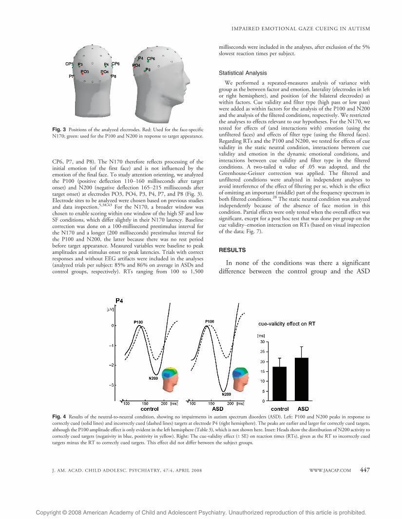
CP6, P7, and P8). The N170 therefore reflects processing of theinitial emotion (of the first face) and is not influenced by theemotion of the final face. To study attention orienting, we analyzedthe P100 (positive deflection 110Y160 milliseconds after targetonset) and N200 (negative deflection 165Y215 milliseconds aftertarget onset) at electrodes PO3, PO4, P3, P4, P7, and P8 (Fig. 3).Electrode sites to be analyzed were chosen based on previous studiesand data inspection.5,38,43 For the N170, a broader window waschosen to enable scoring within one window of the high SF and lowSF conditions, which differ slightly in their N170 latency. Baselinecorrection was done on a 100-millisecond prestimulus interval forthe N170 and a longer (200 milliseconds) prestimulus interval forthe P100 and N200, the latter because there was no rest periodbefore target appearance. Measured variables were baseline to peakamplitudes and stimulus onset to peak latencies. Trials with correctresponses and without EEG artifacts were included in the analyses(analyzed trials per subject: 85% and 86% on average in ASDs andcontrol groups, respectively). RTs ranging from 100 to 1,500
milliseconds were included in the analyses, after exclusion of the 5%slowest reaction times per subject.
Statistical Analysis
We performed a repeated-measures analysis of variance withgroup as the between factor and emotion, laterality (electrodes in leftor right hemisphere), and position (of the bilateral electrodes) aswithin factors. Cue validity and filter type (high pass or low pass)were added as within factors for the analysis of the P100 and N200and the analysis of the filtered conditions, respectively. We restrictedthe analyses to effects relevant to our hypotheses. For the N170, wetested for effects of (and interactions with) emotion (using theunfiltered faces) and effects of filter type (using the filtered faces).Regarding RTs and the P100 and N200, we tested for effects of cuevalidity in the static neutral condition, interactions between cuevalidity and emotion in the dynamic emotional conditions, andinteractions between cue validity and filter type in the filteredconditions. A two-tailed " value of .05 was adopted, and theGreenhouse-Geisser correction was applied. The filtered andunfiltered conditions were analyzed in independent analyses toavoid interference of the effect of filtering per se, which is the effectof omitting an important (middle) part of the frequency spectrum inboth filtered conditions.28 The static neutral condition was analyzedindependently because of the absence of face motion in thiscondition. Partial effects were only tested when the overall effect wassignificant, except for a post hoc test that was done per group on thecue validityYemotion interaction on RTs (based on visual inspectionof the data; Fig. 7).
RESULTS
In none of the conditions was there a significantdifference between the control group and the ASD
Fig. 3 Positions of the analyzed electrodes. Red: Used for the face-specificN170; green: used for the P100 and N200 in response to target appearance.
Fig. 4 Results of the neutral-to-neutral condition, showing no impairments in autism spectrum disorders (ASD). Left: P100 and N200 peaks in response tocorrectly cued (solid lines) and incorrectly cued (dashed lines) targets at electrode P4 (right hemisphere). The peaks are earlier and larger for correctly cued targets,although the P100 amplitude effect is only evident in the left hemisphere (Table 3), which is not shown here. Inset: Heads show the distribution of N200 activity tocorrectly cued targets (negativity in blue, positivity in yellow). Right: The cue-validity effect (T SE) on reaction times (RTs), given as the RT to incorrectly cuedtargets minus the RT to correctly cued targets. This effect did not differ between the subject groups.
IMPAIRED EMOTIONAL GAZE CUEING IN AUTISM
WWW.JAACAP.COM 447J . AM. ACAD. CHILD ADOLESC. PSYCHIATRY, 47:4, APRIL 2008
Copyright @ 2008 American Academy of Child and Adolescent Psychiatry. Unauthorized reproduction of this article is prohibited.
group in absolute RTs or number of correct responses.In the static neutral condition, the cue-validity effects onRTs, P100, and N200 latency did not differ betweenthe subject groups. On N200 amplitude, the ASDgroup showed a larger cue-validity effect (Fig. 4). Inboth groups, correctly cued targets elicited earlier andlarger P100 and N200 peaks and faster RTs than
incorrectly cued targets (significant effects and statisticalvalues are shown in Table 3).
Effects of Emotion on Face Processing (N170 Peak)
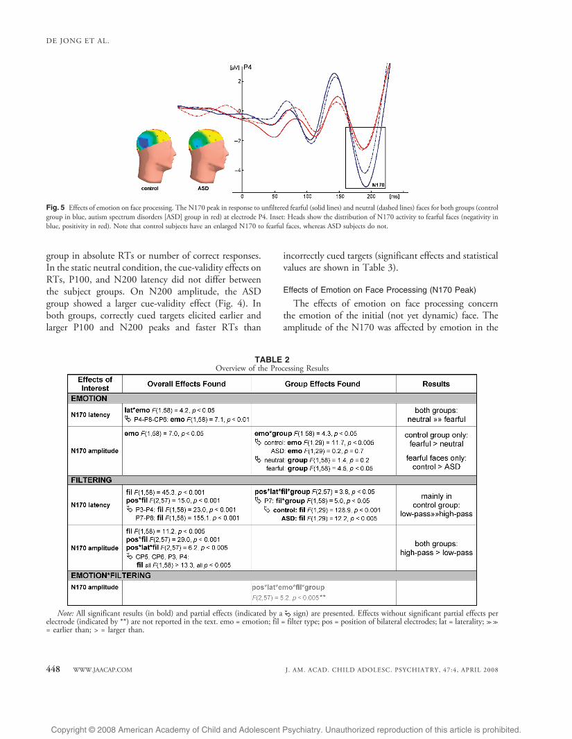
The effects of emotion on face processing concernthe emotion of the initial (not yet dynamic) face. Theamplitude of the N170 was affected by emotion in the
Fig. 5 Effects of emotion on face processing. The N170 peak in response to unfiltered fearful (solid lines) and neutral (dashed lines) faces for both groups (controlgroup in blue, autism spectrum disorders [ASD] group in red) at electrode P4. Inset: Heads show the distribution of N170 activity to fearful faces (negativity inblue, positivity in red). Note that control subjects have an enlarged N170 to fearful faces, whereas ASD subjects do not.
TABLE 2Overview of the Processing Results
Note: All significant results (in bold) and partial effects (indicated by a sign) are presented. Effects without significant partial effects perelectrode (indicated by **) are not reported in the text. emo = emotion; fil = filter type; pos = position of bilateral electrodes; lat = laterality; >>>>= earlier than; > = larger than.
DE JONG ET AL.
448 WWW.JAACAP.COM J. AM. ACAD. CHILD ADOLESC. PSYCHIATRY, 47:4, APRIL 2008
Copyright @ 2008 American Academy of Child and Adolescent Psychiatry. Unauthorized reproduction of this article is prohibited.
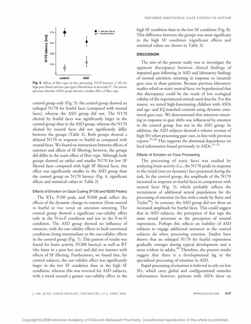
control group only (Fig. 5): the control group showed anenlarged N170 for fearful faces (compared with neutralfaces), whereas the ASD group did not. The N170elicited by fearful faces was significantly larger in thecontrol group than in the ASD group, whereas the N170elicited by neutral faces did not significantly differbetween the groups (Table 2). Both groups showed adelayed N170 in response to fearful as compared withneutral faces.We found no interactions between effects ofemotion and effects of SF filtering; however, the groupsdid differ in the main effect of filter type. Although bothgroups showed an earlier and smaller N170 for low SFfiltered faces compared with high SF filtered faces, thiseffect was significantly smaller in the ASD group thanthe control group on N170 latency (Fig. 6, significanteffects and statistical values in Table 2).
Effects of Emotion on Gaze Cueing (P100 and N200 Peaks)
The RTs, P100 peak, and N200 peak reflect theeffects of the dynamic change in emotion (from neutralto fearful or vice versa) on attention orienting. Thecontrol group showed a significant cue-validity effectonly in the N-to-F condition and not in the F-to-Ncondition. The ASD group showed no influence ofemotion, with the cue-validity effects in both emotionalconditions being intermediate to the cue-validity effectsin the control group (Fig. 7). This pattern of results wasfound for brain activity (N200 latency) as well as RT(the latter in a post hoc test) and did not interact witheffects of SF filtering. Furthermore, we found that, forcontrol subjects, the cue-validity effect was significantlylarger in the low SF condition than in the high SFcondition, whereas this was reversed for ASD subjects,with a trend toward a greater cue-validity effect in the
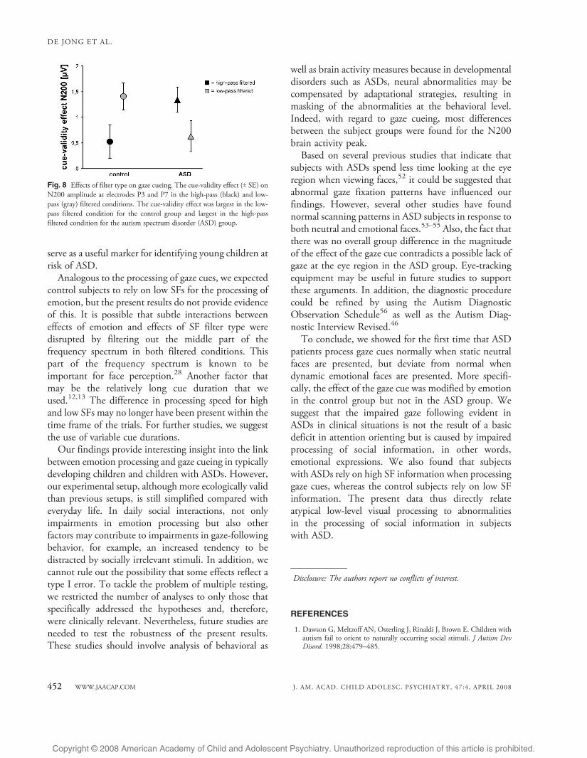
high SF condition than in the low SF condition (Fig. 8).This difference between the groups was most significantin the high SF condition (significant effects andstatistical values are shown in Table 3).
DISCUSSION
The aim of the present study was to investigate theapparent discrepancy between clinical findings ofimpaired gaze following in ASD and laboratory findingsof normal attention orienting in response to (neutral)gaze cues in these patients. Because previous laboratorystudies relied on static neutral faces, we hypothesized thatthis discrepancy could be the result of low ecologicalvalidity of the experimental stimuli used thus far. For thisreason, we tested high-functioning children with ASDsand age- and IQ-matched controls using dynamic emo-tional gaze cues. We demonstrated that attention orient-ing in response to gaze shifts was influenced by emotionin the control group, but not in the ASD group. Inaddition, the ASD subjects showed a relative overuse ofhigh SFs when processing gaze cues, in line with previousreports.35,36 This supports the abnormal dependence onlocal information found previously in ASDs.23Y25
Effects of Emotion on Face Processing
The processing of static faces was studied byanalyzing brain activity (i.e., the N170 peak) in responseto the initial (not yet dynamic) face presented during thetask. In the control group, the amplitude of the N170increased in response to fearful faces in comparison withneutral faces (Fig. 5), which probably reflects therecruitment of additional neural populations for theprocessing of emotion (in line with a study by Batty andTaylor40). In contrast, the ASD group did not show anincreased amplitude for fearful faces. This could suggestthat in ASD subjects, the perception of fear taps thesame neural processes as the perception of neutralexpressions. Perhaps this reflects an inability of ASDsubjects to engage additional resources as the controlsubjects do when processing emotion. Studies haveshown that an enlarged N170 for fearful expressionsgradually emerges during typical development and ismost evident in adults.48 Therefore, the present resultssuggest that there is a developmental lag in thespecialized processing of emotion in ASD.Rapid processing of emotion is believed to rely on low
SFs, which carry global and configurational stimulusinformation; however, patients with ASDs show an
Fig. 6 Effects of filter type on face processing. N170 latencies (T SE) forhigh-pass (black) and low-pass (gray) filtered faces at electrode P7. The autismspectrum disorder (ASD) group showed a smaller effect of filter type.
IMPAIRED EMOTIONAL GAZE CUEING IN AUTISM
WWW.JAACAP.COM 449J . AM. ACAD. CHILD ADOLESC. PSYCHIATRY, 47:4, APRIL 2008

Copyright @ 2008 American Academy of Child and Adolescent Psychiatry. Unauthorized reproduction of this article is prohibited.
overuse of local information, mediated by high SFs.22,23
We hypothesized that this hinders emotion processing inASDs. Although the present results do not provide directsupport for this hypothesis, there were indications thatthe ASD group has a diminished specialization in thedifferential processing of high and low SFs (Fig. 6).Supporting a diminished specialization in the processinghigh and low SFs via different neural pathways,Boeschoten et al. 36 found that the amplitude of the P1peak in response to grating stimuli differed for differentSFs in control subjects, but not in ASD subjects. Inaddition, when processing faces, control subjects werefound to activate different sources in the brain for high-
pass and low-pass filtered faces, whereas ASD subjects didnot.49 The relative overreliance on high SF informationseen in ASDs may be a consequence of a disruptedspecialization process during visual development. Thiscould well be a basic neural mechanism of ASD worthy ofclinical and scientific interest.
Effects of Emotion on Gaze Cueing
To study attention orienting in response to gazeshifts, we analyzed RTs as well as brain activity (theP100 and N200 peaks) in response to the appearance oftargets. We replicated previous findings of normalattention orienting in response to static neutral gaze cues
Note: Overview of the results on the effect of cue validity, i.e., the effect of the gaze shift. RT = reaction time; val = cue validity. See Table 2footnotes for further explanations.
TABLE 3Overview of the Results on the Effect of the Gaze Cue
DE JONG ET AL.
450 WWW.JAACAP.COM J. AM. ACAD. CHILD ADOLESC. PSYCHIATRY, 47:4, APRIL 2008
Copyright @ 2008 American Academy of Child and Adolescent Psychiatry. Unauthorized reproduction of this article is prohibited.
in ASDs (Fig. 4), confirming that the processing ofunemotional gaze cues is not impaired in patients withASDs. We even found an increased, rather thandecreased, effect of the gaze cue on the N200 amplitudein the ASD group, which could reflect an increasedeffect of (expected) target onset or a superior attention tothe task in ASD subjects.50 A similar result was observedin a study by Kemner et al.,5 although it was notstatistically significant in this study, perhaps due to thesmaller number of subjects.
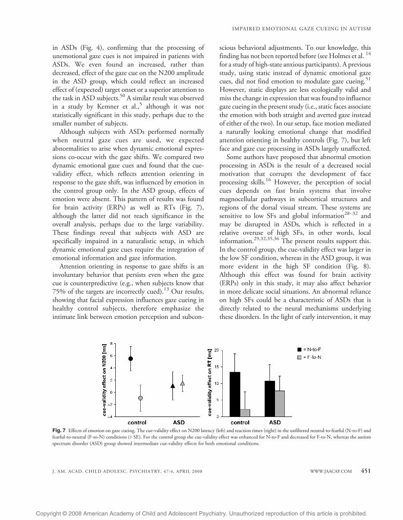
Although subjects with ASDs performed normallywhen neutral gaze cues are used, we expectedabnormalities to arise when dynamic emotional expres-sions co-occur with the gaze shifts. We compared twodynamic emotional gaze cues and found that the cue-validity effect, which reflects attention orienting inresponse to the gaze shift, was influenced by emotion inthe control group only. In the ASD group, effects ofemotion were absent. This pattern of results was foundfor brain activity (ERPs) as well as RTs (Fig. 7),although the latter did not reach significance in theoverall analysis, perhaps due to the large variability.These findings reveal that subjects with ASD arespecifically impaired in a naturalistic setup, in whichdynamic emotional gaze cues require the integration ofemotional information and gaze information.
Attention orienting in response to gaze shifts is aninvoluntary behavior that persists even when the gazecue is counterpredictive (e.g., when subjects know that75% of the targets are incorrectly cued).13 Our results,showing that facial expression influences gaze cueing inhealthy control subjects, therefore emphasize theintimate link between emotion perception and subcon-
scious behavioral adjustments. To our knowledge, thisfinding has not been reported before (see Holmes et al. 14
for a study of high-state anxious participants). A previousstudy, using static instead of dynamic emotional gazecues, did not find emotion to modulate gaze cueing.51
However, static displays are less ecologically valid andmiss the change in expression that was found to influencegaze cueing in the present study (i.e., static faces associatethe emotion with both straight and averted gaze insteadof either of the two). In our setup, face motion mediateda naturally looking emotional change that modifiedattention orienting in healthy controls (Fig. 7), but leftface and gaze cue processing in ASDs largely unaffected.Some authors have proposed that abnormal emotion
processing in ASDs is the result of a decreased socialmotivation that corrupts the development of faceprocessing skills.16 However, the perception of socialcues depends on fast brain systems that involvemagnocellular pathways in subcortical structures andregions of the dorsal visual stream. These systems aresensitive to low SFs and global information28Y32 andmay be disrupted in ASDs, which is reflected in arelative overuse of high SFs, in other words, localinformation.29,32,35,36 The present results support this.In the control group, the cue-validity effect was larger inthe low SF condition, whereas in the ASD group, it wasmore evident in the high SF condition (Fig. 8).Although this effect was found for brain activity(ERPs) only in this study, it may also affect behaviorin more delicate social situations. An abnormal relianceon high SFs could be a characteristic of ASDs that isdirectly related to the neural mechanisms underlyingthese disorders. In the light of early intervention, it may
Fig. 7 Effects of emotion on gaze cueing. The cue-validity effect on N200 latency (left) and reaction times (right) in the unfiltered neutral-to-fearful (N-to-F) andfearful-to-neutral (F-to-N) conditions (T SE). For the control group the cue-validity effect was enhanced for N-to-F and decreased for F-to-N, whereas the autismspectrum disorder (ASD) group showed intermediate cue-validity effects for both emotional conditions.
IMPAIRED EMOTIONAL GAZE CUEING IN AUTISM
WWW.JAACAP.COM 451J . AM. ACAD. CHILD ADOLESC. PSYCHIATRY, 47:4, APRIL 2008
Copyright @ 2008 American Academy of Child and Adolescent Psychiatry. Unauthorized reproduction of this article is prohibited.
serve as a useful marker for identifying young children atrisk of ASD.Analogous to the processing of gaze cues, we expected
control subjects to rely on low SFs for the processing ofemotion, but the present results do not provide evidenceof this. It is possible that subtle interactions betweeneffects of emotion and effects of SF filter type weredisrupted by filtering out the middle part of thefrequency spectrum in both filtered conditions. Thispart of the frequency spectrum is known to beimportant for face perception.28 Another factor thatmay be the relatively long cue duration that weused.12,13 The difference in processing speed for highand low SFs may no longer have been present within thetime frame of the trials. For further studies, we suggestthe use of variable cue durations.Our findings provide interesting insight into the link
between emotion processing and gaze cueing in typicallydeveloping children and children with ASDs. However,our experimental setup, although more ecologically validthan previous setups, is still simplified compared witheveryday life. In daily social interactions, not onlyimpairments in emotion processing but also otherfactors may contribute to impairments in gaze-followingbehavior, for example, an increased tendency to bedistracted by socially irrelevant stimuli. In addition, wecannot rule out the possibility that some effects reflect atype I error. To tackle the problem of multiple testing,we restricted the number of analyses to only those thatspecifically addressed the hypotheses and, therefore,were clinically relevant. Nevertheless, future studies areneeded to test the robustness of the present results.These studies should involve analysis of behavioral as
well as brain activity measures because in developmentaldisorders such as ASDs, neural abnormalities may becompensated by adaptational strategies, resulting inmasking of the abnormalities at the behavioral level.Indeed, with regard to gaze cueing, most differencesbetween the subject groups were found for the N200brain activity peak.Based on several previous studies that indicate that
subjects with ASDs spend less time looking at the eyeregion when viewing faces,52 it could be suggested thatabnormal gaze fixation patterns have influenced ourfindings. However, several other studies have foundnormal scanning patterns in ASD subjects in response toboth neutral and emotional faces.53Y55 Also, the fact thatthere was no overall group difference in the magnitudeof the effect of the gaze cue contradicts a possible lack ofgaze at the eye region in the ASD group. Eye-trackingequipment may be useful in future studies to supportthese arguments. In addition, the diagnostic procedurecould be refined by using the Autism DiagnosticObservation Schedule56 as well as the Autism Diag-nostic Interview Revised.46
To conclude, we showed for the first time that ASDpatients process gaze cues normally when static neutralfaces are presented, but deviate from normal whendynamic emotional faces are presented. More specifi-cally, the effect of the gaze cue was modified by emotionin the control group but not in the ASD group. Wesuggest that the impaired gaze following evident inASDs in clinical situations is not the result of a basicdeficit in attention orienting but is caused by impairedprocessing of social information, in other words,emotional expressions. We also found that subjectswith ASDs rely on high SF information when processinggaze cues, whereas the control subjects rely on low SFinformation. The present data thus directly relateatypical low-level visual processing to abnormalitiesin the processing of social information in subjectswith ASD.
Disclosure: The authors report no conflicts of interest.
REFERENCES
1. Dawson G, Meltzoff AN, Osterling J, Rinaldi J, Brown E. Children withautism fail to orient to naturally occurring social stimuli. J Autism DevDisord. 1998;28:479Y485.
Fig. 8 Effects of filter type on gaze cueing. The cue-validity effect (T SE) onN200 amplitude at electrodes P3 and P7 in the high-pass (black) and low-pass (gray) filtered conditions. The cue-validity effect was largest in the low-pass filtered condition for the control group and largest in the high-passfiltered condition for the autism spectrum disorder (ASD) group.
DE JONG ET AL.
452 WWW.JAACAP.COM J. AM. ACAD. CHILD ADOLESC. PSYCHIATRY, 47:4, APRIL 2008

Copyright @ 2008 American Academy of Child and Adolescent Psychiatry. Unauthorized reproduction of this article is prohibited.
2. Leekam S, Baron-Cohen S, Perrett D, Milders M, Brown S. Eye-direction detection: a dissociation between geometric and joint attentionskills in autism. Br J Dev Psychol. 1997;15:77Y95.
3. Baird GFRC, Charman T, Baron-Cohen S, et al. A screening instrumentfor autism at 18 months of age: a 6-year follow-up study. J Am Acad ChildAdolesc Psychiatry. 2000;39:694Y702.
4. American Psychiatric Association. Diagnostic and Statistical Manual ofMental Disorders, 4th edition (DSM-IV). Washington, DC: AmericanPsychiatric Association; 1994.
5. Kemner C, Schuller A-M, van Engeland H. Electrocortical reflections offace and gaze processing in children with pervasive developmentaldisorder. J Child Psychol Psychiatry. 2006;47:1063Y1072.
6. Senju A, Tojo Y, Dairoku H, Hasegawa T. Reflexive orienting inresponse to eye gaze and an arrow in children with and without autism.J Child Psychol Psychiatry. 2004;45:445Y458.
7. Swettenham J, Condie S, Campbell R, Milne E, Coleman M. Does theperception of moving eyes trigger reflexive visual orienting in autism?Philos Trans R Soc Lond B Biol Sci. 2003;358:325Y334.
8. Chawarska K, Klin A, Volkmar F. Automatic attention cueing througheye movement in 2-year-old children with autism. Child Dev. 2003;74:1108Y1122.
9. Kylliainen A, Hietanen JK. Attention orienting by another`s gazedirection in children with autism. J Child Psychol Psychiatry. 2004;45:435Y444.
10. Okada T, Sato W, Murai T, Kubota Y, Toichi M. Eye gaze triggersvisuospatial attentional shift in individuals with autism. Psychologia.2003;46:246Y254.
11. Klin A, Jones W, Schultz R, Volkmar F, Cohen D. Defining andquantifying the social phenotype in autism. Am J Psychiatry. 2002;159:895Y908.
12. Friesen CK, Kingstone A. The eyes have it! Reflexive orienting istriggered by nonpredictive gaze. Psych Bull Rev. 1998;5:490Y495.
13. Friesen CK, Ristic J, Kingstone A. Attentional effects of counter-predictive gaze and arrow cues. J Exp Psychol Hum Percept Perform.2004;30:319Y329.
14. Holmes A, Richards A, Green S. Anxiety and sensitivity to eye gaze inemotional faces. Brain Cogn. 2006;60:282Y294.
15. Pourtois G, Dan ES, Grandjean D, Sander D, Vuilleumier P.Electrophysiological correlates of rapid spatial orienting towards fearfulfaces. Cereb Cortex. 2004;14:619Y633.
16. Sasson NJ. The development of face processing in autism. J Autism DevDisord. 2006;36:381Y394.
17. Teunisse J-P, de Gelder B. Face processing in adolescents with autis-tic disorder: the inversion and composite effects. Brain Cogn. 2003;52:285Y294.
18. Dalton KM, Nacewicz BM, Johnstone T, Schaefer HS, GernsbacherMA, Goldsmith HH, et al. Gaze fixation and the neural circuitry of faceprocessing in autism. Nat Neurosci. 2005;8:519Y526.
19. Pierce K, Muller RA, Ambrose J, Allen G, Courchesne E. Face processingoccurs outside the fusiform Fface area` in autism: evidence fromfunctional MRI. Brain. 2001;124:2059Y2073.
20. Wang AT, Apretto M, Hariri AR, Sigman M, Bookheimer SY. Neuralcorrelates of facial affect processing in children and adolescents with autismspectrum disorder. J Am Acad Child Adolesc Psychiatry. 2004;43:481Y490.
21. Behrmann M, Thomas C, Humphreys K. Seeing it differently: visualprocessing in autism. Trends Cogn Sci. 2006;10:258Y264.
22. Dakin S, Frith U. Vagaries of visual perception in autism. Neuron.2005;48:497Y507.
23. Happe F, Frith U. The weak coherence account: detail-focusedcognitive style in autism spectrum disorders. J Autism Dev Disord. 2006;36:5Y25.
24. Lahaie A, Mottron L, Arguin M, Berthiaume C, Jemel B, Saumier D.Face perception in high-functioning autistic adults: evidence for superiorprocessing of face parts, not for a configural face-processing deficit.Neuropsychology. 2006;20:30Y41.
25. Mottron L, Burack JA, Iarocci G, Belleville S, Enns JT. Locally orientedperception with intact global processing among adolescents with high-functioning autism: evidence from multiple paradigms. J Child PsycholPsychiatry. 2003;44:904Y913.
26. Shah A, Frith U. Why do autistic individuals show superiorperformance on the block design task? J Child Psychol Psychiatry.1993;34:1351Y1364.
27. Shulman GL, Sullevan MA, Gish K, Sakoda WJ. The role of spatial-frequency channels in the perception of local and global structure.Perception. 1986;15:259Y273.
28. Ruiz-Soler M, Beltran FS. Face perception: an integrative review of therole of spatial frequencies. Psychol Res. 2006;70:273Y292.
29. Merigan WH, Katz LM, Maunsell JHR. The effects of parvocellularlateral geniculate lesions on the acuity and contrast sensitivity of macaquemonkeys. J Neurosci. 1991;11:994Y1001.
30. Johnson MH. Subcortical face processing. Nat Rev Neurosci. 2005;6:766Y774.
31. Vuilleumier P, Armony JL, Driver J, Dolan RJ. Distinct spatial frequencysensitivities for processing faces and emotional expressions. Nat Neurosci.2003;6:624Y631.
32. Vuilleumier P, Pourtois G. Distributed and interactive brain mechanismsduring emotion face perception: evidence from functional neuroimaging.Neuropsychologia. 2007;45:174Y194.
33. Winston JS, Vuilleumier P, Dolan RJ. Effects of low-spatial frequencycomponents of fearful faces on fusiform cortex activity. Curr Biol.2003;13:1824Y1829.
34. Whalen PJ, Rauch SL, Etcoff NL, McInerney SC, Lee MB, JenikeMA. Masked presentations of emotional facial expressions modulateamygdala activity without explicit knowledge. J Neurosci. 1998;18:411Y418.
35. Deruelle C, Rondan C, Gepner B, Tardif C. Spatial frequency and faceprocessing in children with autism and Asperger syndrome. J Autism DevDisord. 2004;34:199Y210.
36. Boeschoten MA, Kenemans JL, Van Engeland H, Kemner C.Abnormal spatial frequency processing in high functioning childrenwith Pervasive Developmental Disorder (PDD). Clin Neurophysiol.2007;118:2076Y2088.
37. Tipples J. Orienting to eye gaze and face processing. J Exp Psychol HumPercept Perform. 2005;31:843Y856.
38. Eimer M. The face-specific N170 component reflects late stages in thestructural encoding of faces. Neuroreport. 2000;11:2319Y2324.
39. Itier RJ, Taylor MJ. Source analysis of the N170 to faces and objects.Neuroreport. 2004;15:1261Y1265.
40. Batty M, Taylor MJ. Early processing of the six basic facial emotionalexpressions. Brain Res Cogn Brain Res. 2003;17:613Y620.
41. Goffaux V, Gauthier I, Rossion B. Spatial scale contribution to earlyvisual differences between face and object processing. Brain Res CognBrain Res. 2003;16:416Y424.
42. Holmes A, Winston SJ, Eimer M. The role of spatial frequencyinformation for ERP components sensitive to faces and emotional facialexpression. Brain Res Cogn Brain Res. 2005;25:508Y 520.
43. Schuller A-M, Rossion B. Spatial attention triggered by eye gazeincreases and speeds up early visual activity. Neuroreport. 2001;12:2381Y2386.
44. Achenbach TM, Edelbrock C. Manual for the Child Behavior Checklistand Revised Child Behavior Profile. Burlington: University of Vermont,Department of Psychiatry; 1983.
45. Verhulst FC, Van der Ende J, Koot HM.Manual for the Teacher`s ReportForm [in Dutch]. Rotterdam, the Netherlands: Department of Child andAdolescent Psychiatry, Erasmus Medical Centre Sophia; 1997.
46. Lord C, Rutter M, Le Couteur A. Autism Diagnostic Interview-Revised:a revised version of a diagnostic interview for caregivers of individualswith possible pervasive developmental disorders. J Autism Dev Disord.1994;24:659Y685.
47. Gratton G, Coles MG, Donchin E. A new method for off-line removalof ocular artifact. Electroencephalogr Clin Neurophysiol. 1983;55:468Y484.
48. Batty M, Taylor MJ. The development of emotional face processingduring childhood. Dev Sci. 2006;9:207Y220.
49. Boeschoten MA, Kenemans JL, Van Engeland H, Kemner C. Faceprocessing in Pervasive Developmental Disorder (PDD): the rolesof expertise and spatial frequency. J Neural Transm 2007;114:1619Y1629.
IMPAIRED EMOTIONAL GAZE CUEING IN AUTISM
WWW.JAACAP.COM 453J . AM. ACAD. CHILD ADOLESC. PSYCHIATRY, 47:4, APRIL 2008

Copyright @ 2008 American Academy of Child and Adolescent Psychiatry. Unauthorized reproduction of this article is prohibited.
50. Luck SJ, Woodman GF, Vogel EK. Event-related potential studies ofattention. Trends Cogn Sci. 2000;4:432Y440.
51. Hietanen JK, Leppanen JM. Does facial expression affect attentionorienting by gaze direction cues? J Exp Psychol Hum Percept Perform.2003;29:1228Y1243.
52. Klin A, Jones W, Schultz R, Volkmar F, Cohen D. Visual fixationpatterns during viewing of naturalistic social situations as predictors ofsocial competence in individuals with autism. Arch Gen Psychiatry.2002;59:809Y816.
53. Van der Geest JN, Kemner C, Verbaten MN, van Engeland H. Gazebehavior of children with pervasive developmental disorder toward
human faces: a fixation time study. J Child Psychol Psychiatry. 2002;43:669Y678.
54. Anderson CJ, Colombo J, Shaddy DJ. Visual scanning and pupillaryresponses in young children with autism spectrum disorder. J Clin ExpNeuropsychol. 2006;26:1238Y1256.
55. Bar-Haim Y, Shulman C, Lamy D, Reuveni A. Attention to eyes andmouth in high-functioning children with autism. J Autism Dev Disord.2006;36:131Y137.
56. Lord C, Rutter M, Good S, et al. Autism Diagnostic ObservationSchedule: a standardized observation of communicative and socialbehavior. J Autism Dev Disord. 1989;19:185Y212.
Barriers to the Identification and Management of Psychosocial Issues in Children and Maternal Depression S.M. Horwitz,Kelleher KJ, Stein RE, et al.
Context: Child psychosocial issues and maternal depression are underidentified and undertreated, but we know surprisingly littleabout the barriers to identification and treatment of these problems by primary care pediatricians. Objectives: The purpose of thiswork was to determine whether (1) perceived barriers to care for children`s psychosocial issues and maternal depression aggregateinto patient, physician, and organizational domains, (2) barrier domains are distinct for mothers and children, and (3) physician,patient, and practice/organizational characteristics are associated with different barrier domains for children and mothers. Methods:We conducted a cross-sectional survey of the 50,818 US nonretired members of the American Academy of Pediatrics. Of a randomsample of 1600 members, 832 (745 nontrainee members) responded. This was a mailed 8-page survey with no patients and nointervention. We measured physician assessment of barriers to providing psychosocial care for children`s psychosocial problems andmaternal depression. Results: Pediatricians frequently endorse the lack of time to treat mental health problems (77.0%) and longwaiting periods to see mental health providers (74.0%) as the most important barriers to the identification and treatment ofchildren`s psychosocial problems. For maternal depression, pediatricians most often endorsed lack of training in treatment (74.5%)and lack of time to treat (64.3%) as important barriers. Pediatricians` reports of barriers clustered into physician and organizationaldomains. Physician domains were distinct for children and mothers, but organizational domains were not. Several physician andpractice characteristics are significantly associated with the 4 barrier scales, and different characteristics (eg, sociodemographic,attitudinal, and practice features) were related to each barrier area. Conclusions: Pediatricians endorse a wide range of barriers withrespect to the diagnosis and treatment of children`s mental health problems and maternal depression. The specificity of factorsrelating to various barrier areas suggests that overcoming barriers to the identification and treatment of child mental health problemsand maternal depression in primary care pediatrics is likely to require a multifaceted approach that spans organizational, physician,and patient issues. In addition, comprehensive interventions will likely require social marketing approaches designed to engagediverse audiences of clinicians and their patients to participate. Reproduced with permission from Pediatrics 2007;119(1):e208Y218, copyright 2007 by the AAP.
DE JONG ET AL.
454 WWW.JAACAP.COM J. AM. ACAD. CHILD ADOLESC. PSYCHIATRY, 47:4, APRIL 2008