atherosclerosis in rats induced by repeated breedings...
TRANSCRIPT

Atherosclerosis in Rats Induced by Repeated Breedings,ACTH and Unilateral Nephrectomy
Acid Mucopolysaccharides, Fibroplasia, Elastosis and Other Changesin Early Lesions
By BERNARD C. WEXLER, P H . D . , THOMAS E. BROWN, B.S., AND
BENJAMIN F. MILLER, CHEM. ENG., M.D.
Repeatedly-bred rats of both sexes develop subintimal accumulations of acid mucopoly-saccharides and fibroblastic proliferation followed by distortion and destruction of elasticlamellae. Fibrosis, endothelial hyperplasia, lipid deposition, calcification and athero-mata are also observed. Changes in the elastic tissue occur independently, and later thanthe alterations of the ground substance and the fibroblasts. Calcification is found inmore advanced lesions, showing an affinity for areas rich in mucopolysaeeharides. Exoge-nous ACTH and unilateral nephrectomy augment early atherosclerotic changes associatedwith multiple breedings.
ONE of the initial alterations occurring inthe development of atherosclerosis is
manifested in the acid mucopolysaccharidecontent of the vascular ground substance.1"4
Moon and Rinehart,1 in investigations ofhuman lesions, noted an early, marked ac-cumulation of acid mucopolysaccharides inthe intima, and emphasized that these lesionswere not accompanied by deposition of lipid.Similar observations have been made in otherspecies.5"8
Wexler and Miller9 have described a severefulminating arteriosclerosis in repeatedly-bred Sprague-Dawley rats which had under-gone unilateral nephrectomy and had beentreated with ACTH. A great variety of lesionswere observed in both coronary10 and systemicvascular beds. The experimentally-producedcoronary lesions were correlated with thosefound in the human disease. The lesions in allvessels were unusual in that they containedmicroscopic lipid droplets rather than the
From the Ma}' Institute for Medical Research ofthe Cincinnati Jewish Hospital; and the Departmentsof Pathology and Medicine, University of CincinnatiCollege of Medicine, Cincinnati, Ohio.
Supported by the John A. Hartford Foundation,the Wm. C. Merrell Pharmaceutical Company, and bya Grant (H-3874) from the National Heart Institute,TJ. S. Public Health Service.
Eoceived for publication July 23, 1959.
typical foam cell aggregates commonly seenin cholesterol-fed rabbits.11 In our experi-ments the serum cholesterol levels remainednormal. Extensive morphological and chemi-cal alterations of the connective tissue ele-ments characterized both early and advancedlesions. The degrees of adrenal hypertrophyand thymic involution served as indices ofthe severity of the arteriopathy.
Eats after repeated breeding show a mildtype of arteriosclerosis spontaneously with anincidence of 20 to 40 per cent. The combinedtreatment of unilateral nephrectomy andACTH greatly intensifies the severity and in-cidence of the lesions. In order to ascertainthe precise role of the repeated pregnanciesin these rats we have conducted an investiga-tion in which animals were bred at frequentintervals and in some instances treated withACTH during the entire breeding period. Wefound that there was a definite relationshipbetween the number of times the animal hadborne litters and the severity of the arterio-sclerosis produced by unilateral nephrectomyand administration of ACTH (unpublishedresults).
This study provided the opportunity to ob-tain many animals which had the very earlyform of atherosclerosis, thus making possibleobservations on the mucopolysaccharides in
278 Circulation Research, Volume VIII, January 1960
by guest on May 30, 2018
http://circres.ahajournals.org/D
ownloaded from

INDUCED ATHEROSCLEROSIS 279
the ground substance before extensive tissuealteration had set in.
MethodsMale (200) and female (200), Sprague-Dawley
rats constituted the experimental group. The ageof the control and experimental animals approxi-mated 2 months. The animals were divided into 1control group, consisting of 12 females and 12males, and 4 experimental groups in which themembers were paired for mating. The experi-mental animals were then bred repeatedly. Themating pairs were kept in separate cages. Noperiods of rest were allowed between successivepregnancies. The original pairing-ofl: of mateswas maintained throughout the experiment. The4 experimental groups were subdivided as follows:(a) non-treated animals, (b) animals subjectedto unilateral nephreetomy, (c) rats treated withACTH, and, (d) rats having both unilateralnephreetomy and ACTH treatment.
Animals subjected to unilateral nephreetomywere permitted 10 days for operative recovery andand for the development of compensatory hyper-trophy of the contralateral kidney before initiationof treatment with ACTH. On the day that malesand females were paired the injections of ACTHwere begun. The dose of ACTH (in gel) was 1/3I. U./100 Gm. of body weight, subeutaneously, 3times a week until the animal was sacrificed. There-fore, the one-time breeders in Group I were underthe influence of ACTH* for a minimum period of54 days, i.e., the time required for mating, gesta-tion, nursing, and a few days after weaning;whereas some animals, carried to the maximumnumber of breedings in Group V (5 breedings),were exposed to ACTH as long as 240 days. Ani-ninls (males and females) were sacrificed after thefinal litter had been weaned. All rats were housedin air-conditioned quarters. They were fed Rock-land Rat Chow ad libitum. No salt or fat wasadded to the diet.
The animals were autopsied immediately afterdecapitation. No anesthesia was used because ofpossible effects on the chemical analyses. Threesegments from the arch, thoracic, and abdominalportions of the aorta were obtained. Each of thesegments was then divided equally and fixed in10 per cent buffered neutral formalin (Lillie).Tissues for histological study were embedded inparaffin and sectioned at 5 JX. Adjacent sectionswere then stained by the modified Hale's stain forthe demonstration of acid mucopolysaccharides,12
Alcian Blue and P.A.S. stain for metachromatic
•We assume that the ACTH was acting continuouslybecause of the use of the long-acting gel preparation.
Circulation Research, Volume VIII, January 1960
material, hematoxylin and eosin, protein-boundlipid,13 and Gomori's elastic tissue method.14
Throughout the paper, we employ the nomen-clature recommended by the World Health Orga-nization Study Group on classification of athero-sclerotic lesions.15 The Study Group defined"atherosclerosis": "Atherosclerosis is a variablecombination of changes of the intima of arteries(as distinguished from arterioles) consisting ofthe focal accumulation of lipids, complex carbo-hydrates, blood and blood products, fibrous tissueand calcium deposits, and associated with medialchanges." We have used the term "atherosclerosis"since the lesions described herein contained markedaccumulation of intimal mucopolysaccharides, fi-brous tissue, calcium deposits, and associatedmedial changes.
ResultsAortaEarly Intimal Changes, e.g., acid mucopolysaccharidedeposition and fibroplasia
The earliest lesion observed in aortic tissuewas found 23 days after the first breeding.This was evidenced by accumulation of acidmucopolysaccharide granules in the subin-tinial and adjacent medial areas. The sites inwhich acid mueopolysaecharides were in-creased were either of a focal or diffuse nat-ure. Very often in the early lesions, there wasobserved hypertrophy and hyperplasia offibroblasts in the subintimal and medial lay-ers. Increased intercellular metachromatic de-posit often formed a blue "halo" around thealtered fibroblasts. The fibroblasts exhibitedprominent and enlarged nuclei which are out-lined by Hale's stain (fig. 1). Marked focalaccumulation of the mucoid ground substancewas commonly seen in areas of fibroblastic pro-liferation. Often, however, lesions showing amore diffuse metachromasia with Alcian Blueand P.A.S. stain were not found to be relatedto morphologically altered fibroblasts. Vacu-oles of various sizes were sometimes seen inareas of greatly swollen ground substance. Nolesions of any land were found in aortae takenfrom control animals.
With successive breedings, the number ofareas showing fibroplasia and Hale-positivematerial increased, these 2 changes beingclosely related on a morphologic basis (figs. 2and 3). The lesions tended to extend in the
by guest on May 30, 2018
http://circres.ahajournals.org/D
ownloaded from
280 WBXLBR, BROWN, MILLER
subintima around the lumen as well as intothe subjacent media. Mild symmetric narrow-ing of the aorta was often observed. As theseverity of the lesions intensified, collagen ap-peared in large amounts. Gross lesionswere observable after the fourth breeding,especially in ACTH-treated female rats. Al-though male rats exhibited no lesions grossly,microscopic examination showed a surprisingnumber of intimal atheromata that stainedintensely with hematoxylin and Hale:s stain.The luminal orientation of the atheromatouslesions was such as to preclude visual de-tection by external gross inspection. Lesionsin the females invaded medial layers to agreater extent, causing obvious raised lesionsvisible on the external surface.
In all of the breeding groups, a radial align-ment of the proliferating fibroblasts in plaquesbecame more frequent with subsequent breed-ings, eventually culminating in the formationof a fibrous plaque with heavy accumulationof acid mucopolysaccharides at its base. Pro-liferating fibroblasts in these lesions were seento contain acid mucopolysaccharide granulesdispersed in their cytoplasm (fig. 4). Occa-sional giant cells were encountered in the le-sions, close to the intimal surface. The degreeof fibrosis within the plaques was qualitativelygreater in animals receiving ACTH. It wasless severe in non-treated breeders than in ratswith unilateral nephrectomy which had beensimilarly bred. The combination of unilateralnephrectomy and ACTPI-treatment producedintimal plaques of a very fibrous and rela-tively acellular nature.
Males suffered more severe involvement ofthe aorta than did the females of the corre-sponding groups, as evidenced by the greaterincidence and severity of microscopic lesions.During the fourth and fifth breeding it wasalso clear that ACTH injections were associ-ated with an intensification of the pre-existingatherosclerosis.Elastic Tissue Alterations
Alterations in elastic lamellae of the sub-intima and media were consistentlv found.
The abdominal aorta was the most severelyaffected in all cases. In areas of marked de-posit of intercellular acid mucopolysaccha-rides, the distortion and fragmentation of theelastic lamellae appeared to be caused by thephysical pressure of the pools of metachrornat-ic substance (fig. 5). Delicate interlamellar fib-rils became visible between the proliferatingfibroblasts, the degree of interlamination in-creasing somewhat with the size of the plaque.In the middle portion of the media, smallvacuoles surrounded by a material whichstained an intense red by the Hale techniquewere occasionally seen. In such areas thenecrosis of elastic tissue was most severe (fig.10).
Endothelial Cells
Changes in the endothelial cells could becorrelated with the severity of the underlyingintimal lesions. Very early, where mild tomoderate subendothelial fibroblasts prolifera-tion had occurred, the nucleus of the endo-thelial cell appeared increased in size, andalso stained with undue intensity. Mild endo-thelial hyperplasia unattended by prolifiera-tion occurreed at this time (fig. 1). In laterbreedings, endothelial cells were enlarged andtheir cytoplasm became greatly distended withmueopolysaccharide-staining granules. Cellswere occasionally 4 to 5 times normal size(fig. 8). In the most advanced lesions, thenuclei were often irregularly oval and pyk-notic, suggestive of degeneration (fig. 9).
Lipids and Calcium
Lipophages were not observed in the earlylesions, whereas occasional cells of this typecould be found near the intimal surface ofthe more advanced lesions. Calcification couldnot be detected in early lesions of the intimaor media. However, sections stained withhematoxylin and eosin occasionally showedmoderately basophilie streaks on the intimalsurface of the aorta, especially near itsbranches. Hale's method disclosed that theseareas exhibited evidence of marked muco-polysaccharide changes. The areas which wereconsistently stained by Hale's method were
Circulation Research, Volume VIII, January 1960
by guest on May 30, 2018
http://circres.ahajournals.org/D
ownloaded from

INDUCED ATHEROSCLEROSIS 281
irregularly stained with hematoxylin.* Cal-cium salts may exist in higher concentrationsin these doubly staining areas.
Multiphasic Changes
After 4 breedings, many of the animals re-ceiving ACTH showed massive increases ofacid mucopolysaccharide in the media andintima. Marked discontinuity of the medialarchitecture occurred in various sections ofthe aorta, especially in the aortic arch. In suchsites, complete elastosis had occurred, associ-ated with necrosis of the medial elements. Inone animal, this degeneration had extendedto the intimal surface of the aorta and a largeaneurysmal outpocketing of the endothelialsurface had occurred (fig. 6).
Muscular ArteriesA wide variety of lesions was found in this
group of vessels. The most common featurewas moderate to severe stenosis even withcomplete occlusion of many smaller arteries(figs. 11 and 12). The lumen was encroachedupon by an intimal outgrowth containingmarked deposits of acid mucopolysaccharide(fig. 7). The Hale positive material was neverobserved in the meda and adventitia of thesevessels. Occasionally, obliterative stenosis inthe small muscular arteries was of a hyper-plastic variety, showing some similarities tothe cellular alterations noted in polyarteritisnodosa (P.A.N.).f Lesions of the type de-scribed above were more common in males.
DiscussionThe earliest lesions observed in repeatedly-
bred rats was the accumulation of acid muco-polysaccharides in close association withsubintimal fibroblastic hypertrophy and hy-perplasia. This atherosclerotic alterationwhich appeared inconstantly after the first
*The inability of most metaehromatic areas to bedemonstrated by hematoxylin suggests a chemical al-teration within the tissue which may govern therelative alkalinity and the subsequent tinctorial re-actions.
tThe microscopic appearance of affected vesselsresembled atherosclerosis, lacking the usual muralalterations and conspicuous periadventitial reaction ofpolyarteritis nodosa.
Circulation Research, Volume VIII, January 1960
breeding was consistently found in all animalsafter the second breeding. The increasedamount of acid mucopolysaccharide may bethe result of enhanced fibroblastic activity.This agrees in part with the detailed accountsby Moon16 and Moon and Rinehart1 in theirstudies of human coronary atherosclerosis.
The origin of intercellular acid mucopoly-saccharides in mast cells has been suggestedby Asboe-Hansen;17 on the other hand, thefibroblast is considered to be the source forthis substance bj' Gersh and Catchpole.is Inour studies tissue mast cells were not en-countered either in or near the atheroina. Acidmucopolysaccharide granules were often ob-served in fibroblasts or in areas of markedfibroblastic proliferation. Similar observa-tions in fibroblasts of non-atherosclerotic tis-sues have been reported by others.18-19 Ourresults appear to agree with those of Taylorand Saunders20 who demonstrated that a cor-relation existed between the accumulation ofintercellular acid mucopolysaccharides andactive fibroblasts in granulation tissue.
Perhaps the acid mucopolysaccharides giverise to the collagen-like material found in thecenter of the fibrous plaques. Metachromasiaor acid mucopolysaccharides (Hale positivematerial) in those instaances was restricted tothe base of the plaques where intense fibro-blastic proliferation was found. Furthermore,the collagen-like material always appearedwell after the initiation of the atherogenicprocess. These morphologic observations areconsistent with the reported relationship offibroblastic response and collagen formationfollowing implantation of gel foam pledgetsin rat skin-0 and chemical assays in healingwounds in guinea pigs.21
Lipids appeared in the atheromata only inlater stages of development; therefore, it doesnot appear likely that lipids are major factorsin the initial stages of atherosclerosis in theseanimals, especially since normal serum cho-lesterol levels were maintained through thisstage of development. Although only minuteextracellular fatty droplets were present inthe very early lesions, occasional foam cells
by guest on May 30, 2018
http://circres.ahajournals.org/D
ownloaded from
282 WBXLEB, BROWN, MILLER
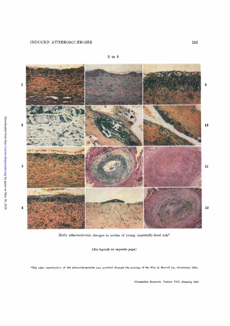
Figure 1
Aortic arch of young female breeder (non-treated)(Group I). Subintimal and medial fibroblastichypertrophy and hyperplasia. Surrounding thecells are "halos" of acid mucopolysaccharide de-posits. Note also the mild elastic distortion andappearance of int c rla initiation between adjacentelastic fibers. The endothelium exhibits earlyhyperplasia and cytoplasmic metaeliromasia. Mod-ified Rale's stain, X470.
Figure 2
A high-power view of aortic arch to show closemorphologic association of increased intercellularacid, mucopolysaccliarid.es with adjacent fibroblasts.Modified Male's stain. X970.
Figure 3
Aortic arch of a male breeder treated with ACTHtmd unilateral nephrectomy (Group II). General-ized fibroplasia and increased acid mucopolysac-eharides of snbintima and adjacent media. Endo-thelial hyperplasia has proceeded to layering andcharacterized by marked cytoplasmic vacuolation("hydropic degeneration"). Modified Hale's stain.X470.
Figure 4
Aortic arch of unilaterally nephrectomized male(Group II). The arterial wall is greatly thickenedby the intense fibroplasia and increased acid muco-polysaccharides of the intima and media, givinga "cobblestone" appearance to the section. Thecytoplasm of many fibroblasts contains metachro-matic granules. The normally orderly elastic tis-sue architecture has been distorted by reduplica-tion. Modified Hale's stain. X-170.
Figure 5
Aortic arch of unilaterally nephrectomized malebreeder (Group TV). At the base of the two earlyfibrotic alheromuta is fibroblastic hyperplasia,showing radial orientation toward the luminal sur-face. The pools of red-staining material in theintima may be collagen or pro-collagen. Mostsevere elastic distortion is in subintimal and. ad-jacent medial areas, associated with fibroplasia.Modified Hale's stain, X21O.
Figure 6
Aortic arch of a 5-times bred female (unilaterallynephrectomized). Severe destruction of medialelastic tissue, and marked accumulation of inter-
cellular acid mucopolysaccharides. Overlying thisarea is an intimal protrusion of cellular and. otherunindentified debris. Modified Hale's stain. X1OO.
Figure 7
Medium artery near abdominal aorta' of a non-treated male breeder (Group II). Note the markedendothelial hyperplasia and metacliromasia. Manyof the cells are pyknotic. The underlying internalelastic membrane is intact. The arterial lumen isalmost completely occluded. Modified Hale's stain.X21O.
Figure 8
Abdominal aorta of unilaterally nephrectomizedmale (Group IV). Peculiar giant, endothelial celloverlying subintimal fibroplasia. Its cytoplasm isfilled with coarse metachromatk grannies, and itsnucleus is somewhat vesicular. Modified Hale'sstain. X970.
Figure 9
Abdominal aorta of a non-treated, male breeder(Group II). ~\Vell-developed atheroma character-ized by intense metacliromasia and fibroplasia incentral and basal portions of plague. Marked ir-regular vacuolization at luminal side of atheroma.Normal underlying deeper layers of the media notshown. Modified Hale's stain. X-170.
Figure 10
Aortic arch. Lesion frequently encountered char-acterized by intimal pools of acid mucopolysaccha-ride subjacent to central pools of red-stainingmaterial which may be glycoprotein, (pro-) colla-gen, fibrin, or other types of mucopolysaccharides.Modified Hale's stain. X210.
Figure 11
Medium-sized artery near abdominal aorta of uni-laterally nephrectomized male rat (Group III).Marked intimal sclerosis with, complete occlusionof lumen and revascularization. Much of the in-ternal elastic membrane is destroyed, these areasbeing adjacent to sites of medial hyperplasia, H& E. X21O.
Figure 12
Same artery as above stained with Hale's stain.Note that acid mucopob/saccharide accumulationis mild. The intercellular ground substance is ap-parently glycoprotein or (pro-) collagenous innature. Modified Hale's stain, X21O.
Circulation Research, Volume VIII, January 1060
by guest on May 30, 2018
http://circres.ahajournals.org/D
ownloaded from
INDUCED ATHEROSCLEROSIS 283
5 to 8
10
11
12
Early atherosclerotic changes in aortae of young repeatedly-bred rats0
(See legends on opposite page)
•The color reproduction of the photomicrographs was provided through the courtesy of the Wm. S. Merrell Co., Cincinnati, Ohio.
Circulation Research, Vohtmc VIII, January I960
by guest on May 30, 2018
http://circres.ahajournals.org/D
ownloaded from

284 WEXLER, BROWN, MILLER
appeared in the more advanced ones. It isdifficult to believe that the fatty deposits en-countered bear any relationship to the choles-terol-induced atherosclerosis of rabbits.11 Thevacuolated endothelial cells encountered in therat aorta suggest that degeneration of thesecells may contribute lipid to the atheroma.Since cell degeneration is often accompaniedby increased lipid content (among other sub-stances), the phagocytosis of these lipids ex-truded upon disintegration during normal re-parative processes might account for the oc-currence of lipophages at, or just beneath, thesurface of the atheroma.
The reasons for the accumulation of lipidin atheromata is still unclarified.22 Perhapsthe accumulation of excess lipid in arterial tis-sue overcomes those metabolic processes con-cerned with the removal of fats, a possibilitysuggested by Shore et al.23 Faber3 believesthat lipids have an affinity for regions inwhich increased amounts of acid mucopoly-saccharides are manifest. However, this ob-servation was not verified in our studies norin investigations of rabbit atherogenesis.24 Amore likely possibility in our experimental le-sions is that plasma lipids tend to localize inareas of damaged tissue. This is suggested byappearance of lipids only where subintimallesions were advanced, or where fibrosis wassevere.
Certain effects of ACTH require comment.During initial breedings ACTH appeared tomodify the developing atherosclerosis by re-ducing the severity of metachromasia inACTH-treated animals. Increased fibrosis ofthe lesions might indicate that ACTH en-hanced reparative processes in its early stages.As the number of breedings increased, how-ever, ACTH administration was associatedwith augmented atherosclerosis. To date, wehave been unable to describe any distinctivelesions attributable only to exogenous ACTH.The possibility exists that exogenous ACTHenhances the atherogenic process induced bysuccessive breedings. Pregnancy and hyper-adrenocortical activity are closely interre-lated.25 It would be interesting to know
whether the excess endogenous production ofACTH in pregnancy is responsible for the"spontaneous" atherosclerosis observed inyoung, repeatedly-bred rats. Support for thishypothesis is found in a previous report fromthis laboratory0 in which is was shown thatACTH augmented pre-existing atherosclerosisin such subjects. Further, Gillman and Ha-thorn26 have also shown that repeated preg-nancy will produce arteriosclerosis in the rat.
The similarity of atherosclerosis observedwhen only exogenous ACTH, or unilateralnephrectomy, or repeated breedings were theexperimental variables, would seem to indi-cate that each of these mediates its action onthe connective tissue components by a similarmechanism.
The marked difference in the severity of ratatherosclerosis in relation to sex is interestingfrom a general biological standpoint. This isalso the case in respect to the well-known di-chotomy between the sexes in relation to coro-nary artery and other types of atherosclerosis.Atheromata in male rats grow inwardlytoward the lumen; in the female rats theygenerally extend in the opposite direction, ortend to remain localized in subintimal areas.This may account for our earlier observationsthat gross appraisal of lesions indicated amarked sex difference,0 i.e., female rats onlywere reported as showing the atherosclerosis.The centrifugal outgrowth of lesions in fe-males causes them to appear more severegrossly than the centripetal growth in males.One could speculate that female rats, althoughsuffering greater connective tissue damage inthe subintimal and medial areas, are in someway protected from the lethal effects of athero-.sclerosis. The male rats, showing lesions on amicroscopic scale, develop rnyocardial infarc-tion, age prematurely, and succumb at a morerapid rate than the females (unpublished ob-servations). Therefore, this sex difference maybe due to the degree and chronological appear-ance of intimal involvement.
It is possible that there may be a funda-mental difference in the reaction of connectivetissue of male and female rats to ACTH. It is
Circulation Research, Volume VI11, Januuary 19G0
by guest on May 30, 2018
http://circres.ahajournals.org/D
ownloaded from

INDUCED ATHEROSCLEROSIS 285
also conceivable that male and female sex hor-mones may condition the connective tissue ele-ments of the arterial wall to respond differ-ently 1o the effects of other hormones.
SummaryEarly atherosclerotic lesions are described
in aortas of repeatedly-bred rats of both sexes.Characteristically, subintimal accumulationsof acid mucopolysaccharides and fibroblasticprolifieration marked the onset of athero-sclerosis, followed by distortion and destruc-tion of elastic lamellae.
Several other pathologic alterations oc-curred later, notably fibrosis, endothelial hy-perplasia, lipid deposition, and calcification.Elastic tissue changes occurred independentlyand usually secondary to alterations of theground substance and fibroblasts. Calcificationand lipid deposits are found in more advancedlesions, showing an affinity for areas rich inluucopolysacharides. Some of these animalswere subjected to unilateral nephrectomy, in-jections of ACTH, or combinations of both.Exogenous ACTH and unilateral nephrec-tomy augmented the early atherosclerotic le-sions associated with multiple breedings.
Atheromata in the male rats progressedtoward the lumen, whereas those in femalestended to extend toward the adventitia. De-spite the equal incidence of lesions in eithersex, their severity as judged by distribution,mortality rate, and myoeardial infarction wasgreater in the males. This sex difference isdiscussed in terms of a possible relationshipbetween the arterial connective tissues andcertain hormonal imbalances.
AcknowledgmentThe Authors wish to express their appreciation for
technical assistance given by Janice Doron, BarbaraSollborger, Myra Springs, Lionel Staples, EobertLeBlond, and James Hensen.
Summario in InterlinguaEs describite precoce lesiones atherosclerotic con-
statate in le aortas de repetitemente copulate rattosde ambe sexos. Characteristicainente, aceumulationessubintimal de mucopolysaccharidos acide e prolifera-tion fibroblastic marcava le declaration del athero-sclerosis, sequite per distortion e destruction del la-mellas elastic.
Plure altere alterationes pathologic occurrcva plustarde, notabilcmente fibrosis, hyperplasia endothelial,deposition de lipido, e calcification. Alterationes dohisto elastic occurreva. independenteniento e usual-mente secundafi a alterationes del substantia funda-mental e fibroblastos. Calcification e deposition delipido es trovate in plus avantiate lesiones c moustraun afnnitiate pro areas que es ric in nmeopolysac-charidos. Certes de iste animates esseva subjicite anephrectomia unilateral, injectiones de ACTH, o com-binationcs del duo. ACTH exogene e nephrectomiaunilateral augmentava le prccoce lesiones dc athero-sclerosis associate con multiple copulationes.
Atheromas in le rattos mascule l>rogredeva versole luinine, durante que illos in rattos feininhi tendevaa progreder verso le adventitia. In despecto delequalitate de incidentia del lesiones in le duo sexos,le severitate de illos—a. judicar per le distribution, lemortalitate, e le infarcimentos del myocardio—essevaplus grande in le masculos. Iste differentia, inter lesexos es discutitc con referontia al relation possibileinter le histos conjunctive del arterias o certo iin-balancias hormonal.
References.1. MOON, H. D., AND RINEHART, J. F.: Histogenesis
of coronary arteriosclerosis. Circulation 6:481, 1952.
2. TAYLOR, H. E.: The role of mucopolysaccharidesin the pathogenesis of intimal fibrosis andatherosclerosis of the human aorta. Am. .1.Path. 29: 871, 1953.
3. FABER, M.: The human aorta. Sulfate-contain-ing polyuronides and the deposition of chol-esterol. Arch. Path. 48: 342, 1949.
4. ALTSIIULER, C. H., AND ANGEVINE, D. M.: Acid
mucopolysaccharides in degenerative disease ofconnective tissue, with special reference toserous inflammation. Am. J. Path. 27: 141,1951.
5. RINEHART, J. F., AND GREENBERG, L. D. : Patho-genesis of experimental arteriosclerosis in pyri-doxine deficiency. Arch Path. 51: 12, 1951.
6. LINDSAY, S., CHAIKOFF, I. L., AND GILMORE, J.
W.: Arteriosclerosis in the dog. I. Spontaneouslesions of the aorta and coronary arteries.Arch. Path. 53: 281, 1952.
7. LINDSAY, S., AND CHAIKOFF, I. L.: Arteriosclerosis
in the baboon. Arch. Path. 63: 460, 1957.8. SCHWARTZ, C. J., PETERS, J. A., AND DAY, A. J'.:
The effects of hypercholesterolemia on thearterial ground substance in rabbit athero-sclerosis. Australian J. Exper. Biol. & Med.Sci. 36: 109, 1958.
9. WEXLER, B. C, AND MILLER, B. F.: Severe arterio-sclerosis and other diseases in the rat pro-duced by corticotrophin. Science 127: 590,1958.
Circulation Research, Volume VIII. January 1960
by guest on May 30, 2018
http://circres.ahajournals.org/D
ownloaded from

286
10. WEXLER, B. C, AND MILLER, B. F. : Coronary
arteriosclerosis and thrombosis in the rat.Proc. Soc. Exptl. Biol. & Mecl. 100: 573, 19139.
11. DUFF, G. L., MCMILLAN, G. C, AND RITCHIE,
A. C.: The morphology of early atheroscleroticlesions of the aorta demonstrated by the sur-face technique in rabbits fed cholesterol. Am.,T. Path. 33: 845, 1957.
12. BINEHART, J. F., AND ABUL-HAJ, S. K.: An im-
proved method for histologic demonstration ofacid mucopolysaccharides in tissues. Arch.Path. 52: 189, 1951.
13. BERENBAUM, M. C.: The histochemistry of bound
Hpids. Quart. J. Micr. Sci. 99: 231, 1958.14. GoxiORi, G.: Aldehyde-fuchsin: A new stain for
elastic tissue. Am. J. Clin. Path. 20: 666, 1950.15. WORLD HEALTH ORGANIZATION TECHNICAL RE-
PORT: Classification of Atherosclerotic Lesions,Series No. 143, Geneva, 1958.
16. MOON, H. D.: Coronary arteries in fetuses, in-fants, and juveniles. Circulation 16: 263, 1957.
17. ASBOE-HANSEN, G.: On the studies and function
of the mast cell, in Connective Tissue. Tun-bridge R. E. (Ed.) Charles C Thomas, Spring-field. 1957. pp. 12-26.
18. GERSH, I., AND CATCHPOLE, H. R.: The organiza-
tion of ground substance and basement mem-brane and its significance in tissue injury,
WEXLER, BROWN, MILLER
disease and growth. Am. J. Anat. 85: 457,1949.
19. SHOENBERG, M. D., AND MOORE, R. D.: Studies
on Connective Tissue. Arch. Path. 65: 115,1958.
20. TAYLOR, H. E., AND SAUNDERS, A. M.: The asso-
ciation of metaehromatie ground substance withfibroblastie activity in granulation tissue. Am.J. Path. 33: 525, 1957.
21. GOULD, B. S., AND WOESSNER, J. F. : Biosyn-
thesis of collagen: Influence of ascorbic acid onproline, hydroxyproline glyeine and collagencontent of regenerating guinea pig skin. J.Biol. Chem. 226: 289, 1957.
22. MOSCHOWITZ, E.: Hyperplastie arteriosclerosis
versus atherosclerosis. J.A.M.A. 143: 861,1950.
23. SHORE, H. L., ZILVERSMIT, D., AND ACKERMAN,
R. F. : Phospholipid synthesis by arterial wall.Am. J. Physiol. 181: 527, 1955.
24. ADLERSBERG, D., WANG, C, AND STRAUSS, L.: The
role of the ground substance in atherogenesis.J. Mt. Sinai Hosp. 24: 655, 1957.
25. VENNING, E. H.: Adrenal function in pregnancy.Endocrinology 39: 203, 1946.
26. GILLMAN, T., AND HATHORN, M.: Sex incidence
of vascular lesions in aging rats in relation toprevious pregnancies. Nature 183: 1139, 1959.
Circulation Research, Volume VIII, January 1960
by guest on May 30, 2018
http://circres.ahajournals.org/D
ownloaded from

BERNARD C. WEXLER, THOMAS E. BROWN and BENJAMIN F. MILLEREarly Lesions
Nephrectomy: Acid Mucopolysaccharides, Fibroplasia, Elastosis and Other Changes in Atherosclerosis in Rats Induced by Repeated Breedings, ACTH and Unilateral
Print ISSN: 0009-7330. Online ISSN: 1524-4571 Copyright © 1960 American Heart Association, Inc. All rights reserved.is published by the American Heart Association, 7272 Greenville Avenue, Dallas, TX 75231Circulation Research
doi: 10.1161/01.RES.8.1.2781960;8:278-286Circ Res.
http://circres.ahajournals.org/content/8/1/278World Wide Web at:
The online version of this article, along with updated information and services, is located on the
http://circres.ahajournals.org//subscriptions/
is online at: Circulation Research Information about subscribing to Subscriptions:
http://www.lww.com/reprints Information about reprints can be found online at: Reprints:
document. Permissions and Rights Question and Answer about this process is available in the
located, click Request Permissions in the middle column of the Web page under Services. Further informationEditorial Office. Once the online version of the published article for which permission is being requested is
can be obtained via RightsLink, a service of the Copyright Clearance Center, not theCirculation Research Requests for permissions to reproduce figures, tables, or portions of articles originally published inPermissions:
by guest on May 30, 2018
http://circres.ahajournals.org/D
ownloaded from