atherosclerosis, autoimmunity, and vascular-associated lymphoid
TRANSCRIPT
0892-6638/97/0011-11 99/$02.25 © FASEB 1199
Atherosclerosis, autoimmunity, and vascular-associated
lymphoid tissue
G. WICK,*.t’ M. ROMEN, A AMBERGER,* B. METZLER,* M. MAYR,*G. FALKENSAMMER,t 1J Q XTJ**Institute for Biomedical Aging Research of the Austrian Academy of Sciences; and tlnstitute for
General and Experimental Pathology, University of Innsbruck, Medical School, Innsbruck, Austria
CLASSICAL CONCEPTS FOR THEDEVELOPMENT OF ATHEROSCLEROSIS
Atherosclerosis is a multifactorial disease induced bythe effects of various risk factors on appropriate ge-netic backgrounds. It is characterized by vascularareas containing mononuclear and proliferating
smooth muscle cells,as well as extracellularmatrix
(ECM)2 components resulting in hardening and
thickening (arteriosclerosis) of the arterial wall. In astrict sense, atherosclerotic lesions are localized inthe inlima; they also contain foam cells and depositsof cholesterolcrystalsmanifested as fatty streaks and,finally, as atheroscleroticplaques.
Fatty streaks are whitish, cushion-like lesions of the
arterial intima harboring abundant lipid-laden mac-rophages, so-called foam cells, which are consideredprecursors of the fully developed plaques that may
finally exulcerate and even calcif’. The main theoriesof atherogenesis are the “response to injury” (1) andthe “response to altered lipoprotein” (2) hypothe-
ses. The response to injury hypothesis postulates analteration of the intima by various risk factors (me-chanical injury, chemically altered low density lipo-proteins [LDL], viruses, toxins) that initiates a
primary endothelial dysfunction and subsequent
changes in permeability, expression of adhesion mol-ecules, and release of chemotactic and growth fac-tors. Consequently, platelets and monocytes becomeactivated and attach to these endothelial cells. Blood-derived monocytes transmigrate into the suben-
dothelial space and transform into macrophages;
smooth muscle cells (SMC) are attracted from themedia to the same site. Both monocytes/macro-
phages and SMC possess the so-called scavenger re-
ceptor, which binds chemically altered (oxidizedLDL {oxLDL]), but not native, LDL in a nonsatura-ble fashion. By uptake of oxLDL, macrophages and
SMC develop into foam cells, and the deposition ofcollagenous and noncollagenous ECM components,especially in the peripheral “shoulder” region andthe superficial “cap” area, complete the pathohis-tological appearance of fatty streaks and atheroscle-rotic plaques, respectively.
The response to altered lipoprotein hypothesis is
based on the concept that lipoproteins can be chem-ically modified and are then able to induce foam cellformation by monocytes/macrophages and SMC. Inrecent years it has become evident that modification
of lipoproteins does not occur primarily in the cir-culation or during transgression through the endo-thelium, but they rather accumulate in native formin the subendothelial space, where the lipoproteinsare retained and oxidized. Accumulation of oxLDL,therefore, is not only the result of increased influxfrom the serum into the arterial intima but, con-versely, is also due to a diminished efflux with sub-
sequent foam cell formation. This “response to LDLretention” hypothesis (3) is thus a special variant ofthe response to altered LDL concept. Thus far, theselatter theories have not explained why atheroscle-rotic lesions develop at certain arterial predilectionsites or why the disease does not affect the venousvascular bed.
THE “IMMUNOLOGICAL HYPOTHESIS” FORATHEROGENESIS
During the past few years we have developed a newimmunological hypothesis for atherogenesis (4-6)based on observations in humans and in animal ex-periments that encompass major components ofboth the response to injury and the response to al-tered lipoprotein hypotheses. Our studies were orig-inally triggered by three factors: our work on theimmunomodulatory role of lipids (7, 8), our long-
‘Correspondence: Institute for Biomedical Aging Researchof the Austrian Academy of Sciences, Rennweg 10, A-6020
Innsbruck, Austria.2Abbreviation: hsp, heat shock proteins; ECM, extracellular
matrix; LDL, low density hipoprotein; SMC, smooth muscle
cells; oxLDL, oxidized LDL; IL, interleukin; MHC, major his-tocompatibility complex; IFN, interferon; TNF, tumor necro-
sis factor; MALT, mucosa-associated lymphoid tissue; GALT,
gut-associated lymphoid tissue; ICAM-1, intercellular adhesionmolecule 1; VCAM-1, vascular cell adhesion molecule-i;ELAM-i, E-selectin; HIA, human MHC; VALT, vascular-asso-
ciated lymphoid tissue.
1200 Vol. 11 November 1997 The FASEB Journal WICK ET AL.
standing interest in autoimmunity (9, 10), and the
increasing number of reports in the literature point-ing to the possible primary or secondary contributionof humoral and cellularimmune reactions to athero-
sclerosis (11-15). Thus, immunoglobulin and com-plement deposits can be demonstrated in advancedlesions, and evidence for complement activation viathe alternative pathway has also been provided (16).A considerable number of lymphoid cells can be
demonstrated within atherosclerotic plaques, most ofwhich are CD4 and interleukin-2(IL-2)receptor,
i.e., activated, although CD8 cells are also present(17).Major histocompatibilitycomplex (MHC) class
II (HLA-DR) antigens are expressed by the majorityof T cells as well as by neighboring macrophages and
SMC, indicating the production of interferon y (IFN-‘y) by the former (17). This fact is also supported byincreased levels of neopterin, an IFN-y-inducedmonocyte/macrophage product, in the serum of ath-erosclerotic patients (18). B cells are scarce in allstages of atherosclerotic lesions, rendering local an-tibody production rather improbable. Also, the num-ber of intralesional granulocytes is surprisingly low.
The cells constituting atherosclerotic lesions producemany differentcytokines that stimulate growth, dif-
ferentiation, chemotaxis, and cytotoxicity, includingIL-i (endothelial cells, SMC, macrophages), IL-8 (en-dothelial cells, macrophages), tumor necrosis factora (TNF-a) (SMC, macrophages, T cells), monocytechemotactic peptide 1 (endothelialcells,SMC, mac-
rophages), platelet-derived growth factor (PDGF)(endothelial cells, SMC, macrophages), and IFN-y (Tcells)(19).
In contrast to general belief, quantitative immu-nohistochemical analyses of the mononuclear cell in-filtrate in the earliest stage of atherosclerotic lesions-in the transition zone between normal intima and
fatty streaks-revealed a clear preponderance of Tcells over macrophages, thus supporting the primaryrole of immune reactions in disease development
(17). Furthermore, HLA-DR expression by endothe-
hal cells was observed only in areas overlying T cellinfiltrates in the intima, excluding the possibility ofendothelial cells acting as primary antigen-present-ing cells. We postulated sensitization of T cells atmore distant sites, such as draining lymph nodes.However, Bobryshev et al. (20-22) have clearlyshown that the presence of dendritic cells in the nor-mal arterial intima suggests T cell sensitization at thatsite as well.
The majority of T cells in early lesions carry the T
cell receptor a/13 (TCR-a/), but a surprisingly highpercentage (10-15%) express TCR-y/#{246}at levels farexceeding that in the peripheral blood of the samedonors (23). Whereas the proportion of V#{246}9/#{246}2TCR cellsin peripheral blood remained low andconstant throughout all lesional stages, the TCRV1 +
subpopulation made up most of the TCR-y/ cells in
vel-y early lesions. These cells are characteristic formucosa-associated lymphoid tissue (MALT), such asthe gut-associated lymphoid tissue (GALT), and pref-erentially react to heat shock proteins (hsp) (24).
Whereas the T cell repertoire in early atheroscle-rotic lesions has not yet been determined, no indi-cations of restricted TCR V-gene usage have beenfound in analyses of T cell clones derived from ath-erosclerotic plaques (25).
Candidate antigens that may lead to atherosclero-sis-associated humoral and/or cellular immune re-actions include (12): 1) modified lipoproteins; 2)partly degraded or denatured macromolecules de-rived from the necrotic core of plaques; 3) intracel-lular cryptic antigens exposed through cytolysis ornecrosis; 4) neoantigens: hsp or cytokine-induced
cell surface antigens; and 5) antigens of infectiousorganisms such as herpes virus, cytomegalovirus, andClamydia pneumoniae.
To date, definitive experimental proof that one of
these antigens may initiate the pathogenesis of ath-erosclerosis is available only for hsp (26). Thus, au-toantibodies to oxLDL are now considered to have aprotective effect (27), and induction of disease byim-
munization of experimental animals with oxLDL hasnot been achieved. There may, of course, be excep-tions to the concept that immune reactions are in-strumental in atherogenesis, especially whereexcessive overloading of the system with cholesteroloccurs due to severe genetic conditions, such as thelack of certain lipoproteins or hipoprotein receptors
with or without concomitant immune deficiency(28), homocystinurea, etc.
ATHEROSCLEROSIS: AN AUTOIMMUNE
DISEASE
Our own clinical and experimental data provide evi-dence that atherosclerosis starts as an inflammatoryimmunological disease due to an autoimmune reac-tion against the stress protein hsp 60.
Stress proteins (or hsp) comprise a family of pro-teins and its cognates that are induced by variousforms of stress such as temperature, mechanical irri-tation, infections, and toxins (29). Hsp are classifiedaccording to their molecular mass into four maingroups: hsp 90, hsp 70, hsp 60 kDa, and low molec-
ular mass families.They fulfill various physiological functions such as
the assembly, intracellulartransport,and controlled
breakdown of proteins;many hsp act as chaperonesto protect other proteins from denaturation whencells are subjected to stress. Hsp are phylogeneticallyhighly conserved. Thus, mycobacterial hsp 65 showgreater than 50% sequence homology with human
hsp 60 on the DNA and protein levels. Hsp constitutemajor antigenic determinants of microorganisms and
Lt) 0 0 4CD (0 W (00.. 0 0.Cl) Cl) Cl)
C.,C) 2>- 4 4
-JDI C.)
ATHEROSCLEROSIS 1201
are thus important for induction of protective hu-moral and cellularimmune responses, although we“pay” for this protection by risking consecutive au-toimmune reactions that may resultin the develop-ment of autoimmune disease (30, 3i).
Evidence that the initial stages of atherosclerosis
are mediated by an immune reaction against hsp 60
can be summarized as follows: Immunization of rab-bits with hsp 65-containing material, such as com-plete Freund’s adjuvant alone or recombinantmycobacterial hsp 65, develop arteriosclerotic lesions
at typical predilection sites that are subject to in-creased hemodynamic stress, including the aorticarch and major arterial bifurcations. Pathohistologi-cally, these lesions consist of activated T cells, mac-rophages, SMC and ECM deposition, but no foamcells (17). Concomitant feeding of a cholesterol-richdiet induces the development of classical fatty streaksand atherosclerotic plaques, including foam cells,that are more severe than those induced by choles-terol feeding alone and completely parallel the hu-man situation (26). Whereas the first inflammatorystage is reversible, the later cholesterol-augmentedstage is not, at least under our experimental timeschedule (32). The peripheral blood of hsp 65-im-munized rabbits contains hsp 65-specific antibodiesand T cells. T cells derived from arterial lesions ofthese rabbits show a significantly increased reactivityto hsp 65 compared to T cells from peripheral blood
of the same animals. T cells isolated from atheroscle-rotic lesions of rabbits that were not immunized, butreceived a cholesterol-rich diet, also revealed a pref-erential reactivity with hsp 65 (33).
Since hsp 65/60 has been incriminated as a possi-ble (auto)antigen in a variety of autoimmune dis-
eases (rheumatoid arthritis, diabetes, systemic lupuserythematosus, scleroderma) (30, 34-37), it is of rel-evance that immunization of rats with hsp 65 led toadjuvant arthritiswithout arteriosclerosis(34; our
own unpublished observations), whereas the reverseistrue for rabbits (26). Inducing hsp 65-dependentarteriosclerosis in mice has also met with limited suc-cess. These differing responses may be due to bothrecognition of differenthsp 65/60 epitopes by theimmune system and differing target cell susceptibility(i.e., endothelial cells vs. synovial cells) to different
stressors in different species.Establishing T cell lines and clones from very early
human lesions has so far been complicated by tech-
nical problems such as the relative paucity of infil-trating cells, difficulties excluding T cells adhering toarterial endothelium or contained in vasa vasorum,
or insufficient supply of antigen-presenting cells.Nevertheless, we have demonstrated significantlyhigher titers of anti-hsp 65 antibodies in the sera ofmany clinically healthy volunteers with sonographi-cally proven atherosclerotic carotid artery lesions vs.controlswithout such lesions(38),an observation re-
cently corroborated in coronary atherosclerosis pa-tients. Immediately after myocardial infarction, thesepatients experience a drop in anti-hsp 65 titers, prob-ably due to the sudden liberation of hsp 60 and sub-
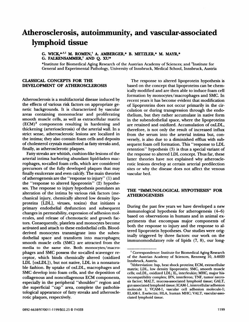
sequent immune complex formation (39). Theanti-mycobacterial hsp 65 antibodies cross-react withrecombinant human hsp 60 and also yield a 60 kDa
band in Western blots with extracts of human athero-sclerotic plaques. Furthermore, these antibodies re-act with GroEL, the Escherichia coli hsp 60 homologueand Chiamydial hsp 60 (Fig. 1). The presence of anti-hsp 65/60 antibodies has been defined as a new riskfactor for atherosclerosis independent of classical risk
factors such as high blood cholesterol values, smok-ing, overweight status, and gender. So far, three lin-ear hsp 65 epitopes have been shown to berecognized by human anti-hsp 65 antibodies, twowith high homology for human hsp 60 (40). Thereactivity with-probably more important-confor-
mational epitopes is still under investigation.Certain hsp 60 epitopes could be demonstrated on
the surface of stressed (heat, TNF-a, H202, bacterial
endotoxin), but not unstressed, cells (41-44). Themechanism whereby hsp 60 or portions of this intra-
cellular, mainly mitochondrial protein are trans-ported to the cell surface is still under investigationin several laboratories (45). It has, however, beenfirmly established that anti-hsp 65/60 antibodies ex-ert a cytotoxic effect on stressed, but not unstressed,endothelial cells and macrophages in a complement-mediated fashion or via antibody-dependent cellularcytotoxicity (46, 47). This effect can also be blockedby E. coli GroEL.
To allow for interaction of hsp 65/60-specific Tcells with endothelial cells, the expression of adhe-sion molecules and hsp 60 is required. The latter
Figure 1. Western blot showing the cross-reactivity of human
anti-hsp antibodies (affinity chromatography purified from a
pool of 5 high titer sera of clinically healthy persons with sono-graphically demonstrable atherosclerotic lesions in the A. car-
otiswith recombinant mycobacterial hsp 65, human hsp 60, E.
coli GroEL, and hsp 60 from Chiamydia pneumoniae. Bovine se-
rum albumin (BSA) was used as a negative control antigen.
IMMUNE SYSTEM
VENOUSENDOTHELIAL
CELLS
ARTERIALENDOTHELIAL
CELLS
I
T1
IC-1
lJ.c-)zU
C,)
0iii I
rLDL oxLDL LOL OxLOL
1202 Vol. 11 November 1997 The FASEB Journal WICK ET AL.
must be presented in association with H1A class I orclass II molecules. However, as mentioned earlier, aprimary aberrant HIA class II expression has onlybeen observed on human endothelial cellsafterin-
timal T cell infiltration(17). On the other hand,
HLA class Il-positivemacrophages and dendriticcells
are present at predilection sites in the intima, andmay stimulate hsp 65/60-specific T cells after theirtransmigration to this site (22). Since the affinity ofthe TCR for the MHC-peptide complex is far too lowfor interaction of hsp 65/60-specific T cells withstressed endothelial cells under arterial blood flowconditions, adhesion molecule expression iscritical
to the initiationof an immune response. Applicationof various types of stress to human arterial and venous
endothehial cells leads to the simultaneous expres-sion of hsp 60 and adhesion molecules (intercellularadhesion molecule 1, ICAM-1; vascular cell adhesion
2.5
2
0.5
0
3
2.5
2
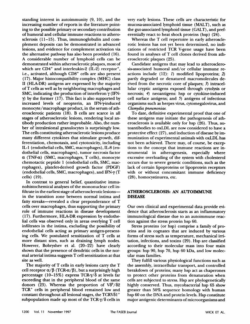
Figure 2. Fluorescence activated cellsorter (FACS) analysisof
adhesion molecule expression by human venous and arterial
enthothelial cells after stress by incubation with oxLDL andnative LDL as a control.Confluent arterialendothelialcellmonolayers were treated with 80 Lg/ml native or oxLDL. The
fluorescence index was calculated as the ratio of the meanfluorescence intensity of stressed vs. unstressed endothelial
cells.Expression of ICAM-1, VCAM-1, and ELAM-l in termsof mean values and standard deviations of four independent
experiments are shown. Preparation and characterization of
oxLDL as well as incubation conditions are detailed in ref 49.
Incubation for 12 h (0); 24 h (); 48 h (#{149}).*significant
difference from control cells P < 0.05.
-TARGET.
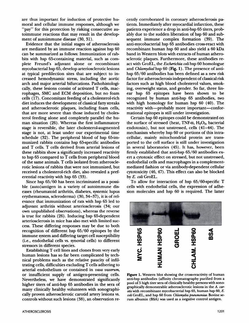
Figure 3. The development of autoimmune disease depends
on the presence of essential genes coding for autoreactivity ofthe immune system and susceptibility of the target cell for the
autoimmune attack,respectively.Nonessentialfactorsmodu-lating the final outcome of the disease are symbolized by ar-
rows. Essential genes influencing autoreactivity may, for
example, code for certain MHC haplotypes, T cell receptorspecificity, and interleukin-2 (IL-2) hyperproduction. Essen-
tial genes affecting a target cell or target organ may code for
susceptibility to virus infections. Modulatory factors include
dietary components (such as iodine in the case of Hashimotothyroiditis) and hormones (glucocorticoids, sex hormones,and others).
molecule-i, VCAM-i; E-selectin, ELAM-i) (48, 49).There are distinctdifferencesin the susceptibility of
arterialand endothehial cellsto different stressors,
the former generally giving a more sustained re-sponse, especially with respect to ELAM-i. In fact,
VCAM-.1 oxLDL acts as a potent inducer of adhesion mole-cules and hsp 60 exclusively on arterial endothehialcells, and the efficiency of oxLDL as a stressor de-
pends on its degree of oxidation (Fig. 2). However,the group of Fogelman (50) has clearly shown thatprolonged action of minimally modified LDL is alsoatherogenic.
ELAM4 Based on our experience with animal models ofspontaneous or experimentally induced organ-spe-
cific and systemic autoimmune diseases, we earlierdeveloped a general concept for the development of
autoimmune diseases postulating that two sets of es-sential genes must be present for autoimmune dis-ease development in an individual or an inbred strainof animals (Si). One set of genes codes for an ab-normal autoimmune reactivity of the immune system,
the other for a primary susceptibility of the targetorgan/structure to attackby humoral and/or allular
autoimmune effector mechanisms. The definitiveoutcome of a given disease isthen fine-tunedby mod-
ulatory factors such as diet and hormones (Fig. 3).This concept also applies to our immunological hy-
pothesis for atherogenesis, which can be summarizedas follows (Fig. 4). Most human beings possess hsp65-reactivehumoral antibodiesand T cellsthatafford
ImmunologicHypothesisforAtherogenesis
Sfresaorag. oxLDL,tree radicals,oxlns (LPS),heatshock (fever,hypertension(shearstress)
Antigens(1) InfectIonswith agents
that produce haphomologues;
(2 tissuedamage-Iln
Damage to endothellalcell proteins
Increased hap production
oxLDL
Immuneresponseagelnstheps
Foamcellswithsw1aceexpoaed
heps
ATHEROSCLEROSIS 1203
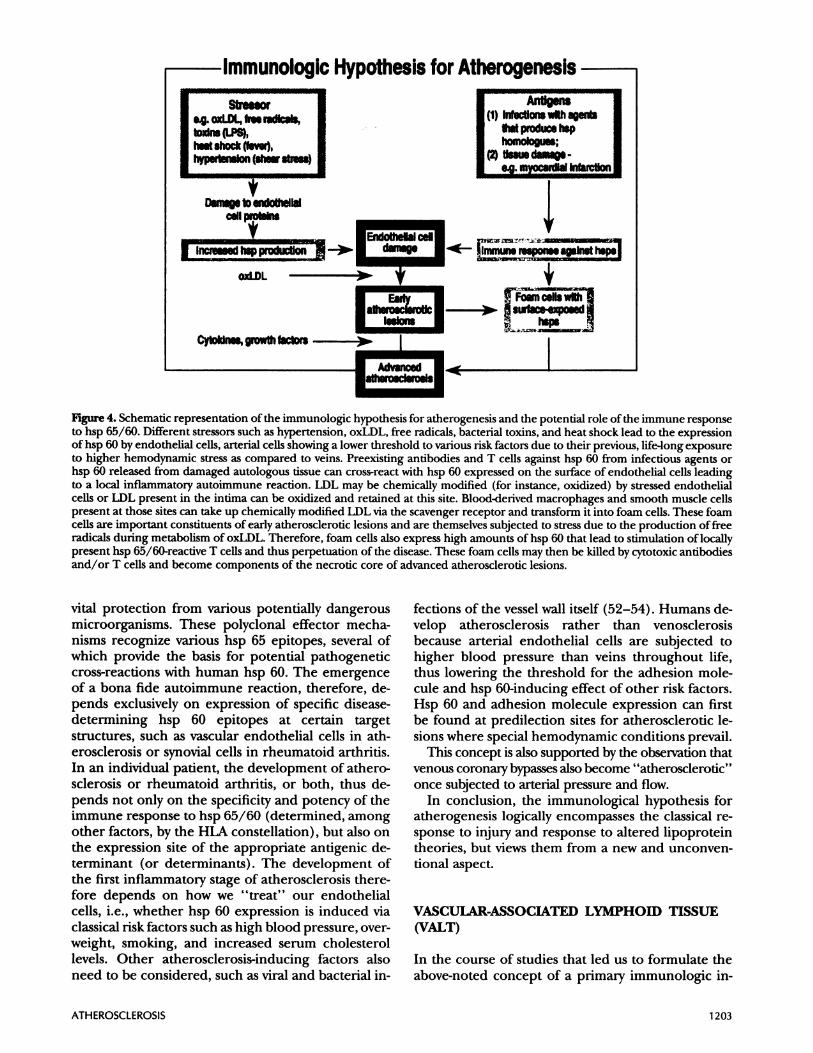
Figure 4. Schematic representation of the immunologic hypothesis for atherogenesis and the potential role of the immune responseto hsp 65/60. Different stressors such as hypertension, oxLDL, free radicals, bacterial toxins, and heat shock lead to the expressionof hsp 60 by endothelial cells, arterial cells showing a lower threshold to various risk factors due to their previous, life-long exposureto higher hemodynamic stressas compared to veins. Preexisting antibodies and T cells against hsp 60 from infectious agents orhsp 60 released from damaged autologous tissue can cross-react with hsp 60 expressed on the surface of endothelial cells leadingto a local inflammatory autoimmune reaction. LDL may be chemically modified (for instance, oxidized) by stressed endothelialcells or LDL present in the intima can be oxidized and retained at this site. Blood-derived macrophages and smooth muscle cellspresent at those sites can take up chemically modified LDL via the scavenger receptor and transform it into foam cells. These foamcells are important constituents of early atherosclerotic lesions and are themselves subjected to stress due to the production of freeradicals during metabolism of oxLDL. Therefore, foam cells also express high amounts of hsp 60 that head to stimulation of locallypresent hsp 65/60-reactive T cells and thus perpetuation of the disease. These foam cells may then be killed by cytotoxic antibodiesand/or T cells and become components of the necrotic core of advanced atherosclerotic lesions.
vital protection from various potentiallydangerous
microorganisms. These polyclonal effector mecha-nisms recognize various hsp 65 epitopes, several ofwhich provide the basis for potential pathogenetic
cross-reactions with human hsp 60. The emergenceof a bona fide autoimmune reaction, therefore, de-pends exclusively on expression of specific disease-determining hsp 60 epitopes at certain targetstructures, such as vascular endothelial cells in ath-
erosclerosis or synovial cells in rheumatoid arthritis.In an individual patient, the development of athero-sclerosis or rheumatoid arthritis, or both, thus de-
pends not only on the specificity and potency of theimmune response to hsp 65/60 (determined, among
other factors, by the HLA constellation), but also onthe expression site of the appropriate antigenic de-terminant (or determinants). The development ofthe first inflammatory stage of atherosclerosis there-fore depends on how we “treat” our endothehialcells, i.e., whether hsp 60 expression is induced via
classical risk factors such as high blood pressure, over-weight, smoking, and increased serum cholesterollevels. Other atherosclerosis-inducing factors alsoneed to be considered, such as viral and bacterial in-
fections of the vessel wall itself (52-54). Humans de-velop atherosclerosis rather than venosclerosis
because arterial endothehial cells are subjected tohigher blood pressure than veins throughout life,thus lowering the threshold for the adhesion mole-cule and hsp 60-inducing effect of other risk factors.Hsp 60 and adhesion molecule expression can firstbe found at predilection sitesfor atheroscleroticle-
sions where special hemodynamic conditions prevail.This concept isalsosupported by the observationthat
venous coronaiy bypasses also become “atherosclerotic”once subjectedto arterial pressure and flow.
In conclusion, the immunological hypothesis foratherogenesis logically encompasses the classical re-
sponse to injury and response to altered lipoproteintheories, but views them from a new and unconven-tional aspect.
VASCULAR-ASSOCIATED LYMPHOII) TISSUE
(VALT)
In the course of studiesthat led us to formulate the
above-noted concept of a primary immunologic in-
B
CD8 ____________________CD4
C03
0 50 100 150 200 250 300 350
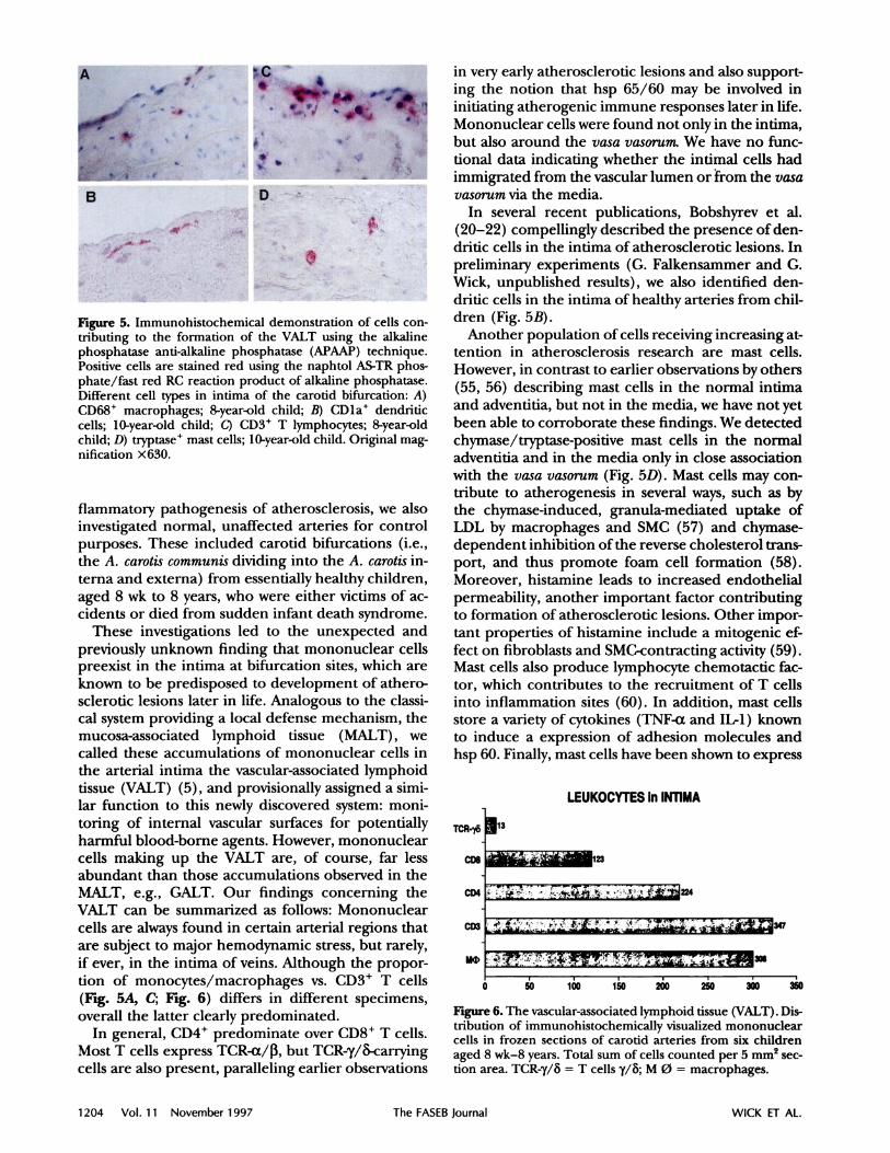
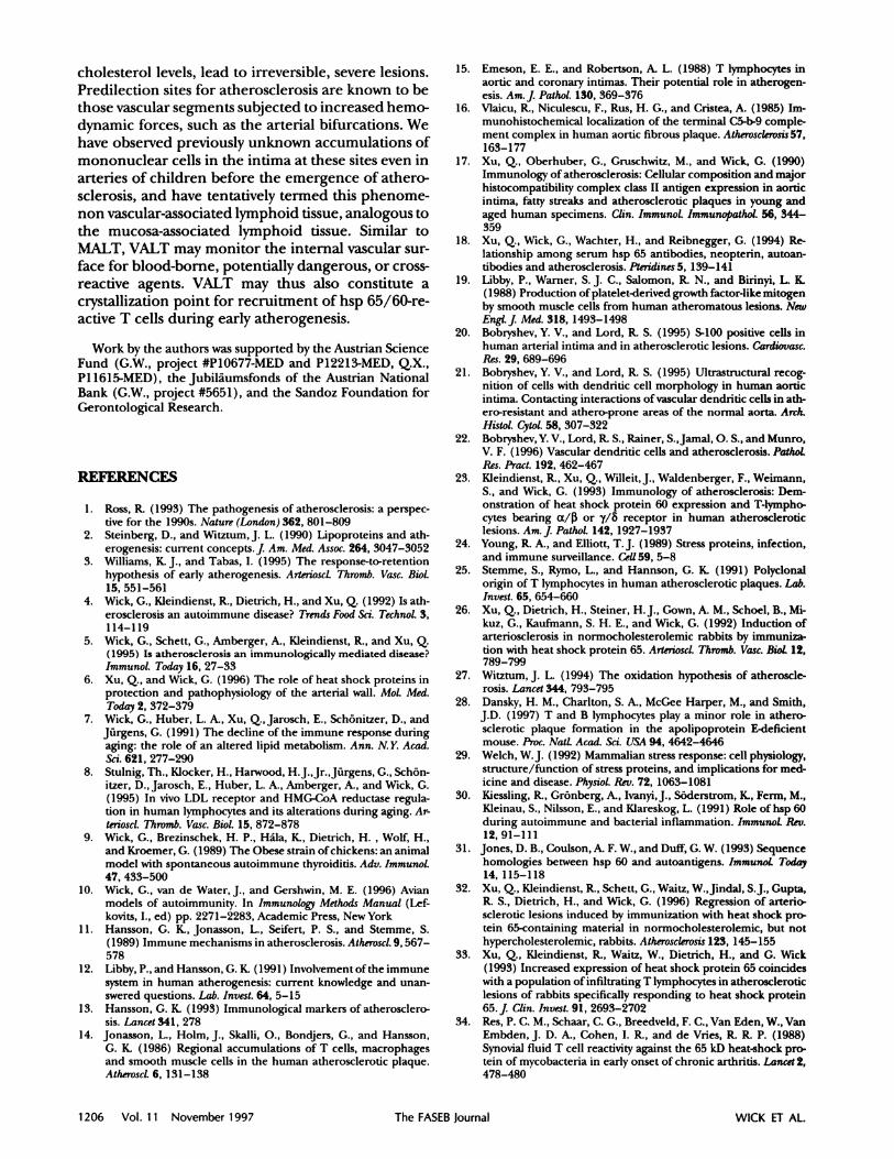
Figure 6. The vascular-associated lymphoid tissue (VALT). Dis-tribution of immunohistochemically visualized mononuclearcells in frozen sections of carotid arteries from six childrenaged 8 wk-8 years. Total sum of cells counted per 5 mm2 sec-tion area. TCR-7/#{246}= T cells y/#{246};M 0 = macrophages.
1204 Vol.11 November1997 The FASEB Journal WICK ET AL.
LD -
-
Figure 5. Immunohistochemical demonstration of cells con-tributing to the formation of the VALT using the alkalinephosphatase anti-alkaline phosphatase (APAAP) technique.Positive cells are stained red using the naphtol AS-TR phos-phate/fast red RC reaction product of alkaline phosphatase.Different cell types in intima of the carotid bifurcation: A)CD68 macrophages; 8-year-old child; B) CDla dendriticcells; 10-year-old child; C) CD3* T lymphocytes; 8-year-old
child; D) tryptase mast cells; 10-year-old child. Original mag-nification x630.
flammatory pathogenesis of atherosclerosis, we alsoinvestigatednormal, unaffected arteriesfor control
purposes. These included carotid bifurcations (i.e.,the A. carotis communis dividing into the A. carotis in-terna and externa) from essentially healthy children,aged 8 wk to 8 years, who were either victims of ac-cidents or died from sudden infant death syndrome.
These investigationsled to the unexpected and
previously unknown finding that mononuclear cellspreexist in the intima at bifurcation sites, which are
known to be predisposed to development of athero-sclerotic lesions later in life. Analogous to the classi-
cal system providing a local defense mechanism, themucosa-associated lymphoid tissue (MALT), wecalled these accumulations of mononuclear cells in
the arterial intima the vascular-associated lymphoidtissue (VALT) (5), and provisionally assigned a simi-lar function to this newly discovered system: moni-
toring of internal vascular surfaces for potentiallyharmful blood-borne agents. However, mononuclearcells making up the VALT are, of course, far lessabundant than those accumulations observed in theMALT, e.g., GALT. Our findings concerning theVALT can be summarized as follows: Mononuclear
cells are always found in certain arterial regions thatare subject to major hemodynamic stress,but rarely,
ifever, in the intima of veins.Although the propor-
tion of monocytes/macrophages vs. CD3 T cells(Fig. 5A, C; Fig. 6) differs in different specimens,overall the latter clearly predominated.
In general, CD4 predominate over CD8 T cells.Most T cells express TCR-a/3, but TCR-y/&-carryingcells are also present, paralleling earlier observations
in very early atherosclerotic lesions and also support-ing the notion that hsp 65/60 may be involved ininitiating atherogenic immune responses later in life.
Mononuclear cells were found not only in the intima,but also around the vasa vasorum. We have no func-tional data indicating whether the intimal cells hadimmigrated from the vascular lumen or from the vasavasorum via the media.
In several recent publications, Bobshyrev et al.(20-22) compellingly described the presence of den-dritic cells in the intima of atherosclerotic lesions. Inpreliminary experiments (G. Falkensammer and G.
Wick, unpublished results), we also identified den-driticcellsin the intima of healthy arteriesfrom chil-dren (Fig. SB).
Another population of cells receiving increasing at-tention in atherosclerosis research are mast cells.
However, in contrast to earlier observations by others(55, 56) describing mast cells in the normal intimaand adventitia, but not in the media, we have not yetbeen able to corroborate these findings. We detectedchymase/tryptase-positive mast cells in the normaladventitiaand in the media only in close association
with the vasa vasorum (Fig. SD). Mast cells may con-tribute to atherogenesis in several ways, such as bythe chymase-induced, granula-mediated uptake ofLDL by macrophages and SMC (57) and chymase-
dependent inhibition of the reverse cholesterol trans-port, and thus promote foam cell formation (58).Moreover, histamine leads to increased endothelial
permeability, another important factor contributingto formation of atherosclerotic lesions. Other impor-tant properties of histamine include a mitogenic ef-
fect on fibroblasts and SMC-contracting activity (59).Mast cells also produce lymphocyte chemotactic fac-
tor, which contributes to the recruitment of T cellsinto inflammation sites (60). In addition, mast cellsstore a variety of cytokines (TNF-a and IL-i) knownto induce a expression of adhesion molecules andhsp 60. Finally, mast cells have been shown to express
TCR-’8 13
LEUKOCYTES inINTIMA
ATHEROSCLEROSIS 1205
Figure 7. Demonstration (usingthe APAAP technique) of P-selectin in endothelial cells at the bifurcation area of the ca-rotid artery of an 8-wk-old child. A) Staining of the carotidendothelium as well as the endothelium of vasa vasorum. Orig-inal magnification x100. B) Staining of the endothelium ofvasa vasorum. Original magnification x400.
MHC class IImolecules and may function as antigen-presenting cells(6i). Thus, this type of cells will beincluded in ongoing and future studies of the VALT
and early atherosclerotic lesions.
K/NK cells,polymorphonuclear granulocytes,and
B cells were virtually absent from the carotid arteriesof allchildren. Hsp 60 expression was observed in
endothelial cellsonly at sitessubjected to increased
mechanical stress, such as the side walls of the bifur-
cations, and also the endothelium of the vasa vaso-
rum. Furthermore, hsp 60-positive macrophages thatsimultaneously express MHC classII molecules were
found in the intima.
Immunohistochemical analyses of adhesion mole-cules revealed the following: There seems to be a con-
stitutive basic expression of ICAM-i and P-selectin inendothelial cellsof normal carotid arteries,confirm-
ing data published by others (62-64), as well as thevasa vasorum (Fig. 7), but not of any other cell typewithin the intima, media, or adventitia. This expres-sion is independent from underlying elements of theVALT.
ELAM-1 was absent throughout all specimens in-
vestigated. Since adhesion of neutrophils is depen-dent mainly on the presence of this molecule, thispartly explains the lack of this cell type within theVALT.
VCAM-l can be demonstrated on both endothelial
cells and macrophages within the VALT, and may beinstrumental in recruitingfurther mononuclear cells
and the later possible development of atheroscleroticlesions.
The simultaneous up-regulation of adhesion mol-
ecules ICAM-1, ELAM-l, and VCAM-1, together withhsp 60 in endothelial cells subjected to classical riskfactorsfor atherogenesis (mechanical stress,oxLDL,
cytokines)shown in previous experiments in our lab-oratory, is considered the primary prerequisite forinitiation of the inflammatory-immunological pro-
cess and subsequent lesion development.
CONCLUSIONS
Our new immunological hypothesis for the develop-ment of atherosclerosis postulates an autoimmunereaction against hsp 60 as a main initiating factor.Since essentially all individuals possess cellular and/or humoral immune reactivityagainst bacterialhsp
65 due to previous infectionsor vaccinations,thisprotectiveimmunity harbors the riskof cross-reactiv-
ity with the highly homologous autologous hsp 60.
The emergence of diseasebased on thiscross-reactiv-
itydepends on the specificityof hsp 65/60-reactive Tcellsand antibodies (i.e.,a given individual’s immuneresponse genes and the type of infection,respec-
tively); on the other hand, it also depends on theorgan-specific or systemic expression of thecross-reactive hsp 60 epitopes. For unknown reasons,in rheumatoid arthritis the relevant epitopes seem tobe expressed by synovial cells, whereas in atheroscle-rosis, the target is the endothelial cell. The latter celltype has also been shown to express hsp 60 epitopeson the cellsurface,rendering it susceptible to cyto-toxic antibodies.We do not dispute the importanceof classical risk factors for atherogenesis such as high
blood pressure, oxLDL, smoking, and even infec-tions, but assign a different role to them: the simul-
taneous induction of adhesion molecule and hsp 60expression by endothelial cells. The fact that arterialendothelial cellsare subjected to higher hemody-
namic stress during their lifetime lowers their thresh-old for the induction of adhesion molecules and hsp60 by other risk factors. Therefore, it depends essen-tially on how we “treat” our vascular endothelial cellsif the preexisting immune reactions entail intimal Tcell infiltrations and antibody-mediated cytotoxicdamage. Even though this first inflammatory stage of
atherosclerosis has been shown to be reversible, con-tinuous presence of risk factors, especially high blood
1206 Vol. 11 November 1997 The FASEB Journal WICK ET AL.
cholesterol levels, lead to irreversible, severe lesions.Predilection sites for atherosclerosis are known to bethose vascular segments subjected to increased hemo-
dynamic forces, such as the arterial bifurcations. Wehave observed previouslyunknown accumulations of
mononuclear cells in the intima at these sites even inarteries of children before the emergence of athero-sclerosis, and have tentatively termed this phenome-non vascular-associated lymphoid tissue, analogous tothe mucosa-associated lymphoid tissue. Similar toMALT, VALT may monitor the internal vascular sur-face for blood-borne, potentially dangerous, or cross-reactive agents. VALT may thus also constitute a
crystallizationpoint for recruitment of hsp 6/60-re-active T cells during early atherogenesis.
Work by the authors was supported by the Austrian Science
Fund (G.W., project #P10677-MED and P12213-MED, Q.X.,
P11615-MED), the Jubil#{227}umsfonds of the Austrian NationalBank (G.W., project #5651), and the Sandoz Foundation for
Gerontological Research.
REFERENCES
I. Ross, R. (1993) The pathogenesis of atherosclerosis: a perspec-tive for the 1990s. Nature (London) 362, 801-809
2. Steinberg. D., and Witztum,J. L. (1990) Lipoproteins and ath-erogenesis: current concepts.]. Am. Med. Assoc. 264, 3047-3052
3. Williams, K. J.. and Tabas, I. (1995) The response-to-retentionhypothesis of early atherogenesis. Arteriosci. Thromb. Vase. Biol.15. 551-561
4. Wick, G., Kleindienst, R., Dietrich, H., and Xu, Q. (1992) Is ath-erosclerosis an autoimmune disease? Trends Food Sri. Technol. 3,114-119
5. Wick, G., Schett, G., Amberger, A., Kleindienst, R., and Xu, Q.(1995) Is atherosclerosis an immunologically mediated disease?
Immunol, Today 16, 27-336. Xu, Q., and Wick, C. (1996) The role of heat shock proteins in
protection and pathophysiology of the arterial wall. Mol. Med.Today 2, 372-379
7. Wick, C., Huber, L. A., Xu, Q., Jarosch, E., Sch#{246}nitzer,D., andJurgens, C. (1991) The decline of the immune response duringaging: the role of an altered lipid metabolism. Ann. N.Y. Acad.Sci. 621, 277-290
8. Stulnig, Th., Kiocker, H., Harwood, H.J.,Jr.,J#{252}rgens, G., Sch#{244}n-itzer, D., Jarosch, E., Huber, L. A., Amberger, A., and Wick, C.(1995) In vivo LDL receptor and HMG-CoA reductase regula-tion in human lymphocytes and its alterations during aging. Ar-terioscl. Thromb. Vase.Biol.15, 872-878
9. Wick, G., Brezinschek, H. P., H#{225}la,K., Dietrich, H. . Wolf, H.,and Kroemer, G. (1989) The Obese strain of chickens: an animalmodel with spontaneous autoimmune thyroiditis. Adv. immunol.47, 433-500
10. Wick, G., van de Water,J., and Gershwin, M. E. (1996) Avianmodels of autoimmunity. In Immunology Methods Manual (Lef-kovits, I., ed) pp. 2271-2283, Academic Press, New York
11. Hansson, G. K., Jonasson, L., Seifert, P. S., and Stemme, S.(1989) Immune mechanisms in atherosclerosis. Atheroscl. 9,567-578
12. Libby, P., and Hansson, C. K. (1991) Involvement of the immunesystem in human atherogenesis: current knowledge and unan-swered questions. Lab. Invest. 64, 5-15
13. Hansson, C. K. (1993) Immunological markers of atherosclero-sis. Lance!341, 278
14. Jonasson, L., Holm,J., Skalli, 0., Bondjers, G., and Hansson,G. K. (1986) Regional accumulations of T cells, macrophagesand smooth muscle cells in the human atherosclerotic plaque.Atheroscl. 6, 131-138
15. Emeson, E. E., and Robertson, A. L. (1988) T lymphocytes inaortic and coronary intimas. Their potential role in atherogen-esis. Am. j Pathol. 130, 369-376
16. Vlaicu, R., Niculescu, F., Rus, H. G., and Cnstea, A. (1985) Im-munohistochemical localization of the terminal C5-b-9 comple-ment complex in human aortic fibrous plaque. Atherosclerosis 57,163-177
17. Xu, Q., Oberhuber, G., Gruschwitz, M., and Wick, G. (1990)Immunology of atherosclerosis: Cellular composition and majorhistocompatibility complex class II antigen expression in aorticintima, fatty streaks and atherosclerotic plaques in young andaged human specimens. Gun. Immunol. Immunopathol. 56, 344-359
18. Xu, Q., Wick, G., Wachter, H., and Reibnegger, G. (1994) Re-lationship among serum hsp 65 antibodies, neopterin, autoan-tibodies and atherosclerosis. Pleridines5, 139-141
19. Libby, P., Warner, S. J.C., Salomon, R. N., and Birinyi, L K.(1988) Production of platelet-denved growth factor-like mitogenby smooth muscle cells from human atheromatous lesions. NewEngl.J.Med. 318, 1493-1498
20. Bobryshev,Y.V., and Lord, R. S. (1995) S-100 positive cells inhuman arterial intima and in atherosclerotic lesions. Gardiovasc.Res.29, 689-696
21. Bobryshev, Y. V., and Lord, R. S. (1995) Ultrastructural recog-nition of cells with dendritic cell morphology in human aorticintima. Contacting interactions of vascular dendritic cells in ath-ero-resistant and athero-prone areas of the normal aorta. Arch.Histol. Cytol.58, 307-322
22. Bobryshev, Y. V., Lord, R. S., Rainer, S.,Jamal, 0. S., and Munro,V. F. (1996) Vascular dendntic cells and atherosclerosis. Pat hol.Res. Pract. 192, 462-467
23. Kleindienst, R., Xu, Q., Willeit,J., Waldenberger, F., Weimann,S., and Wick, G. (1993) Immunology of atherosclerosis: Dem-onstration of heat shock protein 60 expression and T-lympho-cytes bearing a/3 or y/b receptor in human atheroscleroticlesions. Am.]. Pathol. 142, 1927-1937
24. Young, R. A., and Elliott, T. J. (1989) Stress proteins, infection,and immune surveillance. Cell 59, 5-8
25. Stemme, S., Rymo, L., and Hannson, G. K. (1991) Polyclonalorigin of T lymphocytes in human atherosclerotic plaques. Lab.Invest. 65, 654-660
26. Xu, Q., Dietrich, H., Steiner, H.J., Gown, A. M., Schoel, B., Mi-kuz, G., Kaufmann, S. H. E., and Wick, G. (1992) Induction ofarteriosclerosis in normocholesterolemic rabbits by immuniza-tion with heat shock protein 65. Arterioscl. Thromb. Vase. BioL 12,789-799
27. Witztum, J.L. (1994) The oxidation hypothesis of atheroscle-rosis. Lance! 344, 793-795
28. Dansky, H. M., Charlton, S. A., McGee Harper, M., and Smith,J.D. (1997) T and B lymphocytes play a minor role in athero-sclerotic plaque formation in the apolipoprotein E-deficientmouse. Proc. Nat!. Acad. Sri. USA 94, 4642-4646
29. Welch, W.J. (1992) Mammalian stress response:cellphysiology,structure/function of stress proteins, and implications for med-icine and disease. Physiol. Rev. 72, 1063-1081
30. Kiessling, R., Cr#{244}nberg,A., Ivanyi,J., S#{246}derstrom,K., Ferm, M.,Kleinau, S., Nilsson, E., and Klareskog, L. (1991) Role of hip 60during autoimmune and bacterial inflammation. Immunol. Rev.12, 91-111
31. Jones, D. B., Coulson, A. F. W., and Duff, C. W. (1993) Sequencehomologies between hsp 60 and autoantigens. ImmunoL Today14, 115-118
32. Xu, Q., Kleindienst, R., Schett, C., Waitz, W.,Jindal, S.J., Gupta,R. S.,Dietrich,H., and Wick, G. (1996) Regression of arterio-sclerotic lesions induced by immunization with heat shock pro-tein 65-containing material in normocholesterolemic, but nothypercholesterolemic, rabbits. Atherosclerosis 123, 145-155
33. Xu, Q., Kleindienst, R., Waitz, W., Dietrich,H., and G. Wick(1993) Increased expression of heat shock protein 65 coincideswith a population of infiltrating T lymphocytes in atheroscleroticlesions of rabbits specifically responding to heat shock protein65. j Clin. Invest. 91, 2693-2702
34. Res, P. C. M., Schaar, C. C., Breedveld, F. C., Van Eden, W., VanEmbden, J.D. A., Cohen, I. R., and de Vries, R. R. P. (1988)Synovial fluid T cell reactivity against the 65 kD heat-shock pro-tein of mycobacteria in early onset of chronic arthritis. Lance!2,478-480
ATHEROSCLEROSIS 1207
35. Elias, D., Markovits, D., Reshef, T., van der Zee, R., and Cohen,I. R. (1990) Induction and therapy of autoimmune diabetes inthe non.obese diabetic (NOD/Lt) mouse by a 65-kDa heat shockprotein. Proc.NatL Acad. Sci. USA 87, 1576-1580
36. Danieli, M. G., Candela, M., Ricciatti, A. M., Reginelli, R., Dan-ieli, G., Cohen, I. R., and Gabrielli, A. (1992) Antibodies to my-cobacterial 65 kDa heat shock protein in systemic sclerosis(scleroderma) . J. Autoimmun. 5, 443-452
37. van Eden, W., Thole,J. E. R.,van der Zee, R., Noordzij, A., vanEmbden,J. D. A., Hensen, E.J., and Cohen, I. R. (1988) Cloningof the mycobacterial epitope recognized by T lymphocytes inadjuvant arthritis. Nature (London) 331, 171-173
38. Xu, Q., Willeit,J.,Marosi,M., Kleindienst,R., Oberhollenzer, F.,Kiechl, S., Stulnig, T., Luef G., and Wick, G. (1993) Associationof serum antibodies to protein 65 with carotid atherosclerosis.Lance! 341, 255-259
39. Hoppichler, F., Lechleitner, M., Tragweger, C., Schett, C., Dzien,A., Stunn, W., and Xu, Q. (1996) Changes of serum antibodiesto heat-shock protein 65 in coronary heart disease and acutemyocardial infarction. Atherosclerosis 126, 333-338
40. Metzler, B., Schete, C., Kleindienst, K., Xu, Q., van der Zee, R.,Ottenhoff, T., Hajeer, A., Bernstein, R., and Wick, G. (1997) Ep-itope specificity of anti-heat shock protein 65/60 serum antibodiesin atherosclerosis. Ailerioscl. Thromb. Vasc. BioL 17, 536-541
41. Cesare, S. D., Poccia, F., Mastino, A., and Colizzi, V. (1992) Sur-face expressed heat-shock proteins by stressed or human im-munodeficiency virus (HIV)-infected lymphoid cells representthe target for antibody-dependent cellular cytotoxicity. Immu-nology76, 341-343
42. Xu, Q., Luef, G., Weimann, S., Gupta, R. S., Wolf, H., and Wick,C. (1993) Staining of endothelial cells and macrophages in ath-erosclerotic lesions with human heat-shock protein reactive an-tisera. Arteriosci. Thromb. Vasc. Biol. 13, 1763-1769
43. Xu, Q., Schett, G., Seitz, C. S., Hu, Y., Gupta, R. S., and Wick, C.(1994) Surface staining and cytotoxic activity of heat-shock pro-tein 60 antibody on stressed aortic endothelial cells. Girc. Res. 75,1078-1085
44. Wand-W#{252}rttenberger, A., Schoel, B., Ivanyi, J., and Kaufmann,S. H. E. (1991) Surface expression by mononuclear phagocytesof an epitope shared with mycobacterial heat shock protein 60.Eur.]. ImmunoL 21, 1089-1092
45. Soltys, B., and Gupta, R.W. (1996) Immunoelectron localizationof the 60-kDa heat shock chaperonin protein (Hip 60) in mam-malian cells. E4. CellRes.222, 16-17
46. Schett, G., Xu, Q., Amberger, A., van der Zee, R., Recheis, H.,Willeit, J., and Wick, G. (1995) Autoantibodies against heatshock protein 60 mediate endothelial cytotoxicity.]. Glin. Invest.96, 2569-2577
47. Schett, G., Metzler, B., Mayr, M., Amberger, A., Gupta, R. S.,Mizzen, L., Xu, Q., and Wick, C. (1997) Macrophage-lysis me-diated by autoantibodies to heat shock protein 65/60. Atheroscle-rosis 128, 27-38
48. Seit.z, C. S., Kleindienst, R., Xu, Q., and Wick, C. (1996) Coex-pression of intercellular adhesion molecule-i and heat shockprotein 60 is related to increased adherent monocytes and Tcells on aortic endothelium of rats in response to endotoxin.Lab. Invest. 74, 241-252
49. Amberger, A., Maczek, C., JUrgens, C., Michaelis, D., Schett, C.,Trieb, K.,Jindal, S., Xu, Q., and Wick, G. (1997) Co-expressionof ICAM-1, VCAM-1, ELAM-1 and hsp 60 in human arterial andvenous endothelial cells in response to cytokines and oxidizedlow density lipoproteins. Cell Stress & Chaperones 2, 94-103
50. Navab, M., Berliner, J. A., Watson, A. D.. Hama, S. Y., Tento,M. C., Lusis, A. J., Shih, D. M., Van Lenten, B. J.,Frank,J.S.,Demer, L. L., Edwards, P. A., and Fogelman, A. M. (1996) TheYin and Yang of oxidation in the development of the fatty streak:A review based on the 1994 George Lyman Duff Memorial Lec-ture. ArterioscL Thromb. Vasc. Biol. 16, 831-842
51. Wick, G., Kr#{244}mer,G., Neu, N., F#{227}ssler,R., Ziemiecki, A., Muller,R. G., Cinzel, M., Beladi, I., K#{252}hr,T., and H#{225}la,K. (1987) Themultifactorial pathogenesis of autoimmune disease. ImmunoLLelt. 16, 249-257
52. Hajjar, D. P., Fabricant, C. C., Minick, C. R., and Fabricant, J.(1986) Virus-induced atherosclerosis. Am.]. PathoL 122, 62-70
53. Kuo, C. C., Shor, A., Cambell, L. A., Fukushi, H., Patton, D. L.,and Grayston,J. T. (1993) Demonstration of Chlamydiapneumon-iaein atherosclerotic lesions of coronary arteries. j Infrct. Dis.167, 841-849
54. Thom, D. H., Grayston,J. T., Siscovick, D. S., Wang, S. P., Weiss,N. S., and Daling,J. R. (1992) Association of prior infection withCh!amydia pneumoniae and angiographically demonstrated coro-nary artery disease.j Am. Med. Assoc. 268, 68-72
55. Kaartinen, M., Pentill#{227},A., and Kovanen, P. T. (1994) Mast cellsof two types differing in neutral protease composition in thehuman aortic intima. Arterioscl. Thromb. Vase. BioL 14, 966-972
56. Lindstedt, K. A. (1993) Inhibition of macrophage-mediated ox-idation of LDL by rat serosal mast cells. Arteiioscl. Thromb. Vase.
Biol. 13, 23-3257. Kokkonen,J. 0., Lindstedt, K. A., and Kovanen, P. T. (1993)
Metabolism of LDL in mast cells recovering from degranulation.Arterioscl. Thromb. Vase. BioL 13, 276-285
58. Kovanen, PT. (1995) Role of mast cells in atherosclerosis. Chem.
Immunol. 62, 132-17059. Inoue, Y., King, T. E.,Jr., Tinkle, S. S., Dockstader, K., and Newman,
L S. (1996) Human Mast cell basic fibroblast growth fctor in pul-monaxy fibrotic disorders. Am.] PathoL149, 2037-2054
60. Laberge, S., Cruikshank, W. W., Kornfeld, H., and Center, D. M.(1995) Histamin-induced secretion of lymphocyte chemoattrac-tant factor from CD8 T cells is independent of transcriptionand translation. Evidence for constitutive protein synthesis andstorage.]. Immunol. 155, 2902-2910
61. Fox, C. C., Jewell, S. D., and Whitacre, C. C. (1994) Rat perito-neal mast cells present antigen to a PPD-specific T cell line. Cell.
ImmunoL 158, 253-26462. Tokunaga, 0. (1995) Endothelial adhesion molecules. Ann. N.Y.
Acad. Sci. 748, 498-50063. van der Wal, A. C., Das, P. K., Tigges, A. J.,and Becker, A. E.
(1992) Adhesion molecules on the endothelium and mononu-clear cells in human atherosclerotic lesions. Am.]. PathoL 141,1427-1433
64. Wood, K. M., Cadogan, M. D., Ramshaw, A. L., and Parums,D. V. (1993) The distribution of adhesion molecules in humanatherosclerosis. Histopathology 22,