astrocytes control synaptic strength by two distinct v
TRANSCRIPT

© 2
017
Nat
ure
Am
eric
a, In
c., p
art
of
Sp
rin
ger
Nat
ure
. All
rig
hts
res
erve
d.
nature neurOSCIenCe advance online publication
a r t I C l e S
Brain communication via vesicular exocytosis has long been con-sidered to be specific to neuronal synapses. Accumulating evidence, however, suggests that astrocytes, beside their supportive functions in the brain, actively modulate synaptic transmission1–7. A single astrocyte can ensheath thousands of synapses8, an arrangement that demands mutual interactions between neurons and astrocytes. The concept of the ‘tripartite synapse’ comprises presynaptic and post-synaptic elements together with an astroglial perisynaptic sheath, which receives input from and feeds backward on synaptic signal-ing9. Astrocytes detect neurotransmitters released during neuronal activity and, in turn, may modulate synaptic transmission by releasing neuroactive substances termed gliotransmitters9–11. Gliotransmitters such as glutamate, ATP, neuropeptide Y (NPY), d-serine or TNF-α1,4,7,9,12–14 have the potential to regulate synaptic transmission and plasticity by either neuronal excitation or inhibition1–6. Yet the precise mode of gliotransmission and its impact on fast neuronal signaling has remained poorly understood. Indeed, even fundamental questions as to whether and how astrocytes release transmitters (for example, vesicular versus nonvesicular release) have remained controver-sial10,11,15. Previous studies have shown that astrocytes contain various exocytosis-competent intracellular organelles, such as glutamatergic small synaptic-like microvesicles (SLMVs), larger peptidergic vesicles and secretory lysosomes11. Yet no consensus has been reached on the expression and impact of major vesicular SNARE proteins like syn-aptobrevin II (VAMP2; sybII) or its homolog cellubrevin (VAMP3; ceb)16–20. While functional analyses with tetanus neurotoxin (TeNT) or the dominant-negative mouse model (dnSNARE) provide evidence for exocytotic release of gliotransmitters, they failed to discriminate between sybII- or ceb-mediated exocytosis, because both v-SNARE proteins are similarly cleaved by the endoprotease TeNT or hindered by truncated v-SNARE fragments in mediating fusion1,2,5.
Here we show that astrocytes express sybII and ceb on distinct vesicle populations, where they act in a functionally non-overlapping fashion, mediating glutamate and NPY release, respectively, through distinct secretion pathways. Taking advantage of single and double v-SNARE knockout mice, in combination with electrophysiological and imaging analyses of synaptic transmission, we show that sybII- and ceb-mediated release of gliotransmitters oppositely regulates synaptic strength by influencing fundamental presynaptic parameters, such as the number of readily releasable vesicles and the release probability. Taken together, our results elucidate previously unrecognized molec-ular mechanisms of two independent astrocytic secretion pathways that are able to fine-tune neuronal signaling and may represent an integral part of bidirectional neuron–astrocyte communication.
RESULTSSybII and ceb mediate distinct secretion pathways in astrocytesIn a first set of experiments, we investigated whether the major v-SNARE proteins sybII and ceb are expressed in cultured hippocam-pal astrocytes. Co-immunolabeling revealed that both v-SNAREs are expressed in astrocytes but are sorted to distinct subcellular organelles (Supplementary Fig. 1a). While sybII was preferentially found at the cell’s periphery, ceb was scattered throughout the cytoplasm. No staining could be observed in null-mutants for the corresponding antigens, confirming the specificity of the affinity-purified antibod-ies (Supplementary Fig. 2). Line-scan analyses, cytofluorograms and a low Mander′s coefficient confirmed the distinct localization of both v-SNAREs in astrocytes (Supplementary Fig. 1d–f). Notably, ceb strongly co-localized with the endogenous large dense core vesicle (LDCV) marker protein NPY13, whereas sybII was sorted to vGlut+ glutamatergic vesicles (Supplementary Fig. 1b–d). Taken together, astrocytes express the two v-SNARE isoforms, sybII and ceb,
1Molecular Neurophysiology, Center for Integrative Physiology and Molecular Medicine, Saarland University, Homburg, Germany. 2Molecular Physiology, Center for Integrative Physiology and Molecular Medicine, Saarland University, Homburg, Germany. Correspondence should be addressed to D.B. ([email protected]).
Received 30 January; accepted 24 August; published online 25 September 2017; doi:10.1038/nn.4647
Astrocytes control synaptic strength by two distinct v-SNARE-dependent release pathwaysYvonne Schwarz1, Na Zhao2, Frank Kirchhoff2 & Dieter Bruns1
Communication between glia cells and neurons is crucial for brain functions, but the molecular mechanisms and functional consequences of gliotransmission remain enigmatic. Here we report that astrocytes express synaptobrevin II and cellubrevin as functionally non-overlapping vesicular SNARE proteins on glutamatergic vesicles and neuropeptide Y-containing large dense-core vesicles, respectively. Using individual null-mutants for Vamp2 (synaptobrevin II) and Vamp3 (cellubrevin), as well as the corresponding compound null-mutant for genes encoding both v-SNARE proteins, we delineate previously unrecognized individual v-SNARE dependencies of astrocytic release processes and their functional impact on neuronal signaling. Specifically, we show that astroglial cellubrevin-dependent neuropeptide Y secretion diminishes synaptic signaling, while synaptobrevin II–dependent glutamate release from astrocytes enhances synaptic signaling. Our experiments thereby uncover the molecular mechanisms of two distinct v-SNARE-dependent astrocytic release pathways that oppositely control synaptic strength at presynaptic sites, elucidating new avenues of communication between astrocytes and neurons.

© 2
017
Nat
ure
Am
eric
a, In
c., p
art
of
Sp
rin
ger
Nat
ure
. All
rig
hts
res
erve
d.
advance online publication nature neurOSCIenCe
a r t I C l e S
on distinct vesicle populations that contain either classical or pepti-dergic cargo molecules, respectively.
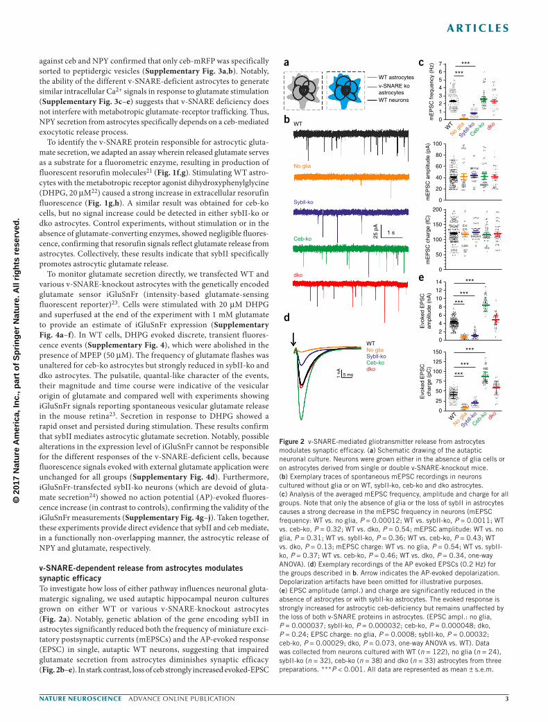
To investigate the v-SNARE dependence of vesicular exocytosis, we transfected astrocytes with fluorescently tagged NPY (using mono-meric teal fluorescent protein). These tagged NPY puncta were visible throughout the cell (Fig. 1a), agreeing well with endogenous NPY staining (Supplementary Fig. 1b). Superfusion of astrocytes with glutamate strongly decreased the number of NPY puncta in wild-type (WT) cells, providing evidence for exocytotic release (Fig. 1a–c). No NPY release was observed in the absence of glutamate stimulation (Fig. 1b) or during simultaneous incubation with a metabotropic
glutamate-receptor antagonist, MPEP (2-methyl-6-(phenylethynyl)-pyridine, 10 µM; Fig. 1d). For sybII-knockout (sybII-ko) astrocytes, NPY secretion was indistinguishable from WT cells, whereas it was completely abolished in ceb-knockout (ceb-ko) astrocytes. These functional results agree well with the high degree of co-localization between NPY and ceb (Supplementary Fig. 1b). Accordingly, in sybII and ceb double-knockout (dko) cells, NPY release was simi-larly abrogated (Fig. 1d,e). In accordance with the phenotypes of the single v-SNARE deficiencies, expression of ceb but not sybII in dko astrocytes led to a gain-of-function phenotype by restoring NPY secretion to the level of WT cells (Fig. 1d,e). Co-immunolabeling
WT
dko
WT + NPYmTFP
Ceb-ko
SybII-ko
WTno stim
Before stim
After stim
SybII
ba***
Ceb
***
WT+
MPEP
***
dko+
Before stimAfter stim
100 µm
******
d e
WT
dko + Ceb
Time (s)
dko + SybII
0 2 4 6 8 100
20
40
60
80
100
120
140
160
180
WT + MPEP
Time (s)
0
t = 8 min
1 mM glutamate
Resorufin
ht = 0 min
150
200 DHPG
0 1 2 3 4 5 6 70
50
100
No substrate
Time (min)
No stim
dko
8
gf
WTCeb-koSybII-ko
250 µm
WT
dko
SybII-koCeb-ko
0 2 4 6 8 100
20
40
60
80
100
120
140
160
180 1 mM Glutamatec
10
20
30
40
Num
ber
of N
PY
pun
cta
Num
ber
of N
PY
pun
cta
NP
Ym
TF
P ∆
F (
a.u.
)N
PY
mT
FP
∆F
(a.
u.)
Res
oruf
in ∆
Fat
8 m
in (
a.u.
)
50
Before stimAfter stim
0
10
20
30
40
50
0
50
100
150
200
250
Res
oruf
in ∆
F (
a.u.
)
No su
bstra
te
No sti
mdko
Ceb-k
o
SybII-
koWT
Figure 1 SybII and ceb govern distinct release pathways in astrocytes. (a) Example confocal images of a WT astrocyte expressing NPY tagged with monomeric teal fluorescent protein (NPYmTFP), illustrating the stimulation-dependent (1 mM glutamate) disappearance of NPYmTFP puncta. Dotted line represents the outline of the cell. Inset shows areas outlined in white dashed boxes. (b) Quantification of the number of NPY puncta before and after stimulation in a 50 × 50-µm region of interest (ROI). NPY secretion is abolished in ceb-ko and dko astrocytes (WT, n = 19; P = 0.00078; sybII-ko, n = 11; P = 0.00034; ceb-ko, n = 13; P = 0.121; WT, no stim = 6; P = 0.65, Student’s t test). (c,e) Time course of NPYmTFP secretion from cells stimulated with 1 mM glutamate. (d) Expression of ceb but not sybII restores NPY secretion in dko astrocytes (Data was collected from three preparations; before stim vs. after stim: dko n = 7; P = 0.00012; dko + sybII, n = 12; P = 0.43; dko + ceb, n = 13; P = 0.00054; WT + MPEP, n = 8; P = 0.124, Students t test). (f) Exemplary images of resorufin fluorescence before and after stimulation with 10 µM DHPG (left, before stimulation; right, after stimulation). (g) Time-courses of the resorufin signal in astrocytes for the indicated groups. Note that glutamate secretion is abolished in sybII-ko but unchanged in ceb-ko astrocytes. Cells were stimulated using 10 µM DHPG (arrow). (h) Quantification of the resorufin signal 8 min after onset of stimulation (data was collected from three preparations; WT, n = 8; sybII-ko, n = 7, P < 0.001; ceb-ko, n = 9, P = 0.35; dko, n = 8, P < 0.001; no stim, n = 9, P < 0.001; no substrate, n = 5, P < 0.001, ***P < 0.001, one-way ANOVA vs. WT). All data are represented as mean ± s.e.m.
© 2
017
Nat
ure
Am
eric
a, In
c., p
art
of
Sp
rin
ger
Nat
ure
. All
rig
hts
res
erve
d.
nature neurOSCIenCe advance online publication
a r t I C l e S
against ceb and NPY confirmed that only ceb-mRFP was specifically sorted to peptidergic vesicles (Supplementary Fig. 3a,b). Notably, the ability of the different v-SNARE-deficient astrocytes to generate similar intracellular Ca2+ signals in response to glutamate stimulation (Supplementary Fig. 3c–e) suggests that v-SNARE deficiency does not interfere with metabotropic glutamate-receptor trafficking. Thus, NPY secretion from astrocytes specifically depends on a ceb-mediated exocytotic release process.
To identify the v-SNARE protein responsible for astrocytic gluta-mate secretion, we adapted an assay wherein released glutamate serves as a substrate for a fluorometric enzyme, resulting in production of fluorescent resorufin molecules21 (Fig. 1f,g). Stimulating WT astro-cytes with the metabotropic receptor agonist dihydroxyphenylglycine (DHPG, 20 µM22) caused a strong increase in extracellular resorufin fluorescence (Fig. 1g,h). A similar result was obtained for ceb-ko cells, but no signal increase could be detected in either sybII-ko or dko astrocytes. Control experiments, without stimulation or in the absence of glutamate-converting enzymes, showed negligible fluores-cence, confirming that resorufin signals reflect glutamate release from astrocytes. Collectively, these results indicate that sybII specifically promotes astrocytic glutamate release.
To monitor glutamate secretion directly, we transfected WT and various v-SNARE-knockout astrocytes with the genetically encoded glutamate sensor iGluSnFr (intensity-based glutamate-sensing fluorescent reporter)23. Cells were stimulated with 20 µM DHPG and superfused at the end of the experiment with 1 mM glutamate to provide an estimate of iGluSnFr expression (Supplementary Fig. 4a–f). In WT cells, DHPG evoked discrete, transient fluores-cence events (Supplementary Fig. 4), which were abolished in the presence of MPEP (50 µM). The frequency of glutamate flashes was unaltered for ceb-ko astrocytes but strongly reduced in sybII-ko and dko astrocytes. The pulsatile, quantal-like character of the events, their magnitude and time course were indicative of the vesicular origin of glutamate and compared well with experiments showing iGluSnFr signals reporting spontaneous vesicular glutamate release in the mouse retina23. Secretion in response to DHPG showed a rapid onset and persisted during stimulation. These results confirm that sybII mediates astrocytic glutamate secretion. Notably, possible alterations in the expression level of iGluSnFr cannot be responsible for the different responses of the v-SNARE-deficient cells, because fluorescence signals evoked with external glutamate application were unchanged for all groups (Supplementary Fig. 4d). Furthermore, iGluSnFr-transfected sybII-ko neurons (which are devoid of gluta-mate secretion24) showed no action potential (AP)-evoked fluores-cence increase (in contrast to controls), confirming the validity of the iGluSnFr measurements (Supplementary Fig. 4g–j). Taken together, these experiments provide direct evidence that sybII and ceb mediate, in a functionally non-overlapping manner, the astrocytic release of NPY and glutamate, respectively.
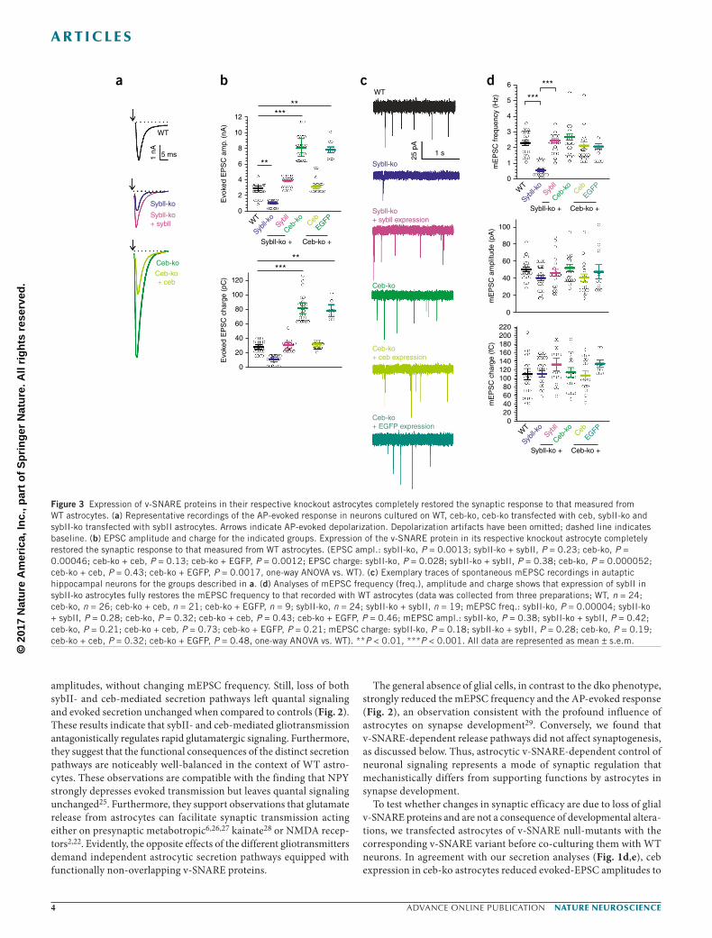
v-SNARE-dependent release from astrocytes modulates synaptic efficacyTo investigate how loss of either pathway influences neuronal gluta-matergic signaling, we used autaptic hippocampal neuron cultures grown on either WT or various v-SNARE-knockout astrocytes (Fig. 2a). Notably, genetic ablation of the gene encoding sybII in astrocytes significantly reduced both the frequency of miniature exci-tatory postsynaptic currents (mEPSCs) and the AP-evoked response (EPSC) in single, autaptic WT neurons, suggesting that impaired glutamate secretion from astrocytes diminishes synaptic efficacy (Fig. 2b–e). In stark contrast, loss of ceb strongly increased evoked-EPSC
7
WT astrocytes
No glia
Sybll-ko
Ceb-ko
dko
WT
WT neurons
v-SNARE koastrocytes
******6
543210
100
80
60
40
20
200
150
100
50
0
14
12
10
8
6
4
2
0
150
WT1
nA 5 ms
No gliaSybll-koCeb-kodko
125
100
75
50
25
0
0
mE
PS
C fr
eque
ncy
(Hz)
mE
PS
C a
mpl
itude
(pA
)m
EP
SC
cha
rge
(fC
)E
voke
d E
PS
Cam
plitu
de (
nA)
Evo
ked
EP
SC
char
ge (
pC)
25 p
A
1 s
***
******
***
******
WT
No gli
a
Sybll-k
o
Ceb-k
odk
o
WT
No gli
a
Sybll-k
o
Ceb-k
odk
o
a
b
c
d
e
Figure 2 v-SNARE-mediated gliotransmitter release from astrocytes modulates synaptic efficacy. (a) Schematic drawing of the autaptic neuronal culture. Neurons were grown either in the absence of glia cells or on astrocytes derived from single or double v-SNARE-knockout mice. (b) Exemplary traces of spontaneous mEPSC recordings in neurons cultured without glia or on WT, sybII-ko, ceb-ko and dko astrocytes. (c) Analysis of the averaged mEPSC frequency, amplitude and charge for all groups. Note that only the absence of glia or the loss of sybII in astrocytes causes a strong decrease in the mEPSC frequency in neurons (mEPSC frequency: WT vs. no glia, P = 0.00012; WT vs. sybII-ko, P = 0.0011; WT vs. ceb-ko, P = 0.32; WT vs. dko, P = 0.54; mEPSC amplitude: WT vs. no glia, P = 0.31; WT vs. sybII-ko, P = 0.36; WT vs. ceb-ko, P = 0.43; WT vs. dko, P = 0.13; mEPSC charge: WT vs. no glia, P = 0.54; WT vs. sybII-ko, P = 0.37; WT vs. ceb-ko, P = 0.46; WT vs. dko, P = 0.34, one-way ANOVA). (d) Exemplary recordings of the AP evoked EPSCs (0.2 Hz) for the groups described in b. Arrow indicates the AP-evoked depolarization. Depolarization artifacts have been omitted for illustrative purposes. (e) EPSC amplitude (ampl.) and charge are significantly reduced in the absence of astrocytes or with sybII-ko astrocytes. The evoked response is strongly increased for astrocytic ceb-deficiency but remains unaffected by the loss of both v-SNARE proteins in astrocytes. (EPSC ampl.: no glia, P = 0.000037; sybII-ko, P = 0.000032; ceb-ko, P = 0.000048; dko, P = 0.24; EPSC charge: no glia, P = 0.0008; sybII-ko, P = 0.00032; ceb-ko, P = 0.00029; dko, P = 0.073, one-way ANOVA vs. WT). Data was collected from neurons cultured with WT (n = 122), no glia (n = 24), sybII-ko (n = 32), ceb-ko (n = 38) and dko (n = 33) astrocytes from three preparations. ***P < 0.001. All data are represented as mean ± s.e.m.
© 2
017
Nat
ure
Am
eric
a, In
c., p
art
of
Sp
rin
ger
Nat
ure
. All
rig
hts
res
erve
d.
advance online publication nature neurOSCIenCe
a r t I C l e S
amplitudes, without changing mEPSC frequency. Still, loss of both sybII- and ceb-mediated secretion pathways left quantal signaling and evoked secretion unchanged when compared to controls (Fig. 2). These results indicate that sybII- and ceb-mediated gliotransmission antagonistically regulates rapid glutamatergic signaling. Furthermore, they suggest that the functional consequences of the distinct secretion pathways are noticeably well-balanced in the context of WT astro-cytes. These observations are compatible with the finding that NPY strongly depresses evoked transmission but leaves quantal signaling unchanged25. Furthermore, they support observations that glutamate release from astrocytes can facilitate synaptic transmission acting either on presynaptic metabotropic6,26,27 kainate28 or NMDA recep-tors2,22. Evidently, the opposite effects of the different gliotransmitters demand independent astrocytic secretion pathways equipped with functionally non-overlapping v-SNARE proteins.
The general absence of glial cells, in contrast to the dko phenotype, strongly reduced the mEPSC frequency and the AP-evoked response (Fig. 2), an observation consistent with the profound influence of astrocytes on synapse development29. Conversely, we found that v-SNARE-dependent release pathways did not affect synaptogenesis, as discussed below. Thus, astrocytic v-SNARE-dependent control of neuronal signaling represents a mode of synaptic regulation that mechanistically differs from supporting functions by astrocytes in synapse development.
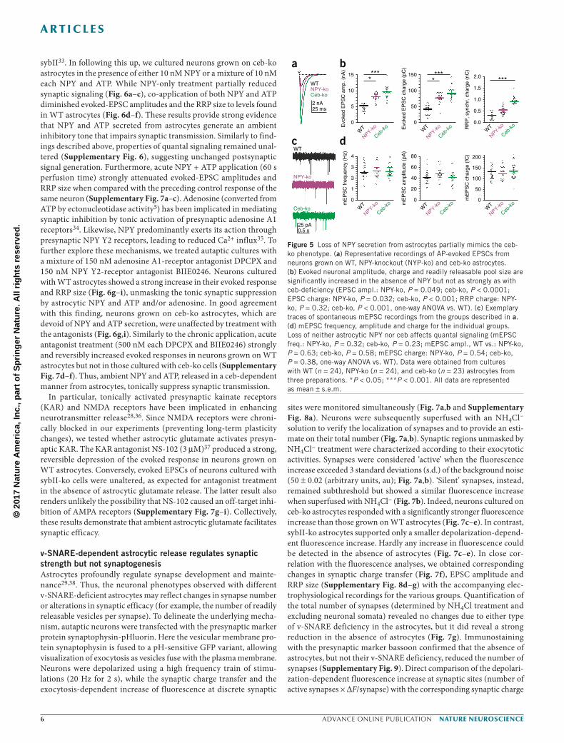
To test whether changes in synaptic efficacy are due to loss of glial v-SNARE proteins and are not a consequence of developmental altera-tions, we transfected astrocytes of v-SNARE null-mutants with the corresponding v-SNARE variant before co-culturing them with WT neurons. In agreement with our secretion analyses (Fig. 1d,e), ceb expression in ceb-ko astrocytes reduced evoked-EPSC amplitudes to
12
WT
WT
25 p
A
Sybll-ko
Sybll-ko+ sybll expression
Ceb-ko
Ceb-ko+ ceb expression
Ceb-ko+ EGFP expression
1 nA
**
**
*****
******
***
10
8
Evo
ked
EP
SC
am
p. (
nA)
Evo
ked
EP
SC
cha
rge
(pC
)
mE
PS
C fr
eque
ncy
(Hz)
mE
PS
C a
mpl
itude
(pA
)m
EP
SC
cha
rge
(fC
)
6
4
2
0Sybll-ko
Ceb-ko
Ceb-ko+ ceb
Sybll-ko+ sybll
120
100
80
60
40
20
0
6
5
4
3
2
1
0
100
80
60
40
20
0
220200180160140120100806040200
Sybll-ko + Ceb-ko +Syb
ll-ko
Sybll
Ceb-ko
EGFPCebW
TSybll-ko + Ceb-ko +
Sybll-k
oSyb
ll
Ceb-ko
EGFPCebW
T
Sybll-ko + Ceb-ko +Syb
ll-ko
Sybll
Ceb-ko
EGFPCebW
T
5 ms 1 s
a b c d
Figure 3 Expression of v-SNARE proteins in their respective knockout astrocytes completely restored the synaptic response to that measured from WT astrocytes. (a) Representative recordings of the AP-evoked response in neurons cultured on WT, ceb-ko, ceb-ko transfected with ceb, sybII-ko and sybII-ko transfected with sybII astrocytes. Arrows indicate AP-evoked depolarization. Depolarization artifacts have been omitted; dashed line indicates baseline. (b) EPSC amplitude and charge for the indicated groups. Expression of the v-SNARE protein in its respective knockout astrocyte completely restored the synaptic response to that measured from WT astrocytes. (EPSC ampl.: sybII-ko, P = 0.0013; sybII-ko + sybII, P = 0.23; ceb-ko, P = 0.00046; ceb-ko + ceb, P = 0.13; ceb-ko + EGFP, P = 0.0012; EPSC charge: sybII-ko, P = 0.028; sybII-ko + sybII, P = 0.38; ceb-ko, P = 0.000052; ceb-ko + ceb, P = 0.43; ceb-ko + EGFP, P = 0.0017, one-way ANOVA vs. WT). (c) Exemplary traces of spontaneous mEPSC recordings in autaptic hippocampal neurons for the groups described in a. (d) Analyses of mEPSC frequency (freq.), amplitude and charge shows that expression of sybII in sybII-ko astrocytes fully restores the mEPSC frequency to that recorded with WT astrocytes (data was collected from three preparations; WT, n = 24; ceb-ko, n = 26; ceb-ko + ceb, n = 21; ceb-ko + EGFP, n = 9; sybII-ko, n = 24; sybII-ko + sybII, n = 19; mEPSC freq.: sybII-ko, P = 0.00004; sybII-ko + sybII, P = 0.28; ceb-ko, P = 0.32; ceb-ko + ceb, P = 0.43; ceb-ko + EGFP, P = 0.46; mEPSC ampl.: sybII-ko, P = 0.38; sybII-ko + sybII, P = 0.42; ceb-ko, P = 0.21; ceb-ko + ceb, P = 0.73; ceb-ko + EGFP, P = 0.21; mEPSC charge: sybII-ko, P = 0.18; sybII-ko + sybII, P = 0.28; ceb-ko, P = 0.19; ceb-ko + ceb, P = 0.32; ceb-ko + EGFP, P = 0.48, one-way ANOVA vs. WT). **P < 0.01, ***P < 0.001. All data are represented as mean ± s.e.m.

© 2
017
Nat
ure
Am
eric
a, In
c., p
art
of
Sp
rin
ger
Nat
ure
. All
rig
hts
res
erve
d.
nature neurOSCIenCe advance online publication
a r t I C l e S
levels found with WT astrocytes, without changing the properties of quantal signaling (Fig. 3). In contrast, transfection with enhanced GFP (EGFP) failed to reduce the evoked response, demonstrating that ceb expression, rather than off-target effects of lentiviral trans-fection, was responsible for the observed phenotype. SybII expres-sion in sybII-ko astrocytes, instead, elevated the mEPSC frequency and rescued the AP-evoked response to that observed with WT cells (Fig. 3). Collectively, these results indicate that expression of either v-SNARE variant in astrocytes restored the corresponding secretion pathway and its functional impact on synaptic signaling.
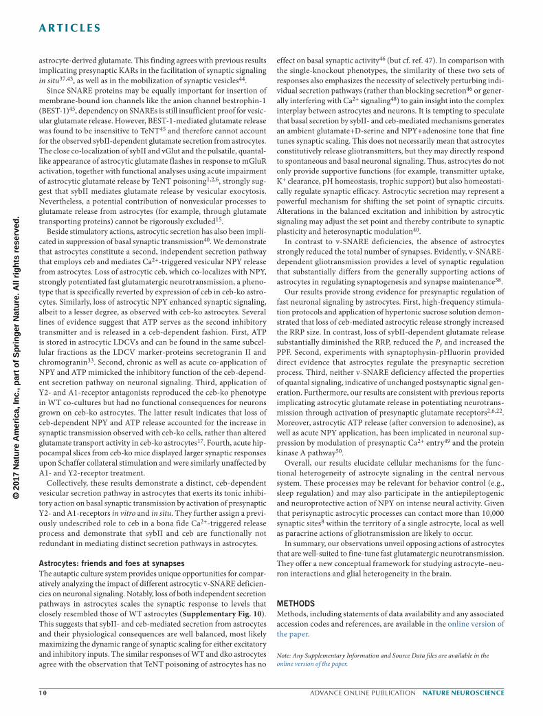
Astrocytes regulate the readily releasable pool size and release probability of hippocampal neuronsTo elucidate the mechanisms by which synaptic release is regulated by gliotransmission, we used high-frequency train stimulations (20 Hz for 2 s) and comparatively analyzed the readily releasable pool (RRP) size, release probability (Pr) and synaptic depression. In good agreement with the results obtained with basal stimulation (0.2 Hz; Fig. 2d,e), the first AP-evoked response of the train was substan-tially decreased for neurons growing on sybII-ko astrocytes, whereas it was strongly enhanced with ceb-ko and unchanged for dko cells (Fig. 4a,b). Evoked synaptic responses of neurons grown on WT, ceb-ko and dko astrocytes displayed depression over the first ten APs before reaching an apparent steady state for the remaining stimuli of the train (Fig. 4a–c). In contrast, neurons cultured with sybII-ko astrocytes displayed a slower time-course and reduced synaptic depression, indicative for changes in vesicular Pr (Fig. 4c). To deter-mine the RRP size, the plot of the cumulative synchronous EPSC charge was approximated with a linear regression fitting the last five stimuli (Fig. 4e). Back-extrapolating the linear component of the steady-state phase renders an estimate of the initial RRP size30 (Fig. 4e,f). For sybII-ko astrocytes, the RRP size was significantly
reduced, whereas it was strongly increased or unchanged with ceb-ko and dko astrocytes, respectively (Fig. 4e,f). Similarly, the asynchro-nous release component, which increased during the stimulus train due to accumulation of presynaptic intracellular [Ca2+], was found to be diminished with sybII-ko and increased with ceb-ko astrocytes (Fig. 4d,g,h). In contrast, Pr (determined by the ratio of the first EPSC charge to the RRP charge) was significantly reduced with sybII-ko astrocytes but remained unchanged for all other groups (Fig. 4i). Consistently, the paired-pulse facilitation (PPF), which is inversely related to Pr, only increased for neurons grown on sybII-ko astrocytes (Supplementary Fig. 5d,e). Experiments with hypertonic sucrose to determine the RRP size31 with Ca2+-independent stimulation con-firmed the different impacts of sybII- and ceb-mediated release from astrocytes on RRP size and Pr (Supplementary Fig. 5a–c). Taken together, these v-SNARE-dependent release pathways from astrocytes oppositely regulate fundamental parameters of synaptic efficacy.
NPY and ATP mimic the inhibition by ceb-mediated gliotransmissionTo explore whether loss of astrocytic NPY secretion can mimic the phenotype of ceb-ko astrocytes, we analyzed the impact of astro-cytes derived from NPY−/− mice32 (Fig. 5). Loss of astrocytic NPY significantly increased evoked-EPSC amplitude and charge, as well as the RRP size, albeit to a lesser degree than that found for ceb-ko cells (Fig. 5a,b). In contrast, mEPSC properties remained unchanged (Fig. 5c,d). While these results agree with previous reports showing that NPY diminishes neuronal transmission25,32, they also suggest that other inhibitory gliotransmitters are additionally released in a ceb-dependent manner. Astrocytic ATP has been implicated in het-erosynaptic depression5,7 and was found to be present in the same subcellular fractions as the LDCV marker-proteins secretogranin II and chromogranin, which were distinct from fractions containing
WTWT
10
8
6
4
AP
evo
ked
ampl
itude
(nA
)
Nor
m. a
mpl
itude
Res
pons
e ch
arge
(pC
)
Cum
. syn
chr.
cha
rge
(nC
)
Cum
. asy
nchr
. cha
rge
(nC
)
Rel
ease
pro
babi
lity
Asy
nchr
. cha
rge
(pC
)
RR
P, s
ynch
r. c
harg
e (n
C)
2
0
2.0 80 0.8
0.6
0.4
0.2
0.0
60
40
20
0
1.5
1.0
0.5
00 10 20
Pulse30 40
60 1.4
***
***
*****
**
1.2
1.0
0.8
0.6
0.4
0.2
0
1.2
1.0
0.8
0.6
0.4
0.2
0.0
1.0
0.8
0.6
0.4
0.2
0.0
50
Synchr. chargeAsynchr. charge
30
40
20
10
00 10 20
Pulse30 40 0 10 20
Pulse30 40 0 10 20
Pulse30 40 0 10 20
Pulse30 40
2 nA
50 msSybll-ko
Sybll-ko
Ceb-ko
Ceb-ko
dko
dko
WT
Sybll-k
o
Ceb-k
odk
oW
T
Sybll-k
o
Ceb-k
odk
oW
T
Sybll-k
o
Ceb-k
odk
o
a b c
g h i
d e
f
Figure 4 Gliotransmission modulates the readily releasable pool size and release probability in hippocampal neurons. (a) Representative EPSCs triggered by high-frequency stimulation (HFS; 20 Hz, 40 AP per 2 s) of autaptic neurons cultured on WT, sybII-ko, ceb-ko and dko astrocytes. (b) Averaged EPSC amplitudes plotted over the stimulus number. (c) The time-course of synaptic depression was reduced in neurons grown on sybII-ko astrocytes (data were normalized to the initial peak EPSC amplitude). (d) Estimates of synchronous and asynchronous release plotted against the stimulus. (e) Mean cumulative synchronous release components during a 20-Hz train. Data points from the linear component were back extrapolated to pulse = 0 to estimate the initial RRP size. (f) The RRP recorded in neurons cultured on sybII-ko astrocytes was reduced, whereas it was increased with ceb-ko astrocytes (sybII-ko, P < 0.001; ceb-ko, P < 0.001; dko, P = 0.46, one-way ANOVA vs. WT). (g,h) Mean cumulative asynchronous release for the indicated groups. Neuronal asynchronous release is also affected by v-SNARE-dependent secretion processes from astrocytes. Asynchronous release was determined as the mean charge over the last five stimuli. (sybII-ko, P < 0.001; ceb-ko, P < 0.001; dko, P = 0.32, one-way ANOVA vs. WT). (i) The release probability (first EPSCcharge/RRPcharge) was specifically decreased in sybII-ko astrocytes (sybII-ko, P = 0.021; ceb-ko, P = 0.18; dko, P = 0.13, one-way ANOVA vs. WT). Data was collected from WT, n = 62; sybII-ko, n = 28; ceb-ko, n = 35; dko, n = 29 from three preparations. **P < 0.01; ***P < 0.001. All data are represented as mean ± s.e.m.
© 2
017
Nat
ure
Am
eric
a, In
c., p
art
of
Sp
rin
ger
Nat
ure
. All
rig
hts
res
erve
d.
advance online publication nature neurOSCIenCe
a r t I C l e S
sybII33. In following this up, we cultured neurons grown on ceb-ko astrocytes in the presence of either 10 nM NPY or a mixture of 10 nM each NPY and ATP. While NPY-only treatment partially reduced synaptic signaling (Fig. 6a–c), co-application of both NPY and ATP diminished evoked-EPSC amplitudes and the RRP size to levels found in WT astrocytes (Fig. 6d–f). These results provide strong evidence that NPY and ATP secreted from astrocytes generate an ambient inhibitory tone that impairs synaptic transmission. Similarly to find-ings described above, properties of quantal signaling remained unal-tered (Supplementary Fig. 6), suggesting unchanged postsynaptic signal generation. Furthermore, acute NPY + ATP application (60 s perfusion time) strongly attenuated evoked-EPSC amplitudes and RRP size when compared with the preceding control response of the same neuron (Supplementary Fig. 7a–c). Adenosine (converted from ATP by ectonucleotidase activity5) has been implicated in mediating synaptic inhibition by tonic activation of presynaptic adenosine A1 receptors34. Likewise, NPY predominantly exerts its action through presynaptic NPY Y2 receptors, leading to reduced Ca2+ influx35. To further explore these mechanisms, we treated autaptic cultures with a mixture of 150 nM adenosine A1-receptor antagonist DPCPX and 150 nM NPY Y2-receptor antagonist BIIE0246. Neurons cultured with WT astrocytes showed a strong increase in their evoked response and RRP size (Fig. 6g–i), unmasking the tonic synaptic suppression by astrocytic NPY and ATP and/or adenosine. In good agreement with this finding, neurons grown on ceb-ko astrocytes, which are devoid of NPY and ATP secretion, were unaffected by treatment with the antagonists (Fig. 6g,i). Similarly to the chronic application, acute antagonist treatment (500 nM each DPCPX and BIIE0246) strongly and reversibly increased evoked responses in neurons grown on WT astrocytes but not in those cultured with ceb-ko cells (Supplementary Fig. 7d–f). Thus, ambient NPY and ATP, released in a ceb-dependent manner from astrocytes, tonically suppress synaptic transmission.
In particular, tonically activated presynaptic kainate receptors (KAR) and NMDA receptors have been implicated in enhancing neurotransmitter release28,36. Since NMDA receptors were chroni-cally blocked in our experiments (preventing long-term plasticity changes), we tested whether astrocytic glutamate activates presyn-aptic KAR. The KAR antagonist NS-102 (3 µM)37 produced a strong, reversible depression of the evoked response in neurons grown on WT astrocytes. Conversely, evoked EPSCs of neurons cultured with sybII-ko cells were unaltered, as expected for antagonist treatment in the absence of astrocytic glutamate release. The latter result also renders unlikely the possibility that NS-102 caused an off-target inhi-bition of AMPA receptors (Supplementary Fig. 7g–i). Collectively, these results demonstrate that ambient astrocytic glutamate facilitates synaptic efficacy.
v-SNARE-dependent astrocytic release regulates synaptic strength but not synaptogenesisAstrocytes profoundly regulate synapse development and mainte-nance29,38. Thus, the neuronal phenotypes observed with different v-SNARE-deficient astrocytes may reflect changes in synapse number or alterations in synaptic efficacy (for example, the number of readily releasable vesicles per synapse). To delineate the underlying mecha-nism, autaptic neurons were transfected with the presynaptic marker protein synaptophysin-pHluorin. Here the vesicular membrane pro-tein synaptophysin is fused to a pH-sensitive GFP variant, allowing visualization of exocytosis as vesicles fuse with the plasma membrane. Neurons were depolarized using a high frequency train of stimu-lations (20 Hz for 2 s), while the synaptic charge transfer and the exocytosis-dependent increase of fluorescence at discrete synaptic
sites were monitored simultaneously (Fig. 7a,b and Supplementary Fig. 8a). Neurons were subsequently superfused with an NH4Cl− solution to verify the localization of synapses and to provide an esti-mate on their total number (Fig. 7a,b). Synaptic regions unmasked by NH4Cl− treatment were characterized according to their exocytotic activities. Synapses were considered ‘active’ when the fluorescence increase exceeded 3 standard deviations (s.d.) of the background noise (50 ± 0.02 (arbitrary units, au); Fig. 7a,b). ‘Silent’ synapses, instead, remained subthreshold but showed a similar fluorescence increase when superfused with NH4Cl− (Fig. 7b). Indeed, neurons cultured on ceb-ko astrocytes responded with a significantly stronger fluorescence increase than those grown on WT astrocytes (Fig. 7c–e). In contrast, sybII-ko astrocytes supported only a smaller depolarization-depend-ent fluorescence increase. Hardly any increase in fluorescence could be detected in the absence of astrocytes (Fig. 7c–e). In close cor-relation with the fluorescence analyses, we obtained corresponding changes in synaptic charge transfer (Fig. 7f), EPSC amplitude and RRP size (Supplementary Fig. 8d–g) with the accompanying elec-trophysiological recordings for the various groups. Quantification of the total number of synapses (determined by NH4Cl treatment and excluding neuronal somata) revealed no changes due to either type of v-SNARE deficiency in the astrocytes, but it did reveal a strong reduction in the absence of astrocytes (Fig. 7g). Immunostaining with the presynaptic marker bassoon confirmed that the absence of astrocytes, but not their v-SNARE deficiency, reduced the number of synapses (Supplementary Fig. 9). Direct comparison of the depolari-zation-dependent fluorescence increase at synaptic sites (number of active synapses × ∆F/synapse) with the corresponding synaptic charge
15
a b
c d
150 2.0
1.5
1.0
0.5
0.0
100
50
0
WT
WT
NPY-ko
NPY-ko
Ceb-ko
Ceb-ko
WT
NPY-ko
Ceb-k
o
WT
NPY-ko
Ceb-k
oW
T
NPY-ko
Ceb-k
oW
T
NPY-ko
Ceb-k
o
WT
NPY-ko
Ceb-k
oW
T
NPY-ko
Ceb-k
o
****
10
52 nA25 ms
25 pA0.5 s
Evo
ked
EP
SC
am
p. (
nA)
mE
PS
C fr
eque
ncy
(Hz)
mE
PS
C a
mpl
itude
(pA
)
mE
PS
C c
harg
e (f
C)
Evo
ked
EP
SC
cha
rge
(pC
)
RR
P, s
ynch
r. c
harg
e (n
C)
0
4 80
60
40
20
0
200
150
100
50
0
3
3
1
0
****
***
Figure 5 Loss of NPY secretion from astrocytes partially mimics the ceb-ko phenotype. (a) Representative recordings of AP-evoked EPSCs from neurons grown on WT, NPY-knockout (NYP-ko) and ceb-ko astrocytes. (b) Evoked neuronal amplitude, charge and readily releasable pool size are significantly increased in the absence of NPY but not as strongly as with ceb-deficiency (EPSC ampl.: NPY-ko, P = 0.049; ceb-ko, P < 0.0001; EPSC charge: NPY-ko, P = 0.032; ceb-ko, P < 0.001; RRP charge: NPY-ko, P = 0.32; ceb-ko, P < 0.001, one-way ANOVA vs. WT). (c) Exemplary traces of spontaneous mEPSC recordings from the groups described in a. (d) mEPSC frequency, amplitude and charge for the individual groups. Loss of neither astrocytic NPY nor ceb affects quantal signaling (mEPSC freq.: NPY-ko, P = 0.32; ceb-ko, P = 0.23; mEPSC ampl., WT vs.: NPY-ko, P = 0.63; ceb-ko, P = 0.58; mEPSC charge: NPY-ko, P = 0.54; ceb-ko, P = 0.38, one-way ANOVA vs. WT). Data were obtained from cultures with WT (n = 24), NPY-ko (n = 24), and ceb-ko (n = 23) astrocytes from three preparations. *P < 0.05; ***P < 0.001. All data are represented as mean ± s.e.m.

© 2
017
Nat
ure
Am
eric
a, In
c., p
art
of
Sp
rin
ger
Nat
ure
. All
rig
hts
res
erve
d.
nature neurOSCIenCe advance online publication
a r t I C l e S
WT + DPCPX+BIIE0246
15
*****
**
** ****
*****
***
****** ***
***
******
10
5
AP
evo
ked
ampl
itude
(nA
)A
P e
voke
d am
plitu
de (
nA)
RR
P, s
ynch
r. c
harg
e (n
C)
AP
evo
ked
char
ge (
pC)
AP
evo
ked
char
ge (
pC)
0Ceb-ko
2 nA
50 ms
2 nA
50 ms
2 nA
50 ms
WT
WT WTWT
WT
WT WT
WT WT
+ vehicle + NPY
Ceb-ko
Ceb-ko
Ceb-ko + NPY
15
10
5
AP
evo
ked
ampl
itude
(nA
)
0
0
2
4
6
8
10
12
150
100
50
0
150
100
50
0
AP
evo
ked
char
ge (
pC)
120
100
80
60
40
0
20
1.0
0.8
0.6
0.4
0.2
0.0
RR
P, s
ynch
r. c
harg
e (n
C)
1.0
0.8
0.6
0.4
0.2
0.0
RR
P, s
ynch
r. c
harg
e (n
C)
1.0
0.8
0.6
0.4
0.2
0.0
Ceb-ko
+ vehicle + NPY
Ceb-ko Ceb-ko
+ vehicle + NPY
Ceb-ko
Ceb-ko
WT
+ vehicle + ATP+NPY
Cebko
Ceb-ko
Cebko + ATP+NPY
Ceb-ko
+ vehicle + ATP+NPY
Ceb-ko Ceb-ko
+ vehicle + ATP+NPY
Ceb-ko
Ceb-ko
WT
Ceb-ko WT WTCeb-ko Ceb-ko WT WTCeb-ko Ceb-ko
+ vehicle + DPCPX+BIIE0246Ceb-ko
Ceb-ko + DPCPX+BIIE0246
+ vehicle + DPCPX+BIIE0246
+ vehicle + DPCPX+BIIE0246
a b c
d e f
g h i
Figure 6 Extracellular application of both NPY and ATP mimics the inhibitory effect of ceb-mediated gliotransmission on neuronal signaling. (a) Representative traces of AP-evoked EPSC recordings in autaptic neurons plated on WT and ceb-ko astrocytes treated daily with 10 nM NPY. Arrow indicates action potential evoked depolarization. Depolarization artifacts have been omitted. (b,c) Daily application of NPY only partially restored the EPSC amplitude, charge and RRP size in neurons grown on ceb-ko astrocytes when compared to recordings from WT astrocytes (data were collected from three preparations; WT + vehicle, n = 19; ceb-ko + vehicle, n = 20; ceb-ko + NPY, n = 21; EPSC ampl.: WT vs. ceb-ko (vehicle), P < 0.001; WT vs. ceb-ko (NPY), P = 0.01; EPSC charge: WT vs. ceb-ko (vehicle), P < 0.001; ceb-ko (vehicle) vs. ceb-ko (NPY), P = 0.01; RRP synchronous (sync) charge: WT vs. ceb-ko (vehicle), P = 0.0018; WT vs. ceb-ko (NPY), P < 0.001; one-way ANOVA). (d) Exemplary traces of the AP-evoked EPSC recorded in neurons treated daily with 10 nM NPY and 10 nM ATP grown on either WT or ceb-ko astrocytes. (e,f) Evoked EPSC amplitude, charge and RRP shows that co-application of NPY and ATP fully restored the WT response (data was obtained from three preparations; WT + vehicle, n = 24; ceb-ko + vehicle, n = 25; ceb-ko + NPY+ATP, n = 26; AP-evoked ampl.: WT vs. ceb-ko (vehicle), P < 0.001; WT (vehicle) vs. ceb-ko (ATP+NPY), P = 0.673; ceb-ko (vehicle) vs. ceb-ko (ATP+NPY), P < 0.001; AP-evoked charge: WT vs. ceb-ko (vehicle), P < 0.001; WT (vehicle) vs. ceb-ko (ATP+NPY), P = 0.943; ceb-ko (vehicle) vs. ceb-ko (ATP+NPY), P < 0.001; RRP sync. charge: WT vs. ceb-ko (vehicle), P < 0.001; WT (vehicle) vs. ceb-ko (ATP+NPY), P = 0.938; ceb-ko (vehicle) vs. ceb-ko (ATP+NPY), P < 0.001, one-way ANOVA). (g) Sample traces of AP-evoked EPSCs for neurons cultured with WT or ceb-ko astrocytes treated daily with 150 nM adenosine-receptor antagonist DPCPX and 150 nM NPY-receptor antagonist BIIE0246. (h,i) Evoked EPSC amplitude, charge and RRP show that co-application of the antagonists increased the synaptic response recorded for WT astrocytes to that of ceb-ko cells but left the ceb-ko response unchanged (data was obtained from three preparations; WT + vehicle, n = 24; ceb-ko + vehicle, n = 19; WT+DPCPX+BIIE0246, n = 20; ceb-ko + DPCPX+BIIE0246, n = 19; AP-evoked ampl.: ceb-ko (vehicle), P < 0.001; WT (DPCPX+BIIE0246), P < 0.001; ceb-ko (DPCPX+BIIE0246), P < 0.001; AP-evoked charge: ceb-ko (vehicle), P < 0.001; WT (DPCPX+BIIE0246), P < 0.001; ceb-ko (DPCPX+BIIE0246), P < 0.001; RRP synchronous charge: ceb-ko (vehicle), P < 0.001; WT (DPCPX+BIIE0246), P < 0.001; ceb-ko (DPCPX+BIIE0246), P < 0.001, one-way ANOVA vs. WT). **P < 0.01; ***P < 0.001. All data are represented as mean ± s.e.m.

© 2
017
Nat
ure
Am
eric
a, In
c., p
art
of
Sp
rin
ger
Nat
ure
. All
rig
hts
res
erve
d.
advance online publication nature neurOSCIenCe
a r t I C l e S
transfer of the same neuron revealed a strict correlation between both types of secretion measurements (Fig. 7i), corroborating the validity of our analyses (ceb-ko, r2 = 0.92; WT, r2 = 0.83; sybII-ko, r2 = 0.81; no glia, r2 = 0.62). In comparison with controls, ceb-ko astrocytes moderately increased their number of active synapses, which were marginally diminished in sybII-ko astrocytes but strongly reduced in cultures without glia cells (Fig. 7h). Notably, the fluorescence increase per synapse strongly and proportionally changed with the magnitude
of the synaptic charge transfer for the different v-SNARE dependen-cies (slope of exocytotic ∆F/charge transfer = 0.96), but the number of active synapses was found to be only slightly altered (slope of active synapse number/charge transfer = 0.32; Fig. 7j).
These results indicate presynaptic control of neuronal signaling by ceb- and sybII-dependent release processes from astrocytes, regulat-ing predominantly the number of releasable vesicles per synapse. They counter the possibility of v-SNARE-dependent alterations in synapse
F0Before stim
50 µm
Tot
al n
umbe
r of
syn
apse
s
Ceb-koWTSybII-ko
20 HzNH4Cl
Time (s)
0 5 10 15 20 25 30
0
0.5
1.0
1.5
2.0
2.5
3.0
∆F
Active synapseSilent synapse
Cum
ulat
ive
freq
uenc
y of
∆F
0 5.0 10.0 15.0 20.0
Num
ber
of a
ctiv
e sy
naps
es ****
Ceb-koWTSybII-ko
No stim
b
e f
g h j
d
Total charge (nC)
0 2 4 6 80
i
10
20
30
40
50 Relative num
ber of active synapses
∆FActive synapses
Act
ive
syna
pses
∆F
(au
, 1 ×
103 )
0 1 2 3 40.0
0.5
1.0
1.5
2.0
2.5
3.0
3.5
4.0
0.0
0.5
1.0
1.5
2.0
2.5
3.0
3.5
4.0
∆F (au, 1 × 10³)
20 Hz NH4Cl
0 5 10 15 20 25 300
1.0
2.0
3.0
4.0
WTCeb-ko
SybII-ko
c
No glia
0.0
0.2
0.4
0.6
0.8
1.0
No gliaNo glia
a∆FStim
∆FNH4Cl
0
100
200
300
0
2
4
6
8
10
0
2
4
6
8
0
50
100
150
200
250
***
******
******
*****
***
∆F p
er R
OI (
au, 1
× 1
03 )
Threshold
Time (s)
∆F p
er R
OI (
au, 1
× 1
03)
∆F (
au, 1
× 1
03)
Tot
al c
harg
e (n
C)
Rel
ativ
e ∆F
(au
, 1 ×
103 )
Total charge (nC)
WT
No gli
a
SybII-
ko
Ceb-k
o
WT
No gli
a
SybII-
ko
Ceb-k
o
WT
No gli
a
SybII-
ko
Ceb-k
o
WT
No gli
a
SybII-
ko
Ceb-k
o
Figure 7 Astrocytes regulate synaptic strength by distinct v-SNARE-dependent secretion pathways. (a) Exemplary difference images (∆F) from a WT neuron transfected with synaptophysin-pHluorin (syn-pH) before stimulation (left), during stimulation (middle) and after NH4Cl treatment (right, scaled to 70% of ∆F). High-magnification insets illustrate exemplary active (red circle) and silent synapses (gray circle); scale bar, 5 µm. The stimulation-dependent (20 Hz for 2 s) increase of fluorescence at synaptic sites was monitored (10 Hz) simultaneously with the AP-evoked charge transfer. (b) Exemplary traces of an active and silent (nonresponding) synapse. Synaptic sites were determined by NH4Cl treatment to unquench all syn-pH fluorescence. Dashed line marks 3× s.d. of background noise. (c) Astrocytic v-SNARE deficiency differentially regulates exocytotic activity at individual synapses (∆F) but does not affect the subsequent NH4Cl−-dependent unquenching response. Exocytotic activity was strongly reduced without an astrocytic feeder layer (no glia). (d) Mean ∆F at the end of stimulation as shown in c (gray bar). Note the strong increase in ∆F in neurons grown with ceb-ko astrocytes (no glia, P < 0.001; sybII-ko, P < 0.001; ceb-ko, P < 0.001, one-way ANOVA vs. WT). (e) Cumulative frequency distribution of ∆F for the indicated groups. (f) The total charge in neurons cultured with ceb-ko astrocytes is strongly increased, whereas it is decreased in cells grown with sybII-ko astrocytes or without glia cells (no glia, P < 0.001; sybII-ko, P = 0.0032; ceb-ko, P < 0.001, one-way ANOVA vs. WT). (g) Synaptic charge transfer for the indicated culture conditions (no glia, P = 0.000048; sybII-ko, P = 0.688; ceb-ko, P = 0.719, one-way ANOVA vs. WT). (h) Astrocytic v-SNARE deficiency affects the number of active synapses (no glia, P < 0.001; sybII-ko, P = 0.001; ceb-ko, P < 0.001, one-way ANOVA vs. WT). (i) The syn-pH response strongly correlates with the synaptic charge transfer (ceb-ko, r2 = 0.92; WT, r2 = 0.83; sybII-ko, r2 = 0.81; no glia, r2 = 0.62). (j) The exocytotic activity per synapse, rather than the number of active synapses, changes proportionally with the magnitude of the synaptic charge transfer (slopes: exocytotic ∆F/charge transfer, 0.96; active synapse number per charge transfer, 0.32). Data was collected from four independent preparations from WT n = 20; sybII-ko n = 17; ceb-ko n = 16; no glia, n = 9; not stimulated, n = 5). **P < 0.01, ***P < 0.001. All data are represented as mean ± s.e.m.

© 2
017
Nat
ure
Am
eric
a, In
c., p
art
of
Sp
rin
ger
Nat
ure
. All
rig
hts
res
erve
d.
nature neurOSCIenCe advance online publication
a r t I C l e S
number and confirm that the general presence of astrocytes is cru-cial for synapse maintenance and formation, as previously shown by others29. In summary, the combined data demonstrate that v-SNARE-mediated release from astrocytes potently regulates the efficacy of synaptic transmission at presynaptic sites.
Astrocytes modulate synaptic transmission in acute hippocampal slicesTo determine whether loss of NPY and ATP secretion also affects syn-aptic transmission in a more intact system, we used acutely isolated hippocampal slices from age-matched WT and Ceb−/− mice (8–11 weeks old). Notably, ceb expression in the brain is restricted mostly to non-neuronal cells, such as astrocytes or vascular cells39. The slope of the Schaffer-collateral-evoked field excitatory postsynaptic poten-tials (fEPSPs) was significantly larger in Ceb−/− mice than in WT controls (Fig. 8a,b). These results, recorded in a global Ceb−/− mouse, are in excellent agreement with our findings in vitro, which were caused by astrocyte-specific ceb deficiency (Figs. 2–7). Furthermore, co-application of DPCPX (2.4 µM) and BIIE0246 (2.4 µM) increased the fEPSP slope in WT but not in ceb-ko hippocampal slices (Fig. 8c,d), substantiating the results obtained with acute and chronic applications of the antagonists in glia–neuron cultures (Fig. 6 and Supplementary Fig. 7). Collectively, these observations suggest that astrocytes mediate
an ambient ATP and NPY level in the brain that tonically suppresses excitatory synaptic transmission.
DISCUSSIONAstrocytes contribute to virtually all aspects of brain function, includ-ing ionic homeostasis, energy metabolism and synaptic signaling. Still, substantial advances toward understanding the molecular mechanisms of gliotransmission and its impact on neuronal signaling have been often hampered by conceptual and technical limitations11. Taking advantage of individual null-mutants for sybII and ceb, as well as the compound null-mutant for both v-SNARE proteins, we unraveled previously undetected individual v-SNARE dependencies of astrocytic release processes and their functional impact on neuro-nal signaling (Supplementary Fig. 10). Specifically, we present strong evidence for the co-existence of independent secretion pathways in astrocytes that employ sybII and ceb as functionally non-overlapping v-SNARE proteins that antagonistically regulate fast glutamatergic neurotransmission. Thus, our work unveils molecular mechanisms by which astrocytes communicate with neurons and effectively control elementary steps of synaptic communication, providing new insights into fundamental aspects of brain function.
v-SNARE-mediated vesicular glutamate and NPY release from astrocytesKey properties of synapses are their ability to process and spread information between neurons in an ever-changing manner. Given their close proximity and intimate contact with synapses, astrocytes are known to contribute to the regulation of the neuronal micro-environment9. Gliotransmission is one of several postulated ways how astrocytes can actively modulate synaptic properties40, yet gen-eral principles regarding their release mechanisms remain unclear and controversial10,11. Previous studies have aimed at interfering with astrocytic SNARE proteins by using either the (dn)SNARE mouse model (which overexpresses a truncated sybII fragment5) or the ‘iBot’ mouse expressing the botulinum neurotoxin B light chain21. Neither these genetic mouse models nor poisoning with TeNT allowed researchers to identify the relevant v-SNARE variant or to discriminate between distinct astrocytic secretion pathways, because sybII and ceb are similarly affected by these perturbations. Taking advantage of individual null-mutants for ceb and sybII, we unraveled a bimodal regulation of synaptic signaling by astrocytes and provide direct evidence for v-SNARE-mediated release of vesicu-lar NPY and glutamate.
Using independent experimental strategies (resorufin and iGluSnFr assay), we show that stimulation of astrocytes with DHPG evokes glutamate release, which is abolished in sybII-deficient astrocytes but unchanged in ceb-ko astrocytes. Furthermore, sybII co-local-ized with vGlut in the astrocytic periphery, consistent with previous reports showing that vGlut+ SLMVs are found in perisynaptic proc-esses of astrocytes12,41. In agreement with these results, biochemical analyses revealed that astrocytic SLMVs are positive for vGlut and vesicle-associated membrane proteins (sybII and ceb) and that they store glutamate as well as D-serine42. Our experiments provide the first evidence we are aware of that specifically sybII-dependent secre-tion in astrocytes potentiates glutamatergic neurotransmission. The differential impact of the KAR antagonist on neuronal signaling in the presence of WT (reduced EPSC amplitude) and sybII-ko astro-cytes (unchanged EPSC amplitude), together with the observation that loss of astrocytic sybII specifically affects presynaptic param-eters (diminished RRP, reduced Pr and increased PPF), indicate that presynaptic KARs28 mediate the enhancement of synaptic signaling by
2.0
1.5
1.0
0.5
0.00.0 0.2 0.4 0.6 0.8
Stimulus (mA)
WTCeb-ko
3.0
2.5
2.0
1.5
1.0
0.5
0.0
WTCeb-ko
1 mV
2 ms
***
*
*
***
******
WT Ceb-ko
3.0
2.5
2.0
1.5
1.0
–10 0 10 20 30Time (min)
ACSF 2.4 µM DPCPX+BIIE0246
Nor
m. f
EP
SP
slo
pe
5
4
3
2
1
0WT Ceb-ko WT Ceb-ko
ACSF DPCPX+BIIE0246
***
Nor
m. f
EP
SP
slo
pe
a b
c d
fEP
SP
slo
pe (
–mV
per
s)
fEP
SP
slo
pe (
–mV
per
s)
Figure 8 Ceb-mediated NPY and ATP release modulates synaptic efficacy. (a) The stimulation-intensity-dependent field excitatory postsynaptic potential (fEPSP) slope increase in the stratum radiatum was larger in ceb-ko mice than in WT controls (dashed line represents the stimulus strength used in c and d. (b) Exemplary recordings (mean of ten single traces, 0.2 Hz, stimulus 0.3 mA) illustrate the significantly increased fEPSP slope in ceb-ko mice compared with controls. (ANOVA vs. WT: 0.1 mA, P = 0.044; 0.2 mA, P = 0.037; 0.3 mA, P = 0.002; 0.5 mA, P < 0.001; 0.8 mA, P < 0.001). (c) DPCPX and BIIE0246 (2.4 µM each simultaneously) steepened the fEPSP slope in slices from WT mice but not in those from ceb-ko mice. (d) DPCPX+BIIE0246 significantly increased the slope of the fEPSP in WT slices. fEPSP slopes were determined at the end of the recording and normalized to responses before drug application (as indicated by the gray bars in c; ANOVA vs. WT, artificial cerebrospinal fluid (ACSF), P = 0.436; DPCPX+BIIE0246, P < 0.001). Data was collected from 20 slices prepared from 7 WT mice and from 29 slices prepared from 8 ceb-ko mice. * P < 0.05, **P < 0.01, ***P < 0.001. All data are represented as mean ± s.e.m.
© 2
017
Nat
ure
Am
eric
a, In
c., p
art
of
Sp
rin
ger
Nat
ure
. All
rig
hts
res
erve
d.
0 advance online publication nature neurOSCIenCe
a r t I C l e S
astrocyte-derived glutamate. This finding agrees with previous results implicating presynaptic KARs in the facilitation of synaptic signaling in situ37,43, as well as in the mobilization of synaptic vesicles44.
Since SNARE proteins may be equally important for insertion of membrane-bound ion channels like the anion channel bestrophin-1 (BEST-1)45, dependency on SNAREs is still insufficient proof for vesic-ular glutamate release. However, BEST-1-mediated glutamate release was found to be insensitive to TeNT45 and therefore cannot account for the observed sybII-dependent glutamate secretion from astrocytes. The close co-localization of sybII and vGlut and the pulsatile, quantal-like appearance of astrocytic glutamate flashes in response to mGluR activation, together with functional analyses using acute impairment of astrocytic glutamate release by TeNT poisoning1,2,6, strongly sug-gest that sybII mediates glutamate release by vesicular exocytosis. Nevertheless, a potential contribution of nonvesicular processes to glutamate release from astrocytes (for example, through glutamate transporting proteins) cannot be rigorously excluded15.
Beside stimulatory actions, astrocytic secretion has also been impli-cated in suppression of basal synaptic transmission40. We demonstrate that astrocytes constitute a second, independent secretion pathway that employs ceb and mediates Ca2+-triggered vesicular NPY release from astrocytes. Loss of astrocytic ceb, which co-localizes with NPY, strongly potentiated fast glutamatergic neurotransmission, a pheno-type that is specifically reverted by expression of ceb in ceb-ko astro-cytes. Similarly, loss of astrocytic NPY enhanced synaptic signaling, albeit to a lesser degree, as observed with ceb-ko astrocytes. Several lines of evidence suggest that ATP serves as the second inhibitory transmitter and is released in a ceb-dependent fashion. First, ATP is stored in astrocytic LDCVs and can be found in the same subcel-lular fractions as the LDCV marker-proteins secretogranin II and chromogranin33. Second, chronic as well as acute co-application of NPY and ATP mimicked the inhibitory function of the ceb-depend-ent secretion pathway on neuronal signaling. Third, application of Y2- and A1-receptor antagonists reproduced the ceb-ko phenotype in WT co-cultures but had no functional consequences for neurons grown on ceb-ko astrocytes. The latter result indicates that loss of ceb-dependent NPY and ATP release accounted for the increase in synaptic transmission observed with ceb-ko cells, rather than altered glutamate transport activity in ceb-ko astrocytes17. Fourth, acute hip-pocampal slices from ceb-ko mice displayed larger synaptic responses upon Schaffer collateral stimulation and were similarly unaffected by A1- and Y2-receptor treatment.
Collectively, these results demonstrate a distinct, ceb-dependent vesicular secretion pathway in astrocytes that exerts its tonic inhibi-tory action on basal synaptic transmission by activation of presynaptic Y2- and A1-receptors in vitro and in situ. They further assign a previ-ously undescribed role to ceb in a bona fide Ca2+-triggered release process and demonstrate that sybII and ceb are functionally not redundant in mediating distinct secretion pathways in astrocytes.
Astrocytes: friends and foes at synapsesThe autaptic culture system provides unique opportunities for compar-atively analyzing the impact of different astrocytic v-SNARE deficien-cies on neuronal signaling. Notably, loss of both independent secretion pathways in astrocytes scales the synaptic response to levels that closely resembled those of WT astrocytes (Supplementary Fig. 10). This suggests that sybII- and ceb-mediated secretion from astrocytes and their physiological consequences are well balanced, most likely maximizing the dynamic range of synaptic scaling for either excitatory and inhibitory inputs. The similar responses of WT and dko astrocytes agree with the observation that TeNT poisoning of astrocytes has no
effect on basal synaptic activity46 (but cf. ref. 47). In comparison with the single-knockout phenotypes, the similarity of these two sets of responses also emphasizes the necessity of selectively perturbing indi-vidual secretion pathways (rather than blocking secretion46 or gener-ally interfering with Ca2+ signaling48) to gain insight into the complex interplay between astrocytes and neurons. It is tempting to speculate that basal secretion by sybII- and ceb-mediated mechanisms generates an ambient glutamate+D-serine and NPY+adenosine tone that fine tunes synaptic scaling. This does not necessarily mean that astrocytes constitutively release gliotransmitters, but they may directly respond to spontaneous and basal neuronal signaling. Thus, astrocytes do not only provide supportive functions (for example, transmitter uptake, K+ clearance, pH homeostasis, trophic support) but also homeostati-cally regulate synaptic efficacy. Astrocytic secretion may represent a powerful mechanism for shifting the set point of synaptic circuits. Alterations in the balanced excitation and inhibition by astrocytic signaling may adjust the set point and thereby contribute to synaptic plasticity and heterosynaptic modulation40.
In contrast to v-SNARE deficiencies, the absence of astrocytes strongly reduced the total number of synapses. Evidently, v-SNARE-dependent gliotransmission provides a level of synaptic regulation that substantially differs from the generally supporting actions of astrocytes in regulating synaptogenesis and synapse maintenance38.
Our results provide strong evidence for presynaptic regulation of fast neuronal signaling by astrocytes. First, high-frequency stimula-tion protocols and application of hypertonic sucrose solution demon-strated that loss of ceb-mediated astrocytic release strongly increased the RRP size. In contrast, loss of sybII-dependent glutamate release substantially diminished the RRP, reduced the Pr and increased the PPF. Second, experiments with synaptophysin-pHluorin provided direct evidence that astrocytes regulate the presynaptic secretion process. Third, neither v-SNARE deficiency affected the properties of quantal signaling, indicative of unchanged postsynaptic signal gen-eration. Furthermore, our results are consistent with previous reports implicating astrocytic glutamate release in potentiating neurotrans-mission through activation of presynaptic glutamate receptors2,6,22. Moreover, astrocytic ATP release (after conversion to adenosine), as well as acute NPY application, has been implicated in neuronal sup-pression by modulation of presynaptic Ca2+ entry49 and the protein kinase A pathway50.
Overall, our results elucidate cellular mechanisms for the func-tional heterogeneity of astrocyte signaling in the central nervous system. These processes may be relevant for behavior control (e.g., sleep regulation) and may also participate in the antiepileptogenic and neuroprotective action of NPY on intense neural activity. Given that perisynaptic astrocytic processes can contact more than 10,000 synaptic sites8 within the territory of a single astrocyte, local as well as paracrine actions of gliotransmission are likely to occur.
In summary, our observations unveil opposing actions of astrocytes that are well-suited to fine-tune fast glutamatergic neurotransmission. They offer a new conceptual framework for studying astrocyte–neu-ron interactions and glial heterogeneity in the brain.
METHODSMethods, including statements of data availability and any associated accession codes and references, are available in the online version of the paper.
Note: Any Supplementary Information and Source Data files are available in the online version of the paper.

© 2
017
Nat
ure
Am
eric
a, In
c., p
art
of
Sp
rin
ger
Nat
ure
. All
rig
hts
res
erve
d.
nature neurOSCIenCe advance online publication
a r t I C l e S
AcknowledgmentSThe authors thank J. Rettig, D. Stevens, M. Dhara and R. Mohrmann for valuable discussions. We thank W. Frisch, V. Schmidt and M. Wirth for excellent technical assistance. The work was supported by grants from the DFG (SFB 894, TRR 152 and SPP 1757) to D.B. and F.K. and from HOMFOR (to Y.S.).
AUtHoR contRIBUtIonSY.S. performed in vitro and in situ experiments; N.Z. and F.K. performed slice recordings and commented on the manuscript. Y.S. and D.B. designed the research and wrote the manuscript.
comPetIng FInAncIAl InteReStSThe authors declare no competing financial interests.
Reprints and permissions information is available online at http://www.nature.com/reprints/index.html. Publisher’s note: Springer Nature remains neutral with regard to jurisdictional claims in published maps and institutional affiliations.
1. Henneberger, C., Papouin, T., Oliet, S.H. & Rusakov, D.A. Long-term potentiation depends on release of D-serine from astrocytes. Nature 463, 232–236 (2010).
2. Jourdain, P. et al. Glutamate exocytosis from astrocytes controls synaptic strength. Nat. Neurosci. 10, 331–339 (2007).
3. Panatier, A. et al. Glia-derived D-serine controls NMDA receptor activity and synaptic memory. Cell 125, 775–784 (2006).
4. Parpura, V. et al. Glutamate-mediated astrocyte-neuron signalling. Nature 369, 744–747 (1994).
5. Pascual, O. et al. Astrocytic purinergic signaling coordinates synaptic networks. Science 310, 113–116 (2005).
6. Perea, G. & Araque, A. Astrocytes potentiate transmitter release at single hippocampal synapses. Science 317, 1083–1086 (2007).
7. Zhang, J.M. et al. ATP released by astrocytes mediates glutamatergic activity-dependent heterosynaptic suppression. Neuron 40, 971–982 (2003).
8. Halassa, M.M., Fellin, T., Takano, H., Dong, J.H. & Haydon, P.G. Synaptic islands defined by the territory of a single astrocyte. J. Neurosci. 27, 6473–6477 (2007).
9. Araque, A. et al. Gliotransmitters travel in time and space. Neuron 81, 728–739 (2014).
10. Sahlender, D.A., Savtchouk, I. & Volterra, A. What do we know about gliotransmitter release from astrocytes? Phil. Trans. R. Soc. Lond. B 369, 20130592 (2014).
11. Verkhratsky, A., Matteoli, M., Parpura, V., Mothet, J.P. & Zorec, R. Astrocytes as secretory cells of the central nervous system: idiosyncrasies of vesicular secretion. EMBO J. 35, 239–257 (2016).
12. Bezzi, P. et al. Astrocytes contain a vesicular compartment that is competent for regulated exocytosis of glutamate. Nat. Neurosci. 7, 613–620 (2004).
13. Ramamoorthy, P. & Whim, M.D. Trafficking and fusion of neuropeptide Y-containing dense-core granules in astrocytes. J. Neurosci. 28, 13815–13827 (2008).
14. Stellwagen, D. & Malenka, R.C. Synaptic scaling mediated by glial TNF-alpha. Nature 440, 1054–1059 (2006).
15. Hamilton, N.B. & Attwell, D. Do astrocytes really exocytose neurotransmitters? Nat. Rev. Neurosci. 11, 227–238 (2010).
16. Crippa, D. et al. Synaptobrevin2-expressing vesicles in rat astrocytes: insights into molecular characterization, dynamics and exocytosis. J. Physiol. (Lond.) 570, 567–582 (2006).
17. Li, D. et al. Astrocyte VAMP3 vesicles undergo Ca2+ -independent cycling and modulate glutamate transporter trafficking. J. Physiol. (Lond.) 593, 2807–2832 (2015).
18. Martineau, M. Gliotransmission: focus on exocytotic release of L-glutamate and D-serine from astrocytes. Biochem. Soc. Trans. 41, 1557–1561 (2013).
19. Schubert, V., Bouvier, D. & Volterra, A. SNARE protein expression in synaptic terminals and astrocytes in the adult hippocampus: a comparative analysis. Glia 59, 1472–1488 (2011).
20. Wolfes, A.C. et al. A novel method for culturing stellate astrocytes reveals spatially distinct Ca2+ signaling and vesicle recycling in astrocytic processes. J. Gen. Physiol. 149, 149–170 (2017).
21. Slezak, M. et al. Relevance of exocytotic glutamate release from retinal glia. Neuron 74, 504–516 (2012).
22. Fellin, T. et al. Neuronal synchrony mediated by astrocytic glutamate through activation of extrasynaptic NMDA receptors. Neuron 43, 729–743 (2004).
23. Marvin, J.S. et al. An optimized fluorescent probe for visualizing glutamate neurotransmission. Nat. Methods 10, 162–170 (2013).
24. Guzman, R.E., Schwarz, Y.N., Rettig, J. & Bruns, D. SNARE force synchronizes synaptic vesicle fusion and controls the kinetics of quantal synaptic transmission. J. Neurosci. 30, 10272–10281 (2010).
25. Bacci, A., Huguenard, J.R. & Prince, D.A. Differential modulation of synaptic transmission by neuropeptide Y in rat neocortical neurons. Proc. Natl. Acad. Sci. USA 99, 17125–17130 (2002).
26. Gómez-Gonzalo, M. et al. Endocannabinoids induce lateral long-term potentiation of transmitter release by stimulation of gliotransmission. Cereb. Cortex 25, 3699–3712 (2015).
27. Navarrete, M. & Araque, A. Endocannabinoids potentiate synaptic transmission through stimulation of astrocytes. Neuron 68, 113–126 (2010).
28. Sihra, T.S. & Rodríguez-Moreno, A. Presynaptic kainate receptor-mediated bidirectional modulatory actions: mechanisms. Neurochem. Int. 62, 982–987 (2013).
29. Ullian, E.M., Sapperstein, S.K., Christopherson, K.S. & Barres, B.A. Control of synapse number by glia. Science 291, 657–661 (2001).
30. Otsu, Y. et al. Competition between phasic and asynchronous release for recovered synaptic vesicles at developing hippocampal autaptic synapses. J. Neurosci. 24, 420–433 (2004).
31. Rosenmund, C. & Stevens, C.F. Definition of the readily releasable pool of vesicles at hippocampal synapses. Neuron 16, 1197–1207 (1996).
32. Baraban, S.C., Hollopeter, G., Erickson, J.C., Schwartzkroin, P.A. & Palmiter, R.D. Knock-out mice reveal a critical antiepileptic role for neuropeptide Y. J. Neurosci. 17, 8927–8936 (1997).
33. Coco, S. et al. Storage and release of ATP from astrocytes in culture. J. Biol. Chem. 278, 1354–1362 (2003).
34. Dunwiddie, T.V. & Masino, S.A. The role and regulation of adenosine in the central nervous system. Annu. Rev. Neurosci. 24, 31–55 (2001).
35. Parker, S.L. & Balasubramaniam, A. Neuropeptide Y Y2 receptor in health and disease. Br. J. Pharmacol. 153, 420–431 (2008).
36. Pinheiro, P.S. & Mulle, C. Presynaptic glutamate receptors: physiological functions and mechanisms of action. Nat. Rev. Neurosci. 9, 423–436 (2008).
37. Chittajallu, R., Braithwaite, S.P., Clarke, V.R. & Henley, J.M. Kainate receptors: subunits, synaptic localization and function. Trends Pharmacol. Sci. 20, 26–35 (1999).
38. Pfrieger, F.W. Role of glial cells in the formation and maintenance of synapses. Brain Res. Rev. 63, 39–46 (2010).
39. Zhang, Y. et al. An RNA-sequencing transcriptome and splicing database of glia, neurons, and vascular cells of the cerebral cortex. J. Neurosci. 34, 11929–11947 (2014).
40. De Pittà, M., Brunel, N. & Volterra, A. Astrocytes: orchestrating synaptic plasticity? Neuroscience 323, 43–61 (2016).
41. Bergersen, L.H. et al. Immunogold detection of L-glutamate and D-serine in small synaptic-like microvesicles in adult hippocampal astrocytes. Cereb. Cortex 22, 1690–1697 (2012).
42. Martineau, M. et al. Storage and uptake of D-serine into astrocytic synaptic-like vesicles specify gliotransmission. J. Neurosci. 33, 3413–3423 (2013).
43. Contractor, A. et al. Loss of kainate receptor-mediated heterosynaptic facilitation of mossy-fiber synapses in KA2-/- mice. J. Neurosci. 23, 422–429 (2003).
44. Gelsomino, G. et al. Kainate induces mobilization of synaptic vesicles at the growth cone through the activation of protein kinase A. Cereb. Cortex 23, 531–541 (2013).
45. Woo, D.H. et al. TREK-1 and Best1 channels mediate fast and slow glutamate release in astrocytes upon GPCR activation. Cell 151, 25–40 (2012).
46. Lee, H.S. et al. Astrocytes contribute to gamma oscillations and recognition memory. Proc. Natl. Acad. Sci. USA 111, E3343–E3352 (2014).
47. Panatier, A. et al. Astrocytes are endogenous regulators of basal transmission at central synapses. Cell 146, 785–798 (2011).
48. Agulhon, C., Fiacco, T.A. & McCarthy, K.D. Hippocampal short- and long-term plasticity are not modulated by astrocyte Ca2+ signaling. Science 327, 1250–1254 (2010).
49. Moore, K.A., Nicoll, R.A. & Schmitz, D. Adenosine gates synaptic plasticity at hippocampal mossy fiber synapses. Proc. Natl. Acad. Sci. USA 100, 14397–14402 (2003).
50. Wang, S. et al. Adenosinergic depression of glutamatergic transmission in the entorhinal cortex of juvenile rats via reduction of glutamate release probability and the number of releasable vesicles. PLoS One 8, e62185 (2013).
© 2
017
Nat
ure
Am
eric
a, In
c., p
art
of
Sp
rin
ger
Nat
ure
. All
rig
hts
res
erve
d.
nature neurOSCIenCe doi:10.1038/nn.4647
ONLINE METHODSethics statement. All experimental procedures were approved and performed in accordance with EU guidelines and the ethic regulations of the animal wel-fare committee at the University of Saarland. All efforts were made to minimize animal suffering and to reduce the number of animals used.
Hippocampal astrocyte and neuronal cultures. Hippocampal astrocytes were prepared from E18.5 SynaptobrevinII−/−, Cellubrevin−/−, Synaptobrevin II/Cellubrevin−/−, NPY−/−32 and age-matched wild-type mice of either sex as described previously13. Mice were housed at a 12-h light/12-h dark cycle (3–4 animals per cage). Autaptic cultures of hippocampal neurons were prepared from WT, SybII−/− or NPY−/− mice at E18.5–P1 as described previously24. Only islands with single neurons were used for electrophysiological recordings after 7–10 DIC.
Viral constructs/lentiviral transfection. cDNAs encoding for sybII, ceb, NPY, syn-pH and iGluSnFr (Addgene #41732) were subcloned into the pRRL.sin.CPPT.CMV.WPRE lentiviral transfer vector24. All constructs were verified by DNA sequence analysis (MWG Germany). Lentiviral particles were produced as previously described24. Primary astrocytes or neurons were transfected with 50–100 µL of viral suspension 1–2 DIC. For rescue experiments in autaptic cul-tures, astrocytes were transfected before plating of neurons.
Immunostaining. Astrocytes, plated on collagen-coated (1 mg/mL) coverslips, were fixed for 10 min at room temperature (20–22 °C) in PBS containing 2% para-formaldehyde. Cells were quenched for 10 min with 50 mM NH4Cl in PBS and blocked for 30 min in PBS containing 3% BSA and 0.1% Triton-X 100. Primary antibodies (anti-sybII, 1:1,000, mouse monoclonal 69.1, kindly provided by R. Jahn, MPI for Biophysical Chemistry, Göttingen; anti-cellubrevin, 1:1,000, polyclonal, rabbit TG21, kindly provided by T. Galli (University of Paris, France); anti-NPY, 1:500, mouse monoclonal Santa Cruz, USA, Cat. No. sc-133080; anti-bassoon, 1:500, Synaptic Systems, Cat. No. 141 011; and anti-vGlut, 1:500, rabbit polyclonal, anti-vGlut1/2, Synaptic Systems, Cat. No. 135 503) and secondary antibodies (1:1,000, Alexa Fluor 555- and 488-conjugated goat anti-mouse or goat anti-rabbit; Invitrogen) were diluted in blocking buffer. Cells were incubated with primary and secondary antibodies overnight and for 1.5 h at RT, respectively. After mounting in glycerol, cells were imaged by confocal microscopy (LSM 710; Carl Zeiss) using AxioVision 2008 software (Carl Zeiss) and a 100× 1.3-NA oil objective. For LSM images, pixel size and pixel dwell time was 0.110 µm and 3.15 µs. The following filter sets were used: Zeiss 09 (BP 450-490, FT 510, LP 515), Zeiss 15 (BP 546/12, FT 580, LP 590) and Zeiss 38 (BP 470/40, FT 495, LP BP 525/50). Images were analyzed with the software package ImageJ (version 1.45) and SigmaPlot 8.0 (Systat Software, Inc.). To determine the number of synapses, the images were subjected to a uniform background (25 ± 1.2 au) subtraction. Presynaptic bassoon+ puncta were manually counted (using a grid of 3 × 3-pixel regions of interest) on the entire autaptic neuron. The antibody specificity was validated in Supplementary Figure 2 (anti-sybII, anti-ceb and anti-NPY) and for anti-vGlut and anti-bassoon by the supplier.
nPY secretion experiments. Astrocytes from the indicated genotypes were plated onto collagen-coated (1 mg/mL) coverslips and transfected with NPYmTFP. For rescue experiments, either sybII-mRFP or ceb-mRFP was co-transfected with NPYmTFP. Cells were imaged 7–9 DIC by confocal microscopy (LSM 710; Carl Zeiss) using AxioVision 2008 software (Carl Zeiss) and a 100× 1.3-NA oil objec-tive at room temperature. To elicit NPY release, astrocytes were stimulated by superfusion with Ringer’s solution containing 1 mM glutamate using a custom-made perfusion pipette. Signals were recorded at 2.5 Hz using a time-series pro-tocol in AxioVision2008. Images were background-subtracted and analyzed with the software package ImageJ (version 1.45) and SigmaPlot 12 (Systat Software, Inc.). NPY puncta were determined within a predefined region of interest (50 × 50 µm) and tracked over consecutive images to follow the exocytotic loss of NPY in response to stimulation.
glutamate secretion experiments. Glutamate secretion was measured according to the previously described procedure21 using a modified Amplex Red Glutamic Acid Kit (Invitrogen) protocol. Cells were incubated in extracellular solution con-taining (in mM): 140 NaCl, 2.4 KCl, 2 CaCl2, 2 MgCl2, 10 HEPES, 10 d-glucose,
pH 7.3 with NaOH, osmolarity 310 mOsm, with 100 mM Amplex Red reagent, 0.5 U/mL horse radish peroxidase, 0.16 U/mL l-glutamate oxidase, 1.0 U/mL l-glutamate pyruvate transaminase and 400 mM l-alanine. Cells were stimulated using 10 µM DHPG and imaged with a Zeiss LSM 710 using AxioVision 2008 software (Carl Zeiss) and a 40× oil objective at RT. To capture fluorescent resoru-fin molecules, cells were coated with 0.1% agarose immediately after stimulation. Resorufin fluorescence (488-nm excitation, 515-nm emission) was recorded at 1 Hz using a time-series protocol. Subsequent images were analyzed with the software package ImageJ (version 1.48) and SigmaPlot 12 (Systat Software, Inc.). Peak amplitudes were calculated as the difference between mean fluorescence intensities before and after the stimulus within the cell.
For the iGluSnFr experiments, transfected astrocytes were visualized with a Zeiss Plan Apochromat 40× oil immersion objective (NA 1.3) at 488 nm exci-tation (PolychromeV, TILL Photonics) using an EMCCD camera (Visitron, Germany). Cells were stimulated with 20 µM DHPG and were subjected to 1 mM glutamate at the end of the experiment to determine the expression level of the sensor. Images were analyzed with the software package ImageJ (version 1.48). Images were background-subtracted and normalized to the initial fluorescence (F0 = average of three prestimulus images). Regions of interest (2 × 2 µm) were identified and fluorescence changes (∆F/F0) were tracked throughout the entire stack. Fluorescence signals were further analyzed with a custom-written routine in IgorPro 6. Only events exceeding 5× s.d. of the noise (WT s.d.: 0.0156 ± 1.3 × 10⁻³; ceb-ko s.d.: 0.0181 ± 3.2 × 10⁻³) were analyzed.
ca2+ measurements in astrocytes. Astrocytes (7–10 DIC) were incubated in extracellular solution containing 10 µM Fluo4am (Invitrogen) for 20 min at 37 °C. Afterwards, cells were washed with extracellular solution and imaged using a Zeiss LSM 710 and AxioVision 2008 software (Carl Zeiss) and a 40× oil objective at room temperature. Fluorescence changes were recorded at 1 Hz using a time-series protocol. Cells were stimulated with 1 mM glutamate at the indi-cated time point. Images were analyzed with the software package ImageJ (version 1.48) and SigmaPlot 12 (Systat Software, Inc.). Fluorescence amplitudes were calculated as difference between mean fluorescence intensities before and after the stimulus in a cell; the nucleus was omitted from the quantification.
drug treatment. All chemicals were purchased from Sigma-Aldrich unless stated otherwise. For chronic agonist and antagonist treatment ATP (10 nM), NPY (10 nM), DPCPX (150 nM) and BIIE0246 (150 nM) were bath applied to the autaptic neurons at 1 DIC and subsequently added daily. For acute treatment, ATP/NPY (10 µM), DPCPX/BIIE0246 (500 nM) and NS-102 (3 µM, Tocris, Germany) neurons were rapidly superfused using a gravity-fed fast-flow system.
electrophysiological measurements of synaptic currents. Whole-cell volt-age-clamp recordings of synaptic currents were obtained from isolated autaptic neurons. All experiments were performed on age-matched neurons derived from mice of the same litter. Patch pipettes (Rtip 4–5.5 MΩ) were filled with intracellu-lar solution containing (in mM): 137.5 potassium-gluconate, 11 NaCl, 2 MgATP, 0.2 Na2GTP, 1.1 EGTA, 11 HEPES, 11 D-glucose, pH 7.3 with KOH. We used the standard extracellular solution, containing (in mM) 130 NaCl, 10 NaHCO3, 2.4 KCl, 2 CaCl2, 2 MgCl2, 10 HEPES, 10 D-glucose, pH 7.3 with NaOH, osmolar-ity 299 mOsm. D-2-amino-5-phosphonopentanoate (APV; 50 µM) was added to inhibit NMDA receptors and prevent synaptic plasticity changes. To minimize the potential contribution of GABAergic currents, the reversal potential of chloride-mediated currents was adjusted to the holding potential. To verify the recording of AMPA-receptor (AMPAR) currents, exemplary neurons were superfused with 25 µM DNQX (data not shown). Neurons were voltage clamped at −70 mV with an EPC10 amplifier (HEKA Electronic) under the control of Pulse 8.5 software (HEKA Electronic) and stimulated by membrane depolarizations to +10 mV for 0.7 ms every 5 s (0.2 Hz). We analyzed cells with an average access resistance of 6–15 MΩ, with 75–80% resistance compensation and >100 pA leak-current. Current signals were low-pass filtered at 2.9 kHz (four-pole Bessel filter EPSC10) and digitized at a rate of 10 or 50 kHz. Spontaneous mEPSCs were recorded before stimulation. AP-evoked EPSC amplitudes and charges were determined from the average of 10 EPSCs recorded at 0.2 Hz.
The readily releasable pool (RRP) was determined by a 5-s application of hyper-tonic sucrose solution (500 mM) using a gravity-fed fast-flow system. To calculate the RRP size, the current integral was corrected by subtracting the steady-state

© 2
017
Nat
ure
Am
eric
a, In
c., p
art
of
Sp
rin
ger
Nat
ure
. All
rig
hts
res
erve
d.
nature neurOSCIenCedoi:10.1038/nn.4647
current component (determined at the end of the of hypertonic sucrose applica-tion) that probably reflects steady-state vesicle replenishment and exocytosis dur-ing hypertonic challenges31. The charge of an AP-evoked response within a train was quantified by integrating the current for 46.5 ms following each pulse after subtracting a baseline value measured 1–2 ms before the stimulus. Synchronous release was determined by subtracting the integral of the steady-state current component (i.e., the asynchronous charge) determined at the end of the response. To determine mEPSC properties with reasonable fidelity and to prevent the detec-tion of ‘false events’ (due to random noise fluctuations), spontaneous mEPSCs with a peak amplitude >15 pA and a charge criterion >25 fC were analyzed using commercial software (Mini Analysis, Synaptosoft, Version 6.0.3).
Slice preparation. Coronal hippocampal slices 350 µm thick were prepared from WT and ceb-ko mice (8–11 weeks) of either sex. Mice were deeply anes-thetized with isoflurane (AbbVie, Germany) and decapitated at the second cervical vertebra. The brain was rapidly removed from the skull and chilled with ice-cold sucrose-based artificial cerebrospinal fluid (ACSF) containing (in mM): 87 NaCl, 3 KCl, 1.25 NaH2PO4, 0.5 CaCl2, 3 MgCl2, 25 NaHCO3, 25 glucose and 75 sucrose, saturated with 95% O2/5%CO2. The brain was cut with a vibratome (Leica, Germany), the hippocampus was microdissected and slices were incubated in ACSF containing (in mM): 126 NaCl, 3 KCl, 25 NaHCO3, 15 glucose, 1.2 NaH2PO4, 2 CaCl2 and 2 MgCl2 for 30 min at 30 °C. Subsequently, slices were kept at room temperature (RT) with continuous oxy-genation before use.
Slice electrophysiology. Slices were transferred into a recording chamber, mounted on an upright microscope (Zeiss, Germany) and continuously super-fused (2 mL/min) with ACSF containing 1 mM MgCl2 and 2.5 mM CaCl2, satu-rated with 95% O2/5% CO2. Field potentials from the CA1 stratum radiatum were recorded with an extracellular low resistance glass pipette (2–2.5 M) filled with ACSF. To block inhibitory transmission, recordings were made in the presence of 50 µM picrotoxin. To stimulate the Schaffer collaterals, a concentric bipolar tungsten electrode (MicroProbes, USA) was placed in the stratum radiatum near the CA1 border. fEPSP slopes of evoked synaptic potentials were measured from averaged (n = 8–12) sweeps recorded at 0.1 Hz. Recordings were performed with
an EPSC 10 amplifier (HEKA, Germany). Signals were filtered at 2 kHz and digi-tized at 5 kHz. Recordings were analyzed offline using custom-written macros in IgorPro 6.3 (WaveMetrics). Genotypes of the animals were confirmed by PCR from tail biopsies after each experiment. For drug application, the fEPSP slopes were normalized to the average slope before drug application.
Imaging synaptophysin-pHluorin responses. Syn-pH fluorescence was acquired with an Evolve EMCCD camera (Visitron, Germany) using a Zeiss Plan Apochromat 40× oil immersion objective (NA 1.3). Autaptic neurons were stimulated with 20 Hz for 2 s. At the end of the experiment, the neuron was sub-jected to alkaline imaging buffer (50 mM NH4Cl substituted for 50 mM NaCl) to reveal total syn-pH fluorescence. Fluorescent images were captured at 10 Hz with custom-written macros in VisiView (Visitron, Germany), processed offline using ImageJ 1.43 and SigmaPlot 12. The background subtraction was done by sub-tracting the F0 image (average of three prestimulus images) from all subsequent images (∆Fn = Fn – F0). Regions of interest of identical size were placed over single synapses visible upon NH4Cl treatment to track the fluorescence intensity over time. Regions that responded to the AP stimulation with a fluorescence increase 3× s.d. of noise (3 × 50 au) were considered active synapses. Somatic regions were omitted from the analysis.
Statistical analysis. Values are given as means ± s.e.m. To determine statistically significant differences, one-way ANOVA and Student’s t test were used for com-paring groups, unless otherwise indicated. The similarities of variances between groups were tested when performing statistics in SigmaPlot, and statistical tests were chosen accordingly. Normality was tested (Shapiro-Wilk). Multiple com-parisons were performed using the Tukey-Kramer post hoc test. No statistical methods were used to predetermine sample sizes; however, sample sizes are similar to those reported in previous publications24. Data collection and analy-ses were not performed blind to the experimental condition, and no method of randomization was applied.
data availability. The data that support the findings of the study are available from the corresponding author upon reasonable request.
A life Science Reporting Summary is available.
1
nature research | life sciences reporting summ
aryJune 2017
Corresponding author(s): Dieter Bruns
Initial submission Revised version Final submission
Life Sciences Reporting SummaryNature Research wishes to improve the reproducibility of the work that we publish. This form is intended for publication with all accepted life science papers and provides structure for consistency and transparency in reporting. Every life science submission will use this form; some list items might not apply to an individual manuscript, but all fields must be completed for clarity.
For further information on the points included in this form, see Reporting Life Sciences Research. For further information on Nature Research policies, including our data availability policy, see Authors & Referees and the Editorial Policy Checklist.
Experimental design1. Sample size
Describe how sample size was determined. No statistical methods were used to predetermine sample sizes; however, sample sizes were similar to those reported in previous publications (Guzman et al., 2010; Borisovska et al., 2012; Dhara et al., 2016).
2. Data exclusions
Describe any data exclusions. No data exclusion was done.
3. Replication
Describe whether the experimental findings were reliably reproduced.
Experiments were repeated in at least three independent preparations. Individual numbers of replication are described in the Reporting Checklist.
4. Randomization
Describe how samples/organisms/participants were allocated into experimental groups.
No method of randomization was done.
5. Blinding
Describe whether the investigators were blinded to group allocation during data collection and/or analysis.
Data collection and analyses were not performed blind to the experimental condition.
Note: all studies involving animals and/or human research participants must disclose whether blinding and randomization were used.
6. Statistical parameters For all figures and tables that use statistical methods, confirm that the following items are present in relevant figure legends (or in the Methods section if additional space is needed).
n/a Confirmed
The exact sample size (n) for each experimental group/condition, given as a discrete number and unit of measurement (animals, litters, cultures, etc.)
A description of how samples were collected, noting whether measurements were taken from distinct samples or whether the same sample was measured repeatedly
A statement indicating how many times each experiment was replicated
The statistical test(s) used and whether they are one- or two-sided (note: only common tests should be described solely by name; more complex techniques should be described in the Methods section)
A description of any assumptions or corrections, such as an adjustment for multiple comparisons
The test results (e.g. P values) given as exact values whenever possible and with confidence intervals noted
A clear description of statistics including central tendency (e.g. median, mean) and variation (e.g. standard deviation, interquartile range)
Clearly defined error bars
See the web collection on statistics for biologists for further resources and guidance.
Nature Neuroscience: doi:10.1038/nn.4647

2
nature research | life sciences reporting summ
aryJune 2017
SoftwarePolicy information about availability of computer code
7. Software
Describe the software used to analyze the data in this study.
IgorPro 6.37; ImageJ 1.48; SigmaPlot 13
For manuscripts utilizing custom algorithms or software that are central to the paper but not yet described in the published literature, software must be made available to editors and reviewers upon request. We strongly encourage code deposition in a community repository (e.g. GitHub). Nature Methods guidance for providing algorithms and software for publication provides further information on this topic.
Materials and reagentsPolicy information about availability of materials
8. Materials availability
Indicate whether there are restrictions on availability of unique materials or if these materials are only available for distribution by a for-profit company.
No unique materials were used in this study. All materials are comercially available.
9. Antibodies
Describe the antibodies used and how they were validated for use in the system under study (i.e. assay and species).
Anti-SybII, 1:1000, mouse monoclonal 69.1 (specificity verified in Supp.Fig. 2; provided by R.Jahn, MPI Göttingen; Synaptic Systems #104211) Anti-cellubrevin, 1:1000, polyclonal, rabbit TG21 (specificity verified in Supp.Fig. 2; kindly provided by T. Galli, Paris) Anti-NPY, 1:500, mouse monoclonal Santa Cruz, USA, Cat. No. sc-133080 (specificity verified in Supp.Fig.2) Anti-bassoon, 1:500, Synaptic Systems, Cat. No. 141 011 (Specificity verified by supplier) Anti-vGlut, 1:500, Synaptic Systems, Cat. No. 135 503 (Specificity verified by supplier)
10. Eukaryotic cell linesa. State the source of each eukaryotic cell line used. Not applicable
b. Describe the method of cell line authentication used. Not applicable
c. Report whether the cell lines were tested for mycoplasma contamination.
Not applicable
d. If any of the cell lines used are listed in the database of commonly misidentified cell lines maintained by ICLAC, provide a scientific rationale for their use.
Not applicable
Animals and human research participantsPolicy information about studies involving animals; when reporting animal research, follow the ARRIVE guidelines
11. Description of research animalsProvide details on animals and/or animal-derived materials used in the study.
Synaptobrevin II-ko (Schoch et al., 2001), Synabrevin/cellubrevin-dko (Borisovska et al., 2005), Cellubrevin-ko (Yang et al., 2001) and NPY-ko mice (Baraban et al., 1997) were continously back-crossed with C57BL/6N. Mice of either sex were used for cell culture preparations and slice recordings.
Policy information about studies involving human research participants
12. Description of human research participantsDescribe the covariate-relevant population characteristics of the human research participants.
Not applicable
Nature Neuroscience: doi:10.1038/nn.4647