association of basonuclin with ability of keratinocytes to multiply
TRANSCRIPT
Association of Basonuclin with Ability of Keratinocytes to Multiply and with Absence of Terminal Differentiation Hung Tseng and Howard Green Department of Cell Biology, Harvard Medical School, Boston, Massachusetts 02115
Abstract. Basonuclin is a protein possessing three pairs of zinc fingers and a nuclear localization signal. Expression of the gene is largely confined to keratino- cytes of stratified squamous epithelia and hair folli- cles. In the epidermis and in stratified epidermal cul- tures, basonuclin is present in the nuclei of cells in or close to the basal layer but not in the nuclei of cells in more superficial layers. The Ki-67 protein, a nuclear marker for any stage of the multiplication cycle is present in only a subclass of basonuclin-containing cells. In cultured keratinocytes, the disappearance of
basonuclin mRNA is associated with loss of colony- forming ability and the appearance of mRNA for invo- luerin, a protein characteristic of terminal differentia- tion. In hair follicles, the largest reservoir of baso- nuclin-containing cells is the outer root sheath, which contains precursors of differentiated cells of the hair shaft and of the epidermis. Basonuclin is not a cell cy- cle marker but is likely instead to be a regulatory molecule whose presence in the keratinocyte is linked to the maintenance of proliferative capacity and pre- vention of terminal differentiation.
T HE syn the t i c activity and protein composition of the keratinocyte of stratified squamous epithelium de- pends on the location of the cell in the epithelium.
Basal cells synthesize kerafins 5 and 14 (Nelson and Sun, 1983) and proteins with an anchoring function, such as type VII collagen (Sakai et al., 1986), integrin cdi B4 (De Luca et al., 1990; Stepp, 1990; Jones and Watt, 1993), the bullous pemphigoid (BP) 1 antigen of hemidesmosomes (Owaribe et al., 1990; Amagai et al., 1990, and kalinin (Rousselle et al., 1991). When cells become suprabasal, they stop making these proteins and instead make keratins 1 and 10 (Fuchs and Green, 1980; Woodcock-Mitchell et al., 1982; Fuchs, 1990) and other proteins characteristic of terminal differentiation, such as involucrin (Rice and Green, 1979; Watt and Green, 1981), filaggrin (Steinert et al., 1981), loricrin (Mehrel et al., 1990; Steven et al., 1990), cornifin (Kartasova et al., 1988; Marvin et al., 1992), and sciellin (Kvedar et al., 1992). These proteins are all cytoplasmic or organellar, and define the phenotypes of the cells of the different layers.
In addition to structural proteins, there should be others with regulatory function. Basonuclin may be such a protein; as it possesses numerous zinc fingers (Tseng and Green, 1992), it may be presumed to bind DNA and to regulate tran- scription, although neither has yet been demonstrated. We
H. Tseng's present address is Department of Dermatology, University of Pennsylvania, 422 Curie Blvd., Philadelphia, PA 19104.
Address all correspondence to H. Green, Department of Cell Biology, Harvard Medical School, 25 Shattuck St., Boston, MA 02115.
1. Abbreviation~ used in this paper: BP, bullous pemphigoid; BRdU, bromodeoxym'idine.
show here that basonuclin is a nuclear protein specific to ker- atinocytes. In the epidermis, it is mainly confined to ceils of the basal layer. In hair follicles, it is found in cells likely to have the greatest potential for multiplication. Basonuclin disappears from ceils prior to their terminal differentiation.
Materials and Methods
Cell Culture
Epidermal kerafinocytes derived from foreskin of normal newborns (strains YF25 and YF28) were grown with supporting 3"1"3 cells (Rheinwald and Green, 1975), using additives to the culture medium (Allen-Hoffmann and Rheinwald, 1984; Simon and Green, 1985), including 10% fetal calf serum (Hyclone, Logan, UT). For immuno-histochemical staining, cells wore cul- tured in two-chamber slides (Lab-TeL Nunc, Inc., Roskilde, Denmark).
Northern Analysis
RNA was prepared from skin that had been quick-frozen and powdered. The powder was ¢~tracted with 6 M guanidinium thiocyanate (Fluka, Bucks, Switzerland) and the solution was placed on top of a CsCI cushion and cen- trifuged at 35,000 rpm in a Beckman SW41 rotor, as described previously (Chirgwin et al., 1979; "rseng and Green, 1992). PolyA + RNA was isolated using a PolyA-Quik column (Stratagene Corp., La Jolla, CA). RNAs pre- pared from the other tissues were purchased from Clontech (Palo Alto, CA).
Antiserum to Basonuclin
A rabbit antiserum was raised against a fusion protein between glutathi- one-S uansfemse and basonuclln amino acid sequence 72-203 0ocated ]qI"]2=tl~rmilmJ of the flIst ~ of ~ ~ ) . This psrt of the bssolRIclin sequence was chosen because no homologous sequence was found in Gen- Bank orthe Swiss-Prot data bank. The resulting antiserum detected a band
© The Rockefeller University Press, 0021-9525/94/07/495/12 $2.00 The Jotlrnal of Cell Biology, Volume 126, Number 2, July 1994. 495-506 495
on February 4, 2018
jcb.rupress.orgD
ownloaded from
corresponding to a molecular weight 120K on Western blots prepared from cell lysates of cultured keratinocytes.
For immunofluorescence studies, the antiserum to the fusion protein was purified (Harlow and Lane, 1988) by attaching the protein to a solid sup- port, applying the antiserum and eluring it at pH 2.5. The purified antiserum stained basal epidermal keratinocytes, whereas affinity purified pre-immune serum did not.
Indirect Immunofluorescent Staining Cultured cells or frozen tissue sections were fixed with acetone-methanol (1:1) at -20°C for 10 rain and the specimens were then air dried. Antisera were diluted in isotonic phosphate buffer (PBS) with or without 5 % bovine serum albumin and filtered through a Nalgene filter unit (0.2 t~m). Rabbit antibodies were combined with biotin-labeled goat antiserum to rabbit IgG (Tago Inc., Burlingame, CA) and labeled with Texas red Streptavidin (Pierce Chemical Co., Piscataway, NJ). Mouse monocionai antibody to in- volucrin was detected with goat antiserum to mouse IgG coupled to FITC (Tago Inc.). All washes were done with 0.1% NP-40 in PBS. DNA was stained with Hoechst 33258 (Fluka) at 1 tLg/ml for 2 rain. Anti-BrdU and anti Ki-67 monoclonal antibodies were purchased from Becton Dickinson & Co. (Mountain View, CA), and Dako Corp. (Carpinteria, CA), respec- tively. Mouse monoclonal antibody to acidic keratins (AE-1) and to involu- crin (SYS) were gifts from Dr. T.-T. Sun (New York University School of Medicine, New York, NY) (Tseng et ai., 1982) and Dr. E Watt (Imperial Cancer Research Fund, London, U.K.) (Hudson et al., 1992), respectively. Micrographs were taken with Kodak Ektachrome 400, Ektar 1000 or Fujicolor 200.
BrdU Labeling and Staining A concentrated stock solution of BrdU (Sigma Chemical Co,, St. Louis, MO) was made at 1 mM in distilled water. Cultures were incubated with a 1:100 dilution for 20 h and fixed. After fixation with acetone-methanol, cultures were stained with antiserum to basonuclin and secondary antibody, and the specimen was fixed with 3.7 % formaldehyde in PBS for 10 min. The DNA was then denatured with 4 N HCI in 0.1% solution of Triton X-100. The denatured specimen was washed with PBS and incubated with anti- BrdU antibody (Leonhardt et al., 1992).
Centrifugal Elutriaaon Human keratinocytes were grown to confluence in flasks, trypsinized, and coUected by centrifugation. The cells were fractionated according to cell size by methods described earlier (D~nna et al., 1988; Teumer et ai., 1993). The ceils were resuspended at 2 × 10⁷ cells/ml in the Dulbecco- Vogt modification of Eagle's medium supplemented with 5 % calf serum and 0.004% of DNase. The separation chamber and the connecting tubing of a JE-6B elutriation rotor assembled in a Beckman J2-21M induction drive centrifuge was s~erilized with 6% hydrogen peroxide for 30 rain. The hydro- gen peroxide was removed by flushing with sterile water and the chamber of the rotor was filled with culture medium. With the rotor speed at 1,000 rpm, the cells were loaded at a constant flow rate of 4.2-4.6 nil/re_in. At the end of the loading, the flow rate was increased stepwise and at each new flow rate, a 150-ml fraction was collected and placed on ice. The increments of flow rate were determined empirically and varied from experiment to experiment. Usually a total of six fractions were collected. Cells in each fraction were centrifuged and resuspended in a smaller volume (2-5 ml) of complete medium. A drop of cell suspension was photographed in a hema- cytometer for size measurement and cell counts. To assess colony-forming ability, 1,000 cells from each fraction were inoculated into 100 ram Falcon tissue culture dishes. The rest oftbe cells were lysed for RNA preparation.
Experiments on Megacolonies Megacolonies are keratinocyte colonies of area greater than 1 c m 2 (Barran- don and Green, 1987a). To compare the colony-forming ability of ceils from the central and the peripheral regions of a megacolony, single cells were cloned, placed in the center of 60 mm dishes and cultivated for 17 d. The peripheral and central regions of a colony were defined as the regions con- taining the outer one third and the inner two thirds of the radius, respec- tively. For wounding experiments, megacolonies were grown on 150 rnm dishes by inoculating 10-20 cells/dish and cultivating for 16-18 d. Well- isolated colonies were then chosen for experiments. The outline of a colony was marked on the bottom of the dish with a hypodermic needle. The colony was bisected with a teflon policeman and the cells of half of the colony were
then removed with the policeman. The free edge was marked with a needle. Some half-colonies were fixed immediately (control) and others were culti- vated for various times before fixation.
Results
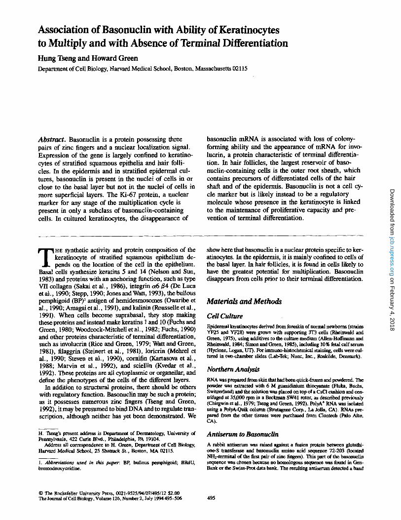
BasonucUn Is Found in the Nuclei of Basal Cells of Epidermis and Other Squamous Epithelia W h e n a col lect ion o f t issue samples was examined by North- ern analysis, it was found that ep idermis was the only tissue o f nine examined that gave a strong signal for basonucl in m R N A (Fig. 1). A faint signal cor responding to a slightly different mobi l i ty was detected in p lacenta and in spleen. The presence o f the protein was tested by extracting ker- atinocytes wi th a solut ion o f SDS and separat ing the pro- teins e lec t rophoret ica l ly for Western analysis. A rabbit anti- s e rum to the sequence of amino acid residues 72-203 of basonucl in , a region lacking zinc fingers, detected a protein wi th mobi l i ty cor responding to 120 kD. Cul tures o f epi- dermal , corneal , and esophageal epithelial cells conta ined basonucl in by this assay, but nei ther the prote in nor its m R N A was detected in cul tured fibroblasts. W h e n cul tured kerat inocytes were stained with both the rabbit an t i serum to basonucl in and a mouse an t i -kera t in monoc lona l antibody AE-1 (Tseng et al . , 1982), all the cel ls conta ining basonucl in also conta ined abundant keratins, conf i rming their identity as keratinocytes.
w h e n frozen sections o f human epidermis were stained
Figure 1. Presence of basonuclin mRNA in various tissues. Approx- imately 2 #g of polyA + RNA of human keratinocytes were loaded in each lane and fractionated on a 1% formaldehyde-agarose gel. The RNAs were transferred to nylon membrane, whose upper part (containing molecules >3 kb) was probed with basonuclin eDNA and whose lower part (containing molecules <3 kb) was probed with a/~-actin eDNA to check the integrity of the RNAs. The sources of the RNAs are indicated. The exposure times for the up- per part and lower part were 3 d and overnight, respectively. Epidermis gave a strong signal for basonuclin mRNA, placenta gave a weak signal (possibly corresponding to a smaller mRNA) and spleen gave a trace signal. All other tissues were completely negative.
The Journal of Cell Biology, Volume 126, 1994 496
on February 4, 2018
jcb.rupress.orgD
ownloaded from
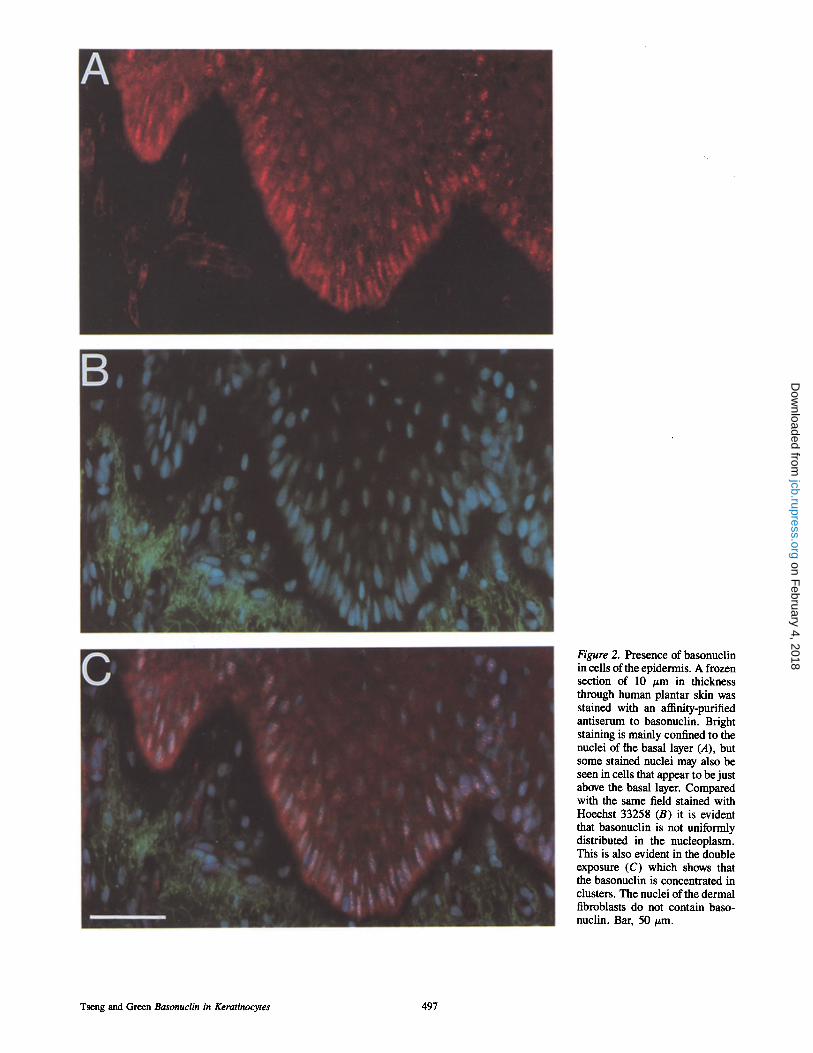
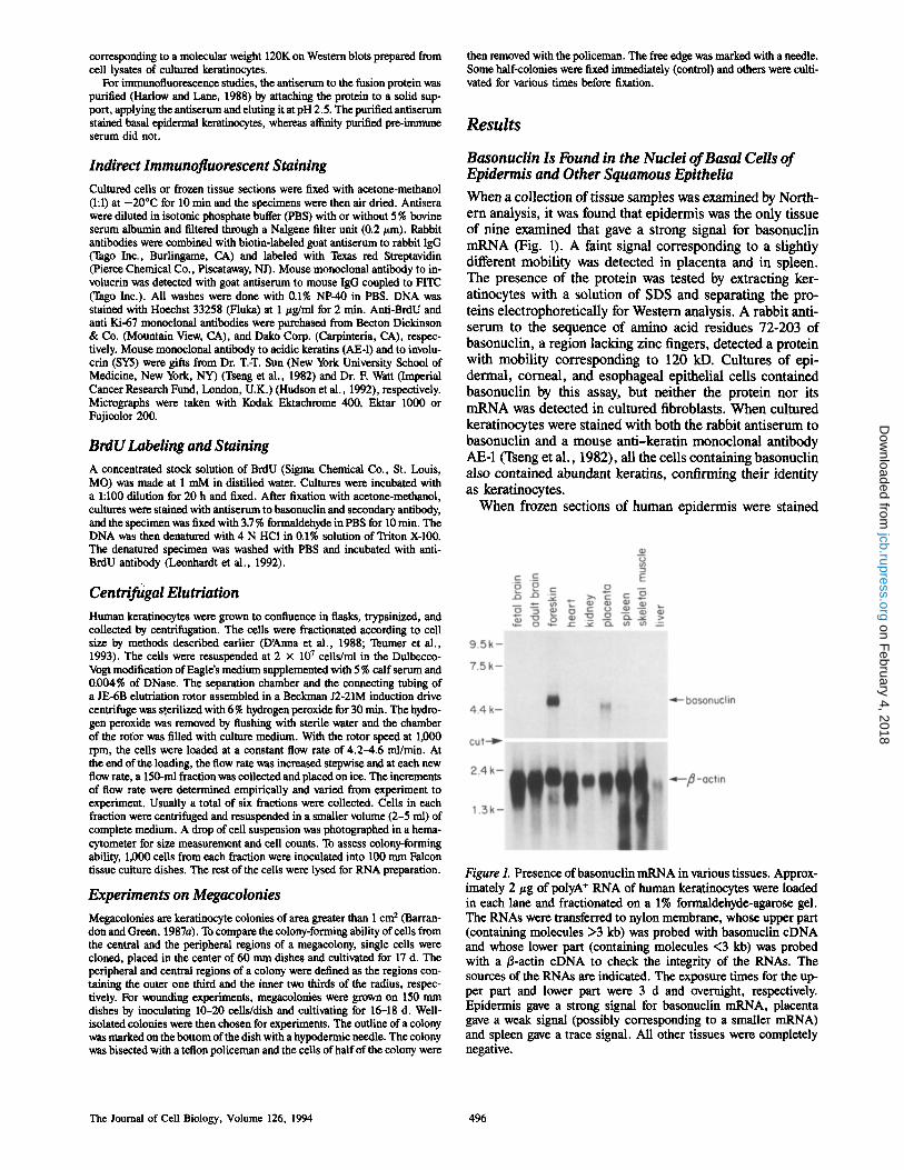
Figure 2. Presence of basonuclin in cells of the epidermis. A frozen section of 10 #m in thickness through human plantar skin was stained with an affinity-purified antiserum to basonuclin. Bright staining is mainly confined to the nuclei of [he basal layer (A), but some stained nuclei may also be seen in cells that appear to be just above the basal layer. Compared with the same field stained with Hoechst 33258 (B) it is evident that basonuclin is not uniformly distributed in the nucleoplasm. This is also evident in the double exposure (C) which shows that the basonuclin is concentrated in clusters. The nuclei of the dermal fibroblasts do not contain baso- nuclin. Bar, 50/~m.
Tseng and Green Basonuclin in Keratinocytes 497
on February 4, 2018
jcb.rupress.orgD
ownloaded from

Figure 3. Comparison of dis- tribution of basonuclin and in- volucrin in the epidermis. Frozen sections of plantar epidermis were fixed and stained for basonuclin and in- voluerin (A) and for DNA as well (B). The involucrin (green) is typically located in the peripheral cytoplasm, close to the cell membrane. Above the boundary at which basonuclin (red) d i s a ~ from suprabasal cell nuclei, there is a zone of cells that lack basonuelin but have not yet acquired involucrin. Bar, 20 #m.
with the same antiserum, basonuclin was found to be largely confined to the nuclei of basal or immediately suprabasal cells (Figs. 2 and 3). The basonuclin did not appear uni- formly distributed in the nucleoplasm, but was mostly con- centrated in nuclear aggregates. Apart from some immedi- ately suprabasal cells, the spinous layer, where terminal differentiation begins, was free of basonuclin. Double stain- ing for basonuclin and for involucrin, a marker of terminal differentiation, with the mouse monoclonal antibody Sy5 (Hudson et al., 1992) showed that the basonuclin disap- peared before involucrin made its appearance (Fig. 3); this resulted in a narrow zone of cells that no longer possessed basonuclin, but did not yet possess involucrin. The use of preimmune serum as primary antibody gave no visible stain- ing of the epidermis.
Basonuclin and the Cells of Hair Follicles
Stem cells of the hair follicle differ from those of the epi- dermis in that they are able to give rise to terminally differ- entiated cells of the hair shaft, while retaining the capacity to generate epidermis. Fig. 4 shows a microdissected hair follicle of human scalp after frozen sectioning and staining for basonuclin. In the upper half of the follicle, the baso- nuclin-containing cells are distributed in the basal layer of the outer root sheath, which is dontinuous with the epidermis. Below the mid-point of the follicle, basonuclin-containing ceils appear suprabasally as well and eventually occupy all layers of the outer root sheath. In the region close to the bulb, where the Outer root sheath narrows into a single layer of cells, these cells retain basonuclin. In all cells of the outer root sheath, basonuclin is clearly nuclear: the outline of the stain follows closely that of the DNA.
In contrast to the outer root sheath, much of the bulb does not contain basonuclin. Keratinocytes possessing basonuclin tend to be located close to the surface of the follicular papilla. In some follicles, basonuclin-containing keratino- cytes surround most of the surface of the papilla, but in other follicles they tend to cluster in certain regions close to that surface, particularly in the deepest part of the matrix, which
has been thought to contain germinal cells (Reynolds and Ja- hoda, 1991). Basonuclin is absent from the cells that form the hair shaft and inner root sheath and from the connective tissue cells within the papilla or surroundirlg the follicle. The sebaceous gland shows some staining in its peripheral part, which is known to contain cells capable of division.
Basonuclin Is Present in Colony-forming Keratinocytes but Absent from Noncolony-forming Keratinocytes
The basal layer of a squamous epithelium contains most of the cells capable of proliferation and these cells are the smallest in the epithelium (Meyer et al., 1970; Rowden, 1975; Yardley and Goldstein, 1976; Barrandon and Green, 1985). The same is true for the basal layer of stratified cul- tures (Sun and Green, 1976; Green, 1980; Barrandon and Green, 1985; Albers et al., 1987). To examine the relation between the presence of basonuclin, the potential for mul- tiplication and the process of terminal differentiation, we trypsinized cultures consisting of growing and terminally differentiating epidermal cells, and separated the cells ac- cording to their size, by centrifugal elutriation. Six fractions were collected and RNA preparations were made from each. To monitor proliferative potential, cells of each fraction were tested for their ability to form colonies by plating them at low density, and each fraction was scored for the abun- dance of cells in terminal differentiation by Northern analy- sis of RNA using, as probe, the coding region of the gene for human involucrin (Eckert and Green, 1986). Fig. 5 shows that basonuclin mRNA was confined to the first two fractions; these fractions also contained nearly all the colony-forming cells, but virtually no involucrin mRNA. In the third fraction, the basonuclin mRNA content dropped sharply, the involucrin mRNA content rose sharply, and the cells lost virtually all colony-forming ability. This experi- ment clearly demonstrated that mRNA for basonuclin was associated with the small cells able to form colonies, and was absent from the larger cells that were terminally differentiat- ing and unable to form colonies.
The Journal of Cell Biology, Volume 126, 1994 498
on February 4, 2018
jcb.rupress.orgD
ownloaded from
Basonuclin and the Departure of Cells from the Basal Layer
To further relate the presence of basonuclin to the growth cy- cle, keratinocyte cultures were allowed to incorporate bromodeoxyuridine (BrdU) for 20 h to label the nuclei of all cells that had entered the S period during a period approxi- mately equal to a generation time. When such a culture was stained with antisera, most of the cells were found to contain both BrdU and basonuclin (Fig. 6). Quantitative scoring showed that 95 % of cells containing BrdU also contained basonuclin (Table I). Probably the 5 % of cells containing BrdU but lacking basonuclin became permanently postmi- totic after completing the cycle during which they incorpo- rated the BrdU. 25% of cells lacking BrdU contained basonuclin, indicating that presence of this protein is not confined to cells that had traversed the S period during the previous 20 h.
In areas of cell stratification in the cultures, it could be seen that suprabasal nuclei usually lacked both BrdU and basonuclin. Cells occupying basal or suprabasal positions by the criterion of overlapping nuclei were scored for these two markers. Scored nuclei were usually those of pairs of cells of which one was basal and the other suprabasal, but in a few cases one suprabasal nucleus overlapped more than one basal nucleus, in which case all were scored.
The results (Table II) showed that nearly all cells lacking both BrdU and basonuclin were suprabasal. 76% of cells containing both BrdU and basonuclin were basal. The rela- tion between cell position and loss of basonuclin in epider- mal cultures is similar to that in the epidermis.
Relation of Basonuclin to the Ki-67 Protein
Ki-67 protein, when it is localized to the nucleus, is a well- known marker for keratinocytes in any stage of the growth cycle (Miyauchi et al., 1990). When small epidermal colo- nies were examined for the presence of Ki-67, virtually all nuclei were found to contain this protein, as well as basonu- clin, consistent with the fact that nearly all the cells of such colonies are in exponential growth.
Keratinocyte colonies that are 10-d old consist of a mixture of multiplying and terminally differentiating ceils. In such colonies, cells containing nuclear Ki-67 were always found to also contain basonuclin (Table I); but cells containing basonuclin often did not contain Ki-67. This demonstrates
Figur e 4. Basonuclin in cells of the hair follicle. Microdissected hair follicles from a 29-yr-old human were frozen and sectioned longitudinally by Dr. Yann Barrandon (Ecole Normale Sup~rieure, Paris, France). The section illustrated extends from beneath the bulb below through the region of the sebaceous gland above. The follicle has been stained for basonuclin and for DNA. Basonuclin is present in ceils of the outer root sheath and below the mid-point of the follicle it is present in most or all layers of this stratified epi- thelium. It is present in the single layer of cells of outer root sheath surrounding the bulb. Basonuclin is present in cells of discrete regions of the bulb close to the follicular papilla. Variability in these regions is shown in A-D, where the DNA has not been stained. Basonuclin is absent from keratinocytes of most of the inner root sheath as well as from the hair shaft and from the connective tissue cells. Continuity of the outer root sheath with the epidermis is inter- rupted because of the microdissection.
Tseng and Green Basonuclin in Keratinocytes 499
on February 4, 2018
jcb.rupress.orgD
ownloaded from
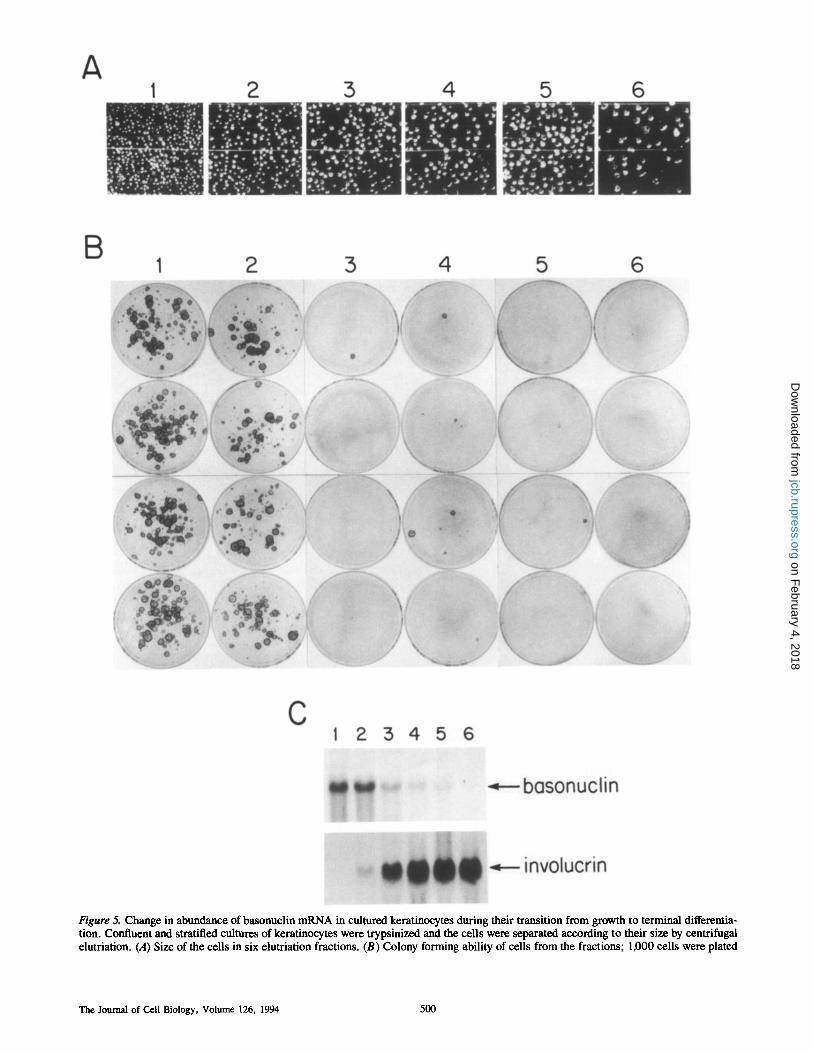
Figure 5. Change in abundance of basonuclin mRNA in cultured keratinocytes during their transition from growth to terminal differentia- tion. Confluent and stratified cultures of keratinocytes were trypsinized and the cells were separated according to their size by centrifugal elutriation. (A) Size of the cells in six elutriation fractions. (B) Colony forming ability of cells from the fractions; 1,000 cells were plated
The Journal of Cell Biology, Volume 126, 1994 500
on February 4, 2018
jcb.rupress.orgD
ownloaded from

Table L Multiplying Cells Contain Basonuclin
Cells containing Cells lacking basonuclin basonuclin Total cells
Cells containing BrdU 270 (95.4%) 13 (4.6%) 283 (100%)
Cells lacking BrdU 24 (25.3%) 73 (74.7%) 97 (100%)
Cells containing Ki67 249 (100%) 0 249 (100%)
Cells lacking Ki67 31 (18%) 142 (82%) 173 (100%)
Table II. Ability to Multiply and the Presence of Basonuclin Are Most Commonly Properties of Basal Cells, Whereas Cells Lacking Both Are Nearly Always Suprabasal
Basal Suprabasal Cells
Cells containing both BrdU and basonuclin 119 (76%) 37 (23%) 156 (100%)
Cells lacking both BrdU and basonuclin 1 (1.6%) 63 (98.4%) 64 (100%)
Figure 6. Basonuclin in relation to cell stratification. 5 d after their inoculation at 5,000 ceils/4 cm 2 chamber, colonies of keratino- cytes merged and began to stratify. Such a culture was exposed to BrdU at 10 #M for 20 h, a period long enough to permit many of the cells incorporating BrdU to progress into 132 and G1. The cul- ture was then stained by double immunofluorescence using antisera to both BrdU (A) and basonuclin (B). The nuclei of most cells in the basal layer (arrowheads) contained both BrdU and basonuclin. Those nuclei containing neither are nearly always suprabasal as they cast a shadow over the fluorescent BrdU-containing nuclei in the basal layer (A, arrows). Occasionally, a nucleus may contain BrdU but not basonuclin (cell 1 ) or basonuclin but not BrdU (cell 2). Metaphase chromatin (cell 3) contains both BrdU and baso- nuclin, which is mainly in the form of clusters or dots. These may be from a few to 20 in number, and are sometimes arranged in strings as in the basal epidermal nuclei (Figs. 2 and 3). In the inter- phase nuclei of the cultures, basonuclih appears to be excluded from the nucleoli, whereas BrdU is not. Bar, 20 #m.
conclusively that basonuclin is not a cell cycle marker, al- though it is present in all multiplying cultured keratinocytes.
The relation between the two proteins was then explored in 17-d megacolonies of 1.3-2.5 cm 2 in area. Expansion of
such colonies is almost entirely due to multiplication and centrifugal migration of the cells located in a rim of 0.2 mm in width at the perimeter of the colonies (Barrandon and Green, 1987a); the multiplication rate internal to this rim is much lower.
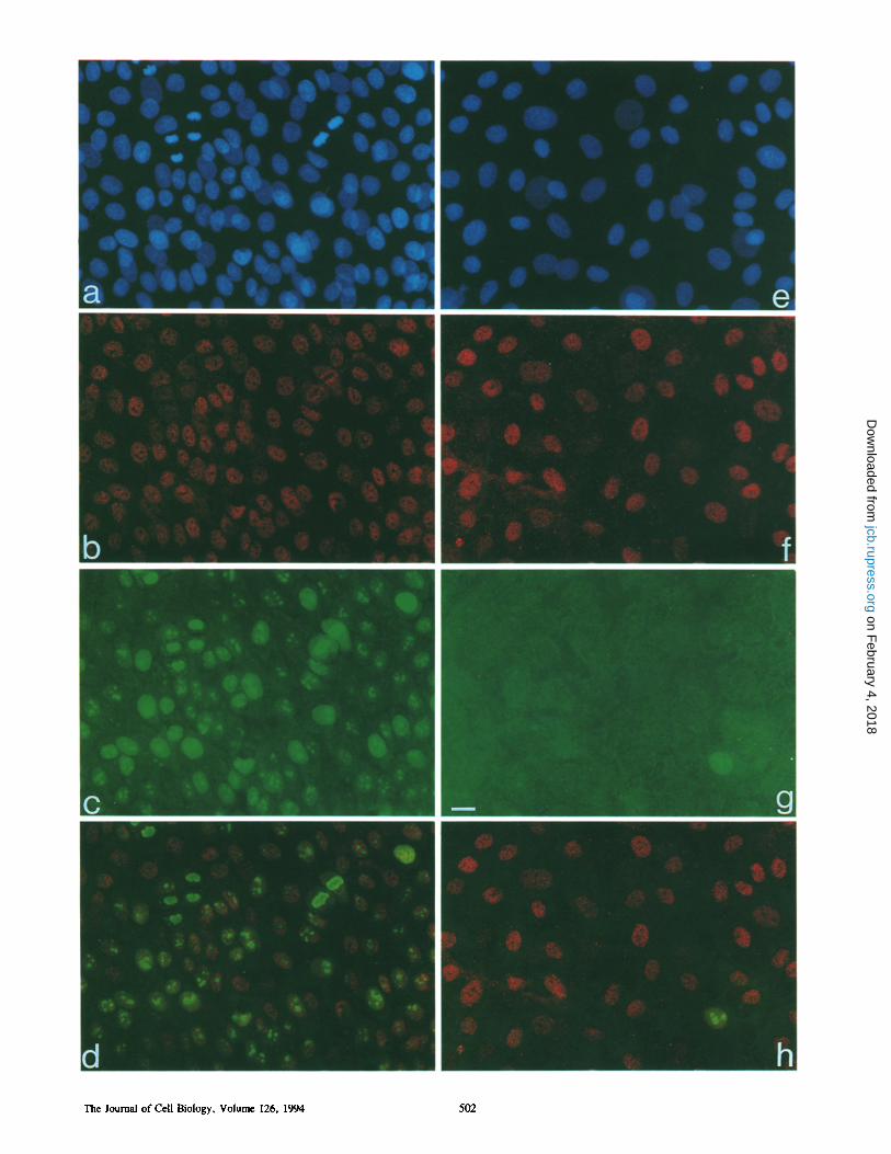
Megacolonies were doubly stained for Ki-67 and basonu- clin and fields located in the central and peripheral regions of each megacolony were examined (Fig. 7). The nuclei of cells located in the peripheral region contained both baso- nuclin and Ki-67. Ki-67 was concentrated in the nucleoli, a site from which basonuclin was excluded. Cells located in the central region usually lacked nuclear Ki-67, while retain- ing basonuclin.
The potential for multiplication by the cells in the two regions was then examined. In one kind of experiment, cen- tral cells were removed from five megacolonies with a teflon policeman and the remaining peripheral cells were tryp- sinized, pooled, and inoculated, while peripheral cells were removed from five additional megacolonies and the central cells were trypsinized, pooled, and inoculated. In another kind of experiment, the central and peripheral cells of a sin- gle megacolony were separated by the application of a clon- ing cylinder, each region was separately trypsinized, and the cells were reinoculated. The results are shown in Table III. Colony formation was extensive by cells in both regions. The colonies formed by cells of central regions were not as numerous as those of the peripheral region in experiment I, whereas in experiment II they were; but in both experiments, most colony-forming cells in the central regions were de- rived from cells lacking Ki-67, since the proportion of cells containing Ki-67 in the central regions was only 4.9 +
on each dish and cultured for fourteen days. (C) Northern analysis of total RNAs prepared from elutriation fractions. 20 #g of total RNA were loaded in each lane, fractionated on a 1% formaldehyde agarose gel and transferred to nylon membranes. The RNA was first probed with basonuclin cDNA and exposed to f i l l for 2 d. This probe was then dehybridized and the RNA was reprobed with genomic clone encoding the human involucrin gene (Eckert and Green, 1986), using an overnight exposure. Small cell fractions contain the colony-forming cells and basonuclin mRNA. Large cell fractions contain neither, but instead contain involucrin mRNA.
Tseng and Green Basonuclin in Keratinocytes 501
on February 4, 2018
jcb.rupress.orgD
ownloaded from
The Journal of Cell Biology, Volume 126, 1994 502
on February 4, 2018
jcb.rupress.orgD
ownloaded from
Table III. Colony Forming Ability of Cells from Central and Peripheral Regions of Megacolonies
Mean number Cells from of colonies Plating efficiency
%
Experiment I* Central region 143 29 Peripheral region 417 84
Experiment II~ Central region 311 62 Peripheral region 320 64
* Pool of central regions and of peripheral regions each of five megacolonies (strain YF28, 3rd passage). ~t Central and peripheral regions isolated with a cloning cylinder from a single 2.5--cm 2 colony. Colonies were counted 10 d after inoculation of two to four dishes with 500 cells. Colonies of all sizes were scored.
4.7%. The frequency of abortive colonies (Barrandon and Green, 1987b) did not appear to be different for the two regions of the megacolony.
Another way of examining the growth potential of the cen- tral ceils of the megacolonies was to divide the colonies in half by bisecting them across their centers and removing the cells of one half with a teflon policeman. Irradiated 3"173 cells were then added to the denuded area. At this time, 2.5% of the ceils along the line of bisection contained nuclear Ki-67; but 3 d later, 43 % of cells in the region migrating outward from the line of bisection contained nuclear Ki-67. It is there- fore concluded that cells of the central region possessing basonuclin, but lacking Ki-67, may revert to the growing state, and form colonies.
Discussion
Relation of Basonuclin to Multiplication of Epidermal Keratinocytes We have used common markers of the growth cycle (Ki-67 and ability to incorporate BrdU) to relate the expression of basonuclin to the growth of cultured keratinocytes. Ki-67 is an interesting protein because it is present in the nuclei of multiplying ceils of any type and at all stages of the growth cycle (Gerdes et al., 1983, 1991; Schltiter et al., 1993) and has been used to evaluate the growth fraction of normal and neoplastic cell populations (Gerdes et al., 1984; Burger et al., 1986). Ki-67 is associated with the nuclear matrix (Verheijen et al., 1989a) and especially with the nucleoli (Verheijen et al., 1989b). Only when located in the nucleus is Ki-67 a marker for multiplication in keratinocytes, since nonmultiplying basal epidermal cells and certain cells of the
outer root sheath of hair follicles may contain Ki-67 in the cytoplasm (Miyauchi et al., 1990).
The experiments show that the synthesis of basonuclin is regulated in keratinocytes of squamous epithelium, the pro- tein being present in nearly all basal cells of the epidermis and in some immediately suprabasal cells but absent from more superficial layers. The proliferative compartment of the epidermis should probably be extended to include im- mediately suprabasal cells, which are sometimes in the growth cycle (Penneys et al., 1970; Lavker and Sun, 1982, 1983; Van Neste et al., 1983; Weinstein et al., 1984), but not usually in epidermal cultures (Albers et al., 1987). The immunocytological experiments show that although baso- nuclin is present in all epidermal cells in growth cycle, it is not confined to them. The experiments on megacolonies show that basonuclin is present in cells able to enter the growth cycle. Terminally differentiating cells, lacking this ability, also lack basonuclin. Disappearance of basonuclin from cells seems to coincide with the irreversible exit of the cells from the multiplication cycle. Since all cells containing Ki-67 contained basonuclin (Table I) and basonuclin was not present in terminally differentiated cells, it appears that basonuclin disappears around the time of the final multipli- cation cycle before the cell becomes postmitotic.
Relation of Basonuclin to Multiplication of Keratinocyte of Hair Follicles The distribution of basonuclin in the hair follicle appears related to the growth potential of the cells. In rodents, the stem cells are located largely in the bulge, not far below the site of the sebaceous gland (Cotsarelis et ai., 1990; Koba- yashi et al., 1993). In adult human follicles, which lack a morphologically well-defined bulge, the stem cells have been assigned to the upper region of the follicle (Yang et ai., 1993) or alternatively to a region below the mid-point of the folli- cle (Rochat et al., 1994). In the lower half of the follicle, basonudin is present in all layers of the outer root sheath. As the outer root sheath approaches the bulb and becomes single layered, the cells still contain basonuclin. From the base of the bulb, basonuclin-containing ceils appear to mi- grate into a ring surrounding the base of the follicular papilla and then along the outer surface of the papilla to its apex, Matricai cells located deep in the bulb have the highest proliferation rate (Van Scott et al., 1963; Weinstein, 1979), particularly those located close to the follicular papilla (Phil- pott et al., 1990). Above the papilla, cells lose the ability to multiply, and lack basonuclin.
The outer root sheath is a structure continuous with the epidermis and, like the epidermis, gives rise on cultivation to proliferating keratinocytes (Weterings et al., 1981; Limat and Noser, 1986) but the distribution of cells containing basonuclin is quite different in the two epithelia. In the epi-
Figure 7. Basonuclin in relation to Ki-67 in megacolonies. (a-d) Part of a 17-d megacolony located close to the perimeter, a region in which most of the cells are multiplying (strain YF28). (e-h) Central region of the same colony, a region in which most cells are resting. All fields show the basal cell layer. Stain for DNA shows numerous mitotic figures in the peripheral region of the megacolony (a) but not in the central region (e). Virtually all of the nuclei in the peripheral region (b) as well as the central region (f) contain basonuclin. Most nuclei of the peripheral region of the megacolony also contain Ki-67 (c), whereas in the central region (g) only a single nucleus contains this marker; virtually all ceils in this region are therefore resting. Double exposure for basonuclin and Ki-67 (d and h) shows Ki-67 concentrated in nucleoli, from which basonuclin is excluded. Bar, 20 #m.
Tseng and Green Basonuclin in Keratinocytes 503
on February 4, 2018
jcb.rupress.orgD
ownloaded from

dermis, they are basal, or immediately suprabasal, whereas in the deeper parts of the outer root sheath, whatever the number of cell layers (from 1-5), virtually all nuclei possess basonuclin. A suprabasal location in this part of the outer root sheath is therefore not a signal for loss of basonuclin. It seems possible that the abundance of colony-forming cells in different segments of the hair follicle (Yang et al., 1993; Rochat et al., 1994) could depend, at least in part, on the differing abundance of basonuclin-containing cells resulting from differing degrees of stratification of the outer root sheath in these segments. Even in the hair bulb, the small number of surrounding outer root sheath cells could contrib- ute to the colony-forming cells found in this segment (Yang et al., 1993; Rochat et al., 1994).
But some cells within the bulb also contain basonuclin. Its presence in keratinocytes located close to the follicular papilla might be explained as a response to signals originat- ing from the fibroblasts of the papilla (Reynolds and Jahoda, 1991; Reynolds et al., 1993). The basonuclin-containing suprabasal cells of the outer root sheath, most of which are located at a considerable distance from the papilla, must rely on signals from other sources, yet these signals must be different from those reaching the epidermis and the upper part of the outer root sheath, whose cells lose basonuclin when they become suprabasal.
Role of Basonuclin
Basonuclin is a protein with three pairs of zinc fingers (Tseng and Green, 1992). Since the discovery of the first zinc finger protein (Hanas et al., 1983; Brown et al., 1985; Miller et al., 1985), many more have been found (Coleman, 1992; Rhodes and Klug, 1993). A number of zinc finger proteins bind to DNA by gel retardation assay, DNA footprinting, or structure determination. At least 45 zinc finger proteins have been shown to act as transcription factors (Coleman, 1992). Consequently, a regulatory role in transcription should be suspected for any newly discovered zinc finger protein.
Basonuclin is related to the drosophila protein disco (Hei- lig et al., 1991), in that the three pairs of zinc fingers of the former resemble the single pair of zinc fingers of the latter; but apart from the zinc fingers, the two proteins are quite different (Tseng and Green, 1992). The function of disco has been ascribed to the positioning and connectivity of larval photoreceptor axons (Lee et al., 1991). This function seems very different from any possible function of basonuclin, even though the similarity in amino acid sequence of the zinc fingers of basonuclin and disco make it possible that these fingers recognize a similar DNA sequence (Tseng and Green, 1992). Although it has not yet been shown that disco is a transcription factor, the mutations that lead to loss of func- tion in that protein are located in one or other of the zinc fingers at a cystein residue that should be coordinated with the zinc atom (Heilig et al., 1991).
Further evidence supporting the idea that basonuclin acts as a transcription factor comes from its similarities to the zinc finger protein PRDII-BF1 (Fan and Maniatis, 1990). These similarities are the possession of a serine stripe (a lin- ear array of exposed serine residues on the surface of a puta- tive c~ helix) and an extended spacer between histidine residues in the second finger of each of two pairs (Tseng and Green, 1992). PRDII-BF1 is a transcription factor that binds
to an identified target sequence in the promoter region of the interferon gene (Goodbourn and Maniatis, 1988; Keller
and Maniatis, 1992). If this reasoning is correct, then basonuclin is a cell type
specific transcription factor regulating genes expressed by keratinocytes; the synthesis of basonuclin is itself regulated at the messenger RNA level in the pathway between stem cells and terminally differentiated products, and when baso- nuclin disappears from the nucleus, the cell is either no longer able to multiply or gives rise (especially in the hair follicle) to transit amplifying cells of limited growth poten- tial. More direct tests of the action of basonuclin will be necessary to define the causes and consequences of the dis- appearance of this protein from the keratinocyte.
The expert technical assistance of Mrs. Karen Easley is gratefully ac- knowledged. We wish to thank Drs. Rick Boyce, Olof Sundin, Brian Burke, and Ray Brown for their advice on antibody production and purification; and Dr. Carolyn Compton for providing tissue sections.
This research was supported by a grant from the National Cancer In- stitute.
Received for publication 21 February 1994, and in revised form 29 March 1994.
References
Albers, K. M., F. Greif, R. W. Setzer, and L. B. Taichman. 1987. Cell-cycle withdrawal in cultured keratinocytes. Differentiation. 34:236-240.
Allan-Hoffmann, B. L., and J. G. Rheinwald. 1984. Polycyclic aromatic hydrocarbon mutagenesis of human epidermal keratinocytes in culture. Proc. Natl. Acad. Sci. USA. 81:7802-7806.
Almendral, J. M., D. Huebsch, P. A. Blundell, H. Macdonald-Bravo, and R. Bravo. 1987. Cloning and sequence of the human nuclear protein cyclin: ho- mology with DNA-hinding proteins. Proc. Natl. Acad. Sci. USA. 84: 1575-1579.
Amagal, M., G. W. Elgart, V. Klans-Kovtun, and J. R. Stanley. 1991. South- ern analysis of the 230-kD bullous pemphigoid antigen gene in normal hu- mans, animals, and patients with junctional epidermolysis bullosa. J. Invest. Dermatol. 97:249-253.
Ando, M., T. Kawashima, H. Kobayashi, and A. Ohkawara. 1989. Proliferat- ing ceils in the normal and psoriatic epidermis detected by Ki67 monoclonal antibody. J. Invest. Dermatol. 92:395.
Barrandon, Y., and H. Green. 1985. Cell size as a determinant of the clone- forming ability of human keratinocytes. Proc. Natl. Acad. Sci. USA. 82: 5390-5394.
Barrandon, Y., and H. Green. 1987a. Cell migration is essential for sustained growth of keratinocyte colonies: the roles of transforming growth factor-c~ and epidermal growth factor. Cell. 50:113 l-1137.
Barrandon, Y., and H. Green. 1987b. Three clonal types of keratinocyte with different capacities for multiplication. Proc. Natl. Acad. Sci. USA. 84: 2302-2306.
Bergstresser, P. R., R. J. Pariser, and J. R. Taylor. 1978. Counting and sizing of epidermal cells in normal human skin. J. lnvest. Dermatol. 70:280-284.
Bravo, R., and H. Macdonald-Bravo. 1985. Changes in the nuclear distribution of cyclin (PCNA) but not its synthesis depend on DNA replication. EMBO (Eur. Mol. Biol. Organ.)J. 4:655-661.
Bravo, R., R. Prank, P. A. Blundell, and H. Macdonald-Bravo. 1987. Cy- clin/PCNA is the auxiliary protein of DNA polymerase-d. Nature (Lond.). 326:515-520.
Brown, R. S., C. Sander, and P. Argos. 1985. The primary structure of tran- scription factor TFIIIA has 12 consecutive repeats. FEBS (Fed. Eur. Bio- chem. Soc.) Lett. 186:271-274.
Burger, P. C., T. Shibata, and P. Kleihues. 1986. The use of the monoclonal antibody Ki-67 in the identification of proliferating cells: application to surgi- cal neuropathology. Am. J. Surg. Pathol. 10:611-617.
Chirgwin, J. M., A. E. Przybyla, R. J. MacDonald, and W. J. Rutter. 1979. Isolation of biologically active ribonucleic acid from sources enriched in ribonuclease. Biochemistry. 18:5294-5299.
Coleman, J. E. 1992. ZINC PROTEINS: Enzymes, storage proteins, transcrip- tion factors, and replication proteins. Annu. Rev. Biochem. 61:897-946.
Cotsarelis, G., T.-T. Sun, and R. M. Lavker. 1990. Label-retaining ceils reside in the bulge area of pilosebaceous unit: implications for follicular stem cells, hair cycle, and skin carcinogenesis. Cell. 61:1329-1337,
D'Anna, F., M. De Luca, R. Cancedda, A. Zicca, and A. T. Franzi. 1988. Elutriation of human keratinocytes and melanocytes from in vitro cultured epithelium. Histochem. J. 20:674-678.
The Journal of Cell Biology, Volume 126, 1994 504
on February 4, 2018
jcb.rupress.orgD
ownloaded from
De Luca, M., R. N. Tamura, S. Kajiji, S. Bondanza, P. Rossino, R. Cancedda, P. C. Marcldsin, and V. Quaranta. 1990. Polarized integrin mediates human keratinocyte adhesion to basal lamina. Proc. Natl. Acad. Sci. USA. 87: 6888-6892.
Eckert, R. L., and H. Green. 1986. Structure and evolution of the human in- volucrin gene. Cell. 46:583-589.
Fan, C.-M., and T. Maniatis. 1990. A DNA-binding protein containing two widely separated zinc finger motifs that recognize the same DNA sequence. Genes & Dev. 4:29-42.
Fuchs, E. 1990. Epidermal differentiation. Curr. Opin. Cell Biol. 2: 1028-1035.
Fuchs, E., and H. Green. 1980. Changes in keratin gene expression during ter- minal differentiation of the keratinocyte. Cell. 19:1033-1042.
Gerdes, J., U. Schwab, H. Lemke, and HI. Stein. 1983. Production of a mouse monoclonai antibody reactive with a human nuclear antigen associated with cell proliferation. Int. J. Cancer. 31:i3-20.
G-erdes, J., H. Lemke, H. Baisch, H.-H. Wacker, U. Schwab, and H. Stein. 1984. Cell cycle analysis of a ceil proliferatinn-assoeiated human nuclear an- tigen defined by the monoclonal antibody Ki-67. J. lmmunoL 133: 1710-1715.
Gerdes, J., L. Li, C. Schlueter, M. Duchrow, C. Wohlenberg, C. Gerlach, I. Stahmer, S. Kloth, E. Brandt, and H.-D. Flad. 1991. Immunobiochemical and molecular biologic characterization of the cell proliferation-associated nuclear antigen that is defined by monoclonal antibody Ki-67. Am. J. PathoL 138:867-873.
Goodbourn, S., and T. Maniatis. 1988. Overlapping positive and negative regulatory domains of the human fl-interferon gene. Proc. Natl. Acad. Sci. USA. 85:1647-1451.
Green, H. 1980. The keratinocyte as differentiated cell type. Harvey Lect. 74:101-139.
Harms, J. S., D. J. Hazuda, D. F. Bogenhagen, F. Y.-H. Wu, and C.-W. Wu. 1983. Xenopus transcription factor A requires zinc for binding to the 5 S RNA gene. J. Biol. Chem. 258:14120-14125.
Harlow, E., and D. Lane. 1988. Antibodies: A Laboratory Manual. Chapter 8. Cold Spring Harbor Laboratory Cold Spring Harbor, NY. 313-315.
Heilig, J. S., M. Freeman, T. Laverty, K. J. Lee, A. R. Campos, G. M. Rubin, and H. Steller. 1991. Isolation and characterization of the disconnected gene of Drosophila melanogaster. EMBO (Eur. Mol. Biol. Organ.) J. 10: 809-815.
Hold, D., T. Mehrel, U. Licldi, M. L. Turner, D. R. Roop, andP. M. Steinert. 1991. Characterization of human loricrin. Structure and function of a new class of epidermal cell envelope proteins. J. Biol. Chem. 266:6626-6636.
Hudson, D. L., K. L. Weiland, T. P. Dooley, M. Simon, and F. M. Watt. 1992. Characterization of eight monoclonal antibodies to involucrin. Hybridoma. 11:367-379.
Jones, P. H., and F. M. Watt. 1993. Separation of human epidermal stem cells from transit amplifying cells on the basis of differences in integrin function and expression. Cell. 73:713-724.
Kartasova, T., G. N. P. van Muijen, H. van Pelt-Heerschap, and P. van de Putte. 1988. Novel protein in human epidermal keratinocytes: regulation of expression during differentiation. Mol. Cell. Biol. 8:2204-2210.
Keller, A. D., and T. Maniatis. 1992. Only two of the five zinc fingers of the eukaryotic transcriptional repressor PRDI-BF1 are required for sequence- specific DNA binding. MoL Cell. Biol. 12:1940-1949.
Kobayasld, K., A. Rochat, and Y. Barrandon. 1993. Segregation of keratino- cyte colony-forming cells in die bulge of the rat vibrissa. Proc. Natl. Acad. Sci. USA. 90:7391-7395.
Kurki, P., K. Ogata, and E. M. Tan. 1988. Mnnoclonal antibodies to proliferat- ing cell nuclear antigen (PCNA)/cyclin as probes for proliferating cells by immunofiuorescenee microscopy and flow cytometry. J. lmmunol. Meth. 109:49-59.
Kvedar, J. C., M. Manabe, S. B. Phillips, B. S. Ross, and H. P. Baden. 1992. Characterization of sciellin, a precursor to the cornified envelope of human keratinocytes. Differentiation. 49:195-204.
Lavker, R. M., and T.-T. Sum 1982. Heterogeneity in epidermal basal ker- atinocytes: morphological and functional correlations. Science (Wash. DC). 215:1239-1241.
Lavker, R. M., and T.-T. Sun. 1983. Epidermal stemcells. J. Invest. Dermatol. 81:121s-127s.
lee, K. J., M. Freeman, and H. Steller. 1991. Expression of the disconnected gene during development of Drosophila melanogaster. EMBO (Eur. Mol. Biol. Organ.) J. 10:817-826.
Leonhardt, H., A. W. Page, H.-U. Weier, and T. H. Bestor. 1992. A targeting sequence directs DNA methyltransferase to sites of DNA replication in mam- malian nuclei. Cell. 71:865-873.
Limat, A., and F. K. Noser. 1986. Serial cultivation of single keratinocytes from the outer root sheath of human scalp hair follicles. J. Invest. Dermatol. 87:485-488.
Marvin, K. W., M. D. George, W. Fujimoto, N. A. Sannders, S. H. Bernacki, and A. M. Jetten. 1992. Cornifin, a cross-linked envelope precursor in ker- atinocytes that is down-regulated by retinoids. Proc. Natl. Acad. Sci. USA. 89:11026-11030.
Mathews, M. B., R. M. Bernstein, B. R. Franza Jr., and J. I. Garrels. 1984. Identity of the proliferating cell nuclear antigen and cyclin. Nature (Lond.). 309:374-376.
Mehrel, T., D. Hold, J. A. Rothnagei, M. A. Longley, D. Bundman, C. Cheng, U. Lichti, M. E. Bisher, A. C. Steven, P. M. Steinert, S. H. Yuspa, and D. R. Roop. 1990. Identification of a major keratinocyte cell envelope pro- rein, Ioricrin. Cell. 61:1103-1112.
Meyer, J., O. F. Alvares, and E. P. Barrington. 1970. Volume and dry weight of cells in the epithelium of rat cheek and palate. Growth. 34:57-73.
Miller, J., A. D. McLacldan, and A. Klug. 1985. Repetitive zinc-binding do- mains in the protein transcription factor IlIA from Xenopus oocytes. EMBO (Eur. Mol. Biol. Organ.) J. 4:1609-1614.
Miyauchi, S., K. Hashimoto, and Y. Miki. 1990. The innermost cell layer of the outer root sheath is positive with 1G-67. J. Invest. Dermatol. 95: 393-396.
Nelson, W. G., and T.-T. Sun. 1983. The 50- and 58-kdalton keratin classes as molecular markers for stratified squamous epithelia: cell culture studies. J. Cell Biol. 97:244-251.
Owaribe, K., J. Kartenbeck, S. Stumpp, T. M. Magin, T. Krieg, L. A. Diaz, and W. W. Franke. 1990. The hernidesmosomal plaque. Differentiation. 45:207-220.
Penneys, N. S., J. E. Fulton Jr., G. D. Weinstein, and P. Frost. 1970. Location of proliferating cells in human epidermis. Arch. Derm. 101:323-32%
Philpott, M. P., M. R. Green, and T. Kealey. 1990. Human hair growth in vitro. J. Cell Sci. 97:463-471.
Reynolds, A. J., and C. A. B. Jaboda. 1991. Hair follicle stem cells? A distinct germinative epidermal cell population is activated in vitro by the presence of hair dermal papilla cells. J. Cell Sci. 99:373-385.
Reynolds, A. J., C. M. Lawrence, and C. A. B. Jahoda. 1993. Human hair follicle germinative epidermal cell culture. J. Invest. Dermatol. 101: 634-638.
Rheinwald, J. G., and H. Green. 1975. Serial cultivation of strains of human epidermal keratinocytes: the formation of keratinizing colonies from single cells. Cell. 6:331-334.
Rheinwald, J. G., and H. Green. 1977. Epidermal growth factor and the mul- tiplication of cultured human epidermal keratinocytes. Nature (Lond.). 265:421-424.
Rhodes, D., and A. Klug. 1993. Zinc Fingers. Sc. Amer. 268:56-65. Rice, R. H., and H. Green. 1979. Presence in human epidermal cells of a solu-
ble protein precursor of the cross-linked envelope: activation of the cross- linking by calcium ions. Cell. 18:681-694.
Rochat, A., K. Kobayashi, and Y. Barrandon. 1994. Location of stem cells of human hair follicles by clonai analysis. Cell. 76:1063-1073.
Rousselle, P., G. P. Lunstrnm, D. R. Keene, and R. E. Burgeson. 1991. Kali- nin: an epithelium-specific basement membrane adhesion molecule that is a component of anchoring filaments. J. Cell Biol. 114:567-576.
Rowden, G. 1975. Ultrastructural studies of keratinized epithelia of the mouse. J. Invest. Dermatol. 64:1-3.
Sakai, L. Y., D. R. Keene, N. P. Morris, and R. E. Burgeson. 1986. Type VH collagen is a major structural component of anchoring fibrils. J. Cell Biol. 103:1577-1586.
Sclduter, C., M. Duchrow, C. Wohlenberg, M. H. G. Becker, G. Key, H.-D. Flad, and J. Gerdes. 1993. The cell proliferation-associated antigen of anti- body Ki-67: a very large, ubiquitous nuclear protein with numerous repeated elements, representing a new kind of cell cycle-maintaining proteins. J. Cell Biol. 123:513-522.
Simon, M., and H. Green. 1985. Enzymatic cross-linking of involucrin and other proteins by keratinocyte particulates in vitro. Cell. 40:677-683.
Steinert, P. M., J. S. Cantieri, D. C. Teller, J. D. Lonsdale-Eccles, and B. A. Dale. 1981. Characterization of a class of cationic proteins that specifically interact with intermediate filaments. Proc. Natl. Acad. Sci. USA. 78: 4097-4101.
Stepp, M. A., S. Spurr-Michaud, A. Tisdaie, J. Elwell, and I. K. Gipsnn. 1990. cr6~4 integrin heterodimer is a component of hemidesmosomes. Proc. Natl. Acad. Sci. USA. 87:8970-8974.
Steven, A. C., M. E. Bisher, D. R. Roop, and P. M. Steinert. 1990. Biosyn- thetic pathways of filaggrin and loricrin-two major proteins expressed by terminally differentiated epidermal keratinocytes. J. Struct. Biol. 104: 150-162.
Sun, T.-T., and H. Green. 1976. Differentiation of the epidermal keratinocyte in cell culture: formation of the cornified envelope. Cell. 9:511-521.
Tan, C.-K., K. Sullivan, X. Li, E. M. Tan, K. M. Downey, and A. G. So. 1987. Autoantibody to the proliferating cell nuclear antigen neutralizes the activity of the auxiliary protein for DNA polymerase delta. Nucleic Acids Res. 15:9299-9308.
Teumer, J. T., K. Zezulak, and H. Green. 1993. Measurement of specific mRNA content of keratinocytes of different sizes in relation to growth and differentiation. In A Keratinocyte Handbook. Leigh, I., F. Watt, and B. Lane, editors. Cambridge University Press, Cambridge, UK. In press.
Tseng, H., and H. Green. 1992. Basonuclln: a keratinocyte protein with multi- ple paired zinc fingers. Proc. Natl. Acad. Sci. USA. 89:10311-10315.
Tseng, S. C. G., M. J. Jarvinen, W. G. Nelson, J.-W. Huang, J. Woodcock- Mitchell, and T.-T. Sun. 1982. Correlation of specific keratins with different types of epithelial differentiation: monoclonal antibody studies. Cell. 30: 361-372.
Van Neste, D., M. J. Staquet, J. Viac, J. M. Lachapelle, and J. Thivolet. 1983. A new way to evaluate the germinative compartment in human epidermis, using [3H] thymidine incorporation and immunoperoxidase staining of 67 K
Tseng and Green Basonuclin in Keratinocytes 505
on February 4, 2018
jcb.rupress.orgD
ownloaded from
polypeptide. Brit. J. Dermatol. 108:433--439. Van Scott, E. J., T. M. Ekel, and R. Auerhach. 1963. Determinants of rate
and kinetics of cell division in scalp hair. ,/. Invest. Dermatol. 41:269-273. Verheijen, R., H. J. H. Kuijpers, R. O. Schlingemann, A. L. M. Boehmer,
R. van Driel, G. L Brakenhoff, and F. C. S. Ramaekers. 1989a. Ki-67 de- texts a nuclear matrix-associated proliferation-related antigen. I. Intracellu- lar localization during interphase. J. Cell Sci. 92:123-130.
Verheijen, R., H. J. H. Kuijpem, R. van Driel, J. L. M. Beck, J. H. van Dieren- donck, G. J. Brakenhoff, and F. C. S. Ramaekers. 1989b. Ki-67 detects a nuclear matrix-associated proliferation-related antigen. II. Localization in mitotic cells and association with chromosomes. J. Cell Sci. 92:531-540.
Watt, F. M., and H. Green. 1981. Invoinerin synthesis is correlated with cell size in human epidermal cultures. J. Cell Biol. 90:738-742.
Weinstein, G. D. 1979. Epidermal cell kinetics. In Dermatology in General Medicine. Fitzpatrick, T. B., A. Z. Eisen, K. Wolff, I. M. Freedberg, and
K. F. Austeu, editors. McGraw Hill, Inc., New York. 85-95. Weinstein, G. D., J. L. McCullough, and P. Ross. 1984. Cell proliferation in
normal epidermis. ,7. Invest. Dermatol. 82:623-628. Weterings, P. J. J. M., A. J. M. Vermorkeu, and H. Bloemandal. 1981. A
method for culturing human hair follicle ceils. Brit. J. DermatoL 104:1--5. Woodcock-Mitchell, J., R. Eichner, W. G. Nelson, and T.-T. Sun. 1982. Im-
munolocalization of keratin polypeptides in human epidermis using mono- clonal antibodies. J. Cell Biol. 95:580-588.
Yang, J.-S., R. M. Lavker, and T.-T. Sun. 1993. Upper human hair follicle contains a subpopulation of keratinocytes with superior in vitro proliferative potential. J. Invest. Derm. 101:652-659.
Yardley, H. J., and D. J. Goldstein. 1976. Changes in dry weight and projected area of human epidermal cells undergoing keratinization as determined by scanning interference microscopy. Brit. J. Dermatol. 95:621-626.
The Journal of Cell Biology, Volume 126, 1994 506
on February 4, 2018
jcb.rupress.orgD
ownloaded from