assessment report - european medicines agency€¦ · cft central foveal thickness . ......
TRANSCRIPT

7 Westferry Circus ● Canary Wharf ● London E14 4HB ● United Kingdom
An agency of the European Union Telephone +44 (0)20 7418 8400 Facsimile +44 (0)20 7523 7455 E-mail [email protected] Website www.ema.europa.eu
© European Medicines Agency, 2013. Reproduction is authorised provided the source is acknowledged.
30 May 2013 EMA/716504/2012 Committee for Medicinal Products for Human Use (CHMP)
Assessment report
Lucentis
International non-proprietary name: RANIBIZUMAB
Procedure No. EMEA/H/C/000715/II/0034
Note
Variation assessment report as adopted by the CHMP with all information of a commercially confidential nature deleted.

Lucentis Assessment report EMA/716504/2012 Page 2/60
Table of contents 1. Background information on the procedure .............................................. 5 1.1. Type II variation .................................................................................................. 5 1.2. Steps taken for the assessment of the product ......................................................... 6
2. Scientific discussion ................................................................................ 6 2.1. Introduction......................................................................................................... 6 2.2. Non-clinical aspects .............................................................................................. 8 2.2.1. Ecotoxicity/environmental risk assessment ........................................................... 8 2.2.2. Conclusion on the non-clinical aspects .................................................................. 8 2.3. Clinical aspects .................................................................................................... 8 2.3.1. Introduction ...................................................................................................... 8 2.3.2. Pharmacokinetics............................................................................................... 9 2.3.3. Pharmacodynamics ............................................................................................ 9 2.3.4. Conclusions on clinical pharmacology ................................................................. 10 2.4. Clinical efficacy .................................................................................................. 10 2.4.1. Dose response study(ies) ................................................................................. 10 2.4.2. Main study(ies) ............................................................................................... 10 2.4.3. Supportive study ............................................................................................. 23 2.4.4. Literature ....................................................................................................... 26 2.4.5. Discussion on clinical efficacy ............................................................................ 26 2.4.6. Conclusions on the clinical efficacy ..................................................................... 29 2.5. Clinical safety .................................................................................................... 29 2.5.1. Introduction .................................................................................................... 29 2.5.2. Patient exposure ............................................................................................. 30 2.5.3. Adverse events (AE) ........................................................................................ 30 2.5.4. Discussion on clinical safety .............................................................................. 33 2.5.5. Conclusions on clinical safety ............................................................................ 35 2.5.6. PSUR cycle ..................................................................................................... 35 2.6. Risk management plan ........................................................................................ 35 2.6.1. PRAC advice .................................................................................................... 35 2.7. Update of the Product information ........................................................................ 43
3. Benefit-Risk Balance.............................................................................. 47
4. Recommendations ................................................................................. 51
5. Appendix ............................................................................................... 53

Lucentis Assessment report EMA/716504/2012 Page 3/60
List of abbreviations
ADR adverse drug reaction
AE adverse event
AMD age-related macular degeneration
ANCOVA analysis of variance
ANOVA analysis of variance
BCVA best corrected visual acuity
CFT central foveal thickness
CMH Cochran-Mantel-Haenszel
CNV choroidal neovascularisation
CRT central retinal thickness
D diopter
DBL database lock
DME diabetic macular oedema
EQ-5D EuroQoL
ETDRS Early Treatment Diabetic Retinopathy Study
FA fluorescence angiography
FAS full analysis set
IOP intraocular pressure
IVT intravitreal
LOCF last-observation-carried-forward
NEI-VFQ-25 National Eye institute Visual Function Questionnaire 25
MacTSQ Macular Disease Treatment Satisfaction Questionnaire
OCT optical coherence tomography
PCV polypoidal choroidal vasculopathy
PI product information
PL package leaflet
PM pathological myopia
PPS per-protocol set
PPSM3 per-protocol set Month 3
PPSM6 per-protocol set Month 6
PRN pro re nata “when needed”
RMP Risk Management Plan

Lucentis Assessment report EMA/716504/2012 Page 4/60
RPE retinal pigment epithelium
RVO retinal vein occlusion
SAE serious adverse events
SmPC summary of product characteristics
VA visual acuity
VEGF vascular endothelial growth factor
vPDT Visudyne (verteporfin) photodynamic therapy (PDT)
W-BQ12 Well-Being Questionnaire
WPAI Work Productivity and Activity Impairment Questionnaire

Lucentis Assessment report EMA/716504/2012 Page 5/60
1. Background information on the procedure
1.1. Type II variation
Pursuant to Article 16 for single variation of Commission Regulation (EC) No 1234/2008, Novartis Europharm Ltd. submitted to the European Medicines Agency on 4 September 2012 an application for a variation including an extension of indication.
This application concerns the following medicinal product:
Medicinal product: International non-proprietary name: Presentations:
Lucentis ranibizumab See Annex A
The following variation was requested:
Variation(s) requested Type C.I.6.a C.I.6.a - Change(s) to therapeutic indication(s) - Addition of a new
therapeutic indication or modification of an approved one II
The MAH applied for an extension of the indication for treatment of visual impairment due to choroidal neovascularisation secondary to pathologic myopia. Consequently, the MAH proposed the update of sections 4.1, 4.2, 4.4, 4.5, 4.8, and 5.1 of the SmPC.
The Package Leaflet (PL) was proposed to be updated in accordance. In addition, the MAH proposed changes to the posology recommendations in the PL for the existing indications in order to enhance clarity and reduce complexity of the explanation for the patient as well to harmonise the wording used throughout the different approved indications. Minor editorial amendments were proposed as well.
In addition, the MAH took the opportunity to update the details of the local representative in Malta in the Package Leaflet.
Furthermore, the MAH proposed this opportunity to bring the PI in line with the latest QRD template version 8.1.
The variation proposed amendments to the SmPC, Labelling and Package Leaflet.
Information on paediatric requirements
Pursuant to Article 8 of Regulation (EC) No 1901/2006, the application included an EMA Decision(s) EMEA-000527-PIP02-10 on the granting of a product-specific waiver.
Applicant’s request(s) for consideration
Additional data protection/marketing exclusivity
The applicant requested consideration of its application in accordance with Article 14(11) of Regulation (EC) 726/2004 - one year of market protection for a new indication.

Lucentis Assessment report EMA/716504/2012 Page 6/60
1.2. Steps taken for the assessment of the product
The Rapporteur and Co-Rapporteur appointed by the CHMP were:
CHMP Rapporteur: Kristina Dunder CHMP Co-Rapporteur: Concepcion Prieto Yerro
PRAC Rapporteur: Ulla Wändel Liminga
Submission date: 4 September 2012
Start of procedure: 16 September 2012
Rapporteur’s preliminary assessment report circulated on: 5 November 2012
Co-Rapporteur’s preliminary assessment report circulated on: 15 November 2012
PRAC RMP advice and assessment overview adopted by PRAC on : 29 November 2012
Request for supplementary information and extension of timetable adopted by the CHMP on: 13 December 2012
MAH’s responses submitted to the CHMP on: 21 January 2013
Joint Rapporteur’s updated assessment report on the MAH’s responses circulated on: 15 March 2013
PRAC RMP advice and assessment overview adopted by PRAC on : 7 March 2013
2nd Request for supplementary information and extension of timetable adopted by the CHMP on: 21 March 2013
MAH’s responses submitted to the CHMP on: 27 March 2013
Joint Rapporteur’s updated assessment report on the MAH’s responses circulated on: 21 April 2013
PRAC RMP outcome (PRAC Rapporteur RMP assessment report) adopted by PRAC on: 16 May 2013
CHMP opinion: 30 May 2013
The CHMP adopted a report on the significant clinical benefit for Lucentis in comparison with existing therapies. (Appendix 1) on: 30 May 2013
2. Scientific discussion
2.1. Introduction
Ranibizumab is a recombinant humanised IgG1 κ isotype monoclonal antibody fragment that selectively binds and neutralises vascular endothelial growth factor (VEGF)-A. Binding of VEGF to its receptors VEGFR-1 and VEGFR-2 triggers angiogenesis and neovascularisation by promoting vascular endothelial cell proliferation/ migration and an increased vascular permeability resulting in leakage, which is a mechanism involved in the development of vascular diseases of the retina. The neutralisation of VEGF results in a reduced angiogenesis and vascular leakage.

Lucentis Assessment report EMA/716504/2012 Page 7/60
Lucentis (ranibizumab), was approved in the EU/EEA in 2007 via the centralised procedure for the treatment of neovascular (wet) age related macular degeneration (AMD). The indication was extended in 2011 for the treatment of visual impairment due to diabetic macular oedema (DME) and retinal vein occlusion (RVO). Lucentis is available as 10 mg/ml solution for injection.
The recommended dose is 0.5 mg ranibizumab, which should be administered monthly until visual acuity is stable for three consecutive months. Thereafter, patient is monitored monthly and treatment is resumed when loss of visual acuity (VA) is indicated.
With this application the MAH proposed to extend the indication to include treatment of visual impairment due to choroidal neovascularisation (CNV) secondary to pathologic myopia (PM). For this indication, the MAH proposed that treatment be initiated with a single intravitreal (IVT) injection to be followed by additional injections only when there are signs of disease activity (loss of VA, lesion activity). It was furthermore proposed to extend the interval from the initial monthly monitoring with time.
Problem statement
CNV secondary to PM is considered one of the major causes of legal blindness in several countries and the leading cause of visual impairment in young patients worldwide.
In PM the axial length of the eyeball is abnormally elongated (> 26 mm), which is associated with high myopia refractive errors (usually greater than -6.0 diopters [D]) and changes of the posterior pole of the eye such as posterior staphyloma, atrophy of the retinal pigment epithelium (RPE), Bruch’s membrane cracks, subretinal haemorrhage, retinal detachment, and CNV. CNV is considered the most vision threatening complication in patients with PM.
While the mechanism of CNV formation in PM is still unclear, the stretching of the retina and thinning of the choroid is considered the key pathway leading to CNV, by way of increasing the probability of choroidal ischemia and subsequent atrophy of the adjacent RPE. These alterations are closely associated with the breaking of Bruch’s membrane (“lacquer cracks”), the main predisposing feature for new vessels formation and vessel growth into the subretinal space. During the natural course of CNV secondary to PM, patients progressively lose VA at a rate of approximately 10 to 15 letters (2 to 3 lines) over 2 years.
The association of an axial length greater than 26 mm with a high myopic refractive error increases the probability of developing CNV from 5% to nearly 10% of patients with associated myopic retinal changes, on average, over 10 years. The prevalence of CNV in patients with PM is high in patients under the age of 50 years; thus, due to the occurrence of this pathology at a young age, this condition has a profound impact on patients’ lives, also affecting the productivity of this working age group.
Although considered the most common treatment for non-subfoveal CNV lesions, laser photocoagulation is associated with permanent loss of vision within the treated area, as well as formation of new anomalous vessels. Visudyne (verteporfin) photodynamic therapy (vPDT) is the only approved medication to treat subfoveal CNV in patients with PM.
Treatment with vPDT has been shown to help avoid vision loss in CNV secondary to PM: in the VIP study, 86% of patients treated with vPDT lost less than 15 letters of best-corrected visual acuity (BCVA) at Month 12 compared with 67% of the placebo treated patients. At Month 24, the mean change in BCVA showed maintenance of vision, on average, at the baseline levels for patients treated with vPDT and a loss of 8 letters in placebo-treated patients. While vPDT provides this improvement in efficacy over natural vision loss, vision gain is uncommon. Therefore, maintenance of VA is the current accepted clinically relevant benefit with vPDT.

Lucentis Assessment report EMA/716504/2012 Page 8/60
Rationale for proposed change
Vascular leakage occurs in macular oedema (DME and RVO) as well as in choroidal neovascularisation (CNV) with exudation of intra- and subretinal fluids with subsequent atrophic changes [wet AMD and pathologic myopia (PM)] and can lead to visual impairment. For all these conditions, a role of VEGF has been proposed. In CNV secondary to PM, the choroidal thinning is believed to cause hypoxic changes in the outer retina including the stimulation of VEGF release.
Similar to wet AMD, by inactivating VEGF, ranibizumab is thought to inhibit neovascular growth and associated exudation, with the subsequent suppression of the CNV growth in PM. Treatment with ranibizumab is intended to have a direct and immediate clinical effect of visual improvement.
Clinical development programme
To support the applied extension of indication for treatment of visual impairment due to CNV secondary to PM, the MAH submitted one pivotal, 12-month phase III study with 0.5 mg ranibizumab (RFB002F2301). In addition, results from study RFB002AGB10 (REPAIR), a local, open-label, exploratory, 12-month study in patients with visual impairment due to CNV secondary to PM, were provided.
2.2. Non-clinical aspects
No new clinical data have been submitted in this application, which was considered acceptable by the CHMP.
2.2.1. Ecotoxicity/environmental risk assessment
No Environmental Risk Assessment has been submitted. This is justified with the protein nature of ranibizumab and Lucentis after application to the patient’s eye by injection is completely metabolised and adsorbed in the body. Any medicinal product that reaches water streams via eventual spills during application or after disposal of unused drug is expected to be very rapidly degraded and mineralised to CO2 by microbial activity. Reference is given to Directive 2001/83/EC and Guideline CHMP/SWP/4447/00.
2.2.2. Conclusion on the non-clinical aspects
The lack of additional non-clinical data is acceptable in view of the data submitted with the previous applications. Considering the above, ranibizumab is not expected to pose a risk to the environment.
2.3. Clinical aspects
2.3.1. Introduction
Good Clinical Practice (GCP)
The Clinical trials were performed in accordance with GCP as claimed by the applicant.
The applicant has provided a statement to the effect that clinical trials conducted outside the community were carried out in accordance with the ethical standards of Directive 2001/20/EC.

Lucentis Assessment report EMA/716504/2012 Page 9/60
Tabular overview of clinical studies
Table 1 Overview of clinical studies in support of new indication for treatment of visual impairment of patients with CNV secondary to PM.
Study No. Study Objectives
No. Treateda Study Duration
Treatment Arms
Ranibizumab regimen
RFB002F2301 - PIVOTAL -
Efficacy and safety
222 subjects treated with ranibizumab only 55 subjects treated with vPDT only of which 40 were also treated with ranibizumabb
Phase I: Day 1 to Month 6 Phase II: Month 6 to Month 12
1: Ranibizumab 0.5 mg (stabilization) 2: Ranibizumab 0.5 mg (disease activity) 3: vPDT 6 mg/m2
IVT injections of ranibizumab 0.5 mg on Day 1 (Group I and Group II) and Month 1 (Group I only). Further monthly injections were given based on whether the patient met visual acuity stabilization criteria (Group I) or disease activity criteria (Group II).
CRFB002AGB10 - SUPPORTIVE -
Efficacy (primary), Safety (secondary)
65 treated with ranibizumab
12 months 1: Ranibizumab 0.5 mg
IVT injections Day 1, thereafter when needed (presence of sub/intraretinal fluid or decrease in VA/increased blurring or metamorphopsia with evidence of leakage)
PM = pathologic myopia; vPDT = visudyne (verteporfin) photodynamic therapy. a The number of treated patients is based on the actual treatment received. Note that 3 patients were randomized to receive treatment with vPDT but instead received treatment with ranibizumab and these patients are included in the appropriate ranibizumab treatment group. b Patients in the vPDT group of Study RFB002F2301 could receive treatment with ranibizumab starting at Month 3.
2.3.2. Pharmacokinetics
Following monthly intravitreal (IVT) administration of ranibizumab 0.5 mg/eye to patients with neovascular AMD, serum concentrations of ranibizumab were generally <5 ng/ml and thus below the ranibizumab concentration necessary to inhibit the biological activity of VEGF by 50% (IC50 = 11 to 27 ng/ml). Serum ranibizumab concentrations in DME and RVO patients were similar to those observed in neovascular AMD patients.
Tong et al (2006) compared aqueous humour concentrations of VEGF in patients with active macular polypoidal choroidal vasculopathy (PCV), patients with CNV of AMD patients with CNV secondary to PM and control subjects. VEGF concentrations were significantly higher in the three disease states versus control. The rank order of mean VEGF concentrations from lowest to highest was control < PCV < PM < AMD. Likewise, Chan et al. (2008) observed higher baseline aqueous VEGF concentrations in patients with CNV of AMD compared to patients with myopic CNV before treatment.
From these cross-indication comparisons, the MAH concluded that (i) aqueous humour VEGF concentrations in patients with CNV secondary to PM were not higher than those in patients with CNV in AMD and (2) serum ranibizumab concentrations were similarly low for patients treated with Lucentis for wet AMD, DME, and RVO. Therefore, vascular permeability and ranibizumab transfer from the vitreous to the serum in patients with visual impairment due to myopic CNV should not be greater than what has been previously measured in other diseases (wet AMD, DME, RVO).
As a consequence, no additional pharmacokinetic studies were performed with in the clinical program to support the use of ranibizumab in the treatment of visual impairment due to CNV secondary to PM.
2.3.3. Pharmacodynamics
The mechanism of action of ranibizumab in the approved indications is to decrease permeability of leaking blood vessels via inactivation of VEGF. The same basic mechanism of action applies

Lucentis Assessment report EMA/716504/2012 Page 10/60
independent on whether targeting choroidal vessels in wet AMD or in PM. The formulation used in study RFB002F2301 was identical to the commercially available formulation. No additional clinical pharmacology profiling studies have been conducted.
2.3.4. Conclusions on clinical pharmacology
Ranibizumab is intended to be administered at the same dose and using the same route of administration already approved for the existing indications. The CHMP therefore concluded that no relevant differences in pharmacokinetics of ranibizumab are to be expected in the new population (patients with CNV due to PM) as compared to the group of patients previously studied (wet AMD, DME, and RVO).
In light of the common mechanism of action as well as the low and similar systemic exposure across the approved indications, the lack of additional studies was considered acceptable by the CHMP.
2.4. Clinical efficacy
2.4.1. Dose response study(ies)
The MAH justified the selection of the 0.5 mg dose since this dose was shown to have the best benefit/risk balance in the wet AMD and RVO dose-finding studies and is approved for all indications (RVO, wet AMD, DME). In addition, the minimum treatment interval of 1 month between doses of ranibizumab was chosen based on the available PK data for ranibizumab and previous studies in patients with wet AMD which showed that both monthly dosing and a biweekly regimen have similar efficacy. Furthermore, the MAH referred to the positive outcome achieved with a low number of injections in the pivotal study (see chapter 2.4.2. ). Therefore, no specific dose finding studies were conducted for the proposed new indication, which was considered acceptable by the CHMP.
2.4.2. Main study(ies)
Title of study
Study RFB002F2301: A 12-month, Phase III, randomized, double-masked, multicenter, active-controlled study to evaluate efficacy and safety of two different dosing regimens of 0.5 mg ranibizumab vs verteporfin PDT in patients with visual impairment due to choroidal neovascularization secondary to pathologic myopia.
Methods
The trial was designed as a randomised, double-blind, multi-centre, active-controlled study with three study arms. 76 centres from 12 EU countries, Canada, Switzerland and Middle&East Asia were involved.
Consenting patients participated in an up to 14 day screening period. The duration of the study was 12 months.
The study was divided into two consecutive phases (up to Month 6 and Month 6 – Month 12), after each of which a database lock (DBL) was planned to occur.

Lucentis Assessment report EMA/716504/2012 Page 11/60
Figure 1: General study design of Study RFB002F2301
BSL (baseline); E (enrolment); S (screening); PE (primary endpoint); EOT (end of treatment); EOS (end of study); DBL (database lock); R (randomisation).
Study participants
Main inclusion criteria: The study included adult patients aged 18 years or older with active CNV secondary to PM with greater than -6D of spherical equivalence and anterio-posterior elongation measurement greater than or equal to 26 mm and at least one subfoveal, juxtafoveal and or extrafoveal lesion or lesion at the margin of the optic disc in the study eye. In addition, patients were required to have a BCVA of ≥24 letters and ≤ 78 letters.
If the patient had previously undergone refractive or cataract surgery, the pre-surgery refraction value was to be used (obtained either from medical chart or by calculation).
The main exclusion criteria for this study were: previous surgery or treatment with corticosteroids within 3 months in study eye prior to the randomisation; laser photocoagulation with involvement of the macular area in the study eye, treatment in the study eye with vPDT or anti-VEGF agents, ocular disorders in the study eye that could confound interpretation of study results, compromised VA or required medical or surgical intervention during the 12-month study period, any type of advanced, severe or unstable disease or its treatment, that could interfere with primary and/or secondary outcome evaluations, a history of stroke, presence of confirmed systolic/diastolic blood pressure > 150/> 90 mmHg, history of malignancy of any organ system within the past 5 years and history of hypersensitivity to the study drugs (ranibizumab, verteporfin) or to drugs of similar chemical classes, and fluorescein or any other component of fluorescein formulation.
Treatments
After eligibility confirmation at baseline, patients were randomised in a 2:2:1 ratio to one of the 3 treatment arms, i.e. to ranibizumab by stabilization (group I), ranibizumab by disease activity (group II), and vPDT (group III). In the vPDT treatment group, treatment with 0.5 mg ranibizumab as a treatment option was allowed from Month 3 in case of disease activity.
Patients were monitored every 4 weeks (+/- 7 days) and were re-treated following the stabilisation or disease activity criteria depending on the treatment group:
The stabilisation criterion was defined as: No change in BCVA as compared to two preceding monthly visits as judged by the evaluating physician.

Lucentis Assessment report EMA/716504/2012 Page 12/60
The disease activity criterion was defined as: Vision impairment, attributable to intra or subretinal fluid or active leakage secondary to PM as assessed by optical coherence tomography (OCT) and/or fluorescence angiography (FA).
Only one eye was selected and treated as the study eye.
Treatment groups
Group I (Ranibizumab I)
• 0.5 mg/0.05mL ranibizumab IVT driven by stabilization criteria
Patients received 0.5 mg ranibizumab intravitreal injections on Day 1 and Month 1. The first time point to assess stabilization criteria was Month 2 based on Baseline, Month 1 and Month 2 assessments. Dosing was stopped if the stabilization criterion for VA was fulfilled. Treatment was resumed with monthly injections when there was a loss of VA due to disease activity and continued until stable VA was reached again for three consecutive monthly assessments.
Group II (Ranibizumab II)
• 0.5 mg/0.5 mL ranibizumab (IVT) driven by disease activity
Patients received intravitreal injection of 0.5 mg ranibizumab at Day 1. From Month 1, dosing was stopped if no disease activity was seen. Treatment was resumed when the disease activity criterion was fulfilled and continued until no disease activity was seen.
Group III (vPDT)
• vPDT [Visudyne®/verteporfin for intravenous infusion at 6 mg/m2 followed by a standard fluence rate of 600 mW/cm2 delivered for 83 seconds with light dose of 50 J/cm2 (as per label)]
Patients received vPDT at Day 1. From Month 3 to 11, there were 3 options to treat the patient’s disease activity:
o 0.5 mg ranibizumab or
o vPDT or
o combination of 0.5 mg ranibizumab and vPDT
Treatment was stopped if no disease activity was seen. Treatment was resumed when the disease activity criterion was fulfilled and continued until no disease activity was seen.
Objectives
The primary objective was to demonstrate the superiority (efficacy) of 0.5 mg ranibizumab driven by stabilisation criteria and/or by disease activity re-treatment criteria vs vPDT as assessed by best corrected visual acuity (BCVA).
The key secondary objective was to demonstrate non-inferiority of 0.5 mg ranibizumab driven by disease activity criteria versus 0.5 mg ranibizumab driven by stabilisation criteria.
Other secondary objectives included evaluation of different aspects of BCVA, anatomical markers, number of treatments as well as safety and tolerability while exploratory objectives included evaluation of ethnicity (Japanese vs. non-Japanese), outcomes in different locations of the CNV and quality of life.

Lucentis Assessment report EMA/716504/2012 Page 13/60
Outcomes/endpoints
The primary efficacy endpoint was the difference between the average level of BCVA (letters) over all monthly post-baseline assessments from Month 1 to Month 3 (endpoint) and the baseline level of BCVA.
Subgroups analyses for age, sex, race, ethnicity (Japanese, non-Japanese), baseline BCVA, baseline axial length, baseline location of CNV and its subtypes were conducted.
The key secondary efficacy variable was the difference between the average level of BCVA (letters) over all monthly post-baseline assessments from Month 1 to Month 6 and the baseline level of BCVA.
The following secondary endpoints based on BCVA were:
• the difference between the average level of BCVA over all monthly post-baseline assessments from Month 1 to Month 12 and the baseline level of BCVA.
• the proportion of patients who gain ≥ 1, ≥ 10, ≥ 15 letters compared with baseline, or reach 84 letters by post-baseline visit up to Month 12.
• the proportion of patients who loose ≥ 10, ≥ 15 letters compared with baseline by post-baseline visit up to Month 12.
Secondary efficacy parameters based on anatomical markers were:
• the proportion of patients with presence of active leakage [based on optical coherence tomography (OCT) and/or fluorescence angiography (FA)] over 12 Months in the treatment groups
• change from baseline in CRT over time (OCT)
Exploratory variables evaluated the BCVA outcome measures in different clinical types of macular (subfoveal, juxtafoveal and extrafoveal) and peripapilar CNV lesions related to PM. Health-related quality of life were evaluated with NEI-VFQ-25, Work Productivity and Activity Impairment Questionnaire (WPAI) and EuroQoL (EQ-5D) at Day 1, Month 3, Month 6 and Month 12.
BCVA (tested at all visits) was tested at 4 meters starting distance using Early Treatment Diabetic Retinopathy Study (ETDRS) charts. OCTs (tested at all visits except visit 3 day 8, all machines allowed – influence of differences e.g. between SD-OCT and 3D-OCT evaluated). FA (and colour fundus photography) was performed at baseline and end of study, thereafter as needed. Anatomical parameters were evaluated by a Central Reading Centre CRC.
Sample size
The study aimed at including 110 patients in each of the ranibizumab groups and 55 patients in the vPDT group. Assuming a treatment difference of 8 letters between each of the ranibizumab groups and vPDT and SD=10 letters, this resulted in a power of ≥91%.
Randomisation
Subjects were randomised in a 2:2:1 ratio (ranibizumab stability criteria: ranibizumab disease activity: vPDT). Upon confirmation of eligibility, at Visit 2 (Day 1) eligible patients received the lowest available randomization number. This number assigned the patient to one of the treatment arms.

Lucentis Assessment report EMA/716504/2012 Page 14/60
Masking
For masking purpose there were both sham ranibizumab (empty vial) and sham vPDT applications (of dextrose 5% solution followed by light application PDT). At least two investigators were involved:
• the masked (assessing) investigator who performed all assessments, captured data in the electronic Case Report Forms and provided re-treatment assignments (stability y/n, disease activity y/n, treatment recommendation: ranibizumab or vPDT or ranibizumab+vPDT), and
• an unmasked (treating) investigator who administered the randomised study treatment when needed according to the protocol.
Statistical methods
This study was divided into 2 consecutive phases, after each of which a DBL lock occurred: Phase I from Day 1 (baseline) to Month 6 and Phase II from Month 6 to Month 12.
Efficacy analyses including the analysis of the primary endpoint and the key secondary endpoint as well as safety analyses were based on the Month 6 DBL (all patients completed month 6 or discontinued).
Unless otherwise specified, confidence intervals were 2-sided and at a 95% level and hypothesis tests were evaluated at a two-sided 0.05 / one-sided 0.025 level of significance.
Primary efficacy (average BCVA over month 1-3), i.e. superiority of ranibizumab (either arm) over vPDT, was evaluated at Month 3 (full analysis set – FAS). Comparisons were performed using the stratified CMH test with the observed values as scores. Missing data followed a Last Observation Carried Forward (LOCF) approach where missing values occurring timely between observed values were replaced by the mean of the last observation observed before and the first observation after the missing time-point. Supportive analyses included ANCOVA and analyses of the per protocol set (PPS) at Month 3. Adjustment for multiplicity was performed according to the Hochberg procedure.
Stratification was done based on categories of baseline BCVA: ≤60 letters vs. >60 letters.
Key secondary efficacy (average BCVA over month 1-6), i.e. non-inferiority comparing the two ranibizumab arms, was evaluated with ANOVA (baseline BCVA as co-variates). The non-inferiority margin of 5 letters is based on health authority feedback related to the Visudyne project in 2008.
Other secondary efficacy endpoints focused on the FAS and considered all three pair-wise comparisons up to Month 6 and up to the end of the study (Month 12). For continuous and ordered categorical variables, changes from baseline were compared between treatment arms using ANOVA/ANCOVA models (baseline assessment as covariate) and/or stratified/ un-stratified CMH/exact Fisher tests.
Results
Overall, 99.6% of all patients randomised completed 3 months, 98.9% of all patients completed 6 months and 96.4% completed the 12-months study period.

Lucentis Assessment report EMA/716504/2012 Page 15/60
Table 3 Disposition of patients
Ranibizumab 0.5 mg vPDT Total
Disposition Reason
Group I by
stabilization n (%)
Group II by disease
activity n (%)
Group III
n (%)
Total
n (%) Screened 334 Randomized 106 (100.0) 116 (100.0) 55 (100.0) 277 (100.0) Completed 3 Months 105 (99.1) 116 (100.0) 55 (100.0) 276 (99.6) Discontinued study prior to Month 3 1 (0.9) 0 0 1 (0.4) Protocol deviation 1 (0.9) 0 0 1 (0.4) Completed 6 Months 103 (97.2) 116 (100.0) 55 (100.0) 274 (98.9) Discontinued study prior to Month 6 3 (2.8) 0 0 3 (1.1) Subject withdrew consent 1 (0.9) 0 0 1 (0.4) Lost to follow-up 1 (0.9) 0 0 1 (0.4) Protocol deviation 1 (0.9) 0 0 1 (0.4) Completed study (12 months) 100 (94.3) 112 (96.6) 55 (100.0) 267 (96.4) Discontinued study prior to Month 12
6 (5.7) 4 (3.4) 0 10 (3.6)
Unsatisfactory therapeutic effect 1 (0.9) 0 0 1 (0.4) Subject withdrew consent 1 (0.9) 2 (1.7) 0 3 (1.1) Lost to follow-up 3 (2.8) 1 (0.9) 0 4 (1.4) Protocol deviation 1 (0.9) 1 (0.9) 0 2 (0.7) Percentages are based on the total number of patients in the randomised set.
Table 2 Analysis set
Ranibizumab 0.5 mg vPDT Total
Analysis sets
Group I by stabilization
n (%)
Group II by disease activity
n (%)
Group III
n (%)
Total
n (%) Randomized set 106 (100.0) 116 (100.0) 55 (100.0) 277 (100.0) Full analysis set 105 (99.1) 116 (100.0) 55 (100.0) 276 (99.6) Per protocol set Month 3 98 (92.5) 108 (93.1) 53 (96.4) 259 (93.5) Month 6 96 (90.6) 107 (92.2) 52 (94.5) 255 (92.1) Safety set 106 (100.0) 118 (101.7) * 53 (96.4) 277 (100.0) Percentages are based on the total number of patients in the Randomized set. * Two patients randomized to vPDT received each one ranibizumab injection prior to Month 3.
Protocol deviations with impact on the analyses were reported in 19 (6.9%) of all randomised patients. Important deviations were: absence of CNV secondary to PM, and absence of appropriate lesion type. In a few cases, there were errors related to re-treatment: under-treatment (sham instead of active/no treatment) as well as over-treatment (active/no treatment instead of sham), absence of high myopia and/or elongation (>-6D/>26 mm).
Baseline data
The mean age of all patients was 55.5 years. Overall, 20% of the patients were younger than 45 years and 28.9% were 65 years of age or older. Most of the patients (75.5%) were female. The majority of patients (58.5%) were Caucasian, 41.2% were Asian including 18.1% Japanese.

Lucentis Assessment report EMA/716504/2012 Page 16/60
Mean BVCA score was 55 letters, retinal thickness was 361 microns, the mean spherical equivalent refractive error was -12.5 and the mean axial length was 29 mm. The most common CNV subtype was located subfoveal (2/3 of patients) which is consistent with this being a population with impaired VA, while in 24%, the CNV location was juxtafoveal.
Most common other ocular conditions were cataract surgery (14.1%), cataract (9%), eye laser surgery (3.2%), and keratomileusis (3.2%). Cataract was slightly more common in Group II (7.8%) and III (9%) than in Group I (5.7%). Overall, 62.8% of patients were reported with an active non-ocular medical condition, most frequently related to the SOCs of vascular disorders (28.2%), musculoskeletal and connective tissue disorders (18.1%), and metabolism and nutrition disorders (15.5%).
Exposure
All randomised patients received at least 1 dose of study drug either of ranibizumab or vPDT.
In the ranibizumab treatment groups, the mean number of ranibizumab injections prior to Month 3 was slightly higher in patients treated according to stabilisation criteria (2.5 ± 0.57) when compared to disease activity criteria (1.8 ± 0.82). This difference was maintained over 6 months (3.5 ± 1.46 vs. 2.5 ± 1.56) and 12 months (4.6 ± 2.59 vs. 3.5 ± 2.92) study duration. In the vPDT group, from Month 3 to Month 6, the mean number of ranibizumab injections was 1.9 (±0.86) and after 12 Months 3.2 (±2.54).
All 55 patients in vPDT treatment group III received 1 vPDT treatment at baseline. Re-treatment (or non-treatment) with vPDT was performed as per-protocol except for few cases. Out of 55, two patients received ranibizumab prior to Month 3 (protocol deviation); other 38 patients received ranibizumab from Month 3. Fifteen patients never received ranibizumab and 2 of them had second vPDT treatment during the study.
The two patients who received ranibizumab before Month 3 were reported in the ranibizumab group (Group II) for the safety analysis.
In the evaluation of efficacy, patients are reported as per originally randomised (i.e. n=55).
During the 12-months period of this study the combination of ranibizumab and vPDT was not administered to any patients.
The mean number of ranibizumab injections was consistent in the majority of subgroups categories irrespective of baseline demographics and ocular characteristics with a tendency for a higher number of injections required in the categories with worse baseline BCVA.
Outcome
Primary and key secondary endpoint
The primary objective was to demonstrate the superior efficacy of 0.5 mg ranibizumab driven by stabilisation criteria or by disease activity re-treatment criteria versus vPDT. The results are summarised in Table 3.
Table 3 Visual acuity of the study eye (letters) - average change from baseline to Month 1 through Month 3 (FAS; modified LOCF)
Ranibizumab 0.5 mg Visudyne PDT Parameter Statistic Group I by
stabilization N=105
Group II by disease activity
N=116
Group III N=55
Baseline n 105 116 55 Mean (SD) 55.4 (13.43) 55.8 (12.59) 54.7 (13.84)

Lucentis Assessment report EMA/716504/2012 Page 17/60
Average Month 1 to Month 3 Mean (SD) 66.0 (12.98) 66.4 (12.28) 56.9 (14.49) SE 1.27 1.14 1.95 Median 69.0 69.2 58.3 Min - Max 12.0 - 86.0 30.7 - 90.0 10.3 - 91.0 Average change from baseline Mean (SD) 10.5 (8.16) 10.6 (7.26) 2.2 (9.47) Min - Max -19.3 - 31.0 -8.3 - 32.0 -24.7 - 24.3 Comparison vs vPDT
Difference in LS means (1) 8.5 8.6
95% CI for difference (1) (5.8,11.2) (6.1,11.1) p-value (2) <0.00001 <0.00001 p-value (3) <0.00001 <0.00001
- n is the number of patients with a value for both baseline and average Month 1 to Month 3. - Stratified analysis includes baseline visual acuity (<=60, >60 letters) as factors. (1) Differences in LS means and the two-sided 95% CIs are estimated from pairwise ANOVA (stratified) model. (2) p-value for CMH test (stratified) (3) p-value for ANOVA (stratified) The outcomes of sensitivity analyses with observed data, with LOCF or with first observation carried back were identical to those in the primary analysis (missing data were imputed with the modified LOCF). The outcome was also essentially identical in the PPS with a change from baseline in group I, II and III of 10.4, 11.0 and 2.3 letters, respectively (PPS observed).
The outcomes in subgroups [age (<45, 45-<55, 55-<65, ≥65 years), sex, race, axial length, baseline BCVA and, generally, the location of CNV subtype] were consistent with that of the overall study results. The characteristics of selected subgroups are detailed in the table below.
Table 4 Average change from baseline in visual acuity (letters) of the study eye to Month 1 through Month 3 by baseline characteristics - subgroup analysis (FAS; modified LOCF)
Ranibizumab 0.5 mg Visudyne PDT Subgroup category Group I
by stabilization N=105
Group II by disease activity
N=116
Group III N=55
Baseline BCVA (letters) n n n <45 21 13.5 (11.41) 26 13.9 (7.28) 13 5.5 (8.11) 45-<60 46 12.8 (6.42) 42 11.6 (8.23) 21 2.6 (9.78) 60-<73 29 7.3 (4.40) 39 8.1 (5.25) 17 2.1 (8.01) >=73 9 2.6 (9.01) 9 7.1 (5.37) 4 -10.8 (9.76) Location of CNV subtype n n n Subfoveal 70 11.2 (9.29) 81 11.2 (7.25) 38 2.1 (7.42) Juxtafoveal 26 10.6 (4.80) 24 10.2 (6.81) 16 1.3 (12.92) Extrafoveal 7 6.9 (3.42) 3 4.0 (11.72) 1 19.0
The key secondary objective was to demonstrate non-inferiority of 0.5 mg ranibizumab driven by disease activity re-treatment criteria vs 0.5 mg ranibizumab driven by stabilisation criteria. The results are summarised in Table 5.

Lucentis Assessment report EMA/716504/2012 Page 18/60
Table 5 Visual acuity of the study eye (letters) - average change from baseline to Month 1 through Month 6 (FAS; modified LOCF)
Ranibizumab 0.5 mg Parameter Statistic Group I by stabilization
N=105 Group II by disease
activity N=116 Average change from baseline n Mean (SD) 11.9 (8.81) 11.7 (8.24) SE 0.86 0.76 Median 11.2 11.7 Min - Max -18.7 - 34.5 -9.7 - 35.7 Comparison vs Group I Difference in LS means1 -0.1 95% CI for difference1 (-2.2, 2.0) p-value2 <0.00001
- n is the number of patients with a value for both baseline and average Month 1 to Month 6. - Stratified analysis includes baseline visual acuity (<=60, >60 letters) as a factor. 1 Differences in LS means and the two-sided 95% CIs are estimated from pairwise ANOVA (stratified) model. 2 This p-value for non-inferiority is from a CMH test (stratified), is one-sided and based on the null hypothesis: Group II by disease activity is worse than Group I by stabilization by 5 letters or more, against the alternative hypothesis: Group II by disease activity is less than 5 letters worse than Group I by stabilization.
As for the primary endpoint, sensitivity analyses were consistent with the outcome based on FAS/modified LOCF.
Other secondary variables
• Change from baseline in visual acuity over time
Rapid VA improvement was observed up to Month 1 in Groups I and II and reached nearly maximum effect by Month 2. Thereafter VA was maintained or slightly increased. Patients randomised to vPDT showed an increase in VA from Month 3 onwards, when patients had the option to receive ranibizumab (38 such subjects crossed over), see Figure 2.
Figure 2 Visual acuity of the study eye (letters): Mean change from baseline over time (FAS; modified LOCF)
BL – baseline; SE – standard error of the mean

Lucentis Assessment report EMA/716504/2012 Page 19/60
• Summary of additional VA-based outcomes
Table 6 Additional visual acuity information for the study eye (FAS; modified LOCF)
Ranibizumab 0.5 mg Visudyne PDT (Ranibizumab as of Month 3)
Parameter Statistic Group I by stabilization
N=105
Group II by disease
activity N=116
Group III N=55 (38/55 patients received
ranibizumab as of Month 3) Change from baseline to Month 3 Mean (SD) 12.1 (10.18) 12.5 (8.81) 1.4 (12.21) Comparison vs vPDT
Difference in LS means
10.7 11.1
95% CI for difference
(7.1, 14.3) (7.9, 14.4)
p-value <0.00001 <0.00001 Change from baseline to Month 6 Mean (SD) 13.7 (10.16) 12.7 (11.01) 7.9 (10.37) Change from baseline to Month 12 Mean (SD) 13.8 (11.42) 14.4 (10.20) 9.3 (11.33) Categorised change from baseline at Month 3 Gain ≥ 10
letters* 65 (61.9) 76 (65.5) 15 (27.3)
Change between -10 and 10 letters
38 (36.2) 39 (33.6) 31 (56.4)
Loss ≥ 10 letters
2 (1.9) 1 (0.9) 9 (16.4)
p-value Rzb vs vPDT (CMH stratified)
<0.00001 <0.00001
Categorised change from baseline at Month 3 Gain ≥ 15
letters* 40 (38.1) 50 (43.1) 8 (14.5)
Change between -15 and 15 letters
63 (60.0) 66 (56.9) 43 (78.2)
Loss ≥ 15 letters
2 (1.9) 0 4 (7.3)
p-value Rzb vs vPDT (CMH stratified)
0.00146 0.00001
Categorised change from baseline at Month 6 Gain ≥ 10
letters* 75 (71.4) 75 (64.7) 25 (45.5)
Change between -10 and 10 letters
28 (26.7) 38 (32.8) 28 (50.9)
Loss ≥ 10 letters
2 ( 1.9) 3 ( 2.6) 2 ( 3.6)
Categorised change from baseline at Month 6 Gain ≥ 15
letters* 49 (46.7) 52 (44.8) 15 (27.3)
Change between -15 and 15 letters
56 (53.3) 63 (54.3) 38 (69.1)
Loss ≥ 15 letters
0 1 (0.9) 2 (3.6)
Categorised change from baseline at Month 12 Gain ≥ 10
letters* 73 (69.5) 80 (69.0) 27 (49.1)
Change between -10 and 10 letters
27 ( 25.7) 34 ( 29.3) 26 (47.3)
Loss ≥ 10 letters
5 (4.8) 2 (1.7) 2 (3.6)

Lucentis Assessment report EMA/716504/2012 Page 20/60
Ranibizumab 0.5 mg Visudyne PDT (Ranibizumab as of Month 3)
Parameter Statistic Group I by stabilization
N=105
Group II by disease
activity N=116
Group III N=55 (38/55 patients received
ranibizumab as of Month 3) Categorised change from baseline at Month 12 Gain ≥ 15
letters* 56 (53.3) 60 (51.7) 18 (32.7)
Change between -15 and 15 letters
47 ( 44.8) 55 ( 47.4) 35 (63.6)
Loss ≥ 15 letters
2 (1.9) 1 (0.9) 2 (3.6)
*gained ≥10/15 letters or reached 84 letters in BCVA
• Visual acuity by subgroups
Consistency among subgroups (Group I and Group II) was observed at Month 3, 6 and 12. At month 12, the mean gain in BCVA vs. baseline was > 10 letters in all subgroups in groups I and II except in the subgroup of subjects with a baseline BCVA of ≥ 73 letters (+6 letters in Groups I and II) and in the group with extrafoveal lesions (+5 letters in Group II).
• Summary of outcomes for anatomical markers
In all three study groups, central retinal thickness had decreased at Month 12 by 60 microns or more compared to baseline.
Most of the intraretinal cysts that were detected at baseline were located in the centre and their centre involvement decreased in all treatment groups up to Month 12 compared to Baseline. Few patients were recorded with new intraretinal cysts.
While the large majority of patients presented with CNV leakage in the study eye at Baseline (Group I: 96.2%, Group II: 93.1%, Group III: 100%) for most of the patients in all 3 treatment groups no CNV leakage was reported at Month 12 (Group I: 68.6%, Group II: 69.8%, Group III: 65.5%).
The size of the area of lesion decreased in the ranibizumab from Baseline to Month 12 with mean changes (SD) from Baseline to Month 12 of -0.310 (± 1.6525) mm2 in Group I and -0.568 (±1.9442) mm2 in Group II, respectively. Patients in Group III were reported with a mean increase from Baseline to Month 12 of 0.279 (±2.9593) mm2. The size of the area of CNV decreased in all treatment groups Baseline to Month 12 with mean changes (SD) from Baseline to Month 12 of -0.248 (±1.8797) mm2 in Group I, -0.979 (±1.6537) mm2 in Group II, and -1.268 (±2.0413) mm2 in Group III, respectively.
• Patient reported outcomes
NEI-VFQ-25
At Month 3, both ranibizumab-treatment arms were favoured over vPDT as regards, for example, general health, general vision, near activities and distance activities (p-values for these ranged from 0.005 to >0.2). The effect was generally maintained or improved at Month 6 and 12, especially for subscales related to vision. For most subscales, Group I (stabilisation) was numerically better than Group II (disease activity).
EQ-5D
Few patients completed the EQ-5D questionnaire. At Month 3, slightly more patients in Group I and II [71.2% (p=0.08) and 64.5% (p=0.05), respectively] improved on the anxiety/depression scale from ‘moderately anxious’ to ‘not anxious’ compared with Group III (51%). In the categories mobility, self-

Lucentis Assessment report EMA/716504/2012 Page 21/60
care, usual activities, and pain/discomfort there were no differences for any group with the majority of patients reported ‘no problems’ in any category for each time point.
WPAI-GH
There was a reduction in percentage of overall work impairment for all 3 treatment groups with the greatest reductions in groups I and II. Mean change from Baseline (SD) of total score at Month 3 were -21.9 (±75.21) in Group I and -22.0 (±55.00) in Group II, compared with -10.2 (±59.89) in Group III.
Ancillary analyses
Posology
During the assessment of the application, the MAH was asked to provide the results of additional analysis with a view to the recommended posology and requirement for monitoring and re-treatment conditions.
In response to the CHMP request, the MAH presented the outcome of an evaluation of the pattern of treatment administration and interruptions by baseline characteristics, the possible predictive value of the length of the treatment-free interval with regards to the need for re-treatment in the future as well as the impact on the VA.
During the first 6 months of the study, the duration of the first treatment-free interval for the majority of patients was ≥2 months. In Group II (disease activity), the duration of the first treatment-free interval was ≥2 months for 82.9% of patients with a treatment interruption.
At Month 12, the average duration of treatment-free intervals increased further and was ≥8 months for almost half of the patients and approximately a third of the patient were not re-treated for 11 months after receiving the single baseline injection. However, there was no consistent pattern in the distribution over time of the treatment-free periods (i.e., relatively consistent percentages of patients in each treatment-free duration category from 1 month to 11 months). When treatment was restarted after an interruption, as above, patients rapidly gained back the previous (generally limited) loss of VA.
Based on the 12-month results, the individual variability of patients needing treatment due to active disease suggest that neither a quarterly fixed monitoring or interruption after one year, as initially proposed, was an optimal recommendation for monitoring the disease in all patients. Instead, an individual monitoring frequency and duration assessed by the treating physician’s clinical judgment, was considered more appropriate to address the individual patient‘s need for treatment.
Further analysis of baseline characteristics indicated that the longest first treatment-free interval was observed in subjects with lower central retinal thickness or central foveal thickness and with less presence of subretinal fluid while no differences were observed for other ocular baseline characteristics. While the length of first treatment-free interval was useful to indicate potential predictors and trends, it did not provide information about the strength of prediction. In a multivariate Poisson regression, disease activity at Month 1 and 2 were identified as the best predictors of future need for treatment of the need for treatment.
Prior treatment with vPDT
Following a request from the CHMP during the assessment, the MAH also submitted VA data separating the non-ranibizumab-treated from the ranibizumab-treated vPDT groups (see Table 7).
Lucentis Assessment report EMA/716504/2012 Page 22/60
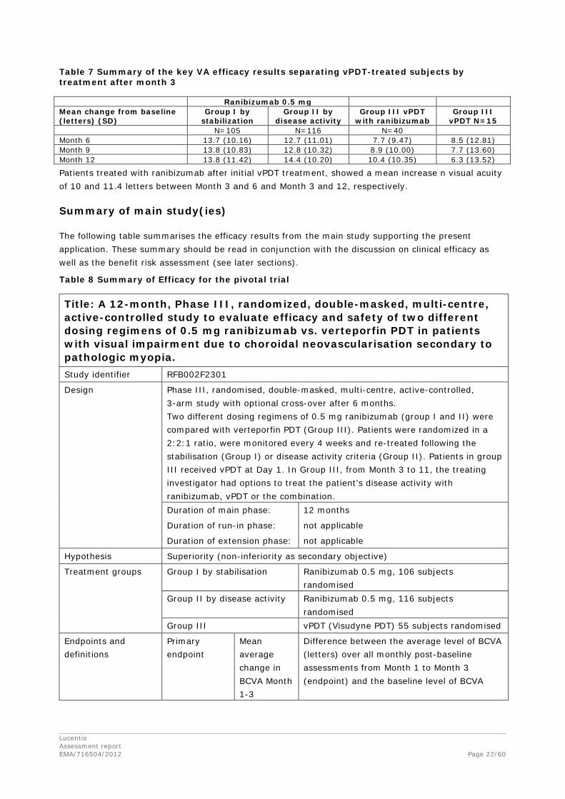
Table 7 Summary of the key VA efficacy results separating vPDT-treated subjects by treatment after month 3
Ranibizumab 0.5 mg Mean change from baseline (letters) (SD)
Group I by stabilization
Group II by disease activity
Group III vPDT with ranibizumab
Group III vPDT N=15
N=105 N=116 N=40 Month 6 13.7 (10.16) 12.7 (11.01) 7.7 (9.47) 8.5 (12.81) Month 9 13.8 (10.83) 12.8 (10.32) 8.9 (10.00) 7.7 (13.60) Month 12 13.8 (11.42) 14.4 (10.20) 10.4 (10.35) 6.3 (13.52)
Patients treated with ranibizumab after initial vPDT treatment, showed a mean increase n visual acuity of 10 and 11.4 letters between Month 3 and 6 and Month 3 and 12, respectively.
Summary of main study(ies)
The following table summarises the efficacy results from the main study supporting the present application. These summary should be read in conjunction with the discussion on clinical efficacy as well as the benefit risk assessment (see later sections).
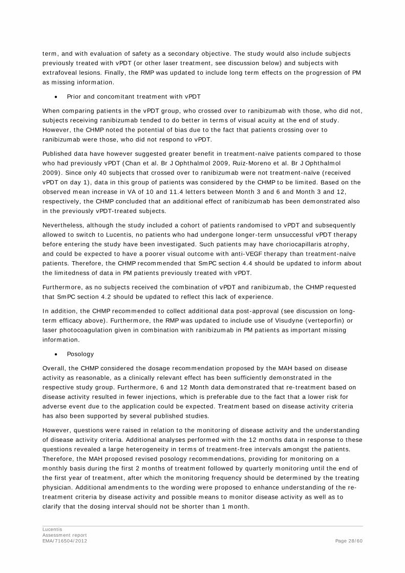
Table 8 Summary of Efficacy for the pivotal trial
Title: A 12-month, Phase III, randomized, double-masked, multi-centre, active-controlled study to evaluate efficacy and safety of two different dosing regimens of 0.5 mg ranibizumab vs. verteporfin PDT in patients with visual impairment due to choroidal neovascularisation secondary to pathologic myopia. Study identifier RFB002F2301
Design Phase III, randomised, double-masked, multi-centre, active-controlled, 3-arm study with optional cross-over after 6 months. Two different dosing regimens of 0.5 mg ranibizumab (group I and II) were compared with verteporfin PDT (Group III). Patients were randomized in a 2:2:1 ratio, were monitored every 4 weeks and re-treated following the stabilisation (Group I) or disease activity criteria (Group II). Patients in group III received vPDT at Day 1. In Group III, from Month 3 to 11, the treating investigator had options to treat the patient’s disease activity with ranibizumab, vPDT or the combination. Duration of main phase: 12 months
Duration of run-in phase: not applicable
Duration of extension phase: not applicable
Hypothesis Superiority (non-inferiority as secondary objective)
Treatment groups Group I by stabilisation Ranibizumab 0.5 mg, 106 subjects randomised
Group II by disease activity Ranibizumab 0.5 mg, 116 subjects randomised
Group III vPDT (Visudyne PDT) 55 subjects randomised
Endpoints and definitions
Primary endpoint
Mean average change in BCVA Month 1-3
Difference between the average level of BCVA (letters) over all monthly post-baseline assessments from Month 1 to Month 3 (endpoint) and the baseline level of BCVA

Lucentis Assessment report EMA/716504/2012 Page 23/60
Key secondary endpoint
Mean average change in BCVA Month 1-6
Difference between the average level of BCVA (letters) over all monthly post-baseline assessments from Month 1 to Month 6 and the baseline level of BCVA.
Database lock DBL 1: After all patients completed 6 months (or discontinued), DBL 2: after all patients completed 12 months (or discontinued)
Results and analysis Analysis description
Primary analysis
Analysis population and time point description
Full analysis set, 3 months (primary endpoint)
Descriptive statistics and estimate variability
Treatment group Ranibizumab Group I by stabilization
Ranibizumab Group II by disease activity
vPDT Group III
Number of subjects
105 116 55
Mean average change in BCVA Month 1-3 (letters ± SD)
10.5 ± 8.2 10.5 ± 7.3 2.2 ± 9.5
Mean average change in BCVA Month 1-6 (letters ± SD)
11.9 ±8.8 11.7 ± 8.2 NA
Effect estimate per comparison
Primary endpoint (CMH test)
Comparison groups (i) Ranibizumab I vs. vPDT (ii) Ranibizumab II vs. vPDT
Difference in mean change in letters
(i) 8.5 (ii) 8.6
95% CI (i) (5.8; 11.2) (ii) (6.1; 11.1)
P-value (i) <0.00001 (ii) <0.00001
Key secondary endpoint (CMH test)
Comparison groups Ranibizumab I vs. Ranibizumab II
Difference in mean change in letters
-0.1
95% CI (-2.2; 2.0) P-value <0.00001
Notes 40 patients (of 55) in the VPDT group (Group III) were treated with ranibizumab.
2.4.3. Supportive study
Study CRFB002AGB10 REPAIR: A phase II, open-label, multicenter, 12-month study to evaluate the efficacy and safety of ranibizumab (0.5 mg) in patients with choroidal neovascularization (CNV) secondary to pathological myopia (PM)

Lucentis Assessment report EMA/716504/2012 Page 24/60
The study was conducted in 12 centres in the United Kingdom.
Objectives
The primary objective was to evaluate the mean change in BCVA from Baseline to Month 12.
The key secondary objectives were to evaluate:
• Mean change in BCVA from Baseline to Month 6 • Mean change in retinal thickness from Baseline to Months 6 and 12 • Time to the first re-treatment and the total number of treatments • Change in lesion size and morphology from screening to Months 6 and 12 • Safety of intravitreal injections of ranibizumab
An exploratory objective was to evaluate the effects of ranibizumab on patient-reported outcomes from baseline to Month 12, assessed by the Macular Disease Treatment Satisfaction Questionnaire (MacTSQ) and the Well-Being Questionnaire (W-BQ12).
Methodology
The study used a prospective, observational, open-label, single-arm, multicentre design. At baseline, all eligible patients received one initial intravitreal injection of ranibizumab 0.5 mg followed by repeated monthly administration as needed for up to a further 11 months based on the presence of sub-/intra-retinal fluid on OCT or decrease in VA/increased blurring or metamorphopsia together with evidence of leakage on FA. Visits to assess efficacy and safety were scheduled at one-monthly intervals.
Number of patients: Recruitment of 64 patients was planned. The sample size was based on outcomes reported in the study by Mones et al (2009). Overall, 65 patients were enrolled.
Diagnosis and main criteria for inclusion
The study included adult patients aged 18 years or older with active primary or recurrent subfoveal or juxtafoveal CNV secondary to PM treated on an outpatient basis. Included patients had a diagnosis of high myopia of at least -6 dioptres in the study eye spherical equivalent and a BCVA (ETDRS chart) between 78 and 24 letters. For subjects who had undergone prior refractive or cataract surgery in the study eye, the preoperative refractive error in the study eye must have been at least -6 dioptres.
The main exclusion criteria for this study were: previous surgery in study eye (within 2 months); laser photocoagulation, intravitreal steroids, vPDT, anti-VEGF agents, external-beam radiation therapy, vitrectomy, or transpupillary thermotherapy in study eye and previous systemic bevacizumab
Endpoints
The primary efficacy variable was the difference from baseline to Month 12 in the level of BCVA (number of letters).
Secondary efficacy variables included the mean change in BCVA from baseline to Month 6, change in lesion size and morphology from screening to Months 6 and 12 and mean change in retinal thickness from baseline to Months 6 and 12. The W-BQ12 questionnaire was administered at baseline then Months 1, 6 and 12. MacTSQ was administered at Months 1, 6 and 12 (exploratory variables).
Safety variables were AEs, ophthalmic examinations and intraocular pressure and vital signs.
Statistical methods
The primary variable was tested to determine if the mean improvement was ≥10 letter BCVA by a paired t-test using the LOCF method. The primary analysis was based on the FAS (all patients who received at least one treatment and had at least one post-baseline assessment for BCVA). For sensitivity purposes, the primary analysis was repeated using the PPS (all patients in the FAS

Lucentis Assessment report EMA/716504/2012 Page 25/60
completing the treatment phase of the trial without clinically significant protocol deviations). Secondary and exploratory efficacy variables were analysed in the FAS and presented descriptively.
Safety variables are presented descriptively based on the safety set (all patients who received at least one injection and had at least one post-baseline safety assessment).
A pre-planned interim analysis was undertaken when 75% of the patients had completed the Month 6 study visit.
Results
Three subjects did not complete the study with one each due to unsatisfactory therapeutic effect, protocol violation and lost to follow-up.
All enrolled patients met the criteria for inclusion in the Safety set and the FAS. Five patients were excluded from the PPS due to protocol deviations including not meeting inclusion and exclusion criteria.
Demographic and background characteristics: Mean (SD) age was 55.5 (14.97) years. The majority of patients were female (70.8%) and Caucasian (90.8%). The mean (SD) duration of CNV prior to study entry was 1.8 (3.26) months, and mean (SD; range) BCVA score in the study eye at baseline was 59.5 (13.6; 26-85) letters. More than half the patients had subfoveal CNV (66.2%), 26% had juxtafoveal and 8% had probably subfoveal/ juxtafoveal CNV.
Exposure: All patients received the first injection. The mean (SD) number of treatments during the 12-month study was 3.6 (2.57). At least one retreatment after the baseline injection was required in 51 patients (78.5%), The median time to first retreatment following the baseline treatment was 2 months (95% CI 1.25, 3.42) (Kaplan-Meier estimates). By Month 12, the cumulative number of treatments was most frequently one (21.5%), two (18.5%) or three (16.9%), with 28 patients (43.1%) receiving four or more treatments in total. Two patients received an injection at each of the 11 successive post-baseline monthly study visits i.e. 12 injections in total.
Efficacy results:
For the primary variable (difference in BCVA from baseline to month 12), the estimated mean (SD) treatment difference was 13.6 (13.9) letters.
The mean difference exceeded 10 letters by Month 2, and remained in the range of 10 to 14 thereafter throughout the 12-month study. The mean (SD) change in BCVA to Month 6 was 11.1 (15.4) letters. During the period from baseline to Month 12, 24 patients (36.9%) achieved a BCVA gain of 15 or more letters in the study eye, and more than half the patients (50.8%, n=33) achieved a gain of 10 or more letters. In the fellow eye, 7.7% and 10.8% of patients, respectively, reported a gain of ≥15 letters or ≥10 letters during the 12 month study period.
There was a significant reduction in CRT from baseline to Month 6, with a mean (SD) change of 128.76 (127.840) μm (p<0.001). This improvement was maintained at Month 12, when the mean (SD) change from baseline was -135.16 (134.117) μm (p<0.001). For patients with evaluable measurements at Baseline and Month 12, the mean (SD) change in lesion size to Month 12 was -0.37 (1.161) (p=0.287). At baseline, intraretinal cysts were absent in 32.3% of patients (21/65), definite in 52.3% (34/65) and questionable in 15.4% (10/65), compared to 80.0% (52/65), 7.7% (5/65) and 4.6% (3/65), respectively, at Month 6 (p<0.001) and 83.1% (54/65), 13.8% (9/65) and 1.5% at Month 12 (1/65) (p<0.001). Subretinal fluid was absent, definite or questionable on OCT in 26.2% (17/65), 67.7% (44/65) and 6.2% (4/65) of patients at baseline, compared to 81.5% (53/65), 12.3% (8/65) and 1.5% (1/65), respectively, at Month 6 (p<0.001) and 89.2% (58/65), 7.7% (5/65) and 1.5% (1/65), respectively, at Month 12 (p<0.001).

Lucentis Assessment report EMA/716504/2012 Page 26/60
The overall General Well-Being score of the W-BQ12 and the MacTSQ questionnaires showed a significant improvement from baseline to Month 12 (p=0.0304 and p=0.0001).
2.4.4. Literature
A recent publication presents the results of a small, 3-year retrospective study investigating the long-term safety and efficacy of ranibizumab in patients (40 eyes of 39 patients) with myopic CNV (Franqueira et al, 2012, Ophthalmologica 227:39–44). Follow-up was at least 30 months for all eyes, and 26 eyes reached the 3 years of follow-up.
The need for re-treatment was determined by a decrease in BCVA and/or signs of disease activity. A mean number of 7.6 (range, 2–19) treatments were performed during the follow-up time. A mean of 4.1 injections were performed in the first year, 2.4 in the second year and 1.1 in the third year.
Mean BCVA was 55.4 ETDRS letters at baseline, 59.7 letters at 12 months (p = 0.07), 61.8 letters at 24 months (p = 0.008) and 63.4 letters at 36 months (p = 0.039). At 6 months, almost 50% of the eyes gained ≥ 5 letters, an effect that was maintained over time. Previous treatment with vPDT (15 eyes) had no impact on the outcome of ranibizumab treatment.
2.4.5. Discussion on clinical efficacy
The CHMP reviewed the data provided from the pivotal trial (study RFB002F2301014) as supported by data from a non-controlled, open-label study (REPAIR).
Pivotal trial
• Study design and baseline data
In general, the CHMP considered the inclusion and exclusion criteria to be relevant with regards for the recruitment of a population representative for the target population. However, the design of the study resulted in a limited proportion of treatment-experienced (laser, vPDT) subjects.
The CHMP furthermore agreed that vPDT (Visudyne photodynamic therapy), the only approved treatment for visual impairment due to myopic CNV, was a reasonable comparator.
However, the CHMP considered that due to the study design only the results from the first 3 months provided a true comparison between treatments. Given the different mechanism of action of both treatments and that the natural course of the condition may be variable, a double-blind, active controlled study period would have been desirable. The MAH justified this by referring to the substantial experience from previous development programmes where all data indicated that the effect of ranibizumab treatment appeared early. The CHMP recognised that considering the current off-label treatment with VEGF-inhibitors in PM together with available publications indicating a promising outcome of such treatment, it would be difficult recruit patients to a study with a longer period restricted to vPDT.
The CHMP furthermore considered the selected endpoints to be relevant for the evaluation of efficacy in the proposed indication. Likewise, the CHMP considered the sample size, randomisation, masking and statistical analysis as well as the general conduct of the study acceptable. However, the CHMP noted that the LOCF approach might not be sufficient conservative in view of the rapid progression of the disease, but since all patients concerned completed 6 months of the study and additional sensitivity analyses were performed, the CHMP concluded that this was of limited concern.
Baseline demographics as well as ocular characteristics were considered to be well balanced and to reasonably represent the spectrum of the target population. PM affects approximately twice as many women than men and this is reflected in the included population. As expected, this population was

Lucentis Assessment report EMA/716504/2012 Page 27/60
younger (from 18 years of age) and had fewer concomitant (ocular and non ocular) conditions when compared to the study populations in the earlier development programmes of ranibizumab.
• Study outcome
Three months after treatment initiation, statistically significant differences of both ranibizumab treatment groups compared to vPDT were shown. The CHMP considered the difference in mean BCVA of more than 8-9 letters to be clinically relevant. The CHMP also noted that for both ranibizumab treatment groups, an effect was apparent within the first month of treatment and increased during the study period, while patients in the vPDT group experienced a minimal and not clinically significant improvement of VA of 2.2 letters. This was considered in line with the expected effect of verteporfin.
Overall, ranibizumab was clearly in favour over vPDT in essentially all subgroups although a lower benefit in elderly patients and those with higher baseline VA was suggested. The benefit of ranibizumab was also less clear in case of extrafoveal lesions, however very few subjects were concerned including only one vPDT-treated subject with such lesion. Based on these limited data, no firm conclusions on the effect of ranibizumab in case of an extrafoveal location of the lesion could be drawn. Therefore, the CHMP considered that SmPC section 4.4 should be updated to inform about this limitation of the available data.
Furthermore, the presence and size of peripapillary atrophy have previously been reported in the scientific literature to be inversely related to the efficacy of anti-VEGF therapy in myopic CNV. However, the MAH had not collected information on the presence and size of peripapillary atrophy. The CHMP therefore recommended that the MAH provided a post-hoc analysis from the existing colour fundus photography and FA images to evaluate the risk for a reduced effect of ranibizumab in patients with CNV due to PM presenting with peripapillary atrophy.
As for the responder analyses, no difference between the two ranibizumab groups was apparent (in both groups, more than 70% of the patients experienced a gain in BCVA of ≥10 letters or reached ≥84 letters overall by the end of the study). However, the proportion of responders was clearly higher for patients treated with ranibizumab as compared to the vPDT treatment group. The differences between treatments observed in the responder analysis was therefore considered by the CHMP to be consistent with those observed in the primary analysis.
At Month 6 and 12, the CHMP noted that the gain in VA further increased compared to Month 3 although to a minor extent with an overall mean change from baseline in the two ranibizumab treatment groups of about 14 letters at the end of the study. No significant difference in BCVA endpoints between the two ranibizumab treatment groups and therefore the re-treatment criteria was observed. The results were stable or increased slightly from Months 6 onwards up until Month 12.
Anatomic markers and patient reported outcomes generally mirrored the results observed for VA, were consistent with ranibizumab’s mechanism of action and in line with previous study results for the approved indications. The CHMP considered these data as supportive.
• Long-term efficacy
Overall, the CHMP considered the efficacy data sufficient to support the proposed new indication. However, concerns were raised regarding the lack of data for long-term efficacy and regarding the possible risk of late reactivation of CNV after years of treatment. To address this concern the CHMP recommended that the MAH collected long-term efficacy data in observational studies post approval. The MAH confirmed that the scope of the ongoing LUMINOUS study would be extended to also include subjects with PM. It was approximated that 100 patients will be followed for a minimum of 2 years. In addition, the MAH agreed to conduct a 3-year observational study in 300 patients with CNV secondary PM with focus on long-term efficacy, including evaluating the risk of reactivation of CNV in the long
Lucentis Assessment report EMA/716504/2012 Page 28/60
term, and with evaluation of safety as a secondary objective. The study would also include subjects previously treated with vPDT (or other laser treatment, see discussion below) and subjects with extrafoveal lesions. Finally, the RMP was updated to include long term effects on the progression of PM as missing information.
• Prior and concomitant treatment with vPDT
When comparing patients in the vPDT group, who crossed over to ranibizumab with those, who did not, subjects receiving ranibizumab tended to do better in terms of visual acuity at the end of study. However, the CHMP noted the potential of bias due to the fact that patients crossing over to ranibizumab were those, who did not respond to vPDT.
Published data have however suggested greater benefit in treatment-naïve patients compared to those who had previously vPDT (Chan et al. Br J Ophthalmol 2009, Ruiz-Moreno et al. Br J Ophthalmol 2009). Since only 40 subjects that crossed over to ranibizumab were not treatment-naïve (received vPDT on day 1), data in this group of patients was considered by the CHMP to be limited. Based on the observed mean increase in VA of 10 and 11.4 letters between Month 3 and 6 and Month 3 and 12, respectively, the CHMP concluded that an additional effect of ranibizumab has been demonstrated also in the previously vPDT-treated subjects.
Nevertheless, although the study included a cohort of patients randomised to vPDT and subsequently allowed to switch to Lucentis, no patients who had undergone longer-term unsuccessful vPDT therapy before entering the study have been investigated. Such patients may have choriocapillaris atrophy, and could be expected to have a poorer visual outcome with anti-VEGF therapy than treatment-naïve patients. Therefore, the CHMP recommended that SmPC section 4.4 should be updated to inform about the limitedness of data in PM patients previously treated with vPDT.
Furthermore, as no subjects received the combination of vPDT and ranibizumab, the CHMP requested that SmPC section 4.2 should be updated to reflect this lack of experience.
In addition, the CHMP recommended to collect additional data post-approval (see discussion on long-term efficacy above). Furthermore, the RMP was updated to include use of Visudyne (verteporfin) or laser photocoagulation given in combination with ranibizumab in PM patients as important missing information.
• Posology
Overall, the CHMP considered the dosage recommendation proposed by the MAH based on disease activity as reasonable, as a clinically relevant effect has been sufficiently demonstrated in the respective study group. Furthermore, 6 and 12 Month data demonstrated that re-treatment based on disease activity resulted in fewer injections, which is preferable due to the fact that a lower risk for adverse event due to the application could be expected. Treatment based on disease activity criteria has also been supported by several published studies.
However, questions were raised in relation to the monitoring of disease activity and the understanding of disease activity criteria. Additional analyses performed with the 12 months data in response to these questions revealed a large heterogeneity in terms of treatment-free intervals amongst the patients. Therefore, the MAH proposed revised posology recommendations, providing for monitoring on a monthly basis during the first 2 months of treatment followed by quarterly monitoring until the end of the first year of treatment, after which the monitoring frequency should be determined by the treating physician. Additional amendments to the wording were proposed to enhance understanding of the re-treatment criteria by disease activity and possible means to monitor disease activity as well as to clarify that the dosing interval should not be shorter than 1 month.

Lucentis Assessment report EMA/716504/2012 Page 29/60
The CHMP considered that the revised wording proposed for SmPC section 4.2 was reasonable and consistent with the available data, reflecting the large heterogeneity in the need for re-treatment as well as the risk of insufficient monitoring/treatment. Considering that half of the patients were stable for at least 8 months, the CHMP agreed that a less frequent monitoring than in the other, approved indications for ranibizumab, was acceptable. In addition, the CHMP was reassured by the fact that in case of loss of VA after treatment interruption, the loss of VA was not rapid and was quickly regained when treatment was re-initiated.
The CHMP furthermore noted that any attempts by the MAH to identify predictors aiding in further recommendations regarding the frequency of monitoring were unsuccessful. While the longest average first treatment-free interval was observed in subjects with lower CRT or CFT, as well as a lower proportion of presence of subretinal fluid, there were no differences with regards to other ocular parameters (e.g. baseline BCVA, IOP, axial length, refraction sphere, CNV location).
Supportive study
With regards to the open-label, non-controlled REPAIR study in 65 patients, the CHMP considered the data generally in line with results seen in the pivotal trial. Both VA-based and anatomic outcomes supported the results observed in the pivotal study.
2.4.6. Conclusions on the clinical efficacy
In summary, the CHMP considered that the data presented by the MAH in support of the proposed extension of indication showed that, while patients on vPDT followed a VA stabilisation pattern, patients receiving ranibizumab experienced an improvement in vision. These results are in line with what has been previously described. The gain in VA with ranibizumab treatment was rapidly achieved (near maximum effect by month 2 of treatment) and was maintained throughout 12 months. Overall, the effect size was considered by the CHMP clinically significant and highly relevant although long term data was lacking.
Concerns were raised as regards the limitedness of data in patients previously treated with vPDT and in patients with extrafoveal lesions as well as in relation to the lack of experience of concomitant use of vPDT and ranibizumab treatment as well as regards efficacy in patients with extrafoveal lesions.
In light of these concerns, the CHMP considered that amendments to the posology recommendations as well as the warnings in the SmPC were necessary compared to the original proposal of the MAH to inform about the limitations of the efficacy data.
In view of the convincing efficacy of Lucentis in the proposed new indication of vision impairment due to CNV secondary to PM, additional efficacy data were not considered necessary by the CHMP prior to approval of the new indication. However, the CHMP recommended that additional efficacy data should be collected post approval in observational studies, in particular with a view to long-term efficacy and the efficacy concerns raised above.
2.5. Clinical safety
2.5.1. Introduction
Safety profile in the approved indications
The MAH reported that the available safety database for ranibizumab in the already approved indications included over 38,000 subjects (up to PSUR 8, 2011) in clinical trials from the previous development programmes as well as post marketing data.

Lucentis Assessment report EMA/716504/2012 Page 30/60
The majority of adverse reactions reported following administration of Lucentis are related to the intravitreal injection procedure. The most frequently reported ocular adverse reactions are eye pain, ocular hyperaemia, increased intraocular pressure, vitritis, vitreous detachment, retinal haemorrhage, visual disturbance, vitreous floaters, conjunctival haemorrhage, eye irritation, foreign body sensation in eyes, increased lacrimation, blepharitis, dry eye and eye pruritus.
The most frequently reported non-ocular adverse reactions are headache, nasopharyngitis and arthralgia.
Less frequently reported, but more serious, adverse reactions include endophthalmitis, blindness, retinal detachment, retinal tear and iatrogenic traumatic cataract.
The overall frequency of non-ocular haemorrhages, potentially related to systemic VEGF inhibition, was slightly increased in ranibizumab-treated subjects in the wet AMD phase III studies. There is also a theoretical risk of arterial thromboembolic events, including stroke and myocardial infarction, following intravitreal use of VEGF inhibitors. A low incidence rate of arterial thromboembolic events was observed in the Lucentis clinical trials in patients with AMD, DME and RVO and there were no major differences between the groups treated with ranibizumab compared to control.
Safety data for myopic CNV indication
The MAH submitted safety data from the pivotal and supportive trial of the new indication of VA impairment due to myopic CNV and the assessment is provided in the following chapters.
2.5.2. Patient exposure
In the pivotal study, 224 subjects in Group I and II have been treated with ranibizumab for up to 12 months and 38 subjects that were treated with vPDT (Group III) received ranibizumab between Month 3 and Month 12. In addition, 65 subjects in the supportive REPAIR study were treated with ranibizumab for up to 12 months. Thus, a total of 327 subjects have received ranibizumab during the 12 months of the studies and 289 patients received ranibizumab from study start.
In the pivotal study, subjects in Group I and II received 2.5 and 1.8 injections, respectively, between baseline and Month 3. Between baseline and Month 6, the mean number of injections in Group I and II was 3.5 and 2.5, respectively and 1.8 injections in Group III. Between baseline and Month 12, the mean number of injections in Group I and II was 4.6 and 3.5, respectively and 3.2 injections in Group III. In REPAIR, the mean number of injections was 3.6 in 12 months.
All subjects in Group III received 1 treatment of vPDT at baseline and 2 subjects that did not cross-over to receive ranibizumab received 1 additional vPDT treatment each after Month 3.
2.5.3. Adverse events (AE)
2.5.3.1. Study RFB002F2301
• Ocular Adverse Events
Up to Month 3, the frequency of ocular AEs in the study eye was 27.4% in Group I, 13.6% in Group II, and 9.4% in Group III. Most AEs were suspected to be related to the injection rather than the study drug and the trend in reporting frequencies, i.e. highest frequency reported in Group I, followed by Group II with the lowest reporting in Group III, mirrored the number of injections given in the respective groups (mean 2.5 injections in Group I vs. mean 1.8 injections in Group II vs. no treatment with ranibizumab up to month 3 in Group III). Only vitreous floaters, retinal tear and conjunctivitis allergic were AEs not considered related to the injection.

Lucentis Assessment report EMA/716504/2012 Page 31/60
Overall, up to Month 6, ocular AEs considered by the investigator as suspected to be related to study drug and/or ocular injection were reported for a total of 46 (16.6%) patients [19.8%, 16.1%, 11.8%, and 10.5% of the patients in ranibizumab Group I and II, Group III (with ranibizumab) and Group III (without ranibizumab), respectively].
The frequencies of ocular AEs in the study eye up to Month 12 was 43.4% in Group I, 37.3% in Group II, and 42.1% and 26.7% in Group III with ranibizumab and without ranibizumab, respectively (see table below).
Table 9 Number (%) of patients with ocular adverse events of the study eye up to Month 12 (at least 2 patients in any group), by preferred term (safety set)
Ranibizumab 0.5 mg Visudyne PDT Group I
by stabilization
Group II by disease
activity
Group III with ranibizumab from
Month 3
Group III without ranibizumab from
Month 3 N=106 N=118 N=38 N=15
Preferred term n (%) n (%) n (%) n (%) Total 46 (43.4) 44 (37.3) 16 (42.1) 4 (26.7)
Conjunctival haemorrhage 12 (11.3) 12 (10.2) 2 (5.3) 0 Punctate keratitis 8 (7.5) 3 (2.5) 2 (5.3) 0 Vitreous floaters 5 (4.7) 1 (0.8) 0 0 Dry eye 4 (3.8) 2 (1.7) 0 1 (6.7) Eye pain 4 (3.8) 4 (3.4) 1 (2.6) 1 (6.7) Injection site haemorrhage 3 (2.8) 3 (2.5) 2 (5.3) 0 IOP increased 3 (2.8) 7 (5.9) 4 (10.5) 0 Blepharitis 2 (1.9) 2 (1.7) 0 0 Conjunctivitis 2 (1.9) 1 (0.8) 0 0 Eyelid oedema 2 (1.9) 0 0 0 Retinal tear 2 (1.9) 1 (0.8) 0 0 Cataract 1 (0.9) 2 (1.7) 0 1 (6.7) Conjunctivitis allergic 1 (0.9) 5 (4.2) 1 (2.6) 0 Ocular hyperaemia 1 (0.9) 2 (1.7) 1 (2.6) 0 Retinal haemorrhage 1 (0.9) 3 (2.5) 0 0 Metamorphopsia 0 3 (2.5) 0 0 Visual impairment 0 0 2 (5.3) 0
Up to month 12, the highest proportion of subjects reporting increased intraocular pressure (IOP) was observed in the previously vPDT-treated subjects that received ranibizumab. A suspected relationship to ocular injection was reported up to Month 12 in 24.5% (Group I) and 20.3% of patients in the ranibizumab groups and in 18.4% and 13.3% patients in the vPDT with and without ranibizumab groups, respectively.
The majority of AEs were of mild severity. A total of 18 patients experienced ocular AEs of moderate severity up to Month 12 (Group I: punctate keratitis, photopsia, uveitis, visual acuity reduced, vitreous detachment; Group II: vitreous floaters, blepharitis, cataract, retinal haemorrhage, eye haemorrhage, metamorphopsia, ocular hypertension, retinoschisis, IOP increased; Group III with ranibizumab: macular oedema, ocular hypertension; Group III without ranibizumab: orbital myositis). All except three of the moderate ocular AEs occurred in patients treated with ranibizumab. Dacryocystitis (n=1, group I) and conjunctivitis allergic (n=1, group II) were classified as severe AEs.
• Non-Ocular Adverse Events
At least one non-ocular AE during the first 3 months of treatment was recorded in 25.5% of patients of Group I, in 25.4% of Group II, and in 11.3% of the vPDT group. The most frequent non-ocular AEs in the ranibizumab groups were nasopharyngitis, headache, upper respiratory tract infection, back pain, and hypertension.

Lucentis Assessment report EMA/716504/2012 Page 32/60
Up to Month 12, five out of 8 cases of upper respiratory tract infection were reported from the same site.
Non-ocular AEs reported by the investigator as suspected to be related to study drug and/or ocular injection were reported for a total of 3 (2.5%) patients in Group II up to Month 12 (1 patient each with headache, hepatic function abnormal and nausea). The report of hepatic function abnormal is further addressed under laboratory findings below.
Up to Month 12, 9 patients were reported with severe non-ocular AEs (Group I: 3 patients with myocarditis, gastritis erosive, gastrointestinal haemorrhage, melaena, depression, Group II: 6 patients with atrial tachycardia, bronchitis, subdural hematoma, muscle spasms, spinal column stenosis, lung adenocarcinoma, hypertension).
• Serious adverse event (SAE) and deaths
No patient died during the 12-month study period.
A single patient in Group I experienced an ocular SAE of the study eye of corneal erosion during the first 3 months. A second patient (Group II) was reported with an ocular SAE of the study eye of retinoschisis in the study eye as a SAE on Day 309. The event was not suspected to be related to study drug/ocular injection and no action was taken.
In addition, 11 serious non-ocular AEs were reported up to Month 12. None of the serious events was suspected by the investigator to be related to study drug or injection.
• Targeted Adverse Events
Endophthalmitis: No cases were reported.
Intraocular pressure (IOP) increased: During the 12-months study there were 15 patients for whom IOP increase was reported as an AE in the study eye: 4 patients in Group I, 7 patients in Group II and 4 patients in Group III. For 8 of these patients, a relation to the injection was suspected. All was of mild severity except one that was regarded as moderate. In summary, the reported cases of IOP increase were transiently experienced post-injection and did not require chronic IOP lowering treatment. Newly diagnosed glaucoma was not reported.
Retinal tear/Retinal detachment: Patients with PM have theoretically a higher risk of retinal tears and retinal detachments. Three patients (2 in Group I, 1.9% and 1 in Group II, 0.8%) were reported with retinal hole, retinal break and retinal tears with operculum. All three events were coded as retinal tear and all of these AEs were of mild severity. There were no cases of “retinal detachment” reported.
• Laboratory findings
Overall, there were no clinically relevant changes from baseline in haematology values and urine analysis values. Largely, there were also no clinically relevant changes from baseline in clinical chemistry values. AEs due to hepatic function abnormal were however reported for 2 patients.
• Immunological events
Given the low incidence and lack of a relationship to clinical outcome in previous studies, immunogenicity to ranibizumab was not assessed in the current clinical program.
• Safety in special populations
The RMP-identified AEs that could represent potential safety concerns were analysed in demographic subgroups (ethnicity, age, sex). While the number of patients was too low to allow an analysis in all subgroups, there was no notable differences in the incidence of any of the defined RMP safety concerns between age groups (i) <50, ≥50 years and (ii) 65 and ≥65 years of age or gender.

Lucentis Assessment report EMA/716504/2012 Page 33/60
• Safety related to drug-drug interactions and other interactions
No specific drug interaction studies were performed.
• Discontinuation due to adverse events
No patient discontinued the study drug treatment prematurely due to an AE or SAE during the 12-months study period. However, 6 patients temporarily interrupted treatment with ranibizumab due to an AE or abnormal laboratory test.
2.5.3.2. Supportive Study CRFB002AGB10 (REPAIR)
The overall incidence of AEs was 70.8% (n=46). Ocular AEs, regardless of study drug relationship, were reported by 29 patients (44.6%).
Among the 29 patients with one or more ocular AE, the severity of AEs was mild in 22 patients (33.8%), moderate in five patients (7.7%) and severe in two patients (3.1%). Ocular AEs with a suspected relation to study drug were reported in four patients (6.2%), comprising conjunctival haemorrhage (n=1), vitreous floaters (n=2) and endophthalmitis (n=1).
No deaths occurred during the study. Three patients experienced one serious AE (SAE) each. One of these was reported as endophthalmitis. The other two SAEs were non-ocular (depression and dislocated shoulder) and were not suspected to be related to study drug.
Mean IOP in the study eye remained stable throughout the study, with no consistent change from baseline at successive study visits when measured either pre-injection or post-injection maximum.
No subjects discontinued treatment due to an AE or SAE. However, in one case, an AE (reduced VA, mild of severity and not suspected related to treatment) led to dose adjustment/interruption.
Post marketing experience
Lucentis is indicated for the treatment of wet AMD, the treatment of visual impairment due to DME or due to macular oedema secondary to RVO (branch RVO or central RVO). The product was first registered in the US on 30-Jun-2006 for wet AMD by Genentech. Novartis is currently Marketing Authorization Holder for wet AMD, DME and RVO in more than 100 countries worldwide.
For a summary of the known safety profile and overall exposure in clinical trials please see chapter 2.5.1. .
2.5.4. Discussion on clinical safety
The CHMP noted that the size of the safety database from the pivotal trial was limited although supported with an additional 65 subjects from the REPAIR study. The limited size of the safety set only allowed detection of common AEs with sufficient precision. However, the CHMP agreed that the pattern of use in the proposed new indication could be considered similar to the already approved indications wet-AMD and DME, as all of them are chronic and progressive conditions. Thus, the safety database from these already authorised indications was considered as supportive for this application.
At the same time, compared to the already authorised indications, the target population of CNV secondary to PM relates to patients without general systemic risk factors and less ocular co-morbidities. Therefore, the CHMP recommended to collect additional safety data. In view of the substantial experience with Lucentis in wet AMD, DME and RVO indications, the CHMP considered it acceptable to provide these data post-authorisation. To this end, the MAH had planned to extend the ongoing Luminous study to include subjects with CNV secondary to PM (see also discussions in relation

Lucentis Assessment report EMA/716504/2012 Page 34/60
to long-term efficacy in chapter 2.4.5. ). Furthermore, the MAH agreed to perform an additional 3-year, observational post-approval study in 300 subjects in which safety will be investigated as a secondary objective.
Furthermore, the CHMP noted that data on long-term safety were missing. Notably, long-term safety was already included in the RMP as important missing information.
With regards to ocular adverse events, the CHMP considered that most of the reported events observed after ranibizumab administration could be expected considering the ocular injection procedure.
When comparing AEs reported early in the study to those reported later on, a generally similar pattern was observed except for some AEs not detected during the first 6 months: vitreous floaters, retinal tear (Group I) and conjunctivitis allergic, 3 events of metamorphopsia (Group II) and 2 cases of visual impairment (Group III with ranibizumab). In an additional analysis conducted by the MAH upon request of the CHMP, metamorphopsia and visual impairment events appeared to be associated with the underlying condition of PM rather than study drug treatment. The CHMP agreed with the MAH’s conclusions and considered that these AEs did not give rise to any new safety concern. As for events of vitreous floaters and retinal tear, while retinal tear is an identified injection-related risk, the CHMP noted that patients suffering an abnormal elongation of the axial length of the eyeball tend to have an increased risk of both of these ocular events. In fact, the incidence of cases of retinal tear (3 cases) was slightly higher in comparison to the incidence observed in previous development programmes. However, it is expected that fewer injections will be required to maintain VA as compared to the other approved indications and this would balance a potentially increased risk.
Based on the observation that IOP increase occurred most frequently in patients treated with ranibizumab following previous vPDT treatment, the CHMP requested that the MAH investigated if there were any mechanistic grounds that could predispose subjects previously receiving vPDT for an increase in IOP. Data provided by the MAH in response to this request indicated that IOP increase was likely associated with the intravitreal injection procedure, which was agreed by the CHMP.
In general, there was limited experience of the safety for subjects previously receiving vPDT and there were no data available for concomitant treatment with Visudyne and Lucentis in this indication. In this context, the CHMP noted that the update of the product information to inform about the lack of data for concomitant treatment with vPDT and the limited available data for PM patients previously treated with vPDT in relation to efficacy concerns (see chapter 2.4.5. for details). In addition, the update of the RMP to include use of Visudyne (verteporfin) or laser photocoagulation given in combination with ranibizumab in PM patients as important missing information was recommended. Finally, the CHMP anticipated that the two planned post-marketing studies would provide useful safety data for this population. Otherwise, reporting of IOP increase was considered by the CHMP to be in line with the previous experience in the approved indications.
Overall, the CHMP noted that ocular AEs occurred with a higher incidence in patients of study Group I and II compared to those in Group III, and were mainly injection-related AEs. However, the frequency of ocular AEs was generally lower or reported to a similar extent (e.g. hypersensitivity) compared to the previous development programmes and no new AEs were identified. This was considered reassuring by the CHMP despite the limited number of patients and injections.
As for non-ocular adverse events, the CHMP agreed that the incidence was low and no specific pattern raising a safety concern with the active treatment was revealed. Up to month 12, there were however 7 AEs of non-ocular haemorrhages with the highest frequency in Group II (5 reports), i.e. the group that received fewer injections compared to Group I. None of the reports was serious; however a potential relation to study drug cannot be excluded for all cases. This theoretical risk of

Lucentis Assessment report EMA/716504/2012 Page 35/60
thromboembolic event and non-ocular haemorrhage was however considered by the CHMP to be already adequately addressed in both the product information and the RMP.
While the safety profile was generally reassuring, the CHMP recommended that the MAH explored further reasonable criteria to stop therapy to avoid unnecessary injections. However, following amendments to the posology recommendations in order to better define treatment cessation based on disease activity criteria (see discussions on posology in chapter 2.4.5. for details), the CHMP considered this concern to be satisfactorily addressed.
2.5.5. Conclusions on clinical safety
The limited size of the safety database in the proposed new indication was the main concern raised by the CHMP. However, the CHMP considered that this limitation would not prevent the approval of the applied indication in myopic CNV considering that the new population does not have general systemic risk factors, has limited ocular co-morbidities and fewer injections were given compared to previous studies and already approved indications. Furthermore, the CHMP considered the clinical trial safety database of previous development programmes consisting of more than 38,000 subjects plus post marketing reporting as supportive of the application.
Overall, the CHMP was of the opinion that consistency with previous development programmes has been demonstrated. No new safety concerns were identified and the CHMP considered the safety profile of ranibizumab in the treatment of VA following CNV due to pathologic myopia as reassuring.
However, in order to enrich the limited safety set in the new indication and to obtain longer-term safety data, the CHMP agreed that the MAH included also subjects with PM in the ongoing Luminous study and recommended to collect safety data in the planned additional observational study.
In view of the reassuring safety profile and the considerable experience in the previously approved indications, additional safety data are not needed for the approval of the new indication.
2.5.6. PSUR cycle
The 1-year PSUR cycle remains unchanged. The next data lock point will be 30 June 2013.
The Annex II related to the PSUR, refers to the EURD list which remains unchanged.
2.6. Risk management plan
2.6.1. PRAC advice
The CHMP received the following PRAC advice on the submitted Risk Management Plan.
PRAC Advice
Based on the PRAC review of the Risk Management Plan version 11.2, the PRAC considered by consensus that the risk management system for ranibizumab (Lucnetis) in the approved indications of
• neovascular (wet) age-related macular degeneration (AMD)
• visual impairment due to diabetic macular oedema (DME)
• visual impairment due to macular oedema secondary to retinal vein occlusion (branch RVO or central RVO)
and the proposed indication of

Lucentis Assessment report EMA/716504/2012 Page 36/60
• visual impairment due to choroidal neovascularisation (CNV) secondary to pathologic myopia (PM)
is acceptable.
This advice is based on the following content of the Risk Management Plan:
Safety concerns
The MAH identified the following safety concerns in the RMP:
Table - Summary of the Safety Concerns
The PRAC agreed.
Summary of safety concerns
Important identified risks
• Hypersensitivity reactions • RPE tear • Endophthalmitis • Retinal detachment • Retinal tear • Cataract • Intraocular inflammation • Intraocular pressure increase • Vitreous haemorrhage
Important potential risks
• Hypertension • Non-ocular haemorrhage • Proteinuria • Myocardial infarction • Non-myocardial Arterial thromboembolic events • Venous thromboembolic events • Deterioration of retinal blood flow including CRAO • Glaucoma
Important missing information
• Systemic AEs related to bilateral treatment and overdose • AEs related to off-label use, including potential local and systemic AEs
related to paediatric off-label use (e.g. retinopathy of prematurity [ROP])
• Long-term safety two years and beyond • Intraocular antibody formation • Long term effects on the progression of diabetic retinopathy including
the potential effect on diabetic retinopathy of stopping periodic anti-VEGF injections (DME)
• Effects of ranibizumab on the deterioration of retinal blood flow including macular ischemia (DME)
• Systemically unstable patients (DME) • Age greater than 75 years (DME) • Ethnicities other than Caucasian (DME and RVO) • Long term effects on the progression of the condition (PM)
• Visudyne (verteporfin plus PDT) or laser photocoagulation given in combination with ranibizumab (PM)

Lucentis Assessment report EMA/716504/2012 Page 37/60
Pharmacovigilance plan
Table - Ongoing and planned studies in the pharmacovigilance development plan
Activity/Study title
(type of activity, study title [if known] category 1-3)*
Objectives Safety concerns addressed
Status
Date for submission of interim or final reports
CRFB002A2401 (Epi-COHORT): A 24-month observational study to evaluate the safety of Ranibizumab (0.5 mg) in a cohort of patients with choroidal neovascularization secondary to AMD.
Category 3
To describe the practice patterns and characteristics and compliance of patients treated with ranibizumab.
• Hypersensitivity reactions • RPE tear • Endophthalmitis • Retinal detachment • Retinal tear • Cataract • Intraocular inflammation • Intraocular pressure increase
• Vitreous haemorrhage • Hypertension • Non-ocular haemorrhage • Proteinuria • Myocardial infarction • Non-myocardial Arterial thromboembolic events
• Venous thromboembolic events
• Deterioration of retinal blood flow including CRAO
• Glaucoma • Systemic AEs related to bilateral treatment and overdose
• Long-term safety two years and beyond
• Intraocular antibody formation
Ongoing Final: Q3/2012
CRFB002D2301E1 (RESTORE extension): An open-label, multi-center, 24-month extension study to evaluate the safety of ranibizumab as symptomatic treatment for visual impairment due to DME in patients who have completed the RESTORE trial.
Category 3
To evaluate ocular and non-ocular AEs during the two-year period of the extension, during which patients with visual impairment due to DME will be treated with ranibizumab 0.5 mg.
• Hypersensitivity reactions • RPE tear • Endophthalmitis • Retinal detachment • Retinal tear • Cataract • Intraocular inflammation • Intraocular pressure increase
• Vitreous haemorrhage • Hypertension • Non-ocular haemorrhage • Proteinuria • Myocardial infarction • Non-myocardial Arterial thromboembolic events
• Venous thromboembolic events
• Deterioration of retinal blood flow including CRAO
Ongoing Final: Q3/2012

Lucentis Assessment report EMA/716504/2012 Page 38/60
• Glaucoma • Long-term safety two years and beyond
• Intraocular antibody formation
• Long term effects on the progression of diabetic retinopathy including the potential effect on diabetic retinopathy of stopping periodic anti-VEGF injections (DME)
• Effects of ranibizumab on the deterioration of retinal blood flow including macular ischemia (DME)
• Age greater than 75 years (DME)
CRFB002D2304 (RETAIN): A 2-year randomized, single-masked, multicenter, controlled phase IIIb trial assessing the efficacy and safety of 0.5 mg ranibizumab in two “treat and extend” treatment algorithms vs. 0.5 mg ranibizumab as needed in patients with macular oedema and visual impairment secondary to diabetes mellitus.
Category 3
To demonstrate stepwise that the mean average change from baseline in BCVA over a 12-month treatment period obtained with either a 0.5 mg ranibizumab “Treat and Extend” (TE) dosing regimen with adjunctive laser, and/or with 0.5 mg ranibizumab TE dosing regimen alone, is non-inferior to 0.5 mg ranibizumab alone given PRN in patients with visual impairment due to DME and – if successful in the first step – to demonstrate superiority.
• Hypersensitivity reactions • RPE tear • Endophthalmitis • Retinal detachment • Retinal tear • Cataract • Intraocular inflammation • Intraocular pressure increase
• Vitreous haemorrhage • Hypertension • Non-ocular haemorrhage • Proteinuria • Myocardial infarction • Non-myocardial Arterial thromboembolic events
• Venous thromboembolic events
• Deterioration of retinal blood flow including CRAO
• Glaucoma • Systemic AEs related to bilateral treatment and overdose
• Long-term safety two years and beyond
• Intraocular antibody formation
• Long term effects on the progression of diabetic retinopathy including the potential effect on diabetic retinopathy of stopping periodic anti-VEGF injections (DME)
• Effects of ranibizumab on the deterioration of retinal blood flow including macular ischemia (DME)
• Age greater than 75 years (DME)
Ongoing Final: Q4/2013 (planned)
CRFB002A2406 (LUMINOUS): Study to observe the
To capture of long-term safety and efficacy data in a broad spectrum of
• Hypersensitivity reactions • RPE tear
Final: Q4/2016

Lucentis Assessment report EMA/716504/2012 Page 39/60
effectiveness and safety of ranibizumab through individualized patient treatment and associated outcomes.
Category 3
patient experience with ranibizumab treatment, as well as to provide for safety data mining opportunities.
• Endophthalmitis • Retinal detachment • Retinal tear • Cataract • Intraocular inflammation • Intraocular pressure increase
• Vitreous haemorrhage • Hypertension • Non-ocular haemorrhage • Proteinuria • Myocardial infarction • Non-myocardial Arterial thromboembolic events
• Venous thromboembolic events
• Deterioration of retinal blood flow including CRAO
• Glaucoma • Systemic AEs related to bilateral treatment and overdose
• Long-term safety two years and beyond
• Intraocular antibody formation
• Systemically unstable patients (DME)
• Age greater than 75 years (DME)
• Ethnicities other than Caucasian (DME and RVO)
• Long term effects on the progression of the condition (PM)
• Visudyne (verteporfin plus PDT) or laser photocoagulation given in combination with ranibizumab (PM)
(planned)
CRFB002E2402 (BRIGHTER): A 24-month, phase IIIb, open-label, randomized, active-controlled, three-arm, multicentre study assessing the efficacy and safety of an individualized, stabilization criteria-driven PRN dosing regimen with 0.5 mg ranibizumab intravitreal injections applied as monotherapy or with adjunctive laser photocoagulation in comparison to laser photocoagulation in patients with visual impairment due to macular oedema secondary to branch retinal vein occlusion (BRVO).
To evaluate ocular and non-ocular AEs.
• Hypersensitivity reactions • RPE tear • Endophthalmitis • Retinal detachment • Retinal tear • Cataract • Intraocular inflammation • Intraocular pressure increase
• Vitreous haemorrhage • Hypertension • Non-ocular haemorrhage • Proteinuria • Myocardial infarction • Non-myocardial Arterial thromboembolic events
• Venous thromboembolic events
• Deterioration of retinal blood
Final: Q4/2015 (planned)

Lucentis Assessment report EMA/716504/2012 Page 40/60
Category 3 flow including CRAO • Glaucoma • Systemic AEs related to bilateral treatment and overdose
• Long-term safety two years and beyond
• Intraocular antibody formation
CRFB002E2401 (CRYSTAL): A 24-month, phase IIIb, open-label, single arm, multicenter study assessing the efficacy and safety of an individualized, stabilization criteria-driven PRN dosing regimen with 0.5 mg ranibizumab intravitreal injections applied as monotherapy in patients with visual impairment due to macular oedema secondary to central retinal vein occlusion (CRVO).
Category 3
To evaluate ocular and non-ocular AEs.
• Hypersensitivity reactions • RPE tear • Endophthalmitis • Retinal detachment • Retinal tear • Cataract • Intraocular inflammation • Intraocular pressure increase
• Vitreous haemorrhage • Hypertension • Non-ocular haemorrhage • Proteinuria • Myocardial infarction • Non-myocardial Arterial thromboembolic events
• Venous thromboembolic events
• Deterioration of retinal blood flow including CRAO
• Glaucoma • Systemic AEs related to bilateral treatment and overdose
• Long-term safety two years and beyond
• Intraocular antibody formation
Final: Q3/2015 (planned)
*Category 1 are imposed activities considered key to the benefit risk of the product. Category 2 are specific obligations Category 3 are required additional PhV activity (to address specific safety concerns or to measure effectiveness of risk minimisation measures)
The PRAC, having considered the data submitted, was of the opinion that the proposed post-authorisation pharmacovigilance development plan was sufficient to identify and characterise the risks of the product.
The PRAC also considered that the studies in the post-authorisation development plan were sufficient to monitor the effectiveness of the risk minimisation measures.
Risk minimisation measures
Table - Summary table of Risk Minimisation Measures
Safety concern Routine risk minimisation measures
Additional risk minimisation
measures
Identified risks

Lucentis Assessment report EMA/716504/2012 Page 41/60
Hypersensitivity reactions Careful evaluation of medical history of hypersensitivity reactions prior to administration of Ranibizumab by the prescribing doctor (SmPC Section 4.2)
Ranibizumab is contraindicated fo rpatients with hypersensitivity to the active substance or to any of the excipients of Ranibizumab (SmPC Section 4.3)
Allergic reactions (rash, urticaria, pruritus, and erythema) are identified as common undesirable effects in SmPC Section 4.8
None.
RPE tear RPE tear is identified as a common undesirable effect in SmPC Section 4.8
None.
Endophthalmitis Instructions for proper aseptic and recommended injection techniques are provided in SmPC Section 4.2 and Section 6.6 Contraindication for patients with active or suspected ocular or periocular infections or with active intraocular inflammation (SmPC Section 4.3)
Endophthalmitis is included in the SmPC Section 4.4 Special warning and precautions for use as well as an identified uncommon undesirable effect in SmPC section 4.8.
Patients should be monitored during the week following the injection to permit early treatment if an infection occurs (SmPC Section 4.4).
Patients should be instructed to report any symptoms suggestive of endophthalmitis without delay (SmPC Section 4.4).
Healthcare professional educational programme (Physician Leaflet, IVT injection pictogram and self-test questionnaire, IVT injection video)
Educational plan for patients (patient booklet, also available in spoken form in audio-CD format)
Retinal detachment Retinal detachment is described in SmPC Section 4.4 Special warnings and precautions for use.
It is also identified as a common undesirable effect in SmPC Section 4.8.
Instructions for proper aseptic and recommended injection techniques are provided in SmPC Section 4.2 and Section 6.6.
Patients should be instructed to report any symptoms suggestive of this event without delay (SmPC Section 4.4).
None.
Retinal tear Retinal tear is described in SmPC Section 4.4 Special warnings and precautions for use.
It is also identified as a common undesirable effect in SmPC Section 4.8.
Instructions for proper aseptic and recommended injection techniques are provided in SmPC Section 4.2 and Section 6.6.
Patients should be instructed to report any symptoms suggestive of this event without delay (SmPC Section 4.4).
None.
Cataract Traumatic cataract is described in SmPC Section 4.4 Special warnings and precautions for use.
It is also identified as an undesirable effect in SmPC Section 4.8.
Instructions for proper aseptic and
Healthcare professional educational programme (Physician Leaflet, IVT injection pictogram and self-test questionnaire, IVT injection video)
Educational plan for

Lucentis Assessment report EMA/716504/2012 Page 42/60
recommended injection techniques are provided in SmPC Section 4.2 and
Section 6.6.
Patients should be instructed to report any symptoms suggestive of this event without delay (SmPC Section 4.4).
patients (patient booklet, also available in spoken form in audio-CD format)
Intraocular inflammation Intraocular inflammation is included in the SmPC section 4.4 Special warning and precautions for use as well as an identified very common undesirable effect (vitritis) in SmPC Section 4.8.
Instructions for proper aseptic and recommended injection techniques are provided in SmPC Section 4.2 and Section 6.6.
Contraindication for patients with active intraocular inflammation (SmPC Section 4.3).
Patients should be instructed to report any symptoms suggestive of this effect without delay (SmPC Section 4.4).
None.
Intraocular pressure increase Transient and sustained IOP increases are described in SmPC Section 4.4.
Special warnings and precautions for use IOP increased is also identified as a very common undesirable effect in SmPC Section 4.8
Instructions for proper aseptic and recommended injection techniques are provided in SmPC Section 4.2 and Section 6.6.
Patients should be instructed to report any symptoms suggestive of this event without delay (SmPC Section 4.4).
Healthcare professional educational programme (Physician Leaflet, IVT injection pictogram and self-test questionnaire, IVT injection video).
Educational plan for patients (patient booklet, also available in spoken form in audio-CD format).
Vitreous haemorrhage It is identified as a common undesirable effect in SmPC Section 4.8.
Instructions for proper aseptic and recommended injection techniques are provided in SmPC Section 4.2 and Section 6.6.
None.
Potential risks
Hypertension None. None.
Non-ocular haemorrhage None. None.
Proteinuria None. None.
Myocardial infarction None. None.
Non-myocardial arterial thromboembolic events (ATEs)
ATEs are described in SmPC Section4.4 Special warnings and precautions for use.
None.
Venous thromboembolic events None. None.
Deterioration of retinal blood flow including CRAO
None. None.
Glaucoma None. None.
Important missing information
Systemic AEs related to bilateral treatment and overdose
The lack of bilateral treatment data is described in SmPC Section 4.4: The safety and efficacy of ranibizumab therapy administered to both eyes concurrently have
None.

Lucentis Assessment report EMA/716504/2012 Page 43/60
not been studied.
AEs related to off-label use, including potential local and systemic AEs related to paediatric off-label use (e.g. retinopathy of prematurity [ROP])
The paucity of pediatric data is described in SmPC Section 4.2: Ranibizumab is not recommended for use in children and adolescents due to a lack of data on safety and efficacy in these sub-populations.
None.
Long-term safety two years and beyond
None. None.
Intraocular antibody formation None. None.
Long term effects on the progression of diabetic retinopathy including the potential effect on diabetic retinopathy of stopping periodic anti-VEGF injections (DME)
None. None.
Effects of ranibizumab on the deterioration of retinal blood flow including macular ischemia (DME)
None. None.
Systemically unstable patients (DME)
None. None.
Age greater than 75 years (DME) None. None.
Ethnicities other than Caucasian (DME and RVO)
None. None.
Long term effects on the progression of the condition (PM)
None. None.
Visudyne (verteporfin plus PDT) or laser photocoagulation given in combination with ranibizumab (PM)
The lack of data on combination of Lucentis with Visudyne or laser treatment of pathologic myopia is described in SmPC Section 4.2: Lucentis and Visudyne photodynamic therapy in CNV secondary to PM: There is no experience of concomitant administration of Lucentis and Visudyne.
None.
The PRAC considered that the proposed risk minimisation measures were sufficient to minimise the risks of the product in the proposed indications.
The CHMP endorsed this advice without changes. However, the CHMP considered that with the next RMP update, the MAH should update the Pharmacovigilance Plan to reflect the recommended additional analyses and post-marketing study (see also chapter 3. ).
2.7. Update of the Product information
As a consequence of this new indication, sections 4.1, 4.2, 4.4, 4.5, 4.8 and 5.1 of the SmPC have been updated. The Package Leaflet has been updated accordingly.
The following changes were agreed by the CHMP (newly added text is shown as bold and underlined; deleted text is shown as strikethough):
SmPC:
– SmPC section 4.1 - Therapeutic indications
Lucentis is indicated in adults for:
• Tthe treatment of neovascular (wet) age-related macular degeneration (AMD) (see section 5.1)

Lucentis Assessment report EMA/716504/2012 Page 44/60
• Tthe treatment of visual impairment due to diabetic macular oedema (DME) (see section 5.1)
• Tthe treatment of visual impairment due to macular oedema secondary to retinal vein occlusion (branch RVO or central RVO) (see section 5.1)
• The treatment of visual impairment due to choroidal neovascularisation (CNV) secondary to pathologic myopia (PM)
– SmPC section 4.2 - Posology and method of administration
(…)
Posology for the treatment of visual impairment due to CNV secondary to PM
Treatment is initiated with a single injection.
If monitoring reveals signs of disease activity, e.g. reduced visual acuity and/or signs of lesion activity, further treatment is recommended.
Monitoring for disease activity may include clinical examination, optical coherence tomography (OCT) or fluorescein angiography (FA).
While many patients may only need one or two injections during the first year, some patients may need more frequent treatment (see section 5.1). Therefore, monitoring is recommended monthly for the first two months and at least every three months thereafter during the first year. After the first year, the frequency of monitoring should be determined by the treating physician.
The interval between two doses should not be shorter than one month.
Lucentis and Visudyne photodynamic therapy in CNV secondary to PM
There is no experience of concomitant administration of Lucentis and Visudyne.
(…)
– SmPC section 4.4 - Special warnings and precautions for use
Populations with limited data
(…)
In patients with PM, there are limited data on the effect of Lucentis in patients who have previously undergone unsuccessful vPDT treatment. Also, while a consistent effect was observed in subjects with subfoveal and juxtafoveal lesions, there are insufficient data to conclude on the effect of Lucentis in PM subjects with extrafoveal lesions.
(…)
Systemic effects following intravitreal use
(…)
There are limited data on safety in the treatment of DME, and macular oedema due to RVO and CNV secondary to PM patients with prior history of stroke or transient ischaemic attacks. Caution should be exercised when treating such patients (see section 4.8).
– SmPC section 4.5 - Interaction with other medicinal products and other forms of interaction
(…)

Lucentis Assessment report EMA/716504/2012 Page 45/60
For the adjunctive use of verteporfin photodynamic therapy (PDT) and Lucentis in wet AMD and PM, see section 5.1.
– SmPC section 4.8 – Undesirable effects
Product-class-related adverse reactions
(…) A low incidence rate of arterial thromboembolic events was observed in the Lucentis clinical trials in patients with AMD, DME, and RVO and PM and there were no major differences between the groups treated with ranibizumab compared to control.
– SmPC section 5.1 – Pharmacodynamic properties
Please see full PI attached to this assessment report for changes to section 5.1.
Package Leaflet:
– Section 1 - What Lucentis is and what it is used for
What Lucentis is
Lucentis is a solution which is injected into the eye. Lucentis belongs to a group of medicines called antineovascularisation agents. It contains the active substance called ranibizumab. , which is a part of an antibody. Antibodies are proteins which specifically recognise and bind to other unique proteins in the body. Ranibizumab binds selectively to a protein called human vascular endothelial growth factor A (VEGF-A). VEGF-A causes abnormal blood vessel growth and swelling in the macula which damage the macula and can impair vision. The macula is the central part of the light-sensitive back part of the eye called the retina that is responsible for sharp, central vision and is important for activities such as reading or recognising faces. By binding to VEGF-A, Lucentis can block its actions and prevent this abnormal growth and swelling.
Lucentis is used in adults to treat damage to the macula caused by growth of leaky, abnormal blood vessels in diseases such as wet age-related macular degeneration (wet AMD). It is also used to treat diseases such as macular oedema (swelling) caused by diabetes (called diabetic macular oedema, DME) or macular oedema due to a blockage of the veins behind the retina (retinal vein occlusion, RVO) where fluid accumulates into the back of the eye. Lucentis reduces leaking of the blood vessels, swelling in the macula and damage to your retina which, on average, results in improved vision, as measured on an eye chart.
These diseases affect the central part of the retina (called the macula) at the back of the eye. The macula provides central vision and damage to the macula causes loss of “straight-ahead” vision.
What Lucentis is used for
Lucentis is used in adults to treat several eye diseases causing vision impairment.
These diseases result from damage to the retina (light-sensitive layer at the back of the eye) caused by:
- Growth of leaky, abnormal blood vessels (choroidal neovascularisation, CNV). This is observed in diseases such as age-related macular degeneration (AMD) or pathologic myopia (PM).
- Macular oedema (swelling of the centre of the retina). This swelling can be caused by diabetes (a disease called diabetic macular oedema (DME)) or by the blockage of retinal veins of the retina (a disease called retinal vein occlusion (RVO)).

Lucentis Assessment report EMA/716504/2012 Page 46/60
How Lucentis works
Lucentis specifically recognises and binds to a protein called human vascular endothelial growth factor A (VEGF-A) present in the eye. In excess, VEGF-A causes abnormal blood vessel growth and swelling in the eye which can lead to impairment of vision in diseases like AMD, PM, DME or RVO. By binding to VEGF-A, Lucentis can block its actions and prevent this abnormal growth and swelling.
In these diseases, Lucentis can help to stabilise and in many cases improve your vision.
– Section 3 - How Lucentis is given
If you are being treated for wet age-related macular degeneration (wet AMD)
The injection is treatment is initially given once a month in the first 3 months. Afterwards, your doctor will monitor your vision on a monthly basis. If your vision remains the same while you are being given Lucentis treatmentcondition is found to be worsening, your doctor may decide to stop the treatment with Lucentis. Your doctor will continue to monitor your vision and decide if treatment with Lucentis should be resumed or notwill administer Lucentis to your affected eye again.
If you are being treated for visual impairment due to diabetic macular oedema (DME) or macular oedema secondary to retinal vein occlusion (RVO)
The injection is treatment is initially given once a month. Your doctor will monitor your vision monthly. If your vision remains the same while you are being given Lucentis treatment, your doctor may decide to stop the treatment with Lucentis. Your doctor will continue to monitor your vision monthlyand will decide if treatment with Lucentis should be resumed or not. Your doctor may decide that you also need to be treated with laser for this condition. If so, laser can be administered together with Lucentis.
If you are being treated for visual impairment due to choroidal neovascularisation (CNV) secondary to pathologic myopia (PM)
The treatment is started with one injection of Lucentis. Your doctor will continue to monitor the condition of your eye and, depending on how you respond to the treatment, will decide if and when you need to receive further treatment.
– Information intended for healthcare professionals only
How to prepare and administer Lucentis
(…)
In wet AMD and in, visual impairment due to DME, to andmacular oedema secondary to RVO or to CNV secondary to PM, the recommended dose for Lucentis is 0.5 mg given monthly as a single intravitreal injection. This corresponds to an injection volume of 0.05 ml.
(…)
Visual impairment due to CNV secondary to PM
Treatment is initiated with a single injection.
If monitoring reveals signs of disease activity, e.g. reduced visual acuity and/or signs of lesion activity, further treatment is recommended.
Monitoring for disease activity may include clinical examination, optical coherence tomography (OCT) or fluorescein angiography (FA).

Lucentis Assessment report EMA/716504/2012 Page 47/60
While many patients may only need one or two injections during the first year, some patients may need more frequent treatment (see section 5.1). Therefore, monitoring is recommended monthly for the first two months and at least every three months thereafter during the first year. After the first year, the frequency of monitoring should be determined by the treating physician.
The interval between two doses should not be shorter than one month.
Lucentis and Visudyne photodynamic therapy in CNV secondary to PM
There is no experience of concomitant administration of Lucentis and Visudyne.
Changes were also made to the PI to bring it in line with the current Agency/QRD template and SmPC guideline. In this context, the CHMP requested that the MAH implemented QRD template version 9.0, which became available during the evaluation procedure and required additional changes to Annex I, Annex II and IIIB. The QRD changes were reviewed and accepted by the CHMP.
In addition, changes to the wording of the posology recommendations in the PL for the existing indications in order to enhance clarity and reduce complexity of the explanation for the patient as well to harmonise the wording used throughout the different approved indications were endorsed by the CHMP as were minor editorial amendments introduced by the MAH. Finally, the MAH took the opportunity to update the details of the local representative in Malta in the Package Leaflet and included the local representative of Croatia.
3. Benefit-Risk Balance
Benefits
Beneficial effects
The main objectives in the treatment of CNV secondary to PM are to avoid vision loss, which occurs without therapy in the natural course of the disease, and to regain vision in already visually impaired patients. This can be measured as visual acuity and indirectly via anatomic markers for active leakage and lesions using OCT and FA. In the pivotal and supportive study conducted by the MAH in support of this application, mean changes in best-corrected visual acuity (BCVA) in the study eye, in form of letters on a vision chart, before and after treatment, was used as endpoint to determine a beneficial effect of ranibizumab on the vision of visually impaired myopic CNV patients compared to vPDT treatment. This together with the additional structural endpoints and patient reported outcomes was considered by the CHMP as suitable means to determine a beneficial treatment effect.
In the pivotal study, superiority of ranibizumab over vPDT treatment was demonstrated after 3 months (primary endpoint) with regards to visual acuity [difference in mean change in BCVA was 8.5 (95% CI: 5.8; 11.2) and 8.6 letters (95% CI: 6.1; 11.1)] for the two ranibizumab treatment groups when compared to the vPDT group. While with vPDT maintenance of visual acuity was achieved (BCVA change from baseline: 2.2±9.5), ranibizumab treatment resulted in a clinically relevant gain in vision (BCVA change from baseline: 10.5±8.2and 10.5±7.3 for treatment group I and II, respectively), which was more pronounced in patients with a lower BCVA baseline value. After 6 months, there was no clinically relevant difference between the two groups treated with ranibizumab (key secondary efficacy endpoint, mean average change of 12 letters in both groups; difference in mean BCVA was -0.1 letters; 95% CI: -2.2, 2.0). However, during the 6 months, subjects in the ranibizumab treatment group applying a regimen of re-treatment based on disease stability criteria received on average 1
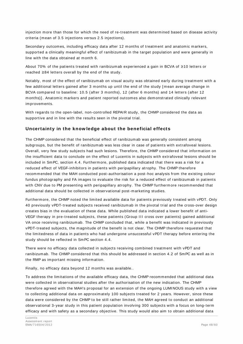
Lucentis Assessment report EMA/716504/2012 Page 48/60
injection more than those for which the need of re-treatment was determined based on disease activity criteria (mean of 3.5 injections versus 2.5 injections).
Secondary outcomes, including efficacy data after 12 months of treatment and anatomic markers, supported a clinically meaningful effect of ranibizumab in the target population and were generally in line with the data obtained at month 6.
About 70% of the patients treated with ranibizumab experienced a gain in BCVA of ≥10 letters or reached ≥84 letters overall by the end of the study.
Notably, most of the effect of ranibizumab on visual acuity was obtained early during treatment with a few additional letters gained after 3 months up until the end of the study [mean average change in BCVA compared to baseline: 10.5 (after 3 months), 12 (after 6 months) and 14 letters (after 12 months)]. Anatomic markers and patient reported outcomes also demonstrated clinically relevant improvements.
With regards to the open-label, non-controlled REPAIR study, the CHMP considered the data as supportive and in line with the results seen in the pivotal trial.
Uncertainty in the knowledge about the beneficial effects
The CHMP considered that the beneficial effect of ranibizumab was generally consistent among subgroups, but the benefit of ranibizumab was less clear in case of patients with extrafoveal lesions. Overall, very few study subjects had such lesions. Therefore, the CHMP considered that information on the insufficient data to conclude on the effect of Lucentis in subjects with extrafoveal lesions should be included in SmPC, section 4.4. Furthermore, published data indicated that there was a risk for a reduced effect of VEGF-inhibitors in patients with peripapillary atrophy. The CHMP therefore recommended that the MAH conducted post-authorisation a post-hoc analysis from the existing colour fundus photography and FA images to evaluate the risk for a reduced effect of ranibizumab in patients with CNV due to PM presenting with peripapillary atrophy. The CHMP furthermore recommended that additional data should be collected in observational post-marketing studies.
Furthermore, the CHMP noted the limited available data for patients previously treated with vPDT. Only 40 previously vPDT-treated subjects received ranibizumab in the pivotal trial and the cross-over design creates bias in the evaluation of these data. While published data indicated a lower benefit of anti-VEGF therapy in pre-treated subjects, these patients (Group III cross over patients) gained additional VA once receiving ranibizumab. The CHMP concluded that, while a benefit was indicated in previously vPDT-treated subjects, the magnitude of the benefit is not clear. The CHMP therefore requested that the limitedness of data in patients who had undergone unsuccessful vPDT therapy before entering the study should be reflected in SmPC section 4.4.
There were no efficacy data collected in subjects receiving combined treatment with vPDT and ranibizumab. The CHMP considered that this should be addressed in section 4.2 of SmPC as well as in the RMP as important missing information.
Finally, no efficacy data beyond 12 months was available..
To address the limitations of the available efficacy data, the CHMP recommended that additional data were collected in observational studies after the authorisation of the new indication. The CHMP therefore agreed with the MAH’s proposal for an extension of the ongoing LUMINOUS study with a view to collecting additional data on approximately 100 subjects treated for 2 years. However, since these data were considered by the CHMP to be still rather limited, the MAH agreed to conduct an additional observational 3-year study in this patient population involving 300 subjects with a focus on long-term efficacy and with safety as a secondary objective. This study would also aim to obtain additional data

Lucentis Assessment report EMA/716504/2012 Page 49/60
in subjects previously treated with vPDT (or other laser treatment), in subjects with extrafoveal lesions and evaluate whether there is a risk of late reactivation of CNV in the long term.
Risks
Unfavourable effects
Overall, few AE were reported and the CHMP considered the safety profile arising from the study data to be reassuring. No new AEs were identified.
Ocular AEs were mainly associated with the injection (conjunctival haemorrhage, punctuate keratitis), and were generally of mild to moderate severity. The incidence of AEs in the treatment groups of the pivotal trial reflected the number of injections and was highest in the group with the most injections. Only vitreous floaters, retinal tear and conjunctivitis allergic were AEs not considered related to the injection. None of the few reported SAEs were considered related to treatment.
The incidence of non-ocular AEs was low and no specific pattern indicating safety risks with the active treatment was revealed. The most frequent non-ocular AEs in ranibizumab-treated patients (both studies) were nasopharyngitis, headache, upper and lower respiratory tract infection, back pain, and hypertension. There were no fatalities reported and no discontinuations from study treatment due AEs.
Safety concerns raised during the assessment in relation to cases of metamorphopsia and the potential IOP increase in patients previously treated with vPDT were addressed in additional analyses and supportive explanations by the MAH and eventually considered by the CHMP to be likely related to the underlying disease (metamorphopsia) and the route of administration (IOP increase) rather than the study drug itself.
Overall, the CHMP considered that all relevant AEs observed in the clinical trials were already adequately addressed in the product information and the RMP.
Uncertainty in the knowledge about the unfavourable effects
The overall size of the safety database (290 subjects being treated with ranibizumab over 12 months) and in particular in relation to previously vPDT-treated subjects was considered by the CHMP to be rather limited. Furthermore, long-term safety data and safety data for the concomitant use of vPDT or other laser treatments and ranibizumab were lacking. However, the safety database from previous development programmes was considered by the CHMP to provide supportive safety information for the treatment with ranibizumab of chronic and progressive conditions.
There were also uncertainties with regards to less frequent AEs, since the size of the safety database did only allow detection of common AEs with sufficient precision. Some concerns were raised with regard to cases of retinal detachment and tear. Subjects with high myopia are predisposed for retinal detachments, and the risk for detachments and tears may increase due to the injection procedure. However, the study data suggested that relatively few injections would be needed, which could counterbalance this possible increased risk.
The CHMP considered it necessary to provide information on the limitations of the safety database in the product information and the RMP. Therefore, SmPC sections 4.2 and 4.4 were updated and important missing information was added to the RMP.
Furthermore, whilst no additional data were considered necessary prior approval of the new indication, the CHMP recommended collection of additional safety data from post-marketing observational studies. Both the extended LUMIOUS study and the additional planned observational study will address safety of ranibizumab in the myopic CNV population.

Lucentis Assessment report EMA/716504/2012 Page 50/60
Benefit-Risk Balance
Importance of favourable and unfavourable effects
The CHMP considered the observed mean gain in visual acuity of more than 10 letters achieved with ranibizumab treatment as highly clinically meaningful. Subjects with CNV secondary to PM generally have a poor prognosis if being untreated. Published studies report that more than 90% of subjects progress to a VA of 20/200 or worse after 10 years. The currently approved treatment option, vPDT, essentially halts the progression of the disease, but in average, no improvement in VA is observed. The effect of ranibizumab seen in the clinical trials in support of this application was therefore considered to present a relevant clinical advantage over vPDT. The effect was achieved rapidly and the fact that the gain in vision was maintained throughout the study duration was considered promising.
No new adverse reaction or other differences in the safety profile compared to previously approved indications arose from the clinical trials supporting the application for the new indication. Most events were considered related to the intravitreal injection procedure and therefore the treatment regimen with the least number of injections was considered most preferable.
Benefit-risk balance
Whilst a clear beneficial effect of ranibizumab was demonstrated in patients with visual impairment due to CNV following pathologic myopia, the safety profile in this condition was considered consistent with the already known profile in the approved indications. All known and suspected safety concerns were considered to be adequately addressed and managed in the product information and the RMP. Furthermore, the treatment regimen with the least number of injections was chosen for the new indication.
Considering that ranibizumab has been shown to stop and partially invert vision loss in the new target population, who untreated is likely to suffer progressive visual impairment, the CHMP considered that the benefits outweighed the treatment risks.
Discussion on the Benefit-Risk Balance
Overall, the CHMP considered that the data presented by the MAH demonstrated a clinically relevant and maintained beneficial effect of ranibizumab in patients with CNV secondary to pathologic myopia.
The effect was present independent of whether the need for re-treatment was determined based on disease stability or activity. However, as patients treated according to disease activity criteria received on average one injection less, the CHMP agreed that this approach was preferable and considered the recommendations for re-treatment as detailed in SMPC section 4.2 as acceptable. A progressive extension of the monitoring frequency with duration of treatment and in absence of disease activity signs was agreed by the CHMP.
Due to the limited available data for vPDT pre-treated patients and patients with extrafoveal lesions, the benefit-risk balance in these subgroups is less clear. However, the results for vPDT pre-treated patient were promising and the information for patients with extrafoveal lesions was too limited to draw firm conclusions.
No data was collected for the concomitant use of vPDT and ranibizumab therapy, nor in relation to patients with peripapillary atrophy.
Finally, although the clinical trials performed in support of this application were considered by the CHMP to be of adequate size and duration, the CHMP noted that data on long-term safety and efficacy

Lucentis Assessment report EMA/716504/2012 Page 51/60
(i.e. beyond one year of treatment) in the new proposed indication was missing. Furthermore, the safety database was limited. However, data collected in previous development programmes for the already approved indications in chronic and progressive conditions were considered by the CHMP to provide supportive evidence of the safety of ranibizumab.
In summary, few limitations were identified concerning small subgroups and long-term effects. However, the CHMP concluded that, in view of the convincing efficacy together with the lack of new safety signals as well as the significant experience with Lucentis at the time of this report, these limitations did not present major concerns and therefore would not prevent approval of the new indication. Nevertheless, the CHMP considered that additional data would help to better characterise the benefit-risk profile in the afore-mentioned patient subgroups and with regards to long-term therapy as well as prior and concomitant vPDT treatment. However, considering that additional information on the limitations of the available data has been included in the product information and that the RMP was updated accordingly, the CHMP was of the opinion that it would be acceptable to collect these data post approval. Furthermore, the post-marketing setting was considered a suitable environment to collect these data as it was anticipated to enable inclusion of a larger number of patients and provide information on the use of ranibizumab in real-life clinical practice.
Therefore, the CHMP considered that the MAH should evaluate the long-term efficacy and safety in subjects with CNV secondary to PM in the LUMINOUS study. The study report for LUMINOUS is due Q4 2017. LUMINOUS is already included as required additional PhV activity in the RMP and the extension of the population to PM patients has been adequately reflected in version 11.2 of the RMP.
The CHMP furthermore recommended that the MAH should perform the following additional studies and analyses:
– An evaluation of the risk for a reduced effect of ranibizumab in patients with CNV due to PM presenting with peripapillary atrophy by end of December 2013.
– Further evaluation of the long-term efficacy and safety in subjects with CNV secondary to PM in a 3-year observational study including at least 300 subjects. Study protocol to be submitted March 2014.
4. Recommendations
The application for an extension of the indication for treatment of visual impairment due to choroidal neovascularisation secondary to pathologic myopia is approvable since other concerns and major objections have all been resolved.
Final Outcome
Based on the review of the submitted data, the CHMP considers the following variation acceptable and therefore recommends the variation to the terms of the Marketing Authorisation, concerning the following change(s):
Variation(s) accepted Type C.I.6.a C.I.6.a - Change(s) to therapeutic indication(s) - Addition
of a new therapeutic indication or modification of an approved one
II
Extension of Indication to include treatment of visual impairment due to choroidal neovascularisation secondary to pathologic myopia for Lucentis.

Lucentis Assessment report EMA/716504/2012 Page 52/60
As a consequence, update of sections 4.1, 4.2, 4.4 , 4.5, 4.8, and 5.1 of the SmPC in order to add information on the limited data available for patients with extrafoveal lesions or pre-treated with vPDT and the lack of experience with concomitant vPDT and ranibizumab treatment as well as to provide a summary of clinical trials data supporting the use in myopic CNV and to supplement all statements in the SmPC valid for all indications with a reference to CNV secondary to PM . The Package Leaflet was updated in accordance.
In addition, changes to the wording of the posology recommendations in the PL were introduced for the existing indications in order to enhance clarity and reduce complexity of the explanation for the patient as well to harmonise the wording used throughout the different approved indications. Minor editorial amendments were made as well. The MAH also took the opportunity to update the details of the local representative in Malta in the Package Leaflet and to introduce the local representative of Croatia.
Furthermore, the PI was brought in line with the latest QRD template version 9.0.
The variation proposed amendments to the SmPC, Annex II, Labelling and Package Leaflet.
Conditions and requirements of the marketing authorisation
The marketing authorisation holder shall submit periodic safety update reports for this product in accordance with the requirements set out in the list of Union reference dates (EURD list) provided for under Article 107c(7) of Directive 2001/83/EC and published on the European medicines web-portal.
Conditions or restrictions with regard to the safe and effective use of the medicinal product
• Risk management plan (RMP)
The MAH shall perform the required pharmacovigilance activities and interventions detailed in the agreed RMP presented in Module 1.8.2 of the Marketing Authorisation and any agreed subsequent updates of the RMP.
An updated RMP should be submitted:
• At request of the European Medicines Agency;
• Whenever the risk management system is modified, especially as the result of new information being received that may lead to a significant change to the benefit/risk profile or as the result of an important (pharmacovigilance or risk minimisation) milestone being reached.
If the submission of a PSUR and the update of a RMP coincide, they can be submitted at the same time.
• Additional risk minimisation measures
Prior to launch in each Member State the MAH shall agree the final educational material with the National Competent Authority.
The MAH shall ensure that, following discussions and agreements with the National Competent Authorities in each Member State where Lucentis is marketed, at launch and after launch all ophthalmological clinics where Lucentis is expected to be used are provided with an up-to-date physician information pack containing the following elements:
• Physician information
• Intravitreal injection procedure video

Lucentis Assessment report EMA/716504/2012 Page 53/60
• Intravitreal injection procedure pictogram
• Patient information packs
The physician information should contain the following key elements:
• The Summary of Product Characteristics
• Sterile techniques, including periocular and ocular disinfection, to minimise risk of infection
• Use of antibiotics
• Use of povidone iodine or equivalent
• Techniques for the intravitreal injection
• Patient monitoring after IVT injection
• Key signs and symptoms of IVT injection related adverse events including increased intraocular pressure, traumatic cataract and endophthalmitis
• Management of IVT injection related adverse events
The patient information pack should be provided in both the form of patient information booklets and an audio-CD that contain following key elements:
• Patient information leaflet
• How to prepare for Lucentis treatment
• What are the steps following treatment with Lucentis
• Key signs and symptoms of serious adverse events including increased intraocular pressure, traumatic cataract and endophthalmitis
• When to seek urgent attention from the health care provider
Additional data exclusivity /market protection
Furthermore, the CHMP reviewed the data submitted by the MAH, taking into account the provisions of Article 14(11) of Regulation (EC) No 726/2004, and considered that the new therapeutic indication brings significant clinical benefit in comparison with existing therapies (see appendix 1).
5. Appendix
1. CHMP assessment report on the significant clinical benefit in comparison with existing therapies in accordance with Article 14(11) of Regulation (EC) No 726/2004, as adopted on 30 May 2013

Lucentis Assessment report EMA/716504/2012 Page 54/60
30 May 2013 EMA/CHMP/317146/2013 Committee for Medicinal Products for Human Use (CHMP)
Assessment report on the significant clinical benefit in comparison with existing therapies in accordance with Article 14(11) of Regulation (EC) No 726/2004
Lucentis
International non-proprietary name: RANIBIZUMAB
Procedure No. EMEA/H/C/000715/II/0034
Note
Annex to variation assessment report as adopted by the CHMP with all information of a commercially confidential nature deleted.

Lucentis Assessment report EMA/716504/2012 Page 55/60
1. Introduction
In accordance with the provisions of Article 14(11) of Regulation (EC) No 726/2004, the Marketing Authorisation Holder (MAH) has applied for an additional one year marketing protection period in the framework of the Lucentis procedure (EMEA/H/C/715/II/34).
The request was based on the MAH’s position that Lucentis represents a significant clinical benefit in the treatment of visual impairment due to choroidal neovascularisation (CNV) secondary to pathologic myopia (PM) in comparison with existing therapies
2. Justification of significant clinical benefit as presented by the MAH
Lucentis has been approved via the centralised procedure in three indications in the European Union/European Economic Area (EU/EEA); namely, in the treatment of neovascular (wet) age-related macular degeneration (AMD), of visual impairment due to diabetic macular oedema (DME), and of visual impairment due to macular oedema secondary to retinal vein occlusion (RVO; branch RVO or central RVO).
The initial marketing authorisation was granted in the EU/EEA for wet AMD in 2007. In line with Article 14(11) of Regulation (EC) No 726/2004, the MAH requested granting of an additional year of marketing protection, provided approval of the new indication in visual impairment due to choroidal neovascularization (CNV) secondary to pathologic myopia (PM), claiming a clinical significant benefit of ranibizumab over existing therapies of this condition.
2.1. Demonstration of new therapeutic indication
Novartis applied for a new indication of ranibizumab in the treatment of visual impairment due to CNV secondary to PM, i.e. the abnormal elongation of the axial length of the eyeball (>26 mm) associated with high myopia refractive errors (usually greater than 6.0 diopters)
In patients with PM, deterioration in vision may occur as a result of rupture and leakage of abnormal vessel formed (CNV) with subsequent damage of the neurosensory retina. CNV in myopic eyes is located between the neurosensory retina and retinal pigment epithelium (RPE) (Type 2), and can be distinguished biomicroscopically from CNV occurring in eyes with AMD, which is usually located in the sub-RPE space (Type 1). Additionally, a typical myopic CNV is less than 1 disc diameter in size, located subfoveal or juxtafoveal, and associated with less subretinal fluid or exudates compared with Type 1 CNV in AMD eyes. During the end stages of natural progression myopic CNV appears as a fibrous pigmented scar (Fuchs’ spot), which causes a blind spot in the centre of the visual field. Therefore, the MAH concluded that CNV in patients with PM is a different pathologic entity compared with CNV in wet AMD.
Visual impairment in diabetic eyes and eyes with RVO are primarily the result of macular oedema, a different pathologic entity than CNV.
Therefore, the MAH considered that visual impairment due to CNV secondary to PM was a new indication for ranibizumab.

Lucentis Assessment report EMA/716504/2012 Page 56/60
2.2. Details of existing therapies
Visudyne® (verteporfin) photodynamic therapy (vPDT) was the only approved medication to treat subfoveal CNV in patients with PM at the time of this application. Treatment with vPDT has been shown to halt vision loss; in the VIP study (Verteporfin in Photodynamic Therapy Study Group-VIP report no.1, 2001 report no.3, 2003), 86% of patients treated with vPDT lost less than 15 letters of best-corrected visual acuity (BCVA) at Month 12 compared with 67% of the patients treated with placebo. At Month 24, the mean change in BCVA showed maintenance of vision, on average, at the baseline levels for patients treated with vPDT and a mean loss of 8 letters in patients treated with placebo.
Evidence of the benefit of vPDT in patients with extrafoveal CNV is limited to non-randomised clinical trials (RCTs) (Pece et al, 2007; Pece et al, 2011), which involved between 20 and 49 eyes of patients observed for a mean of 15 to 35 months. Results indicated that BCVA remained stable or improved with vPDT in most patients; however, mean BCVA at the final assessment did not differ significantly from that at baseline. The percentage of patients with stable or improved BCVA at the final assessment was reported for 2 studies and provided further confirmation that vPDT at least stabilises visual acuity in most patients (Pece et al, 2007, 61%; Hayashi et al. 2008, 86%).
The most common treatment of choice for non-subfoveal CNV lesions is laser photocoagulation, which is associated with permanent loss of vision within the treated area, as well as formation of new abnormal vasculature.
The efficacy of laser photocoagulation to treat non-subfoveal CNV secondary to PM has been assessed in 3 studies involving 27 to 70 eyes (17 to 35 eyes treated with laser) followed for a period of 6 to 48 months (Parodi et al, 2010; Brancato et al, 1988; Fardeau et al, 1992), and in one prospective case series involving 133 eyes that were all treated with laser photocoagulation and followed for 12 to 60 months (Pece et al, 1995). In general, patients treated with laser photocoagulation showed stabilisation of visual acuity, and the majority of patients experienced no change in BCVA over 12 months. Laser photocoagulation treatment led to some safety concerns in the prospective case series. In this study, ocular adverse events (AEs) over the follow-up period of 12 to 60 months included progressive scar enlargement in nearly all eyes (96.0%). This was considered by the MAH of significant concern, as enlargement of the scar could lead to a long-term loss of vision even in eyes that do not experience CNV recurrence.
Similarly, in an RCT of 27 eyes treated with 1 of 3 different laser wavelengths during photocoagulation, the majority of patients reported stable or improved BCVA over the mean 7.2-month follow-up period, and only 19% of patients had actual improvements in their BCVA scores (Brancato et al, 1988). In another study, an RCT comparing laser therapy with no treatment, a final BCVA of 20/100 or worse at the end of the 6- to 48-month follow-up was reported for a smaller proportion of patients who received laser therapy compared with those who received no treatment (46% vs 89%) (Fardeau et al, 1992).
Another study compared laser therapy with off-label intravitreal bevacizumab and vPDT (Parodi et al, 2010). A non-significant mean loss of 5.5 letters from baseline was observed for laser therapy after 24 months of follow-up and 41% of patients achieved stabilisation or improvement in BCVA at this time point.
2.3. Significant clinical benefit based on improved efficacy
The MAH claimed a significant benefit of ranibizumab based on efficacy and justified this claim as follows:
Existing therapies have been shown to maintain levels of visual acuity without significant visual gain.

Lucentis Assessment report EMA/716504/2012 Page 57/60
Available published evidence, as well as the results from the pivotal study RFB002F2301 supporting the application for the new indication of ranibizumab, provided data on a new magnitude of the treatment effect. The treatment benefit of ranibizumab in visually impaired patients due to myopic CNV has been shown with a clinically relevant gain in visual acuity of more than 10 letters on average. Therefore, treatment with ranibizumab provides a significant clinical benefit over existing therapies in patients with visual impairment due to CNV secondary to PM.
3. Assessment of the MAH’s justification of significant clinical benefit1
3.1. Demonstration of new therapeutic indication
Although CNV occurs in eyes with neovascular age related macular degeneration (AMD), CNV in the new proposed indication occurred secondary to PM. The abnormal elongation of the axial length of the eyeball can lead to changes of the posterior pole of the eye such as posterior staphyloma, atrophy of the retinal pigment epithelium (RPE), Bruch’s membrane cracks, subretinal haemorrhage and retinal detachment. This in turn may result in CNV, the most vision threatening complication in patients with PM.
The CHMP considered the justification of the MAH for a new indication to be acceptable and agreed that the proposed use of ranibizumab in patients with visual impairment following myopic CNS was distinct from the previously approved indications for Lucentis.
3.2. Details of existing therapies
The CHMP agreed that the only existing pharmacological therapy for CNV secondary to PM is Visudyne (verteporfin) plus photodynamic therapy (vPDT). Verteporfin is a photosensitiser administered intravenously which after exposure to visible light causes the in situ generation of cytotoxic reactive oxygen species.
During the clinical development (VIP study, see chapter 2.2. ), on average, vision was stabilised in the Visudyne treatment group while the placebo group lost a mean of 8 letters, i.e. there was no mean gain in visual acuity achieved with vPDT. This outcome, i.e. that vision is mainly stabilised and no relevant improvement is to be expected, has been confirmed in two additional published studies.
In addition to Visudyne, the CHMP noted that laser photocoagulation is used to treat non-subfoveal CNV lesions in this disease. As demonstrated in a number of rather small, published studies, vision was mainly stabilised and there is a concern regarding long-term progressive scar enlargements and loss of vision after treatment.
3.3. Significant clinical benefit based on improved efficacy
The pivotal study (RFB002F2301) in Support of the application for the new indication included subjects with active CNV secondary to PM with greater than -6 diopters of spherical equivalence and anterio-posterior elongation measurement greater than or equal to 26 mm and with at least one of the following lesion types in the study eye: subfoveal, juxtafoveal, extrafoveal, margin of the optic disc. This was a randomised, double-masked, multi-centre, active-controlled study to evaluate the efficacy and safety of two different dosing regimens of 0.5 mg ranibizumab (Group I and II) versus vPDT
1 In accordance with guidance on elements required to support the significant clinical benefit in comparison with existing therapies of a new therapeutic indication in order to benefit from an extended (11-year) marketing protection period http://ec.europa.eu/health/files/eudralex/vol-2/c/guideline_14-11-2007_en.pdf

Lucentis Assessment report EMA/716504/2012 Page 58/60
(Group III) over 12 months. Patients in Group III were allowed to cross over to ranibizumab treatment after Month 3.
The primary objective was to demonstrate the superiority (efficacy) of 0.5 mg ranibizumab driven by stabilisation criteria and/or by disease activity re-treatment criteria vs vPDT as assessed by best corrected visual acuity (BCVA).
The primary efficacy endpoint was the difference between the average level of BCVA (letters) over all monthly post-baseline assessments from Month 1 to Month 3 (endpoint) and the baseline level of BCVA is summarised below.
Ranibizumab 0.5 mg Visudyne PDT Parameter Statistic Group I by
stabilization N=105
Group II by disease activity
N=116
Group III N=55
Baseline Mean (SD) 55.4 (13.43) 55.8 (12.59) 54.7 (13.84) Average change from baseline Month 1 to Month 3 Mean (SD) 10.5 (8.16) 10.6 (7.26) 2.2 (9.47) Comparison vs vPDT Difference in LS means (1) 8.5 8.6 95% CI for difference (1) (5.8,11.2) (6.1,11.1) p-value <0.00001 <0.00001
A rapid VA improvement was observed in the ranibizumab treatment groups (Groups I and II) and most of the effect was achieved by Month 2. Thereafter VA was essentially maintained or slightly increased during the course of the study up to Month 12. For patients randomised to vPDT an increase in VA from Month 3 onwards was shown (see below Figure). However, Group III included 38 patients (plus 2 patients who received ranibizumab prior to Month 3 due to a protocol deviation) who received ranibizumab after Month 3.

Lucentis Assessment report EMA/716504/2012 Page 59/60
Figure Visual acuity of the study eye (letters): Mean change from baseline over time (FAS; modified LOCF)
Additional visual acuity-based efficacy outcome measures are summarised below.
Ranibizumab 0.5 mg Visudyne PDT (Ranibizumab allowed
as of Month 3) Parameter Statistic Group I by
stabilization N=105
Group II by disease activity N=116
Group III N=55 (38/55 patients received
ranibizumab as of Month 3)
Categorised change from baseline at Month 3 Gain ≥ 10
letters* 65 (61.9) 76 (65.5) 15 (27.3)
p-value Rzb vs vPDT (CMH stratified)
<0.00001 <0.00001
Gain ≥ 15 letters*
40 (38.1) 50 (43.1) 8 (14.5)
Loss ≥ 15 letters
2 (1.9) 0 4 (7.3)
p-value Rzb vs vPDT (CMH stratified)
0.00146 0.00001
Categorised change from baseline at Month 6 Gain ≥ 10
letters* 75 (71.4) 75 (64.7) 25 (45.5)
Gain ≥ 15 letters*
49 (46.7) 52 (44.8) 15 (27.3)
Loss ≥ 15 letters
0 1 (0.9) 2 (3.6)
Categorised change from baseline at Month 12 Gain ≥ 10
letters* 73 (69.5) 80 (69.0) 27 (49.1)
Gain ≥ 15 56 (53.3) 60 (51.7) 18 (32.7)

Lucentis Assessment report EMA/716504/2012 Page 60/60
Ranibizumab 0.5 mg Visudyne PDT (Ranibizumab allowed
as of Month 3) letters*
Loss ≥ 15 letters
2 (1.9) 1 (0.9) 2 (3.6)
*gained ≥10/15 letters or reached 84 letters in BCVA
In the pivotal trial, a direct comparison with Visudyne was performed after three months of treatment. During this time period ranibizumab-treated subjects gained on average 10-11 letters versus mean 2 letters in the vPDT group. In addition, approximately 40% of ranibizumab-treated subjects gained ≥15 letters of visual acuity. The mean gain was maintained or slightly improved throughout the 12 months duration of the study. Likewise, the proportion of subjects that gained ≥10 or ≥15 letters increased further in the course of the study. Very few subjects treated with ranibizumab lost ≥15 letters during the study.
The study demonstrated superiority of both ranibizumab treatment regimens compared to vPDT at Month 3 based on a significantly higher gain in visual acuity observed with ranibizumab at Month 3. At month 12, the mean gain in BCVA achieved with ranibizumab was 14 letters, which was considered highly clinically relevant by the MAH
In an indirect comparison with the data from the VIP-study, which showed no mean increase in visual acuity during the first 12 months of the study, the effect of ranibizumab demonstrated in the study supporting the application for the new indication was considered highly relevant by the CHMP.
Taken together, the CHMP concluded that a significant clinical benefit of Lucentis over the previously approved therapy, vPDT, has been demonstrated.
4. Conclusion
The CHMP agreed that the proposed use of ranibizumab in patients with visual impairment following myopic CNV was distinct from the previously approved indications for Lucentis.
The CHMP furthermore considered that the data submitted in support of the application as well as the justification for the extension of the marketing protection period demonstrated a clinically significant benefit of ranibizumab in terms of a clinically meaningful increase in visual acuity in visually impaired patients with myopic CNV. No such effect has been shown for vPDT or laser photocoagulation therapy in this condition. The CHMP therefore considered that the treatment with Lucentis in this new therapeutic indication brings significant clinical benefit in comparison with existing therapies.
5. Recommendation
The CHMP reviewed the data submitted by the MAH, taking into account the provisions of Article 14(11) of Regulation (EC) No. 726/2004 and the “Guidance on elements required to support the significant clinical benefit in comparison with existing therapies of a new therapeutic indication in order to benefit from an extended (11-year) marketing protection period”, and considers by consensus that the new therapeutic indication brings a significant clinical benefit in comparison with existing therapies.