assessment questions
TRANSCRIPT

Assessment QuestionsAssessment Questions
Raymond G Buick FRCS
The Axilla• The anterior wall of the axilla is formed by the pectoralis major
and minor muscles and the pectoral and clavipectoral fascia.
• The posterior wall by the scapula and subscapularis muscle.
• Inferiorly by teres major and latissimus dorsi
• The medial wall is the thoracic cage and the serratus anterior
• The lateral wall is the intertubercular sulcus of the humerus.
the long thoracic nerve
• The first branch of the posterior aspect of the anterior rami of C5 C6 (and C7) forms the long thoracic nerve, and supplies serratus anterior

infrahyoid muscle group
• The omohyoid, thyrohyoid and the sternohyoid muscles are part of the infrahyoid muscle group.
• The mylohyoid muscle is a suprahyoid muscle attached to the hyoid and a mylohyoid raphe.
• It elevates the hyoid bone, the floor of the mouth and the tongue, during swallowing and speaking
the brain stem
• The brain stem lies in the anterior part of the posterior cranial fossa in front of the cerebellum
• The basilar artery ascends in front of it
• The temopral lobes of the cerebral hemispheres occupy the middle cranial fossa

The radial nerve• The radial nerve is vunerable in a mid-shaft fracture of the humerus.
• The radial nerve runs with the profunda brachii artery, in the radial groove
• It passes between the lateral and medial heads of the triceps brachii muscle, before perforating the lateral intermuscular septum, to enter the cubital fossa
• Damage to the radial nerve, may affect the muscles in the posterior compartment of the upper arm and forearm, skin of the posterior and inferolateral arm and posterior aspect of forearm and hand, to axial line of digit 4.
muscles of mastication• The masseter and temporalis muscles are supplied by the anterior trunk of
the mandibular branch of the trigeminal nerve (V3)
• Temporalis muscle is supplied by deep temporal nerves
• The masseter muscle is supplied by the masseteric nerve.
• V3 supplies branches to the 4 muscles of mastication: temporalis, masseter and medial and lateral pterigoids.
• It does not supply the buccinator muscle which is supplied by the facial nerve (VII).
orbicularis oris
• The contraction of the orbicularis oris muscle closes the mouth, the eye is closed by orbicularis oculi.

deep cervical fascia
• the deep cervical fascia has 3 fascial layers – the investing, pretracheal and prevertebral layers
• the sternocleidomastoid muscle is surrounded (you cannot be ‘surrounded’ on one side) by the investing layer of the deep cervical fascia
The flexor retinaculum
• The flexor retinaculum is a specialised form of the deep fascia, at the wrist.
• It is formed by the deep layers of the antibrachial fascia.
There are two tutorials on the carpal There are two tutorials on the carpal tunnel syndrome on meducation premiumtunnel syndrome on meducation premium

The clavicle
• The acromion of the scapula articulates laterally with the clavicle, at the acromioclavicular joint.
• The clavicle articulates medially with the manubrium sterni
• The coracoid process does not have a bony articulation.
• The short head of the biceps brachii is attached to it.
Colles’ fracture
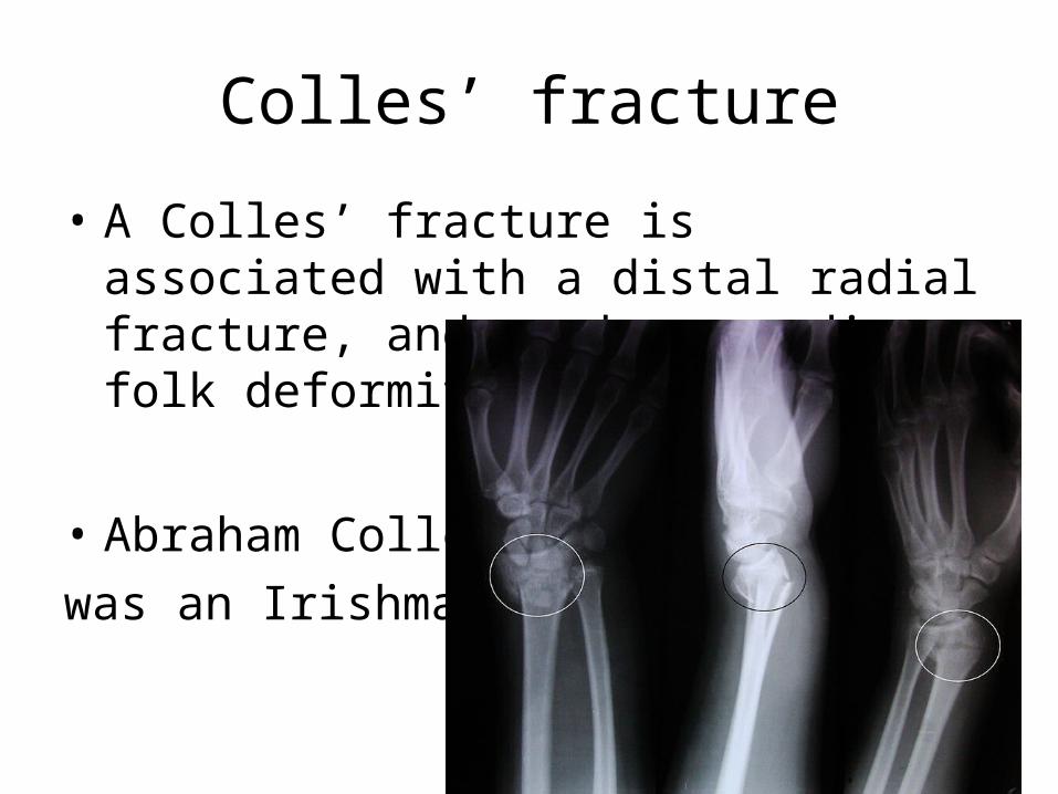
• A Colles’ fracture is associated with a distal radial fracture, and produces a dinner folk deformity.
• Abraham Colles was an Irishman!
The median nerve
• The median nerve, passes through the carpal tunnel with 9 tendons.
• they are flexor digitorum profundus x 4, flexor digitorum superficialis x4, flexor pollicis longus.
• The tendon of palmaris longus passes over, superficial to, the flexor retinaculum, to join the palmer aponeurosis

the cubital fossa
• The boundaries of the cubital fossa include the pronator teres muscle on the ulnar side, brachioradialis muscle on the radial side.
• The superior boundry is an imaginary line between the epicondyles of the humerus.
• The floor is formed by brachialis and supinator muscles.
• The roof is the brachial fascia and bisceps brachii aponurosis.
the foramen rotundum
• The nerve which passes through the foramen rotundum is the maxillary branch of the trigeminal nerve V2
• It carries sensation from the skin of the face, including the upper lip, maxillary teeth, mucosa of nose, maxillary sinuses and palate
• The 4 muscles of mastication are supplied by V3, and the facial nerve supplies the buccinator muscle.
A typical cervical vertebra
• A typical cervical vertebra (C3-C5) has a short bifid spinous process.
• C1 – C6 have a large vertebral foramen, through which runs the vertebral artery accompanied by venous and sympathetic plexuses.
• The vertebral artery is a branch of the subclavian artery which enters the skull through the foramen magnum and they join in front of the brain stem to form the basilar artery.
End
• no time to finish these tonight
• If sufficient interest I will complete them later in the week

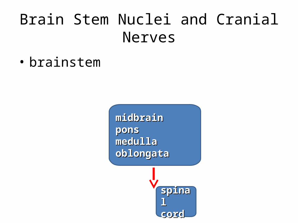
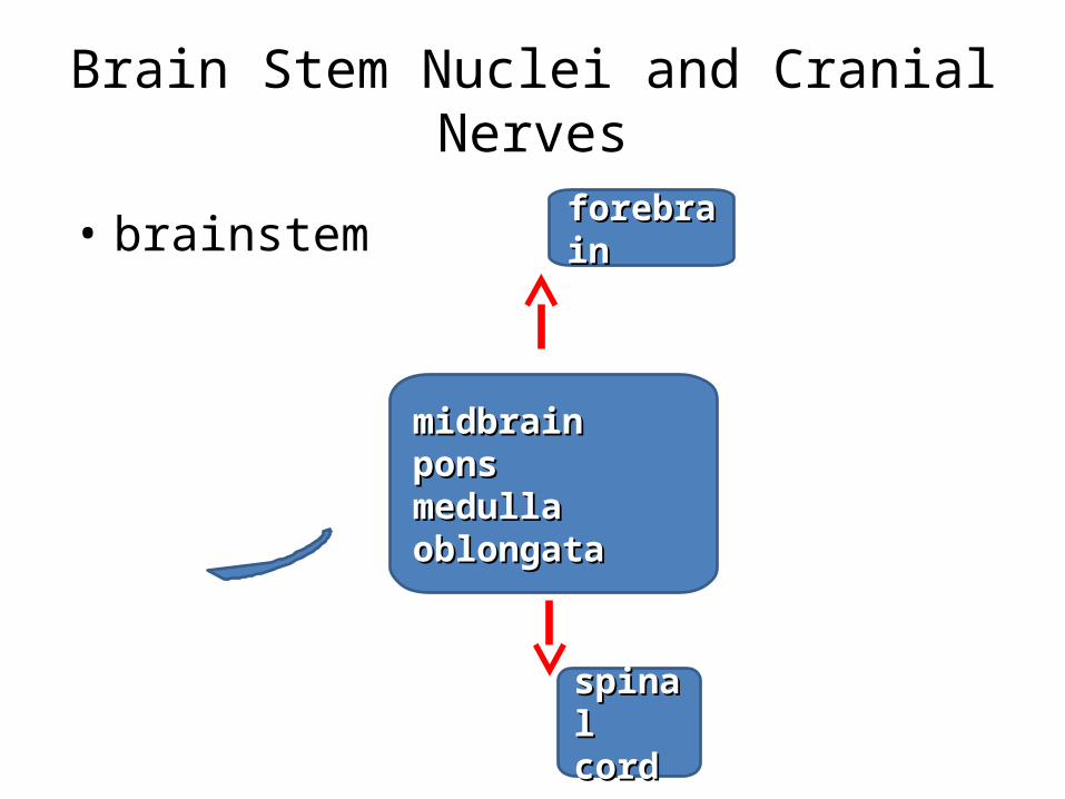
Brain Stem Nuclei and Cranial Nerves
• brainstem
midbrainmidbrainponsponsmedulla oblongatamedulla oblongata
spinal spinal cordcord
Brain Stem Nuclei and Cranial Nerves
• brainstem
midbrainmidbrainponsponsmedulla oblongatamedulla oblongata
spinal spinal cordcord
Brain Stem Nuclei and Cranial Nerves
• brainstem
midbrainmidbrainponsponsmedulla oblongatamedulla oblongata
spinal spinal cordcord
forebrainforebrain