assessment of the outcome of lower limb amputations as
TRANSCRIPT

i
ASSESSMENT OF THE OUTCOME OF LOWER LIMB AMPUTATIONS AS SEEN
IN KENYATTA NATIONAL HOSPITAL.
A ONE YEAR PROSPECTIVE STUDY.
A DISSERTATION SUBMITTED IN PART FULFILMENT FOR THE REQUIREMENTS OF THE DEGREE OF MASTER OF MEDICINE IN SURGERY, UNIVERSITY OF NAIROBI.
M E D I C A L LIBRARY W m V P R S l T Y O P N A I R O B I
By DR. KIRSTEEN ONDIKO AWORI
M.B.,Ch.B. (Nairobi]
2004.
University of NAIROBI UDrary
0390061 0

II
DECLARATION.
This dissertation is my original work, and has not been presented for a degree in any other University.
l^rH M) r tmG C-fU Date: day of , 2004
DR KIRSTEEN ONDIKO AWORI
(M.B.,Ch.B. (NAIROBI)
CANDIDATE.
This dissertation has been submitted for examination with my approval as the University supervisor.
A? S i g n e c ^ - — ~ ~ / Date: ^ day o f ^ ^ ' , 2004. PROFESSOR J O H N E.O. ATING'A, M/B, Ch.B., M.Med (SURGERY) (Nairobi), MCH, ORTHO (Liverpool). CONSULTANT ORTHOPAEDIC SURGEON, KENYATTA NATIONAL
HOSPITAL AND ASSOCIATE PROFESSOR. DEPARTMENT OF ORTHOPAEDIC SURGERY,
FACULTY OF MEDICINE, UNIVERSITY OF NAIROBI,
P.O. BOX 30197-00100 GPO, NAIROBI, KENYA.

iii
DEDICATION
To my wife Victoria.
i

iv
ACKNOWLEDGEMENT
The completion of this study would not have been possible without the contribution
of my supervisor, Professor John E Ating'a for guidance and corrections in the
preparation of this document, Professor Stephen O Ogendo, Consultant Thoracic and
Cardiovascular surgeon, Kenyatta National Hospital and Associate professor, Department
of Surgery, University of Nairobi for accepting to read and correct the study proposal,
and offering suggestions that contributed significantly to the success of this study. Mr.
Hassan Saidi, General surgeon and senior lecturer, Department of Human anatomy,
University of Nairobi, and Dr Kiptoon for their help in the data management and finally
relatives of the amputees, who agreed and sacrificed to bring them and even wait for me
for hours, to enable a successful follow up.

V
TABLE OF CONTENTS
1. TOPIC (i)
2. DECLARATION (ii)
3. DEDICATION (iii)
4. ACKNOWLEDGEMENT (iv)
5. TABLE OF CONTENTS (v)
6. LIST OF TABLES (vi)
7. LIST OF FIGURES (vii)
8. LIST OF ABBREVIATIONS (viii)
9. SUMMARY 1
10. INTRODUCTION 2
11. STUDY JUSTIFICATION 26
12. STUDY OBJECTIVES 27
13. MATERIALS AND METHODS 28
14. RESULTS 32
15. DISCUSSION 52
16. CONCLUSIONS 56
17. RECOMMENDATIONS 57
18. REFERENCES 58
19. APPENDICES 64
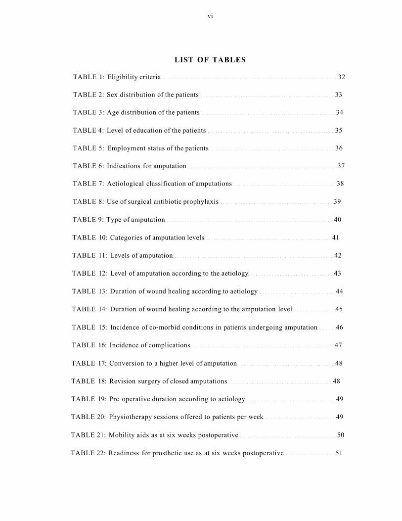
vi
LIST OF TABLES TABLE 1: Eligibility criteria 32 TABLE 2: Sex distribution of the patients 33 TABLE 3: Age distribution of the patients 34
TABLE 4: Level of education of the patients 35
TABLE 5: Employment status of the patients 36 TABLE 6: Indications for amputation 37 TABLE 7: Aetiological classification of amputations 38
TABLE 8: Use of surgical antibiotic prophylaxis 39
TABLE 9: Type of amputation 40
TABLE 10: Categories of amputation levels 41
TABLE 11: Levels of amputation 42
TABLE 12: Level of amputation according to the aetiology 43
TABLE 13: Duration of wound healing according to aetiology 44
TABLE 14: Duration of wound healing according to the amputation level 45
TABLE 15: Incidence of co-morbid conditions in patients undergoing amputation 46
TABLE 16: Incidence of complications 47
TABLE 17: Conversion to a higher level of amputation 48
TABLE 18: Revision surgery of closed amputations 48
TABLE 19: Pre-operative duration according to aetiology 49
TABLE 20: Physiotherapy sessions offered to patients per week 49
TABLE 21: Mobility aids as at six weeks postoperative 50
TABLE 22: Readiness for prosthetic use as at six weeks postoperative 51

vii
LIST OF FIGURES
FIGURE 1: Sex distribution of the patients 33
FIGURE 2: Age distribution of the patients 34
FIGURE 3: Level of education of the patients 35
FIGURE 4: Employment status of the patients 36
FIGURE 5: Indications for amputation 37
FIGURE 6: Aetiological classification of amputations 38
FIGURE 7: Use of surgical antibiotic prophylaxis 39
FIGURE 8: Type of amputation 40
FIGURE 9: Categories of amputation levels 41
FIGURE 10: Levels of amputation 42
FIGURE 11: Level of amputation according to the aetiology 43
FIGURE 12: Pattern of wound healing according to the aetiology ...44
FIGURE 13: Pattern of wound healing according to the amputation level 45
FIGURE 14: Incidence of co-morbid conditions in patients undergoing amputation 46
FIGURE 15: Incidence of complications 47
FIGURE 16: Pre-operative duration according to aetiology 49
FIGURE 17: Mobility aids as at six weeks postoperative 50

Vlll
LIST OF ABBREVIATIONS
AK Above knee AKA Above-the-knee amputation BK Below knee
BKA Below-the-knee amputation
DVT Deep vein thrombosis
ELISA Enzyme-linked immunosorbent assay
EWA Early Walking Aid(s)
FBC Full blood count
HBsAg Hepatitis B surface antigen
HIV Human immunodeficiency virus
KNH Kenyatta National Hospital
LLA Lower limb amputation(s)
PEM Protein Energy Malnutrition
PVD Peripheral vascular disease
TLC Total lymphocyte count
UEC Urea electrolytes and creatinine
1
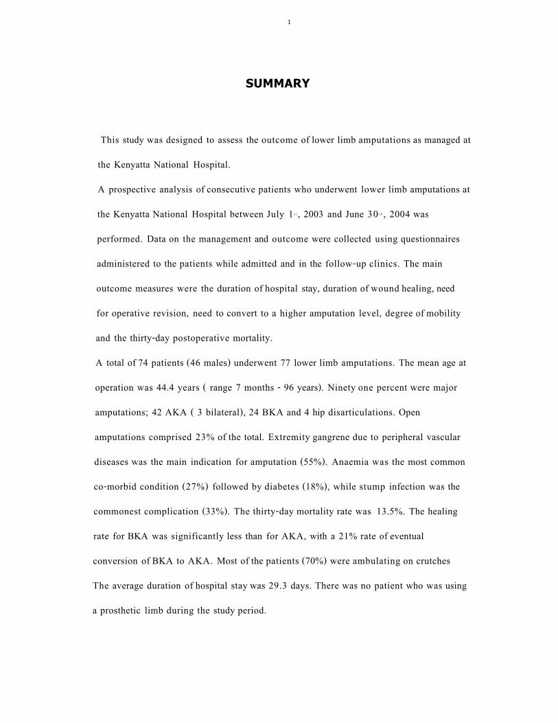
SUMMARY
This study was designed to assess the outcome of lower limb amputations as managed at
the Kenyatta National Hospital.
A prospective analysis of consecutive patients who underwent lower limb amputations at
the Kenyatta National Hospital between July 1 s t , 2003 and June 30 t h , 2004 was
performed. Data on the management and outcome were collected using questionnaires
administered to the patients while admitted and in the follow-up clinics. The main
outcome measures were the duration of hospital stay, duration of wound healing, need
for operative revision, need to convert to a higher amputation level, degree of mobility
and the thirty-day postoperative mortality.
A total of 74 patients (46 males) underwent 77 lower limb amputations. The mean age at
operation was 44.4 years ( range 7 months - 96 years). Ninety one percent were major
amputations; 42 AKA ( 3 bilateral), 24 BKA and 4 hip disarticulations. Open
amputations comprised 23% of the total. Extremity gangrene due to peripheral vascular
diseases was the main indication for amputation (55%). Anaemia was the most common
co-morbid condition (27%) followed by diabetes (18%), while stump infection was the
commonest complication (33%). The thirty-day mortality rate was 13.5%. The healing
rate for BKA was significantly less than for AKA, with a 21% rate of eventual
conversion of BKA to AKA. Most of the patients (70%) were ambulating on crutches
The average duration of hospital stay was 29.3 days. There was no patient who was using
a prosthetic limb during the study period.

2
INTRODUCTION
Amputation is the removal of an extremity in whole or in part, while disarticulation is amputation through a joint. 1
HISTORICAL BACKGROUND
Amputation is one of the most ancient of all surgical procedures. It was also a common
punishment in some societies on religions grounds.
Early in the 16 l h century, amputation surgery and prosthetics were much improved by
Ambroise Pare. Pare created more functional stumps and was the first to use ligatures to
control bleeding after amputation. Morel introduced the tourniquet, and with the
development of anaesthesia and aseptic technique, surgeons could carefully fashion
sturdy and functional amputation stumps and reasonably anticipate healing without
infection. 2 5 Without true anaesthesia, it was not possible to perform other than the
Guillotine-type amputations. It was Younge and Loudham's addition of a shot flap in
1679, which made closure easier. Today, Guillotine-type amputations are performed on
grossly contaminated or infected limbs prior to definitive flap amputation, with control of
the sepsis. 1 3

3
EPIDEMIOLOGY
Incidence and prevalence.
Rates of lower extremity amputation vary significantly both between and within countries
due to in part by variations in clinical decision making.4 There are no statistics on
incidence and prevalence of amputations in Kenya. 5 ' 6 Denmark has a National registry
with more accurate data on amputations. The overall annual incidence quoted is 25.5 per
100,000 population. Published estimates in the United States of America put their annual
incidence at 20,000 to 30,000 and prevalence at 350,000 to 1 million. 7 A study
comparing the epidemiology of lower extremity amputations in centres in Europe, North
America and Asia by The Global Lower Extremity Amputation Study Group found
the highest incidence rate at the Najavo area (U.S.A.) at 43.9 per 100 000 population per
year, while Madrid, Spain had the lowest rate at 2.8 per 100 000 per year, for first major
amputation in men. 8
Age and sex incidence
Mbindyo 5 in a seven-year retrospective study between 1970 and 1976 on lower
limb amputations in KNH, found two peaks in age incidence; a teenage peak mainly due
to trauma and osteogenic sarcoma and another one at 50 years and above, due to vascular
diseases. The Global Lower Extremity Amputation Study Group, found age and sex
distribution to be similar, despite great variation in the overall rates in the centres
the study was carried out. The incidence of both major and minor amputations in both
men and women rose steeply with age; the largest increases being between 40-59
and 60-79 years. Overall amputation rates were higher in men than in women except in
one centre where crude rates were higher in women than men.

4
INDICATIONS
European and American literature site peripheral vascular insufficiency as the main indication for lower limb amputations. 1" 4 , 9 Mbindyo 3 in a study of LLA at the KNH however, found neoplasms followed by trauma as the main indications. Similar results, were reported by Hassan 1 0 in a retrospective study conducted at the Maiduguri Teaching
Hospital in Nigeria. The pattern seen in studies conducted in the African setting has been attributed to late presentation of patients with malignancies partly due to poverty, stigmatisation of amputees and the eventual inability to afford a prosthesis, therefore making limb-preserving surgery impossible. Over the years however, there has been an increase in peripheral vascular diseases with or without diabetes in our setting , which is likely to have altered the above pa t te rn . 6 ' 1 1 1 4
1. Trauma 2 j i
Injuries related to road traffic accidents (RTA) are the leading causes of LLA.
Other traumatic causes include, industrial and farm accidents, assaults and burns.
The injuries to extremities that necessitate amputation are usually associated with
complete damage of the main blood and /or nerve supply of the extremity. When
discussing massive lower extremity trauma, the prototype injury is the severe open
tibial fractures.1 ,13 Gustillo et al 16 sub classified these type (iii) open fractures to
better differentiate on basis of prognosis the outcome of these injuries. Type (iii)c
fractures are those associated with arterial injury requiring repair.

5
Caudle et al 17 found that long-term results were poor with type (iii) c fractures of the tibia, and that most limbs (78%) eventually required amputation. Lange 15 has shown that in extreme cases, the criteria of prolonged ischaemia (of more than six hours) and /or absent posterior tibial nerve function, are absolute indications of LLA. He however, found out that majority of patients do not fit the absolute criteria yet a decision to salvage or amputate the limb has to be made. He therefore, came up with a categorised system of patient variables like age and occupation, extremity ones like location of vascular injury and the associated variables like warm ischaemia time, to be used in decision making. Various authors have come up with a number of scoring systems to help determine which limbs can be salvaged and the ones to be amputated. 1* 2 2 Dirschl et a l 2 3 reviewed these systems and while they appreciated that all the systems have short-comings, and that none has been validated in studies involving large numbers of patients, they recommend their use, preferably the mangled extremity scoring system (MESS), as guides to the
treatment of mangled lower extremities, but cautioned substituting these systems for the treating physician's clinical judgment and experience. In making the decision to salvage or to amputate a limb, the surgeon must also give strong consideration to the functional outcome and the cost of limb salvage attempts, as compared with amputation. 2 ' Few
published studies however, have directly compared the functional outcome and the hospital cost of early amputation and limb salvage in the lower extremity. 1 5 26

6
Bondurant et a l 2 4 showed a definite cost saving if the decision to amputate can be made early in the treatment of major extremity trauma. Hansen 27 came up with similar conclusions and goes on to state that old patients, patients with multiple injuries, patients who have been in shock and those suffering from diabetes or other vascular diseases who generally have a smaller chance of survival and successful reconstruction of a badly damaged limb, should preferably be amputated. Williams 2 ! i on the other hand looks at the long-term costs of salvage versus amputation and considers the cost of modem prosthetic devices to make amputation in some patients a more expensive treatment option during their lifespan versus the Ilizarov technique. He however, finds this technique to be very labour intense and to increase total treatment time.
(2) Neoplasms
Benign tumours rarely require amputation, but occasionally one is so large or is of such a
nature that excising it locally would result into a functionless limb. For malignant
tumours, amputation is done in the absence of metastatic spread or sometimes indicated
even after metastatic spread has occurred to relieve pain or as a toilet procedure. 2 ' 3 5
Studies performed in Africa, have found advanced skin cancers, especially squamous cell
carcinoma as the leading cause of lower limb amputations followed by osteosarcoma of
the femur and tibia. 5 , 1 0 This pattern has been attributed to late presentation of patients
suffering from skin cancers.

7
Up to the recent past, amputation has been the most common surgical treatment for osteosarcoma of an extremity. 1 1 , 2 9 In most anatomical sites, the amputation is done through the bone or joint that lies proximal to the bone in which the tumour is developing. However, when the tumour is in the distal part of femur, some authors have recommended a trans-medullary amputation with neo-adjuvant and/or adjuvant chemotherapy. 1 1 , 2 9" 3 1 Limb salvage resection has recently become more popular than when it was first developed, and it is often chosen by patients who have
osteosarcoma. 1 2 ' 1 4 , 3 2 Enneking et al 13 came up with a system of staging musculoskeletal sarcoma. Based on this system, different varieties of tumour excision techniques have been described, from radical excision (disarticulation at proximal joint above the affected bone) wide and marginal excisions, to intralesional amputation. Although all authors agree that the primary tumour should be removed, there is controversy regarding the appropriate surgical margin and the indication for, or against limb salvage resection as compared with amputation. 2 9 3 1 , 3 3 Some authors have recommended a wide en bloc surgery with neoadjuvant or adjuvant chemotherapy for high-grade malignant sarcomas among the several forms of treatment. 3 3 , 3 4 Lane et a l 3 4 have found that while limb-salvage procedures that use endoprosthestic implants are being performed more frequently, even in the best of circumstances, anywhere from 15-25% of patients with osteosarcoma still require amputation to achieve disease-free surgical margin.
0 P

8
3. Ischaemia
This is usually related to arteriosclerotic disease with or without diabetes. Other causes include, thromboembolic disease and in our setting, trauma cases with delayed presentation resulting in compartment syndrome.5 It should be remembered that both arteriosclerosis and diabetes mellitus are systemic diseases that tend to occur in the elderly; making it of necessity to evaluate the status of the heart, kidneys and the cerebral circulation in this category of patients before surgery. Amputation is indicated in these patients either because of failed salvage vascular surgery or due to late presentation. Majority, present with rest pain, neuropathy, sepsis or gangrene of an extremity. These patients are a challenge to the surgeon because primary wound healing is a the main objective. However, due to lack of oxygen and nutrients delivered to tissues and therefore poor neo-vascularisation in granulation tissue, the wound heals slowly and also, there is reduced resistance against infection. Co-existent poor cardiac function and respiratory disease and anaemia or limb infection makes the situation worse. 1
4) Infection
Infection necessitating amputation may be acute or chronic, and must have been
unresponsive to medical or other surgical measures. Fulminating gas gangrene is the most
dangerous. Hyperbaric oxygen therapy may eliminate need for amputation or make
amputation level more distal in this condition. In chronic infections, amputation is
indicated because function has been so impaired by chronic osteomyelitis or an infected
non-united fracture, that amputation and prosthetic fitting will improve function and
allow more normal activities. 2 ' 3

9
(5) Congenital
Surgical excision of part or all of a congenitally deficient limb may be indicated in
infancy or early childhood, as a primary conversion for those conditions whose natural
history is so well known that the surgeon can predict better prosthetic use and overall
function after surgery; like in complete fibular hemimelia or tibia hemimelia.
In other conditions such as proximal femoral focal deficiency, surgery may be best
performed as a secondary conversion after a period of prosthetic fitting around the
deformity has proved that prosthetic use and overall function will be better after
surgery. Almost 50% of congenital lower limb deficiences will require either primary or
secondary conversion. 2

10
CHOICE OF AMPUTATION LEVEL
The most important requirement for successful amputation is skin healing, preferably by first intention. A major surgical effort should be directed to creating a 'stump' that is able to tolerate total contact and partial end-bearing. The ideal levels in the lower limb are those that allow an element of weight bearing. The decision about the level and type of amputation should not be taken during operation, since the degree of bleeding noted on incision of tissues at operation is a very poor guide to subsequent healing, being greatly affected by anaesthetic agents and transient hypotension. 3 ' 3 5
Knee preservation affords greater mobility after rehabilitation.3 5
Considering the increased energy consumption required to use a prosthesis, pre-existing
medical conditions such as severe respiratory or cardiac disease, neurological problems,
the presence of previous amputation (which now makes a patient a bilateral amputee),
and the ability of the tissues to heal adequately enough to allow weight bearing, may
make prosthetic fitting an unrealistic goal. Therefore, all the factors influencing outcome
including physical and mental capacity of the patient to withstand re-amputation, and
whether the patient is likely to make use of the increased mobility that a joint-saving
amputation may afford should be considered in the final decision. 3 , 3 6
11
FACTORS IN LEVEL SELECTION 1. Prosthetic fitting
In the past, the case of fitting the limb for a prosthetic was a major factor. However, with the steady advance in material and design of prosthetics this is less so. It is also important to note that not all amputees are prosthetic candidates. The so-called 'ideal' stump, which rarely exists, consists of adequate length, sufficient strength, adequate proprioception, and full joint range of motion, sufficient nerve supply, intact circulation and sufficient skin sensation. 3
Enough stump length allows for adequate leverage of the stump within the socket. It also ensures a more intimate socket fit during ambulation and sitting because the stump will maintain its socket position. Very short stumps have a tendency, particularly during sitting, to partially slip out of the socket, which then causes stump replacement problems when the patient stands to walk. The stump must have sufficient muscle strength within it to enable the patient achieve a stable and efficient gait through controlled forward motion of the socket and the ability to carry the weight of the prosthesis. 3
2. The indication for amputation.
Amputations for malignancies in most anatomical sites, are done through the bone or
joint that lies proximal to the bone or compartment in which the tumour is developing.
For trauma however, you aim generally at a site as distal as possible, subject to the
possibility of fitting a usable prosthesis. The open type of amputation is done when there
is sepsis or gross wound contamination and the level chosen is through the healthy tissue,
with fashioning of the flaps later when sepsis has settled.2
12
Ischaemia offers a great challenge to the surgeon. However, the site should be a balance between removing non-viable tissue and proximal a site sufficient enough to allow wound healing. 2 , 3
By and large, the indication for amputation is the main determinant of the level.
3. Nutritional status. Mullen et al 37 found malnutrition to be rampant among surgical patients, while Dickhaut
38
et al " have shown that even sub-clinical malnutrition makes wound healing slow. In
addition to socio-economic deprivation, disease, injury and some diagnostic tests and
even medical and surgical treatment may adversely affect patient's nutritional status.
Laboratory and arthropometric measurements have been used to calculate body
composition. 3 9 ' 4 0 The triceps skin fold is used to estimate caloric fat reserve while serum
protein deficiency is determined by measuring the levels of serum albumin, transferrin,
pre-albumin or retinel binding protein. Serum albumin is used more frequently because of
its ready availability and multiple physiological functions. Levels less than 30 grams/litre
imply serum depletion. Total lymphocyte count (TLC) on the other hand, is an important
indicator of the degree of anergy associated with malnutrition and loss of immune-
competence as measured by anergy to recall antigens. Values less than 1500
lymphocytes/cm3 on the full blood count, are associated with anergy. Kay S.P. et a l 3 9 in a
study of 41 patients who underwent LLA proximal to the Syme level, showed using
serum albumin levels and TLC, that malnutrition adversely affects the prognosis for
healing of LLA, but it seems to be less detrimental to wound healing of the more
proximal amputations.

13
Due to the increased risk of sepsis and poor healing in malnourished patients undergoing distal LLA, pre-operative diagnosis of PEM enhances the surgeons ability to accommodate the high risk of stump failure or correct malnutrition as part of the patient management.
METHODS OF DETERMINING THE LEVEL OF AMPUTATION.
1. Clinical assessment
Clinical judgement has the accuracy of 85% in the hands of an experienced surgeon in
determining the level of amputation. 4 1 4 2
The skin to form the amputation flap must be viable. Signs of demarcation, fixed staining
or anaesthesia of the flap skin are obvious contraindications to its use. Excoriation,
cellulitis and frank ulceration are relative contraindications, subject to adequate treatment
of the infection. Subcutaneous tissues and muscle must be healthy, as the presence of
deep tenderness or loss of function may signal necrosis of deep muscles or sepsis. The
absence of a pulse at the next proximal palpation point has been said to be a contra-
indication to more distal amputation and vice versa, but this is certainly an unreliable
sign. 3 ' 4 3
2. Use of diagnostic tests to determine level of amputation. Because of recent interest in preserving the knee joint and a significant failure rate of wound healing after the BKA, numerous non-invasive tests have been proposed as objective methods of assessing likely non-healing after various amputations.

14
These tests include transcutaneous oxygen tension measurement, doppler ultrasound,
digital plesthymography, fluorescein dye studies and Xenon 133 clearance.
There is no doubt that in the hands of enthusiasts, these techniques can allow the correct
choice of amputation site in a larger proportion of cases than is probably achieved by
clinical judgement alone. 3 Tanzer et a l 4 4 reported the accuracy of fluorescein dye studies
in predicting amputation healing in BKA to be 85%. Oi sh i 4 5 in evaluating a number of
these tests, concluded that transcutaneous oxygen tension measurement was the most
accurate with sensitivity of 90%. Doppler studies have long had their proponents, but
other studies have refuted the accuracy of this measurement in predicting the success of
an amputation. 4 6 M o o r e 4 7 reported on the use of radioactive Xenon to measure pretibial
blood flow in the skin. This method requires special radionuclear equipment and has
therefore, not gained extensive clinical u se . 4 8 , 4 9 Despite transcutaneous oxygen tension
measurement being probably the most valuable, no single technique has been shown to be
sufficiently superior to the other, to attain widespread acceptance . In summary, these
techniques require some degree of technical expertise not readily available in many
hospitals.3
15
Arteriography and Duplex ultrasonography.
Arteriography is the go -standard investigation for peripheral arterial disease. 5 0
While it gives an anatomical representation, it does not assess the functional obstruction presented by stenotic lesions. Because atherosclerotic lesions are often eccentric, the angiographic appearance may be misleading, especially only if unipolar views are
obtained. 5 1
The use of Duplex ultrasonography to assess peripheral arterial stenoses using the peak
systolic velocity ratio (PSVR) across the lesion has been described , and is reliable in the
aortoiliac and femoropopliteal regions in comparison with arteriography. Using this
study however, there are occasional difficulties in differentiating between very tight
stenoses and total occlusion. 5 0 , 5 1 Kohler 5 1 concluded that a normal duplex study
virtually excludes significant occlusive disease (negative predictive value of 93%).

16
POST-OPERATIVE MANAGEMENT AND REHABILITATION Many people regard amputation of a limb as a catastrophic disability; one which produces a profound emotional response due to its effect on the body image as well as a serious loss of functional ability. Therefore, it is crucial to managing prospective amputees that counselling be given prior to amputation on the possibilities of functional restoration and outlook for the future. The success of the procedure depends on the input of a multidisciplinary team and close liaison with the surgeon. The patient's knowledge of the rehabilitation process improves participation and helps determine outcome goals. 5 2
1. Wound and s tump management.
There are considerable differences in opinion on the dressing, bandaging and prosthetic
fitting of amputation stumps. These differences stem from experiences of different
specialists dealing with either predominantly amputations performed for vascular disease
or for non-vascular conditions, and the management of these groups should probably be
different. 3
The dressings used are classified as either soft or rigid (plaster):
a). Soft dressing
This involves use of sterile gauzes and fluff, then an elastic bandage for compression of
the stump. Some surgeons however, avoid compression in vascular disease as this may
produce skin necrosis through tourniquet effect, or are used once wound healing is
assured. The dressing is best left undisturbed if at all is well, and is viewed earlier if there
is any sign of infection. 3

17
Gentle exercises, should be encouraged by the physiotherapist within the limits of pain. The stump should be left until primary wound healing is well underway, usually two weeks or more, before trying a few steps under supervision in a pneumatic pylon or an early walking aid (EWA) to regain bipedal balance. Subsequent progression to an adjustable training prosthesis encourages early learning of correct gait with moulding and shrinkage of the stump and the final prosthesis can be fitted by six weeks. 3 , 5 3
b) Rigid dressing.
In this type, a plaster cast is moulded over the stump in theatre, and mobilization is
commenced on the second day on pylon. Proponents of this type of dressing site better
control of stump oedema and prevention of complications related to orthostasis as its
advantages. The cast is changed usually by day 14 for BKA and Syme amputation, unless
there is excessive pre-operative oedema or in obese patient; and day 7 to 10 in AKA and
hip disarticulation. Stitches are removed at this opportunity. 4 8 5 4
MEDICAL LIBRAKY WWIVERSITY OF N A I R O l l
2. Exercises
Physical therapy activities start immediately following amputation surgery and exercise
intensity gradually increases in order to prepare the patient for gait training and
independence in activities of daily living. Exercise tolerance and energy consumption are
interlinked with the patient's cardiovascular system and the body's metabolism. Patients
with a peripheral vascular disease therefore, have to be monitored carefully to avoid
undue fatigue following exercise. The amputee's basic needs include sitting, standing and
walking balance.

18
These objectives can be achieved by selective general exercises for trunk and upper extremity strengthening, specific stump exercises and exercises for the remaining leg. The exercises are done at individual level or as a group to increase the amputee's morale especially after discharge from the hospital.
The physical therapist can apply other modalities to promote wound healing, to reduce stump pain, to provide the amputee with a feeling for body coordination, to increase circulation, and to encourage proprioception. These modalities include: Transcutaneous electrical nerve stimulation, use of vibrators, hydrotherapy, ultrasound, ultraviolet radiation, whirlpool and frictions.55 In the KNH, physical therapy is often delayed and the exercise program is erratic, however, due to the relatively younger patients, outcomes are adverse.
3. Transfers.
These involve the earliest attempts to getting the patient out of bed. A firm bed permits
correct body alignment and positioning. A soft mattress encourages a hip flexion
contracture when the patient is supine, which will give the patient a stooped position
when he is upright. Prone lying while resting is intended for the AK amputee. The
overhead T-bar is generally not favoured because pulling on the bar strengthens the
biceps rather than the triceps, which are necessary for use of any aids required for
ambulation. Without this bar, any movements in bed strengthen these muscles. It is
however, useful in bilateral amputees having difficulties attaining sitting position or when
decubitus ulcers are a problem, when one arm can be used to hold the bar while the other
acts as a pushing arm. 5 5

19
The most ideal chair for an amputee is an amputee wheelchair with the axis of the rear wheels set back to compensate for body weight loss in the front. The chair should have a firm sitting surface for pelvic support to prevent a pelvic drop and spinal scoliotic compensation, both which can lead to fixed contractures. The AK amputee merely needs a square sitting board, while the BK amputee needs a board with an extension, either left or right, and a spring mechanism under the extension, which will allow the board to be in a flexed or extended position. 5 5Early walking aids (EWA) allow the patient to stand and start walking early with the physiotherapist. These aids can assist in control of stump oedema, improve standing and walking balance and morale. The maximum pressure allowed with the commonly used pneumatic post amputation mobility (PPAM) aid is 40mmHg. The stump should be examined before and after use of the aid. The amputation mobility aid (AID) allows knee flexion, and the femurette is used by patients who have had trans-femoral amputations and as an assessment tool. 5 3
4. Gait training
This is an indispensable phase in amputation rehabilitation that is preceded by an
intensive and conditioning program. A controlled prosthetic gait is the safest; requires the
least amount of energy output; and is, therefore, the most efficient. Although one always
analyses and strives to improve gait, one has to realize that moderate gait deviations are
acceptable provided the gait is safe and requires a minimum amount of energy
consumption. These deviations may have been part of the amputee's previous gait
habits. 5 5

20
5. Prostheses Pylons have been superseded by EWAs, and early prescription of a modular prosthesis,
cast as soon as wound healing or stump maturation has been achieved. Gait re-education
and prosthetic supervision for adjustment to socket fit to accommodate the stump volume
changes are essential. 5 3
Prosthetic development
Significant developments in prosthetic hardware have occurred, especially for younger
and more active patient. Modem materials (e.g. flexible thermoplastic and urethane) and
designs (e.g. total contact or suction) have been used in fabrication of sockets and liners.
Knee mechanisms, incorporating pneumatic or hydraulic swing phase control in gait or
the use of microchips and Energy storage feet have been developed. For elderly
dysvascular patients, however, simpler but lighter and modular components are
recommended. If necessary, an automatic knee lock should be incorporated for safety in
prostheses to be used by patients who have had trans-femoral amputations. Other
advances include the use of prostheses for activities such as running, golf or water sport
or for cosmesis. 5 3

21
OUTCOME ASSESSMENT
The assessment of outcomes has been made necessary by the dramatic increase in health
care costs and practice-pattern variations. 5 6 The outcome measures include
generic measures; which are used to assess health status or health-related quality of life,
condition-specific measures that are used to assess aspects of a specific condition or body
system and measures of satisfaction used to assess various components of care for
instance quality of care, health care delivery, patient-centred model of care and
continuous quality improvement. 5 7
Following amputation, quality of life is assessed using variables such as the use of
prosthesis, level of independence and the degree of ambulation 5 8 through specific
mobility measures such as the Harold Wood- Stanmore Mobility Grades or the recently
revised Special Interest Group Amputee Medicine (SIGAM) grades.5 ' Since these
disability tools are used to assess outcome in a walking amputee who is using a
prosthesis, they are not applicable to majority of our patients who cannot afford these
gadgets. Measures of satisfaction used include variables such as operative mortality,
follow-up, survival, time to incision healing, secondary operative procedures for wound
management and conversions to a higher level of amputa t ion . 5 8 - 5 9 - 6 0 , 6 1 - 6 2
22
COMPLICATIONS OF AMPUTATIONS.
1. General complications
It is important to note that many elderly patients undergoing amputations have concurrent
cardiac or pulmonary disease that affects the general and surgical management.
Amputations are also associated not only with social stigma, but also functional disability
that makes the patient anxious about the surgery. Psychological preparation, examination
and investigation for cardiac, pulmonary and renal reserve especially for elderly patients,
should be done routinely to reduce intra-operative and post-operative morbidity.
Risks related to general anaesthesia such as aspiration, heart dysrhythmias, anaesthetic
drug-related complications or even death have fortunately been reduced with advances in
anaesthesia that include the use of regional techniques. Post-operative problems such as
pneumonia, deep vein thrombosis and pulmonary embolism, dementia in the elderly can
be prevented by early ambulation and prophylactic treatment."1
2. Local complications
Early pre-prosthetic complications.
These include primary haemorrhage and haematoma formation usually due to venous
oozing. Skin ulcers and flap necrosis occur due to delayed healing of the surgical incision
especially in amputations performed for peripheral vascular disease. If the necrotic area is
less than half of an inch wide, healing by secondary intention is usually sufficiently rapid
to obviate the necessity for further surgery. Larger areas however, require surgical
closure through wedge resection or re-amputation. Sometimes skin sloughing is only
superficial and, a revision is not necessary other than a split thickness skin graft.' 6 3 - 6 4

23
Infection, is usually due to Staphylococcus aureus, but other organisms such as Clostridium spp.,may be found particularly in ischaemic limbs. Phantom sensations are experienced by nearly every amputee, and usually disappear over a period of months to years, especially if the prosthesis is worn regularly. Phantom pain in contrast to phantom sensation, does not normally occur after amputation. Phantom pain may be precipitated or intensified by any contact, not necessarily painful, with the stump or with the trigger area on the trunk contralateral limb or head. Nothing needs to be done about phantom sensations except reassurance to the patient. Phantom pain management on the other hand is challenging, but good stump care and psychological therapy is what is necessary initially. In a few instances, local excision of neuromas or myoplastic stump revision is done especially in cases where there are areas on the stump, which trigger phantom pain and which respond to local anaesthetic/steroid injections, but consistently recur. 6 3 , 6 4 Disabling cases require complete psychological and physical evaluation including use of diagnostic tests such as local nerve blocks and differential spinal anaesthesia. Use of drugs such as Amitriptyline at bedtime, has been found to provide restful sleep. Elliott et a l 6 5 found Carbamazepine to be useful in the management of patients who have disabling phantom symptoms. Other modalities of treatment include transcutaneous or direct electrical stimulation of nerves, which acts by spinal afferent nerve traffic modulation. 6 6
Stump oedema is common in amputation stumps that are not treated with rigid plaster
dressing in the pre prosthetic period. Maintenance of a compression wrap on the stump,
replaceable every six hours until a definitive prosthetic fitting is advised. Other methods
include elastic stockinette drawn on the stump or using air inflated air boots. 6 4

24
Late pre-prosthetic complications.
These include adherent scar, stump neuroma, contracture and bone spur formation.
Adherent scar, is prevented by fashioning of good skin flaps over bone cushioned well by
muscle. Frequent use of massages daily by the therapist and the patient sometimes
decreases the adherence by promoting the development of subcutaneous fat. Some cases
require wedge resection, and stump re-fashioning. 6 4 , " Stump neuromas are common, but
unless they lie under a point of pressure and a trigger is demonstrable, they rarely need
excision. They can usually be managed by appropriate alterations in the prosthetic socket
to avoid pressure or traction to the lesion. When conservative treatment fails, the
neuroma should be excised and the nerve should be divided at a more proximal level.
Martini et al 63 proposes use of tissue adhesive put at the nerve stump end and sealed with
the perineurium to prevent neuromas. Contractures on the other hand are preventable
with aggressive and early physiotherapy involving measures to achieve proper stump
positioning, and exercises to strengthen muscles and mobilize the joints. Mild to
moderate contractures are treated, by appropriate positioning of the stump, gentle passive
stretching of the joints and strengthening muscles around these joints. Severe and fixed
ones may require wedging casts or surgical release. Fifteen degrees of flexion contracture
at the knee is the maximum that is permissible for a reasonable prosthetic fitting. , > 4

25
Bone spurs are usually palpable underneath the stump skin. If small and are not presenting any problem such as pain, they are not an obstacle to the fitting of a definitive prosthesis. They are thought to arise from retained periosteal tags in the distal stump as a result of uneven trimming of the periosteum with the bone during surgery. Very rarely, they necessitate resection in a healed amputation stump. 6 4
Other complications include excess stump bulbosity and osteomyelitis with formation of ring sequestra that usually presents as a chronic sinus in a healed stump.
Post-prosthetic complications.
Skin complications constitute the majority of complications associated with prosthetic
use. They include skin blistering and breakdown to ulceration. Stumps with undue
prominences, grafted or adherent skin are at risk. Treatment involves ensuring good
circulation through massage and wrapping to prevent local oedema. Persistent stump
oedema may result in the so called verrucose hyperplasia especially after BKA. 6 4
Bony overgrowth occurs to a greater extent in every amputation in children, and in 8-
12% of patients, one may require one or more stump revisions. It is rare following
congenital amputations, but never occurs after disarticulation. Terminal overgrowth of a
sectioned bone is caused by the apposition of new bone and is not related to growth of the
physis at the proximal end of the bone. Treatment is by surgical excision of excess bone
and it may help if you cap the resected bone end with a bone graft. Amputee children
may also encounter problems related to growth such as anterior bowing in BKA, and
hemiatrophy of the pelvis associated with coxa valga and elongation of the lesser • AS trochanter in AKA.

26
STUDY JUSTIFICATION
Lower limb amputations are associated with increased morbidity and mortality and therefore have a negative socio-economic impact in this country. There is an increase in the incidence of lower limb amputations in Kenya despite efforts to contain the risk factors such as diabetes mellitus.
This study was set to provide a database on the outcome of lower limb amputations in this country on whose foundation, strategies aiming at reducing the morbidity and mortality among patients undergoing amputations can be made and also help improve the rehabilitation of these patients.

27
STUDY OBJECTIVES.
Main objective
This was to determine the outcome of lower limb amputations in the KNH.
Specific objectives
1. To establish the current pattern of indications of lower limb amputations as
seen in the KNH.
2. To establish how the following management factors affected the outcome of
lower limb amputations in the KNH:
a). Pre-operative preparation.
b). Indication of amputation.
c). Amputation level determination.
d). Post-operative care.

28
MATERIALS AND METHODS.
Study design and setting.
This was a descriptive one year prospective study carried out between July l", 2003 and June 30 t h , 2004 at the Kenyatta National Hospital, Nairobi, Kenya; the largest teaching and referral hospital in the country. Data management. Collection.
The study was approved by the Kenyatta National Hospital Research and Ethics
Committee (KNH-ERC) before data collection. A pre-tested questionnaire was
administered to consecutive patients by the author, once a decision to amputate a lower
limb was made. Data collected included date of admission, details of pre-operative
preparation, operative date and procedures. Other data included general and wound care
in regard to complications, date of discharge and progress in terms of stump condition
and mobility on follow up in the outpatient clinic.
All patients who were amputated at the KNH during the study period were included in
the study. Criteria for exclusion from the study were: all patients who were operated
outside the KNH and required either a revision procedure or conversion to a higher level,
those who were lost to follow up before the stump wounds healed, and those operated on
while admitted in the hospital amenity wards as the author could not be able to follow
them up post operatively. There were no patients who declined to participate in the study.
During data collection, whenever a conversion from one level of amputation to another
during the same hospitalisation, the most proximal level was chosen for classification.

29
The Burgess long posterior flap technique 4 8 was used for closed BKA amputation. The timing for wound assessment was arbitrarily fixed by the investigator to fit in the hospital's discharge and clinic attendance routines. Six weeks was chosen for assessment of mobility based on the rehabilitation experience of Burgess4* and Hanspal 5 4. Sample size.
Recruitment involved prospective consecutive enrolment of patients who fulfilled the set eligibility criteria. The sample size (n), was derived by the formula: n >z 2 pq/d 2
Where p is the proportion of patients undergoing LLA in KNH, d the confidence limit, q=(l-p) and z is the standard deviation of the 95 I h percentile (1.96). A confidence limit of 0.1 was used.
The sample size targeted was seventy six patients; from (1,96)2 x (0.27 x 0.73)/ (0.1 )2
Data analysis.
After the data were collected, the information was re entered into a coded data sheet, and
entered into data editor of the Statistical Package of Social Sciences (SPSS) for
Windows, version 11. Chicago, Illinois, U.S.A. 2001. Comparison between variables was
performed by cross tabulation and Pearson's Chi square test. Time to incision healing
was determined with the Kaplan-Meier method. A P value less than 0.05 was considered
significant.
30
Hospital management protocol
At the KNH, any patient who is to undergo surgery must have a full blood count or at least a haemoglobin level in emergencies, and urea, electrolytes and creatinine. Patients on whom arteriography is performed must have had the following prior investigations performed: FBC, UEC, LFTs, Elisa for HbsAg and HIV and a coagulation screen. A lipid profile is performed at the discretion of the managing surgeons. A Chest radiograph is performed on all patients with lower limb tumours who eventually are amputated, and is a requirement for all patients above sixty years who are to undergo surgery. Post-operatively, after ruling out stump sepsis, and especially those patients whose pathology does not require specialized care for instance traumatic amputations in young fit patients, are discharged with an option of being followed up at the nearest residential hospital for removal of sutures, other than the KNH surgical clinics, at the discretion of the managing surgeons. Patients admitted to the Amenity wards are managed by personal surgeons and are followed up in their private clinics.
Ethical considerations
1. Approval to carry out the study was granted by the Kenyatta National Hospital
Ethical and Research Committee(KNH-ERC/01/1866 of 25 , h June, 2003).
2. Patients consented to take part in the study freely and voluntarily.
3. The information collected about the patients was kept confidential, and can be
made available to interested party/parties only with permission of the investigator.

31
Study limitations
1. Discharge of some patients through the nearest health facilities made follow-up of some patients impossible.
2. Poor handling of patients' laboratory investigations records between the laboratories and the wards made some records go missing.
3. The follow-up period was not long enough to identify further morbidity and survival.
4. The study was limited to the KNH, therefore its outcome can not entirely be representative of what happens in the whole country.
Study definitions Minor amputations refers to amputations distal to the ankle joint while Major amputations refers to those proximal to the ankle joint to the hip joint. Below knee refers to a trans-tibial and 'Above knee' to a trans-femoral amputation plane. Hip disarticulation refers to amputation through the hip joint.
Stump sepsis was defined by a stump, which clinically was discharging pus and/or had a positive bacteriological culture.

32
RESULTS
This chapter presents the results of the study on assessment of the outcome of lower limb amputations that was carried out at the Kenyatta National Hospital between J u l y l s \ 2003 and June 3 0 ' \ 2004.
A total of 91 patients underwent 94 lower limb amputations during the study period.
TABLE 1: EXCLUSION CRITERIA
PATIENTS EVALUATED 74 PATIENTS EXCLUDED FROM STUDY Lost to follow up 8 PATIENTS EXCLUDED FROM STUDY
Private patients in Amenity wards 9 TOTAL NO. OF AMPUTEES OVER STU DY PERIOD 91
Seventeen patients were excluded from the study. Eight of these, were lost to follow up
after discharge from the wards before stump wounds had healed, while nine were
managed in the Amenity wards where they were under the care of private surgeons.

33
TABLE 2: SEX DISTRIBUTION OF THE PATIENTS
FREQUENCY PERCENTAGE Male 46 62 Female 28 38 TOTAL 74 100
FIGURE 1: SEX DISTRIBUTION OF THE PATIENTS
Majority of the patients were male (62%), while females made 38% of the total study
population.

34
TABLE 3: AGE DISTRIBUTION OF THE PATIENTS
AGE CLASS (YEARS) 0-14 15-30 31-45 46-60 >60 NO. OF PATIENTS (n=74) 7 14 21 11 21 PERCENT OF THE TOTAL 9 19 28.5 15 28.5
oy<y V i u
FIGURE 2: AGE DISTRIBUTION OF THE PATIENTS
No. of patients
0-14 15-30 31-45 46-60
AGE CLASSES(YEARS)
>60
The mean age of the patients was 44.4 years (range of 7 months to 96 years).
The distribution of patients was bimodal, with majority (28.5%) being in the 31-45 year and the 'over 60 years' age classes, followed by the 15-30 years age class.

35
TABLE 4: LEVEL OF EDUCATION OF THE PATIENTS
LEVEL OF EDUCATION NO. OF PATIENTS No formal education 23 Primary education 43 Secondary education 8 TOTAL 74
FIGURE 3: LEVEL OF EDUCATION OF THE PATIENTS
Secondary education 11% No formal education
31%
Primary education 58%
Most of the patients had primary education and below. Forty-three patients
(58% n=74) had primary education while 23 (31%) had no formal education. The
minority, 8 ( 11%) had secondary education and none with tertiary education.

36
TABLE 5: EMPLOYMENT STATUS OF THE PATIENTS
EMPLOYMENT STATUS NO. OF PATIENTS Unemployed 41 Self-employed 29 Formal employment 4 TOTAL 74
FIGURE 4: EMPLOYMENT STATUS OF THE PATIENTS
5%
39% 56%
• Unemployed
• Self-employed
• Formal employment
Majority of the patients (56% n= 74) were unemployed. 39% were self employed in the
informal sector and only 5% had formal employment.

37
TABLE 6: INDICATIONS FOR AMPUTATION
INDICATION NO. OF PATIENTS PERCENTAGE (n=74) RTA 10 13.5 Burns 4 5.4 DM PVD 13 17.6 Non DM PVD 28 37.8 Skin tumour 2 2.7 Bone tumour 12 16.2 ST Tumour 4 5.4 Infection 1 1.4
FIGURE 5: INDICATIONS FOR AMPUTATION.
• R T A • Burns • DM PVD • Non DM PVD • Skin tumour • Bone tumour • ST Tumour • infection
KEY: 1 .RTA - Road traffic accident, 2. DM - Diabetes mellitus, 3. ST - Soft tissue. 4. PVD -Peripheral vascular disease.
Non diabetic peripheral vascular disease was the most common indication for amputation
(37.8%) followed by diabetic peripheral vascular disease (17.6%) and closely by bone
tumours, mainly osteosarcoma (16.2%). Traumatic amputations were mainly secondary
to crush injuries from road traffic accidents ( 10%). Soft tissue tumours included
Kaposi's sarcoma, rhabdomyosarcoma and liposarcoma. There was only one case of
infection due to a necrotizing fasciitis of the leg that complicated to chronic
osteomyelitis.

38
TABLE 7: AETIOLOGICAL CLASSIFICATION OF AMPUTATIONS
AETIOLOGY NO. OF PATIENTS PERCENTAGE (n=74) Peripheral vascular disease 41 55 Tumours 19 26 Trauma 14 19
FIGURE 6: AETIOLOGICAL CLASSIFICATION OF AMPUTATIONS.
Trauma 19%
m m h / Periphera Ti imnnr*:^^^ ^ 1 vascu lar 1 UMIUUI O ^ ^ ^
26% d i s e a s e
55%
Forty one patients (55% n=74) were amputated due to peripheral vascular disease.
Twenty six percent of the amputations were due to tumours, while trauma contributed
19%.

39
TABLE 8: USE OF SURGICAL ANTIBIOTIC PROPHYLAXIS
SURGICAL PROPHYLAXIS NO. OF PATIENTS PERCENTAGE (n=74) Yes 17 23 No 57 77
FIGURE 7: USE OF SURGICAL ANTIBIOTIC PROPHYLAXIS
Yes 23%
No 77%
No patient was administered with formal preoperative surgical antibiotic prophylaxis.
However, seventeen patients (23%) were on antibiotic treatment courses for either
underlying sepsis or severe trauma before amputation surgery was performed.

40
TABLE 9: TYPE OF AMPUTATION
TYPE NO. OF PATIENTS PERCENTAGE (n=77) Closed 62 81 Open 15 19
FIGURE 8: TYPE OF AMPUTATION
Open 1 9 %
Closed 81%
A total of 15 (19% n=77) open amputations were performed. Thirteen of these were for
major amputations. Five open amputations were secondary to peripheral vascular disease,
four due to tumours and the rest due to trauma and infection.

41
TABLE 10: CATEGORIES OF AMPUTATION LEVELS
CATEGORY NO. OF PATIENTS PERCENTAGE (n=77) Major amputation 70 91 Minor amputation 7 9
FIGURE 9: CATEGORIES OF AMPUTATION LEVELS
Minor 9%
Major 91%
There were 70 (91% n=77) major amputations and 7 (9%) minor amputations. Three
patients underwent bilateral major amputations.

42
TABLE 11: LEVELS OF AMPUTATION
LEVEL OF AMPUTATION Toe TMA Tarsal BKA AKA Hip TOTAL NO. OF AMPUTATIONS 3 3 1 24 42 4 77 PERCENTAGE 4 4 1 31 55 5 100 FIGURE 10: LEVELS OF AMPUTATION
Key: TMA- Transmetatarsal BKA- Below knee AKA- Above knee Hip— Hip disarticulation
Forty-two patients (55%) underwent above knee amputation, 3 of which were bilateral.
Those who underwent below knee amputation were 31% and 5% had hip disarticulation.
The rest were minor amputations.

43
TABLE 12: LEVEL OF AMPUTATION ACCORDING TO THE AETIOLOGY
LEVEL OF AMPUTATION AETIOLOGY/ NO. OF AMPl JTATIONS (%) LEVEL OF AMPUTATION Trauma Tumour PVD
Below knee 2(12.5) 2(11) 20 (47.6) Above knee 10(62.5) 12(67) 19 (45.3) Hip disarticulation 0 3(17) 1 (2.4) Minor amputation 4 (25) 1 (5) 2 (4.7) TOTAL 16 18 42 FIGURE 11: LEVEL OF AMPUTATION ACCORDING TO THE AETIOLOGY
No. of patients
20 18 16 14 12 10 8 6 4 2 0 1 I
CO E D (D
I— • j i mm n
fill D O E Z5
o > CL
] Below knee
I Above knee
• Hip disarticulation
• Minor amputation
AETIOLOGY
Key: PVD- Peripheral vascular disease Below knee and above knee amputations were performed almost equally in patients with
peripheral vascular disease at 47.6% and 45.3% respectively. AKA was the main level for
both trauma and tumour-related amputations (62.5% and 67% respectively).
One patient underwent AKA due to infection. m E H ' C AI ' J R A R Y WWTVFR'? , " r v n F NAIROBI

44
TABLE 13: DURATION OF WOUND HEALING ACCORDING TO THE AETIOLOGY
AETIOLOGY Number of
duration of p healing (in da]
patients as per ostoperative wound
Total no. of patients. (n)
Percentage of the total of wounds not healed after 21 days(X x 100/n)
AETIOLOGY
7th day
14th day
21st day
More than 21st day (X).
Total no. of patients. (n)
Percentage of the total of wounds not healed after 21 days(X x 100/n)
Trauma 2 3 2 7 14 50
Diabetic PVD 0 3 1 9 13 69
Non- diabetic PVD
0 5 3 16 24 67
Tumours 0 11 4 3 18 17
FIGURE 12:PATTERN OF WOUND HEALING ACCORDING TO THE AETIOLOGY
POSTOPERATIVE DAY
Key: PVD- Peripheral vascular disease
Majority (67-69%) of the wounds had not healed by the twenty-first postoperative day
following amputation for peripheral vascular disease. Only 17% had not healed for
neoplastic causes and 50% following traumatic amputations at the same point in time.

45
TABLE 14: DURATION OF WOUND HEALING AS ACCORDING TO THE AMPUTATION LEVEL
LEVEL Number of
duration of p healing (in da>
patients as per ostoperative wound
Total no. of patients, (n)
Percentage of the total of wounds not healed after 21 days(X x 100/n)
LEVEL
7th day
14th day
21st day
More than 21st day (X).
Total no. of patients, (n)
Percentage of the total of wounds not healed after 21 days(X x 100/n)
Below knee 1 6 2 14 23 61
Above knee 0 12 6 18 36 50
Minor amputation
1 1 1 4 7 57
Hip disarticulation
0 3 1 0 4 0
FIGURE 13: PATTERN OF WOUND HEALING ACCORDING TO THE LEVEL OF AMPUTATION
POSTPERATIVE DAY
Below knee amputation stumps took the longest time to heal, as sixty one percent of the wounds had not healed by the twenty-first postoperative day. Half the number of stumps had healed following both above knee and minor amputations, 50% and 57% respectively by the same time. Seven amputation stumps were not assessed because the patients died before the wounds healed and before the twenty-first postoperative day.

46
TABLE 15: INCIDENCE OF CO-MORBID CONDITIONS IN PATIENTS UNDERGOING AMPUTATIONS
DISEASE/CONDITION NO. OF PATIENTS PERCENTAGE (n=74) Anaemia 20 27 Diabetes 13 18 Cardiac 3 4 Other 1 1 None 37 50
FIGURE 14: INCIDENCE OF CO-MORBID CONDITIONS IN PATIENTS UNDERGOING AMPUTATION
• Anaemia • Diabetes • Cardiac • Other • None
Half the patients had no co-morbid illness or condition.
Anaemia was the most common co-morbid condition (27%), contributing significantly
towards the prolongation of the preoperative in-hospital stay (p= 0.005).
Thirteen patients (18%) had underlying diabetes, while cardiac conditions in 4%. None
had ischaemic heart disease.
50%
27%
1 %4% 1 8 %

47
TABLE 16: INCIDENCE OF COMPLICATIONS.
COMPLICATION NO. OF PATIENTS PERCENTAGE (n=75) Stump infection 25 34 Flap necrosis 7 9 Septicaemia 9 12 Others 7 9 None 27 36 Mortality 12 16 (n=74)
FIGURE 15: INCIDENCE OF COMPLICATIONS
Stump None infection
Others Septicaem F l aP 9% ja necrosis
12% 9 %
Stump infection was the most common complication (33%), followed by septicaemia
(12%). All the patients who developed septicaemia died; half of them had flap necrosis.
Other complications included contractures, bone erosion of stump and phantom pain
(9%). The overall mortality for the duration the patients were followed-up was 16%.
Ten patients (13.5%) died within thirty postoperative days after BKA and AKA in
equal proportions.

48
TABLE 17: CONVERSION TO A HIGHER LEVEL OF AMPUTATION
LEVEL CONVERSION
NO. OF STUMPS CONVERTED
STUMPS AMPUTATED AT PRECEDING LEVEL (n)
PERCENTAGE (%)
FOOT TO BKA 3 7 43 BKA TO AKA 5 24 21
All patients who required stump conversions had peripheral vascular disease. A total of 8 patients (12.9% n=62) had conversions. The rate of conversion was highest at foot level with 43% (n=7) being converted to BKA. Twenty-one percent were converted from BKA to AKA.
TABLE 18: REVISION SURGERY OF CLOSED MAJOR AMPUTATIONS
LEVEL OF AMPUTATION
NO. OF STUMPS (n=no. of closed amputations as per the level)
PERCENTAGE
BELOW KNEE 2 (n=18) 11 ABOVE KNEE 8 (n=35) 23 TOTAL 10
A total of 59 closed amputations were done, 5 for minor amputations and 4 for hip
disarticulation. Ten of the stumps that required revision were after a major amputation.
Eleven percent of BKA required revision compared to 23% of AKA.

49
TABLE 19: PRE-OPERATIVE DURATION ACCORDING TO AETIOLOGY
AETIOLOGY NO. OF PATII sNTS AMPUTAT ED /days after admission
AETIOLOGY Day 0-7 Day 8-14 Day 16 & over Trauma 8 1 5 PVD 12 5 24 Tumours 4 4 10 Infections 1 0 0
FIGURE 16: PRE-OPERATIVE DURATION ACCORDING TO AETIOLOGY
-Trauma -PVD Tumours Infections
Day 0-7 Day 8-14 Day16&over
P R E O P E R A T I V E DURATION
KEY: PVD. Peripheral vascular diseases.
Patients with PVD stayed the longest while awaiting amputation.
TABLE 20: NUMBER OF PHYSIOTHERAPY SESSIONS OFFERED TO PATIENTS IN A WEEK
NO. OF SESSIONS PER WEEK NO. OF PATIENTS PERCENTAGE (n=74)
None 11 15 Less than 7 62 84 Equal or more than 7 1 1
Most of the patients had in-patient physiotherapy sessions (85%) despite majority not
getting the sessions on a daily basis. One patient among those who did not get the
services died hours after surgery before physiotherapy could commence while 9 patients
had undergone minor amputations.

50
TABLE 21: MOBILITY AIDS AS AT SIX WEEKS POST-OPERATIVE
WALKING AID NO. OF PATIENTS PERCENTAGE (n=74) Cane or Crutches 52 70 Wheel chair 5 7 Walking frame 2 3 Bed-ridden 12 16 No need of aid 3 4
FIGURE 17: MOBILITY AID AS AT SIX WEEKS POST-OPERARATIVE
®Cane or Crutches
• Wheel chair
• Walking frame
• Bed-ridden
• No need of aid
Majority (70%) of the patients were ambulating using crutches by six weeks
postoperative. Two patients were using a walking frame despite minor amputations as
they were still on the wards and had not secured canes. Of the 12 patients that were bed-
ridden, 6 had undergone AKA (two were bilateral). One patient who was wheel chair-
bound had bilateral AKA.
51
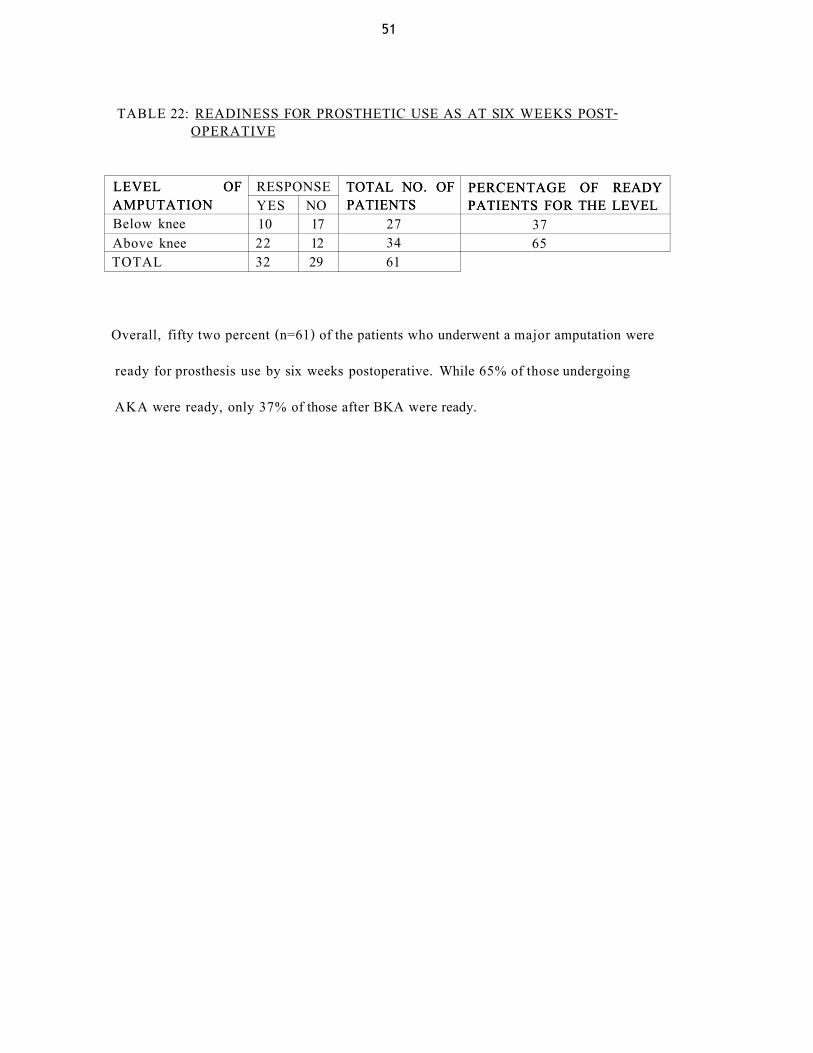
TABLE 22: READINESS FOR PROSTHETIC USE AS AT SIX WEEKS POST-OPERATIVE
LEVEL OF AMPUTATION
RESPONSE TOTAL NO. OF PATIENTS
PERCENTAGE OF READY PATIENTS FOR THE LEVEL
LEVEL OF AMPUTATION YES NO
TOTAL NO. OF PATIENTS
PERCENTAGE OF READY PATIENTS FOR THE LEVEL
Below knee 10 17 27 37 Above knee 22 12 34 65 TOTAL 32 29 61
Overall, fifty two percent (n=61) of the patients who underwent a major amputation were
ready for prosthesis use by six weeks postoperative. While 65% of those undergoing
AKA were ready, only 37% of those after BKA were ready.

52
DISCUSSION
A total of 74 patients were recruited into the study. There were 46 males and 28 females accounting for 62% and 38% respectively.
The study found the age distribution to be bimodal; one peak in the 31 -45 years and the other in the 'over 60 years' age classes. This distribution is more or less similar to that reported by Mbindyo 5 in a local previous study who found a teenage peak due mainly to trauma and osteogenic sarcoma and '50 years and above' due to vascular disease. In this study however, PVD contributes significantly to the peak in 31 -45 years age class. At a mean age of 44.4 years at operation, patients in this study arc quite young compared to other ser ies , 5 8 - 5 9 , 6 0 , 6 2 because a significant proportion of amputations due to tumours and trauma are carried out on young patients.
Most of the patients had primary education and below and were either unemployed or
self-employed in the informal sector. They therefore, could not afford to pay for the cost
of investigations for amputation level assessment in PVD. This either prolonged the pre-
operative duration while waiting for decision on amputation, or increased the number of
patients amputated on clinical level assessment alone. The end result was a prolonged
hospital stay (29.3 days in this study).

53
PVD whether diabetic or non-diabetic was the main indication for LLA as in the other s e r i e s . 5 8 , 5 9 , 6 0 , 6 1 , 6 2 , 6 9 , 7 0 , 7 1 This is however different from previous studies in the loco-regional set t ing, 5 1 0 where tumours were the main indication for LLA. This may be attributed to the rise in diabetic-related complications requiring amputations 6 and probably an increase in the number of patients with PVD referred by lower level hospitals to the KNH Thoracic and Cardiovascular surgical unit for limb salvage surgery or for special radiological studies, but end up being amputated in the KNH.
This study found an overall AKA:BKA ratio of 1.75:1 unlike the reported 1:1 in the other
major se r i e s / 8 ' 5 9 ' 7 0 , 7 1 There was a relatively higher proportion of AKA than BKA
performed due to trauma and tumours, which explains the difference. However, when
you consider that most reports are based on vascular amputations alone, this study had a
similar ratio as 47.5% and 45.3% of PVD amputations were BKA and AKA respectively.
Half the patients had no co-morbid conditions. Anaemia, followed by diabetes, was the
most common co-morbid condition. Cardiac disease was rare unlike in other reports.
58.59,61.70 m a y b e d u e t Q t h e r e i a t j v e i y y 0 U n g age and the low socio-economic status
of the patients in this study. The high prevalence of anaemia in the pre-operative period
considering that far much fewer patients had traumatic amputations suggests other factors
other than blood loss to be responsible. This study did not evaluate the aetiology or
degree of anaemia. However, determining the causes of anaemia and how to prevent or
treat them early will reduce the hospital stay, as anaemia significantly increased the pre-
operative duration in this study (p=0.005).

54
The healing rate of BKA was significantly less than for AKA (p=0.036), with a 21% rate
of eventual conversion of BKA to AKA, in accord with published se r ies . 6 , 4 3 , 5 8 , 6 2
The study noted a relatively high proportion of patients with minor amputations (43%),
who required conversion to BKA. All the patients who underwent minor amputations that
required conversion in this study had PVD. It may therefore be that delays in performing
arteriography for amputation level determination made surgeons carry out amputations on
clinical assessment alone in situations where BKA would have been ideal..
MEDICAL L l b R A K Y WWVPPSITY O P N A I R O i f
The most common complication was stump infection (33%), a rate much higher than in
other ser ies . 6 , 5 8 , 6 2 In this study, majority of the patients were not administered with
surgical antibiotic prophylaxis (77%). This and other un-investigated hospital-based
factors such as delays in scheduling operations due to shortage of theatre space,
congestion on general wards and irregular wound dressing habits could be the reason for
this high rates of sepsis. This high rate of sepsis further explains the reason why the study
found a higher proportion of revisions in AKA than BKA unlike other studies, 5 8 ' 6 1 ' 6 2
as more AKA were performed compared to BKA.
The overall mortality for the duration the patients were followed up was 16%, with a
thirty-day postoperative mortality rate of 13.5% which is similar to that in other
s tud ies . 5 8 , 5 9 , 6 1 , 6 2 Unlike in these studies where death is usually related to acute cardiac
events, majority of the patients died from septicaemia also in keeping with high sepsis
rates.

55
Majority of the patients (70%), were ambulating using crutches at six weeks after surgery despite irregular physiotherapy sessions and poor rehabilitation protocol. The younger age at amputation could explain the higher rate of ambulation compared to other studies. 5 8 , 7 0 None of the patients wore a prosthesis during the study period mainly due to the cost of securing prostheses. Majority of the patients are lost to follow-up therefore, long-term rehabilitation is not carried out or is there a laid-down program for the same.

56
CONCLUSIONS
1. Peripheral vascular disease is the main indication for lower limb amputations in
the KNH.
2. One of the most affected age class (31 -45 years) comprises people in the
economic prime age.
3. Majority of the patients undergoing LLA at the KNH have primary school
education and below and are unemployed.
4. Diabetes mellitus and anaemia are the most common co-morbid conditions in
patients undergoing LLA at the KNH.
5. The pre-operative in-hospital duration is long due to delays in pre-operative
assessment for level of amputation in patients with PVD..
6. Stump infection is the most common postoperative complication.
7. Majority of the patients are able to ambulate outdoors despite lack of established
rehabilitation programs and availability of prostheses.

57
RECOMMENDATIONS
LLA are costly to the nation and are associated with high morbidity and mortality. In the Kenyatta National Hospital, the following recommendations will help in improving the outcome of patients undergoing LLA.
1. Health education to patients attending clinics and the Accident and Emergency
department on the risk factors for amputation.
2. Expand the Thoracic and Cardiovascular unit of the hospital with full support of
diabetologists, cardiologists and physiotherapists for better management and
rehabilitation of patients with PVD.
3. Increase the number or utility of operating theatres to help reduce pre-operative
waiting time.
4. Encouraging the use of Duplex ultrasonography in amputation level determination
especially in patients already with gangrene, will reduce the waiting time and
provide more space for arteriography to be used in evaluating especially patients
requiring revascularisation procedures to save limbs from amputation.
5. Equip laboratories to facilitate constant availability of tests that identify and help
manage known risks such diabetes, cardiac, renal and liver disease.
58
REFERENCES
1. Kirk N.T. Amputations. Clin. Orthop. 1989; 243, 3-16. 2. Tooms R.E. Amputations. Canale T.S. (Editor), Campbell's Operative
Orthopaedics, volume two. St Louis: Moshy Inc. 1999; 689 - 702.
3. Gray D.VV.R. Limb Amputation. Morris. P.J., Maly, R.A. (Editors), Oxford Textbook of Surgery. Oxford: Oxford University Press, 1994; 488-500.
4. Cornelly J., Airey M., Chell S. Variation in clinical decision is a partial explanation for geographical variation in lower extremity amputation rates. Br J Surg 2001; 88: 529-535
5. Mbindyo B.S. Lower limb amputations in Kenyatta National Hospital. EastAfrMedJ. 1978; 55: 10. 458-461.
6. Maganga H.M. Lower extremity amputations in diabetic patients as seen in Kenyatta National Hospital. University of Nairobi 2001; Master of Medicine (Surgery), Dissertation.
7. Micheals J.A. Major amputations of the lower limb. Surgery International 1995; 13(7): 151-156.
8. The Global Lower Extremity Amputation Study Group. Epidemiology of lower extremity amputation in centres in Europe, North America and East Asia. Br J Surg 2000; 87: 328-337.
9. Mooney V., Wagner W.F jr., Waddell J., et al. The below-knee amputation for vascular disease. J Bone Joint Surg Am 1976; 58: 365-368.
10. Hassan A.W. Major limb amputations at Maiduguri Teaching Hospital. Medicare 1992; 5(4): 13-15.
11. Cortes E.P., Holland J.F., Glidewell O. Amputation and Adriamycin in primary osteosarcoma. A 5-year report. Cancer Treat Rep. 1978; 62: 271-277.
12. Gilber F.R., Gand T., Morton D.L. Adjuvant therapy for osteosarcoma; Pre-operative and post-operative treatment. Cancer Treat Rep. 1978; 62: 213-216.
59
13. Enneking W.F., Spanier S.S., Goodman, M.A. A system for the surgical staging of musculoskeletal sarcoma. Clin Orthop 1980; 531: 106-120.
14. Jaffe N., Goorin A., Link M., Watts H., et al. High dose Methotrexate in osteosarcoma adjuvant chemotherapy and limb salvage results. Cancer Treat Rep 1981; 6S(supple.): 99-106.
15. Lange R.H. Limb reconstruction versus amputation; Decision making in massive lower extremity trauma. Clin Orthop 1989; 249: 92-99.
16. Gustillo R.B., Mendoza R.M., Williams D.N. Problems in the management of type III (severe) open fractures; a new classification of type III open fractures. J Trauma 1984; 24: 742.
17. Caudle R.J., Stern P.J. Severe open fractures of the tibia. J Bone Joint Surg Am 1987; 69: 801-807.
18. Gregory R.T., Gould R.J., Peclet M., et al. The Mangled Extremity Syndrome; A severity grading system for multi system injury of the extremity. J Trauma 1985; 25:1147-1150.
19. Howe H.R jr., Poole G.V jr., Hansen K.J., et al. Salvage of lower extremities following combined orthopaedic and vascular trauma: A predictive salvage index. Am Surg 1987; 53: 205-208.
20. Johansen K., Darines M., Howe T., et al. Objective assessment criteria for accurately predicting amputation following lower extremity trauma. J Trauma 1990; 30: 568-573.
21. Mc Namara M.G., Heckman J.D., Corley F.G. Severe open fractures of the lower extremity: A retrospective evaluation of the Mangled Extremity Severity Score (MESS). J Orthop Trauma 1994: 8: 81-87.
22. Russell W.L., Sailors D.M., Whittle T.B et al. Limb salvage versus traumatic amputation: A decision based on a seven-part predictive index. Ann Surg 1991; 213: 473-481.
23. Dirschl D.R., Dahners L.E., The Mangled extremity: When should it be amputated? J Am Acad Orthop Surg 1996; 4: 182-190.
24. Bondurant F.J., Cotler H.B., Buckle R, et al. The medical and economic impact of severely injured lower extremities. J Trauma 1988; 28: 1270-1273.

60
25. Georgiadis G.M., Behrens F.F., Joyce J.M., et al. Open tibial fractures with severe soft tissue loss; Limb salvage compared with below-knee amputation. J Bone Joint Surg Am 1993; 75: 1431-1441.
26. Helfet D.L., Howey T., Sanders R., et al. Limb salvage versus Amputation; Preliminary results of the Mangled Extremity Severity Score. Clin Orthop 1990; 256: 80-86.
27. Hansen S.T. Overview of the severely traumatized lower limb; Reconstruction versus Amputation. Clin Orthop 1989; 243:17(editorial).
28. Williams M.O. Long term cost comparison of major limb salvage using Ilizarov method versus amputation. Clin Orthop 1994; 301: 156-158.
29. Campanacci M., Laus M. Local recurrence after amputation for osteosarcoma. J Bone Joint Surg Br (2) 1980; 62: 201-207.
30. Glasser D.B., Lane J.M. Osteosarcoma. Evarts M. (Ed), Surgery of Musculoskeletal system. London: Churchill Livingstone, 2nd edition, 1990; 4851-4882.
31. Lewis R.J., Lotz J.M. Medullary extension of osteosarcoma; Implication for rational therapy. Cancer 1974; 33: 371-375.
32. Marcove R.C. En bloc resection of osteosarcoma. Cancer Treat Rep 1978; 62: 225-231.
33. Springfield D.S., Schmidt R., Graham P.G., et al. Surgical treatment of osteosarcoma. J Bone Joint Surg Am 1988; 70: 1124-1130
34. Lane J.M., KroII M.A., P.T., et al. New advances and concepts in amputee management after treatment for bone and soft tissue sarcomas. Clin Orthop 1990; 256. 22-28.
35. Keblish P.A. Amputation alternatives in the lower limb; stressing combined management of a traumatized extremity. Clinics in Plastic Surg 1986; 13(4): 595-618.
36. Waters R.L. Energy costs of walking amputees' influence of amputation. J Bone Joint Surg Am 1976; 58: 42-47.
37. Mullen J.C., Gertner M.H., Buzby G.P., et al. Implications of malnutrition in the surgical patient. Arch Surg 1979; 114: 121.
61
38. Dickhaut S.C., De lee J.C., Page C.P. Nutritional status; Importance in predicting wound healing after amputation. J Bone Joint Surg Am 1984; 66: 71.
39. Kay S.P., Mooreland J.R. Nutritional status and wound healing in lower extremity amputations. Clin Orthop 187; 217: 253-256.
40. Jensen J.E., Jensen T.G., Smith T.K., et al. Nutrition in orthopaedic surgery. J Bone Joint Surg Am 1982; 64: 1263-1272.
41. Burgess E.M., Matsen F.A., Wyss C.R., et al. Segmental cutaneous measurements of p02 in patients requiring below-knee amputation for peripheral vascular disease. J Bone Joint Surg Am 1982; 64:1493
42. Cederberg P.A., Pritchard D.J., Joyce J.W. Doppler determined segmental pressures and wound healing in amputations for vascular disease. J Bone Joint Surg Am 1983; 65: 363-365.
43. O'dwyer K.J., Edwards M.H. The association between lowest palpable pulse and wound healing in below knee amputations. Ann R Coll Surg 1985;67: 232-234.
44. Tanzer T.L., Horne J.G. The assessment of skin viability using fluorescein angiography prior to amputation. J Bone Joint Surg Am 1983; 65: 599.
45. Oishi C.S., Fronek A., Golbranson L.F. The role of non-invasive vascular studies in determining levels of amputation level. J Bone Joint Surg Am 1988; 70: 1520-1530.
46. Dean R.H., Yao J.S.T., Thompson R.G., et al. Predictive value of ultra sonically derived arterial pressures in determination of amputation levels. Am Surg 1975; 41: 731-737.
47. Moore W.S. Determination of amputation level: Measurement of skin blood flow with Xenon 133. Arch Surg 1973; 107: 798-802.
48. Burgess E.M., Romano R.L., Zettl F.H. The management of lower extremity amputations: Surgery, immediate post surgical prosthetic fitting patient care. Revision for The World Wide Web. Laving L, Boone DCP (editors). Seattle, Washington: Prosthetics Research Study 1996.
49. Holoway B.A., Burgess E.M. Cutaneous blood flow and its relation to healing of below-knee amputations. Surg Gynaecol Obstet 1978; 146: 750.
50. Aly S., Sommerville K., Adiseshia M., et al. Comparison of duplex imaging and arteriography in the evaluation of lower limb arteries. Br J Surg 1998; 85: 1099-1102.

62
51. Kohler T.R., Nance D.R., Cramer M.M. Duplex scanning for diagnosis of aortoiliac and femoropopliteal disease. Circulation 1987; 76 (5): 1074-1080.
52. Burgess E.M. Amputation surgery and postoperative care. Banerjee S.N (Editor). Rehabilitation management of amputees. Baltimore, Williams and Wilkins, 1982; 17-44.
53. Hanspal R.S. Medical aspects of amputations and rehabilitation. Surgery International 2001; 56:292-294.
54. Mooney V., Harvey J.P., McBride E., et al. Comparison of postoperative stump management; Plaster versus soft dressing. J Bone Joint Surg Am 1971;53:241-249.
55. Mensch G., Ellis P. Physical therapeutic management for lower extremity amputees. Banerjee S.N. (Editor). Rehabilitation management of amputees. Baltimore. Williams and Wilkins, 1982; 167-236.
56. Keller R.B. Outcomes Research in Orthopaedics. J Am Acad Orthop Surg 1993; 1:122-129.
57. Kocher M.S., Zurakowski D. Clinical epidemiology and biostatistics: A primer for orthopaedic surgeons. Current concepts review. J Bone Joint Surg Am 2004; 86 (3): 607-620.
58. Nehler M.R., Coll J.R., Hiatt W.R., et al. Functional outcome in a contemporary series of major lower extremity amputations. J Vase Surg 2003; 38 (1): 7-14.
59. Kazmers A., M.S.P.H., Perkins J.A., et al. Major lower extremity amputations in Veterans Affairs Medical Centers. Ann Vase Surg 2000; 14: 216-222.
60. Kald A., Carlsson R., Nilsson E. Major amputation in a defined population: incidence, mortality and results of treatment. Br J Surg 1989; 76: 308-310.
61. Keagy B.A., Schwartz J.A., Kotb M., et al. Lower extremity amputation: the control series. J Vase Surg 1986; 4(4): 321-326.
62. Ahmed M.A Jr. , Theodore H.T., K. David K., et al. Major lower extremity amputation in an Academic Vascular Center. Ann Vase Surg 2003; 17: 86-89.
63. Martini A., Fromm B. A new operation for the prevention and treatment of amputation neuromas. J Bone Joint Surg Br 1989; 71: 379-382.

63
64. Kumar V.N. Stump complications and management. Banerjee S.N. (Editor). Rehabilitation management of amputees. Baltimore. Williams and Wilkins. 1982: 372-390.
65. Elliott F., Little A., Milbrandt N. Carbamazepine for phantom limb phenomena. N Engl. J. Med. J976; 678.
66. Ganong W.F. Cutaneous, deep and visceral sensation. Review of medical physiology. Norwalk. Appleton and Lange, 16th edition, 1993: 120-131.
67. Hadden W., Marks R., Murdoch G. Wedge resection of amputation stumps: A valuable salvage procedure J Bone Joint Surg Br 1987; 69: 306-308.
68. Marquardt E., Correll J. Amputation and prosthesis for the lower limb. Int Ortho 1984; 8: 139.
69. Feinglass J., Pearse W.H., Gary J., et al. Postoperative and late survival outcomes after major amputation: Findings from the Department of Veterans Affairs National Surgical Quality Improvement Program. Surgery 2001; 130: 21-29
70. Mayfield J.A., Reiber G.E., Maynard C., et al. Survival following lower-limb amputation in a veteran population. J Rehabil Res Dev 2001; 38(3): 39-43.
71. Houghton A.D., Taylor P.R., Thurlow S., et al. Success rates for rehabilitation of vascular amputees: implications of preoperative assessment and amputation level. Br J Surg 1992; 79: 753-755.

64
APPENDIX 1.
QUESTIONNAIRE Patient 's name: Age ( ) IP No Date of admission: Date of discharge: Date of operation: 1. Age group (in years)
a) <15 b) 15-30 c) 31-45 d) 46-60 W e) >60
2. Sex a) Male b) Female
3. Education level a) Illiterate b) Primary c) Secondary d) Tertiary o
4. Employment status a) Not employed b) Self employed c) Employed by other d) Retired o
5. Co-morbid medical illness a) Cardiac b) Respiratory c) Neuropsychiatry d) Diabetes e) Anaemia f) None
series
^OMJ
65
Blood tests a) White cell count (TLC) b) Liver function tests (albumin levels) c) Urea & Electrolytes d) Random blood sugar e) P24 Elisa 0 Lipid profile g) None
Radiological tests a). Doppler ultrasonography of the limb b). Chest X-ray c). Arteriography
Indication of amputation
a) Trauma (i). RTA (ii). Industrial accident .
(iii). Bums (iv). Assault (v). Other
b) Peripheral vascular disease (i). Diabetic
(ii). Non diabetic Category
c) Neoplastic (i) Skin (ii) Bone (iii) Other
d) Infection (i) Soft tissue (ii) Bone
e) Congenital
Surgical prophylaxis a) Yes b) No o

66
Level of amputation (i). Toe
(ii). Transmetatarsal (iii). Transtarsal (iv). BKA O (v). AKA W
(vi). Hip disarticulation
Technique of amputation a) Open ^ b ) Closed ( J
Pre-operative physiotherapy a) Yes b) No
Duration taken for wound to heal a) By day 7 b) 7 to 14 days c) 14 to 21 days d) >21 days Day post-operative physiotherapy was started a) Day 2 b) After day 2, but before day 7 c) After day 7 post-operative
o
o
o Number of physiotherapy sessions in a week a) None b) < 7 c) 7 d) > 7 o

67
16. Complications a) None
b) Systemic i). Cardiac
ii). Pulmonary iii). Neuropsychiatry Category iv). Anaemia v). Septicaemia
i). Phantom pain ii). Stump infection
iii). Flap necrosis iv). Significant contracture Category
v). Bone erosion/stump ulcer vi). Adherent scar
vii). Stump neuroma viii). Stump bulbosity/ 'dog ears'
c) Local
d) Death
17. Method of mobility
a). Wheel chair b). Walking frame c). Crutches/ Cane d). Bedridden e). Not applicable
18. Is the patient ready to use a prosthesis by six weeks? a). Yes b). No
19. Revision surgery 20. Conversion
a). None b). Foot o BKA c). BKA to AKA
a). None b). Refashioning c). Skin graft or flap

68
APPENDIX 2
CONSENT BY THE PARTICIPATING PATIENT (English)*
I have understood the explanation by Dr. Awori K.O, who is carrying out a study on
lower limb amputations, and hereby give consent to participate in the study.
I agree to participate in the study on my on free will and also to do the following:
> Be interviewed concerning my illness and subsequent amputation and the
answers to be recorded by Dr. Awori.
> To be examined physically.
I have understood that my participation is completely voluntary, and that I can
withdraw my consent at any point of the study, and that such act will not affect my
treatment in any way.
That the information I give will be treated with utmost confidence and that my name will
not be included in the results
PARTICIPANT'S NAME (OR GUARDIAN/PARENT/CONSULTANT)
Signature/thumb print
WITNESS Signature/thumb print
INVESTIGATOR DR. KIRSTEEN ONDIKO A WORl Signature
* There was a Kiswahili version for patient who did not understand English
69
APPENDIX 3
KENYATTA NATIONAL HOSPITAL Hospital R<3 along Ngong Rd
P.O Box 20723, Na-rut* To! 726300-9 Fax 725272
Telegrams- MEDSUP-. Kuarobi Frrwl KNHnlanf lKt i .Haa l t lma t .om
Rcf: KNH-ERC/01 / 1866 Date: 25 June 2003
Dr K Ondiko Awoii Dcpt of Surgery Pacultv of Medicine
Dear Dr. Oitdiko,
RESEARCH PROPOSAL "ASSESSMENT OF THE DUTCOMh AMPUTATIONS AS SEEN IN KENYA1TA NATIONAL HOSPITAL"
OF LOWER LIMB (P53/5/2Q03)
This is to intbnn you dial the Kcnyatta National Hospital Ftliics aud Research Committee has reviewed aud approved your above cited rexraicli p<oposal
On behalf uf ihe Committee 1 wish you fiuitl'ul rcscaicb and look forward to receiving a summary of the research findings upon completion of the study I his information will form part ol database that will he consulted in Rilmc when proceswng
related research study so as to minimise chances of study duplication
Yours sincerely.
PROF. A N G DA NT AI SKCHH I ARV. KNH-ERC Cc Prof KM Hhatt. Chairperson, KNII-CRC
I he Deputy Director (C/S), KNH The Dean, Faculty of Medicine, I JON The Chairman, O p t af Surgery. UON CMRO Supeivisor Prof J F. O Ating'a. Depl. of Oithopaodic Surgery. UON