assessment of resistance to antibiotics in pseudomonas
TRANSCRIPT

Assessment of Resistance to Antibiotics in Pseudomonas syringae pv. syringae
Phytotoxin Secretion Mutants
Renee L. Schaeffer University Honors Thesis
September 14, 2000
Honors Thesis ************************* PASS WITH DISTINCTION

INTRODUCTION Pseudomonas syringae pv. syringae is a well-known bacterial plant pathogen of
many agronomically important crops such as sweet cherry, beans, and corn (2). P. s. pv.
syringae produces two necrosis inducing phytotoxins: syringomycin (syr) and
syringopeptin (syp). These phytotoxins are virulence factors that extensively contribute
to the pathogen's ability to cause disease (9). The genes responsible for syringomycin
and syringopeptin production are clustered on the bacterial chromosome and include
genes coding for biosynthesis, regulation, and secretion of both syringomycin and
syringopeptin (10). Interestingly, genes were identified that code for two complete
secretion systems. Mutations in these genes result in the reduction or inability of P. s. pv.
syringae to secrete syringomycin or syringopeptin (11). Secretion is the transportation of
products such as toxins, metabolic waste products, or antibiotics out of a cel!. Secretion
is thought to occur for two reasons: prevent toxic levels of substances from accumulating
in the cell and give the bacterium a competitive advantage by releasing toxins into the
environment. It is hypothesized that the mechanisms of secretion in P. s. pv. syringae are
similar to those of related bacteria of medical importance such as Pseudomonas
aeruginosa (13). Most bacterial plant pathogens have one or more of four secretory
pathways that utilize a combination of proteins to form transporters that work in the
cytoplasm, inner membrane, and outer membrane (9).
Mutants were constructed by "knocking out" the genes of interest in P. s. pv.
syringae. These mutants were assessed to determine which genes are more significant in
secretion of various compounds. All three genes exhibit homology to genes involved in
secretion. Three mutants were tested in this study and compared to the wild type strain
B301DR: two ABC Transporters (containing the ATP-Binding Cassette motif) (9), and
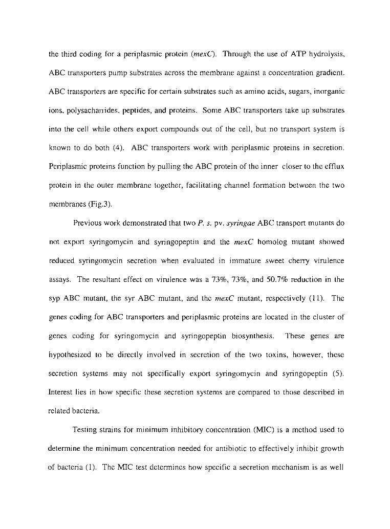
the third coding for a periplasmic protein (mexC). Through the use of ATP hydrolysis,
ABC transporters pump substrates across the membrane against a concentration gradient.
ABC transporters are specific for certain substrates such as amino acids, sugars, inorganic
ions, polysacharrides, peptides, and proteins. Some ABC transporters take up substrates
into the cell while others export compounds out of the cell, but no transport system is
known to do both (4). ABC transporters work with peri plasmic proteins in secretion.
Periplasmic proteins function by pulling the ABC protein of the inner closer to the efflux
protein in the outer membrane together, facilitating channel formation between the two
membranes (Fig.3).
Previous work demonstrated that two P. s. pv. syringae ABC transport mutants do
not export syringomycin and syringopeptin and the mexC homolog mutant showed
reduced syringomycin secretion when evaluated in immature sweet cherry virulence
assays. The resultant effect on virulence was a 73%, 73%, and 50.7% reduction in the
syp ABC mutant, the syr ABC mutant, and the mexC mutant, respectively (11). The
genes coding for ABC transporters and periplasmic proteins are located in the cluster of
genes coding for syringomycin and syringopeptin biosynthesis. These genes are
hypothesized to be directly involved in secretion of the two toxins, however, these
secretion systems may not specifically export syringomycin and syringopeptin (5).
Interest lies in how specific these secretion systems are compared to those described in
related bacteria.
Testing strains for minimum inhibitory concentration (MIC) is a method used to
determine the minimum concentration needed for antibiotic to effectively inhibit growth
of bacteria (1). The MIC test determines how specific a secretion mechanism is as well

as identify proteins involved in secretion. Six mutants were constructed by "knocking
out" individual genes for secretion in P. s. pv. syringae (11). These mutants disrupt
genes having homology with the following genes: oprM (syr), oprN (syp), ABC
~ transporter (syrD), ABC transporter (sypD), mexC (syr), and mexC (syp) (4). MIC
~
experiments were performed on three of these P. s. pv. syringae secretion mutants to ~
I evaluate their role in secretion of various antibiotic compounds.
I The MICs were determined using several different methods: a broth dilution
~
~ method in microtiter plates, and two agar dilution methods. Media containing the lowest ~
concentration of antibiotic resulting in no growth was considered the MIC. The ~
~ minimum concentration for each antibiotic was determined and compared to the wild ~
type P. s. pv. syringae strain B301DR. Two media were chosen for the agar plate ~
~ dilution methods. Potato dextrose agar (PDA) was chosen because PDA does not inhibit ~
the production of syringomycin and syringopeptin, while Mueller-Hinton Agar (MHA) ~
~ was chosen because it is commonly used in related MIC research. Observing the effects
~
of different classes of antibiotics on the mutants offers a key to understanding the types
• ~
of compounds they secrete. Eleven antibiotics were chosen for this MIC study by
considering related literature. One antibiotic from each class of antibiotic, with the
exception of gentamycin and kanamycin, were chosen to use in this study. Both
gentamycin and kanamycin are aminoglycosides and both were used in this study because
two of the mutant strains carry kanamycin cassettes, making them resistant to kanamycin.
It was necessary to have a different aminoglycoside available to test these strains. Using
MIC data for P. s. pv. syringae secretion mutants, we can make conclusions about the

mechanism of secretion of the phytotoxins as well as the relationship to secretion systems
of related bacteria.
Materials and Methods
Bacterial strains and media. All P. s. pv. syringae strains used in this study are listed in
Table 1. Strains were maintained on nutrient-broth yeast extract (NBY) agar (15).
Antibiotics were added to the media in the following concentrations: (pipperacillin, 25
Ilg/ml), (kanamycin, 100 Ilg/ml), and (kanamycin, 50 Ilg/ml) for maintenance of strains
BR105, B301D.67.18, and B301D.SLA, respectively. MIC assays were performed on
potato dextrose agar (PDA) supplemented with 1.5% (w/v) glucose and 0.4% (w/v)
casamino acids (20 ml/plate) (2) and Difco Mueller-Hinton Agar (Sigma Chemical Co.,
St. Louis, MO) was prepared as directed by the manufacturer (20 ml/plate) amended with
different concentrations of antibiotic. All antibiotics were supplied by Sigma Chemical
Co., and were stored at -20·C at the following stock concentrations: carbenicillin, 50
/lg/ml; chloramphenicol, 50 /lg/ml; cefoperazone, 50 /lg/ml; erythromycin, 50 /lg/ml;
gentamycin, 25 /lg/ml; kanamycin, 25 /lg/ml; novobiocin, 150 /lg/ml; ofloxacin, 1.67
/lg/ml; polymixin B, 25 /lg/ml; tetracycline, 25 /lg/ml; and trimethoprim, 5 /lg/ml.
Glycerol Storage. P. s. pv. syringae strains were inoculated to agar plates and incubated
at 25°C overnight. A single colony was selected from each plate and incubated at 25°C in
5ml NBY broth cultures overnight with agitation. In cryogen tubes, 0.75 ml of culture
and 0.75 ml of 75% glycerol were added, mixed, and were placed at -80·C. Fresh
cultures were started each week from cryogen storage tubes.

.'
Microtiter Plate assays. Cells were grown overnight in liquid NBY cultures at 25°C
I with agitation and washed with water. In water cells were suspended to an optical
I density (a.D.) of 0.3 (approximately 2 x 10⁸ CFU/ml) using a spectrophotometer 21 at
I
I 1..=420. AI: 10 dilution series was done to achieve approx 2 x 10⁵ CFU/ml and 100JlI of
I the last dilution (-2 x 10⁴ CFU) were inoculated into 100 Jlg/ml of a 1:2 dilution series of
I
~ PDA and antibiotic. When approximate MIC ranges was determined, a 1: 1.2 dilution
I series was completed for each antibiotic. The a.D. for each well in the microtiter plate
~
~ was measured using an a.D. reader to detect the amounts of growth.
~ Inoculation and analysis of agar plate assays. Liquid cultures (5 ml) were inoculated
~
~ with each strain and incubated overnight at 25°C with agitation. A 1.5 ml aliquot of each
• culture was transferred to a 1.5 ml microcentrifuge tube and cells were collected by
centrifugation. The supernatant was aspirated and cells were resuspended in 1 ml sterile
water. The cell suspension was added to 10 ml of sterile water until an a.D. of 0.3 (-2 x
108 CFU) was achieved. Both MHA and PDA were used for plate assays. Each plate
was inoculated with a 5 JlI droplets containing 1 x lif and 1 x 10⁶ CFU and the plates
were counted after day two. Colonies were counted first by a visual MIC and then by
using a dissecting microscope at 20X. The MIC was defined as the lowest concentration
of antibiotic inhibiting visible growth after two days of incubation at 2YC.
Results
Evaluation of antibiotic susceptibilities of P. s. pv. syringae secretion mutants
in broth media using microtiter plate assays. P. s. pv. syringae strains B301DR,
BR105, B301D.67.18, and B301D.SlA were evaluated for their ability to resist various
t'

antibiotics in broth culture. The results of the mitrotiter plate assays are listed in Table 2
and Fig. 4. The observed MJC for carbenicillin, cefoperazone, and ofloxacin were the
same for each strain. The wild type strain B301DR was the most resistant to
erythromycin, and kanamycin. BR105 was more resistant to tetracycline,
chloramphenicol, and polymixin B and was less resistant to gentamycin, kanamycin,
novobiocin, and trimethoprim. Unlike BR105, B301D.67.18 was more resistant to
erythromycin, gentamycin, novobiocin, and trimethoprim, and was less resistant to
chloramphenicol and tetracycline. B301D.SL4 exhibited low MJCs to most antibiotics
relative to the other strains, being consistently less or equal to B301DR. B301D.SL4 was ~
~ most susceptible to erythromycin and tetracycline with MJCs at 37.86 {Lg/ml and 0.76
~
~ {Lg/ml, respectively, as opposed to B301D.67.18 with MlCs at 120 {Lg/ml and 3.05 {Lg/ml,
respectively.
Evaluation of P. s. pv. syringae strains for antibiotic susceptibility on PDA
plate assays. Assays were perfonned on PDA plates amended with various antibiotics to
detennine the MIC for each of the P. s. pv. syringae secretion mutants. PDA plate assays
indicated that while differences were observed among the strains, MlC data was quite ~
••••
•
•, ~
•similar for all antibiotics tested (Table 3 and Fig. 5). BR105 exhibited an MlC for
chloramphenicol above the wild type strain, with an observed MIC at 500 {Lg/ml and 300
~ {Lg/ml, respectively. In addition, the mutant strains consistently exhibit greater or equal
MICs then the wild type strain for the antibiotics evaluated.
~ Evaluation of antibiotic sensitivities of P. s. pv. syringae strains on Mueller~
Hinton agar plate assays. Mueller-Hinton agar plate assays were perfonned to ~
~ detennine MJCs of several P. s. pv. syringae secretion mutants. Mueller-Hinton agar
• ~
~
•

plate assays (Table 4 and Fig. 6) were similar to the PDA plate assay results. When
B301DR was tested, the MICs were either nonnal or low for most of the antibiotics
relati ve to the mutant strains. It is interesting to note that the mutant strains had higher
MICs than the wild type strain for chloramphenicol, novobiocin, tetracycline, and
trimethoprim (Table 4 and Fig. 6).
Conclusions
J The microtiter plate MIC data reflected other published MIC experiments (6).
Differences were observed between the wild type strain and the mutants in respect to ~
their MICs to various antibiotics. More variation was seen among the wild type and the
mutants with respect to the MICs. Our observed B301DR microtiter MICs for ofloxacin,
~ gentamycin, and trimethoprim were 1.03 JLglml, 13.1 JLglml , and 355.56, respectively.
Maseda et al reported such MICs for the related wild type P. aeruginosa as 0.78 JLglml,
~ 3.13 JLglml, and 200 JLglml for ofloxacin, gentamycin and trimethoprim, respectively (6).
Higher MICs were observed for PDA plates compared to the microtiter plate
assays (Fig. 3 and 4). The microtiter plates were originally completed to detennine a
range for the PDA plate assays and to conserve antibiotics and media, though it appears
the data is not comparable. We chose to use a dissecting microscope at 20X to detennine
I
I
I
••
•
••t
••
••
if colonies were fonning on PDA plates amended with various antibiotics. This is in
contrast to literature which used a visible evaluation, but we felt this would result in a
more conservative and accurate measurement of growth. Using the microscope may have
distorted our data because most plates showed small amounts of growth at very high
concentrations of antibiotics that were not '/isually apparent. We did not observe the
• ••

range in MlC data for P. s. pv. syringae strains in the PDA plate assays as we did in the
microtiter plates because at least one very small colony would form after two days of
incubation at 25°C. This is shown in Fig 1 where two different antibiotics react very
differently in PDA at 1 x 10⁶ CFU, but both had MlCs at 1 x 10⁴ CFU. In addition, small
amounts of growth were found at high concentrations of antibiotic, which may result
from the presence of a highly resistant variant form within the population. These same
results were seen for MBA.
Higher MICs were observed on PDA as compared to the MHA plate assays. This
is interesting because PDA does not inhibit syringomycin and syringopeptin production
in P. s. pv. syringae and complex media such as NBY inhibits syringomycin and
syringopeptin production. This can be seen in Figure 2, where the same concentration of
chlormaphenicol inhibits growth on MBA, but colonies form on PDA. There is a
significant difference in growth for some of the antibiotics in the two medias (Fig. 5 and
6). MBA was chosen because it is most commonly used in related MIC literature with P.
aeruginosa, in particular. This media is suggested for MlC testing by the National
Committee for Clinical Laboratory Standards because of the wide use of MHA in MIC
research (7). However, they do suggest that the media be supplemented with cation
supplements or other buffers such as EDTA, which was not completed in these
experiments. MHA was not amended because it is known that some of these
supplements increase the permeability of the outer membrane, which may interfere with
our study (14).
In the microtiter plate assays, an increased resistance of B301D.67.18 to
gentamycin was observed. This was interesting because the strain carries a kanamycin

•
••
••
•
cassette, giving the strain resistance to kanamycin. Increased resistance to gentamycin
may be because kanamycin and gentamycin are both aminoglycosides, therefore
increased gentamycin resistance may be the result of another secretion systems exporting
the similar compounds. BR 105 is more resistant to tetracycline, chloramphenicol, and
polymixin B. What is interesting here is that BR105 carries resistance to piperacillin, a
ureidopenicillin in the penicillin family. BRI05 was expected to have increased
resistance to carboxypenicillins like carbenicillin, but in the microtiter plate study it did
not exhibit resistance above the other strains. BR105 does appear to have susceptibility
~ to gentamycin, kanamycin, novobiocin, and trimethoprim, which were the same
~
~ antibiotics B30ID.67.I8 attained higher MICs. BR105 and B30ID.67.I8 are both ABC ~
transport mutants, but the proteins are structurally mirror images of each other. Even
~ though the differences were slight, the strains did exhibit opposite MICs to the ~
antibiotics. B30ID.SiA exhibited either normal or low MICs for most antibiotics, and ~
~ the lowest MICs were to erythromycin and tetracycline, which is opposite of ~
B30ID.67.I8. This is significant because both strains have kanamycin cassettes, but one
~ is an ABC transport mutant and the other is a periplasmic protein mutant. B30 ID.SiA
~
has a periplasmic protein that may not allow the antibiotics to get from the inner~
~ membrane protein (ABC transporter) to the outer membrane protein (efflux protein) as
~
easily (Fig 3). Antibiotic may slowly accumulate in the periplasm and take longer for the
cells to export the antibiotics, therefore making the cells abnormally susceptible to many ~
~ of the antibiotics.
The mutant strains in general exhibited increased MICs above the wild type
~ strain. This could indicate that the syr and syp this secretion systems are not directly
~
~
~
~

involved in the export of antibiotics and export may occur through other secretion
systems in the cell. It has been hypothesized that these secretion systems work together,
but it is possible that the secretion systems for syringomycin and syringopeptin are
specific.

•••
•••
TABLE 1. Bacterial strains and Elasmids Strain or Plasmid Relevant characteristics a Source P. s. pv. syringae
B3010 Wild type from pear Cody et al. 1987 B3010-R Spontaneous RiF derivative of B3010 Xu and Gross 1988 BR105 syrD::Tn3HoHol derivative of B3010-R; Pipr Quigley et al. 1993
RiF B3010.67.18 sypD::miniTn5 derivative of B3010; Kmr This study B3010.SLA mexC::miniTn5 derivative ofB3010; Kmr This study
a Kmr, pipr, and RiF, resistance to kanamycin, piperacillin, and rifampicin, respectively.
Table 2. Microtiter plate assays in POB.
Microtiter Carbenicillin Chloramphenicol Cefoperazone Erythromycin Gentamycin
B301DR 100 38.97 100 100 13.1 BRI0S 100 46.8 100 56.5 7.8 B301D.67.18 100 32.47 100 120 25 B301D.sL4 100 32.47 100 37.86 13.1
~
Table 2. (cont.) MiL (Jl 1m!)
Microtiter Kanamycin Novobiocin Ofloxacin Polymixin B Tetracycline Trimethoprim
B301DR 7.92 2178.6 1.03 4.13 3.9 355.56 BRI05 3.96 1512.9 1.03 5.95 5.67 296.3 B301D.67.18 Not tested 2614.37 1.03 4.13 3.05 500 B301D.sL4 Not tested 1512.9 1.03 4.13 0.76 355.56
Table 3. Agar plate assays on POA.
»•
POA Carbenicillin Chloramphenicol Cefoperazone Erythromycin Gentamycin B301DR 65 300 400 400 250 BRI0S Not tested 500 375 375 275 B301D.67.18 65 400 425 400 250 B301D.SL4 45 300 400 400 175
Table 3. (cont.)
20X MIC IJl.g!mJ) Kanamycin Novobiocin Ofloxacin Polymixin B Tetracycline TrimethoprimPOA 300 4800B301DR 95 350 275 950 250BRI0S 5400 75 300 300 950
B301D.67.18 Not tested 5400 95 325 275 900 B301D.SL4 Not tested 5100 95 250 275 950
•t

Table 4. Agar plate assays in Mueller-Hinton agar.
---- - -- - ,,-r-c:;r ----f
Erythromycin GentamycinCarbenicillin Chloramphenicol CefoperazoneMBA 42575 250 400 300B3010R
Not tested 325 375 350 300BR105 400 375 275B3010.67.18 75 300
45 325 350 375 275B3010.SL4
Table 4. (cont.)
20X MIC IJl.g/ml) MHA Kanamycin Novobiocin Ofloxacin Polymixin B Tetracycline Trimethoprim
B3010R 250 4500 90 150 250 775 BR105 225 4900 80 150 300 825 B3010.67.18 Not tested 4900 90 125 250 750 B3010.SL4 Not tested 4700 90 125 300 825


• •
_~__.....;.....,;;;:;uter Membrane
:J
Fig. 3. Model for ABC Transporters
Plasma Membrane
• • ...
0 Ii • •
'" - ,
OprM . ;. '\ , MexC monomer SypD/ SyrD homo/ heterodimer
Syr/ Syp Toxin • ATP


• •m m ~ ~ c ~ en ;"'J-co


LITERATURE CITED:
1. Aires, J. R., T. Kohler, H. Nikaido, and P. Plesiat. 1999. Involvement of an active efflux system in the natural resistance of Pseudomonas aeruginosa to aminoglycosides. Antimicrob. Agents Chemother. 43:2624-2628.
2. Bradbury, J. F. 1986. Guide to plant pathogenic bacteria. CAB Int.Mycol.Inst. Ferry Lane, Kew, Surrey, England. 175-177.
3. Gross, D. C. and DeVay, J. E. 1977. Population dynamics and pathogenesis of Pseudomonas syringae in maize and cowpea in relation to the in vitro production of syringomycin. Phytopathol. 67:475-483.
4. Higgins, C.F. 1992. ABC transporters: from microorganisms to man. Annu. Rev. Cell BioI. 8:67-113.
5. Johnson, J.M. and G.M. Church. 1999. Alignment and structure prediction of divergent protein families: periplasmic and outer membrane proteins of bacterial efflux pumps. J. Mol. BioI. 287:695-715.
6. Mesada, H., H. Yoneyama, T. Nakae. 2000. Assignment of the substrate-selective subunits of the MexEF-OprN multidrug efflux pump of Pseudomonas aeruginosa. Antimicrob. Agents Chemother. 44:658-664.
7. National Committee for Clinical Laboratory Standards. 2000. Methods for dilution antimicrobial susceptibility tests for bacteria that grow aerobically; approved standard, 5th ed. Document M7-A5. National Committee for Clinical Laboratory Standards, Wayne Pa.
8. National Committee for Clinical Laboratory Standards. 2000. MIC testing Supplemental Tables. Document MI00-SIO (M7). National Committee for Clinical Laboratory Standards, Wayne Pa.
9. Salmond, G.P. 1994. Secretion of extracellular virulence factors by plant pathogenic bacteria. Annu. Rev. Phyopathol. 32: 181-200.
10. Scholz-Schroeder, B. K., M. L. Hutchison, I. Grgurina, and D. C. Gross. 2000. The contribution of syringopeptin and syringomycin to virulence of Pseudomonas syringae pv. syringae strain B301D based on analysis of sypA and syrBl biosynthesis mutants. Mol. Plant-Microbe Interact. (submitted July 2000).
11. Scholz-Schroeder, B. K., J. Soule, S. Lu, D.C. Gross. 2000. Unpublished data.
12. Soule, J. 2000. Unpublished data.

13. Tommassen, J., A. Filloux, M. Bally, M. Murgier, and A. Lazdunski. 1992. Protein secretion in Pseudomonas aeruginosa. FEMS Microbiol. Rev. 103:73-90.
14. Vaara, M. 1992. Agents that increase the permeability of the outer membrane. Microbio. Rev. 56:395-411.
15. Vidaver, A. K. 1967. Synthetic and complex media for the rapid detection of fluorescence of phytopathogenic pseudomonads: Effect of the carbon source. Appl. Microbiol. 15: 1523-1524.