assessment of hazard risk associated with the use of … of hazard risk associated with the use of...
TRANSCRIPT

Assessment of hazard risk associated with the use of viral
vectors in rodents
Daniel Gibbs, PhD
The Salk InstituteGene transfer, targeting and therapeutics (GT3) Facility
UCSDTranslational Neurosciences Institute

Salk Animal Research Dept (ARD) Salk GT3 Core FacilityJon Reuter – Co‐PI Helen Fang – RA III
Karen Suter – Senior technician Christina Ly – RA II
Funding: NEI 1P30EY019005‐01A1 (DG), NIA 5P01AG010435‐18 (DG), NCI 5P30CA014195‐39 (DG) and the ACLAM foundation (JR)

Shedding of viral vectors from experimental animals
• What are viruses and what makes a viral vector?
• Basic features of the viral vectors used in this study
• Evaluation of the risk of shedding of three common viral vectors in transduced rodents
• Assessment of the potential for human exposure to these vectors from treated animals.
Wikipedia (http://en.wikipedia.org/wiki/Virus):
“A virus is a nanometer scale infectious agent that can replicate only inside the living cells of organisms. Viruses infect all types of organisms, from animals and plants to Bacteria and Achaea. Viruses are found in almost every ecosystem on Earth and may be the most abundant type of biological entity.”
The vector‐centric viewpoint
Viruses are evolutions “magic bullets” for nucleic acid delivery

Viral Vectors
• Genetically engineered viruses capable of delivering specific DNA or RNA sequences to cells or tissues of interest
• Goff SP and Berg P. (1976) Construction of hybrid viruses containing SV40 and lambda phage DNA segments and their propagation in cultured monkey cells. Cell. 9:695‐705
http://vectorcore.salk.edu

Commonly used Viral Vectors
• Lentivirus (HIV‐1, FIV, SIV, EIAV)• Adeno‐associated virus (AAV2)• Adenovirus (Ad5, Ad35, CAV‐2)• Rabies virus (SADb19, SADb19G)• Retrovirus (MMLV, MSCV, RCAS, FV)• Herpesvirus (HSV, PrV)• Poxvirus (vaccinia)• Vesicular stomatitis virus (VSV, VSVG)• Sendai Virus

Viral Vectors In This Study
• Lentivirus (HIV‐1)• Adeno‐associated virus (rAAV2)• Adenovirus (Ad5)

Shedding of viral vectors from experimental animals
• What are viruses and what makes a viral vector?
• Basic features of the viral vectors used in this study
• Evaluation of the risk of shedding of three common viral vectors in transduced rodents
• Assessment of the potential for human exposure to these vectors from treated animals.

Lentiviral Vectors
• Pseudotyped HIV‐1 vectors
Phospholipid envelope
dsRNA genome
core
matrix
Reverse transcriptase integrase
Glycoprotein

• Advantages• 3rd generation SIN vectors have good biosafety record• Expression of large inserts (~8‐10KB)• Integrating virus ‐ Stable transgene expression• Tailored tropism by pseudotyping (VSV.G, RabiesG, EnvA, EnvB)• Transduction of non‐dividing cells• Probability of replication competent vector extremely low
• Disadvantages• WT HIV‐1 is a known human pathogen• Integration has potential risk of insertional effects on endogenous genes
• Variable expression level• Broad tropism of VSV.G can reduce specificity• Some tissues are not transduced well (e.g. Airway epithelia)• Expression of oncogenic transgenes in integrating vector presents a significant and as yet uncharacterized biohazard risk

Lentiviral vector mediated neuronal labeling in the rat hippocampus
LV‐HsynP‐EGFP Injection into the dentate gyrus at P5. Image collected at P14.
200um

Adeno‐associated Viruses
• rAAV2 pseudotyped vectors• Non‐enveloped virus – 20 nm diameter virions
• Single stranded DNA genome
• wtAAV requires co‐infection with adenovirus, herpesvirus or vaccinia for replication
• >100 different serotypes predicted

Serotype Known Tropism
AAV1 Muscle, heart, CNS AAV2 In vitro, CNS, retina (RGCs), LiverAAV5 Lung (airway, alveoli), RPE, retina, CNSAAV6 Lung, heart, Spinal cord, retinaAAV7 Muscle, liverAAV8 Liver, muscle, eye, CNSAAV9 Lung (alveoli), liver, muscle, heart, CNSAAVrh10 Pleura, CNS, amacrine and bipolar cellsAAV‐DJ hESC, CNS, Liver, Muscle
• Tropism is highly dependent on: (A) Purification method, (B) Experimental system,( C) Viral titer, (D) Expressed transgene and (D) Regulatory elements

• Advantages• AAV is not a known human pathogen• Wide choice of capsids allow some tissue specific expression• Low immunogenicity• Non‐integrating• Long term expression in vivo in non‐dividing cells• Transduction of non‐dividing cells
• Disadvantages• Small packaging capacity• rAAV typically non‐integrating ‐ lost from dividing tissues• Neutralizing antibodies to some AAV serotypes common in human population
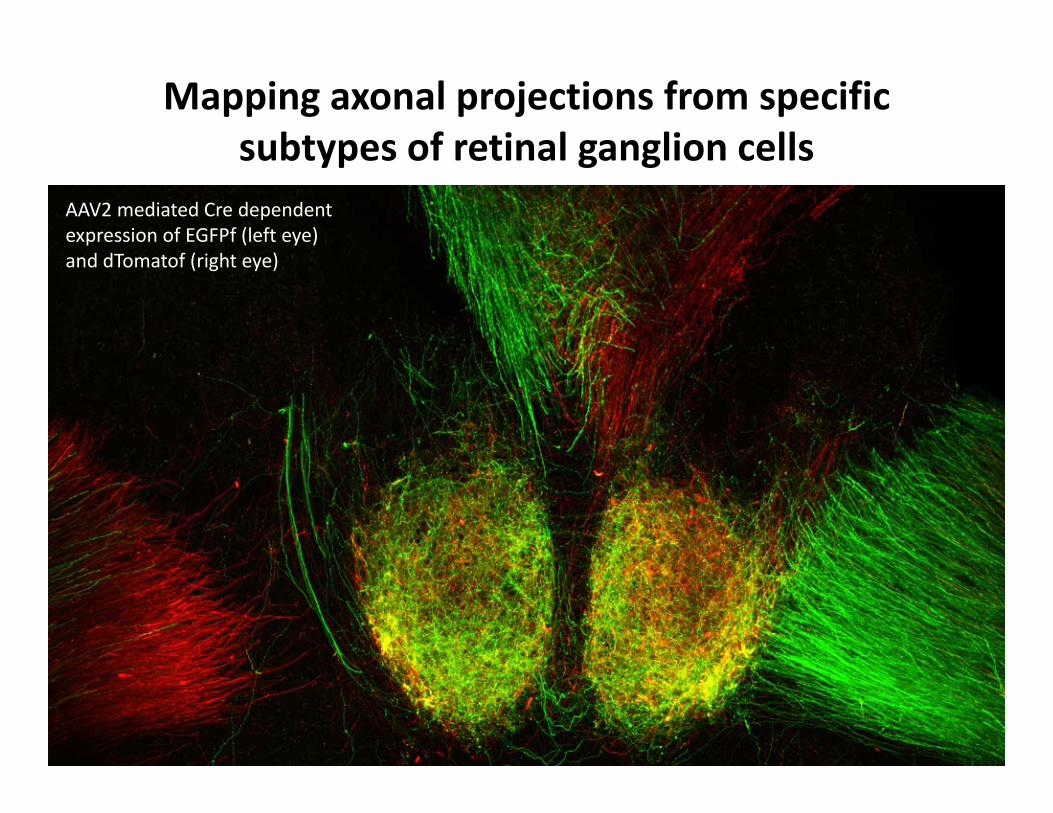
Mapping axonal projections from specific subtypes of retinal ganglion cells
AAV2 mediated Cre dependent expression of EGFPf (left eye) and dTomatof (right eye)

Adenoviral vectors (Ad5)
• Non‐enveloped virus ‐ 90nm diameter virions
• Double stranded DNA genome
• Infection mediated by fiber binding to the host cell CAR receptor
•Many different human and non‐human serotypes identified
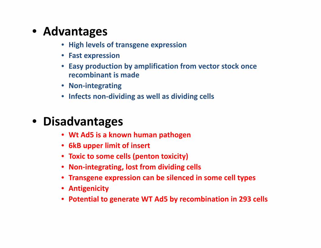
• Advantages• High levels of transgene expression• Fast expression• Easy production by amplification from vector stock once recombinant is made
• Non‐integrating• Infects non‐dividing as well as dividing cells
• Disadvantages• Wt Ad5 is a known human pathogen• 6kB upper limit of insert• Toxic to some cells (penton toxicity)• Non‐integrating, lost from dividing cells• Transgene expression can be silenced in some cell types• Antigenicity• Potential to generate WT Ad5 by recombination in 293 cells

Shedding of viral vectors from experimental animals
• What are viruses and what makes a viral vector?
• Basic features of the viral vectors used in this study
• Evaluation of the risk of shedding of three common genetically modified viral vectors from transduced rodents
• Assessment of the potential for human exposure to these vectors from treated animals.

Study Design –(A) shedding of infectious vectors from animals
• Naïve CD1 and NOD/SCID mice were inoculated with 100ul of each viral vector via tail vein injection.
• Vector dose per animal was: • LV‐CMV‐EGFP = 9.51x10⁸ IU • AAV2‐CMV‐EGFP = 1.03x10⁹ IU• Ad5‐CMV‐EGFP, 1.9x10⁹ IU
• Sampling time points were ‐1, 0, 1, 2, 3 and 7 days post inoculation• Transduction of target tissue confirmed histologically and by qPCR• Levels of infectious vector at the injection site were assessed using a cell
infectivity assay• Presence of vector genomes in blood, urine and feces was assessed by
endpoint PCR using primers against the EGFP transgene• Sentinel animals housed with the treated animals were also assessed

Vector transduction of mouse liver 7d post inoculation– native EGFP expression
4x whole mountstereoscopy

Vector transduction of mouse tissue 7d post inoculation– QPCR quantification

Recovery of infectious vector from injection site

Recovery of LV vector genomes from injection site
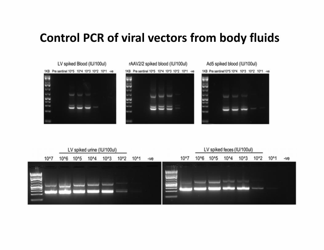
Control PCR of viral vectors from body fluids

PCR of viral vector genomes from infected animals
Shedding of viral vectors from experimental animals
• What are viruses and what makes a viral vector?
• Basic features of the viral vectors used in this study
• Evaluation of the risk of shedding of three common genetically modified viral vectors from transduced rodents
• Assessment of the potential for human exposure to these vectors from treated animals.

Study Design –(B) infectious vector in the environment
• Control experiments tested the stability of viral vectors spotted onto animal cage plastic and allowed to dry.
• Time points were days 0, 1, 3, 5 and 14 post inoculation
• Recovered vectors were used in a cell based infectivity assay and compared to fresh titer matched stocks
• Stability of vectors on clean and soiled animal bedding was also assessed by cell infectivity assay
• Time points were 0, 30min, 24hr and 72hr post inoculation
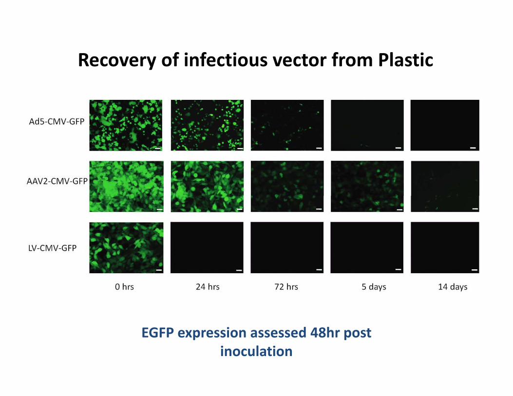
Recovery of infectious vector from Plastic
EGFP expression assessed 48hr post inoculation

Recovery of infectious vector from soiled corn cob bedding
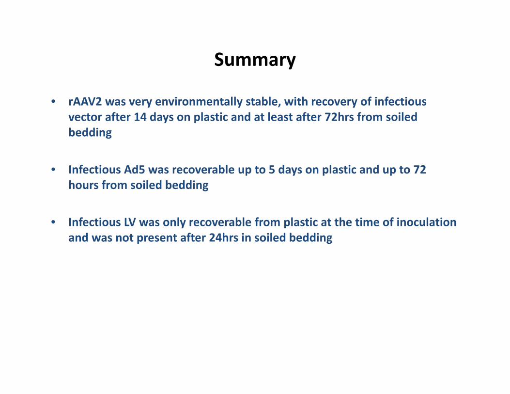
Summary
• rAAV2 was very environmentally stable, with recovery of infectious vector after 14 days on plastic and at least after 72hrs from soiled bedding
• Infectious Ad5 was recoverable up to 5 days on plastic and up to 72 hours from soiled bedding
• Infectious LV was only recoverable from plastic at the time of inoculation and was not present after 24hrs in soiled bedding

Summary
Following IV tail vein administration:
• Infectious Ad5 and LV were present at the injection site up to 24hrs post inoculation. Infectious rAAV2 was present up to 72hrs
• No infectious rAAV2, Ad5 or LV was found in samples of soiled bedding taken from cages group housing treated animals
• rAAV2 genomic DNA was detectable by PCR up to 24hrs post inoculation in blood and feces, but not urine from treated animals
• No Ad5 or LV vector genomes were detectable by PCR in blood, urine or feces at any time point post inoculation
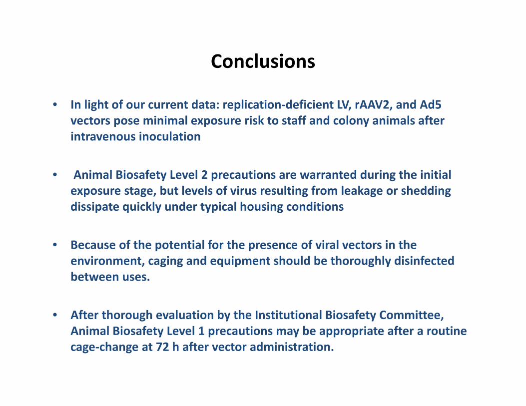
Conclusions
• In light of our current data: replication‐deficient LV, rAAV2, and Ad5 vectors pose minimal exposure risk to staff and colony animals after intravenous inoculation
• Animal Biosafety Level 2 precautions are warranted during the initial exposure stage, but levels of virus resulting from leakage or shedding dissipate quickly under typical housing conditions
• Because of the potential for the presence of viral vectors in the environment, caging and equipment should be thoroughly disinfected between uses.
• After thorough evaluation by the Institutional Biosafety Committee, Animal Biosafety Level 1 precautions may be appropriate after a routine cage‐change at 72 h after vector administration.

Thank you for your attention
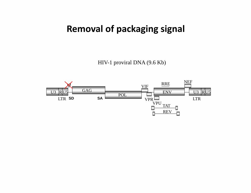
HIV-1 proviral DNA (9.6 Kb)
POLGAG
ENV
LTRLTR
NEF
VPU TATVPR
VIF
REV
U3 RU5U3 RU5
RRE
SD SA
Removal of packaging signal

HIV-1 proviral DNA (9.6 Kb)
POLGAG
ENV
LTRLTR
NEF
VPU TATVPR
VIF
REV
U3 RU5U3 RU5
RRE
SD SA
Deletion of Vpu

HIV-1 proviral DNA (9.6 Kb)
POLGAG
LTRLTR
NEF
VPU TATVPR
VIF
REV
U3 RU5U3 RU5
RRE
SD SA
Deletion of Vpu and truncation of env N‐terminus
ENV

HIV-1 proviral DNA (9.6 Kb)
POLGAG
NEF
VPU TATVPR
VIF
REV
RRE
SD SAENVCMV polyA
Removal of LTRs and addition of CMV promoterand poly A signal for expression in 293T cells
1st Generation packaging construct

HIV-1 proviral DNA (9.6 Kb)
POLGAG
NEF
TATVPR
VIF
REV
RRE
SD SA
Removal of accessory proteins except tat and revAnd complete deletion of env
CMV polyA
VPU
2nd Generation packaging construct

Separation of GAG/POL from REV and Deletion of TAT
POLGAG
REV
CMV
RRE
SD SA
polyARSV
polyA
polyA
TAT
3rd Generation split genome packaging constructs

1st Generation Adeno Vectors

Adeno Vector Generation

rAAV production
• rAAV2 genomes can be cross‐packaged into vectors using natural AAV capsids (e.g. serotypes 1‐9, rh10) as well as molecularly evolved synthetic capsids (e.g. AAV‐DJ, AAV 2i8)
• Typical yields: ~ 3ml of 1011 ‐ 1014 GC/ml - in vivo grade

HIV-1 proviral DNA (9.6 Kb)
POLGAG
ENV
LTRLTR
NEF
VPU TATVPR
VIF
REV
U3 RU5U3 RU5
RRE
SD SA
Design of packaging constructs for safety

CMV
PackagingConstructs
Envelope
Self‐InactivatingTransfer Vector
RSV polyAREV
CMV GAG
PRO POLRRE polyA
polyAVSV.GCMV
GA RRE
transgene
SD SA
U3 deletion U3 deletion
LTR LTR
3rd Generation Lentiviral Vector SystemWPREcPPT
The genome of the virus is split and overlapping regions eliminatedto minimize the possibility of recombination and regeneration of a WT virus

Sequence overlap between wild‐type HIV‐1 and 3rd gen lentiviral vector genomes
gag 366 bp dU3 15 bprre 238 bp R 56 bpcppt 121 bp U5 83 bpppt 15 bp
total: 977 bp
= 9.9% of the HIV-1 genome (9719 bp)