assessment of acute antipsychotic poisoned cases admitted
TRANSCRIPT

113
Ain Shams Journal of Forensic Medicine and Clinical Toxicology
July 2019, 33: 113-125
Assessment of Acute Antipsychotic Poisoned Cases Admitted to Tanta University Poison Control Unit
Marwa A. Mubarak, Enas I. El Madah, Doaa M. El Gharbawy and Magdy M. Ashmawy1 1 Department of Forensic Medicine and Clinical Toxicology, Faculty of Medicine, Tanta University, Tanta, Egypt.
All rights reserved.
Abstract Background: Sedative hypnotic/antipsychotic poisoning represented 5.84% of cases according to
national poison data system in 2017. In Egypt, studies about antipsychotic poisoning are scarce.
Objectives: is to assess the pattern of toxicity and prognosis of cases with acute antipsychotic
poisoning admitted to Tanta University Poison Control Unit.
Patients and Methods: This study was conducted on cases who admitted to Tanta University Poison
Control Unit with acute antipsychotics poisoning. All cases were subjected to clinical evaluation,
laboratory investigations and electrocardiogram. Severity was assessed by poison severity score.
Results: Majority of cases were represented equally in age groups (0-10) and (11-20) years with
33.3% for each age group, 65% of cases were females, most of cases were from urban areas (71.7%)
and singles (71.7%). History of mental disorder found in 31.6% of cases and suicidal attempts
represented the most common manner of poisoning (71.7%). Clozapine was the highest antipsychotic
drug taken (35%) and 6.7% of cases developed extrapyramidal manifestations. Miosis was found in
56.7% of cases. Tachycardia was recorded in 46.7% of cases and QTc interval was prolonged in 43%
of cases. According to poison severity score, most studied cases were either mild (51.7%) or moderate
(33.7%). One case was admitted to ICU. Multiple regression analysis showed that decreased GCS,
PCO2, O2 saturation were associated with increased hospital stay.
Conclusion: Antipsychotic poisoning was commonly mild to moderate. Neurological and
cardiovascular manifestations were the predominant. Sinus tachycardia and prolonged QTc were the
most common electrocardiographic changes. Antipsychotic poisoning usually had a good prognosis.
Key words antipsychotics, accidental poisoning, suicidal poisoning
Introduction rug overdose is the most common cause of acute
poisoning worldwide. Moreover, poisoning is the
most frequent method of suicide attempts in
which psychotropic drugs as antipsychotics,
antidepressants and benzodiazepines are significantly
used (Kim et al., 2015, Mowry et al., 2016).
Antipsychotics are primary used to treat
schizophrenia, manic phase of bipolar disorders and
agitated behavior; however they are often used to treat
nausea, vomiting, headache, and various neurological
conditions (chorea, dystonia and tics). Antipsychotics
toxic effects include anticholinergic and extrapyramidal
syndromes as well as CNS and cardiovascular depression
(Georgiev et al., 2015).
Antipsychotics are classified into two groups:
typical and atypical. The typical one or the first
generation includes the older drugs as butyrophenones,
dibenzoxazepines, diphenylbutylpiperidine and
phenothiazines. Atypical group or the second generation
includes the newer drugs as benzopines, indoles,
quinolinone (DeSilva et al., 2006).
Diagnosis of antipsychotic overdose is based on
history coupled with predictable symptoms and physical
findings. Plasma concentrations of antipsychotics are not
widely available and not useful in management of acute
antipsychotic overdose, this demonstrates the importance
of clinical evaluation in diagnosing these cases (Minns
and Clark, 2012).
Supportive care is the cornerstone for treatment
of antipsychotic overdose. Gastrointestinal
decontamination with activated charcoal should be done.
No specific antidote is present. Cardiovascular
complications are managed by intravenous fluids,
vasopressors if needed and sodium bicarbonate in cases
of ventricular dysrhythmia. Seizures are treated with
benzodiazepines followed by phenobarbital.
Hemoperfusion and hemodialysis are not effective
(Isbister et al., 2007, Minns and Clark, 2012).
D

114 Mubarak et al., / Ain Shams J Forensic Med Clin Toxicol, July 2019 (33): 113-125
Aim of the work This study aimed to assess the pattern of toxicity in acute
antipsychotic poisoned cases admitted to Tanta
University Poison control Unit at emergency hospital.
Patients and Methods This study was conducted on cases admitted to Tanta
University Poison Control Unit with history, symptoms
and signs of acute antipsychotics poisoning. Cases were
selected by convenience sampling (this means that all
cases with antipsychotic poisoning admitted at this
period were included in this study without
randomization) in the period from the 1st of November
2016 to 1st of November 2017. The study was carried out
after approval of the medical research ethical committee
of Tanta Faculty of Medicine. Written informed consent
was signed by the cases or their guardians after
explaining the aim and method of the study. The
exclusion criteria included patients who refused to sign
the informed consent and patients with chronic
poisoning, co-ingestion, chronic illness or patients with
history of drug addiction.
Methods
All cases were subjected to sociodemographic evaluation
including age, sex, occupation, educational level,
residence, marital status and special habits; smoking,
coffee and alcohol. Additionally, past medical history,
history of previous admission to hospital, toxicological
history including name of drug, its form, route of intake,
amount of drug taken, manner of poisoning, reason in
case of intentional poisoning, time of drug intake, time of
hospital admission and delay time were recorded.
Clinical examination was conducted through physical
examination; vital signs, level of consciousness using
Glasgow coma scale and systemic examination. Severity
of cases was assessed by poison severity score (Persson
et al., 1998). Laboratory investigations were measured
including arterial blood gases, liver enzymes, kidney
function tests, complete blood count, electrolytes
(sodium, potassium and magnesium) and random blood
sugar. Electrocardiogram was done for the studied
patients. Medical treatment was done according to
protocol of antipsychotic treatment in Tanta poison
control unit.
Outcome measures (prognosis) were recorded
including duration of hospital stay, complete recovery,
complications, need for intubation, ventilation, intensive
care unit or death.
Statistical analysis Was conducted by IBM SPSS software package version
21 including descriptive statistics (number, percentage,
minimum, maximum, mean, standard deviation). Chi-
square, Kruskal-Wallis, median, t test and one-way
ANOVA were done also to identify factors influencing
manner of poisoning, effect of antipsychotic poisoning
on clinical and biochemical parameters of cases and
regression analysis to test factors affecting outcome.
Results
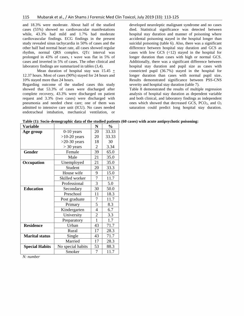
Socio-demographic study showed that most of cases
were in age groups (0-10) and (11-20) years with 33.3%
for each age group followed by age group (21- 30) years
representing 30% of cases. More than half of the studied
cases were females (65%). Most of cases were
unemployed (35%), while, 33.3% were students, and
50% were secondary educated. The majority of cases
were from urban areas and singles (71.7%). Only 11.7%
were smokers. (Table 1)
Toxicological findings demonstrated that 66.7%
had no past medical history, while 31.6% were mentally
ill. Manner of poisoning in 71.7% of cases was suicidal
followed by accidental (28.3%). Intentional poisoning
was due to family troubles (40%) followed by psychiatric
illness (28.3 %). Clozapine was the drug used in 35% of
cases, followed by risperidone (20%), chlorpromazine
(15%), quetiapine (11.7%), olanzapine (6.7%),
haloperidol (3.3%), and aripiprazole (3.3%). All cases
were poisoned orally by tablets (91.7%) followed by
syrup (8.3%). Most of cases were admitted within 5
hours from drug ingestion (75%) (Table 2).
Analysis of the above data showed highly
significant association between manner of poisoning and
age groups (p value <0.001). All cases of accidental
poisoning were less than 10 years while most of cases
with suicidal poisoning were focused in two age groups
(>10-20 and >20-30 years). Additionally, there was a
significant association between manner of poisoning and
educational level as most of cases with accidental
poisoning were in preschool and kindergarten children
while most cases with suicidal poisoning were
encountered among patients of secondary school and post
graduates (p = 0.001). Also, a significant association was
found between manner of poisoning and residence (p
value = 0.002) where both accidental and suicidal
poisoning were more common in urban than rural areas.
Regarding history of medical disease; most of cases with
accidental poisoning had no past medical history
(82.35%) while no history of medical diseases was found
in 60.5% in cases of suicidal poisoning. This difference
is statistically significant (p=0.026) (Table 5).
There was a statistical significant difference
between cases with suicidal and accidental poisoning
regarding past medical history as most of cases with
accidental poisoning had no history of medical diseases
(82.35%) and 11.76% of them suffered from mental
disorders. while, in cases with suicidal poisoning 60.5%
had no history of medical diseases and 39.5%/ gave a
history of mental diseases (Table 5).
The present study demonstrated that 51.7% of
cases were classified as mild poisoning according to PSS
while 33.3% were moderate and only 5% showed severe
toxicity on admission. Clinical evaluation of the studied
cases showed that 5% of cases suffered from mild GIT
manifestations, 3.3% were presented with moderate
respiratory manifestations. Regarding CNS
manifestations, 43.3% suffered from mild manifestations
115 Mubarak et al., / Ain Shams J Forensic Med Clin Toxicol, July 2019 (33): 113-125
and 18.3% were moderate. About half of the studied
cases (55%) showed no cardiovascular manifestations
while, 43.3% had mild and 1.7% had moderate
cardiovascular findings. ECG findings in the present
study revealed sinus tachycardia in 50% of cases and the
other half had normal heart rate, all cases showed regular
rhythm, normal QRS complex. QTc interval was
prolonged in 43% of cases, t wave was flat in 5% of
cases and inverted in 5% of cases. The other clinical and
laboratory findings are summarized in tables (3,4).
Mean duration of hospital stay was 15.45 +
12.37 hours. Most of cases (90%) stayed for 24 hours and
10% stayed more than 24 hours.
Regarding outcome of the studied cases this study
showed that 53.3% of cases were discharged after
complete recovery, 43.3% were discharged on patient
request and 3.3% (two cases) were discharged with
pneumonia and needed chest care; one of them was
admitted to intensive care unit (ICU). No cases needed
endotracheal intubation, mechanical ventilation, or
developed neuroleptic malignant syndrome and no cases
died. Statistical significance was detected between
hospital stay duration and manner of poisoning where
accidental poisoning stayed in the hospital longer than
suicidal poisoning (table 6). Also, there was a significant
difference between hospital stay duration and GCS as
cases with low GCS (<12) stayed in the hospital for
longer duration than cases with high or normal GCS.
Additionally, there was a significant difference between
hospital stay duration and pupil size as cases with
constricted pupil (36.7%) stayed in the hospital for
longer duration than cases with normal pupil size.
Results demonstrated significance between PSS-CNS
severity and hospital stay duration (table 7).
Table 8 demonstrated the results of multiple regression
analysis of hospital stay duration as dependent variable
and both clinical, and laboratory findings as independent
ones which showed that decreased GCS, PCO2, and O2
saturation could predict long hospital stay duration.
Table (1): Socio-demographic data of the studied patients (60 cases) with acute antipsychotic poisoning:
Variable N %
Age group 0-10 years
>10-20 years
>20-30 years
> 30 years
20
20
18
2
33.33
33.33
30
3.34
Gender Female 39 65.0
Male 21 35.0
Occupation Unemployed 21 35.0
Student 20 33.3
House wife 9 15.0
Skilled worker 7 11.7
Professional 3 5.0
Education Secondary 30 50.0
Preschool 11 18.3
Post graduate 7 11.7
Primary 5 8.3
Kindergarten 4 6.7
University 2 3.3
Preparatory 1 1.7
Residence Urban 43 71.7
Rural 17 28.3
Marital status Single 43 71.7
Married 17 28.3
Special Habits No special habits 53 88.3
Smoker 7 11.7 N: number

116 Mubarak et al., / Ain Shams J Forensic Med Clin Toxicol, July 2019 (33): 113-125
Table (2): Medical and toxicological findings of the studied cases (60 cases) with acute antipsychotic poisoning:
Medical and Toxicological history N %
Past medical history No history of special medical disease 40 66.7
History of mental disorders 19 31.6
History of bronchial asthma 1 1.7
Manner of poisoning Suicidal 43 71.7
Accidental 17 28.3
Reasons of intentional poisoning Family troubles 24 40.0
Psychiatric illness 17 28.4
Learning problems 2 3.3
Drugs taken
Clozapine 21 35.0
Risperidone 12 20.0
Chlorpromazine 9 15.0
Quetiapine 7 11.7
Olanzapine 4 6.6
Aripiprazole 2 3.3
Haloperidol 2 3.3
Amisulpride 1 1.7
Combined (Risperidone and Quetiapine) 1 1.7
Combined (Chlorpromazine and Risperidone) 1 1.7
Route of intake Oral 60 100
Form of the drug taken Tablet 55 91.7
Syrup 5 8.3
Delay time 1-5 hours 45 75
>5-10 hours 9 15
>10-15 hours 5 8.3
>15-20 hours 0 0
>20 hours 1 1.7 N: number.

117 Mubarak et al., / Ain Shams J Forensic Med Clin Toxicol, July 2019 (33): 113-125
Table (3): Clinical evaluation of the studied cases (60 cases) with acute antipsychotic poisoning:
N %
Pulse rate (beats/min)
Normal 32 53.33
Tachycardia 28 46.67
Bradycardia 0 0.00
Blood pressure (mmHg)
Normotensive 54 90.00
Hypotensive 3 5.00
Hypertensive 3 5.00
Respiratory rate (Cycles/min)
Normal 32 53.33
Tachypnea 28 46.67
Bradypnea 0 0.00
Temperature (oc) (axillary) Normal 57 95.00
Hyperthermia 3 5.00
Hypothermia 0 0.00
GCS score 3-8 5 8.3
9-12 4 6.7
13-15 51 85.00
Speech Normal 35 58.3
Slurred comprehensible 22 36.7
Slurred un comprehensible 3 5.0
Head and neck Free 56 93.3
Dystonia or dyskinesia 4 6.7
Pupil Constricted 34 56.7
Normal 24 40.0
Dilated 2 3.3
Chest Free 56 93.3
Crepitation 2 3.3
Diminished air entry 1 1.7
Wheezing 1 1.7
Abdomen Free 60 100.0
Extremities
Free 59 98.3
Involuntary movements 1 1.7
Poison Severity Score None
Mild
Moderate
Severe
6
31
20
3
10.0%
51.7%
33.3%
5.0% N: number, GCS: Glasgow Coma Scale

118 Mubarak et al., / Ain Shams J Forensic Med Clin Toxicol, July 2019 (33): 113-125
Table (4) Laboratory findings of studied cases (60 cases) with acute antipsychotic poisoning.
N %
Arterial Blood gases Normal 40 66.7
Respiratory acidosis 2 3.3
Respiratory alkalosis 16 26.7
Mixed disorder 2 3.3
Sodium (mEq/L) Normal 59 98.3
Hypernatremia 1 1.7
Hyponatremia 0 0
Potassium (mEq/L) Normal 51 85
Hypokalemia 9 15
Hyperkalemia 0 0
Magnesium (mg/dL) Normal 60 100
Random blood sugar (mg/dL) Normal 51 85
Hyperglycemia 9 15
Hypoglycemia 0 0
Liver enzymes
(U/L)
Normal 59 98.3
Increased 1 1.7
Decreased 0 0
Kidney Function (mg/dl) Normal 60 100
Complete Blood Count
Hb (gm/dl) Normal 4 6.7
Abnormal 56 93.3
RBCs (mcL) Normal 47 78.3
Abnormal 13 21.7
WBCs (mcL) Normal 48 80
Abnormal 12 20
Platelets (mcL)
Normal 59 98.3
Abnormal 1 1.7
n= Number, mEq/L= milliEquivalents per Liter, mg/dL= milligrams per deciliter, U/L= Unit per Liter gm/dl= gram per
deciliter, mcL: microliter.

119 Mubarak et al., / Ain Shams J Forensic Med Clin Toxicol, July 2019 (33): 113-125
Table (5): Association between manner of poisoning and sociodemographic data in the studied cases of (60 cases)
acute antipsychotic poisoning:
Manner of poisoning Tests of significance
Accidental
(N = 17)
Suicidal
(N = 43)
Test Statistic p
Age (Years) Mean ± SD 4+2 21+6 -14.961 a <0.001*
N % N %
Age groups
0-10 17 100.0% 3 6.98%
46.891 b <0.001* > 10 - 20 0 0.0% 20 46.51%
> 20 - 30 0 0.0% 18 41.86%
> 30 0 0.0% 2 4.65%
Gender
Female 9 52.9% 30 69.8% 1.516 c 0.218
Male 8 47.1% 13 30.2%
Education
KG1 4 23.5% 0 0.0%
4.090 b 0.001*
Preschool 11 64.7% 0 0.0%
Post graduate 0 0.0% 7 16.28%
Preparatory school 0 0.0% 1 2.3%
Primary school 2 11.8% 3 6.97%
Secondary School 0 0.0% 30 69.8%
University 0 0.0% 2 4.65%
Marital status
Married 0 0.0% 17 39.5% 0.270 b 0.604
Single 17 100.0% 26 60.5%
Residence
Urban 13 76.5% 30 69.8% 9.378 c 0.002*
Rural 4 23.5% 13 30.2%
Special Habits
None 17 100.0% 36 83.7% 3.133 b 0.077
Smoker 0 0.0% 7 16.3%
Past medical history
No Past medical history 14 82.35% 26 60.5%
6.135 b 0.026* Mental disorders 2 11.76% 17 39.5%
Bronchial asthma 1 5.89% 0 0.0%
N: number; SD: standard deviation; a: Independent samples t test; b: Fisher-Freeman-Halton Exact Test; *significant at
p<0.05.
Table (6): Association between hospital stay duration and age groups, gender, past medical history, mode of
poisoning and ingested dose in the studied cases (60 cases) of acute antipsychotic poisoning.
Hospital Stay (hours) Tests of significance
≤ 13
(N = 30)
> 13
(N = 30) Test Statistic p
Age groups
0-10 6 20.0% 14 46.7% 5.551 a 0.120
> 10 - 20 11 36.7% 9 30.0%
> 20 - 30 12 40.0% 6 20.0%
> 30 1 3.3% 1 3.3%
Gender Female 22 73.3% 17 56.7% 1.832b 0.176
Male 8 26.7% 13 43.3%
Past medical history
No history 20 66.7% 20 66.7% 1.015 a 1.000
Mental disorders 10 33.3% 9 30.0%
Bronchial asthma 0 0.0% 1 3.3%
Manner of poisoning Accidental 5 16.7% 12 40.0% 4.022 b 0.045*
Suicidal 25 83.3% 18 60.0%
Ingested dose Median (IQR) 500 (200 - 1000) 150 (14 - 900) - 1.496 c 0.135
Mean ranks 30.1 23.8
N: number; IQR: interquartile range; a: Fisher-Freeman-Halton Exact Test; b: Pearson's Chi test; c: Mann-Whitney test; *
significant at p<0.05.

120 Mubarak et al., / Ain Shams J Forensic Med Clin Toxicol, July 2019 (33): 113-125
Table (7): Association between hospital stay duration and poison severity score in the studied cases (60 cases) of
acute antipsychotic poisoning.
Hospital Stay in hours Tests of significance
≤ 13 > 13 Total Test statistic p
n % n % n %
GIT 0 28 93.3% 29 96.7% 57 95.0% FE 1.000
1 2 6.7% 1 3.3% 3 5.0%
Respiratory 0 30 100.0% 28 93.3% 58 96.7% FE 0.492
3 0 0.0% 2 6.7% 2 3.3%
CNS 0 17 56.7% 7 23.3% 24 40.0% X2ChS= 11.570 0.003*
1 12 40.0% 13 43.3% 25 41.7%
2 1 3.3% 10 33.3% 11 18.3%
CVS 0 17 56.7% 16 53.3% 33 55.0% X2FFH =1.138 0.795
1 12 40.0% 14 46.7% 26 43.3%
2 1 3.3% 0 0.0% 1 1.7%
Metabolic 0 18 60.0% 21 70.0% 39 65.0% X2FFH =1.517 0.539
1 11 36.7% 7 23.3% 18 30.0%
2 1 3.3% 2 6.7% 3 5.0%
Liver 0 30 100.0% 30 100.0% 60 100.0%
Kidney 0 30 100.0% 30 100.0% 60 100.0%
Blood 0 30 100.0% 30 100.0% 60 100.0%
Muscular 0 30 100.0% 30 100.0% 60 100.0% n: number; FE: Fisher's exact test; X2ChS: Pearson's Chi square test; X2FFH: Fisher-Freeman-Halton test; * significant
at p <0.05.
Table (8): Multiple regression analysis test
ANOVA test Adjusted
R square
Variables Unstandardized
Coefficients
t p 95.0%
Confidence
Interval for B
F p B SE Lower Upper
6.689 <0.001* 0.354 (Constant) 241.1 61.7 3.906 <0.001* 116.9 365.2
Manner of
poisoning
(suicidal)
-9.0 3.5 -
2.550
0.014* -16.0 -1.9
GCS -3.3 0.8 -
4.300
<0.001* -4.9 -0.5
O2
saturation
-1.7 0.6 -
2.845
0.007* -3.0 -0.5
PCO2 -0.6 0.2 -
1.680
0.100 -1.2 0.1
HCO3 1.1 0.6 1.787 0.080 -0.1 2.4 R square: square of residuals; SE: standard error.
Discussion
Antipsychotic poisoning is one of the top five substances
most frequently involved in human poisoning as sedative
hypnotics/antipsychotics poisoning represented 5.84% of
cases in 2017. However, sedative hypnotic/antipsychotic
poisoning increased most rapidly by 10.7% per 2017
(2088 cases/year) according to national poison data
system (Gummin et al., 2017). Meanwhile, in Egypt,
there is limited data about antipsychotic poisoning.
Therefore, this study aimed to assess the pattern of
toxicity in acute antipsychotic poisoned cases admitted to
Tanta University Poison control Unit at emergency
hospital throughout the period from 1st of November
2016 to the end of October 2017.
In the present study, childhood and adolescence
less than 20 year of age represented the majority of cases
(66.7%) which could be attributed to the rapid increase in
the use and prescription of antipsychotic medication for
children and adolescent in the last two decades for

121 Mubarak et al., / Ain Shams J Forensic Med Clin Toxicol, July 2019 (33): 113-125
managing sleep disorders, anxiety and mood disorders so
it became more available at home (Berling et al., 2016).
Furthermore, suicidal attempt by psychiatric medications
in adolescence were reported in previous study (Sheridan
et al., 2017). This result was explained by factors like
emotional disturbance, puberty changes, educational
problems, family relationship and media influence
(Bazrafshan et al., 2016). Also, the unsafe medications
storage in addition to unsafe environment and
insufficient supervision of children behavior can explain
the increased children rate in the sample. Pediatric
poisoning with antipsychotics is remarkably increasing in
the last years and became a significant cause of
morbidity (Meli et al., 2014, Dayasiri et al., 2017).
The higher female ratio (65%) could be
explained by the concept that females are socially and
psychologically more vulnerable to drug poisoning than
males in our culture (Boukatta et al., 2014, Shojaei et al.,
2014, Georgiev et al., 2015, Borg et al., 2016, Toft et al.,
2017). Most of our cases were unemployed (35%)
followed by students (33.3%). The high rate of suicide
among unemployed was reported to be due to their
socioeconomic and psychological problems (Milner et
al., 2014), while the poor family communication,
parent’s economic problems, educational problems and
failure in love were reported to be the causes of suicide
among students (Mohammadkhani et al., 2006).
Most of the studied cases with both suicidal and
accidental poisoning were from urban areas (71.7%),
while 28.3% were from rural areas. This was also
reported by some authors (Anthony and Kulkarni, 2012).
Additionally, urban areas are at a higher risk of suicide
due to more stressful life, family conflicts, job
competitions and more liability for psychiatric disorders
(Qin, 2005). Singles were more in our sample (71.7%)
than married. The decrease in social integration and
maturity of single than married could explain their
suicidal vulnerability (Griffiths et al., 2008).
Most of cases were poisoned by atypical
antipsychotics (81.7%) which coincided with the results
founded by the study of Berling and his colleagues
(Berling et al., 2016). This reflects the increase in the use
of atypical antipsychotics than typical ones due to less
neurological side effects as extrapyramidal
manifestations (Ucok and Gaebel, 2008). The worldwide
increase of suicide may explain the increase of suicidal
poisoning in our cases (71.7%) which coincided with
other studies (Georgiev et al., 2015, Borg et al., 2016).
Most of cases admitted to the hospital within 5 hours
from ingestion (75%) which could be attributed to the
location of Tanta Poison Control Unit in the center of
Delta with easy availability of transportation for it.
The clinical profile of antipsychotic poisoning
in our cases demonstrated that about half of the studied
cases had mild general symptoms like tachycardia
(46.67%), tachypnea (46.67%) and miosis (56.7%) while,
small number were presented with serious
manifestations like GCS less than 8 (8.3%) and dystonia
(6.7%). This was in agreement with the previous reports
of lower morbidity and lower lethal effect of
antipsychotic poisoning in most of cases specially the
atypical ones (Capel et al., 2000, Rasimas and Liebelt,
2012, Meli et al., 2014). Most of our cases didn’t
consume big doses and most of those who poisoned
themselves intended only to seek attention not to kill
themselves or ingested the drug accidentally.
CNS depression manifestations in the studied
cases were related to acute intoxication with clozapine
(35%), it is known that tachycardia and CNS depression
are common manifestations of its toxicity (Pickford,
2000, Kramer et al., 2010). This could be explained by
the fact the that clozapine has a wide profile of receptor
binding affinity (Sackey et al., 2017). Blood pressure
changes were not common in our cases which were
similar to previous studies (Tan et al., 2009, Gugger,
2011, Mucci et al., 2016, Ramnarine, 2017).
Tachypnea was observed in 46.67% of cases
and no cases had bradypnea which could be attributed to
stress and anxiety. Moreover, the absence of bradypnea
could be explained in most of our cases by the absence of
marked CNS depression. Studies showed that significant
respiratory depression is not common with antipsychotic
poisoning (Rasimas and Liebelt, 2012). Slurred speech
was observed in 41.7% of the studied cases which agreed
with the study of Kramer and his colleagues (Kramer et
al., 2010) who reported dysarthria in 15.1% of cases
intoxicated with clozapine.
The present study showed that pupil was
constricted in more than half of cases (56.7%), dilated in
3.3% and normal in 40% of cases. Pupil size changes
were recorded by some previous researches (Pickford,
2000, Meli et al., 2014). Miosis was explained by the α1
adrenergic receptor blocking effect of antipsychotics
while mydriasis was explained by the anticholinergic
effect of these drugs (Stahl, 2013).
Dystonia was found in 6.7% of the studied cases
who were intoxicated by chlorpromazine, haloperidol
and risperidone. The low incidence of extrapyramidal
manifestations in our study could be due to most of our
cases were intoxicated by atypical antipsychotics
(81.7%) which are known to have a lower incidence of
extrapyramidal manifestations than typical antipsychotics
because of their lower dopamine receptor binding affinity
(Divac et al., 2014). Poison severity score (PSS) revealed
that most of cases showed mild severity (51.7%), 33.7%
were moderate, 5% were severe and 10% had non grade
poison severity score. No fatalities were recorded in our
cases. This was similar to previous studies (Rasimas and
Liebelt, 2012, Meli et al., 2014).
This study revealed ECG changes as tachycardia
(50%), QTc interval prolongation (43%) which was
reported to be the most common ECG changes in
antipsychotic poisoning (Tan et al., 2009). The heart
rhythm was regular with normal QRS morphology.
Previous studies showed low risk of ventricular
arrhythmia and cardiac arrest (Liperoti et al., 2005). T
wave flattening and inversion was detected in 10 % of
cases which agreed with previous researches (Marano et

122 Mubarak et al., / Ain Shams J Forensic Med Clin Toxicol, July 2019 (33): 113-125
al., 2011). Also, the majority of cases had prolonged QT
interval and were poisoned by atypical antipsychotics
(78%) while 22% were poisoned with typical agents. On
the other hand, Chohan and his colleagues stated that
atypical antipsychotics were less commonly to prolong
QTc interval than typical ones (Chohan et al., 2015). This
difference could be attributed to over representation of
atypical (81.7%) than typical antipsychotics (18.3%) in
our sample. In this study, no cases presented with torsade
de points which coincided with previous studies
(Wenzel-Seifert et al., 2011).
Most of cases in this study had normal arterial
blood gas (66.7%) followed by respiratory alkalosis
(26.7%) then respiratory acidosis and mixed disorder
were equally represented (3.3% for each). This coincided
with the results reported by Capel and his colleagues
(Capel et al., 2000). Hypokalemia was found in 15% of
our cases. Malik et al. reported hypokalemia with
risperidone and quetiapine toxicity (Malik et al., 2005).
Antipsychotics block potassium efflux channel and
inhibit shifting of potassium from intracellular to
extracellular space (Pal et al., 2015). Furthermore,
Hyperglycemia was detected in 15% of our cases which
could be attributed to the decrease in insulin sensitivity
due to 5-HT2A receptor antagonism by antipsychotics
(Müller et al., 2009, Yam et al., 2013).
One case of acute clozapine toxicity (1250 mg)
showed high level of AST (51 U/L) and ALT (73 U/L).
Other studies (Erdogan et al., 2004, Chou et al., 2014)
reported also the elevation of liver enzymes with
clozapine intake but this effect is usually asymptomatic
and usually transient phenomenon. Leukocytosis was
detected in 20% of cases while other cases had normal
WBCs count. 50% of cases who had leukocytosis were
intoxicated by clozapine and the mechanism is still
unknown but one possibility is that clozapine may induce
the release of some cytokines as TNF, IL-2, IL-6, and G-
CSF (Fehsel et al., 2005). Another explanation of
leukocytosis could be attributed to presence of infection
such as pneumonia which developed in 2 cases in our
study. Also, the results showed no abnormality in
sodium, magnesium level and kidney function tests
which coincided with the results reported by other
authors (Capel et al., 2000).
The present study revealed that the median
duration of hospital stay was 13 hours which is close to
the results demonstrated by Berling and his colleagues
(Berling et al., 2016). Most of the studied sample had just
mild toxicity (51.7%) with 53.3% of cases were
discharged after complete recovery. Cases with GCS <12
stayed in the hospital for longer duration than other
cases. This coincided with other studies (Abe et al.,
2008). Regression analysis was done to find out risk
factors that could predict hospital stay showed that
decreased GCS, PCO2, and O2 saturation were associated
with increased hospital stay in our studied cases. Abe and
his colleagues also performed multiple regression
analysis for the same purpose and they stated that cases
stayed in the hospital longer were presented with
tachycardia, lower blood pressure, altered consciousness
and elevated white blood cells count (Abe et al., 2008).
This difference in risk factors from our study could be
explained by the fact that they studied all psychotropic
drugs not only antipsychotics.
Conclusion Acute antipsychotic poisoning is more in age group less
than 30 years particularly females with mild to moderate
severity of poisoning. Neurological and cardiovascular
manifestations were the predominant manifestations.
Sinus tachycardia and prolonged QTc were the most
common electrocardiographic changes in antipsychotic
poisoning. Most of cases were discharged after complete
recovery which could reflect the good prognosis of
antipsychotic poisoning. The decrease in GCS, PCO2,
and O2 saturation were associated with increased
hospital stay duration. These results should be considered
in the view of inevitable limitation of our results due to
the short duration of the study, the convenience nature of
the sample and the restriction on one centre. So, larger
sample from multiple centers with multi-staging
randomization is recommended.
Recommendations
Cases with susceptibility of acute antipsychotic overdose
should be referred to poison control unit as soon as
possible as this will minimize the complications and
improve the outcome. On the other hand, awareness
campaigns for families about poison prevention strategies
should be done regularly. These include; antipsychotic
drugs should be away from the reach of children to
prevent accidental poisoning. Also, mentally ill patients
who treated with antipsychotics should have proper
supervision to avoid overdose.
References
Abe, t., Tokuda, y., Stein, g. h.,.et al. 2008. risk
factors associated with prolonged hospital stay in
admitted patients with psychotropic drug overdose. open
critical care medicine journal, 1, 12-16.
Anthony, L. & Kulkarni, C. 2012. Patterns of poisoning
and drug overdosage and their outcome among
in-patients admitted to the emergency medicine
department of a tertiary care hospital. Indian
journal of critical care medicine, 16, 130.
Bazrafshan, M.-R., Sharif, F., Molazem, Z.,.et al. 2016.
Exploring the risk factors contributing to suicide
attempt among adolescents: A qualitative study.
Iranian Journal of nursing and midwifery
research, 21, 93-99.
Berling, I., Buckley, N. A. & Isbister, G. K. 2016. The
antipsychotic story: changes in prescriptions and
overdose without better safety. Br J Clin
Pharmacol, 82, 249-254.
Borg, L., Julkunen, A., Rorbaek Madsen, K.,.et al. 2016.
Antidepressant or antipsychotic overdose in the
intensive care unit - Identification of patients at
risk. Basic clinical pharmacology toxicology,
119, 110-114.
123 Mubarak et al., / Ain Shams J Forensic Med Clin Toxicol, July 2019 (33): 113-125
Boukatta, B., El Bouazzaoui, G. R., Houari, N.,.et al.
2014. An epidemiological study of adult acute
poisoning in Fez: Morocco. Journal of
toxicology. Clinical toxicology, 4, 219.
Capel, M. M., Colbridge, M. G. & Henry, J. A. 2000.
Overdose profiles of new antipsychotic agents.
Int J Neuropsychopharmacol, 3, 51-54.
Chohan, P. S., Mittal, R. & Javed, A. 2015.
Antipsychotic medication and QT prolongation.
Pakistan journal of medical sciences, 31, 1269.
Chou, A. I. W., Lu, M.-L. & SHEN, W. W. 2014.
Hepatotoxicity induced by clozapine: a case
report and review of literature. Neuropsychiatric
disease and treatment, 10, 1585.
Dayasiri, M., Jayamanne, S. F. & Jayasinghe, C. Y.
2017. Risk factors for acute unintentional
poisoning among children aged 1-5 years in the
rural community of Sri Lanka. International
journal of pediatrics, 2017, 1-9.
Desilva, P., Fenton, M. & Rathbone, J. 2006. Zotepine
for schizophrenia. Cochrane Database Syst Rev,
Cd001948.
Divac, N., Prostran, M., Jakovcevski, I.,.et al. 2014.
Second-generation antipsychotics and
extrapyramidal adverse effects. BioMed
research international, 2014, 6.
Erdogan, A., Kocabasoglu, N., Yalug, I.,. et al. 2004.
Management of marked liver enzyme increase
during clozapine treatment: a case report and
review of the literature. The International
journal of psychiatry in medicine, 34, 83-89.
Fehsel, K., Loeffler, S., Krieger, K.,.et al. 2005.
Clozapine induces oxidative stress and
proapoptotic gene expression in neutrophils of
schizophrenic patients. Journal of clinical
psychopharmacology, 25, 419-426.
Georgiev, K., Georgieva, M., Marinov, P., et al. 2015.
Acute poisonings with neuroleptics in clinic of
toxicology of military medical academy
Varna/Bulgaria registered for 20 year period.
Scripta scientifica medica, 47, 70-72.
Griffiths, C., Ladva, G., Brock, A.,.et al. 2008. Trends in
suicide by marital status in England and Wales,
1982-2005. Health statistics quarterly, 8.
Gugger, J. J. 2011. Antipsychotic pharmacotherapy and
orthostatic hypotension. CNS drugs, 25, 659-
671.
Gummin, D. , Mowry, J. B., Spyker, D. A. and et al.,
2017. 2016 Annual report of the American
Association of Poison Control Centers’ National
Poison Data System (NPDS): 34th Annual
Report. Clinical toxicology, 55, 1072-1254.
Isbister, G. K., Friberg, L. E., Hackett, L. P.,.et al.
Pharmacokinetics of quetiapine in overdose and
the effect of activated charcoal. Clinical
pharmacology and therapeutics, 81, 821- 827.
Kim, J., Kim, M., Kim, Y. R.,.et al. 2015. High
prevalence of psychotropics overdose among
suicide attempters in Korea. Clin
Psychopharmacol Neurosci, 13, 302-307.
Kramer, I., Rauber-Luthy, C., Kupferschmidt, H.,.et al.
2010. Minimal dose for severe poisoning and
influencing factors in acute human clozapine
intoxication: a 13-year retrospective study.
Clinical neuropharmacology, 33, 230-234.
Liperoti, R., Gambassi, G., Lapane, K. L.,.et al. 2005.
Conventional and atypical antipsychotics and
the risk of hospitalization for ventricular
arrhythmias or cardiac arrest. Arch Intern Med,
165, 696-701.
Malik, A. R., Wolf, P. K. & Ravasia, S. 2005.
Hypokalemia from risperidone and quetiapine
overdose. The Canadian journal of psychiatry,
50, 76-76.
Marano, G., Traversi, G., Romagnoli, E.,.et al. 2011.
Cardiologic side effects of psychotropic drugs.
Journal of geriatric cardiology 8, 243-253.
Meli, M., Rauber-lüthy, C., Hoffmann-walbeck, P.,.et al.
2014. Atypical antipsychotic poisoning in young
children: a multicentre analysis of poisons
centres data. European journal of pediatrics,
173, 743-750.
Milner, A., Morrell, S. & Lamontagne, A. D. 2014.
Economically inactive, unemployed and
employed suicides in Australia by age and sex
over a 10-year period: what was the impact of
the 2007 economic recession? International
journal of epidemiolology, 43, 1500-1507.
Minns, A. B. & Clark, R. F. 2012. Toxicology and
overdose of atypical antipsychotics. J Emerg
Med, 43, 906-913.
Mohammadkhani, P., Mohammadi, M., Delavar, A.,.et
al. 2006. Predisposing and precipitating risk
factors for suicide ideations and suicide attempts
in young and adolescent girls. Medical journal
of The Islamic Republic of Iran (MJIRI), 20,
123-129.
Mowry, J. B., Spyker, D. A., Brooks, D. E.,.et al. 2016.
2015 Annual report of the American
Association of Poison Control Centers’ National
Poison Data System (NPDS): 33rd Annual
Report. Clinical Toxicology, 54, 924-1109.
Mucci, N., Giorgi, G., De Pasquale Ceratti, S.,.et al.
2016. Anxiety, stress-related factors, and blood
pressure in young adults. Frontiers in
psychology, 7, 1682.
Müller, C., Reuter, H. & Dohmen, C. 2009. Intoxication
after extreme oral overdose of quetiapine to
attempt suicide: pharmacological concerns of
side effects. Case reports in medicine, 2009.
Pal, A., Samanta, S., Samanta, S. & Wig, J. 2015.
Sustained ventricular tachycardia after
electroconvulsive therapy: Can it be prevented?
Indian journal of psychological medicine, 37,
247.
Persson, H. E., Sjoberg, G. K., Haines, J. A.,.et al. 1998.
Poisoning severity score. Grading of acute

124 Mubarak et al., / Ain Shams J Forensic Med Clin Toxicol, July 2019 (33): 113-125
poisoning. Journal of toxicology. clinical
toxicology, 36, 205-213.
Pickford, M. 2000. Antipsychotic drug overdose.
Emergency nurse, 7, 17-22.
Qin, P. 2005. Suicide risk in relation to level of
urbanicity—a population-based linkage study.
International journal of epidemiology, 34, 846-
852.
Ramnarine, M. 2017. Anticholinergic Toxicity [Online].
medescape. Available:
https://emedicine.medscape.com/article/812644-
overview [Accessed].
Rasimas, J. J. & Liebelt, E. L. 2012. Adverse effects and
toxicity of the atypical antipsychotics: What is
Important for the pediatric emergency medicine
practitioner. Clinical pediatric emergency
medicine, 13, 300-310.
Sackey, B., Miller, L. J. & Davis, M. C. 2017. Possible
clozapine overdose–associated thromboembolic
event. Journal of clinical psychopharmacology,
37, 364-366.
Sheridan, D. C., Hendrickson, R. G., Lin, A. L.,.et al.
2017. Adolescent suicidal ingestion: national
trends over a decade. Journal of adolescent
health, 60, 191-195.
Shojaei, A., Moradi, S., Alaeddini, F.,.et al. 2014.
Association between suicide method, and
gender, age, and education level in Iran over
2006–2010. Asia-Pacific psychiatry, 6, 18-22.
Stahl, S. M. 2013. Antipsychotic agents. In: STAHL, S.
M. (ed.) Stahl's essential psychopharmacology.
4th ed. New York: Cambridge University press.
Tan, H. H., Hoppe, J. & Heard, K. 2009. A systematic
review of cardiovascular effects following
atypical antipsychotic medication overdose. The
American journal of emergency medicine, 27,
607-616.
Toft, S., Horwitz, H. & Dalhoff, K. P. 2017. Long-term
mortality after poisoning with antipsychotics.
Clin Toxicol (Phila), 55, 267-274.
Ucok, A. L. P. & Gaebel, W. 2008. Side effects of
atypical antipsychotics: a brief overview. World
Psychiatry, 7, 58-62.
Wenzel-Seifert, K., Wittmann, M. & Haen, E. 2011. QTc
prolongation by psychotropic drugs and the risk
of Torsade de Pointes. Deutsches Ärzteblatt
International, 108, 687-693.
Yam, M. F.-C., Kiew, C.-F. & Chong, C.-P. 2013.
Hyperglycemia and late onset seizures
associated with quetiapine overdose. Tzu Chi
medical journal, 25, 119-121.

125 Mubarak et al., / Ain Shams J Forensic Med Clin Toxicol, July 2019 (33): 113-125
الملخص العربي
تقييم حالات التسمم الحاد بمضادات الذهان بوحدة علاج التسمم بجامعة طنطا
مروة أحمد مبارك و إيناس إبراهيم المداح و دعاء محمد الغرباوى و مجدى محمد عشماوى1
لمنومات / مضادات الذهان با يمثل التسمم حيث يعد التسمم بمضادات الذهان واحد من أكثر خمسة مواد يتسمم بهم الإنسان . و بالرغم من ذلك فإنه لا 2017من حالات التسمم على مستوى العالم وفقًا لإحصائيات النظام العالمي للبيانات السمية في عام 5.84٪
يوجد بمصر العديد من الدراسات الخاصة بالتسمم بهذه الأدوية.ييم نمط التسمم وتطور حالات التسمم الحاد بمضاد الذهان والذين تم كان الهدف من إجراء هذا البحث تق: الهدف من الدراسة
استقبالهم وعلاجهم بوحدة علاج التسمم في مستشفى الطوارئ بجامعة طنطا.واشتكت من تناول جرعات أجريت هذه الدراسة على الحالات التي تم استقبالها في وحدة علاج التسمم بجامعة طنطا طرق البحث:
1حتى 2016نوفمبر 1ات الذهان وظهرت عليها أعراض وعلامات التسمم الحاد بمضاد الذهان خلال الفترة الزمنية من زائدة من مضادوتم عمل الفحص الطبي الشامل للمرضى والفحوصات المعملية و رسم القلب وتم علاجهم و تتبع تطور هذه الحالات و معرفة 2017نوفمبر
مصيرها. وأكثر من لكل فئة منهم %33.3( سنة بنسبة 20-11( سنوات ومن )10-1كانت معظم الحالات في الفئة العمرية من )النتائج:
(. وكانت نسبة الحالات المصابة باضطرابات ٪ 71.7( وغير متزوجين )٪ 71.7المدن ) هم منمعظم 65نصف الحالات كانت من الإناث )( %35وقد سجل الكلوزابين أعلى نسبة مضاد للذهان تم تناوله )(، ٪ 71.7الانتحار )وغالبية المرضى تسمموا بقصد %31.6عقلية
من %46.7من الحالات. وكان %56.7من الحالات وضيق حدقة العين في %6.7وظهرت الأثار الجانبية الحركية للجهاز خارج الهرمي في من الحالات. %43يو تى سى طويلة في المرضى يعانون من زيادة عدد نبضات القلب وكانت المدة الزمنية ك
( من درجات %33.7( إلى معتدلة )%51.7ووفقا لمقياس شدة التسمم كانت معظم الحالات تظهر عليها درجة تسمم بسيطة )ل من درجة الوعي نتائج اختبار تحليل الانحدار المتعدد أن انخفاض ك التسمم، وتم دخول حالة واحدة أثناء الدراسة إلى العناية المركزة. وأظهرت
في طبقا لمقياس الغيبوبة جلاسكو و ثانى أكسيد الكربون في البلازما و نسبة تشبع الهيموجلوبين بالأكسجين كان مرتبطا بزيادة مدة الإقامة المستشفى لحالات التسمم الحاد بمضاد الذهان.
طبقا لمقياس شدة التسمم، وتعد معتدلة السميةإن معظم حالات التسمم الحاد بمضادات الذهان تكون بسيطة إلى الاستنتاج:الجهاز العصبي المركزى والقلب و الأوعية الدموية هي الأكثر شيوعا في هذه الحالات، و تعد زيادة سرعة نبضات القلب و المدة الزمنية أعراض
ية جيد. كيو تى سى الأكثر شيوعا في رسم القلب، و غالبا ما يكون مصير حالات التسمم بهذه الأدو .مضادات الذهان، التسمم العرضى، التسمم الإنتحارىالكلمات الدالة :
، مصر.طنطا ةقسم الطب الشرعي والسموم الإكلينكية، كليه الطب البشرى ، جامع 1