assessing response in malignant lymphoma lieselot brepoels sigrid stroobants uz gasthuisberg,...
TRANSCRIPT

Assessing Response Assessing Response in Malignant in Malignant LymphomaLymphoma
Lieselot Brepoels Lieselot Brepoels
Sigrid StroobantsSigrid StroobantsUZ Gasthuisberg, Leuven, BelgiumUZ Gasthuisberg, Leuven, Belgium

IntroductionIntroduction
Wide range of pathobiologically distinct Wide range of pathobiologically distinct diseases (REAL/WHO classification)diseases (REAL/WHO classification) 1. Hodgkin’s disease/lymphoma1. Hodgkin’s disease/lymphoma 2. Non-Hodgkin’s lymphoma2. Non-Hodgkin’s lymphoma
Difficulties:Difficulties:• Multiple sites Multiple sites • Different entities have different FDG avidity, different Different entities have different FDG avidity, different
clinical behaviour, different treatment modalitiesclinical behaviour, different treatment modalities
Working diagnosis -“Aggressive” subtypes - “Indolent” subtypes

Classification of lymphoma Classification of lymphoma (WHO-classification)(WHO-classification)
Hodgkin's Lymphoma (Hodgkin's Disease) (B Cell Origin)Hodgkin's Lymphoma (Hodgkin's Disease) (B Cell Origin)a.a. Nodular lymphocyte predominance Hodgkin's lymphoma Nodular lymphocyte predominance Hodgkin's lymphoma b.b. Classical Hodgkin's lymphoma Classical Hodgkin's lymphoma
Nodular sclerosis Hodgkin's lymphoma Nodular sclerosis Hodgkin's lymphoma Lymphocyte-rich classical Hodgkin's lymphoma Lymphocyte-rich classical Hodgkin's lymphoma Mixed cellularity Hodgkin's lymphoma Mixed cellularity Hodgkin's lymphoma Lymphocyte depletion Hodgkin's lymphoma Lymphocyte depletion Hodgkin's lymphoma
B-Cell Neoplasms B-Cell Neoplasms I. Precursor B-cell neoplasm:I. Precursor B-cell neoplasm:
a. Precursor B-lymphoblastic leukemia/lymphoma a. Precursor B-lymphoblastic leukemia/lymphoma II. Mature (peripheral) B-cell neoplasms II. Mature (peripheral) B-cell neoplasms
a.a. B-cell chronic lymphocytic leukemia / B-cell chronic lymphocytic leukemia / small lymphocytic lymphoma small lymphocytic lymphoma
b.b. B-cell prolymphocytic leukemia B-cell prolymphocytic leukemia c.c. Lymphoplasmacytic lymphoma Lymphoplasmacytic lymphoma d.d. Splenic marginal zone B-cell lymphoma Splenic marginal zone B-cell lymphoma
(+/- villous lymphocytes) (+/- villous lymphocytes) e.e. Nodal marginal zone lymphoma Nodal marginal zone lymphoma
(+/- monocytoid B-cells) (+/- monocytoid B-cells) f.f. Extranodal marginal zone B-cell lymphoma ofExtranodal marginal zone B-cell lymphoma of
mucosa-associated lymphoid tissue (MALT) type mucosa-associated lymphoid tissue (MALT) type g.g. Hairy cell leuekmia Hairy cell leuekmia h.h. Plasma cell myeloma/plasmacytoma Plasma cell myeloma/plasmacytoma i.i. Follicular lymphoma, follicle center Follicular lymphoma, follicle center j.j. Mantle cell lymphoma Mantle cell lymphoma k.k. Diffuse large cell B-cell lymphoma Diffuse large cell B-cell lymphoma
Mediastinal large B-cell lymphoma Mediastinal large B-cell lymphoma Intravascular large B-cell lymphoma Intravascular large B-cell lymphoma Primary effusion lymphomaPrimary effusion lymphoma
l.l. Burkitt's lymphoma/Burkitt's cell leukemia Burkitt's lymphoma/Burkitt's cell leukemia
B-cell proliferations of uncertain malignant potential B-cell proliferations of uncertain malignant potential Lymphomatoid granulomatosis Lymphomatoid granulomatosis Post-transplant lymphoproliferative disorder Post-transplant lymphoproliferative disorder
Childhood lymphomaChildhood lymphomalymphoblastic lymphoma (30%)lymphoblastic lymphoma (30%)Burkitt's lymphoma (40%)Burkitt's lymphoma (40%)Anaplastic Large Cell Lymphoma (10%)Anaplastic Large Cell Lymphoma (10%)large B-cell lymphoma (20%)large B-cell lymphoma (20%)
T-Cell and Natural Killer Cell Neoplasms T-Cell and Natural Killer Cell Neoplasms I. Precursor T cell neoplasm:I. Precursor T cell neoplasm:
a. Precursor T-lymphoblastic lymphoma/leukemia a. Precursor T-lymphoblastic lymphoma/leukemia b. Blastic NK lymphoma b. Blastic NK lymphoma
II. Mature (peripheral) T cell and NK-cell neoplasms II. Mature (peripheral) T cell and NK-cell neoplasms a. T cell prolymphocytic leukemia a. T cell prolymphocytic leukemia b. T-cell granular lymphocytic leukemia b. T-cell granular lymphocytic leukemia c. Aggressive NK Cell leukemia c. Aggressive NK Cell leukemia d. Adult T cell lymphoma/leukemia (HTLV1+) d. Adult T cell lymphoma/leukemia (HTLV1+) e. Extranodal NK/T-cell lymphoma, nasal type e. Extranodal NK/T-cell lymphoma, nasal type f. Enteropathy-type T-cell lymphoma f. Enteropathy-type T-cell lymphoma g. Hepatosplenic gamma-delta T-cell lymphoma g. Hepatosplenic gamma-delta T-cell lymphoma h. Subcutaneous panniculitis-like T-cell lymphoma h. Subcutaneous panniculitis-like T-cell lymphoma i. Mycosis fungoides/Sézary's syndrome i. Mycosis fungoides/Sézary's syndrome j. Primary Cutaneous Anaplastic large cell lymphoma T/null cell j. Primary Cutaneous Anaplastic large cell lymphoma T/null cell k. Peripheral T cell lymphoma, unspecified k. Peripheral T cell lymphoma, unspecified l. Angioimmunoblastic T cell lymphoma l. Angioimmunoblastic T cell lymphoma m.Primary Systemic Anaplastic large cell lymphoma, T/null cell m.Primary Systemic Anaplastic large cell lymphoma, T/null cell
T-cell proliferation of uncertain malignant potential T-cell proliferation of uncertain malignant potential Lymphomatoid papulosis Lymphomatoid papulosis
Lymphomas by Location Lymphomas by Location Central Nervous System (CNS) Lymphoma Central Nervous System (CNS) Lymphoma Lymphoma of Bone Lymphoma of Bone Eyelid Lymphoma (new) Eyelid Lymphoma (new) Digestive Tract Lymphoma Digestive Tract Lymphoma Ocular (Eye) Lymphoma (new) Ocular (Eye) Lymphoma (new)
Working diagnosis (NHL)-“Aggressive” subtypes: fast progression, potentially curable-“Indolent” subtypes: slow progression, considered incurable

Positron emission tomography Positron emission tomography
511 KeV
511 KeV
180o+-
-
0
+
0
+
+
0
+
+
00
-
-
-
-
-
0
+
-+
18F
> 60 min 45 min
Injection FDGfastingNo glucose containing infuses
start scan from pelvis upto head
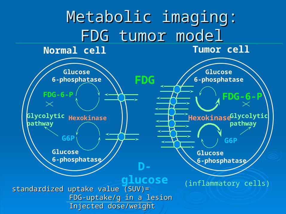
Metabolic imaging:Metabolic imaging:FDG tumor modelFDG tumor model
Glucose6-phosphatase
Glucose6-phosphatase
HexokinaseGlycolyticpathway
FDG
D-glucose
G6P
FDG-6-P
Glucose6-phosphatase
Glucose6-phosphatase
Hexokinase Glycolyticpathway
G6P
FDG-6-P
Normal cell Tumor cell
(inflammatory cells)standardized uptake value (SUV)=standardized uptake value (SUV)=
FDG-uptake/g in a lesionFDG-uptake/g in a lesionInjected dose/weightInjected dose/weight

Indication for FDG-PET in Indication for FDG-PET in LymphomaLymphoma
Staging and restaging Staging and restaging afterafter treatmenttreatment Revised reponse criteria IWC+PETRevised reponse criteria IWC+PET
PET as a prognostic markerPET as a prognostic marker early early during treatmentduring treatment

Residual massesResidual masses at the end of therapy are frequent at the end of therapy are frequent (70% HD, 50% NHL)(70% HD, 50% NHL)
but only a minority of pts relapse (<20% HD, 25% NHL)but only a minority of pts relapse (<20% HD, 25% NHL)
late toxic effectslate toxic effects of treatment are increasingly of treatment are increasingly recognized recognized
Patients in apparent complete remission also relapse Patients in apparent complete remission also relapse
Early treatment of residual disease may improve Early treatment of residual disease may improve survivalsurvival
End-of-treatment PET in Lymphoma
Need for an accurate and sensitive tool to Need for an accurate and sensitive tool to detect residual diseasedetect residual disease

Author Year Number sensitivity specificity accuracy PPV NPV Median follow-up
of patients % % % % % (months)
Naumann 2001 43 100 93 93 25 100 35
Heultenschmidt 2001 47 95 89 91 86 96 20
Spaepen 2001 60 50 100 92 100 91 32
Dittman 2001 24 88 94 92 88 94 >6
Weihrauch 2001 28 67 80 76 60 84 28
de Wit 2001 37 91 69 74 46 96 26
Lavely 2003 20 ns 81 36
Guay 2003 48 79 97 92 92 92 16
Jerusalem 2003 36 100 81 83 100 94 >36
Friedberg 2004 32 80 85 84 50 96 24
Panizo 2004 29 100 85 90 75 100 28
Rigacci 2005 28 100 83 86 50 100 45
Abbreviations: PPV: positive predictive value; NPV: negative predictive value
PET for evaluation of residual PET for evaluation of residual disease in Hodgkin’s Lymphomadisease in Hodgkin’s Lymphoma

Systematic review Zijlstra et al, Haematologica 2006NHL sub-analysis, n=201
End-of-treatment PET in NHL

New Revised Response Criteria New Revised Response Criteria in Lymphomain Lymphoma
Aim = Revision of the 1999 guidelines (CT-based) for Aim = Revision of the 1999 guidelines (CT-based) for
response assessment and outcome measurementsresponse assessment and outcome measurements
1. Standardization of PET-response1. Standardization of PET-response
2. Update of CT-based guidelines2. Update of CT-based guidelines
3. Combination of PET and CT-results3. Combination of PET and CT-results
International Harmonisation Working GroupJuweid et al, JCO February 2007Juweid et al, JCO February 2007
Bruce Cheson et al., JCO February 2007

PET in Lymphoma PET in Lymphoma standardization and interpretationstandardization and interpretation
Image acquisition for PET and PET/CTImage acquisition for PET and PET/CT Image interpretationImage interpretation
PET+ = uptake > local background and incompatible PET+ = uptake > local background and incompatible with normal anatomy/physiologywith normal anatomy/physiology
Exception:Exception: • residualresidual mass > 2 cmmass > 2 cm, , uptake uptake ≤ mediastinal BG≤ mediastinal BG = PET = PET
negativenegative
additional guidelinesadditional guidelines• Liver and SpleenLiver and Spleen
Diffuse uptake in spleen is positive if > liver uptakeDiffuse uptake in spleen is positive if > liver uptake Focal lesions > 1.5 cm must appear photopenic to be PET Focal lesions > 1.5 cm must appear photopenic to be PET
negativenegative• Bone marrowBone marrow
Focal higher than BG (no BMB+ necessary to confirm Focal higher than BG (no BMB+ necessary to confirm involvement)involvement)
Juweid et al, JCO February 2007Juweid et al, JCO February 2007

Positivity on PET?Positivity on PET?
residual mass > 2 residual mass > 2 cm cm uptake ≤ mediastinal uptake ≤ mediastinal BG BG = PET = PET negativenegative
residual mass < 2 cmresidual mass < 2 cmuptake > local BG uptake > local BG = PET positive= PET positive
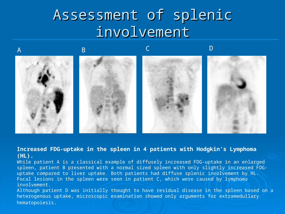
A B C D
Increased FDG-uptake in the spleen in 4 patients with Hodgkin’s Lymphoma (HL).While patient A is a classical example of diffusely increased FDG-uptake in an enlarged spleen, patient B presented with a normal sized spleen with only slightly increased FDG-uptake compared to liver uptake. Both patients had diffuse splenic involvement by HL.Focal lesions in the spleen were seen in patient C, which were caused by lymphoma involvement.Although patient D was initially thought to have residual disease in the spleen based on a heterogenous uptake, microscopic examination showed only arguments for extramedullary hematopoiesis.
Assessment of splenic Assessment of splenic involvementinvolvement

Combination of PET and CTCombination of PET and CTIWC+PET criteriaIWC+PET criteria
PR if > 50% reduction in SPD
SD if < 50% reduction in SPD
PD or Relapse if new lesion or > 50% increase of a known lesion
Only adviced for end-of-treatment evaluation in routinely FDG-avid and potentially curable lymphoma !
CR regardless of response on CT
CT+
-PET
Cheson et al, JCO February 2007Cheson et al, JCO February 2007
IWCCheson et al.JCO 1999
CRu if > 75% reduction in SPD

Patiënt 1
Patiënt 2PR (SPD – 55%)
CRu (SPD – 84%)

IWC+PET in Hodgkin’s Lymphoma IWC+PET in Hodgkin’s Lymphoma
Brepoels, Stroobants et al. 2007Brepoels, Stroobants et al. 2007 Data Spaepen, Br J Haematol. 2001Updated and IWC + PET responsein 56 pts with Hodgkin’s lymphomaPET at the end of first line R/ (after additional radiotherapy)

IWC+PET in Aggressive NHLIWC+PET in Aggressive NHL
CR
CRuPR
SD
PD
IWC
PR
IWC+PET
CR
SDPD
Brepoels, Stroobants et al., 2007Brepoels, Stroobants et al., 2007Data Spaepen, JCO 2000PET after first line R/Updated and IWC + PET responsein 55 pts with routinely FDG-avid and potentially curable (aggressive) NHL

No additional value of No additional value of IWC+PET in Indolent NHL ?IWC+PET in Indolent NHL ?
Brepoels, Stroobants et al., 2007Brepoels, Stroobants et al., 2007
Data Spaepen, JCO 2000 PET after first line R/Updated and IWC + PET responsein 14 pts with not-routinely FDG-avid and incurable NHL (8 FL, 4 MCL, 2 MZL)

FDG uptake and gradingFDG uptake and gradingSchöder et al. JCO 2005
Aggressive N=63 DLBCL 55 FL gr III 7 PTCL 1
Indolent N=28 FL gr I 11 FL gr II 4 MZL 4 small cell 8
13

Histology N° pts SUV mean range
Agressive 63new 44 19.6 (3.2 - 35)recurrent 19 18.1 (6.1 - 43)
MCL 6new 4 6 (4.7 - 8.2)recurrent 2 7 (5.3 - 8.7)
Indolent 28new 20 7.0 (2.3 - 13)recurrent 8 6.3 (3.1 - 10.6)
FDG uptake and gradingFDG uptake and grading
Schöder et al. JCO 2005
MCL:-Tendency to relapse- PET presentation as indolent- agressive disease course

no. of patientstotal n=37
histologycommon MCL 30MCL-BV 7
age at diagnosis (years)median 58range 36-77
follow-up (months)median 39range 14-144
sexman 28women 9
bone marrow involvementyes 31no 6
stage at initial diagnosisIII 1III 2IV 34
B-symptomsyes 19no 18
Clinical outcomedeath 12
Table I. Patients characteristics
Common MCL MCL-BV n = 13 n = 4
0
2
4
6
8
10
12
14
16
18
20
SU
Vm
ax
Median 25%-75%
Min-Max
*P = 0.000062
*
Common MCL MCL-BV n = 13 n = 4
0
2
4
6
8
10
12
14
16
18
20
SU
Vm
ax
Median 25%-75%
Min-Max
*P = 0.000062
*
MCL-BV (median SUV 16.88) versus common MCL (median 6.79) (p=0.00006).
Staging of Mantle cell lymphomaStaging of Mantle cell lymphoma

Response criteria in MCL? Response criteria in MCL?
CT-based criteria (IHP) CR: disappearance of all detectable diseasePR: decrease > 50% SPDSD: decrease < 50% SPDPD: new lesion or >50% SPD
PET-based criteria (EORTC)CMR: PET negativePMR: SUV decrease > 25% SMD: SUV decrease < 25%PMD: SUV increase >25% or new lesion
IWC+PET criteriaCR: everything if PET negativePR/SD/PD idem as CT-based criteria if PET is positive

0 6 12 18 24 30 36 42 48
Time (months)
0
1
Cu
mu
lativ
e P
rop
ort
ion
Su
rviv
ing
0 6 12 18 24 30 36 42 48
Time (months)
0
1
Cu
mu
lativ
e P
rop
ort
ion
Su
rviv
ing
0 6 12 18 24 30 36 42 48
Time (months)
0
1
Cu
mu
lativ
e P
rop
ort
ion
Su
rviv
ing
A
B
CT-based
CT-based
PET-based
PET-based
IWC+PET
IWC+PET
CR n=18
PR n=18
SD n=7
PD n=10
CRn=32
PRn=4
SD n=7
PD n=10
PMD n=8
PMR n=9
CMR n=31
SMD n=5
SD n=5
SMD n=3PMD n=2
CR n=5
PR n=7
PMR n=5
CMR n=10
CR n=11PD
n=3
SD n=5
PD n=3PR n=1
*
* p=0.002
*
* p=0.011
*
* p=0.036
0 6 12 18 24 30 36 42 48
Time (months)
0
1
Cu
mu
lativ
e P
rop
ort
ion
Su
rviv
ing
0 6 12 18 24 30 36 42 48
Time (months)
0
1
Cu
mu
lativ
e P
rop
ort
ion
Su
rviv
ing
0 6 12 18 24 30 36 42 48
Time (months)
0
1
Cu
mu
lativ
e P
rop
ort
ion
Su
rviv
ing
A
B
CT-based
CT-based
PET-based
PET-based
IWC+PET
IWC+PET
CR n=18
PR n=18
SD n=7
PD n=10
CRn=32
PRn=4
SD n=7
PD n=10
PMD n=8
PMR n=9
CMR n=31
SMD n=5
SD n=5
SMD n=3PMD n=2
CR n=5
PR n=7
PMR n=5
CMR n=10
CR n=11PD
n=3
SD n=5
PD n=3PR n=1
*
* p=0.002
*
* p=0.011
*
* p=0.036
Additional value of Additional value of IWC+PET in MCL ?IWC+PET in MCL ?
Different response categories have different prognosis
No prognostic difference between respondersNon-responders are the same as with CT alone

0 6 12 18 24 30 36 42 48
Time (months)
0
1
Cu
mu
lativ
e P
rop
ort
ion
Su
rviv
ing
0 6 12 18 24 30 36 42 48
Time (months)
0
1
Cu
mu
lativ
e P
rop
ort
ion
Su
rviv
ing
0 6 12 18 24 30 36 42 48
Time (months)
0
1
Cu
mu
lativ
e P
rop
ort
ion
Su
rviv
ing
0 6 12 18 24 30 36 42 48
Time (months)
0
1
Cu
mu
lativ
e P
rop
ort
ion
Su
rviv
ing
0 6 12 18 24 30 36 42 48
Time (months)
0
1
Cu
mu
lativ
e P
rop
ort
ion
Su
rviv
ing
0 6 12 18 24 30 36 42 48
Time (months)
0
1
Cu
mu
lativ
e P
rop
ort
ion
Su
rviv
ing
A
B
CT-based
CT-based
PET-based
PET-based
IWC+PET
IWC+PET
CR n=18
PR n=18
SD n=7
PD n=10
CRn=32
PRn=4
SD n=7
PD n=10
PMD n=8
PMR n=9
CMR n=31
SMD n=5
SD n=5
SMD n=3PMD n=2
CR n=5
PR n=7
PMR n=5
CMR n=10
CR n=11PD
n=3
SD n=5
PD n=3PR n=1
*
* p=0.002
*
* p=0.011
*
* p=0.036
0 6 12 18 24 30 36 42 48
Time (months)
0
1
Cu
mu
lativ
e P
rop
ort
ion
Su
rviv
ing
0 6 12 18 24 30 36 42 48
Time (months)
0
1
Cu
mu
lativ
e P
rop
ort
ion
Su
rviv
ing
0 6 12 18 24 30 36 42 48
Time (months)
0
1
Cu
mu
lativ
e P
rop
ort
ion
Su
rviv
ing
0 6 12 18 24 30 36 42 48
Time (months)
0
1
Cu
mu
lativ
e P
rop
ort
ion
Su
rviv
ing
0 6 12 18 24 30 36 42 48
Time (months)
0
1
Cu
mu
lativ
e P
rop
ort
ion
Su
rviv
ing
0 6 12 18 24 30 36 42 48
Time (months)
0
1
Cu
mu
lativ
e P
rop
ort
ion
Su
rviv
ing
A
B
CT-based
CT-based
PET-based
PET-based
IWC+PET
IWC+PET
CR n=18
PR n=18
SD n=7
PD n=10
CRn=32
PRn=4
SD n=7
PD n=10
PMD n=8
PMR n=9
CMR n=31
SMD n=5
SD n=5
SMD n=3PMD n=2
CR n=5
PR n=7
PMR n=5
CMR n=10
CR n=11PD
n=3
SD n=5
PD n=3PR n=1
*
* p=0.002
*
* p=0.011
*
* p=0.036
Additional value of Additional value of IWC+PET in MCL ?IWC+PET in MCL ?

Use of IWC+PET criteriaUse of IWC+PET criteria essential at the end of treatment in routinely FDG-essential at the end of treatment in routinely FDG-
avid and avid and potentially curable lymphoma (DLBCL and HD)potentially curable lymphoma (DLBCL and HD)
Limitations Limitations Only recommended for end-of-treatment evaluationOnly recommended for end-of-treatment evaluation value has to be established in the other histological value has to be established in the other histological
subtypes, subtypes, no encouraging results in Mantle cell no encouraging results in Mantle cell lymphomalymphoma
Only assessment of negative or positive on PET, no Only assessment of negative or positive on PET, no quantitative quantitative evaluation of response !evaluation of response !
Revised Response Criteria in Revised Response Criteria in LymphomaLymphoma

PET as a prognostic marker early PET as a prognostic marker early during therapyduring therapy
A substantial number of patients are not cured with standard A substantial number of patients are not cured with standard chemotherapychemotherapy
early change of therapyearly change of therapy might improve outcome might improve outcome
Late treatment related morbidity and mortality especially Late treatment related morbidity and mortality especially after combination chemoradiationafter combination chemoradiation
reduce therapyreduce therapy without compromising outcome without compromising outcome
Individualized patient managementIndividualized patient management
- risk adapted- risk adapted
- response adapted- response adapted

4x CHOP-R 8 x CHOP-R
prior 1x CHOP-R
Early Response Assessment

Early PET in HLEarly PET in HLHutchings et al, Blood 2005Prospective study, 77 pts, 48 stage I/IIPET after 2 cycles

Prognostic value of PET for early response assessment during first line or induction treatment
Author Year Number histology timing PET sensitivity specificity accuracy PPV NPV Follow-up
of patients (cycles) % % % % % (months)
Jerusalem 2000 28 NHL 2-5 42 100 73 100 67 17
Mikhaeel 2000 23 NHL 2-4 100 94 96 88 100 24
Mikhaeel 2002 32 HD 2-3 75 100 94 100 92 36
Kostakoglu 2002 30 HD/NHL 1 87 87 87 87 87 19
Spaepen 2002 70 NHL 3-4 85 100 91 100 84 36
Zijlstra 2003 26 NHL 2 64 75 69 75 75 25
Torizuka 2004 20 HD/NHL 1-2 87 50 80 87 50 24
Friedberg 2004 22 HD 3 80 94 91 80 94 24
Haioun 2005 90 NHL 2 63 71 68 55 77 24
Mikhaeel 2005 121 NHL 2-3 5-year PFS of 16.2% when PET+, 88.8% when PET-, 59.3% for MRU
Hutchings 2005 85 HD 2-3 5-year PFS of 38.5% when PET+, 91.5% when PET-, MRU considered PET-
Hutchings 2006 77 HD 2 79 92 90 69 95 23
Gallamini 2006 108 HD 2 86 98 95 90 97 20
Abbreviations: PPV: positive predictive value; NPV: negative predictive value, HD: Hodgkin's disease; NHL: Non-Hodgkin's lymphoma; PFS: progression-free survival; PET+: PET positive; PET-: PET negative; MRU: minimal residual uptake
Early Response Assessment by PET
Optimal timing? Optimal timing?
Definition of PET response (quantification)?Definition of PET response (quantification)?

Baseline PET-CT PET-CT after 1 cycle CHOP-R
Timing of early PET?? Timing of early PET?? Need for quantificationNeed for quantification
The patient with DLBCL obtained a complete remission on PET after 3 The patient with DLBCL obtained a complete remission on PET after 3 cycles of CHOP and is still progression free after 16 months follow-upcycles of CHOP and is still progression free after 16 months follow-up
very early after initiation of very early after initiation of therapy: need therapy: need for quantification???for quantification???

Baseline PET-CT
Timing of early PET?? Timing of early PET?? Need for quantificationNeed for quantification
The patient became clinical progressive during treatment The patient became clinical progressive during treatment and died of lymphoma only 3 months after initial diagnosis. and died of lymphoma only 3 months after initial diagnosis.
very early after initiation of therapy: very early after initiation of therapy: need for quantification???need for quantification???
PET-CT after 1 week CHOP-R

Cycles of chemo
ChemosensitivityChemoresistance
Num
ber
of
malig
nant
cells
Time
Tim
e o
f d
iag
nosis
S
tart
ch
em
oth
era
py
Symptoms
Death
PET-detection

Quantification of early PET? Quantification of early PET? Hutchings, Blood 2005
SUVmax = 4 is optimal? HD after 2-3 cycles
Kostakoglu, Cancer 2006Kostakoglu, Cancer 2006 SUVmax = 1.75 SUVmax = 1.75 (sens 92%, spec 84%)(sens 92%, spec 84%)
HD/DLBCL after 1 cycleHD/DLBCL after 1 cycle
Torizuka, EJNM 2004Torizuka, EJNM 2004 Decrease SUV of -60%Decrease SUV of -60%HD/NHL after 1-2 cyclesHD/NHL after 1-2 cycles

PET after 1 week CHOP-R in PET after 1 week CHOP-R in DLBCL DLBCL (29 patients)(29 patients)
Prospective study Prospective study Newly diagnosed Newly diagnosed
DLBCLDLBCL PET after 7 days PET after 7 days
CHOP-RCHOP-R 12 patients negative 12 patients negative
on early PETon early PET 12/12 disease free 12/12 disease free
after a median after a median follow-up of 19 follow-up of 19 monthsmonths
A. Pretreatment D. End-of-treatmentC. InterimB. EarlyA. Pretreatment D. End-of-treatmentC. InterimB. Early
Figure V.1. Complete remission on early FDG-PET.(A) Pretreatment PET/CT shows an intense FDG-avide lesion left hilair.
(B) Early PET/CT shows a slighly increased FDG-uptake but not exceeding mediastinal blood pool uptake. Also an important response is seen on CT.
(C) Interim and (D) end-of-treatment PET/CT show persistent complete remission.This patient is in sustained complete remission after 30 months follow-up

PET after 1 week CHOP-R in PET after 1 week CHOP-R in DLBCL DLBCL (29 patients)(29 patients)
17 patients positive 17 patients positive on early PETon early PET
2/17 refractory 2/17 refractory diseasedisease
2/17 relapsed (12, 2/17 relapsed (12, 23 mts) 23 mts)
Interim End-of-treatmentPretreatment Early
A
B
SUVmean_all = 7.42SUVmax_all = 14.05Volmet_all = 695 ml
SUVmean_all = 4.23SUVmax_all = 7.94Volmet_all = 269 ml
SUVmean_all = 3.27SUVmax_all = 3.87Volmet_all = 58 ml
SUVmean_all = 3.34SUVmax_all = 3.88Volmet_all = 28 ml
SUVmean_all = 8.55SUVmax_all = 18.34Volmet_all = 658 ml
SUVmean_all = 5.66SUVmax_all = 12.47Volmet_all = 361 ml
CR CR
Residual FDG-uptake on early PET Patient A never obtained a complete remission and refractory disease was histologically proven based on end-of-treatment PET/CT

PET after 1 week CHOP-R in PET after 1 week CHOP-R in DLBCL DLBCL (29 patients)(29 patients)
17 patients positive 17 patients positive on early PETon early PET
13/17 disease free 13/17 disease free after a median after a median follow-up of 20 follow-up of 20 monthsmonths
Quantification???? Quantification???? No significant No significant
difference in absolute difference in absolute uptake valuesuptake values
High variability in High variability in percentage decreasepercentage decrease
Interim End-of-treatmentPretreatment Early
A
B
SUVmean_all = 7.42SUVmax_all = 14.05Volmet_all = 695 ml
SUVmean_all = 4.23SUVmax_all = 7.94Volmet_all = 269 ml
SUVmean_all = 3.27SUVmax_all = 3.87Volmet_all = 58 ml
SUVmean_all = 3.34SUVmax_all = 3.88Volmet_all = 28 ml
SUVmean_all = 8.55SUVmax_all = 18.34Volmet_all = 658 ml
SUVmean_all = 5.66SUVmax_all = 12.47Volmet_all = 361 ml
CR CR
(B) Despite a significant residual uptake on early PET, this patient obtained a complete remission at interim PET, and is still disease free after a follow-up of 29 months

-100
-100
-100
-100
-100
-100
-100 -8
9
-88 -85
-84
-84 -7
7
-77
-76
-75
-75
-75
-73
-71
-70 -6
4 -58
-46
-44
-32
-23
-12
-100
-100
-90
-80
-70
-60
-50
-40
-30
-20
-10
0
-100
-100
-100
-100
-100
-100
-100 -8
9
-73 -6
6
-66
-64
-64 -61
-58
-56
-56
-47
-46
-45
-43
-43
-43 -3
8 -34 -2
6 -20
-4
-100
-100
-90
-80
-70
-60
-50
-40
-30
-20
-10
0
-100
-100
-100
-100
-100
-100
-100
-100
-100
-100
-100
-99
-99
-96
-96
-95
-95
-93
-92 -89 -8
5
-84
-84 -7
9
-64
-61
-50 -4
6
-100
-100
-90
-80
-70
-60
-50
-40
-30
-20
-10
0
-100
-100
-100
-100
-100
-100
-100
-100
-100
-100
-100
-100
-99
-99
-98
-98
-98
-97
-97
-96
-93
-93 -8
9
-88 -8
0
-78
-64
-52
-100
-100
-90
-80
-70
-60
-50
-40
-30
-20
-10
0
-100
-100
-100
-100
-100
-100
-100
-85
-84 -8
0
-77
-77
-76
-75
-75
-75
-74
-73
-71
-70 -6
4 -57
-46
-46
-44
-32
-4
-100
-2
-100
-90
-80
-70
-60
-50
-40
-30
-20
-10
0
-100
-100
-100
-100
-100
-100
-100
-98
-96
-96
-96
-95
-93
-92 -8
5
-85
-84
-84 -7
8 -75 -6
8 -61 -5
5
-53 -4
5
-35
-18
1
-100
-100
-90
-80
-70
-60
-50
-40
-30
-20
-10
0
10
-100
-100
-100
-100
-100
-100
-100
-99
-98
-98
-98
-98
-98
-97
-96
-93
-93 -8
7-8
6-8
5 -81 -78 -7
3 -69 -6
4-3
7-2
7-3
-100
-100
-90
-80
-70
-60
-50
-40
-30
-20
-10
0
Analysis based on tumor bulk
-100
-100
-100
-100
-100
-100
-100
-90
-87
-84
-82
-80 -77
-76
-75
-75
-73
-73
-70 -66
-66
-64 -5
8-4
6-4
4-3
2-1
614
-100
-100
-90
-80
-70
-60
-50
-40
-30
-20
-10
0
10
20
-100
-100
-100
-100
-100
-100
-100 -9
0-6
7 -60 -57 -5
0-5
0-4
9-4
9-4
8 -41
-39 -3
3-3
1-3
1-3
0-1
6-1
6 -95
1745
-100-100
-90-80-70-60-50-40-30-20-10
01020304050
-100
-100
-100
-100
-100
-100
-100
-100
-100
-100
-100
-99
-98
-97
-97
-96
-95
-94
-93
-90
-86
-86
-85
-82
-66
-63
-51
-47
-100
-100
-90
-80
-70
-60
-50
-40
-30
-20
-10
0
-100
-100
-100
-100
-100
-100
-100
-100
-100
-100
-100
-99
-98
-98
-98
-98
-98
-97
-96
-95
-93
-93
-90
-87 -8
0
-78
-64
-48
-100
-100
-90
-80
-70
-60
-50
-40
-30
-20
-10
0
-100
-100
-100
-100
-100
-100
-100 -8
7-8
4 -78
-77
-76
-75
-75
-73
-73
-71 -6
6-6
4-6
2-6
2-4
6-4
6-4
4 -34
-32
126
-100
-100
-90
-80
-70
-60
-50
-40-30
-20
-10
0
10
20
30
-100
-100
-100
-100
-100
-100
-100 -8
7-6
7 -57 -5
2-5
0-4
9-4
9-4
9-4
6 -40 -3
1-3
0-2
9 -26 -22 -1
1-1
1-1
0 -34
24
-100
-100
-90
-80
-70
-60
-50
-40-30
-20
-10
0
10
20
30
-100
-100
-100
-100
-100
-100
-100
-98
-97
-97
-97
-96
-94
-93 -8
7-8
6-8
6-8
5 -78 -7
2-7
0 -63 -5
6-5
4 -47
-35
-17
18
-100
-100
-90
-80
-70
-60
-50
-40
-30
-20
-10
0
10
20
-100
-100
-100
-100
-100
-100
-100
-99
-98
-98
-98
-97
-97
-97
-95
-93
-93 -8
7 -83 -80
-79
-78 -7
2 -65
-63
-34 -2
721
-100
-100
-90
-80
-70
-60
-50
-40-30
-20
-10
0
10
20
30Analysis based on the worst responding lesion after partial volume correction
Analysis based on tumor bulk after partial volume correction
Analysis based on the worst responding lesion
Δ SUVmax_all Δ TLGallΔ Volmet_allΔ SUVmean_all
-100
-100
-100
-100
-100
-100
-100
-85
-73 -6
7
-64
-63 -59
-58
-56
-56 -52 -4
7
-47
-44
-43 -3
7 -34
-33
-17 -1
1
-3 -1
-100
-100
-90
-80
-70
-60
-50
-40
-30
-20
-10
0
Figure IV.7. Overview of the different relative parameters of FDG-decrease for the different analyses with corresponding outcome of the patient.
No evidence of disease Primary refractory diseaseRelapse after 12mths Relapse after 23 mths
Δ SUVmax_worst Δ TLGworstΔ Volmet_worstΔ SUVmean_worst
Δ SUVmax_all Δ TLGallΔ Volmet_allΔ SUVmean_all
Δ SUVmax_worst Δ TLGworstΔ Volmet_worstΔ SUVmean_worst
-100
-100
-100
-100
-100
-100
-100 -8
9
-88 -85
-84
-84 -7
7
-77
-76
-75
-75
-75
-73
-71
-70 -6
4 -58
-46
-44
-32
-23
-12
-100
-100
-90
-80
-70
-60
-50
-40
-30
-20
-10
0
-100
-100
-100
-100
-100
-100
-100 -8
9
-73 -6
6
-66
-64
-64 -61
-58
-56
-56
-47
-46
-45
-43
-43
-43 -3
8 -34 -2
6 -20
-4
-100
-100
-90
-80
-70
-60
-50
-40
-30
-20
-10
0
-100
-100
-100
-100
-100
-100
-100
-100
-100
-100
-100
-99
-99
-96
-96
-95
-95
-93
-92 -89 -8
5
-84
-84 -7
9
-64
-61
-50 -4
6
-100
-100
-90
-80
-70
-60
-50
-40
-30
-20
-10
0
-100
-100
-100
-100
-100
-100
-100
-100
-100
-100
-100
-100
-99
-99
-98
-98
-98
-97
-97
-96
-93
-93 -8
9
-88 -8
0
-78
-64
-52
-100
-100
-90
-80
-70
-60
-50
-40
-30
-20
-10
0
-100
-100
-100
-100
-100
-100
-100
-85
-84 -8
0
-77
-77
-76
-75
-75
-75
-74
-73
-71
-70 -6
4 -57
-46
-46
-44
-32
-4
-100
-2
-100
-90
-80
-70
-60
-50
-40
-30
-20
-10
0
-100
-100
-100
-100
-100
-100
-100
-98
-96
-96
-96
-95
-93
-92 -8
5
-85
-84
-84 -7
8 -75 -6
8 -61 -5
5
-53 -4
5
-35
-18
1
-100
-100
-90
-80
-70
-60
-50
-40
-30
-20
-10
0
10
-100
-100
-100
-100
-100
-100
-100
-99
-98
-98
-98
-98
-98
-97
-96
-93
-93 -8
7-8
6-8
5 -81 -78 -7
3 -69 -6
4-3
7-2
7-3
-100
-100
-90
-80
-70
-60
-50
-40
-30
-20
-10
0
Analysis based on tumor bulk
-100
-100
-100
-100
-100
-100
-100
-90
-87
-84
-82
-80 -77
-76
-75
-75
-73
-73
-70 -66
-66
-64 -5
8-4
6-4
4-3
2-1
614
-100
-100
-90
-80
-70
-60
-50
-40
-30
-20
-10
0
10
20
-100
-100
-100
-100
-100
-100
-100 -9
0-6
7 -60 -57 -5
0-5
0-4
9-4
9-4
8 -41
-39 -3
3-3
1-3
1-3
0-1
6-1
6 -95
1745
-100-100
-90-80-70-60-50-40-30-20-10
01020304050
-100
-100
-100
-100
-100
-100
-100
-100
-100
-100
-100
-99
-98
-97
-97
-96
-95
-94
-93
-90
-86
-86
-85
-82
-66
-63
-51
-47
-100
-100
-90
-80
-70
-60
-50
-40
-30
-20
-10
0
-100
-100
-100
-100
-100
-100
-100
-100
-100
-100
-100
-99
-98
-98
-98
-98
-98
-97
-96
-95
-93
-93
-90
-87 -8
0
-78
-64
-48
-100
-100
-90
-80
-70
-60
-50
-40
-30
-20
-10
0
-100
-100
-100
-100
-100
-100
-100 -8
7-8
4 -78
-77
-76
-75
-75
-73
-73
-71 -6
6-6
4-6
2-6
2-4
6-4
6-4
4 -34
-32
126
-100
-100
-90
-80
-70
-60
-50
-40-30
-20
-10
0
10
20
30
-100
-100
-100
-100
-100
-100
-100 -8
7-6
7 -57 -5
2-5
0-4
9-4
9-4
9-4
6 -40 -3
1-3
0-2
9 -26 -22 -1
1-1
1-1
0 -34
24
-100
-100
-90
-80
-70
-60
-50
-40-30
-20
-10
0
10
20
30
-100
-100
-100
-100
-100
-100
-100
-98
-97
-97
-97
-96
-94
-93 -8
7-8
6-8
6-8
5 -78 -7
2-7
0 -63 -5
6-5
4 -47
-35
-17
18
-100
-100
-90
-80
-70
-60
-50
-40
-30
-20
-10
0
10
20
-100
-100
-100
-100
-100
-100
-100
-99
-98
-98
-98
-97
-97
-97
-95
-93
-93 -8
7 -83 -80
-79
-78 -7
2 -65
-63
-34 -2
721
-100
-100
-90
-80
-70
-60
-50
-40-30
-20
-10
0
10
20
30Analysis based on the worst responding lesion after partial volume correction
Analysis based on tumor bulk after partial volume correction
Analysis based on the worst responding lesion
Δ SUVmax_all Δ TLGallΔ Volmet_allΔ SUVmean_allΔ SUVmax_all Δ TLGallΔ Volmet_allΔ SUVmean_all
-100
-100
-100
-100
-100
-100
-100
-85
-73 -6
7
-64
-63 -59
-58
-56
-56 -52 -4
7
-47
-44
-43 -3
7 -34
-33
-17 -1
1
-3 -1
-100
-100
-90
-80
-70
-60
-50
-40
-30
-20
-10
0
Figure IV.7. Overview of the different relative parameters of FDG-decrease for the different analyses with corresponding outcome of the patient.
No evidence of disease Primary refractory diseaseRelapse after 12mths Relapse after 23 mths
Δ SUVmax_worst Δ TLGworstΔ Volmet_worstΔ SUVmean_worstΔ SUVmax_worst Δ TLGworstΔ Volmet_worstΔ SUVmean_worst
Δ SUVmax_all Δ TLGallΔ Volmet_allΔ SUVmean_allΔ SUVmax_all Δ TLGallΔ Volmet_allΔ SUVmean_all
Δ SUVmax_worst Δ TLGworstΔ Volmet_worstΔ SUVmean_worstΔ SUVmax_worst Δ TLGworstΔ Volmet_worstΔ SUVmean_worst

PET after 1 week of therapyPET after 1 week of therapy
early PET: early PET: sensitivity 100%, specificity 48%, NPV sensitivity 100%, specificity 48%, NPV
100%, PPV 24% 100%, PPV 24%
Negative on early PET: excellent Negative on early PET: excellent prognosis, reduction of therapy?prognosis, reduction of therapy?
Positive on early PET: low specificity, Positive on early PET: low specificity, quantification not better than visual quantification not better than visual interpretationinterpretation

Persistent Persistent FDG uptake after 2-3 cyclesFDG uptake after 2-3 cycles of chemo is of chemo is associated with a associated with a high probability of relapsehigh probability of relapse at the at the end of treatmentend of treatment
Early PET response is a stronger predictor of PFS Early PET response is a stronger predictor of PFS than IPI or IPSthan IPI or IPS
Optimal timing? Definition of PET response Optimal timing? Definition of PET response (quantification)?(quantification)?
PET for Early Therapy response:Conclusion

ConclusionsConclusions PET for RESIDUAL DISEASE in aggressive NHL and HLPET for RESIDUAL DISEASE in aggressive NHL and HL
IWC+PET for end-of-treatment evaluationIWC+PET for end-of-treatment evaluation Persistent FDG uptake after a 2-3 cycles of chemo is associated Persistent FDG uptake after a 2-3 cycles of chemo is associated
with a high probability of relapsewith a high probability of relapse
PET for measuring CHEMOSENSITIVITY in the other PET for measuring CHEMOSENSITIVITY in the other histological subtypes: IWC+PET criteria not recommended in histological subtypes: IWC+PET criteria not recommended in MCLMCL
Early PET at more early time points and Early PET at more early time points and Optimal timing? Optimal timing? Definition of PET response (quantification)?Definition of PET response (quantification)?
Will Will PET-adapted treatmentPET-adapted treatment improve overall survival? improve overall survival? (Dann, Blood 2007, PET after 2 cycles in HL, reduction of (Dann, Blood 2007, PET after 2 cycles in HL, reduction of cumulative dose of chemotherapy without impairment of outcome.)cumulative dose of chemotherapy without impairment of outcome.)
!!!! Need for prospective trials!!!! Need for prospective trials
BaselinePET
EarlyPET
Chemo AChemo A
Chemo B
PFS & OS