assessing alveolar bone height and thickness using cone ... using cone beam computed tomography: are...
TRANSCRIPT
4/17/2017
1
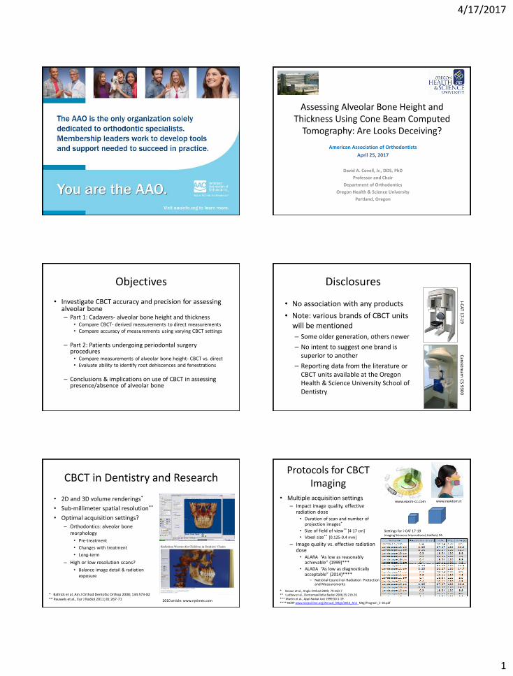
Assessing Alveolar Bone Height and Thickness Using Cone Beam Computed
Tomography: Are Looks Deceiving?
American Association of Orthodontists
April 25, 2017
David A. Covell, Jr., DDS, PhD
Professor and Chair
Department of Orthodontics
Oregon Health & Science University
Portland, Oregon
Objectives
• Investigate CBCT accuracy and precision for assessing alveolar bone – Part 1: Cadavers- alveolar bone height and thickness
• Compare CBCT- derived measurements to direct measurements • Compare accuracy of measurements using varying CBCT settings
– Part 2: Patients undergoing periodontal surgery procedures • Compare measurements of alveolar bone height- CBCT vs. direct • Evaluate ability to identify root dehiscences and fenestrations
– Conclusions & implications on use of CBCT in assessing presence/absence of alveolar bone
Disclosures
• No association with any products
• Note: various brands of CBCT units will be mentioned
– Some older generation, others newer
– No intent to suggest one brand is superior to another
– Reporting data from the literature or CBCT units available at the Oregon Health & Science University School of Dentistry
i-CAT 17-19
C
arestream C
S 9300
CBCT in Dentistry and Research
• 2D and 3D volume renderings*
• Sub-millimeter spatial resolution**
• Optimal acquisition settings?
– Orthodontics: alveolar bone morphology
• Pre-treatment
• Changes with treatment
• Long-term
– High or low resolution scans?
• Balance image detail & radiation exposure
* Ballrick et al, Am J Orthod Dentofac Orthop 2008; 134:573-82 ** Pauwels et al., Eur J Radiol 2011; 81:267-71 2010 article: www.nytimes.com
Protocols for CBCT Imaging
• Multiple acquisition settings – Impact image quality, effective
radiation dose • Duration of scan and number of
projection images*
• Size of field of view** [4-17 cm]
• Voxel size** [0.125-0.4 mm]
– Image quality vs. effective radiation dose • ALARA “As low as reasonably
achievable” (1999)***
• ALADA “As low as diagnostically acceptable” (2014)****
– National Council on Radiation Protection and Measurements
* Brown et al., Angle Orthod 2009; 79:150-7 ** Ludlow et al., Dentomaxillofac Radiol 2006;35:219-26 *** Martin et al., Appl Radiat Isot 1999;50:1-19 **** NCRP www.ncrponline.org/Annual_Mtgs/2014_Ann_Mtg/Program_2-10.pdf
Settings for i-CAT 17-19 Imaging Sciences International, Hatfield, PA
www.newtom.it www.exxim-cc.com

4/17/2017
2
Part 1: Facial Bone Height and Thickness Measurements in Cadaver Study
(Adam Timock, Lane Cook)
• Comparison of alveolar bone measurements:
– Made from CBCT scans acquired using i-CAT 17-19 CBCT unit’s default setting, and settings aimed to produce lesser or greater resolution
– Compare CBCT to direct measurements via dissection
• Timock et al., Am J Orthod Dentofac Orthop 2011;140:734-44 • Cook et al. Orthod Craniofac Res 2015;18(Suppl.1):127-136
Sample Selection
• Protocol reviewed by OHSU’s IRB
• 17 cadavers accessed through Department of Integrative Biosciences
• Screening inclusion criteria: – Teeth with periodontium free of damage
– Sample teeth and adjacent teeth having no metal restorations
• Sample selected:
– 12 cadavers: 5 female, 7 male; all Caucasians
– Mean age: 77 years (range: 55-89 years)
– 65 teeth selected: 48 anterior, 17 posterior teeth
• Timock et al., Am J Orthod Dentofac Orthop 2011;140:734-44 • Cook et al. Orthod Craniofac Res 2015;18(Suppl.1):127-136
CBCT Settings
– 3 scans: default, 2 custom settings • Long scan (LS)**: 26.9 seconds; 619
projection images; 360o rotation; 0.2 mm voxel size
• Default scan (DS)*: 8.9 seconds; 309 projection images; 360o rotation; 0.3 mm voxel size
• Short scan (SS)**: 4.8 seconds; 169 projection images; 180o rotation; 0.3 mm voxel size
– Compared to direct measurements from dissection
i-CAT 17-19 CBCT Unit Imaging Sciences International
• Timock et al., Am J Orthod Dentofac Orthop 2011;140:734-44 • Cook et al. Orthod Craniofac Res 2015;18(Suppl.1):127-136
Direct Measurement of Bone Height
Following acquisition of CBCT scans:
• Gingiva dissected
• Buccal bone height measurement (BBH) – Digital caliper (0.01 mm)
– Distance from coronal-most point of crown to buccal alveolar crest, following the long axis of tooth
Timock et al., Am J Orthod Dentofac Orthop 2011;140:734-44
Direct Measurement of Bone Thickness
• Buccal bone thickness measurement (BBT) – Notch dissected in alveolar crest - 3 mm height
– Measured with a customized depth gauge (0.01 mm)
– Linear distance from cementum to lateral bone surface
• Document site of BBT measurement – With caliper, measure from cusp tip to apical base of
dissection site
– Defined the location where BBT measurements made in CBCT images
• Thickness measurements – Made by 2 calibrated investigators (AT, LC)
– Repeated 3 times, at least one day apart
CBCT Measurements
– DICOM files imported into Dolphin 3D Imaging
– Measurements:
• 0.5 mm slice thickness oriented using a standardized protocol
– BBH: measured in sagittal plane, parallel to long axis of tooth
– BBT: measured in axial plane at vertical location where direct BBT measurements had been made with dissections
– Measurements made independently by 2 calibrated investigators (AT, LC)
– Repeated 3 times, at least 1 day in between

4/17/2017
3
Statistical Analysis
– Comparison of measurements • 3 repeat measurements averaged
• Direct vs. CBCT mean values compared using paired t-tests (p<.05)
• ANOVA: Comparison of measurements from 3 CBCT settings
• Concordance correlation coefficients (CCC): Rater & measurement comparisons
• Bland-Altman plots* : Agreement of CBCT vs. direct measurements
* Bland & Altman. Stat Methods Med Res 1999;8:135-60
Results: Direct Measurements
• Both intra- and inter-rater reliability were very high
Bland-Altman Plot: Example
o Each circle = 1 tooth (n=65)
Y-axis: difference between direct and CBCT measurements
X-axis: average of the direct and CBCT measurements (X-axis)
- - Dashed line = mean difference among all direct and CBCT measurements
Solid lines = 95% limits of agreement
Dif
fere
nce
[Dir
ect
– C
BC
T] (
mm
)
Average [Direct & CBCT] (mm)
Bone Height, Long Scan - CBCT scan - Dissection - 65 teeth measured
Bland-Altman Plots: Bone Height- Long Scan & Short Scan
Long Scan Short Scan
LS: mean difference = -0.02 mm SS: mean difference = 0.00 mm 95% LOA -0.43 to 0.40 mm 95% LOA -1.02 to 1.04 mm
Cook et al. Orthod Craniofac Res 2015;18(Suppl.1):127-136
Dif
fere
nce
[Dir
ect
– C
BC
T] (
mm
)
Dif
fere
nce
[Dir
ect
– C
BC
T] (
mm
)
Bland-Altman Plots: Bone Thickness- Long Scan & Short Scan
Long Scan Short Scan
LS: mean difference = 0.00 mm SS: mean difference = -0.04 mm 95% LOA -0.25 to 0.24 mm 95% LOA -0.37 to 0.28 mm
Cook et al. Orthod Craniofac Res 2015;18(Suppl.1):127-136
Dif
fere
nce
[Dir
ect
– C
BC
T] (
mm
)
Dif
fere
nce
[Dir
ect
– C
BC
T] (
mm
)
Measurement Comparisons
• Accuracy and precision: – Direct measurements: high reliability
• Correlation coefficients close to 1.0
• Valid for use as controls
– CBCT-derived measurements: accurate, but varying precision
• Accuracy: accurate with all 3 settings (no difference statistically from direct measurements)
• Precision: greater with longer scans as shown by Bland-Altman 95% LOA:
– E.g., bone height: Long Scan: +0.40 mm
– Default Scan: +0.78 mm
– Short Scan: +1.03 mm
LS SS Timock et al., Am J Orthod Dentofac Orthop 2011;140:734-44 Cook et al. Orthod Craniofac Res 2015;18(Suppl.1):127-136
4/17/2017
4
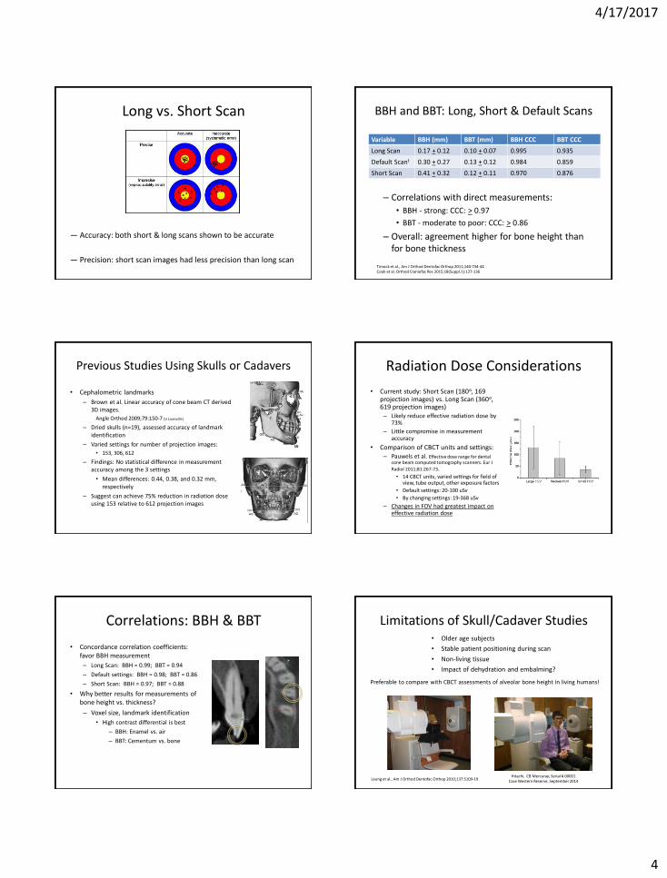
Long vs. Short Scan
— Accuracy: both short & long scans shown to be accurate
— Precision: short scan images had less precision than long scan
BBH and BBT: Long, Short & Default Scans
– Correlations with direct measurements:
• BBH - strong: CCC: > 0.97
• BBT - moderate to poor: CCC: > 0.86
– Overall: agreement higher for bone height than for bone thickness
Variable BBH (mm) BBT (mm) BBH CCC BBT CCC
Long Scan 0.17 + 0.12 0.10 + 0.07 0.995 0.935
Default Scant 0.30 + 0.27 0.13 + 0.12 0.984 0.859
Short Scan 0.41 + 0.32 0.12 + 0.11 0.970 0.876
Timock et al., Am J Orthod Dentofac Orthop 2011;140:734-44 Cook et al. Orthod Craniofac Res 2015;18(Suppl.1):127-136
Previous Studies Using Skulls or Cadavers
• Cephalometric landmarks
– Brown et al. Linear accuracy of cone beam CT derived 3D images.
Angle Orthod 2009;79:150-7 [U Louisville]
– Dried skulls (n=19), assessed accuracy of landmark identification
– Varied settings for number of projection images:
• 153, 306, 612
– Findings: No statistical difference in measurement accuracy among the 3 settings
• Mean differences: 0.44, 0.38, and 0.32 mm, respectively
– Suggest can achieve 75% reduction in radiation dose using 153 relative to 612 projection images
Radiation Dose Considerations
• Current study: Short Scan (180o, 169 projection images) vs. Long Scan (360o, 619 projection images) – Likely reduce effective radiation dose by
73%
– Little compromise in measurement accuracy
• Comparison of CBCT units and settings: – Pauwels et al. Effective dose range for dental
cone beam computed tomography scanners. Eur J
Radiol 2011;81:267-73. • 14 CBCT units, varied settings for field of
view, tube output, other exposure factors
• Default settings: 20-100 uSv
• By changing settings: 19-368 uSv
– Changes in FOV had greatest impact on effective radiation dose
Correlations: BBH & BBT
• Concordance correlation coefficients: favor BBH measurement
– Long Scan: BBH = 0.99; BBT = 0.94
– Default settings: BBH = 0.98; BBT = 0.86
– Short Scan: BBH = 0.97; BBT = 0.88
• Why better results for measurements of bone height vs. thickness?
– Voxel size, landmark identification
• High contrast differential is best
– BBH: Enamel vs. air
– BBT: Cementum vs. bone
Limitations of Skull/Cadaver Studies • Older age subjects
• Stable patient positioning during scan
• Non-living tissue
• Impact of dehydration and embalming?
Leung et al., Am J Orthod Dentofac Orthop 2010;137:S109-19
Preferable to compare with CBCT assessments of alveolar bone height in living humans!
Hitachi, CB Mercuray; Serial # 00001 Case Western Reserve, September 2014

4/17/2017
5
Part 2: Alveolar Bone Height in Patients Scheduled for Periodontal Surgery
(Megan Miller, Vanessa Browne Peterson: iCAT 17-19 Andrew Peterson: Carestream CS 9300)
Objectives:
1. Investigate accuracy and reliability of CBCT compared to direct measurements of facial alveolar bone height in vivo
2. Measurement of bone height only
3. Assess ability to detect naturally occurring dehiscences and fenestrations
Methods and Patient Selection
• IRB approval
• Sample:
– Patients scheduled for surgery involving mucoperiosteal flaps
• Clinics: Periodontology; Oral and Maxillofacial Surgery
– CBCT imaging as part of patient’s treatment plan
– CBCT images acquired within the previous 18 months
• Pilot study conducted to develop methodology
– Provided data for power calculation
Surgical Procedures
Osteotomy
Implant Placement
Sinus Lift
Bone Graft
Crown Lengthening
Connective Tissue Graft
Surgical Extraction
Screening
• Inclusion Criteria
• CBCT scan on record in preparation for surgical procedure
• At least 18 years of age with permanent dentition
• Teeth in the area of surgery having natural tooth structure or small composite restorations
• Region of surgery free from osseous or soft tissue pathology
• Exclusion Criteria
• Alloy restorations (or implants) adjacent to or in the tooth of interest
• Previous surgery in the region
Sample Selection (Miller, Browne: i-CAT 17-19)
177 patients with CBCT images acquired
September 2012 to July 2014 135 patients
excluded
-Duplicate CBCT on same patient -Patient undergoing orthodontic treatment -Private practice referral -Patient did not undergo surgical procedure involving full thickness flap -Patient did not have intact tooth structure in surgical area
12 patients received surgery February –
November 2014
4 patients from pilot study
September -December 2013
16 Patients (Mean age: 64
years) 26 patients not
available for surgical
treatment during study period February –
November 2014
26 patients unavailable for Sx
32 teeth included
Tooth Type Maxilla Mandible Total
Anterior
Central
Incisor 6 6
Lateral
Incisor 4 4
Canine 4 8 12
Anterior
total 4 18 22
Posterior
First
Premolar 2 5 7
Second
Premolar 1 2 3
Posterior
total 3 7 10
Total 7 25 32
CBCT Acquisition and Measurements (Miller, Browne)
• CBCT scan: i-CAT 17-19
• 0.3 mm voxel; either: 10 x 10 cm (two arches), or 10 cm x 5 cm (one arch)
• DICOM File imported into Dolphin 3D Imaging® for analysis
• Standardized protocol: 0.5 mm slice oriented to the M-D center of tooth and parallel to long axis
• 2 calibrated, blinded investigators
1. Measured bone height
2. Assessed presence or absence of dehiscence or fenestration
• Measurements/assessments made 3x
– Minimum 3 days between recordings

4/17/2017
6
Dehiscence and Fenestration
• Dehiscence: alveolar bone height >3.0 mm from the CEJ
• Fenestration: bone defect identified toward apical end of root but not involving the alveolar crest
JuniorDentist.com
Clinical Recordings
• Mucoperiosteal flap reflected
• 2-point bow compass recorded bone height
• Two calibrated investigators independently measured bone height
• Recorded presence or absence of a dehiscence or fenestration
• Photograph made of the surgical region
Results - Bone Height Measurements, Intra- and Inter-Rater Agreement
• Direct and CBCT measurements:
• Intrarater and interrater agreement was high
• CCC >0.98
• As a result, measurements from the two raters were pooled for comparison of direct and CBCT measurements
Direct Measurement CBCT Measurement
Mean Diff ± SD (mm) -0.09±0.58 -0.20±0.99
Mean Abs ± SD (mm) 0.46±0.35 0.64±0.77
CCC (95% CI) 0.98 (0.96, 0.99) 0.98 (0.95, 0.99)
Results: CBCT vs. Direct Bone Height Measurements
• Relative to direct measurements, CBCT measurements showed larger values (reduced bone height)
• Difference: 2.3 mm; p<0.001
• CBCT vs. direct measurement correlation coefficients showed poor agreement • CCC: 0.64
Direct CBCT Difference (direct - CBCT)
Variable Mean ± SD
(mm) Mean ± SD
(mm) Mean
Diff ± SD (mm) Mean Abs ± SD
(mm) CCC (95% CI)
FBH 12.56 ± 3.04 14.84 ± 4.65 -2.28 ± 2.80 2.49 ± 2.61 0.64
(0.45, 0.77)
Bland Altman Plot
• Direct vs. CBCT measurements:
– Mean difference (CBCT-Direct):
-2.28 mm
– 95% level of agreement:
-7.76 to 3.20 mm
• Compare to cadaver short scan:
– Mean difference (CBCT-Direct):
0.0 mm
– 96% level of agreement:
-1.02 to 1.03 mm
Periodontal surgery patients
-10.0
-8.0
-6.0
-4.0
-2.0
0.0
2.0
4.0
5.0 10.0 15.0 20.0 25.0
Mean Diff = -2.28
95% LOA= (-7.76, 3.20)
Dif
fere
nce
: Dir
ect-
CB
CT
(mm
)
Average of Direct and CBCT (mm)
Measurement Differences by Tooth Type (International Tooth Numbering)
Absolute difference, clinical vs. CBCT measurements:
• 0.0 - 0.5 mm: 9 teeth
• Mx and Mn canines
• 0.5 - 1.0 mm: 6 teeth
• Premolars (4); Mn incisors (2)
• 1.0 - 2.0 mm: 5 teeth
• Premolars (4); lower incisor (1)
• 2.0 - 9.0 mm: 12 teeth
• Mn incisors (7); Mn canines (3); Mn 1st Premolars (2)

4/17/2017
7
Dehiscences & Fenestrations 0.3 mm Voxel
Arch
Tooth
#
Pooled
Clinical
Pooled
CBCT
#1
Pooled
CBCT
#2
Mx 14 D D D
Mx 13 None None F
Mx 13 None None None
Mx 13 None None None
Mx 23 None None None
Mx 23 None None None
Mx 23 None None None
Mx 24 D D D
Dehiscences & Fenestrations
Arch
Tooth
#
Pooled
Clinical
Pooled
CBCT
#1
Pooled
CBCT #2
Md L 34 None None F
Md L 33 None None F
Md L 33 D D D
Md L 32 D D D
Md L 32 F F F
Md L 32 D D D
Md L 32 None None None
Md L 32 D D D
Md L 32 F D D
Md L 32 D None F
Md L 31 D D D
Arch Tooth
#
Pooled
Clinical
Pooled
CBCT #1
Pooled
CBCT
#2
Md R 45 None None None
Md R 44 None None D
Md R 44 D D None
Md R 44 None None None
Md R 43 None None None
Md R 43 D D D
Md R 42 D D D
Md R 42 D D D
Md R 42 None D D
Md R 41 D D D
Md R 41 None D D
Md R 41 D D D
Dehiscences & Fenestrations
• Clinically:
– Agreement between raters: 100%
– 58% of teeth were found to have an alveolar bone defect
• 89% of defects: dehiscence
• 11% of defects: fenestration
• CBCT:
– Agreement between raters: 81% of the time
– Confirmed clinical findings: 72% of the time
– Defects either not detected or misidentified in CBCT
– Presence or absence of bone defect was misidentified in total of 9 teeth (28%),
8/32 (25%) were associated with mandibular teeth
• 6 Mn incisors; 2 Mn premolars
• 1 maxillary canine misidentified
Good Agreement
#35 Direct: 9.8 mm CBCT: mean 10.0 mm
Both raters in agreement: No dehiscence/fenestration
Dehiscence/Fenestration
#44 Direct: 10.9 mm; no dehiscence/fenestration CBCT rater 1: 10.5 mm; no dehiscence/fenestration
CBCT rater 2: 15.6 mm; with dehiscence
Fenestration
#43 Direct: 10.5 mm with fenestration CBCT: mean 18.2 mm with dehiscence

4/17/2017
8
Periodontal Surgery – CBCT Study
Strengths • Sample:
• Prospective study: standardized protocol
• Live patients
• Adequately powered
• CBCT:
• Cadaver vs. patients with iCAT 17-19: Same unit used for entire sample
• Raters calibrated
• Surgery:
• Able to directly visualize alveolar bone
• Raters calibrated
• Sample:
• Patients of older age (mean in 60’s)
• Periodontally compromised dentitions
• Time delay between CBCT and surgery (mean: 4.5 months)
• Only 2 teeth with fenestrations
• CBCT:
• 0.3 mm voxel: older CBCT unit (i-CAT 17-19)
• Limited experience of raters
• Surgery:
• Flap surgery can potentially damage bone
Limitations
Methods and Patient Selection (Peterson: Carestream CS 9300)
• IRB approval
• Sample inclusion:
– Age: 18 years or older
– Patients scheduled for surgery involving mucoperiosteal flaps
• Periodontology clinic
– CBCT images planned or on file within previous 18 months
– Teeth in area of interest have intact crowns
• Exclusion: – Medically compromised
– Pregnancy
– Prisoner status
– Unable to consent
Surgical Procedures
Osteotomy
Implant Placement
Sinus Lift
Bone Graft
Soft Tissue Graft
Crown Lenghtening
Sample Selection (Peterson; CS 9300)
26 patients screened from January to September 2016 1 patient excluded
-CBCT Scan not acquire at 0.09 or 0.18 mm voxel
12 patients received surgery February –
November 2014
4 patients from pilot study
September -December 2013
25 Patients Mean age: 63 years 26 patients not
available for surgical
treatment during study period February –
November 2014
56 teeth included
Two calibrated raters assess CBCT & clinical measurements
CBCT Settings
– Carestream CS 9300 • Localized area
– 5 x 5 cm field of view
– 0.09 mm voxel size
– 80 kVp/5 mA
– 360o, 20 seconds scan time
– 524 mGy/cm2 radiation dose
• Full dental arch – 10 x 10 cm field of view
– 0.18 mm voxel size
– 90 kVp/4 mA
– 360o, 8 seconds scan time
– 685 mGy/cm2 radiation dose
Carestream CS 9300 CBCT Carestream Health, Rochester, NY
CBCT Acquisition and Measurements
• DICOM File imported into Carestream’s imaging software
• Standardized protocol: 0.18 mm slice oriented to the M-D center of tooth and parallel to long axis*
• 2 calibrated, blinded investigators
1. Measured bone height
2. Assessed presence or absence of dehiscence or fenestration
• Measurements/assessments made 3x
– Minimum 3 days between recordings
– Minimum 30 days between CBCT and surgery recordings
* Timock et al., Am J Orthod Dentofac Orthop 2011;140:734-44
Measurement Comparisons
Direct Measurement CBCT Measurement Mean Diff±SD (mm) 0.0±0.50 0.07±0.45 Mean Abs±SD (mm) 0.42±0.25 0.32±0.32 CCC (95% CI) 0.98 (0.95,0.99) 0.98 (0.96,0.99)
Direct and CBCT facial bone height measurements for 0.18 voxel size(n=33) - Interrater agreement was high for both direct and CBCT measurements
Direct Measurement CBCT Measurement Mean Diff±SD (mm) 0.07±0.47 -0.10±0.72 Mean Abs±SD (mm) 0.40±0.25 0.38±0.61 CCC (95% CI) 0.97 (0.92, 0.99) 0.94 (0.86, 0.97)
Direct and CBCT Facial Bone Height Measurements for 0.09 Voxel Size (n=23) -Interrater agreement was high for direct measurements; moderate for CBCT
4/17/2017
9
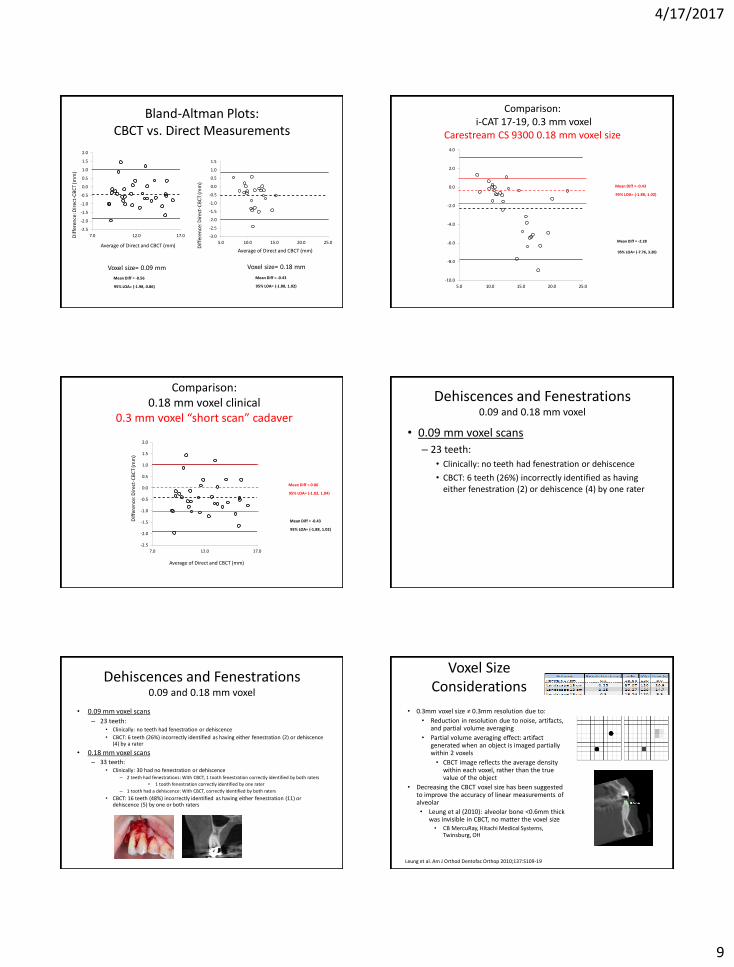
Bland-Altman Plots: CBCT vs. Direct Measurements
-2.5
-2.0
-1.5
-1.0
-0.5
0.0
0.5
1.0
1.5
2.0
7.0 12.0 17.0
Mean Diff = -0.43
95% LOA= (-1.88, 1.02)
-3.0
-2.5
-2.0
-1.5
-1.0
-0.5
0.0
0.5
1.0
1.5
5.0 10.0 15.0 20.0 25.0
Mean Diff = -0.56
95% LOA= (-1.98, 0.86)
Voxel size= 0.18 mm Voxel size= 0.09 mm
Dif
fere
nce
: Dir
ect-
CB
CT
(mm
)
Average of Direct and CBCT (mm)
Dif
fere
nce
: Dir
ect-
CB
CT
(mm
)
Average of Direct and CBCT (mm)
Comparison: i-CAT 17-19, 0.3 mm voxel
Carestream CS 9300 0.18 mm voxel size
-10.0
-8.0
-6.0
-4.0
-2.0
0.0
2.0
4.0
5.0 10.0 15.0 20.0 25.0
Mean Diff = -0.43
95% LOA= (-1.88, 1.02)
Mean Diff = -2.28
95% LOA= (-7.76, 3.20)
Comparison: 0.18 mm voxel clinical
0.3 mm voxel “short scan” cadaver
-2.5
-2.0
-1.5
-1.0
-0.5
0.0
0.5
1.0
1.5
2.0
7.0 12.0 17.0
Dif
fere
nce
: Dir
ect-
CB
CT
(mm
)
Average of Direct and CBCT (mm)
Mean Diff = -0.43
95% LOA= (-1.88, 1.02)
Mean Diff = 0.00
95% LOA= (-1.02, 1.04)
Dehiscences and Fenestrations 0.09 and 0.18 mm voxel
• 0.09 mm voxel scans
– 23 teeth:
• Clinically: no teeth had fenestration or dehiscence
• CBCT: 6 teeth (26%) incorrectly identified as having either fenestration (2) or dehiscence (4) by one rater
Dehiscences and Fenestrations 0.09 and 0.18 mm voxel
• 0.09 mm voxel scans
– 23 teeth: • Clinically: no teeth had fenestration or dehiscence
• CBCT: 6 teeth (26%) incorrectly identified as having either fenestration (2) or dehiscence (4) by a rater
• 0.18 mm voxel scans – 33 teeth:
• Clinically: 30 had no fenestration or dehiscence – 2 teeth had fenestrations: With CBCT, 1 tooth fenestration correctly identified by both raters
• 1 tooth fenestration correctly identified by one rater
– 1 tooth had a dehiscence: With CBCT, correctly identified by both raters
• CBCT: 16 teeth (48%) incorrectly identified as having either fenestration (11) or dehiscence (5) by one or both raters
Voxel Size Considerations
• 0.3mm voxel size ≠ 0.3mm resolution due to:
• Reduction in resolution due to noise, artifacts, and partial volume averaging
• Partial volume averaging effect: artifact generated when an object is imaged partially within 2 voxels
• CBCT image reflects the average density within each voxel, rather than the true value of the object
• Decreasing the CBCT voxel size has been suggested to improve the accuracy of linear measurements of alveolar
• Leung et al (2010): alveolar bone <0.6mm thick was invisible in CBCT, no matter the voxel size • CB MercuRay, Hitachi Medical Systems,
Twinsburg, OH
Leung et al. Am J Orthod Dentofac Orthop 2010;137:S109-19

4/17/2017
10
Dihiscences & Fenestrations in Cadavers • Improve accuracy with smaller voxel size?
– Patcas et al. Accuracy of cone-beam computed tomography at different resolutions assessed on the bony covering of mandibular anterior teeth. Am J Orthod Dentofac Orthop 2012;141:41-50
– Cadaver study: CBCT* 0. 4 and 0.125 mm voxel, followed by dissection
– Conclusion: Even with 0.125 mm voxel scan, thin buccal bone was not reliably imaged
* KaVo 3D eXam, Brugg, Switzerland
Dehiscences & Fenestrations in Patients
• Liangyan et al: Class III patients (n=14 patients, 122 teeth)
• Accelerated osteogenic orthodontic surgery on maxillary and/or mandibular anterior teeth
• Pretreatment CBCT*: 0.125 mm voxel size
– Direct measurement at surgery: Vernier caliper
– Dehiscence (d): > 2 mm from CEJ; fenestration (f): > 0 mm
• Compared direct measurements at surgery to pre-treatment CBCT measurements
Liangyan Sun et al. Accuracy of cone-bean computed tomography in detecting alveolar bone dehiscences and fenestrations. Am J Orthod Dentofac Orthop 2015;147:313-23
* NewTom VG, NewTom, Verona, Italy
Liangyan Sun et al. Am J Orthod Dentofac Orthop 2015;147:313-23
• New-Tom VG; 30 cm (12 inch) FOV; 0.125 mm voxel; 5.4 seconds • Measured bone height from CEJ; Mean difference: -0.7 +/- 4.1 mm 95 % limits of agreement
• Carestream CS 9300; 10 cm (4 inch) FOV; 0.18 mm voxel; 8 seconds • Measured bone height from cusp tip; Mean difference: -0.4 +/- 1.4 mm 95% limits of agreement
Tooth type Sites (n) Direct CBCT
Dehis (n) Fenestr (n) Dehis (n) Fenestr (n)
Maxilla 59 25 4 27 15
Central incisor 20 8 0 7 0
Lateral incisor 19 6 0 8 6
Canine 20 11 4 12 9
Mandible 63 35 3 40 16
Central incisor 20 8 1 10 5
Lateral incisor 21 10 1 14 7
Canine 22 17 1 16 4
Total 122 60 7 67 31
Table VI. Summary of direct and CBCT results for dehiscences and fenestrations by tooth type (N = 122)
Liangyan Sun et al. Accuracy of cone-bean computed tomography in detecting alveolar bone dehiscences and fenestrations. Am J Orthod Dentofac Orthop 2015;147:313-23
Comparisons: OHSU Studies
Cadavers i-CAT 17-19
• Age:
• Mean: 77 years
• Range: 55-89 years
• Mix of posterior and anterior teeth (48):
• Maxilla: posterior: 4 (6%); anterior: 16 (25%)
• Mandible: posterior: 13 (20%); anterior: 32 (49%)
• CBCT considerations:
• O.3 mm voxel
• 0.5 mm slices
• Impact of embalming on scan resolution unknown relative to live tissues
• No movement artifacts
Patients i-CAT 17-19
• Age:
• Mean: 64 years
• Range: 43-81 years
• Mix of posterior and anterior teeth (32):
• Maxillary: posterior: 2 (6%); anterior: 6 (18%)
• Mandible: posterior: 5 (15%); anterior: 19 (59%)
• CBCT considerations:
• 0.3 mm voxel
• 0.5 mm slices
• Presence of living cells: x-ray scatter from cells/organelles, high water content
• Patient movement during scanning
Patients Carestream CS 9300
• Age:
• Mean: 64 years
• Range: 37-81 years
• Mix of posterior and anterior teeth (56):
• Maxillary: posterior: 28 (50%); anterior: 17 (30%)
• Mandible: posterior: 7 (12%); anterior: 4 (7%)
• CBCT considerations:
• 0.18 or 0.09 mm voxel
• 0.18 mm slices
• Presence of living cells: x-ray scatter from cells/organelles, high water content
• Patient movement during scanning
Summary: Cadavers using Various CBCT Settings
• Accuracy: – No difference in accuracy of measurements among scans having
widely varying numbers of projection images (169 to 619) and differing arcs of rotation (360o vs. 180o)
• Precision: – Better with high resolution scans: Perhaps reserve for research
purposes, or when high precision is desired
• Assessing alveolar bone height vs. thickness with CBCT:
– Bone height measurements correlate more closely to direct measurements than do bone thickness measurements

4/17/2017
11
Summary: Live Patients and CBCT @ 0.3 mm Voxel Size
• With older CBCT unit (i-CAT 17-19) and 0.3 mm voxel: – Alveolar bone height was underestimated in CBCT images in areas
where alveolar bone is thin
• Mean of 2.3 mm underestimation
• Particularly an issue with mandibular anterior teeth
– CBCT had notable limitations determining if thin bone or a bone defect
is present
• 28% of teeth studied had either the presence of bone or a boney defect misidentified by one or both examiners
Summary: Live Patients and CBCT @ 0.18 and 0.09 mm Voxel Size
• With newer CBCT unit and 0.18 or 0.09 mm voxel size scans: – Alveolar bone height measurements were more accurate
• Mean of 0.5 mm underestimation
• No particular advantage of 0.09 vs. 0.18 mm voxel size
– CBCT still had notable limitations determining if thin bone or a bone defect is present
• 0.18 mm voxel size: 48% of teeth studied had bone present that was misidentified with CBCT as a defect by one or both assessors
• 0.09 mm voxel size: 26% of teeth had bone present that was misidentified with CBCT as a defect by one assessor or the other
CBCT in Assessing Alveolar Bone Height: Are Looks Deceiving?
Results of investigations at OHSU:
– Skulls/cadavers:
• Show best case scenario
• Measurements from CBCT had high accuracy and precision
• Caution needed translating results to clinical setting
– Clinical CBCT Imaging:
• Scans with small voxel size scans more accurate than with large voxel size
• Even with smallest voxel size, when imaging indicates a lack of alveolar bone along the facial surface of roots, one cannot know if thin bone is present, or if bone is absent
• Newer CBCT unit, smaller voxel size scans more accurate
CBCT in Assessing Alveolar Bone Height: Are Looks Deceiving?
Results of investigations at OHSU:
– Skulls/cadavers and bone height:
• Show best case scenario
• Measurements from CBCT had high accuracy and precision
• Caution needed translating results to clinical setting
– Clinical CBCT Imaging and bone height:
• Larger voxel size - less accuracy, precision
• Smaller voxel size, newer CBCT units – Improved accuracy and precision, but will tend to underestimate
• Even with smallest voxel size, when imaging indicates a lack of alveolar bone, one cannot know if thin bone is present, or if bone is absent
Reflections on CBCT Studies
Clinical considerations:
– What is it that you want to view with CBCT imaging?
• For traditional radiographic records used in orthodontics: – Moderate resolution likely adequate
– Large to medium field of view (medium if use digital lateral cephalogram)
– ALADA: larger voxel size (0.3 – 0.4 mm), “low dose scans”
– Bone height: -2.2 +/- 5.5 mm (95% limit of agreement)*
• For assessing alveolar bone height: – High resolution optimal
– Small field of view (single or both dental arches; localized area)
– ALADA: smaller voxel size (0.18 mm)
– Bone height : -0.5 +/-1.5 mm (95% limit of agreement)**
* i-CAT 17-19; 0.3 mm voxel scan ** Carestream CS 9300; 0.18 voxel scan
Acknowledgements
– Adam Timock (2010)
• Fort Collins, CO
– Lane Cook (2011)
• Garland, Tx
– Megan Miller (2013)
• Lancaster, OH
– Vanessa Browne (2014)
• Portland, OR
– Andrew Peterson (2017) • Resident, Periodontology
– Mansen Wang • Providence Health & Services
– Brion Benninger • Western Univ. of Health Sciences
– Jennifer Crowe
– Harjit Singh Sehgal
– Larry Doyle
– Terry McDonald
– OHSU Orthodontic Alumni Association