aspergilloma caso clinico - moodle@units
TRANSCRIPT

Sinus Aspergilloma
A CASE REPORT

ASPERGILLOSIS
ASPERGILLOSIS = fungal disease, it exists in two forms: a noninvasive form and an invasive form (SECOND IN FREQUENCY ONLY TO CANDIDIASIS)
Noninvasive = affects the normal host appearing as an allergic reaction or a cluster of fungal hyphae (fungal ball)
Invasive = affects the immunocompromised patient (AIDS, SOLID ORGAN TRANSPLANTATION, UNCONTROLLED DIABETES)

ASPERGILLOSIS
ASPERGILLOSIS = fungal disease = SPORES reside
worldwide in soil, water or decaying organic debris.
Released into the air and inhaled by human host
Two most common species are A. FLAVUS and A. FUMIGATUS
CLINICAL MANIFESTATION
Noninvasive
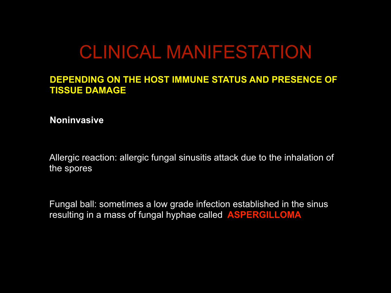
DEPENDING ON THE HOST IMMUNE STATUS AND PRESENCE OF TISSUE DAMAGE
Allergic reaction: allergic fungal sinusitis attack due to the inhalation of the spores
Fungal ball: sometimes a low grade infection established in the sinus resulting in a mass of fungal hyphae called ASPERGILLOMA

CLINICAL MANIFESTATION
Invasive
DEPENDING ON THE HOST IMMUNE STATUS AND PRESENCE OF TISSUE DAMAGE
immunocompromised patients susceptible to oral aspergillosi, portal of entry the marginal gingiva or endodontic treatment or tooth extraction especially in the posterior treatment.
Disseminate aspergillosis (leukemia or high doses of corticosteroids) chest pain fever = vague symptpms = difficult diagnosis, if the infection gains the blood CNS, liver, thyroid damage.
locally: painful gingival ulceration =swelling and violaceus hue = if no treated necrosis.

DEFINITION
FUNGAL BALL or ASPERGILLOMA are extramucosal collections of fungal elements, usually localized to a single sinus cavity, commonly the maxillary sinus. They are usually of the noninvasive variety, and commonly seen in immunocompetent hosts. Occasionally a waning of the immunity can cause them to turn invasive.

DEFINITION
Occasionally, the mass will undergo d y s t r o p h i c c a l c i f i c a t i o n (radiopaque body) called ANTROLITH

CASE HISTORY
CASE HISTORY
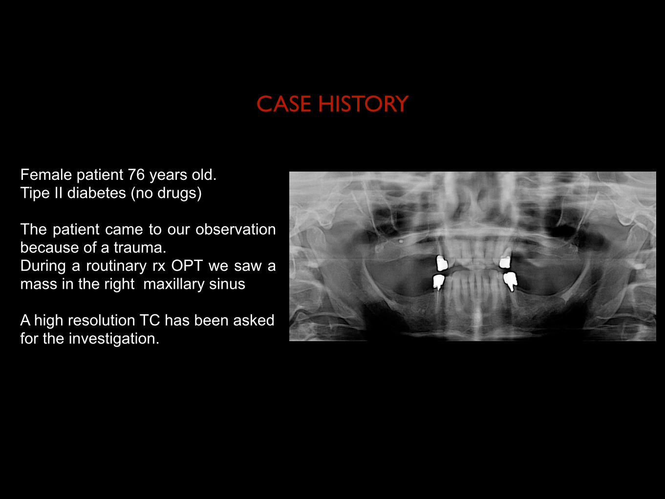
Female patient 76 years old. Tipe II diabetes (no drugs) The patient came to our observation because of a trauma. During a routinary rx OPT we saw a mass in the right maxillary sinus
A high resolution TC has been asked for the investigation.

Patient underwent surgery to remove the lesion compatible with suspected aspergilloma

Allestimento botola ossea a mezzo inserti piezoelettrici. Individuazione membrana di Schneider e accesso al seno mascellare destro mediante interruzione della stessa.
1
2
3

4
5
6Curettage e detersione seno mascellare.
Ispezione con fibroscopio c o n s o n d a f l e s s i b i l e . Drenaggio ascesso seno mascel lare e r imozione corpo estraneo compatibile con sospetta aspergillosi.

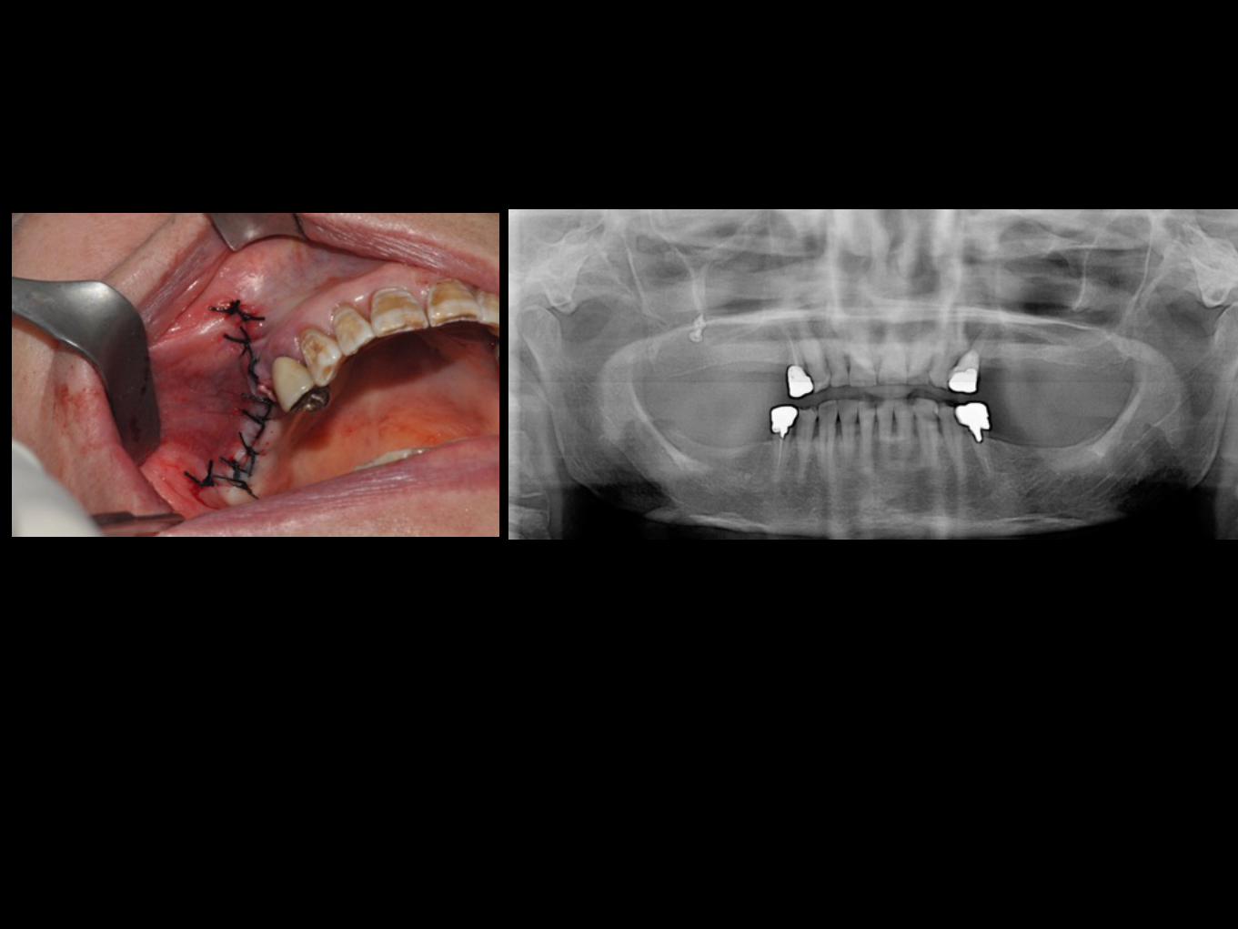
Riposizionamento botola ossea a mezzo viti e placche di osteosintesi. C h i u s u r a p o r z i o n e rimanente del difetto con membrana in collagene riassorbibile. Protezione di botola ossea a mezzo membrana in collagene riassorbibile. Sutura per prima intenzione.
7
8
9

S u t u r a p e r p r i m a intenzione. Consegnata terapia Ab con A m o x i c i l l i n a 1 g d a assumere 3/die per una settimana, antidolorifico al bisogno
R i m o z i o n e s u t u r e e controllo ad una settimana
10
11

HISTOPATOLOGIC
ASPERGILLOMA is characterized by tangle mass of hyphae with no evidence of tissue invasion and presente large pool of eosinophilic inspissated mucin and lymphocytes
noninvasive form are features that differ from invasive
INVASIVE FORM: the hyphae branch in an acute angle, invade small blood vessel = occlusion = necrosis

MANAGEMENTThe goal of treatment for a fungus ball (immunocompetent patient with low kind of aspergillosy) is surgical removal of the hyphal mass with re-establishing the drainage from the affected sinus + corticosteroid.
Endoscopic sinus surgery to remove the fungus ball is the treatment of choice
Irrigation of the sinus is performed to clear the sinus of all the fungal debris.
Recurrence is rare but, has been reported to occur even as late as two years following surgery
A regular follow-up is important as many of these recurrences can be addressed with simple irrigation or suctioning in the outpatient department or conservative endoscopic surgery
Bachi T Hathiram, Vicky S Khattar
34JAYPEE
Symptoms are similar to those seen in chronicrhinosinusitis secondary to inflammation or bacterialinfection. These include nasal obstruction, nasal discharge,cacosmia, facial pain with a history of these symptomsbeing refractory to medication. Symptoms are usually oflong duration(months to years). Occasionally the patientmay present with unusual symptoms such as epistaxis, visualdisturbances, convulsions, fever, cough, and proptosis.4-6
Sometimes, the patient may be asymptomatic and the fungusball may be an incidental finding. 10% of patients haveassociated nasal polyps4 which are infact, a nonspecificresponse to a variety of inflammatory conditions.Approximately 50% of individuals give a history of someendodontic treatment being done prior to maxillary sinusfungus ball indicating it could be a predisposing factor.4
Fungus balls are rarely known to cause bone remodelingwith widening of the affected sinus and distortion ofanatomy. They may also cause bone erosion. Rarely, if duringthe infection, the immunity of the host declines, a fungusball may become invasive. Characteristic imaging findingsand histopathologic examination confirms the diagnosis. Atsurgery, thick inspissated debris forms a mass which fillsthe sinus cavity.5 On histopathological examination, the debrisfound in a fungus ball consists of dense tangles of hyphaewith calcifications and oxalate crystals.5 However, fungalcultures are usually negative. It usually does not invadetissue; however acute or chronic inflammatory infiltrate maybe present in adjacent mucosa but, granulomas are absent.5,9
HISTOPATHOLOGY AND FUNGAL CULTURE
Fungus balls are essentially noninvasive and extramucosalfungal infestations without any granulomatous reaction.Routine hemotoxylin and eosin stains can demonstrate thepresence of fungus but, special stains such as the gomorimethenamine silver (GMS) are helpful in diagnosing theAspergillus species.
Intraoperatively, the gross appearance of the fungus isgritty or cheesy and clay-like, breaking up into fragments,the color of which ranges from brown to black to green oryellow.
The causative fungi include Aspergillus fumigatus,Aspergillus flavus, Alternaria Sp and P Boydii. Only 23-50% cultures result in fungal growth.
RADIOLOGIC FEATURES
Fungus balls have typical characteristics on CT scan. Inover 90% of patients, a single sinus is involved.4 Themaxillary sinus is most commonly involved (Fig. 2) followedby the sphenoid (Fig. 3). Involvement of the ethmoids iscontiguous with the maxillary sinus. The frontal sinus israrely involved. On CT scan, fungus balls show partial orcomplete heterogenous opacification of the sinus.5 Centralareas of hyperattenuation within the fungus ball correspondto fungal debris/hyphae and calcifications. Sclerosis or bonythickening of the sinus wall is commonly seen and bonyerosion of the sinus wall may occur. However, there isminimal or no sinus expansion.
MANAGEMENT
The goal of treatment for a fungus ball is surgical removalof the hyphal mass with re-establishing the drainage fromthe affected sinus. A symptomatic patient with opacificationof the sinus and bone erosion merits surgical evacuation,however the same line of management in a patient who isasymptomatic is controversial. Presence of coexistingasthma in a patient with a fungus ball merits surgery toprevent exacerbation of asthmatic attacks due to the fungalantigen.
Endoscopic sinus surgery to remove the fungus ball isthe treatment of choice today and the erstwhile externalapproaches are obsolete. Irrigation of the sinus is performedto clear the sinus of all the fungal debris.
The maxillary sinus is cleared by widening the naturalostium (middle meatus antrostomy) and a canine puncturewill help in visualizing the entire sinus cavity as well asserve the purpose of irrigation.
FIGURE 1: Endoscopic photograph showing a fungus ball in the leftsphenoid sinus

MANAGEMENT
For the immunocompetent patient with invasive aspergillosi : surgical removal + antifungal medication (voriconazole a triazole antifungal)
For the immunocompromised patient with invasive aspergillosi : agressive surgical removal + sistemic antifungal terapy (triazole antifungal) only one third of this patient survive (usually develops while hospitalized)