aspects of tail muscle ultrastructure and its...
TRANSCRIPT

/ . Embryol. exp. Morph. Vol. 23, 1, pp. 191-207, 1975 191
Printed in Great Britain
Aspects of tail muscle ultrastructure and itsdegeneration in Rana temporaria
By H. FOX1
From The Department of Zoology,University College, London
SUMMARY.1. Tail muscles of larval Rana temporaria and to a lesser extent Xenopus laevis, at pro-
metamorphosis, were investigated by light and electron microscopy. In the case of Ranatheir degeneration is described at metamorphic climax.
2. The non-degenerate tail muscles of both genera are generally similar in their ultra-structure and likewise similar to those striated muscles of other adult vertebrates includingamphibians.
3. At climax tail muscles of Rana degenerate by autolysis. It seems likely that lysosomalenzymes are directly involved in sarcoplasmic degeneration. The relationship betweenlysosomes and myofibrillar degradation is difficult to establish. The subject is discussed inthe light of relevant information (mainly mammalian) on muscle dystrophies.
4. Anuran tail muscle is highly degraded before its probable phagocytosis by mesenchymalmacrophages.
INTRODUCTION
Somitic striated muscle is a major component of the anuran larval tail. Atmetamorphic climax in vivo, under the influence of a high threshold level ofcirculatory thyroid hormones (Etkin, 1964), or in vitro, influenced by a similarlyhigh ambient concentration of thyroxine, thyroxine analogs or triiodothyronine,the tail involutes with consequent degeneration and disappearance of its muscu-lature (see among others Kollros, 1961; Shaffer, 1963; Kaltenbach, 1968;Weber, 1969; Frieden & Just, 1970). As in all vertebrates anuran striated musclehas a complex ultra-structure (Page 1965; Franzini-Armstrong, 1970; Karlson& Anderson-Cedergren, 1971; Eichelberg & Schneider, 1973). Features of thelarval axial and tail musculature have likewise been described by electronmicroscopy (Porter & Palade, 1957; Weber, 1964; Fox, 1972c), but much isobscure, particularly causality when anuran tail muscles degrade at climax.In vertebrates a variety of factors will elicit muscle degeneration (see below)and at this time a number of structural features are similar whatever the cause(see Tomanek & Lund, 1973). Information obtained from the study of climactic
1 Author's address: Department of Zoology, University College, Gower Street, LondonW.C.I.
13 EMB 34

192 H. FOX
anuran tail muscle could well be of importance in relation to the wider aspectsof tissue ageing and disease.
The results in the present work lead to the conclusion that at climax thesarcoplasm degenerates autolytically, probably initiated through the agency oflysosomal enzymes, though the widespread necrosis expressed visually in termsof large cytolysomes positive for acid phosphatase is not always recognizedto the same extent as in some other degenerating tail tissues. Myofibrillardegeneration is described and the vexed question on the possible role playedby lysosomal enzymes is considered.
It is likely that degraded tail muscle is phagocytosed by mesenchymalmacrophages.
MATERIAL AND METHODS
Specimens of Rana temporaria larvae were staged as for Rana dalmatina(Cambar & Marrot, 1964); Xenopus laevis according to the scheme of Nieuw-koop and Faber (1956). In Rana prometamorphosis includes stages 45-49,used in the present work, and climax extends from late stage 49-54.
For examination by light microscopy tails from normal Rana temporaria (intap water) at various stages from 41 (15 mm) to <54 (14 mm) were utilized.Larval length is given in brackets. All specimens were fixed in Bouin's fluid,embedded in paraffin wax, sectioned transversely at 10 ju,m and stained byhaematoxylin and eosin.
For examination by electron microscopy tails at different levels proximo-distally, from Rana at stages 45, 45-47, 48, 48-49, 49, 52, 52-53 and 53 werefixed in osmic acid (Palade, 1952), or glutaraldehyde and post-osmicated (Saba-tini, Bensch & Barrnett, 1963), or in a combined mixture of glutaraldehyde andosmic acid (Hirsch & Fedorko, 1968); each stage was frequently fixed by morethan one method. After dehydration and embedding in Araldite, thick sections(about 1 fim thick) were examined by phase contrast microscopy for tissuelocalization and silver grey sections (about 90-100 nm thick) were stained byuranyl acetate (Watson, 1958) and lead citrate (Reynolds, 1963). Tails ofXenopus laevis at early and late prometamorphic stages 45 and 57 (Nieuwkoop& Faber, 1956) and fixed by the method of Hirsch & Fedorko (1968), werelikewise investigated mainly from sagittal sections.
Deposition of acid phosphatase in tail muscle was registered for electronmicroscopy by the method of Gomori (1952), modified by Barka & Anderson(1962). Incubated hand-cut transverse sections, about 150/*m thick, werepreviously fixed for about 1 h in glutaraldehyde (see Fox, 1974). Controls wererun omitting the sodium /?-glycerophosphate from the incubation medium.Silver grey sections were stained by uranyl acetate alone. All sections wereviewed under an AEI EM 6B electron microscope and to a lesser extent anAEI Corinth 275.

infrastructure o/Rana tail muscles 193
OBSERVATIONS
Examination by light microscopy
The tail musculature of well-developed, active larval amphibians comprisesa series of segmentally arranged muscles, separated by connective tissue, themyocommata, and situated on each side of the tail between the skin and thenerve cord and notochord. Anterior somites are longer than posterior ones, atleast in Xenopus embryos (Hamilton, 1969), and in Rana larvae individual tailsomite size seems to diminish progressively on proceeding distally (see alsoBrown, 1946). Somites disappear just before the tail tip.
Tail muscles of anurans degenerate at metamorphic climax. In Rana atlate stage 49, the tail tip blackens, probably in part due to the accumulation ofpigment and with the exception of macrophages and perhaps some blood cells,shows features of degeneration.
Initially there is widespread necrosis in the distal tail region. Simultaneouslythe tail reduces in length to a tiny sac-like structure, which ultimately disap-pears. At the height of climax (stages 52-53) the posterior third of one tailstub (3-4 mm long measured from the front margin of the cloaca) includeddegraded tissue, much of it presumably derived from muscle suffering phago-cytosis by macrophages. Further forwards the tail includes partially degenerateblocks of myofibrillar tissue, some dispersed into smaller disorganized units.
The recognizable notochord shows features of degeneration 1-6 mm infront of the tail tip, though some muscle components are necrotic even furtherproximally. The nerve cord is still luminated 0-7 mm from the stub end. On thewhole in Rana at climax, the extent of degeneration is more widespread inmuscle and more localized in the nerve cord than in the case of other tail tissues.
Examination by electron microscopy
(a) Preclimactic tail muscle
As in other vertebrates tail striated muscle cells of larval Rana and Xenopustypically are made up of a large number of longitudinally arranged, closelypacked (especially in older larvae), parallel myofibrils (about 1 fim thick); eachis cross-striated and of regularly repeating periodicity, due to the overlappingof constituent thick and thin myofilaments of myosin and actin respectively(Fig. 1). In longitudinal section the Z lines are usually straight. The occasionalwavy Z lines found in young prometamorphic, non-degenerate tail musclesmay represent either oblique sectional orientation or myofibrillar distortionowing to incomplete muscle contraction. The dimensions of the I band,which correspond to the degree of sarcomere contraction, reflect the amountof mutual sliding of the overlapping myofilaments whose lengths remainconstant (see Price, 1969).
The sarcoplasm includes ribosomes, polyribosomes and a modest granular13-2

194 H. FOX
infrastructure o/Rana tail muscles 195endoplasmic reticulum. A prominent sarcoplasmic reticulum embraces themyofibrils throughout their length. Frequently a Golgi complex is recognized.Occasional large lipid droplets are found at the periphery or between themyofibrils and there are pigment bodies and numerous glycogen granules.Mitochondria of varied shape, but usually elongated in the longitudinal plane,frequently occur in groups at the periphery or singly between the myofibrils(Fig. 1). Cristae are moderately numerous and of random orientation. Vesiclesmay indent the sarcolemma and so-called caviolae often occur - whether theyalways open to the extracellular space cannot be decided. Large nuclei oftenwith a prominent nucleolus are situated usually against the outer regions of themyofibrils, or terminally in the cell.
The sarcolemma comprises a trilaminar plasma membrane; externally a
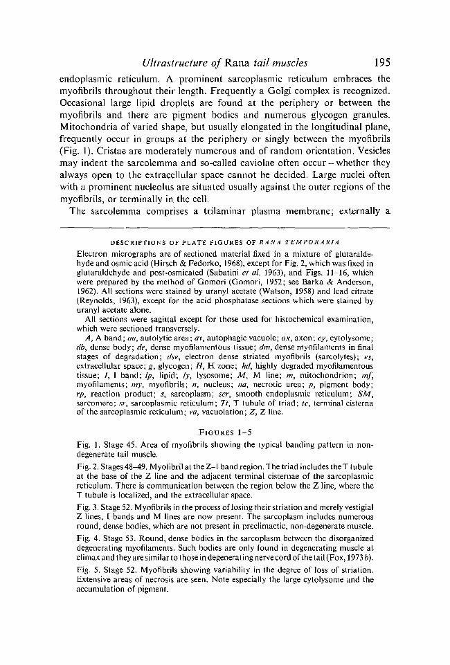
DESCRIPTIONS OF PLATE FIGURES OF RANA TEMPORARIA
Electron micrographs are of sectioned material fixed in a mixture of glutaralde-hyde and osmic acid (Hirsch & Fedorko, 1968), except for Fig. 2, which was fixed inglutaraldehyde and post-osmicated (Sabatini et al. 1963), and Figs. 11-16, whichwere prepared by the method of Gomori (Gomori, 1952; see Barka & Anderson,1962). All sections were stained by uranyl acetate (Watson, 1958) and lead citrate(Reynolds, 1963), except for the acid phosphatase sections which were stained byuranyl acetate alone.
All sections were sagittal except for those used for histochemical examination,which were sectioned transversely.
A, A band; an, autolytic area; av, autophagic vacuole; ax, axon; cy, cytolysome;db, dense body; de, dense myofilamentous tissue; dm, dense myofilaments in finalstages of degradation; dse, electron dense striated myofibrils (sarcolytes); es,extracellular space; g, glycogen; H, H zone; hd, highly degraded myofilamentoustissue; /, I band; Ip, lipid; ly, lysosome; M, M line; m, mitochondrion; mf,myofilaments; my, myofibrils; n, nucleus; na, necrotic area; p, pigment body;rp, reaction product; s, sarcoplasm; ser, smooth endoplasmic reticulum; SM,sarcomere; sr, sarcoplasmic reticulum; Tt, T tubule of triad; tc, terminal cisternaof the sarcoplasmic reticulum; va, vacuolation; Z, Z line.
FIGURES 1-5
Fig. 1. Stage 45. Area of myofibrils showing the typical banding pattern in non-degenerate tail muscle.Fig. 2. Stages 48-49. Myofibril at the Z-I band region. The triad includes the T tubuleat the base of the Z line and the adjacent terminal cisternae of the sarcoplasmicreticulum. There is communication between the region below the Z line, where theT tubule is localized, and the extracellular space.Fig. 3. Stage 52. Myofibrils in the process of losing their striation and merely vestigialZ lines, 1 bands and M lines are now present. The sarcoplasm includes numerousround, dense bodies, which are not present in preclimactic, non-degenerate muscle.Fig. 4. Stage 53. Round, dense bodies in the sarcoplasm between the disorganizeddegenerating myofilaments. Such bodies are only found in degenerating muscle atclimax and they are similar to those in degenerating nerve cord of the tail (Fox, 1973 b).Fig. 5. Stage 52. Myofibrils showing variability in the degree of loss of striation.Extensive areas of necrosis are seen. Note especially the large cytolysome and theaccumulation of pigment.
196 H. FOX
moderately dense basement membrane is intimately associated with collagenfibrils, which often penetrate between blocks of myofibrils, though they arealways separated from them by the sarcolemmal sheath.
In Rana temporaria tail muscles triads are particularly well represented,situated at the levels of the Z lines. The T tubular system opens to the cellexterior, for continuity across the sarcoplasm between the extracellular regionand the base of the Z line, where the T tubule is clearly localized, is occasionallyrecognizable (Fig. 2). T tubules encircle the myofibrils at the Z lines (Fig. 1).They may also extend longitudinally in some places, as in the adult frog sar-torius muscle, though still remain separated from the sarcoplasmic reticulum(Huxley, 1964), a feature likewise recognized in the Rana tail muscle.
In both Rana and Xenopus the diameter of the thick myofilaments is about16 nm and of the thin myofilaments about 6 nm. The recorded maximummitochondrial length and breadth of groups of the organelles, in typicalrandom muscle profiles, each of Rana and Xenopus, showed those of Rana torange between 1-3 and 2-7/tm in length and 0-4 and 0-8 jam in breadth. Com-parable measurements in Xenopus were 1-5-5-5 /im and 0-6-1-5 jim. Clearlythroughout prometamorphosis mitochondria in Xenopus tail muscles appearto be of greater area than those of Rana.
(b) Climactic degeneration of tail muscles
At climax ultrastructural profiles of degenerating cells of the anuran tail arevariable, and the intimate relationships of the sub-epidermal cells and associatedmacrophages are complex. In different cells degeneration phenomena appearseemingly at random. Even at the same transverse level tail cells of like tissuemay vary in their degree of degeneration, a variation which may occur withinthe same cell. The following description, therefore, provides a generalizedaccount of muscle necrosis and of its subsequent phagocytosis during tailinvolution.
At climax the sarcoplasm normally shows signs of degeneration before themyofibrils, though the entire muscle is soon affected at any level but mainlydistally. A number of organelles, not usually present in preclimactic stages,appear in the sarcoplasm. Numerous membrane-bound bodies having a whorledor mottled appearance occur, often near the nucleus. Other large irregular-shaped bodies, frequently appearing fibrous in texture, are often found betweenmyofibrils or in the sarcoplasm peripherally, and a number of smooth-surfaced,roundish, dense bodies occur, either in groups at the periphery or singlybetween myofibrils (Figs. 3, 4). Similar bodies are found in macrophages andthey are often profuse in degenerating tail nerve cord cells (Fox, 1973 b).Myelin figures (occasionally seen in earlier stage-45 specimens of Rana) arepresent, often near the sarcolemma - the sarcolemmasomes (Bone & Ryan,1973). Mitochondria degenerate in two ways: either the cristae disorganizeand disappear leaving an empty vesicle, or the outer membranes rupture and

Ultrastructure o/Rana tail muscles 197the partially disorganized cristae spill out to merge with and subsequentlydisperse amid the ambient cytoplasm (see Fox, 1972c, 1973a). Small auto-phagic vacuoles and somewhat larger cytolysomes are present; these areoccasionally seen between the myofibrils (Figs. 5, 13). Lipid droplets andpigment bodies increase in number, sometimes the latter are grouped in mem-brane-bound structures. Usually the Golgi complex, granular endoplasmicreticulum and ribosomes reduce in amount or have disappeared in highlydegenerate sarcoplasm, but there is an increase in the amount of smoothendoplasmic reticulum and in the number of glycogen granules.
Areas of necrosis gradually become more widespread and large cytolysomesare sometimes recognizable (Figs. 5, 7).
The sarcoplasm ultimately loses its typically recognizable form, the sarco-lemma ruptures and the remains of the sarcoplasm and the partially or whollydegraded myofilaments become intimately associated spatially with envelopingmacrophages (see Fox, 1972c, fig. 9; 1973c, fig. 5).
The muscle cell nucleus usually appears normal even when the myofibrilsand sarcoplasm show pronounced features of degeneration. Nuclear resistanceto necrotizing influences is likewise seen in the anuran tail nerve cord andnotochord and in the apical ridge of the limb-buds of chick and mouse embryos(Jurand, 1965; Fox, 1973 a, b).
The first obvious sign of myofibrillar degeneration is the loss of striation,when only remains of banding are visible (Figs. 3, 5). The myofilaments becomedisorientated (Fig. 4) and lose their parallel, elongate arrangement, seen ideallyin longitudinal sections of preclimactic tail muscles. At climax a sagittal sectionoften includes profiles of myofilaments orientated in all directions. The clarityof delineation of the mass of disorganized myofilaments is reduced due to theblurring of the myofilamentous outlines, which are often still recognizablein highly degraded myofibrillar tissue.
Vestigial Z lines are occasionally straight but more often wavy in appearance.The extensive sarcoplasmic reticulum is sometimes expanded amid the degenera-ting myofibrils. Substantial areas of myofibrillar tissue often become electrondense and merge with degenerate, often electron-translucent, homogeneoussarcoplasmic substance (see also Fox, 1972c, figs. 5, 9). In some cases musclecells include highly electron dense, fragmented myofibrils, with a recognizablestriation (Fig. 6) - the so-called sarcolytes (Brown, 1946; see also Watanabe &Sasaki, 1974) - which may occur simultaneously with less degenerate myofila-ments within the same cell.
At the height of climax profiles of highly degenerate, tail-stub muscle cellsinclude roundish areas of varied size and of lightish, homogeneous substance;frequently they possess a denser occasionally filamentous core (Figs. 7-10).Disorientated myofilaments are often found alongside, or in spatial continuitywith, such homogeneous degraded tissue (Fig. 9); clearly some at least of thelatter is derived from myofilaments. Degenerating muscle cells thus include

198 H. FOX

Ultrastructure o/Rana tail muscles 199partially and highly degraded myofilamentous (and sarcoplasmic) tissue, whichhas degenerated autolytically before it spills out through a ruptured sarcolemmato suffer phagocytosis.
Macrophages likewise may include similar degraded, homogeneous substancein heterophagic vacuoles, or phagosomes, presumably the result of previousengulfment (see also Watanabe & Sasaki, 1974). It is, nevertheless, frequentlydifficult to decide between profiles of highly degenerate muscle cells andmacrophages; whether, for example, autophagic or heterophagic vacuoles arefeatured. Initially during prometamorphosis mesenchymal macrophages arespatially associated with muscle and later, at climax, they closely envelopdegraded tail muscle tissue (Fox, 1972c, 1973 c). Yet many cells containingfragmented, electron-dense, striated myofibrillar tissue (sarcolytes) and classifiedas macrophages (Kerr, Harmon & Searle, 1974), may well be degenerate musclecells, as earlier implied by Brown (1946). Numerous examples were found ofcells possessing myofilaments showing varying degrees of degradation, includingsome which were similar to the so-called heterophagic inclusions (Figs. 6, 10).
Deposition of acid phosphatase represented by reaction product (RP) is noteasily demonstrated in muscle tissue. However, in a number of different speci-mens of Rana, round or oval-shaped, dense, membrane-bound bodies (3-9 ineach group) and positive for RP, were found in tail muscle sarcoplasm near thenucleus of late prometamorphic (stage 49) and climax specimens. They varied indiameter from 0-2 to 0-5 jttm (Fig. 11). RP also occurred in small autophagicvacuoles (often situated between myofibrils), slightly larger cytolysomes andoccasionally alongside or within the sarcoplasmic reticulum (Figs. 12, 13, 16).RP was also found deposited in discrete areas against the outermost myofibrillar
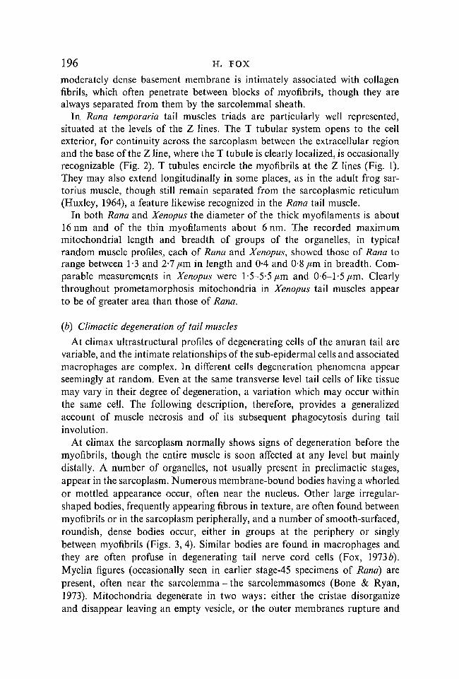
FIGURES 6—10
Fig. 6. Stage 53. Degenerate myofibrils (sarcolytes) of varied electron densityshowing some striation. Some portions of the myofibrils are isolated within mem-brane-bound regions and they are degrading autolytically to a homogeneoussubstance.Fig. 7. Stage 53. Heavy necrosis in the sarcoplasm and varied profiles of myofibrillardegeneration, including disorganized myofilaments, dense myofilaments - allwithout striation - and highly degraded homogeneous myofibrillar substance inwhich vestigial myofilaments may still be present.Fig. 8. Stage 53. Highly degraded myofibrillar tissue with vestigial myofilamentsstill recognizable. Lipid is abundant. Lesser degenerate myofilaments are seennearby.Fig. 9. Stage 53. Origin of the highly degraded homogeneous substance frommyofilaments during the final stages of muscle degeneration.Fig. 10. Stage 53. Varied array of myofibrillar degeneration profiles in tail muscleat climax. They range from striated to unstriated myofibrils, highly electron-densesarcolytes, and fully degraded homogeneous tissue of myofibrillar derivation. Thesarcoplasm includes areas of necrosis with abundant lipid, especially near the topof the illustration between the two nuclei.

200 H. FOX
FIGURES 11-16
Fig. 11. Stage 49. Just preceding metamorphic climax. Acid phosphatase-rich organ-elles near the nucleus. They are not found in younger larval tail muscles andsome of them at least are probably lysosomes.Fig. 12. Stage 49. Deposition of reaction product (RP) in the sarcoplasm andwithin a mitochondrion.Fig. 13. Stage climax. Autophagic vacuole, positive for RP, between myofibrils.Fig. 14. Stage climax. Widespread deposition of RP in the degenerating sarco-plasm. The myofilaments show no deposition of RP.Fig. 15. Higher magnification of a region of Fig. 14 showing the deposition of RPin the sarcoplasm.Fig. 16. Stage 49. Cytolysome positive for RP, situated in the sarcoplasm betweenthe myofibrils.

Ultrastructure o/Rana tail muscles 201surface in the sarcoplasm, around and sometimes within mitochondria (Fig. 12).Deposition of RP in the sarcoplasm was sometimes found to be quite extensive(Figs. 14, 15). RP was never found to impregnate or be freely distributed amidmyofilaments. Controls were always negative for reaction product.
DISCUSSION
Primary lysosomes were not recognized by Weber (1964) in regressing tailmuscles of Xenopus, who concluded that they degenerated in their absence,perhaps without the intervention of lysosomal enzymes.
Yet it is difficult to imagine lysosomal enzymes not to be involved, in someway, when vertebrate muscles degenerate. In anurans tail involution dependsupon the de novo synthesis of protein, probably acid hydrolases, near climax(Tata, 1966; Weber, 1969). A number of them, including cathepsins, acidphosphatase and DNA-ase, increase in amount (together with an increasedpopulation of macrophages) towards climax (Lehman, 1953; Eeckhout, 1969;Weber, 1969; Hickey, 1971). Acid hydrolases are probably synthesized bymacrophages (Hassan & Autuori, 1964; Hassan & Marinucci, 1966), though atclimax in autolysing tissues (including muscle; see below) lysosomal enzymesalso substantially increase in amount in the anuran tail (Kaltenbach, 1971;Fox, 1972a, 1913a, b, 1974; Fry, Leius, Bacher & Kaltenbach, 1973).
Among mammals lysosomes are sparse in normal rat muscle (Gordon, Price& Blumberg, 1967; Buchanan & Schwartz, 1967; Weinstock & Iodice, 1969)and like lipofuscin granules (Shafiq, Milhorat & Gorycki, 1967), they increasein number together with the amount of acid hydrolases (Pennington, 1963;Tappel et al. 1962; Tappel, Sawant & Shibko, 1963; in ageing, congenitalmuscular dystrophy, muscular atrophy from denervation and nutritional defi-ciency (usually vitamin E) in man, various mammals and the chicken (Pellegrino& Franzini, 1963; Smith, 1964; Howes, Price & Blumberg, 1964; Milhorat,Shafiq & Goldstone, 1966; Pearce, 1966; Gordon et al. 1967; Weinstock &Iodice, 1969; Shafiq, Askanas, Asiedu & Milhorat, 1972). Muscle autolysis indystrophic muscles occurs simultaneously with increase in acid hydrolases(Iodice, Chin, Parker & Weinstock, 1972). The increased activity of acidhydrolases in dystrophic muscle was considered due to the damage of musclelysosomal membranes, permitting the release of enzymes to degrade myofibrillarand sarcoplasmic tissue, though the main source of enzymes probably arose frominvasive macrophages (Tappel et al. 1962; Tappel et al. 1963; Zalkin et al. 1962).In contrast, others believed that the main source is from the muscle itself (Wein-stock & Lukacs, 1965; Weinstock & Iodice, 1969), especially in later stages ofdystrophy (Shafiq et al. 1972). Lysosomes (Pearce, 1966) and autophagicvacuoles (Milhorat et al. 1966) occur between myofibrils in human dystrophicmuscles. Furthermore, free reaction product (acid phosphatase) was reported tooccur in the walls of the T tubules and to a lesser extent in the intermyofibrillarsarcoplasmic reticulum of human dystrophic muscle (Pearce, 1966). Pellegrino

202 H. FOX
& Franzini (1963) correlated the appearance of lysosomes with rat muscleatrophy after denervation.
Lysosome-type bodies (positive for RP) and features of autolysis are rarelyfound in tail muscles of young prometamorphic larvae of Rana (stages 45-47),though occasionally autophagic vacuoles are present at stage 49 (and in Xenopusat stage 57) just before climax. Organelles and autophagic vacuoles, positivefor RP, are more common at climax and free reaction product was recognizablein highly degenerate sarcoplasm (see below and Fox, 1972c). In Rana tailmuscle profiles of (sarcoplasmic) degeneration and the acid phosphatase-richorganelles are generally similar in appearance to those in cells of other degenera-ting larval tail tissues (Fox, 1972a, 1973a, b, 1974). In terms of location (oftennear the nucleus), size and appearance the acid phosphatase-rich bodies arecomparable to the lysosomes illustrated in rat muscle (Gordon et al. 1967).Furthermore, deposition of RP also occurred in the sarcoplasmic reticulum ofthe anuran tail at climax as in humans (Pearce, 1966) and rats (Seiden, 1973),in addition to its presence in other regions of the sarcoplasm.
It is of interest that in silk moths during the breakdown of the intersegmentalmuscles a percentage cross section covered by lysosomes increased 4- to 6-fold;acid phosphatase likewise is recognized in them and autophagic vacuolesoccur in later stages (Lockshin & Beaulaton 1974a, b). Actinomycin D andcyclohexamide, which probably act via the genome, inhibit the synthesis ofprotein and of lysosomal enzymes (even when thyroxine is administeredexogenously to anuran tadpoles); in consequence tail involution and muscledegeneration in silk moths are prevented (Tata, 1966; Perriard, 1971; Lockshin& Beaulaton, 1974a, b).
However, other evidence so far available would seem to emphasize cautionin the assessment of the role of lysosomes in muscle degeneration. In thesarcoplasm the prominent cytolysomes seen at climax in tail epidermis,notochord and nerve cord, the pronephros (Fox, 1970, 1971, 1972a, 1973a, b,1974) and the epidermis of the external gills of Rana (Michaels, Albright &Patt, 1971), the intestinal epithelium of the larval Discoglossus (Hourdry,1971a, b) and in the mesonephros of the embryo chick (Salzgeber & Weber,1966) appear to be less common. Again in the involuting tail of Rana pipiensalkaline phosphatase, acid phosphatase, esterase and amino peptidase are gener-ally recognized in the epidermis, notochord, nerve cord, spinal ganglia, con-nective tissue and the endothelium of blood vessels but not in the striatedmuscle (Kaltenbach, 1971; Fry et al 1973).
Though acid phosphatase-rich organelles and free RP occur in climacticsarcoplasm, the extent of the deposition and the amount registered would seemto be inadequate to attribute to lysosomes sole responsibility for the degradationof the relatively massive tail muscle. Moreover, little enzyme deposition is foundamid myofilaments and its absence is unlikely to be due to complete failure ofhistochemical techniques.

Ultrastructure of Rana tail muscles 203Lysosomes were not figured, or mentioned, by Webb (1972) in his description
of degenerating skeletal muscle in normal human foetuses. Again, in denervationatrophy of guinea-pig muscles, though there are fibril and membrane fragments,degenerate mitochondria, myelin figures, an increase in the amount of lipid,enlargement of the sarcoplasmic reticulum and accumulation of dense bodiesand lysosomes, the latter would seem to be inadequate in number to accountfor these widespread changes (Tomanek & Lund, 1973). Furthermore, afterdenervation of rat skeletal muscle intact myofilaments were not found withinautophagic vacuoles, and though the number and size of lysosomes increaseSchiafnno & Hanzlikova (1972) believe that extra-lysosomal enzymes activatemyofibrillar degeneration; those from lysosomes are merely involved in thefinal autolysis.
Perhaps lysosomal enzymes activate proteases already present within myo-fibrils but in an inactive form (Lockshin & Beaulaton, 1974a; Lockshin,personal communication), which heretofore have not been demonstratedbiochemically.
Fibrils of muscle and collagen of the anuran tail ultimately degrade at climaxto a structureless substance, whose derivation in most cases is not distinguish-able before final engulfment (Fox, 1972/?, c). Macrophages ingest these tissues;their enzyme activity increases dramatically, especially in those near musclecells (Kaltenbach, 1971; Watanabe & Sasaki, 1974). They may envelop orintimately associate with degenerate tail muscle and probably a rupturedsarcolemma permits release of the latter which stimulates macrophagicactivity (Weber, 1964). Within the degenerating muscle cell, however, such highlydegraded tissue is frequently recognized together with lesser degraded andrecognizable myofilaments, often still retaining Z lines. Within macrophagessmall quantities of fibrils usually represent heterophagic inclusions ofpartially degraded collagen, seen ideally at climax near the basement lamella(Fox, 1972a, b). Some profiles of anuran tail cells with substantial quantitiesof highly electron-dense, degenerate, yet still recognizable, myofibrillar tissuesare in all probability muscle cells with autolytically degrading myofilaments.Vacuolar membranes may be the remains of the sarcoplasmic reticulum. Whydegenerating myofibrils should show such varied degrees of electron density isnot known.
There is no unequivocable evidence that macrophages can ingest the highlydense, yet still recognizable myofibrils (Kerr et al. 1974), though equally it isdifficult to exclude such activity. It is more likely, however, that like nerveaxons of the tail (Brown, 1946), muscle is phagocytosed only when highlydegraded, in most cases when unrecognizable as muscle.
The circulatory level of thyroid hormones rises during anuran prometa-morphosis to climax (Etkin, 1964; Fox & Turner, 1967) and in various tailtissues there is synthesis of orimary lysosomes in the Golgi cisternae (Novikoff,Essner & Quintana, 1964). At climax lysosomal enzymes are released to
204 H. FOX
autolytically degrade tissue organelles in secondary lysosomes or cytolysomes,subsequently (in some cases) to be phagocytosed by mesenchymal macrophages.The degree of involvement of lysosomal enzymes in different tail tissues maywell be variable, however. Epidermal cells, for example, do not wholly degradeas tonofibrils are retained, keratin is laid down and finally the cells slough(Fox, 1974). Cells of other tissues such as the notochord and nerve cord wouldseem to show a greater degree of autolysis. Initially, therefore, myofilamentsmay possess an independence to lysosomal enzymes of a kind seen with tono-filaments. Unlike the latter, however, they ultimately fully degrade and arephagocytosed, influenced it seems by enzymes, whose origin is unknown andindeed whose presence can only be surmised.
Dr Ruth Beilairs and Dr Sally Page of the Departments of Embryology and Biophysicsrespectively, University College, London, kindly read the manuscript and offered valuedadvice. Thanks are also due to E. Perry, R. Mahoney and P. Howard of the Department ofZoology, University College, for technical assistance.
REFERENCES
BARKA, T. & ANDERSON, P. J. (1962). Histochemical methods for acid phosphatase usinghexazonium pararosanilin as complex. /. Histochem. Cytochem. 10, 741-753.
BONE, Q. & RYAN, K. P. (1973). The structure and innervation of the locomotor muscles ofsalps (Tunicata: Thaliacea). /. mar. biol. Ass. U.K. 53, 873-883.
BROWN, M. E. (1946). The histology of the tadpole during metamorphosis with specialreference to the nervous system. Am. J. Anat. 78, 79-113.
BUCHANAN, W. E. & SCHWARTZ, T. B. (1967). Lysosomal enzyme activity in heart and skeletalmuscle of cortisone treated rats. Am. J. Physiol. 212, 732-736.
CAMBAR, R. & MARROT, B. (1954). Table chronologique du developpement de la Grenouilleagile (Rana dalmatina Bon.). Bull. biol. Fr. Belg. 88, 168-177.
EECKHOUT, Y. (1969). Etude histochimique de la metamorphose caudale des amphibiensanoures. Mem. Acad. roy. Mid. Belg. 38, 1-113.
EICHELBERG, H. & SCHNEIDER, H. (1973). Die Feinstruktur der Kehlkopfmuskeln desLaubfrosches, Hyla arborea (L.), im Vergleich zu einem Skelettmuskeln. Z. Zellforsch.mikrosk. Anat. 141, 223-233.
ETKIN, W. (1964). Metamorphosis. In Physiology of the Amphibia (ed. J. A. Moore),pp. 427-468. New York, London: Academic Press.
Fox, H. (1970). Tissue degeneration; an electron microscopic study of the pronephros ofRana temporaria. J. Embryol. exp. Morph. 24, 139-157.
Fox, H. (1971). Cell death, thyroxine and the development of Rana temporaria larvae withspecial reference to the pronephros. Exp. Geront. 6, 173-177.
Fox, H. (1972 a). Tissue degeneration: an electron microscopic study of the tail skin ofRana temporaria during metamorphosis. Archs Biol., Liege 83, 373-394.
Fox, H. (19726). Sub-dermal and notochordal collagen degeneration in the tail of Ranatemporaria: an electron microscopic study. Archs Biol., Liege 83, 395-405.
Fox, H. (1972c). Muscle degeneration in the tail of Rana temporaria at metamorphic climax:an electron microscopic study. Archs Biol., Liege 83, 407-417.
Fox, H. (1973 a). Degeneration of the tail notochord of Rana temporaria at metamorphicclimax. Examination by electron microscopy. Z. Zellforsch. mikrosk. Anat. 138, 371-386.
Fox, H. (19736). Degeneration of the nerve cord in the tail of Rana temporaria duringmetamorphic climax: a study by electron microscopy. /. Embryol. exp. Morph. 30, 377-396.
Fox, H. (1973 c). Ultrastructure of tail degeneration in Rana temporaria larvae. Folia morph.,Praha 21, 109-112.

Ultrastructure of Rana tail muscles 205Fox, H. (1974). The epidermis and its degeneration in the larval tail and adult body of Rana
temporaria and Xenopus laevis (Amphibia: Anura). /. Zool. Loud. 174, 217-235.Fox, H. & TURNER, S. C. (1967). A study of the relationship between the thyroid and larval
growth in Rana temporaria and Xenopus laevis. Archs Biol., Liege 78, 61-90.FRANZINI-ARMSTRONG, C. (1970). Studies of the triad. I. Structure of the junction in frog
twitch fibers. /. Cell Biol. 47, 488-499.FRIEDEN, E. & JUST, J. J. (1970). Hormonal responses in amphibian metamorphosis. In
Biochemical Action of Hormones, vol. I (ed. G. Litwak,), pp. 1-52. New York, London:Academic Press.
FRY, A. E., LEIUS, V. K., BACHER, B. E. & KALTENBACH, J. C. (1973). Histochemical patternsin the tadpole tail during normal and thyroxine-induced metamorphosis. I. Alkalinephosphatase, acid phosphatase, esterase and aminopeptidase. Gen. comp. Endocr 21,16-29.
GOMORI, G. (1952). Microscopic Histochemistry: Principles and Practice. Ithaca: Universityof Chicago Press.
GORDON, G. B., PRICE, H. M. & BLUMBERG, J. M. (1967). Electron microscopic localizationof phosphatase activities within striated muscle fibers. Lab. Invest. 16, 422-435.
HAMILTON, L. (1969). The formation of somites in Xenopus. J. Embryol. exp. Morph. 22,353-364.
HASSAN, G. & AUTUORI, F. (1964). The behaviour of some acid phosphatases in the tail ofBufo vulgaris tadpole before and during metamorphosis. Acta Embryol. Morph. exp. 1,314-322.
HASSAN, G. & MARINUCCI, G. (1966). Studio istochimico ed autoradiografico della corda edell'intestino della larva di Rana prima e durante la metamorfosi. Riv. Istochim. norm.Patolog. 12, 304-318.
HICKEY, E. D. (1971). Behaviour of DNA, protein and acid hydrolases in response tothroxine in isolated tail tips of Xenopus laevis larvae. Wilhelm Roux Arch. EntwMech. Org.166, 303-330.
HIRSCH, J. & FEDORKO, M. E. (1968). Glutaraldehyde and osmium tetroxide combinedfixation method. / . Cell Biol. 38, 615-637.
HOURDRY, J. (1971 a). Etude histochemique de quelques hydrolases lysomiques de l'epitheliumintestinal, au cours du developpement de la larve de Discoglossus pictus Otth. AmphibienAnoure. Histochemie 26, 142-159.
HOURDRY, J. (19716). Evolution des processus lytique dans l'epithelium intestinal de Disco-glossus pictus Otth. (Amphibien Anoure) au cours de sa metamorphose. / . Microscopie 10,41-58.
HOWES, E. L., PRICE, H. M. & BLUMBERG, J. M. (1964). The effects of diet producing lipo-chrome pigment (ceroid) on the ultrastructure of skeletal muscle in the rat. Am. J. Path.45,599-631.
HUXLEY, H. E. (1964). Evidence for continuity between the central elements of the triadsand extracellular space in frog sartorius muscle. Nature, Lond. 202, 1067-1071.
IODICE, A. A., CHIN, J., PARKER, S. & WEINSTOCK, I. M. (1972). Cathepsins A, B, C and Dand autolysis during development of breast muscle of normal and dystrophic chickens.Archs Biochem. Biophys. 152, 166-174.
JURAND, A. (1965). Ultrastructural aspects of early development in the forelimb buds inthe chicken and the mouse. Proc. R. Soc. Lond. B 162, 387-405.
KALTENBACH, J. C. (1968). Nature of hormone action in amphibian metamorphosis. InMetamorphosis: A Problem of Developmental Biology (ed. W. Etkin & L. I. Gilbert),pp. 399-441. New York: Appleton-Century-Crofts.
KALTENBACH, J. C. (1971). Histochemical patterns in the tail of the metamorphosingtadpole. In Hormones in Development (ed. M. Hamburgh & E. J. W. Barrington),pp. 281-297. New York: Appleton-Century Crofts.
KARLSON, U. & ANDERSON-CEDERGREN, E. (1971). Satellite cells of the frog muscle spindleas revealed by electron microscopy. / . Ultrastruct. Res. 34, 426-438.
KERR, J. F. R., HARMON, B. & SEARLE, J. (1974). An electron microscopic study of celldeletion in the anuran tadpole tail during spontaneous metamorphosis with specialreference to apoptosis of striated muscle fibres. / . Cell Sci. 14, 571-585.

206 H. FOX
KOLLROS, J. J. (1961). Mechanisms of amphibian metamorphosis: hormones. Am. Zool.1, 107-114.
LEHMAN, H. E. (1953). Observations on macrophage behaviour in the fin of Xenopus larvae.Biol. Bull. mar. biol. Lab., Woods Hole 105, 490-495.
LOCKSHIN, R. A. & BEAULATON, J. (1974G). Programmed cell death. Cytochemical evidencefor lysosomes during normal breakdown of the intersegmental muscles. J. Ultrastruct.Res. 46, 43-62.
LOCKSHIN, R. A. & BEAULATON, J. (19746). Programmed cell death. Cytochemical appearanceof lysosomes when the death of the intersegmental muscles is prevented. J. Ultrastruct.Res. 46, 63-78.
MICHAELS, J. E., ALBRIGHT, J. T. & PATT, D. I. (1971). Fine structural observations on celldeath in the epidermis of the external gills of the larval frog Rana pipiens. Am. J. Anat.132, 301-318.
MILHORAT, A. T., SHAFIQ, S. A. & GOLDSTONE, L. (1966). Changes in muscle structure indystrophic patients, carriers and normal siblings seen by electron microscopy: correlationswith levels of serum creatine phosphokinase (CPK). Ann. N. Y. Acad. Sci. 138, 246-292.
NIEUWKOOP, P. D. & FABER, J. (1956). Normal Table o/Xenopus laevis {Daudin). Amsterdam:North Holland Publ. Company.
NOVIKOFF, A. B., ESSNER, E. & QUINTANA, N. (1964). Golgi apparatus and lysosomes.Fedn Proc. Fedn Am. Socs exp. Biol. 23, 1010-1022.
PAGE, S. (1965). A comparison of the fine structure of the frog slow and twitch muscle./ . Cell Biol. 26, 477-497.
PALADE, G. E. (1952). A study of fixation for electron microscopy. J. exp. Med. 95, 285-297.PEARCE, G. W. (1966). Electronmicroscopy in the study of muscular dystrophy. Ann. N.Y.
Acad. Sci. 138, 138-150.PELLEGRINO, C. & FRANZINI, C. (1963). An electronmicroscopic study of denervation
atrophy in red and white skeletal muscle fibers. / . Cell Biol. 17, 327-349.PENNINGTON, R. J. (1963). Biochemistry of dystrophic muscle. 11. Some enzyme changes in
dystrophic mouse muscle. Biochem. J. 88, 64-68.PERRIARD, J. C. (1971). Kennzeichnung und Verhalten lysomaler Enzyme Schwanzegewebe
von Xenopus laevis in Beziehung zur Involution. Wilhelm Roux Arch. EntwMech. Org.168, 39-62.
PORTER, K. R. & PALADE, G. E. (1957). Studies on the endoplasmic reticulum. III. Its formand distribution in striated muscle cells. / . biophys. biochem. Cytol. 3, 269-300.
PRICE, H. M. (1969). Ultrastructure of the skeletal muscle fibre. In Disorders of VoluntaryMuscle (ed. J. N. Walton), pp. 29-56. London: Churchill.
REYNOLDS, E. S. (1963). The use of lead citrate at high pH as an electron opaque stain inelectron microscopy. / . Cell Biol. 17, 208-212.
SABATINI, D., BENSCH, K. & BARRNETT, R. (1963). Cytochemistry and electron microscopy.The preservation of cellular ultrastructure and enzyme activity in aldehyde fixation./ . Cell Biol. 17, 19-58.
SALZGEBER, B. & WEBER, R. (1966). La regression du mesonephros chez Pembryon de poulet./ . Embryol. exp. Morph. 15, 397-419.
SCHIAFFINO, S. & HANZLIKOVA, V. (1972). Studies on the effect of denervation in developingmuscle, II. The lysosomal system. / . Ultrastruct. Res. 39, 1-14.
SEIDEN, D. (1973). Effects of colchicine on myofilament arrangement and the lysosomalsystem in skeletal muscle. Z. Zellforsch. mikrosk. Anat. 144, 467-473.
SHAFFER, B. M. (1963). The isolated Xenopus laevis tail: a preparation for studying the centralnervous system and metamorphosis in culture. / . Embryol. exp. Morph. 11, 77-90.
SHAFIQ, S. A., ASKANAS, V., ASIEDU, S. A. & MILHORAT, A. T. (1972). Structural changesin human and chicken muscular dystrophy. In Muscle Biology (ed. R. G. Cassens),pp. 255-272. New York: Marcel Dekker.
SHAFIQ, S. A., MILHORAT, A. T. & GORYCKI, M. I. (1967). Fine structure of human musclein neurogenic atrophy. Neurology, Minneap. 17, 934-948.
SMITH, B. (1964). Histological and histochemical changes in the muscle of rabbits given thecorticosteroid triamcinolone. Neurology, Minneap. 14, 857-863.

infrastructure of Rana tail muscles 207TAPPEL, A. L., ZALKIN, H., CALDWELL, K. A., DESAI, J. D. & SHIBKO, S. (1962). Increased
lysosomal enzymes in genetic muscular dystrophy. Archs Biochem. 96, 340-346.TAPPEL, A. L., SAW ANT, P. L. & SHIBKO, S. (1963). Lysosomes: distribution in animals.
Hydrolytic capacity and other properties. In Lysosomes. Ciba Found. Symp. pp. 78-113.TATA, J. R. (1966). Requirements for RNA and protein synthesis for induced regression
of the tadpole tail in organ culture. Devi Biol. 13, 77-94.TOMANEK, R. J. & LUND, D. D. (1973). Degeneration of different types of skeletal muscle
fibres. I. Denervation. / . Anat. 116, 395-407.WATANABE, K. & SASAKI, F. (1974). Ultrastructural changes in the tail muscles of anuran
tadpoles during metamorphosis. Cell Tiss. Res. 155, 321-336.WATSON, M. L. (1958). Staining of tissue sections for electron microscopy with heavy metals.
/ . biophys. biochem. Cytol. 4, 475-478.WEBB, J. N. (1972). The development of human skeletal muscle with particular reference to
muscle cell death. / . Path. Bact. 106, 221-228.WEBER, R. (1964). Ultrastructural changes in the regressing tail muscle of Xenopus larvae at
metamorphosis. /. Cell Biol. 22, 481-487.WEBER, R. (1969). Tissue involution and lysosomal enzymes during anuran metamorphosis.
In Lysosomes in Biology and Pathology, vol. 2 (ed. J. T. Dingle & H. B. Fell), pp. 437-461.Amsterdam, London: North Holland Publ. Co.
WEINSTOCK, I. M. & IODICE, A. A. (1969). Acid hydrolase activity in muscular dystrophyand denervation atrophy. In Lysosomes in Biology and Pathology, vol. i (ed. J. T. Dingle& H. B. Fell), pp. 450-468. Amsterdam, London: North Holland Publ. Co.
WEINSTOCK, I. M. & LUKACS, M. (1965). Enzyme studies in muscular dystrophy. V. VitaminE deficiency and denervation atrophy. Enzymol. biol. Clin. 5, 89-102.
ZALKIN, H., TAPPEL, A. L., CALDWELL, K. A., SHIBKO, S., DESAI, J. D. & HOLLIDAY, T. A.(1962). Increased lysosomal enzymes in muscular dystrophy of vitamin E deficient rabbits./ . biol. Chem. 237, 2678-2682.
{Received 28 January 1975, revised 21 March 1975)
14 EMB 34