asian j. med. biol. res. 2016, 2 (1), 82-94; doi: … · bangladesh with isolation, identification...
TRANSCRIPT

Asian J. Med. Biol. Res. 2016, 2 (1), 82-94; doi: 10.3329/ajmbr.v2i1.27573
Asian Journal of
Medical and Biological Research ISSN 2411-4472 (Print) 2412-5571 (Online)
www.ebupress.com/journal/ajmbr
Article
Epidemic behavior of the etiological agent of infectious coryza in layer chicken of
Bangladesh with isolation, identification and pathogenicity study
Mir Rowshan Akter1, Md. Shahidur Rahman Khan
2, Md. Mostafizer Rahman
1, S. M. Lutful Kabir
2 and Md. Abu
Sayed Khan3
1Department of Microbiology, Hajee Mohammad Danesh Science and Technology University, Dinajpur-5200,
Bangladesh 2Department of Microbiology and Hygiene, Bangladesh Agricultural University, Mymensingh, Bangladesh
3Bridge Pharmaceuticals Ltd., House # 231, Lane # 16, Mohakhali DOSH, Dhaka, Bangladesh
*Corresponding author: Dr. Mir Rowshan Akter, Department of Microbiology, Hajee Mohammad Danesh
Science and Technology University, Dinajpur-5200, Bangladesh. E-mail: [email protected]
Received: 07 March 2016/Accepted: 21 March 2016/Published: 31 March 2016
Abstract: The present study was selected as infectious coryza is one of the major problems affecting poultry
industry in the developing country like Bangladesh and the reports regarding infectious coryza are yet not be
documented considering epidemiological investigation, proper isolation, identification and pathogenicity study.
The epidemic behavior of the etiological agent of this disease were studied based on age, sex, breed, spatial and
temporal differences after collection of samples suspected to be infected with infectious coryza in layer chicken
of Bangladesh. The incidence rate of infectious coryza from field cases were recorded as per information
received from farmers by using a structured questionnaire and also clinical signs and symptoms. The disease
was very high in laying hen (18.38%) in Sylhet and growing birds (7.25%) in Khulna in comparing with
prelaying stage (2.07%) also in Sylhet region of Bangladesh. In this study no significant differences was
observed as their location variation except Sylhet (9.2%) in comparison with other areas (Rangpur – 8.76%,
Rajshahi -8.82%, Khulna - 8.83%, Dhaka – 8.72 and Chittagong - 8.65% respectively) of Bangladesh but
significant differences was observed as their age group. However, the incidence rate of this disease was found to
be very high during winter (8.77%) in compare with summer (0.42%) season. Moreover, during investigation a
total of 122 samples were collected from different areas of Bangladesh for the period of March 2011 to February
2014. The higher rate of incidence of A. paragallinarum was found in Sylhet (66.66%) and lowest in Dhaka
(43.75%). The association of A. paragallinarum with different seasons revealed that higher incidence rate was
found in winter season (52.26%) in comparison with summer season (1.85%). The suspected positive isolates
were subjected to experimental pathogenicity study in natural host for there – isolation of A. paragallinarum
was done as per Kotch postulates.
Keywords: epidemiology; incidence; pathogenicity; infectious coryza; layer chicken
1. Introduction
Infectious Coryza (IC) is an infectious and contagious respiratory disease of chickens. The disease is
characterized by nasal discharge, facial swelling, sneezing, coughing, labored breathing, anorexia and fetid odor
of the exudates. The causative agent is Avibacterium (A.) paragallinarum a Gram negative non-motile, rod
shaped organism. The incubation period is 1 to 3 days with duration of the disease 14 days in the infected
individual bird. Transmission occurs by direct bird to bird contact, inhalation of infectious aerosols, coughed
into the air, or through ingestion of contaminated feed and water. The organism can be transferred on
contaminated clothing, equipment and fomites. The greatest economic losses result from poor growth
performance in growing birds and marked reduction (10 – 40%) in egg production in laying hens (Zhang et al.
2003). Early treatment of IC may be of value; however, the infected chickens continue to be carrier of the

Asian J. Med. Biol. Res. 2016, 2 (1)
83
bacterium. One of the reasons for the success of survival for this bacterium is that after recovering from
infection, birds become carriers of the bacterium, therefore aiding the spread of A. paragallinarum (De Blieck,
1948). In Bangladesh, the information on IC is very scanty except Talha et al. 2001, Akter, 2012 and Ali et al.
2013.These investigators focused on preliminary isolation, identification and pathogenicity study for IC. For the
effective control of a specific disease of a specific host in a country must rely on the geographical and
epidemiological information based on age, sex, breed, spatial and temporal differences. As per literature review
in the context of Bangladesh no information as per mention earlier was recorded. Moreover, the prevention and
control of IC depends on strict biosecurity, use of antiseptics, disinfectants, antibiotics and finally specific
vaccines to IC. But the problem is that due to serotype or serovar or strain variation of A. paragallinarum, this
fastidious disease control by using vaccine is sometimes difficult. From the above cited information and
hypothesis in the context of Bangladesh the etiological agent identification based on age, sex, breed, spatial and
temporal differences with confirmatory diagnosis of a specific serotype or serovar or strain of A.
paragallinarum is a must before developing and producing a specific vaccine to control this fastidious disease.
Considering the idea the research work was selected as IC is one of the major problems affecting poultry
industry in the developing country like Bangladesh and the reports regarding IC are yet not be documented
considering epidemiological investigation, proper isolation, identification, characterization and control of this
remedy although this problem has become a constant threat to our poultry industry. By justifying the research in
the context of Bangladesh and neighboring countries of the world, the present study was conducted for the
epidemiological investigation, proper isolation, identification and characterization of field isolate of A.
paragallinarum by using morphological, cultural, biochemical and pathgenicity study considering as entirely a
new work in Bangladesh.
2. Materials and Methods
2.1. Selection of study area This study was conducted at different areas (Rangpur, Rajshahi, Sylhet, Dhaka, Chittagong and Khulna) of
Bangladesh during the period from March, 2011 to February 2014. The epidemic behavior of the etiological
agent of infectious coryza (IC) were studied based on age, sex, breed, spatial and temporal differences. The
samples were collected aseptically from the suspected layer flocks and brought to the Department of
Microbiology (Bacteriology laboratory and Molecular Biology laboratory), Hajee Mohammad Danesh Science
and Technology University (HSTU), Dinajpur.
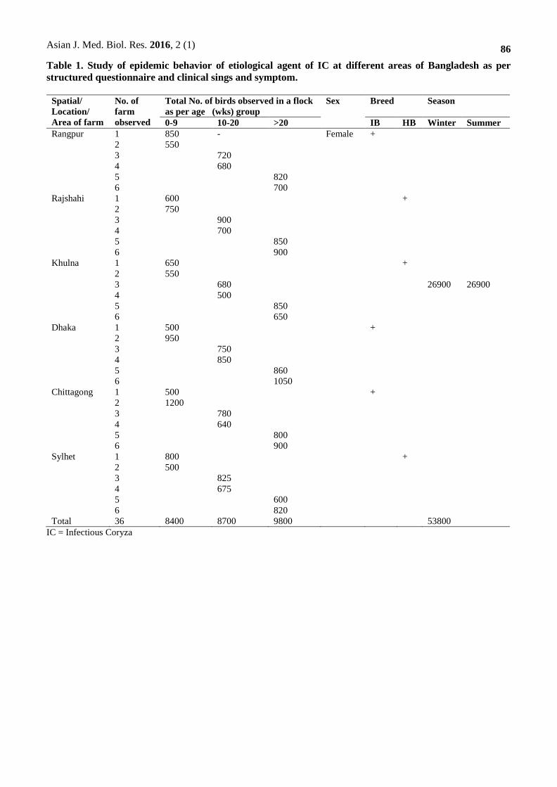
2.2. Collection of data and samples The epidemic behavior of etiological agent of IC from field cases were recorded (Table 1) as per information
received from farmers by using a structured questionnaire and also clinical signs and symptoms. The date of
collection, age, sex, breed, clinical signs and symptoms and environmental history were recorded for each case.
A total of 122 (Table 2) samples; Nasal and tracheal exudates (Figure 1), visceral organs like liver, lung, heart
were collected from birds suspected to be infected with IC during epidemiological investigation at different
areas (Rangpur, Rajshahi, Sylhet, Dhaka, Chittagong and Khulna) of Bangladesh for the proper isolation and
identification of bacterial pathogen by using morphological, cultural, biochemical test and pathogenicity study.
Precautions were taken to avoid contamination of one sample with other.
2.3. Experimental birds The birds were divided into three groups as group - A (0-9 weeks old), group – B (10-20 weeks old) and group -
C (above 20 weeks old) respectively. A total of 36 farms (2 farms for each age group) were studied for
epidemiological investigation of IC in layer chicken of Bangladesh.
2.4. Study of epidemic behavior of the etiological agent of infectious coryza in layer chicken
2.4.1. Visit the selected layer farms Visit the selected layer farms at different areas of Bangladesh (Rangpur, Rajshahi, Sylhet, Dhaka, Chittagong
and Khulna) and the surveillance of epidemic behavior of the etiological agent of infectious coryza (IC) were
studied based on age, sex, breed, spatial and temporal differences.
2.4.2. Collection of information and data The information and data about the outbreak of IC were recorded as per structured questionnaire mentioned in
this study for the surveillance of the etiological agent of IC in layer chicken. The collected data were
summarized for the occurrence or absence of IC in that specific farm.

Asian J. Med. Biol. Res. 2016, 2 (1)
84
2.4.3. Feeding and housing Commercial balanced diet and clean drinking water was supplied ad libitum in deep litter system.
2.4.4. Health status Information about the sources of egg or chicks and biosecurity measures including structural, conceptual and
operational were recorded in structured questionnaire.
2.4.5. History of disease Regular deworming maintained by using anthelmantic at 2 (two) month’s interval in the selected farm was
recorded. The history, clinical signs and symptoms of any respiratory diseases were also observed and recorded
from the suspected to be infected bird.
2.4.6. Vaccination and medication All layer chickens were vaccinated against Newcastle disease, Fowl pox and Infectious Bursal disease according
to the manufacturer recommendation and infected birds were treated by using sulphar drugs and broad spectrum
antibiotics.
2.5. Isolation and identification of causal agent of IC
2.5.1. Cultural characterization Isolation of bacterial pathogen from suspected samples were carried out by culturing the samples on blood agar
and chocolate agar plate cross streaked with Staphylococcus spp. The inoculated plates are then incubated at
37◦C with 5 -10 % CO2 for 24 - 48 hours. Identification of the bacterial agent from the pure culture were carried
out based on their colony characteristics, satellitism phenomenon and hemolysis pattern as described by
Blackall, et al., 1997 and Chen et al., 1998.
2.5.2. Morphological characterization The colonies from pure culture were then studied for its morphological characters by Gram’s stain described by
Buxton and Fraser, 1977.
2.5.3. Biochemical characterization Different biochemical tests were employed to the organism like different Sugar’s fermentation, Indol
production, Voges-Proskauer, Methyl red, Hydrogen sulphide production and Nitrate reduction tests, Catalase,
Dulcitol and Motility test to confirm the pathogen as Avibacterium paragallinarum.
2.6. Experimental pathogenicity tests
2.6.1. Organism The local strain of A. paragallinarum was isolated from IC outbreaks in laying flocks then it was used for the
experimental pathogenicity test in natural host for the determination of type of organisms.
2.6.2. Inoculum preparations One single colony of A. paragallinarum was picked up from blood agar and placed in nutrient broth and
cultured for 48 hours. Inoculation dose of A. paragallinarum (1ml/bird) was prepared according to the
procedure described by Islam 2010 for inoculation on 14 day’s old chicks.
2.6.3. Experimental designs A total of 30 day old layer chicks were collected and equally divided into two groups (A and B, N = 15). On day
14 of age, the chicks of group A were inoculated through the intranasal route with 1 ml of 2 days old culture
broth of A. paragallinarum whereas the chicks of group B were kept as uninoculated control group. Clinical
signs observation, postmortem study and re-isolation of A. paragallinarum were performed at interval of day 3,
5 and 7 of post inoculation.
2.6.4. Gross lesion studies All the internal organs including nasal passage were examined and gross lesions were recorded (nasal discharge,
sneezing, conjunctivitis, swelling of sinuses and facial oedema) as Yamamoto, 1980. The inflammatory lesions
of different organs (congestion, hemorrhage, swelling, mucus, etc.) were graded as ± = almost absence of lesion,
+ = mild lesion, ++ = moderate lesion and +++ = severe lesion.

Asian J. Med. Biol. Res. 2016, 2 (1)
85
2.6.5. Re-isolation of Avibacterium paragallinarum After the development of clinical signs of infectious coryza, the birds were scarified humanely and necropsied
for observation of post-mortem lesions at day 7 of post inoculation. Muciod exudates from nasal cavities and
infraorbital sinus were collected with sterile loop and streaked directly on to blood agar media plates containing
NAD with 5 – 10 % CO2 for re-isolation of the bacteria from the experimentally infected birds according to the
procedures followed by Kridda et al., 2012.
2.7. Maintenance of stock culture During the experiment it was necessary to preserve the isolated organisms for longer periods. For this purpose,
pure culture of the isolated organisms was stored in 20% sterilized glycerin and sealed with paraffin wax and
stored at - 80°C in freezer for future use.
3. Results and Discussion The epidemic behavior of the etiological agent of infectious coryza (IC) was studied for the first time in layer
chicken of Bangladesh as per structured questionnaire. During investigation the birds were observed for clinical
signs and symptoms and 122 samples were collected from birds suspected to be infected with IC based on age,
sex, breed, spatial and temporal differences for proper isolation, identification and pathogenicity study of the
etiological agent of the disease. The results are presented below:
3.1. Results of epidemiological investigations
3.1.1. Study on epidemic behavior of the etiological agent of infectious coryza (IC) in layer chicken of
Bangladesh
3.1.1.1. Study on incidence of IC based on age, sex, breed and spatial differences The epidemic behavior of the etiological agent of IC was studied at different areas of Bangladesh based on age,
sex, breed, spatial and temporal differences. In this study period a total of 26900 (Table 4) layer chickens were
observed on 36 farms for two times considering winter and summer season when the disease is prevailing. The
rate of incidence of IC was recorded very high in laying hen (18.38%) in Sylhet and growing birds (7.25%) in
Khulna in comparing with prelaying stage (2.07%) also in Sylhet region of Bangladesh (BD) are presented in
Table 4. As per location variation the incidence of IC was also recorded slightly high (9.2%) in Sylhet
comparing with other areas of Bangladesh are presented in Table 3. The highest incidence in Sylhet might be
due to marshy environmental factor of this area. This findings supported by the Byarugaba et al. 2007.
3.1.1.2. Study on incidence of IC in stipulations of temporal or seasonal variation In this investigation 26900 birds were observed in 36 farms for each season (winter and summer) at various
areas of BD for recording the rate of incidence of IC in layer chicken. The incidence rate was found to be very
high in winter (8.77%) in compare with summer (0.42%) season are presented in Table 4. Our present findings
supported by Terzolo et al., 1993, Chen et al., 1993 and Blackall et al. 1997.
3.1.2. Study on incidence rate of Avibacterium paragallinarum after collection of samples from suspected
layer chickens during epidemic investigation
3.1.2.1. Determination of incidence rate of A. paragallinarum in suspected birds based on age, sex and
breed A total of 122 samples were screened by epidemiological investigation of which the overall incidence
rate of Avibacterium paragallinarum was detected as 47.54 % (Table 5). The incidence rate was varied in terms
of age (Table 5). In this study it was observed that incidence of Avibacterium paragallinarum was very high in
laying hen (52.8%) and growing birds (42.8) in compare with the prelaying stage (16.6%) are presented in Table
5. This findings supported by the earlier observation of Blackall et al. 1997. This increased incidence rate of
A. paragallinarum in layer chicken might be due to increased length of exposure to pathogens compared to
grower and prelaying stage.
Asian J. Med. Biol. Res. 2016, 2 (1)
86
Table 1. Study of epidemic behavior of etiological agent of IC at different areas of Bangladesh as per
structured questionnaire and clinical sings and symptom.
Spatial/
Location/
Area of farm
No. of
farm
observed
Total No. of birds observed in a flock
as per age (wks) group
Sex Breed Season
0-9 10-20 >20 IB HB Winter Summer
Rangpur 1 850 - Female +
26900
26900
2 550
3 720
4 680
5 820
6 700
Rajshahi 1 600 +
2 750
3 900
4 700
5 850
6 900
Khulna 1 650 +
2 550
3 680
4 500
5 850
6 650
Dhaka 1 500 +
2 950
3 750
4 850
5 860
6 1050
Chittagong 1 500 +
2 1200
3 780
4 640
5 800
6 900
Sylhet 1 800 +
2 500
3 825
4 675
5 600
6 820
Total 36 8400 8700 9800 53800
IC = Infectious Coryza

Asian J. Med. Biol. Res. 2016, 2 (1)
87
Table 2. Samples collected from suspected birds based on age, sex, breed, spatial and temporal
differences during epidemic investigation.
Age (wks) Sex Breed Spatial/
Area
Season Name
of collected
samples
Total
number of
samples
tested
0-9 10-20 >20
Female
IB HB Summer Winter NS TS VO
7 5 30 + Rangpur 9 33 18 8 16 42
5 2 10 + Rajshahi 2 15 4 1 12 17
3 1 10 + Sylhet 6 8 10 4 0 14
2 2 12 + Dhaka 0 16 5 7 4 16
1 0 08 + Chittagong 0 9 9 0 o 9
3 2 19 + Khulna 2 22 15 0 9 24
21 12 89
*NS = Nasal S\swab, TS = Tracheal swab, VO = Visceral organ, IB = Isa Brown, HB = Hyline Brown
Table 3. Study on epidemic behavior of infectious coryza in layer chicken of Bangladesh based on age,
sex, breed and spatial differences.
Location or
area of
farm
No. of
farms
observed
Total No. of birds in a
flock with their age (wks)
group
No. of birds
infected in flock
with respiratory
disorder (IC)
Incidence rate (%) of IC as their
age group (wks) and location
0-9 10-20 >20 0-9 10-20 >20 Location
Rangpur
36
1400 99 7.07 8.76
1400 18 1.2
1520 274 18.03
Rajshahi 1350 91 6.74 8.82
1600 13 1.19
1750 314 17.94
Khulna
1200 87 7.25 8.83
1180 17 1.44
1500 267 17.80
Dhaka 1450 97 6.69 8.72
1600 21 1.31
1910 347 18.17
Chittagong 1700 111 6.53 8.65
1420 22 1.55
1700 304 17.88
Sylhet 1300 93 7.15 9.2
1500 31 2.07
1420 261 18.38
Total 36 8400 8700 9800 2473 6.90 1.46 18.03
Level of sig. 0.375 NS 0.138 NS
IC = Infectious coryza, wks = Weeks, NS = Not Significant
Table 4. Study on incidence of infectious coryza in suspected birds as per seasonal variation.
Seasons No. of farm
observed
No. birds observed in flock No. of birds affected
in flock
Incidence rate (%)
Winter 36 26900 2359 8.77
Summer 36 26900 114 0.42
Total 72 53800 2473
Level of sig. 0.447 NS
NS = Not Significant

Asian J. Med. Biol. Res. 2016, 2 (1)
88
Table 5. Determination of incidence rate of A. paragallinarum in suspected birds based on age, sex and
breed.
Total number of
samples tested as
per age (wks)
group
Sex Breed Spatial or
area
Season Total No. of Positive
isolates
Overall
Incidence
rate
(%)
0-9 10-
20
>20
Female
IB HB Summer Winter 0-9
wks
10-20
wks
>20
wks
7 5 30 + Rangpur 9 33 09b
(42.8)
02c
(16.6)
47a
(52.8)
47.54
5 2 10 + Rajshahi 2 15
3 1 10 + Sylhet 2 12
2 2 12 + Dhaka 0 16
1 0 08 + Chittagong 0 9
3 2 19 + Khulna 1 23
21 12 89 + 14 108 (42.8) (16.6) (52.8)
Level of sig. 0.002 ** 0.001** 0.263 NS
** = Significant at 1% level of probability (p<0.01), NS = Not Significant, IB = Isa brown, HB = Highline brown, wks =
Weeks
Table 6. Determination of incidence rate of A paragallinarum in suspected birds based on spatial and
temporal differences.
Spatial or
area
Name
of collected
samples
Season and total
number of samples
tested
No of positive
isolates (%)
Overall Incidence rate
( % )
NS TS VO Summer Winter Summer Winter Summer Winter
Rangpur 18 8 16 9 33 1 19 (11.11) (57.57)
Rajshahi 4 1 12 2 15 0 8 (53.33)
Sylhet 10 4 0 2 12 0 8 (66.66)
Dhaka 5 7 4 0 16 0 7 (43.75)
Chittagong 9 0 o 0 9 0 4 (44.44)
Khulna 15 0 9 1 23 0 11 (47.82)
Total = 14 108 1 57
122 58 1.85 52.26
Level of sig. 0.112 NS 0.144 NS
NS = Not Significant
Table 7. Determination of ‘V ’factor for the growth of A. paragallinarum by Staphylococcus aureus.
Name of the media Colony characteristics
Blood Agar Small ,dew drop like nonhemolytic colonies
Chocolate Agar Luxuriant growth
Chocolate Agar Cross streaked with Staphylococcus aureus. Satellitic growth

Asian J. Med. Biol. Res. 2016, 2 (1)
89
Table 8. Chracterization of field isolates of A. paragallinarum by staining or morphological and
biochemical examination.
No. of
tested
isolates
Test performed Observation Response Indication
Positive
isolates
% of Positive
isolates
122
Microscopic
examination by
Gram’s staining
Gram negative, coccobacilli
or short rod shaped
58
100%
A. paragallinarum
TSI agar slant
reaction
Ferment Glucose ,
Sucrose & Lactose
A. paragallinarum
Motility test by
MIU medium
Absence of turbidity
A. paragallinarum
Indole test No pink color ring at the
adjacent
A. paragallinarum
MR test Absence of red color indicate
MR test negative
A. paragallinarum
VP test No color change indicate VP
test negative
A. paragallinarum
H2S Production Absence of black coloration
at TSI slant indicate H2S
Production negative
A. paragallinarum
MR = Methyl red; VP = Voges Proskauer; MIU =Motility indole urease
Table 9. Biochemical reactions of the isolate.
Test Result Indication
Glucose +
A. paragallinarum
Sucrose +
Lactose +
Indole –
Vogas Proskauer test –
Methyl Red test –
H2S Production –
Motility –
Catalase _
Dulcitol _
+ = Positive; = Negative; MR = Methyl red; VP = Voges Proskauer; MIU =Motility indole urease
Table 10. Experimental pathogenicity study.
Days Post
Inoculation
Signs Postmortem lesions Group – A
(Inoculated with
A. paragallinarum)
Group – B
(Control group)
3 Facial swelling,
Watery nasal
discharge,
conjunctivitis
Grayish white
exudates in nasal
cavities,
+
-
5 Disappearance of
nasal discharge and
lacrimation, swelling
of face
Yellowish catarrhal
exudates in larynx and
nasal cavities
+
-
7 Swelling of face,
depression and
inability to move
Congestion of lung and
trachea, air sacs became
cloudy and thickened with
foamy exudates
+
-
+ = Positive, - = Negative

Asian J. Med. Biol. Res. 2016, 2 (1)
90
Figure 1. Collection of exudates from sinus cavity.
Figure 2. Facial swelling with nasal and ocular discharge (Right).
Figure 3. Growth of A. paragallinarum on Blood Agar (Right).
Figure 4. Growth of A. paragallinarum on Chocolate Agar (Right).

Asian J. Med. Biol. Res. 2016, 2 (1)
91
Figure 5. Gram’s staining of A. paragallinarum.
Figure 6. A. paragallinarum showing satellitism phenomenon around V factor.
3.1.2.2. Determination of incidence rate of A. paragallinarum in suspected birds based on spatial and
temporal differences
The incidence of A. paragallinarum in Rangpur, Rajshahi, Sylhet, Dhaka, Chittagong and Khulna were found to
be 57.57%, 53.33%, 66.66%, 43.75%, 44.44% and 47.82% respectively (Table 6). The highest incidence was
found in Sylhet (66.66%) and the lowest in Dhaka (43.75%) in comparison with other areas of BD mentioned
earlier. In this study, 58 samples were found to be positive for A. paragallinarum from 122 suspected samples
collected during epidemiological investigation. The association of A. paragallinarum with different seasons
(Table 6) revealed that higher incidence was found in winter season (52.26%) in comparison with summer
season (1.85%). This observed variation in incidence of A. paragallinarum at various areas and seasons of
Bangladesh could be related with several factors such as geoclimatic situation, passive immunity level, infecting
dose, simultaneous infection with other respiratory pathogens, stress, managemental practice, biosecurity failure
and different locations of the study areas.
3.2. Results of isolation and characterization of A. paragallinarum by morphological, cultural and
biochemical properties
3.2.1. Isolation and characterization of A. paragallinarum by cultural properties The birds in the infected flock had facial swelling, nasal and lacrimal discharge, open mouth breathing and
mucoid discharge from the nares. The clinical signs (Figure 2) are common features of infectious coryza. This
present findings supported by the Droual et al. 1990, Horner et al.1992 Mouahid et al. 1992, Calnek et al. 1991
and Sandovel et al. 1994.The bacterium was recovered only from nares on blood agar (Small, dew drop like
nonhemolytic colonies, Figure 3 and Table 7) and chocolate agar (Luxuriant growth, Figure 4 and Table 7). No
growth was recovered from samples like liver, lungs, heart streaked on different agars. The growth and
morphological characteristics indicated that the isolated organism might be A. paragallinarum (Table 8),
which was later confirmed by different biochemical tests (Table 9). This findings supported by the earlier
observation of Terzolo et al. 1993, Rimler et al.1975 and Miflin et al.1999. The bacterium was isolated
from nares on blood agar, chocolate agar and chocolate agar cross streaked with a nursery colony of
Staphylococcus aureus as feeders. It was observed that satellitic growth patterns (Figure 6) of isolated
bacterium might be A. paragallinarum , which was later confirmed by biochemical tests.

Asian J. Med. Biol. Res. 2016, 2 (1)
92
3.2.2. Characterization of A. paragallinarum by morphological and biochemical Properties The isolated organism was characterized by morphological characterization (Gram’s staining technique, Figure
5, Table 8) and different biochemical (Table 9) tests. This observation revealed that the isolated organism was
Gram negative, short rods or cocccobacilli arranged in single or pairs. It was also observed that caseopurulent
air sac lesions in field cases of infectious coryza in layer chickens. This observation supported by Sameera
2001, Fujivara and Konno 1965, Blackall et al. 1989 and Rimler et al. 1975.
3.3. Experimental pathogenicity test Among the infectious diseases of poultry, infectious coryza (IC) is an upper respiratory tract infection of
chickens caused by a bacterial agent called A. paragallinarum, is one of the major problems affecting
commercial poultry industry worldwide (Blackall et al. 1997). No systematic pathogenicity studies have been
conducted by local isolates of A. paragallinarum in conjunction with epidemic behavior study of the etiological
agent of this IC in Bangladesh. The present study describes experimental pathogenicity studies of field isolates
of A. paragallinarum in susceptible layer chicks.
3.3.1. Clinical signs The experimentally infected birds with the A. paragallinarum isolates were examined in detail at regular
intervals of time up to 7days post inoculation for clinical signs and gross postmortem lesions. The clinical signs
of infectious coryza in birds of Group A (inoculated with A. paragallinarum) appeared after 24 hrs of infection
characterized as oedematous swelling of face and infraorbital sinus and secretion of watery nasal discharge
(Table 10). Some birds showed bilateral swelling of face and infraorbital sinuses, conjunctivitis, serous to
mucoid nasal discharge with foul smelling, foamy lacrimation and induration of face after 3 DPI. After 5 days of
infection, disappearance of nasal discharge and lacrimation but the birds still suffered from swelling of face
after 7 DPI (Table10). This observation supported by other researchers; Kridda et al. 12010, Gayatri et al. 2010,
Fujivara and Konno 1965, Blackall 1989 and Page 1962.The birds of group B (uninoculated control group) did
not reveal any conspicuous clinical sign and lesion.
3.3.2. Gross postmortem study A. paragallinarum infected birds of Group A (inoculated with A. paragallinarum) showed, infraorbital sinus
cavities were filled with grayish white watery exudates at 3 DPI. Larger amount of yellowish cattarhal exudates
with necrotic debris were found in upper larynx and nasal cavities after 5 DPI, while at 5 to 7 DPI trachea and
lung revealed mild congestion. The air sacs became cloudy and thickened with foamy exudates after 5 to 7 DPI.
On the other hand, the birds of group B (uninoculated control group) did not reveal any lesion related to the IC
on day 3, 5 and 7 of post inoculation. This observation supported by other researchers; Fujivara and Konno
1965, Blackall 1989 and Page 1962.
3.3.3. Re-isolation of A. paragallinarum at day 7 of post inoculation Re-isolation was performed only in tissues (nasal passage) showing postmortem lesions on day 7 of post
inoculation (PI) according to the procedure described by Rimler et al. 1975.
3.3.4. Gram's stain, biochemical tests, sugarfermentation test and catalase activity test Tentatively identified colonies of A. paragallinarum from nasal passage of day 7 of PI from blood agar media
cross streaked with Staphylococcus aureus or extra supply of NAD were used for morphological study. The
morphology of the isolated bacteria exhibited red (Gram’s stain) color, small rod shaped Gram negative
coccobacilli. A. paragallinarum isolate was able to ferment four basic sugars by producing acid (Table 8). These
findings agreed with Sameera et al. 2001, Yamamoto1991, Sawata et al. 1982, Blackall 1989 and Haunshi et al.
2006.
4. Conclusions Among economically important diseases of poultry, Infectious Coryza (IC) is an infectious and contagious
respiratory bacterial disease in poultry industry causing heavy economic losses through morbidity and reduced
(10 - 40%) in egg production. In this study, the incidence was higher in laying hen (52.8%) in compare with
prelaying stage (16.6%). In the present study a trend in increasing the incidence rate of infectious coryza
was observed as their location (66.66) and seasonal (52.26%) variations. This observed variation in incidence
of infectious coryza in various areas of Bangladesh could be related with several factors such as geoclimatic
situation, passive immunity level, infecting dose, simultaneous infection with other respiratory pathogens,

Asian J. Med. Biol. Res. 2016, 2 (1)
93
stress, manage mental practice, biosecurity failure and different locations of the study areas. To prevent the
spread of IC in laying hen, disease management strategies could be undertaken and introducing a continuous
monitoring of organism by randomized detection of antibody by serological (HI) test, culling of infected and
carrier bird and implementation of good husbandry practice with biosafety plan but it does not eliminate the
carrier status of chickens. It is advisable to vaccinate the chickens with inactivated coryza vaccine to prevent
economic losses. Considering this fact the research work will also extends for the production of vaccine
candidate from the field isolate to control infectious coryza in layer chicken of Bangladesh.
Acknowledgements We thank the Government of the People’s Republic of Bangladesh for providing NSICT fellowship to Mir
Rowshan Akter.
Conflict of interest
None to declare.
References Akter S, 2012. Isolation and identification of Avibacterium paragallinarum from layer chickens. MS Thesis,
Department of Pathology, Faculty of Veterinary Science, Bangladesh Agricultural University, Mymensingh-
2202.
Ali M, MS Hossain, S Akter, MAHNA Khan and MM Hossain, 2013. Pathogenesis of Infectious Coryza in
Chickens (Gallus gallus) by Avibacterium paragallinarum Isolate of Bangladesh. The Agriculturists, 11: 39-
46.
Blackall PJ, M Matsumoto and R Yamamoto, 1997. Infectious coryza. In BW Calnek, HJ Barnes, CW Beard,
LR McDougald, and YM Saif (eds.). Diseases of Poultry, 10th ed. Iowa State University Press: Ames, IA,
pp. 179—190.
Blackall PJ, CJ Morrow, A McInnes, LE Eaves and DG Rogers, 1990. Epidemiologic studies on infectious
coryza outbreaks in northern New South Wales, Australia, using serotyping, biotyping, and chromosomal
DNA restriction endonuclease analysis. Avian dis., 34: 367 -376.
Blackall PJ, 1989.The Avian Haemophili. Clin. Microbiol. Rev., 2: 270-277. Byarugaba DK, UM Minga, PS
Gwakisa, E Katunguka, M Bisgaard and JE Olsen, 2007. Virulence characterization of Avibacterium
paragallinarum isolates from Uganda. Avian Pathol. , 36: 35-42.
Calnek BW, H John Barnes, CW Breed, WM Reid and HW Yodev, 1991. Diseases of Poultry, 9th Ed., Wolfe
Publishing Ltd., USA. pp. 186–92.
Chen X, Zhang P, Blackall PJ and Feng W, 1993. Characterization of Haemophilus paragallinarum Isolates
from China. Avian Dis., 37: 574-576.
Droual R, AA Bickford, BR Charlon, GL Cooper and SE Channing, 1990. Infectious coryza in meat chickens in
the San Joaquin Valley of California. Avian Dis., 34: 1009-1016.
Fujiwara H and S Konno, 1965. Histopathological studies on infectious coryza of chickens. ii. Findings in
experimentally infected cases. Natl. Inst. Anim. Health Q (Tokyo). 5: 86-96.
Gayatri Rr, R Ashish and MY Mahendra, 2010. Antimicrobial sensitivity pattern of Haemophilus
paragallinarum isolated from suspected cases of infectious coryza in poultry. Vet. World, 3:177-181.
Haunshi S, B Dutta and SC Saxena, 2006. An outbreak of infectious coryza in Vanaraja poultry of Meghalaya.
Ind. J. Vet. Pathol., 30: 55.
Horner RF, GC Bishop and C Haw, 1992. An upper respiratory disease of commercial chickens resembling
infectious coryza, but caused by a V factor – idependent bacterium. Avian Pathol., 21: 421–427.
Kridda, Suwit and Niwat, 2010. An Outbreak of Avibacterium paragallinarum serovar B in a Thai Layer Farm.
Th. J. Vet. Med., 40: 441- 444.
Miflin JK , X Chen , RR Bragg, JM Welgemoed , JM Greyling, RF Horner and PJ Blackall, 1999. Confirmation
that PCR can be used to identify NAD-dependent and NAD- independent Haemophilus paragallinarum
isolates. Onderstepoort J. Vet. Res., 66: 55- 57.
Mouahid M, KH Hhinz, Engelhard, R Mutters and W Mannheim, 1992. Characterization of Haemophilus
paragallinarum by analysis of whole cell carbohydrates, fatty acids and phospholipids. Avian Pathol., 21:
127-136.
Page LA, 1962. Haemophilus infections in chickens. Characteristics of 12 Haemophilus isolates recovered from
diseased chickens. Am. J. Vet. Res., 23:85-95.

Asian J. Med. Biol. Res. 2016, 2 (1)
94
Rimler RB, EB Shotts and RB Davis, 1975. A growthmedium for the production of a bacterin for immunization
against infectious coryza. Avian Dis.,19: 318-322.
Roberts DH, BS Hanson and L Timms, 1964. Observations in the incidence and significance of Haemophilus
gallinarum in outbreaks of respiratory disease among poultry in Great Britain. Vet. Rec., 76:1512-1516.
Sameera A, RB Asif and M Khushi, 2001. Clinico-Therapeutic Observations on an Outbreak of Infectious
Coryza. Int. J. Agri. Biol., 3: 531- 532.
Sandoval VE, HR Twerzolo and PJ Blackall, 1994.Complicated infectious coryza cases in Argentina. Avian
Dis., 38: 672-678.
Sawata A, K Kume and Y Nakase, 1982. Hemagglutinin of Haemophilus paragallinarum serotype 2
organisms: Occurrence and immunologic properties of hemagglutinin. Am. J. Vet. Res., 43: 1311-1314.
Talha AFSM, MM Hossain, EH Chowdhury, ASM Bari, MR Islam and PM Das, 2001. Poultry diseases
occurring in Mymensingh district of Bangladesh. Bangladesh Vet., l8: 20-23.
Terzolo HR, FA Paolicchi , VE Sandoval , PJ Blackall , T Yamaguchi and Y Iritani, 1993. Characterization of
isolates of Haemophilus paragallinarum from Argentina. Avian Dis., 37: 310-314.
Yamamoto R, 1991. Infectious coryza: In: Diseases of Poultry, 9th ed. Hofstad MS, HJ Barnes, BW Calnek,
WM Reid and JHW Yoder, Eds. Iowa University Press, Ames, Iowa, USA. pp.178-186.
Zhang PJ, M Miao, H Sun, Y Gong and PJ Blackall, 2003. Infectious Coryza due to Haemophilus
paragallinarum serovar B in China. Aust. Vet. J. ,81:96-97.