article targeting clk3 inhibits the progression of
TRANSCRIPT
ARTICLE
Targeting CLK3 inhibits the progression ofcholangiocarcinoma by reprogramming nucleotidemetabolismQingxin Zhou1,2,3*, Meihua Lin4,5*, Xing Feng6*, Fei Ma7*, Yuekun Zhu8,9, Xing Liu10, Chao Qu11, Hong Sui2, Bei Sun8,9, Anlong Zhu8, Heng Zhang12,He Huang12, Zhi Gao13, Yongxiang Zhao13, Jiangyun Sun14, Yuxian Bai2, Junfei Jin15,16, Xuehui Hong17, Chang Zou18,19, and Zhiyong Zhang1,20
CDC-like kinase 3 (CLK3) is a dual specificity kinase that functions on substrates containing serine/threonine and tyrosine. Butits role in human cancer remains unknown. Herein, we demonstrated that CLK3 was significantly up-regulated incholangiocarcinoma (CCA) and identified a recurrent Q607R somatic substitution that represented a gain-of-functionmutation in the CLK3 kinase domain. Gene ontology term enrichment suggested that high CLK3 expression in CCA patientsmainly was associated with nucleotide metabolism reprogramming, which was further confirmed by comparing metabolicprofiling of CCA cells. CLK3 directly phosphorylated USP13 at Y708, which promoted its binding to c-Myc, thereby preventingFbxl14-mediated c-Myc ubiquitination and activating the transcription of purine metabolic genes. Notably, the CCA-associatedCLK3-Q607R mutant induced USP13-Y708 phosphorylation and enhanced the activity of c-Myc. In turn, c-Myc transcriptionallyup-regulated CLK3. Finally, we identified tacrine hydrochloride as a potential drug to inhibit aberrant CLK3-induced CCA.These findings demonstrate that CLK3 plays a crucial role in CCA purine metabolism, suggesting a potential therapeutic utility.
IntroductionCholangiocarcinoma (CCA) stemming from cholangiocytes is aprimary hepatic malignancy and is divided as extrahepatic andintrahepatic according to its anatomical position (Maroni et al.,2013). The incidence of CCA is increasing worldwide, and itsprognosis has remained dismal. So far, there are no uniquelyidentified markers for CCA, and very few options for its treat-ment are available. Although several risk factors such as ERBB2,FOXM1, and Yap have been shown to promote CCA initiation(Sugihara et al., 2019), they are not typically found in most CCA
patients. Recently, genetic studies have enhanced our under-standing of the molecular mechanisms by which normal biliarycells acquire the properties of malignant transformation in hu-man CCA (Marks and Yee, 2016). Therefore, more efforts in thisdirection might form the basis for developing new diagnosticapproaches and more effective therapy for CCA.
Uncontrolled cell proliferation is a characteristic of humancancer. Purines are the most abundant metabolic substrates byproviding necessary components for DNA and RNA to support
.............................................................................................................................................................................1The Affiliated Hospital of Guilin Medical University, Guangxi Key Laboratory of Brain and Cognitive Neuroscience, Guangxi Neurological Diseases Clinical Research Center,Guilin, Guangxi, China; 2Department of Gastrointestinal Oncology, Harbin Medical University Cancer Hospital, Harbin, China; 3Cancer Institute of New Jersey, RutgersUniversity, New Brunswick, NJ; 4Research Center of Clinical Pharmacy, State Key Laboratory for Diagnosis and Treatment of Infectious Disease, First Affiliated Hospital,Zhejiang University, Hangzhou, China; 5Zhejiang Provincial Key Laboratory for Drug Evaluation and Clinical Research, First Affiliated Hospital, Zhejiang University,Hangzhou, China; 6Department of Immunobiology, Yale University School of Medicine, New Haven, CT; 7Department of General Surgery, The Second Affiliated Hospital ofHarbin Medical University, Harbin, China; 8Department of General Surgery, The First Affiliated Hospital of Harbin Medical University, Harbin, China; 9Key Laboratory ofHepatosplenic Surgery, Harbin Medical University, Ministry of Education, Harbin, China; 10Department of Neurosurgery, Beijing Tiantan Hospital, Capital Medical University,Beijing, China; 11Department of Radiation Oncology, The First Hospital of Jilin University, Changchun, China; 12Department of Histology and Embryology, Xiang Ya Schoolof Medicine, Central South University, Changsha, Hunan, China; 13National Center for International Research of Biological Targeting Diagnosis and Therapy, Guangxi KeyLaboratory of Biological Targeting Diagnosis and Therapy Research, Guangxi Medical University, Nanning, China; 14Department of Acupuncture, The First Affiliated Hospitalof Harbin Medical University, Harbin, China; 15Laboratory of Hepatobiliary and Pancreatic Surgery, Affiliated Hospital of Guilin Medical University, Guilin, China; 16GuangxiKey Laboratory of Molecular Medicine in Liver Injury and Repair, Guilin Medical University, Guilin, China; 17Department of Gastrointestinal Surgery, Zhongshan Hospital ofXiamen University, Xiamen, China; 18Clinical Medical Research Center, The First Affiliated Hospital of Southern University of Science and Technology, The Second ClinicalMedical College of Jinan University, Shenzhen People’s Hospital, Shenzhen, China; 19Shenzhen Public Service Platform on Tumor Precision Medicine and MolecularDiagnosis, The Second Clinical Medical College of Jinan University, Shenzhen People’s Hospital, Shenzhen, China; 20Department of Surgery, Robert Wood Johnson MedicalSchool University Hospital, Rutgers University, The State University of New Jersey, New Brunswick, NJ.
*Q. Zhou, M. Lin, X. Feng, and F. Ma contributed equally to this paper; Correspondence to Zhiyong Zhang: [email protected]; Junfei Jin: [email protected]; Xuehui Hong: [email protected]; Chang Zou: [email protected].
© 2020 Zhou et al. This article is distributed under the terms of an Attribution–Noncommercial–Share Alike–No Mirror Sites license for the first six months after thepublication date (see http://www.rupress.org/terms/). After six months it is available under a Creative Commons License (Attribution–Noncommercial–Share Alike 4.0International license, as described at https://creativecommons.org/licenses/by-nc-sa/4.0/).
Rockefeller University Press https://doi.org/10.1084/jem.20191779 1 of 19
J. Exp. Med. 2020 Vol. 217 No. 8 e20191779
Dow
nloaded from http://rupress.org/jem
/article-pdf/217/8/e20191779/1044596/jem_20191779.pdf by guest on 18 N
ovember 2021

cell proliferation (Yin et al., 2018). Therefore, enhanced purinebiosynthesis is tightly associated with the progression of cancer.We and other groups previously reported that several kinasesand transcriptional factors, for example, mechanistic target ofrapamycin kinase (mTOR), activating transcription factor 4(ATF4), microphthalmia-associated transcription factor (MITF),and c-Myc, dictated cancer-dependent purine biosynthesis (Ben-Sahra et al., 2016; Ma et al., 2019). However, the direct signalingnetwork linking the purine synthesis pathway and CCA devel-opment is entirely unknown.
CDC-like kinase 3 (CLK3) is a nuclear dual-specificity kinasethat functions on substrates containing serine/threonine andtyrosine (Nayler et al., 1997). CLK3 modulates RNA splicing byphosphorylating serine/arginine–rich proteins such as SRSF1and SRSF3 (Cesana et al., 2018). Recently, CLK3 dysregulationwas indicated to be a high-penetrant factor in different types ofhuman tumors even though its functions in tumors were notclearly characterized (Bowler et al., 2018).
In this work, we identified a critical Q607 somatic mutationof CLK3 in CCA patients by exon sequencing. Then, we uncov-ered the importance of this CLK3 mutant as an oncogene inpromoting de novo purine synthesis in CCA. Moreover, throughdrug screening, for the first time we identified tacrine hydro-chloride as a potential drug to inhibit the aberrant CLK3-inducedCCA. Thus, our data provide a new therapeutic strategy for CCAharboring CLK3 dysregulation.
ResultsThe levels of CLK3 in CCA and other digestive system cancersare significantly up-regulated and associated with decreasedoverall survival (OS)It is well known that the kinase families play important roles inthe development of various types of cancer. Therefore, by ana-lyzing openly available databases, we hoped to screen severalcandidate kinases that may have significant effects on carcino-genesis. By thoroughly analyzing gene expression profiles across1,508 digestive system tumors with various histological subtypesin The Cancer Genome Atlas (TCGA), we found that CLK3 wassignificantly up-regulated compared with nontumor controls(Fig. 1 A) and that the survival of patients with high CLK3 ex-pression significantly decreased (Fig. 1 B), suggesting a potentialpro-oncogenic role of CLK3 in the human digestive system.However, very little is known about the physiological functionof CLK3 in cancer. Thus, we focused on CLK3 in this study. Giventhat the change in the level of CLK3 expression was most notablein CCA compared with other tumors (Fig. 1 A), in the followingexperiments, we mainly examined the clinical significance andaction mechanisms of CLK3 in CCA.
Analysis of open GSE26566 data and CCA cell lines confirmedthat CLK3 expression was enhanced compared with their con-trols (Fig. 1 C and Fig. S1 A). Tissue array data indicated that thepercentage of cells with CLK3 expression was positively asso-ciated with the stages of CCA patients (Fig. S1 B), implying thatthe levels of CLK3 tightly correlate with CCA malignancy.Analysis of a cohort of 100 CCA patients further validated thatCLK3 expression was up-regulated and positively related to
tumor size, stage, and metastasis (Fig. 1, D–F; and Table S1).Kaplan-Meier data from two independent cohorts indicated thatCCA patients with higher levels of CLK3 had shorter OS (Fig. 1 Gand Fig. S1 C). Multivariate analyses identified CLK3 as an in-dependent prognostic factor in CCA (Table S2). Together, ourdata uncovered that CLK3 may promote the pathogenesisof CCA.
Silencing CLK3 strongly suppresses the aggressiveness ofCCA cellsTo uncover the exact biological roles of CLK3 in CCA, we firstexamined the effect of CLK3 knockdown or overexpression onCCA cell proliferation. As shown in Fig. 2, A–C, doxycycline(Dox)-induced CLK3 deficiency significantly impaired HuCCT1and RBE cell proliferation and BrdU incorporation. Consistently,CLK3 knockdown suppressed the anchorage-independentgrowth of CCA cells (Fig. 2 D). However, overexpression ofCLK3 in CLK3-low HCCC9810 and HuH-28 cells had the oppo-site effects (Fig. S1, D–Eiii; and data not shown), implicatingCLK3 as a direct oncogene. Wound healing and transwell ex-periments demonstrated the invasive function of CLK3 in CCAcells (Fig. 2, E and F; and Fig. S1, Fi and Fii). In accordance within vitro findings, CLK3 silencing in CLK3-high HuCCT1 and RBEcells significantly inhibited the development of mice xenografttumors, whereas its overexpression in HCCC9810 cells en-hanced this development (Fig. 2 G and Fig. S1 Gi). The knock-down of CLK3 markedly reduced the number of CCA abdominalmetastatic nodules (Fig. 2 H). However, CLK3 overexpressionsignificantly promoted CCA metastasis (Fig. S1, Gii and Giii).Together, our findings confirm the tumor-promoting activity ofCLK3 in CCA.
We also examined the effect of CLK3 knockdown on othercells lines from other cancers and on normal biliary epithelialcells (HiBEC). As shown in Fig. S1, Hi and Hii, we observedsimilar effects on Sk-hep1 cells (hepatocellular carcinoma) andHCT116 cells (colon cancer) compared with CCA cells, whereasthere were few effects on HiBEC cells after CLK3 knockdown(Fig. S1 Hiii).
CLK3 up-regulation promotes CCA development byreprogramming purine metabolismTo further uncover the action mechanism of CLK3 in CCA, weassessed the transcriptomes of TCGA of human CCA to examinethe top 500 differentially expressed genes with varying CLK3expression (Fig. 3 A and Table S3). Gene ontology term enrich-ment suggested that high CLK3 expression in CCA patientsmight primarily reprogram tumor metabolism, particularlypurinemetabolism (Fig. 3 B). Thus, mass spectrometry (MS)wasperformed to collect metabolic profiling of HuCCT1 cells with orwithout Dox-induced CLK3 knockdown. Interestingly, CLK3 si-lencing mainly down-regulated the intracellular pools of purineintermediates (Fig. 3, C and D; and Table S4), suggesting thatsilencing CLK3 in CCA cells primarily inhibits purine synthesis.
Supporting this finding, Dox-induced CLK3 knockdown inHuCCT1 cells significantly inhibited the numbers of 15N-purineintermediates (inosine monophosphate, adenosine monophos-phate, and guanosine monophosphate; Fig. 3 E, upper panel).
Zhou et al. Journal of Experimental Medicine 2 of 19
Targeting CLK3 reprograms nucleotide metabolism https://doi.org/10.1084/jem.20191779
Dow
nloaded from http://rupress.org/jem
/article-pdf/217/8/e20191779/1044596/jem_20191779.pdf by guest on 18 N
ovember 2021

Second, similar findings were observed when the flux of13C-glycine into purine intermediates was analyzed (Fig. 3 E,lower panel). Third, Dox-induced CLK3 knockdown signifi-cantly decreased the levels of DNA and RNA labeled with14C-glycine in HuCCT1 cells (Fig. 3 F). Fourth, Dox-inducedCLK3 knockdown in HuCCT1 cells significantly suppressedthe critical enzymes that are necessary for the de novo purinesynthesis pathway (Fig. 3 G). However, Dox-induced CLK3overexpression in HCCC9810 cells had the opposite effects (Fig.S2, Ai and Bi). Our data strongly confirm that CLK3 mainlyactivates de novo purine synthesis in CCA.
Next, we explored the physiological significance of CLK3-mediated de novo purine synthesis in CCA progression. Asshown in Fig. 3 H and Fig. S2 Bii, although CLK3 silencingstrongly inhibited the proliferative and invasive abilities ofHuCCT1 cells, supplementation of purine or overexpression ofATIC (one critical enzyme in the purine synthesis pathway)significantly rescued these defects. Conversely, ATIC inhibitorsignificantly reverted the proliferation, migration, and invasion
of HCCC9810 cells induced by CLK3 overexpression (Fig. 3 I andFig. S2 Biii).
Taken together, our data indicate that CLK3 up-regulationpromotes CCA development at least partially by reprogram-ming de novo purine metabolism.
CLK3 promotes de novo purine synthesis and CCA progressionthrough enhancing the stabilization and nuclear translocationof c-MycGiven that mTOR, c-Myc, MITF, and ATF4 dictated the de novopurine synthesis pathway (Ben-Sahra et al., 2016), we hypoth-esized that they might mediate the effects of CLK3 on CCA. In-terestingly, the knockdown of c-Myc but not MITF or ATF4 ormTOR inhibition significantly reverted CLK3-driven increase inthe purinemetabolism intermediates in HCCC9810 cells (Fig. S2 C;and data not shown). Conversely, c-Myc introduction revertedthe shCLK3-mediated effects on purine metabolism (Fig. 3 C andFig. 4, A and B). The knockdown of c-Myc also significantlydown-regulated the abundance of several enzymes that are
Figure 1. The levels of CLK3 in CCA and other digestive system cancers are significantly up-regulated and associated with decreased OS. (A) TCGAanalysis of CLK3 mRNA expression in 1,508 human digestive system tumors. Data are mean ± SD. **, P < 0.01; unpaired Student’s t test. (B) The survival ofpatients with high CLK3 expression was poor based on TCGA data in A. **, P < 0.01; Kaplan-Meier analysis. (C) Analysis of CLK3 expression based on openGSE26566 data. Data are mean ± SD. ***; P < 0.001; unpaired Student’s t test. (D) CLK3mRNA expression in 100 human CCA specimens and matched normalsamples was determined by quantitative PCR. (E) CLK3 expression in 100 human CCA specimens was analyzed as follows: level, tumor size, metastasis, andstage. *, P < 0.05; **, P < 0.01; χ-square test. (F) IHC analysis of CLK3 expression in 100 human CCA specimens and matched normal samples. Representativepictures are presented. Bar, 100 µm. **, P < 0.01; χ-square test. (G) Stratified by CLK3 levels in CCA patients (n = 100), Kaplan-Meier analysis of OS ispresented. **, P < 0.01. CHOL, cholangiocarcinoma; ESCA, esophageal carcinoma; HCC, hepatocellular carcinoma; HE, hematoxylin and eosin; PAAD, pancreaticadenocarcinoma; READ, rectum adenocarcinoma; STAD, stomach adenocarcinoma; TPM, transcripts per million.
Zhou et al. Journal of Experimental Medicine 3 of 19
Targeting CLK3 reprograms nucleotide metabolism https://doi.org/10.1084/jem.20191779
Dow
nloaded from http://rupress.org/jem
/article-pdf/217/8/e20191779/1044596/jem_20191779.pdf by guest on 18 N
ovember 2021

Figure 2. Silencing CLK3 strongly suppresses the aggressiveness of CCA cells. (A and B) MTT assays for measuring the proliferation of the HuCCT1 andRBE cells with or without 4 µg/ml Dox-induced knockdown of CLK3, which was confirmed using immunoblotting. (C) BrdU incorporation assay was performed
Zhou et al. Journal of Experimental Medicine 4 of 19
Targeting CLK3 reprograms nucleotide metabolism https://doi.org/10.1084/jem.20191779
Dow
nloaded from http://rupress.org/jem
/article-pdf/217/8/e20191779/1044596/jem_20191779.pdf by guest on 18 N
ovember 2021
necessary for purine metabolism reprogramming in HCCC9810cells with CLK3 overexpression (Fig. S2 D).
Notably, CLK3 overexpression up-regulated the protein lev-els of c-Myc but did not affect its mRNA levels (Fig. 4 C and Fig.S2 Ci). Cycloheximide (CHX) chase assays indicated that CLK3deficiency in HuCCT1 cells significantly reduced the half-time ofc-Myc protein. However, WT-CLK3 but not active-dead CLK3-K186M overexpression in HCCC9810 cells had the opposite effect(Fig. 4, D and E). Furthermore, MG132 reverted c-Myc down-regulation mediated by CLK3 silencing (Fig. 4 F). These dataindicate that overexpression of CLK3 enhances the stability ofc-Myc protein in CCA cells.
We also observed that overexpression of WT-CLK3 but notCLK3-K186M enhanced c-Myc nuclear translocation, while thedeficiency of CLK3 had the opposite effect (Fig. 4, Gi and Gii; andFig. S2, Ei and Eii), suggesting that CLK3 overexpression pro-motes purine synthesis by enhancing the transcriptional activityof c-Myc.
Functionally, silencing c-Myc significantly reverted CLK3-induced HCCC9810 cell proliferation, migration, and invasion(Fig. S2 F). Therefore, we propose that c-Myc is a necessaryeffector downstream of CLK3 in CCA.
To elucidate if other importantly oncogenic pathways regu-lated by c-Myc (such as ribosome biogenesis; van Riggelen et al.,2010) were similarly affected, we reexamined the top 500 genesaffected by CLK3. As shown in Table S5, a set of genes involvedin ribosome biogenesis was present. To further confirm if ri-bosome biogenesis was also affected by CLK3, we determinedthe mRNA levels of several representative genes. As shown inFig. S2 G, their mRNA levels were down-regulated after CLK3depletion, suggesting that CLK3 also affects ribosomal biogenesisin CCA.
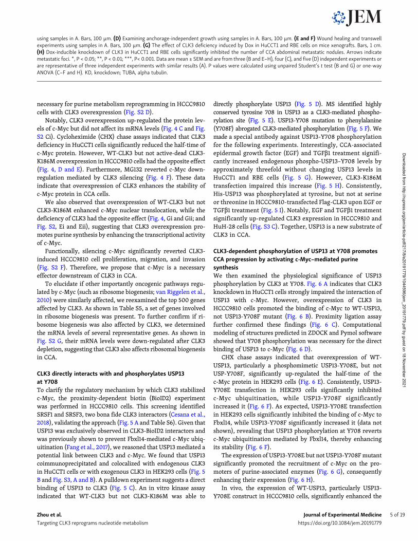
CLK3 directly interacts with and phosphorylates USP13at Y708To clarify the regulatory mechanism by which CLK3 stabilizedc-Myc, the proximity-dependent biotin (BioID2) experimentwas performed in HCCC9810 cells. This screening identifiedSRSF1 and SRSF3, two bona fide CLK3 interactors (Cesana et al.,2018), validating the approach (Fig. 5 A and Table S6). Given thatUSP13 was exclusively observed in CLK3-BioID2 interactors andwas previously shown to prevent Fbxl14-mediated c-Myc ubiq-uitination (Fang et al., 2017), we reasoned that USP13 mediated apotential link between CLK3 and c-Myc. We found that USP13coimmunoprecipitated and colocalized with endogenous CLK3in HuCCT1 cells or with exogenous CLK3 in HEK293 cells (Fig. 5B and Fig. S3, A and B). A pulldown experiment suggests a directbinding of USP13 to CLK3 (Fig. 5 C). An in vitro kinase assayindicated that WT-CLK3 but not CLK3-K186M was able to
directly phosphorylate USP13 (Fig. 5 D). MS identified highlyconserved tyrosine 708 in USP13 as a CLK3-mediated phospho-rylation site (Fig. 5 E). USP13-Y708 mutation to phenylalanine(Y708F) abrogated CLK3-mediated phosphorylation (Fig. 5 F). Wemade a special antibody against USP13-Y708 phosphorylationfor the following experiments. Interestingly, CCA-associatedepidermal growth factor (EGF) and TGFβ1 treatment signifi-cantly increased endogenous phospho-USP13–Y708 levels byapproximately threefold without changing USP13 levels inHuCCT1 and RBE cells (Fig. 5 G). However, CLK3-K186Mtransfection impaired this increase (Fig. 5 H). Consistently,His-USP13 was phosphorylated at tyrosine, but not at serineor threonine in HCCC9810-transfected Flag-CLK3 upon EGF orTGFβ1 treatment (Fig. 5 I). Notably, EGF and TGFβ1 treatmentsignificantly up-regulated CLK3 expression in HCCC9810 andHuH-28 cells (Fig. S3 C). Together, USP13 is a new substrate ofCLK3 in CCA.
CLK3-dependent phosphorylation of USP13 at Y708 promotesCCA progression by activating c-Myc–mediated purinesynthesisWe then examined the physiological significance of USP13phosphorylation by CLK3 at Y708. Fig. 6 A indicates that CLK3knockdown in HuCCT1 cells strongly impaired the interaction ofUSP13 with c-Myc. However, overexpression of CLK3 inHCCC9810 cells promoted the binding of c-Myc to WT-USP13,not USP13-Y708F mutant (Fig. 6 B). Proximity ligation assayfurther confirmed these findings (Fig. 6 C). Computationalmodeling of structures predicted in ZDOCK and Pymol softwareshowed that Y708 phosphorylation was necessary for the directbinding of USP13 to c-Myc (Fig. 6 D).
CHX chase assays indicated that overexpression of WT-USP13, particularly a phosphomimetic USP13-Y708E, but notUSP-Y708F, significantly up-regulated the half-time of thec-Myc protein in HEK293 cells (Fig. 6 E). Consistently, USP13-Y708E transfection in HEK293 cells significantly inhibitedc-Myc ubiquitination, while USP13-Y708F significantlyincreased it (Fig. 6 F). As expected, USP13-Y708E transfectionin HEK293 cells significantly inhibited the binding of c-Myc toFbxl14, while USP13-Y708F significantly increased it (data notshown), revealing that USP13 phosphorylation at Y708 revertsc-Myc ubiquitination mediated by Fbxl14, thereby enhancingits stability (Fig. 6 F).
The expression of USP13-Y708E but not USP13-Y708Fmutantsignificantly promoted the recruitment of c-Myc on the pro-moters of purine-associated enzymes (Fig. 6 G), consequentlyenhancing their expression (Fig. 6 H).
In vivo, the expression of WT-USP13, particularly USP13-Y708E construct in HCCC9810 cells, significantly enhanced the
using samples in A. Bars, 100 µm. (D) Examining anchorage-independent growth using samples in A. Bars, 100 µm. (E and F) Wound healing and transwellexperiments using samples in A. Bars, 100 µm. (G) The effect of CLK3 deficiency induced by Dox in HuCCT1 and RBE cells on mice xenografts. Bars, 1 cm.(H) Dox-inducible knockdown of CLK3 in HuCCT1 and RBE cells significantly inhibited the number of CCA abdominal metastatic nodules. Arrows indicatemetastatic foci. *, P < 0.05; **, P < 0.01; ***, P< 0.001. Data are mean ± SEM and are from three (B and E–H), four (C), and five (D) independent experiments orare representative of three independent experiments with similar results (A). P values were calculated using unpaired Student’s t test (B and G) or one-wayANOVA (C–F and H). KD, knockdown; TUBA, alpha tubulin.
Zhou et al. Journal of Experimental Medicine 5 of 19
Targeting CLK3 reprograms nucleotide metabolism https://doi.org/10.1084/jem.20191779
Dow
nloaded from http://rupress.org/jem
/article-pdf/217/8/e20191779/1044596/jem_20191779.pdf by guest on 18 N
ovember 2021

Figure 3. CLK3 up-regulation promotes CCA development by reprogramming purine metabolism. (A) Heatmap of top 500 up-regulated genes in humanCCA with a high abundance of CLK3. (B) Top 20 biological processes uncovered among CCA patients with high CLK3 expression using gene ontology term
Zhou et al. Journal of Experimental Medicine 6 of 19
Targeting CLK3 reprograms nucleotide metabolism https://doi.org/10.1084/jem.20191779
Dow
nloaded from http://rupress.org/jem
/article-pdf/217/8/e20191779/1044596/jem_20191779.pdf by guest on 18 N
ovember 2021

metastasis and growth of CCA cells, while the USP13-Y708Fconstruct was resistant to carcinogenesis after implanting innude mice (Fig. 6, I and J).
Therefore, we propose that CLK3-induced Y708 phospho-rylation of USP13 promotes CCA progression by activatingc-Myc–mediated purine synthesis.
CLK3 is frequently mutated and activated in human CCATo further uncover the clinical significance of CLK3 in CCA, we se-quenced all exons of CLK3 to identify recurrent somatic mutations ina cohort of 100 CCA patients. As shown in Fig. 7, Ai and Aii, twomissense mutations (Gln607Arg or Q607R and Arg634Cys or R634C)were identified in 8% of patients. Although Polyphen-2 analysispredicted that Q607R andR634Cmutantsmight be benign (Adzhubeiet al., 2013; Table S7), the clinical data analysis indicated that thesemutations were closely associated with a higher level of CA19-9 andmetastasis (Table S8). We verified the two missense mutations inanother cohort of samples (Fig. 7 Aiii and Table S9). Particularly,given that the mutation Q607R happened in the kinase domain, wereasoned that this mutation might affect the kinase activity of CLK3.
As expected, the Q607R mutant greatly increased the activityof CLK3, while R634C had no noticeable effect (Fig. 7 Bi). TheQ607Rmutant also up-regulated USP13 phosphorylation at Y708(Fig. 7 Bii). Importantly, overexpression of WT-CLK3 or CLK3–Q607R/R634C mutant significantly promoted aggressivenesscompared with CLK3-K186M overexpression in HCCC9810 cells,while the enhancing effect of the Q607R but not the R634Cmutant was significantly bigger than in WT (Fig. 7, Ci–Dii). Asexpected, the enhancing effect of the Q607R but not the R634Cmutant on the purine synthesis pathway was significantly big-ger than in WT (Fig. 7, E–H; and Table S10).
Therefore, our findings indicate that the oncogenic effect ofCLK3 is often activated by its Q607R mutation in CCA patients.
c-Myc enhances transcriptional activation of the CLK3promoter in CCA cellsAs an important transcription factor, c-Myc overexpression wasfound to enhance CLK3 promoter activity and expression, whilethe other member of the CLK family was not affected (Fig. S4, Aand B). Silencing c-Myc had the opposite effect (Fig. S4 C).
Although four possible c-Myc–binding E-boxes were in thesequence of the CLK3 promoter (Fig. S4 D), only mutation of site3 or 4 inhibited c-Myc–induced CLK3 expression (Fig. S4 E).Physiologically, EGF or TGFβ1 treatment promoted the recruit-ment of c-Myc to sites 3 and 4 in the CLK3 promoter in HuCCT1cells (Fig. S4 F). Together, our data show that c-Myc is a tran-scriptional activator of CLK3.
Tacrine hydrochloride inhibits CCA with aberrantCLK3 expressionFinally, we tried to screen therapeutic agents against CCA withaberrant CLK3 expression or Q607R mutant. 1,280 compoundsfrom the US drug collection were respectively added toHCCC9810 cells stably expressing EGFP-CLK3-Q607R (Fig. 8 A).The results indicated that 20 compounds decreased the fluo-rescence of CLK3-Q607R, with tacrine hydrochloride being thehighest hit (Fig. 8 B). Further analysis found that tacrine hy-drochloride significantly decreased CLK3-Q607R–enhanced pu-rine production and USP13-Y708 phosphorylation in CCA cells(Fig. 8, C and D). Tacrine hydrochloride also inhibited the pro-liferation and invasion of HCCC9810 cells stably expressing theCLK3-Q607R mutant (Fig. 8, E and F). Similar data were alsoobserved in the EGF-induced increase in CLK3 expression,USP13 phosphorylation at Y708, and proliferation in CCA cells(Fig. S5, A and B). Furthermore, we compared the effect oftacrine hydrochloride on HuCCT1 with HuH-28 and HiBEC. Theresults showed that HuCCT1 was more sensitive to tacrine hy-drochloride than HuH-28 and HiBEC (Fig. S5, C–E).
In vivo, tacrine hydrochloride significantly inhibited thegrowth of CCA in mice with CLK3-Q607R overexpression(Fig. 8 G). Therefore, tacrine hydrochloride might be a candidatecompound for the treatment of human CCA with aberrant CLK3expression or mutant.
Clinical correlations between CLK3, p-USP13–Y708, c-Myc, andATIC in CCA patients’ tissuesTo further reveal the clinical significance of our data, we ex-amined the associations between CLK3, p-USP13–Y708, c-Myc,and ATIC in 103 CCA patients’ samples. The immunohisto-chemistry (IHC) assays indicated a significant positive correla-tion between these markers (Fig. 9 A and Table S11). Thesefindings were further validated using Pearson analysis (Fig. 9 B).Kaplan-Meier data indicated that the high levels of CLK3,p-USP13–Y708, and c-Myc in CCA significantly correlated withpoor OS (Fig. 9 C). Another independent cohort of CCA patientsalso presented similar results (data not shown). Together, thesefindings suggest that targeting the CLK3/USP13/c-Myc feedbackloop might be critical in treating human CCA (Fig. 9 D).
DiscussionThis report uncovered for the first time an important role ofCLK3 kinase in the reprogramming of CCA metabolism. Wedemonstrated that (1) a recurrent Q607R somatic substitution inCLK3 was identified in 8% of 100 human CCAs, particularly in
enrichment. (C) LC-MS/MS was used to examine the metabolites in HuCCT1 cells with or without CLK3 deficiency induced by Dox or with Dox-induced c-Mycintroduction. The data are shown in the heatmap. (D) Schematic representation of the main metabolic pathways. (E) LC-MS/MS analysis was performed tomeasure 15N-glutamine–labeled intermediates of purine synthesis (upper panel) or metabolites labeled with 13C-glycine (lower panel) in HuCCT1 cells withDox-induced CLK3 knockdown. (F) Measuring RNA and DNA with the incorporation of 14C-glycine using samples in E. (G) Quantitative RT-PCR assays wereused to analyze the effects of CLK3 silencing on the genes deciding purine metabolism in HuCCT1 cells. (H) CLK3 silencing significantly decreased HuCCT1 cellproliferation, while overexpressing ATIC or adding purine markedly reverted this defect. (I) ATIC inhibitor significantly reverted the proliferation of HCCC9810cells induced by CLK3 overexpression. *, P < 0.05; **, P < 0.01; ***, P < 0.001; #, not significant; one-way ANOVA. Data are from three independent ex-periments (C and E–I; mean ± SEM). GO, gene ontology; KD, knockdown; PPP, pentose phosphate pathway; AMP, adenosine monophosphate; GMP, guanosinemonophosphate; IMP, inosine monophosphate.
Zhou et al. Journal of Experimental Medicine 7 of 19
Targeting CLK3 reprograms nucleotide metabolism https://doi.org/10.1084/jem.20191779
Dow
nloaded from http://rupress.org/jem
/article-pdf/217/8/e20191779/1044596/jem_20191779.pdf by guest on 18 N
ovember 2021

Figure 4. CLK3 promotes de novo purine synthesis and CCA progression through enhancing the stabilization and nuclear translocation of c-Myc.(A and B) The effects of introducing c-Myc on shCLK3-mediated purine metabolites in HuCCT1 cells. (C) The effect of CLK3 knockdown or overexpression onc-Myc mRNA abundance in HuCCT1 cells (left) or HCCC9810 cells (right), respectively. (D and E) The c-Myc half-life in HuCCT1 (D) and HCCC9810 (E) cellstreated using CHX (20 µg/ml). (F) The effect of MG132 on c-Myc degradation in HuCCT1 cells. (G) The impacts of CLK3 knockdown or overexpression on thesubcellular localization of c-Myc in HuCCT1 (i) or HCCC9810 (ii) cells were analyzed by Western blot and quantified. *, P < 0.05; **, P < 0.01; ***, P < 0.001; #,not significant. Data are representative of three independent experiments with similar results (D and E) or from three independent experiments (A–C, F, and G;mean ± SEM). P values were calculated using unpaired Student’s t test (C and G) or one-way ANOVA (A, B, and F). Cyto, cytoplasmic; KD, knockdown; Nucl,nuclear; PPP, pentose phosphate pathway; AMP, adenosine monophosphate; GMP, guanosine monophosphate; IMP, inosine monophosphate; TUBA, alphatubulin.
Zhou et al. Journal of Experimental Medicine 8 of 19
Targeting CLK3 reprograms nucleotide metabolism https://doi.org/10.1084/jem.20191779
Dow
nloaded from http://rupress.org/jem
/article-pdf/217/8/e20191779/1044596/jem_20191779.pdf by guest on 18 N
ovember 2021

patients with CCA metastasis; (2) the expression of CLK3 wassignificantly up-regulated in CCA compared with matchedcontrol tissues; (3) CLK3 knockdown significantly inhibited CCAaggressiveness in vitro and in vivo; (4) gene ontology term en-richment and MS assays indicated that high CLK3 expression inCCA patients mainly regulated nucleotide metabolism, espe-cially purine biosynthesis; (5) mechanistically, CLK3 directlyphosphorylated USP13 at Y708, which promoted its binding toc-Myc, a critical purine synthesis–associated transcription fac-tor, thereby preventing Fbxl14-mediated c-Myc ubiquitinationand activating the transcription of purine metabolic genes; (6)the CCA-associated CLK3-Q607R mutant induced USP13-Y708
phosphorylation and enhanced the activity of c-Myc; (7) in turn,c-Myc transcriptionally up-regulated CLK3; (8) importantly,levels of CLK3 significantly correlated with the expression ofphospho-USP13-Y708, c-Myc, and ATIC in human CCA speci-mens; and (9) tacrine hydrochloride was identified as a potentialcompound to inhibit the aberrant CLK3-enhanced CCA inva-siveness. Together, our data elucidate a previously unrecognizedmechanism that is operational in CCA, thus providing a new andviable therapeutic strategy for CCA harboring CLK3 mutation.
Many genetic driver mutations in CCA have been identifiedby large-scale parallel sequencing studies, most notably thoseaffecting p53, EGFR, and KRAS (Chong and Zhu, 2016). However,
Figure 5. CLK3 directly interacts with andphosphorylates USP13 at Y708. (A) Volcanoplot showing CLK3 interactors identified inHCCC9810 cells with Empty-BioID2 or CLK3-BioID2 (n = 6). (B) Co-IP assays for endogenousCLK3 and USP13 from HuCCT1 cells. IB, immu-noblot. (C) GST pulldown assays were per-formed. (D) Ni-NTA agarose beads were used toimmobilize bacterially purified His-CLK3 proteinsas indicated. Then, these beads were incubatedwith purified GST-USP13 and [γ-32P] ATP kinasebuffer. Autoradiography was performed. (E) MSwas performed to identify CLK3-induced phos-phorylation site of USP13 (lower panel). Se-quences containing Y708 across species areshown (upper panel). (F) The effect of USP13-Y708F on CLK3-mediated phosphorylation.(G) HuCCT1 and RBE cells were treated with orwithout EGF or TGFβ1. IP with USP13 was per-formed. A specific anti-phospho-USP13-Y708antibody produced by this group was used todetect USP13 phosphorylation. (H) HuCCT1 cellswith or without CLK3-K186M transfection weretreated by EGF (50 ng/ml) or TGFβ1 (10 ng/ml).IP with USP13 was performed. A specific anti-phospho-USP13-Y708 antibody was used to de-tect USP13 phosphorylation. (I) HCCC9810 cellsstably expressing Flag-CLK3 and His-USP13 weretreated with EGF (50 ng/ml) or TGFβ1 (10 ng/ml)for 60 min. Western blots were performed asindicated. Phosphotyrosine (p-Tyr), phospho-serine (p-Ser), and phosphothreonine (p-Thr)were analyzed. Data are representative of threeindependent experiments with similar results(B–D and F–I). TUBA, alpha tubulin.
Zhou et al. Journal of Experimental Medicine 9 of 19
Targeting CLK3 reprograms nucleotide metabolism https://doi.org/10.1084/jem.20191779
Dow
nloaded from http://rupress.org/jem
/article-pdf/217/8/e20191779/1044596/jem_20191779.pdf by guest on 18 N
ovember 2021
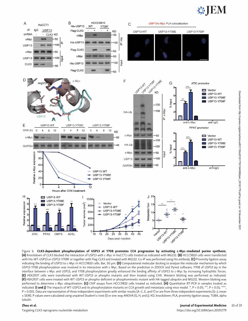
Figure 6. CLK3-dependent phosphorylation of USP13 at Y708 promotes CCA progression by activating c-Myc–mediated purine synthesis.(A) Knockdown of CLK3 blocked the interaction of USP13 with c-Myc in HuCCT1 cells treated as indicated with MG132. (B) HCCC9810 cells were transfectedwith His-WT-USP13 or USP13-Y708F or together with Flag-CLK3 and treated withMG132. Co-IP was performed using His antibody. (C) Proximity ligation assayindicating the binding of USP13 to c-Myc in HCCC9810 cells. Bar, 50 µm. (D) Computational molecular docking to analyze the molecular mechanism by whichUSP13-Y708 phosphorylation was involved in its interaction with c-Myc. Based on the prediction in ZDOCK and Pymol software, Y708 of USP13 lay in theinterface between c-Myc and USP13, and Y708 phosphorylation greatly enhanced the binding affinity of USP13 to c-Myc by increasing hydrophilic forces.(E) HEK293T cells were transfected with WT-USP13 or phospho mutants and then treated using CHX. Western blotting was performed as indicated.(F) HEK293T cells were treated with WT-USP13 or phospho-deficient or phosphomimetic mutant with HA-tagged ubiquitin and MG132. Western blotting wasperformed to determine c-Myc ubiquitination. (G) ChIP assays from HCCC9810 cells treated as indicated. (H) Quantitative RT-PCR in samples treated asindicated. (I and J) The impacts of WT-USP13 and its phosphorylation mutants on CCA growth and metastasis using mice model. *, P < 0.05; **, P < 0.01; ***,P < 0.001. Data are representative of three independent experiments with similar results (A–C, E, and F) or are from three independent experiments (G–J; mean± SEM). P values were calculated using unpaired Student’s t test (I) or one-way ANOVA (G, H, and J). KD, knockdown; PLA, proximity ligation assay; TUBA, alphatubulin.
Zhou et al. Journal of Experimental Medicine 10 of 19
Targeting CLK3 reprograms nucleotide metabolism https://doi.org/10.1084/jem.20191779
Dow
nloaded from http://rupress.org/jem
/article-pdf/217/8/e20191779/1044596/jem_20191779.pdf by guest on 18 N
ovember 2021

Figure 7. CLK3 is frequentlymutated and activated in human CCA. (Ai) Schematic presentation of somatic mutations in CLK3 in 100 CCA patients. (Aii andAiii) Sanger sequencing to identify CLK3 mutations in 100 (ii) and 75 (iii) human CCAs. Arrows indicate the location of the mutation in the tumor, but not in thetumor tissue. (B) IP kinase assay. Briefly, CLK3 was immunoprecipitated with anti-HA antibodies from HEK293 cells expressing WT-CLK3 or its mutants. Theimmunoprecipitated proteins mixed with the SRSF1 protein (a known substrate of CLK3) synthetic peptide and [γ-32P] ATP. The phosphocellulose paper assaywas used to measure kinase activity. The results were normalized to 1.0 for WT-CLK3. (C) The effects of human CCA-associated CLK3 mutants on theproliferation and colony formation of HCCC9810 cells. (D) The impact of human CCA-associated CLK3 mutants on HCCC9810 cell wound healing and invasion.(E) Heatmap showing WT-CLK3 and its mutants in CCA patients on purine metabolism in HCCC9810 cells. (F–H) The effects of WT-CLK3 and its mutants inCCA patients on purine intermediates in HCCC9810 cells. *, P < 0.05; **, P < 0.01; ***, P < 0.001. Data are representative of three independent experimentswith similar results (Bii and E) or are from three independent experiments (Bi, C, D, and F–H; mean ± SEM). P values were calculated using unpaired Student’st test (Ci) or one-way ANOVA (Bi, Cii, D, and F–H). AMP, adenosine monophosphate; GMP, guanosine monophosphate; IMP, inosine monophosphate; TUBA,alpha tubulin.
Zhou et al. Journal of Experimental Medicine 11 of 19
Targeting CLK3 reprograms nucleotide metabolism https://doi.org/10.1084/jem.20191779
Dow
nloaded from http://rupress.org/jem
/article-pdf/217/8/e20191779/1044596/jem_20191779.pdf by guest on 18 N
ovember 2021
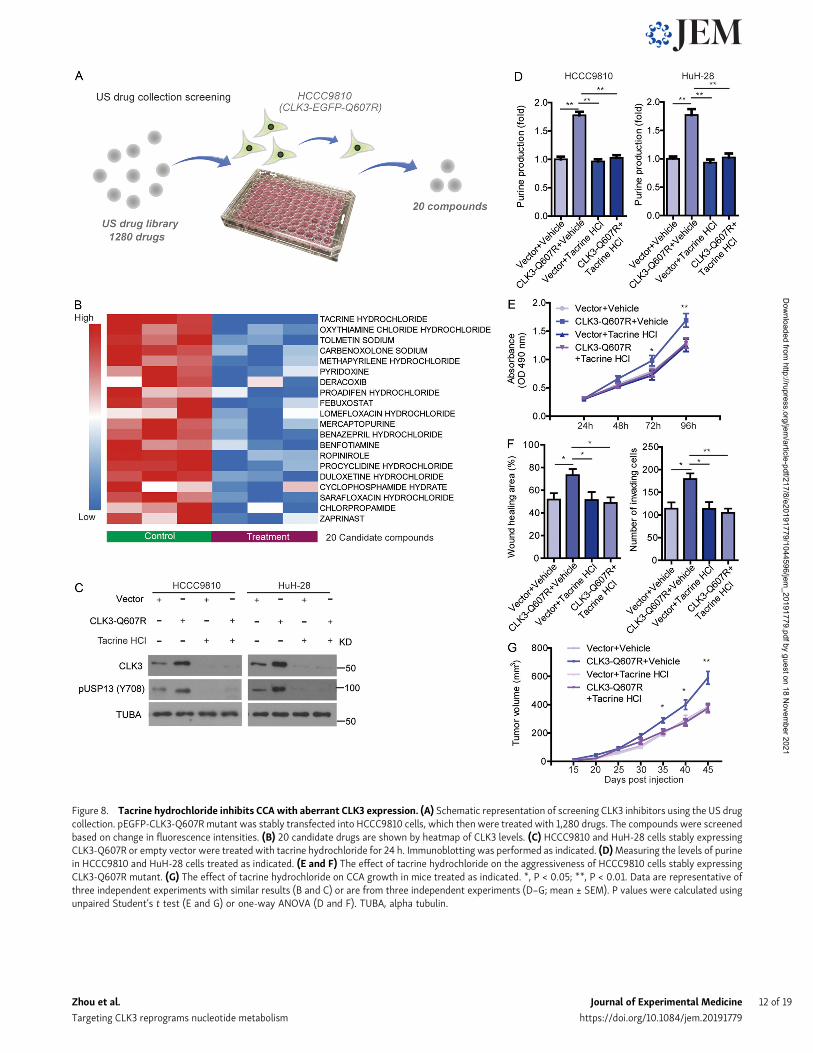
Figure 8. Tacrine hydrochloride inhibits CCA with aberrant CLK3 expression. (A) Schematic representation of screening CLK3 inhibitors using the US drugcollection. pEGFP-CLK3-Q607R mutant was stably transfected into HCCC9810 cells, which then were treated with 1,280 drugs. The compounds were screenedbased on change in fluorescence intensities. (B) 20 candidate drugs are shown by heatmap of CLK3 levels. (C) HCCC9810 and HuH-28 cells stably expressingCLK3-Q607R or empty vector were treated with tacrine hydrochloride for 24 h. Immunoblotting was performed as indicated. (D)Measuring the levels of purinein HCCC9810 and HuH-28 cells treated as indicated. (E and F) The effect of tacrine hydrochloride on the aggressiveness of HCCC9810 cells stably expressingCLK3-Q607R mutant. (G) The effect of tacrine hydrochloride on CCA growth in mice treated as indicated. *, P < 0.05; **, P < 0.01. Data are representative ofthree independent experiments with similar results (B and C) or are from three independent experiments (D–G; mean ± SEM). P values were calculated usingunpaired Student’s t test (E and G) or one-way ANOVA (D and F). TUBA, alpha tubulin.
Zhou et al. Journal of Experimental Medicine 12 of 19
Targeting CLK3 reprograms nucleotide metabolism https://doi.org/10.1084/jem.20191779
Dow
nloaded from http://rupress.org/jem
/article-pdf/217/8/e20191779/1044596/jem_20191779.pdf by guest on 18 N
ovember 2021

our knowledge about the genetic driver genes in CCA re-mains limited. In this study, our cellular and genetic studiesnot only identified CLK3 as an additional significantly mu-tated gene in CCA but also demonstrated the CLK3-Q607Rmutant as a gain-of-function mutation in CCA patients thataccelerates oncogenic CLK3-driven CCA progression. Thesefindings therefore suggest CLK3 mutation is a typical driverthat facilitates CCA development by activation of the purinesynthesis signaling pathway. Future studies should examine
whether this mutation-activating CLK3 happens in othertumor types.
Notably, only one Q607K substitution in the entire COSMICdatabase can be found, and no R634 mutation is reported, eventhough we did not find its functional effect on CCA. Possiblereasons why the R634 mutation was not found before are asfollows. (1) Although >500 CCA samples have been includedin COSMIC, they cannot represent overall CCA incidences.Therefore, it is still possible for undiscovered mutations to be
Figure 9. Clinical correlations between CLK3, p-USP13–Y708, c-Myc, and ATIC in CCA patients’ samples. (A) The pictures show CLK3, p-USP13–Y708,c-Myc, and ATIC protein expression in two human CCA specimens. Bars, 100 µm. (B) Pearson correlation coefficient analysis about the expression of CLK3,p-USP13–Y708, c-Myc, and ATIC protein in patients with CCA (n = 103) by IHC. ***, P < 0.001. (C)OS data from CCA patients stratified by the level of CLK3withp-USP13–Y708 or c-Myc. ***, P < 0.001; Kaplan-Meier analysis. (D) EGF or TGFβ1 or human CCA-associated CLK3mutant activates CLK3 and thereby enhancesp-Y708 levels of USP13; this event significantly increases the association of USP13 with c-Myc and disrupts Fbxl14-mediated c-Myc ubiquitination, whichactivated the de novo purine biosynthesis pathway and promoted the development of CCA. Interestingly, activated c-Myc transcriptionally up-regulated CLK3expression. Finally, tacrine hydrochloride inhibits aberrant CLK3-induced CCA. H, high; L, low.
Zhou et al. Journal of Experimental Medicine 13 of 19
Targeting CLK3 reprograms nucleotide metabolism https://doi.org/10.1084/jem.20191779
Dow
nloaded from http://rupress.org/jem
/article-pdf/217/8/e20191779/1044596/jem_20191779.pdf by guest on 18 N
ovember 2021

identified. In fact, COSMIC, as the largest resource for mutation,has been constantly updated to include new mutation data. (2)Genomic background may affect mutation status, althoughmutation information on CCA in COSMIC comes from threestudies, none of which includes Chinese samples. However, inour study, we used Chinese CCA samples.
Understanding the functional consequences of genetic drivermutations dramatically facilitates the development of targetedcancer therapies. When a US drug collection approved by theFood and Drug Administration (FDA) was exploited, tacrinehydrochloride was identified as one candidate that effectivelydown-regulated the CLK3 level in CCA. Tacrine hydrochloride isa cholinesterase inhibitor and has been approved by the FDA totreat Alzheimer’s disease (de los Rıos and Marco-Contelles,2019). Interestingly, a recent study reported that tacrine or itsderivative displayed strong anticancer ability (Qin et al., 2018),which is consistent with our findings. Given that in a clinicalsetting, tacrine hydrochloride had been demonstrated to havegood safety and high potency, it might become a candidate drugfor treating human CCA. And it also might serve as a useful toolin CLK3-related research. Although our preliminary data indi-cated the effects of anti-CCA by tacrine hydrochloride, the de-tailed mechanism by which tacrine hydrochloride decreasedCLK3 expression and its preclinical application in CCA definitelyneeds to be further explored.
The functional and targeted therapeutic study against CLK3 isreminiscent of the Dyrk kinase family. First, sequence analysisof the catalytic domains of proteins from this superfamilyshowed that CLK3 forms a sister groupwith the Dyrk family, andliterature confirmed that CLK3 kinase shares a high sequencehomology with the Dyrk kinase family (Tomas-Loba et al., 2019).Second, similar to CLK3, the Dyrk family has very importanteffects on the nervous system. Notably, inhibiting Dyrk1A hasbeen shown to be a potential treatment for Alzheimer’s disease(Stotani et al., 2016). Third, the Dyrk family also modulatesmetabolic disorders and cancers. Several Dyrk family inhibitors,such as harmine, have been known to be effective anticanceragents (Uhl et al., 2018). Fourth, our recent study reported thatDyrk3 inhibits liver cancer by impairing purine synthesis (Maet al., 2019). These findings indicate that structural and se-quencing similarities suggest a functional similarity betweenCLK and Dyrk kinase families (Schmitt et al., 2014), which willbe very helpful for us in uncovering the unrecognized functionof the CLK family. Considering their similarities, it is temptingto hypothesize that Dyrk family inhibitors might affect CLK3function. Future studies are needed to test this hypothesis,which may provide new insights into the CLK3 signaling net-work in CCA.
By using the basic online tool Gepia (http://gepia.cancer-pku.cn/) to analyze the CCA TCGA dataset in this study, we indicatethat CLK3 is overexpressed in CCA. However, when usingcBioPortal, another online tool, CLK3 was not up-regulated invirtually all CCAs. How can this discrepancy be explained? Wethink there are at least two reasons. First, the sample number ofCCAs analyzed by using these two basic online tools (Gepia andcBioPortal) was totally different. Second, cBioPortal does notcontain any expression data from normal tissue samples.
Additionally, by analyzing the public Oncomine database, wefound that genomic DNA of CLK3 is also frequently amplified inesophageal carcinoma and gastric cancer (data not shown).However, KEGG (Kyoto Encyclopedia of Genes and Genomes)function pathway analysis did not indicate its involvementin purine synthesis (data not shown). Based on these findings,we speculate that CLK3-mediated purine metabolism may beunique in CCA, while CLK3 may be involved in esophagealcarcinoma and gastric cancer carcinogenesis by other alterna-tive mechanisms. For instance, (1) CLK3 was reported to reg-ulate HMGA2 splicing through SRSF1, which affects stem celldevelopment (Cesana et al., 2018), and (2) CLK3 contributed totumor progression via activating the Wnt/β–catenin signalingpathway (Li et al., 2019). Our MS data also suggest that CLK3may regulate tumor-related autophagy. If this is the case, fu-ture studies should explore other possible Myc-independentmechanisms in diverse tumor types, including CCA.
In summary, CLK3 may represent a unique type of kinasethat is essential for the de novo purine synthesis of CCA cells.This confirmation strongly supports the potential of CLK3 as atherapeutic target in CCA, which will provide a new approachtoward the treatment of this devastating disease. Our researchalso suggests that tacrine hydrochloride, an FDA-approved drugfor Alzheimer’s disease treatment, can be repurposed for CCAtreatment. Future studies in higher-animal models for CCA arerequired for further pharmacological confirmation.
Materials and methodsCell culture, reagents, and antibodiesHEK293T, normal human intrahepatic epithelial cholangiocyte(HiBEC; as a control), and CCA cell lines (HuCCT1, RBE, HuH-28,and HCCC9810) were provided by Shanghai Cell Bank of theChinese Academy of Science, ScienCell, and American TypeCulture Collection or as a gift from Dr. Bing Wang at RutgersUniversity (New Brunswick, NJ). FBS, 13C-glycine, X-film, and3-(4,5-dimethylthiazol-2-yl)-2,5-diphenyltetrazolium bro-mide (MTT) were fromMillipore and Sigma. Protease inhibitorcocktail was obtained from Santa Cruz. 14C-glycine was fromPerkin Elmer. Lipofectamine 2000, PBS, antibiotics, andDMEM were purchased from Invitrogen. Tween 20 and X-filmwere provided by Sigma. The Site-Directed Mutagenesis Kit (Agi-lent) was QuikChange II. Alexa Fluor 488 Phalloidin was fromThermo Fisher. The antibodies were listed as follows: anti–glutathione S-transferase (GST) antibody (Abcam; #ab19256),anti-Flag M2-conjugated agarose was from Sigma, anti-His(Abcam; #ab18184), CLK3 (Santa Cruz Biotechnology; #sc-365225), ADSL (GeneTex; #GTX84956), Fbxl14 (Sigma;#SAB2103691), ATF4 (Santa Cruz Biotechnology; #sc-390063),CLK2 (Abcam; #ab86147), GAPDH (Abcam; #ab9485), c-Myc(Abcam; #ab39688), ATIC (Santa Cruz Biotechnology; #sc-53612), MITF (Abcam; #ab20663), CLK1 (Abcam; #ab74044),GMPS (Abcam; #ab135538), CLK4 (Abcam, #ab67936), PFAS,(Abcam; #ab251740), phosphoserine (Abcam; #ab9332), Tubu-lin (Abcam; #ab18251), USP13 antibody (Bethyl Laboratories;#A302-762A), phosphothreonine (Abcam; #ab9337), phospho-tyrosine (Abcam; #ab10321), phosphoserine/threonine (Abcam
Zhou et al. Journal of Experimental Medicine 14 of 19
Targeting CLK3 reprograms nucleotide metabolism https://doi.org/10.1084/jem.20191779
Dow
nloaded from http://rupress.org/jem
/article-pdf/217/8/e20191779/1044596/jem_20191779.pdf by guest on 18 N
ovember 2021

#ab17464), and secondary antibodies (Bio-Rad; #1706515 and#1706516). P-USP13–Y708–specific antibody was made by thislaboratory using similar methods as described previously(Hong et al., 2018).
Screening of compounds against CLK3CLK3-Q607R mutant was stably transfected into HCCC9810 andHuH-28 cells. Then, these cells were cultured using 96-wellplates to 50% confluence. Then, 1 µM 1,280 drugs from the USdrug collection were individually added to each well. After 12 h,the CCA cells were washed with PBS, and fluorescence intensitywas measured to determine the levels of CLK3 in transfectedCCA cells.
Transfection, constructs, shRNA, and siRNAFor Tet-inducible overexpression of CLK3, we used the Tet-On3G Inducible Expression System (Clontech) following the man-ufacturer’s instructions. Briefly, CLK3 cDNA or flag-CLK3 wascloned into a Tet-inducible vector, pTRE3G (Clontech), andHCCC9810 or HuH-28 cells were stably transfected with pCMV-Tet-3G plasmid (500 µg/ml of G418, for 2 wk) using Xfecttransfection reagent (Clontech) to generate Tet-On 3G cell lines.Then, the Tet-On 3G cell lines were transfected with pTRE3G-CLK3 under puromycin selection (1 µg/ml, for 2 wk) to generatedouble-stable Tet-On 3G inducible cell lines. For Tet-inducibleknockdown of CLK3, two different shRNAs against CLK3(shCLK3-#1 and #2) were respectively cloned into pLVCT-tTR-KRAB (Addgene; plasmid #11643) in which we replaced GFPwithpuromycin. For transfection, as previously reported (Zhu et al.,2019), HuCCT1 and RBE cells were transduced through spino-culation with pLVCT-tTR-KRAB-shCLK3 and pLVCT-tTR-KRAB-shcontrol lentivirus particles at a multiplicity of infection ∼1.About 2 wk of antibiotics selection (1 µg/ml) later, stable poly-clonal CCA cell lines with shcontrol or shCLK3 were established.The cells above were maintained in the absence or presence ofDox conditions depending on the experimental requirement.Lipofectamine 2000 (Invitrogen) was used to perform siRNAtransfection for siATF4, siMyc, siMITF, or scramble negativecontrol (Invitrogen) following the protocol. Immunoblots wereused to determine the efficiency of knockdown at 2 d aftertransfection. All siRNA and shRNA, which were from Santa CruzBiotechnologies, were as follows: shCLK3-#1: 59-AGTCAGACATCAAGACACAC-39; shCLK3-#2: 59-GAUGCUUGAUCUUGCACAATT-39; or shcontrol: 59-AATGCTCGCACAGCACAAG-39; siMITF:59-GAAACUUGAUCGACCUCUACA-39; siATIC: 59-CAGUCUAACUCUGUGUGCUACGCCA-39; siATF4: 59-CCACGUAUGACACUUGdTdT-39; sic-Myc #1: 59-TCCGTACAGCCCTATTTCA-39; and sic-Myc #2: 59-GTTCTAATTACCTCATTGTCT-39. CLK3, c-Myc, andUSP13 constructs were purchased from GeneChem. Mutants ortruncated fragments from different genes, such as CLK3 andUSP13, were constructed as described previously (Hong et al.,2014), and sequencing was used to verify the resulting mutants.
The sample collections of CCA patientsIn this study, tissue samples of all CCA patients and corre-sponding nontumor samples were collected from 2012 to 2017 atthe Affiliated Hospitals, Anhui Medical University and Harbin
Medical University. This study ethic was passed by the HarbinMedical University Institute Research Ethics Committee. EachCCA participant signed the informed consent. All tumor tissuesfor RNA isolation and IHC were histopathologically confirmedby a pathologist. Tumor-node-metastasis, a cancer staging sys-tem, was used to define the histological type and cancer stageaccording to the American Joint Committee on Cancer (seventhedition).
Western blotCCA tissues or cell lines were collected using the lysis bufferradioimmunoprecipitation assay as previously described (Songet al., 2018). bicinchoninic protein reagent (Pierce) was used tomeasure protein concentration. Denaturing 10% SDS-PAGEseparated all samples, which were then transferred to a poly-vinylidene fluoride membrane. After blocking using tris-buffered saline with Tween 20 containing 5% milk for 1 h, theindicated primary antibody was added on polyvinylidene fluoridemembranes at 4°C overnight. As previously described (Qu et al.,2016), the secondary antibody was added, and enhanced chemi-luminescence reagents (Pierce) were applied to visualized blots.
Co-immunoprecipitation (Co-IP)As described (Qu et al., 2016), the indicated antibodies wereadded to 500 µg of precleared samples and were rotated at 4°Cfor ∼8 h Then A&G beads (Sigma) were mixed with the samplesfor 3 h. Finally, the immunoprecipitation (IP) complexes weresubjected to blot analysis.
PCRTRIzol (Thermo Fisher) was purchased to purify indicated RNAfrom CCA cells or CCA tissues. Then, random primers and theReverse Transcription Kit (Invitrogen) were purchased for RT oftotal RNA into cDNA. Subsequently, RT-PCR was finished byApplied Biosystems. Primers for the respective genes weresynthesized by Invitrogen. The relative levels of indicated pro-teins were analyzed through the 2−ΔΔCt method. The endogenouscontrol was GAPDH. All primer sequences are: CLK1: 59-ACAAGACATTATAGAGCACCGGA-39 and 59-GTGGTCCAAGAATCCTTTCCATC-39; USP13: 59-GCGAAATCAGGCTATTCAGG-39 and59-TTGTAAATCACCCATCTTCCTTCC-39; CLK2: 59-CGAACACTATCAGAGCCGAAAG-39 and 59-GAACGTGGTAGCTGTCCTCC-39;CLK3: 59-CGTACCTGAGCTACCGATGGA-39 and 59-TCCCTTCGGGACGGGTATC-39; CLK4: 59-ATGCGGCATTCCAAACGAAC-39and 59-GTACTGCTGTGAGACCTTCTCT-39; ATF4: 59-TTCTCCAGCGACAAGGCTAAGG-39 and 59-CTCCAACATCCAATCTGTCCCG-39; GMPS: 59-ATGGCTCTGTGCAACGGAG-39 and 59-CCTCACTCTTCGGTCTATGACT-39; PFAS: 59-CCCAGTCCTTCACTTCTATGTTC-39 and 59-GTAGCACAGTTCAGTCTCGAC-39; ADSL:59-TAGCGACAGGTATAAATTCC-39 and 59-TCTCCTGCCCTTGCTTTCCT-39; GART: 59-GGAATCCCAACCGCACAATG-39, and 59-AGCAGGGAAGTCTGCACTCA-39; ATIC: 59-CACGCTCGAGTGACAGTG-39 and 59-TCGGAGCTCTGCATCTCCG-39; c-Myc: 59-AATGAAAAGGCCCCCAAGGTAGTTATCC-39 and 59-CGTACTGGAGAGTTCCGGTTTG-39; RPL21: 59-CAAGGGAATGGGTACTGTTCAAA-39 and 59-CTCGGCTCTTAGAGTGCTTAATG-39; RPL18:59-ATGTGCGGGTTCAGGAGGTA-39 and 59-CTGGTCGAAAGT
Zhou et al. Journal of Experimental Medicine 15 of 19
Targeting CLK3 reprograms nucleotide metabolism https://doi.org/10.1084/jem.20191779
Dow
nloaded from http://rupress.org/jem
/article-pdf/217/8/e20191779/1044596/jem_20191779.pdf by guest on 18 N
ovember 2021

GAGGATCTTG-39; RPS15: 59-CCCGAGATGATCGGCCACTA-39and 59-CCATGCTTTACGGGCTTGTAG-39; RPL27: 59-TGGCTGGAATTGACCGCTAC-39 and 59-CCTTGTGGGCATTAGGTGATTG-39; NOP10: 59-CAGTATTACCTCAACGAGCAGG-39 and 59-GGCTGAGCAGGTCTGTTGTC-39; POP5: 59-ATGGTGCGGTTCAAGCACA-39 and 59-GAACTCGGTCATCGAGGCTTA-39; IPM3: 59-CCCTGACGTGGTTACCGAC-39 and 59-CCGCTTGATCTTGGACGAGT-39; and GAPDH: 59-GCCCAATACGACCAAATCC-39 and 59-CACCACATCGCTCAGACAC-39.
In vivo deubiquitinationIndicated constructs were transfected into indicated cells for 48 h.Before these cells were collected, 5 µg/ml MG132 (Bio-Rad) wasadded and incubated for∼4 h. Then, cells were lysed in denaturingbuffer containing 0.1 M NaH2PO4 and Na2HPO4, 6 M guanidine-HCl, 10 mM imidazole, and 400 mM Tris-HCl. These lysates weremixed with nickel beads for 3 h at cold room temperature. Finally,immunoblotting was performed with the indicated antibodies.
Metabolite assaysLiquid chromatography–tandem MS (LC-MS/MS) was used toanalyze intracellular metabolites of indicated cells as describedpreviously (Ben-Sahra et al., 2016). Briefly, glycine-free DMEMwas used to wash the indicated cells, and then cells were addedwith the same medium containing 400 µM [13C1]-glycine for30min. Metabolites were extracted using 4 ml 80%methanol ondry ice. After spinning at 4000 ×g at 4°C, the insoluble pelletswere isolated by 0.5 ml 80%methanol via spinning at 20,000 ×gat 4°C. An N-EVAP from Organomation Associates was used todry the metabolites under nitrogen gas. 10 µl of HPLC-gradewater was added to the resuspended pellets, and then MSanalysis was performed. Finally, AB/SCIEX, a Multi Quant v2.0software program, was used to analyze the metabolite SRM(selected reaction monitoring) transition. The SRMs were usedto analyze the incorporation of 15N or 13C by LC-MS/MS.
Detection of CLK3 mutations by Sanger sequencingWe first isolated DNA from tissues using the High Pure PCRTemplate Preparation Kit (Roche; #11796828001). DNA amplifi-cation was performed by conventional PCR with CLK3 mutationanalysis by direct Sanger sequencing. The PCR primers weresupplied by Sangon Biotech, and the PCR mixture included0.2 mM deoxy-ribonucleoside triphosphate, 0.2 µM primer, 10×PCR buffer, Platinum Taq DNA Polymerase, and 1.5 mM Mg2+
(Invitrogen; #15966005). PCR was performed as follows: 94°Cfor 2 min, 94°C for 30 s, ∼60°C for 30 s (depending on primermelting temperature), and 72°C for 1 min (for 35 cycles). Afterconfirmation of the band of interest, the PCR products werepurified using the QIAquick PCR Purification Kit (Qiagen;#28104) and then sent for sequencing by Tsingke. All of theprimers used for independent amplification of 13 exons of CLK3are listed in Table S12.
Identifying CLK3-binding proteins and USP13 phosphorylationsites by MSBioID2-based screening was used to identify CLK3-bindingproteins as described previously (Kim et al., 2016). Briefly,
myc-BioID2-CLK3 or myc-BioID2 was stably transfected intoHCCC9810 cells. 50mMbiotin was added to themedium of thesecells for 2 d. Then, proteins were extracted by spinning andkeeping the supernatant. Biotinylated proteins were purified byusing AssayMap streptavidin cartridges. Finally, LC-MS/MSanalysis was performed, and USP13 phosphorylation sites wereidentified as described previously (Caporarello et al., 2017).
Soft agar assaySoft agar experiments were performed as previously described(Ma et al., 2019). Briefly, CCA cells were cultured in the top agar(0.4%) in 6-well plates (5,000 cells per well). After∼3 wk, 0.05%crystal violet was used to stain colonies for 1 h. A digital camerawas used to count the colonies. All experiments were repeated inat least triplicates.
Cell invasion assaysFor invasion assays, transwell assay with 8-µm pores and Ma-trigel (Corning) were used to measure the invasive ability ofCCA cell lines. Briefly, after 48 h of transfection, the indicated2 × 104 cells per well were cultured in the upper chamber with100 µl of medium without FBS. Then, 500 µl of medium wasadded to the lower chambers, including 10% FBS, which acted asa chemoattractant. 24 h later, a cotton swab was used to wipe offcells left on the upper membrane while keeping the invadedcells. After fixing in 4% formaldehyde, 1% crystal violet was usedto stain the invaded cells. An inverted microscope (Nikon) wasused to count 10 random visual fields.
MTT assayBriefly, ∼1 × 10⁴ cells were cultured in 12-well plates. 4 µg/mlDox was added to the medium for 3 d. Then, at 37°C, 1 ml of MTTreagent was added to treat the cells for 30min, and 1 ml of acidicisopropanol was added. At 595 nm, the absorbance was analyzedwith background subtraction at 650 nm.
Wound-healing (scratch) experimentsExperiments were done as described previously (Mereness et al.,2018). Briefly, indicated cells were cultured on coated 12-wellplates at 3 × 10⁵ per well and grown to confluence for 24 h. Apipette tip was used to vertically scratch a monolayer in eachwell. At 0 and 24 h, images of the scratch were taken.
GST pulldown assayAs previously described (Song et al., 2014), Escherichia coli wasused to express GST-fusion proteins. Then, isopropyl-β-D-thio-galactoside induced their expression. Proteins were purifiedusing glutathione-sepharose 4B beads purchased from Sigma.GST-tagged CLK3 or USP13 and GST (around 10 µg) were cross-linked by dimethyl pimelimidate dihydrochloride to glutathione-sepharose in reaction buffer, pH 8.0. After elution with samplebuffer, Coomassie staining and Western blot were used to ana-lyze the samples.
Luciferase reporter assaysAs described previously (Song et al., 2019), we culturedHEK293T cells to perform the dual-luciferase reporter assays.
Zhou et al. Journal of Experimental Medicine 16 of 19
Targeting CLK3 reprograms nucleotide metabolism https://doi.org/10.1084/jem.20191779
Dow
nloaded from http://rupress.org/jem
/article-pdf/217/8/e20191779/1044596/jem_20191779.pdf by guest on 18 N
ovember 2021
Briefly, using Lipofectamine 2000 overnight after plating, 0.2µg of the firefly promoter luciferase reporter constructs (WT-CLK3 or CLK3 mutant promoter) was cotransfected with theindicated plasmids. The control group was PGL-TK Renilla lucif-erase plasmid. The activity was monitored via the Dual-LuciferaseSystem bought from Promega, and luciferase activity was aver-aged from three replicates.
Chromatin IP (ChIP)As described (Song et al., 2019), the primers that were adoptedfor c-Myc motif are: 59-GACGGAGTTTTGCTCTCTTG-39 and 59-CTGCCTCCCGGGTTTAAGTG-39; 59-CTCCCACCTCAGCCTCC-39 and59-AGGCGCGTGCCACCACGTCT-39; 59-CATGTTGGCCAGACTGGTCT-39 and 59-GCCTCCCAAAGTACTGGGAT-39; and 59-CTCAAAAGATCCCCACCTCA-39 and 59-GCCTCGTAATCCTGTCCGAC-39.
Mice xenograft experiments and metastasis modelIn accordancewith National Institutes of Health guidelines, miceexperiments were performed; the Institutional Animal Com-mittee at Anhui Medical University and Harbin Medical Uni-versity approved the animal protocols. For the xenograft ormetastasis model, 106 Dox-inducible knockdown or over-expression of CLK3 cells with their corresponding control cellswere subcutaneously or intraperitoneally or intravenously in-jected into 4–6-wk-old BALB/c nu/nu mice (n = 6–10/group).Mice were given drinking water containing 2 mg/ml Dox and10% sucrose for inducible knockdown or overexpression ofCLK3. The xenograft tumors or metastatic peritoneal or meta-static lung tumors were monitored at the indicated time pointsafter injection. At the indicated time, the size and volume of thetumor were calculated as described previously (Qu et al., 2016).
Tissue arrays and IHC stainingCCA tissue microarray was purchased from Alenabio Company,and IHC staining was performed for CLK3 as described previ-ously (Hong et al., 2018). IHC staining was evaluated and scoredusing the scale 0, 1+, 2+, and 3+ representing no staining, weakstaining, moderate staining, and strong staining, respectively.The final H-score was calculated based on the formula reportedpreviously (Ma et al., 2019). The indicated protein levels weredefined by H-score, and then low- and high-expression patientgroups were divided.
IP kinase analysisAs described before (Bankston et al., 2017), an IP kinase assaywas done. Briefly, the lysate (1 mg) was mixed with anti-HAantibody (Santa Cruz Biotechnology; #sc-57592). 24 h later,protein G–agarose beads (Santa Cruz Biotechnology; #sc2002)were added. The immunocomplexes were resuspended in buffercontaining 1 mm Na3VO4. Using the phosphocellulose paperassay, the immunoprecipitated CLK3 activity was examined asfollows. One synthetic peptide (1 mM) derived from the SRSF1protein phosphorylation site (a known substrate of CLK3) wasadded as a substrate in the mixture (0.4 mMATP, 1 mmNa3VO4,20 mMTris, [γ32P] ATP, pH 7.4, and 10mMMgCl2). 30min laterat 30°C, 10% trichloroacetic acid terminated the reaction. Thenthe mixtures were loaded as a dot on the p81 phosphocellulose
paper. Scintillation counting was used to determinate incorpo-ration of 32P into the peptide.
TCGA and Gene Expression Omnibus analysisWhole-genome RNA sequencing data about the CCA TCGA da-taset were downloaded using the Xena Functional GenomicsExplorer website (https://xenabrowser.net). Patients with un-available survival data were excluded. The relevant clinicalcharacteristics, including age, gender, pathological tumor-node-metastasis, disease stage, survival time, and censor, were ob-tained from the TCGA dataset. We used Gepia (http://gepia.cancer-pku.cn/), an interactive web server for analyzing theRNA sequencing expression data of tumors and normal samplesfrom the TCGA and the Genotype Tissues Expression projects(Tang et al., 2017), to examine CLK3 expression in CCA. TheseTCGA data of CCA include 36 tumor samples and nine normalsamples. According to user instructions of this online tool, theresults are presented with log2(TPM+1; TPM means transcriptsper million) and analyzed by using Student’s t test. The mRNAarray data about CLK3 analysis in CCA are publicly available in theGene Expression Omnibus (accession no. GSE26566). The ex-pression pattern was plotted using GraphPad Prism 5 software.
In vitro kinase assay1 µg recombinant CLK3 protein and purified WT-USP13 or itsmutant proteins were mixed with 1X reaction buffer containing10 µM ATP and 0.2 mM Na3VO4 and 10 µCi [γ-32P] ATP. Thereaction proceeded at 30°C for 15 min. Then, the mixtures wereseparated and the incorporated [γ-32P] radioisotope was de-tected by using the imaging plate–autoradiography system.
BrdU assayBriefly, indicated cells (∼4,000 cells/well) were plated into a 96-well plate. After 24 h, the proliferation of CCA cells was exam-ined using a chemiluminescent BrdU kit (Sigma) as described bythe manufacturer.
StatisticsThe results from two groups were compared using Student’st test. When comparing data from groups greater than two, weused one-way ANOVA. To analyze the growth curves, we usedtwo-way ANOVA. Pearson correlation analysis was used toelucidate the correlation of two proteins. Programs for Graph-Pad Prism 5, R software package (version 3.0.0), and SocialSciences software 20.0 were used. Determining Kaplan-Meierdata required the log-rank test. Data were reported through mean± SD, performed in at least triplicates. *, P < 0.05 was consideredstatistically significant, and **, P < 0.01 or ***, P < 0.001 wasconsidered very significant; # indicates no significance.
Online supplemental materialFig. S1 shows that CLK3 in CCA is significantly up-regulated andis associated with decreased OS and acts as an oncogene. Fig. S2shows that CLK3 promotes purine synthesis and CCA progres-sion through enhancing the stabilization and nuclear translo-cation of c-Myc. Fig. S3 shows that CLK3 interacts with USP13and that CLK3 expression was up-regulated on EGF or TGFβ1
Zhou et al. Journal of Experimental Medicine 17 of 19
Targeting CLK3 reprograms nucleotide metabolism https://doi.org/10.1084/jem.20191779
Dow
nloaded from http://rupress.org/jem
/article-pdf/217/8/e20191779/1044596/jem_20191779.pdf by guest on 18 N
ovember 2021

treatment. Fig. S4 documents that c-Myc enhances transcrip-tional activation of the CLK3 promoter in CCA cells. Fig. S5 showsthat tacrine hydrochloride inhibits CCA with aberrant CLK3 ex-pression. Table S1 describes the relationship between CLK3 ex-pression and clinicopathological features of CCA patients. Table S2shows univariate and multivariate analyses of factors associatedwith survival in CCA patients. Table S3 shows the top 500 differ-entially expressed genes with high CLK3 expression. Table S4shows metabolic profiling of CCA cells with or without CLK3knockdown. Table S5 summarizes ribosome-related genes amongthe 500 up-regulated genes in Table S3. Table S6 documents theCLK3-interacting proteins. Table S7 and Table S9 document theCLK3mutations. Table S8 shows the relationship of CLK3mutationwith clinicopathological features of CCA. Table S10 shows purinemetabolite profiling of WT and mutant HCCC9810 cells. Table S11describes the relationship between CLK3 or p-USP13–Y708 orc-Myc expression and clinicopathological features of CCA patients.Table S12 lists the primers for Sanger sequencing.
AcknowledgmentsThis study was supported by the National Natural Science Founda-tion of China (no. 81702387, no. 81702744, and no. 81960520), theScience and Technology Planned Project in Guilin (20190206-1), theGuangxi Distinguished Experts Special Fund (2019-13-12), the Nat-ural Science Foundation of Fujian Province (no. 2017J01368 and no.2017J01369), the Training Program for Young Talents of FujianHealth System (no. 2016-ZQN-85), Fujian Provincial Funds for Dis-tinguished Young Scientists (no. 2018D0016), the Fujian HealthEducation Joint Research Project (WKJ2016-2-17), the HeilongjiangPostdoctoral Science Foundation (LBH-Z17176), the Science andTechnology Foundation of Shenzhen (JCYJ20170412155231633 andJCYJ20180305164128430), the Shenzhen Economic and InformationCommittee “Innovation Chain and Industry Chain” integration spe-cial support plan project (20180225112449943), the Shenzhen PublicService Platform on Tumor Precision Medicine and Molecular Di-agnosis, and the Shenzhen Cell Therapy Public Service Platform.
Author contributions: Q. Zhou, M. Lin, X. Feng, and F. Maacquired and analyzed experimental data. Y. Zhu, X. Liu, C. Qu,H. Sui, H. Huang, B. Sun, H. Zhang, A. Zhu, J. Sun, Z. Gao, Y.Zhao, J. Jin, and Y. Bai provided administrative, technical, ormaterial support. Z. Zhang, X. Hong, and C. Zou designed thestudy, and Z. Zhang drafted the manuscript.
Disclosures: The authors declare no competing interests exist.
Submitted: 22 September 2019Revised: 3 January 2020Accepted: 13 March 2020
ReferencesAdzhubei, I., D.M. Jordan, and S.R. Sunyaev. 2013. Predicting functional ef-
fect of humanmissense mutations using PolyPhen-2. Curr. Protoc. Hum.Genet. Chapter 7:20. https://doi.org/10.1002/0471142905.hg0720s76
Bankston, A.N., L. Ku, and Y. Feng. 2017. Active Cdk5 Immunoprecipita-tion and Kinase Assay. Bio Protoc. 7. e2363. https://doi.org/10.21769/BioProtoc.2363
Ben-Sahra, I., G. Hoxhaj, S.J.H. Ricoult, J.M. Asara, and B.D. Manning. 2016.mTORC1 induces purine synthesis through control of the mitochondrialtetrahydrofolate cycle. Science. 351:728–733. https://doi.org/10.1126/science.aad0489
Bowler, E., S. Porazinski, S. Uzor, P. Thibault, M. Durand, E. Lapointe, K.M.A.Rouschop, J. Hancock, I. Wilson, andM. Ladomery. 2018. Hypoxia leadsto significant changes in alternative splicing and elevated expression ofCLK splice factor kinases in PC3 prostate cancer cells. BMC Cancer. 18:355. https://doi.org/10.1186/s12885-018-4227-7
Caporarello, N., G. Lupo, M. Olivieri, M. Cristaldi, M.T. Cambria, M. Salmeri,and C.D. Anfuso. 2017. Classical VEGF, Notch and Ang signalling incancer angiogenesis, alternative approaches and future directions(Review). Mol. Med. Rep. 16:4393–4402. https://doi.org/10.3892/mmr.2017.7179
Cesana, M., M.H. Guo, D. Cacchiarelli, L. Wahlster, J. Barragan, S. Doulatov,L.T. Vo, B. Salvatori, C. Trapnell, K. Clement, et al. 2018. A CLK3-HMGA2 alternative splicing axis impacts human hematopoietic stemcell molecular identity throughout development. Cell Stem Cell. 22:575–588.e7. https://doi.org/10.1016/j.stem.2018.03.012
Chong, D.Q., and A.X. Zhu. 2016. The landscape of targeted therapies forcholangiocarcinoma: current status and emerging targets. Oncotarget. 7:46750–46767. https://doi.org/10.18632/oncotarget.8775
de Los Rıos, C., and J. Marco-Contelles. 2019. Tacrines for Alzheimer’s diseasetherapy. III. The PyridoTacrines. Eur. J. Med. Chem. 166:381–389. https://doi.org/10.1016/j.ejmech.2019.02.005
Fang, X., W. Zhou, Q. Wu, Z. Huang, Y. Shi, K. Yang, C. Chen, Q. Xie, S.C.Mack, X. Wang, et al. 2017. Deubiquitinase USP13 maintains glioblas-toma stem cells by antagonizing FBXL14-mediated Myc ubiquitination.J. Exp. Med. 214:245–267. https://doi.org/10.1084/jem.20151673
Hong, X., R. Song, H. Song, T. Zheng, J. Wang, Y. Liang, S. Qi, Z. Lu, X. Song,H. Jiang, et al. 2014. PTEN antagonises Tcl1/hnRNPK-mediated G6PDpre-mRNA splicing which contributes to hepatocarcinogenesis. Gut. 63:1635–1647. https://doi.org/10.1136/gutjnl-2013-305302
Hong, X., H. Huang, X. Qiu, Z. Ding, X. Feng, Y. Zhu, H. Zhuo, J. Hou, J. Zhao,W. Cai, et al. 2018. Targeting posttranslational modifications of RIOK1inhibits the progression of colorectal and gastric cancers. eLife. 7.e29511. https://doi.org/10.7554/eLife.29511
Kim, D.I., S.C. Jensen, K.A. Noble, B. Kc, K.H. Roux, K. Motamedchaboki, andK.J. Roux. 2016. An improved smaller biotin ligase for BioID proximitylabeling.Mol. Biol. Cell. 27:1188–1196. https://doi.org/10.1091/mbc.E15-12-0844
Li, H., X. Cui, Q. Hu, X. Chen, and P. Zhou. 2019. CLK3 Is A Direct Target OfmiR-144 And Contributes To Aggressive Progression In HepatocellularCarcinoma. OncoTargets Ther. 12:9201–9213. https://doi.org/10.2147/OTT.S224527
Ma, F., Y. Zhu, X. Liu, Q. Zhou, X. Hong, C. Qu, X. Feng, Y. Zhang, Q. Ding, J.Zhao, et al. 2019. Dual-Specificity Tyrosine Phosphorylation-RegulatedKinase 3 Loss Activates Purine Metabolism and Promotes Hepatocel-lular Carcinoma Progression. Hepatology. 70:1785–1803. https://doi.org/10.1002/hep.30703
Marks, E.I., and N.S. Yee. 2016. Molecular genetics and targeted therapeuticsin biliary tract carcinoma. World J. Gastroenterol. 22:1335–1347. https://doi.org/10.3748/wjg.v22.i4.1335
Maroni, L., I. Pierantonelli, J.M. Banales, A. Benedetti, and M.Marzioni. 2013.The significance of genetics for cholangiocarcinoma development. Ann.Transl. Med. 1:28.
Mereness, J.A., S. Bhattacharya, Q. Wang, Y. Ren, G.S. Pryhuber, and T.J.Mariani. 2018. Type VI collagen promotes lung epithelial cell spreadingand wound-closure. PLoS One. 13. e0209095. https://doi.org/10.1371/journal.pone.0209095
Nayler, O., S. Stamm, and A. Ullrich. 1997. Characterization and comparisonof four serine- and arginine-rich (SR) protein kinases. Biochem. J. 326:693–700. https://doi.org/10.1042/bj3260693
Qin, Q.-P., S.-L. Wang, M.-X. Tan, Z.-F. Wang, D.-M. Luo, B.-Q. Zou, Y.-C. Liu,P.-F. Yao, and H. Liang. 2018. Novel tacrine platinum(II) complexesdisplay high anticancer activity via inhibition of telomerase activity,dysfunction of mitochondria, and activation of the p53 signaling path-way. Eur. J. Med. Chem. 158:106–122. https://doi.org/10.1016/j.ejmech.2018.09.008
Qu, C., D. He, X. Lu, L. Dong, Y. Zhu, Q. Zhao, X. Jiang, P. Chang, X. Jiang, L.Wang, et al. 2016. Salt-inducible Kinase (SIK1) regulates HCC progres-sion and WNT/β-catenin activation. J. Hepatol. 64:1076–1089. https://doi.org/10.1016/j.jhep.2016.01.005
Schmitt, C., P. Miralinaghi, M. Mariano, R.W. Hartmann, andM. Engel. 2014.Hydroxybenzothiophene ketones are efficient pre-mRNA splicing
Zhou et al. Journal of Experimental Medicine 18 of 19
Targeting CLK3 reprograms nucleotide metabolism https://doi.org/10.1084/jem.20191779
Dow
nloaded from http://rupress.org/jem
/article-pdf/217/8/e20191779/1044596/jem_20191779.pdf by guest on 18 N
ovember 2021

modulators due to dual inhibition of Dyrk1A and Clk1/4. ACSMed. Chem.Lett. 5:963–967. https://doi.org/10.1021/ml500059y
Song, R., H. Song, Y. Liang, D. Yin, H. Zhang, T. Zheng, J. Wang, Z. Lu, X.Song, T. Pei, et al. 2014. Reciprocal activation between ATPase inhibi-tory factor 1 and NF-κB drives hepatocellular carcinoma angiogenesisand metastasis. Hepatology. 60:1659–1673. https://doi.org/10.1002/hep.27312
Song, H., X. Feng, M. Zhang, X. Jin, X. Xu, L. Wang, X. Ding, Y. Luo, F. Lin, Q.Wu, et al. 2018. Crosstalk between lysine methylation and phospho-rylation of ATG16L1 dictates the apoptosis of hypoxia/reoxygenation-induced cardiomyocytes. Autophagy. 14:825–844. https://doi.org/10.1080/15548627.2017.1389357
Song, H., X. Feng, H. Zhang, Y. Luo, J. Huang, M. Lin, J. Jin, X. Ding, S. Wu, H.Huang, et al. 2019. METTL3 and ALKBH5 oppositely regulate m6Amodification of TFEB mRNA, which dictates the fate of hypoxia/reox-ygenation-treated cardiomyocytes. Autophagy. 15:1419–1437. https://doi.org/10.1080/15548627.2019.1586246
Stotani, S., F. Giordanetto, and F. Medda. 2016. DYRK1A inhibition as po-tential treatment for Alzheimer’s disease. Future Med. Chem. 8:681–696.https://doi.org/10.4155/fmc-2016-0013
Sugihara, T., H. Isomoto, G. Gores, and R. Smoot. 2019. YAP and the Hippopathway in cholangiocarcinoma. J. Gastroenterol. 54:485–491. https://doi.org/10.1007/s00535-019-01563-z
Tang, Z., C. Li, B. Kang, G. Gao, C. Li, and Z. Zhang. 2017. GEPIA: a web serverfor cancer and normal gene expression profiling and interactive anal-yses. Nucleic Acids Res. 45(W1):W98–W102. https://doi.org/10.1093/nar/gkx247
Tomas-Loba, A., E. Manieri, B. Gonzalez-Teran, A. Mora, L. Leiva-Vega, A.M.Santamans, R. Romero-Becerra, E. Rodrıguez, A. Pintor-Chocano, F.Feixas, et al. 2019. p38γ is essential for cell cycle progression and livertumorigenesis. Nature. 568:557–560. https://doi.org/10.1038/s41586-019-1112-8
Uhl, K.L., C.R. Schultz, D. Geerts, and A.S. Bachmann. 2018. Harmine, a dual-specificity tyrosine phosphorylation-regulated kinase (DYRK) inhibitorinduces caspase-mediated apoptosis in neuroblastoma. Cancer Cell Int.18:82. https://doi.org/10.1186/s12935-018-0574-3
van Riggelen, J., A. Yetil, and D.W. Felsher. 2010. MYC as a regulator of ri-bosome biogenesis and protein synthesis. Nat. Rev. Cancer. 10:301–309.https://doi.org/10.1038/nrc2819
Yin, J., W. Ren, X. Huang, J. Deng, T. Li, and Y. Yin. 2018. Potential mecha-nisms connecting purine metabolism and cancer therapy. Front. Im-munol. 9:1697. https://doi.org/10.3389/fimmu.2018.01697
Zhu, Y., C. Qu, X. Hong, Y. Jia, M. Lin, Y. Luo, F. Lin, X. Xie, X. Xie, J. Huang,et al. 2019. Trabid inhibits hepatocellular carcinoma growth and me-tastasis by cleaving RNF8-induced K63 ubiquitination of Twist1. CellDeath Differ. 26:306–320. https://doi.org/10.1038/s41418-018-0119-2
Zhou et al. Journal of Experimental Medicine 19 of 19
Targeting CLK3 reprograms nucleotide metabolism https://doi.org/10.1084/jem.20191779
Dow
nloaded from http://rupress.org/jem
/article-pdf/217/8/e20191779/1044596/jem_20191779.pdf by guest on 18 N
ovember 2021

Supplemental material
Zhou et al. Journal of Experimental Medicine S1
Targeting CLK3 reprograms nucleotide metabolism https://doi.org/10.1084/jem.20191779
Dow
nloaded from http://rupress.org/jem
/article-pdf/217/8/e20191779/1044596/jem_20191779.pdf by guest on 18 N
ovember 2021

Figure S1. CLK3 in CCA is significantly up-regulated and associated with decreased OS and acts as an oncogene. (A) Examining the mRNA (upper panel)and protein (lower panel) levels of CLK3 in normal cholangiocytes and CCA cell lines. (Bi) Serial slides were incubated with either mouse IgG or CLK3 antibodyfor IHC detection. Bar, 100 µm. (Bii) IHC analysis of CLK3 expression in a CCA patient’s tissue array (n = 100). Representative pictures are presented. Bars, 100µm. (C) Stratified by CLK3 levels in CCA patients (n = 84), Kaplan-Meier analysis of OS is shown. (D and Ei) MTT assays were performed to compare theproliferation of HCCC9810 cells with Dox-induced CLK3 expression or without, which was confirmed using immunoblotting. (Eii) BrdU assay in D. (Eiii) Softagar assays in D. Bar, 100 µm. (Fi and Fii) Transwell assays andwound healing using HCCC9810 treated as in D. (Gi) The effect of CLK3 overexpression inducedby Dox on HCCC9810 xenografts. (Gii and Giii) Dox-inducible expression of CLK3 in HCCC9810 cells significantly promoted the number of CCA abdominalmetastatic nodules (ii) and lung metastatic nodules (iii) compared with their controls. (Hi and Hii)MTT assays were performed to compare the proliferation ofSk-hep1 and HCT116 cells with CLK3 knockdown, which was confirmed using immunoblotting. (Hiii) Transwell assay in Hi. (I)MTT assays and wound healingusing HiBEC treated as in D. *, P < 0.05; **, P < 0.01; ***, P < 0.001; #, not significant. Data are mean ± SEM and are from three (F, G, Hii, Hiii, and I) and four (E)independent experiments or representative of three independent experiments with similar results (A lower panel, D, and Hi). P values were calculated usingunpaired Student’s t test (E–G, Hii, Hiii, and I) or one-way ANOVA (A) or χ-square test (Bii). KD, knockdown; TUBA, alpha tubulin.
Zhou et al. Journal of Experimental Medicine S2
Targeting CLK3 reprograms nucleotide metabolism https://doi.org/10.1084/jem.20191779
Dow
nloaded from http://rupress.org/jem
/article-pdf/217/8/e20191779/1044596/jem_20191779.pdf by guest on 18 N
ovember 2021

Figure S2. CLK3 promotes purine synthesis and CCA progression through enhancing the stabilization and nuclear translocation of c-Myc. (Ai) LC-MS/MS analysis was performed to measure 15N-glutamine–labeled intermediates of purine synthesis in HCCC9810 cells with or without Dox-induced CLK3overexpression. (Aii and Aiii) LC-MS/MS was used to analyze metabolites labeled with 13C-glycine (ii) and 14C-glycine (iii) in HCCC9810 cells with or withoutDox-induced CLK3 overexpression. (Bi) Performing quantitative RT-PCR assays as indicated in HCCC9810 cells with or without Dox-induced CLK3 over-expression. (Bii) CLK3 silencing significantly decreased HuCCT1 cell invasion while overexpressing ATIC or adding purine markedly reverted this defect.(Biii) ATIC inhibitor significantly reverted the invasion of HCCC9810 cells induced by CLK3 overexpression. (Ci)Western blots confirmed CLK3 overexpressionand c-Myc knockdown in HCCC9810 cells. (Cii) The effects of silencing c-Myc on the levels of GMP, AMP, and IMP in HCCC9810 cells with CLK3 overexpression.(Ciii) Silencing c-Myc reduced the levels of U-14C-glycine in HCCC9810 cells with CLK3 overexpression. (D) The knockdown of c-Myc suppressed purine-associated enzymes in HCCC9810 cells with CLK3 overexpression. (Ei and Eii) The impacts of CLK3 knockdown or overexpression on the nuclear translocationof c-Myc in HuCCT1 (i) or HCCC9810 (ii) cells were analyzed by immunofluorescence staining and were quantified. Bars, 50 µm. (F) Silencing c-Myc revertedthe aggressiveness of HCCC9810 cells with CLK3 overexpression. (G) CLK3 knockdown reduced the mRNA levels of genes of ribosome components (i) and theirregulators (ii). *, P < 0.05; **, P < 0.01; ***, P < 0.001; #, not significant. Data are mean ± SEM and are from three (A, B, Cii, Ciii, D, E, and Fii, and Fiii) and four(Fi) independent experiments or are representative of three independent experiments with similar results (Ci). P values were calculated using unpairedStudent’s t test (Ai, Bi, and Ei) or one-way ANOVA (Bii, Biii, Cii, Ciii, D, Eii, and F). KD, knockdown; IB, immunoblot; AMP, adenosine monophosphate; GMP,guanosine monophosphate; IMP, inosine monophosphate; TUBA, alpha tubulin.
Zhou et al. Journal of Experimental Medicine S3
Targeting CLK3 reprograms nucleotide metabolism https://doi.org/10.1084/jem.20191779
Dow
nloaded from http://rupress.org/jem
/article-pdf/217/8/e20191779/1044596/jem_20191779.pdf by guest on 18 N
ovember 2021

Figure S3. CLK3 directly interacted with USP13, and CLK3 expression was up-regulated on EGF or TGFβ1 treatment. (A) Immunofluorescence stainingindicated the nuclear colocalization of CLK3 and USP13 in HuCCT1 cells. Bar, 50 µm. (B) Indicated constructs were transfected into HEK293T cells. Co-IP wasperformed with indicated antibody. (C) HCCC9810 and HuH-28 cells were added with or without EGF (50 ng/ml) or TGFβ1 (10 ng/ml) for indicated time. CLK3expression was analyzed using immunoblotting. Data are representative of three independent experiments with similar results. IB, immunoblot; TUBA, alphatubulin.
Zhou et al. Journal of Experimental Medicine S4
Targeting CLK3 reprograms nucleotide metabolism https://doi.org/10.1084/jem.20191779
Dow
nloaded from http://rupress.org/jem
/article-pdf/217/8/e20191779/1044596/jem_20191779.pdf by guest on 18 N
ovember 2021

Figure S4. c-Myc enhances transcriptional activation of CLK3 promoter in CCA cells. (A) HEK293T cells were overexpressed with empty vector orFlag–c-Myc with or without CLK3-Luc (−1,328 bp) reporter as indicated. Relative luciferase activity was measured and normalized based on CMV-LacZ ex-pression. (B) Flag–c-Myc or empty vector was transfected into HCCC9810 cells. 24 h later, we performed quantitative RT-PCR and immunoblotting (IB) assays.(C) siRNA against c-Myc or scramble was transfected into HuCCT1 cells. 24 h later, we performed quantitative RT-PCR and immunoblotting assays.(D) Positions of four putative c-Myc–binding motifs (E-box) relative to the transcription start in the CLK3 promoter are indicated. (E) The effect of mutatingeach of four putative c-Myc–binding sites on the luciferase reporter of CLK3 promoter in HEK293T cells transfected with a c-Myc. Relative luciferase activitywas measured. (F) EGF- or TGFβ1-treated HuCCT1 cells were used to perform ChIP with c-Myc antibody. PCR was performed using primers flanking fourputative E-box sites of the CLK3 promoter. *, P < 0.05; **, P < 0.01; ***, P < 0.001; #, not significant. Data are mean ± SEM and are from three (A, B, right panel;C, lower panel; and E) independent experiments or are representative of three independent experiments with similar results (B, left panel; C, upper panel; andF). P values were calculated using unpaired Student’s t test (A and B) or one-way ANOVA (C and E). TUBA, alpha tubulin.
Zhou et al. Journal of Experimental Medicine S5
Targeting CLK3 reprograms nucleotide metabolism https://doi.org/10.1084/jem.20191779
Dow
nloaded from http://rupress.org/jem
/article-pdf/217/8/e20191779/1044596/jem_20191779.pdf by guest on 18 N
ovember 2021

Tables S1–S12 are provided online. Table S1 shows the relationship between CLK3 expression and clinicopathological features ofCCA patients (n = 100). Table S2 describes the univariate and multivariate analyses of factors associated with survival in CCApatients (n = 100). Table S3 lists the top 500 differentially expressed genes with CLK3 up-regulation. Table S4. shows MS-basedmetabolic profiling of HuCCT-1 cells expressing shCtrl and shCLK3 with or without c-Myc in the presence or absence of Dox. TableS5 lists genes that are associated with ribosomal biogenesis among the 500 up-regulated genes. Table S6 identifies CLK3-interacting proteins via MS. Table S7 shows the systematic investigation of mutation of CLK3 in 100 CCA samples. Table S8 showsthe relationship of CLK3mutationwith clinicopathological features of CCA (n = 100). Table S9 presents the systematic investigationofmutation of CLK3 in 75 CCA samples. Table S10 listsMS-based purinemetabolites profilingWT andmutant HCCC9810 cells. TableS11 shows the relationship between CLK3 or p-USP13–Y708 or c-Myc expression and clinicopathological features of CCA patients(n = 103). Table S12 lists all primers used for independent amplification of 13 exons of CLK3.
Figure S5. Tacrine hydrochloride inhibits CCA with aberrant CLK3 expression. (A and B) Tacrine hydrochloride inhibited the EGF-induced increase inCLK3 expression, USP13 phosphorylation at Y708 (A), and proliferation in HCCC9810 and HuH-28 cells (B). (C)Measuring the levels of purine in HuCCT1, HuH-28, and HiBEC cells treated as indicated. (D and E) The effect of tacrine hydrochloride on the growth (D) and migration (E, i) of HuCCT1, HuH-28, and HiBECcells and on the invasion (Eii) of HuCCT1 and HuH-28 cells. *, P < 0.05; **, P < 0.01; ***, P < 0.001; #, not significant. Data are mean ± SEM and are from three(B–E) independent experiments or are representative of three independent experiments with similar results (A). P values were calculated using unpairedStudent’s t test (B–E). TUBA, alpha tubulin.
Zhou et al. Journal of Experimental Medicine S6
Targeting CLK3 reprograms nucleotide metabolism https://doi.org/10.1084/jem.20191779
Dow
nloaded from http://rupress.org/jem
/article-pdf/217/8/e20191779/1044596/jem_20191779.pdf by guest on 18 N
ovember 2021