article supplementary information10.1038/s41586-019-0993-x... · supplementary information...
TRANSCRIPT

Articlehttps://doi.org/10.1038/s41586-019-0993-x
Gboxin is an oxidative phosphorylation inhibitor that targets glioblastoma Yufeng Shi1,2, S. Kyun lim3,4,8, Qiren liang3, Swathi V. iyer1,2, Hua-Yu Wang3, Zilai Wang1,2, Xuanhua Xie1,2, Daochun Sun1,2, Yu-Jung chen1,2,5, Viviane tabar1,6, Philip Gutin1,6, Noelle Williams3, Jef K. De Brabander3 & luis F. Parada1,2,6,7*
1Brain Tumor Center, Memorial Sloan Kettering Cancer Center, New York, NY, USA. 2Cancer Biology & Genetics Program, Memorial Sloan Kettering Cancer Center, New York, NY, USA. 3Department of Biochemistry, UT Southwestern Medical Center, Dallas, TX, USA. 4Department of Developmental Biology, UT Southwestern Medical Center, Dallas, TX, USA. 5Louis V. Gerstner Jr. Graduate School of Biomedical Sciences, Memorial Sloan Kettering Cancer Center, New York, NY, USA. 6Department of Neurosurgery, Memorial Sloan Kettering Cancer Center, New York, NY, USA. 7Department of Neurology, Memorial Sloan Kettering Cancer Center, New York, NY, USA. 8Present address: Vivid Biosciences, Boston, MA, USA. *e-mail: [email protected]
N A T U R E | www.nature.com/nature
SUPPLEMENTARY INFORMATIONhttps://doi.org/10.1038/s41586-019-0993-x
In the format provided by the authors and unedited.

Supplementary Information Supplementary Results Harsh denaturing conditions did not disrupt multiple B-Gboxin/OxPhos protein interactions (Extended Data Fig. 3h), indicating a covalent interaction. This view was supported by the fact that preincubation with Gboxin impeded B-Gboxin interaction, but preincubation with B-Gboxin could not be displaced by excess Gboxin despite its lower IC50 (Extended Data Fig. 3i). Furthermore, immunoprecipitation of the OxPhos proteins Sdha, Cox4, Atp5a1, and Atp5b following B-Gboxin treatment, followed by Avidin-HRP blotting, showed that three of the proteins (Sdha, Atp5a1 and Atp5b) retained a biotin tag (Extended Data Fig. 3j). We consider the covalent interaction of B-Gboxin with OxPhos proteins is likely a consequence of the phenolic biotin ester B-Gboxin acting as an acylating agent transferring the biotin moiety to the B-Gboxin interacting partners. First, phenolic acyl transfer is facilitated under basic (high pH) conditions found at the matrix side of the inner mitochondrial membrane as a result of the proton gradient generated by the ETC. Consistent with this, sodium hydroxide buffer modulation of lysate pH causes enhanced B-Gboxin association to cell lysate proteins in a high pH environment (Extended Data Fig. 3k). Second, Gboxin and B-Gboxin are positively charged (Fig. 1a and Extended Data Fig. 3a) and thus will enrich on the matrix side of the inner mitochondrial membrane51. The B-Gboxin matrix concentration and ability to transfer biotin will hence be correlated with the mitochondrial membrane potential and the matrix pH, respectively. Methods Primary Mouse and Human Tumor Cultures Human tumor tissue collection and PDX model generation was conducted with the approval of the Memorial Sloan Kettering Cancer Center Institutional Review Board, Protocol #15-283. Informed consent was obtained from all subjects involved in this study. Primary mouse GBM HTS cells were cultured from symptomatic mutant mice with a genotype of hGFAP-Cre; Nf1flox/+;P53-/flox;Ptenflox/+. Details of these mice were previously described52. For HTS cell isolation, tumor tissues collected from fourteen mutant mice were minced into single cell suspension, grown as neurospheres in serum free medium as previously described52, pooled and frozen down at low passages (<5 passage). Other primary mouse GBM cells used in this study were isolated the same as for HTS cells but from independent mouse tumors #2396, #1661, and #1663, respectively. These tumors have genotype Nestin-CreERT2-P2A-H2B-eGFP-P2A-hDTR;NF1+/flox;Pten+/flox;Trp53+/flox;Luc/+, and tumor suppressor recombination was induced by tamoxifen administration to mice between 7 and 9 weeks of age. Primary mouse malignant peripheral nerve sheath tumors (MPNST) cells were cultured from mouse tumors carrying NF1-/- and Trp53-/- mutations53. Briefly, MPNST samples were collected, minced and disassociated with collagenase type 4 into single cell suspension, and MPNST cells were grown in serum free medium as for mouse GBM cells. The three independent primary human GBM cell cultures (ts12017, ts1156, and ts603) were isolated from tumor tissues of GBM patients in the same way as for mouse GBM cells. Exon sequencing or RT-PCR data showed: GBM (ts12017) positive for EGFRvIII and 3 copies of chromosome 7; and GBM (ts1156) has EGFR amplification and mutations in MET, CDKN2A, and PTEN genes; and GBM (ts603) has IDH1 mutation. All experiments were done at 5% O2 with serum free defined medium and growth factors for primary cells.

Primary Wildtype Cell Cultures and Cell Lines Primary neonatal astrocytes were isolated and cultured as described previously54. Astrocytes were cultured in DMEM with 10% FBS, 1% Pen/Strep for the original counter screen. For subsequent assays in this study, astrocytes were cultured in serum free defined medium as for HTS cells. For primary wildtype MEF cultures, embryos from mice having appropriate genotypes were isolated between E12.5 and E18.5. Following dissociation of heads, tails, limbs, and internal organs, the remaining tissue was minced, trypsinized, and seeded into T-75 cell culture dishes in 15 mL of complete serum media (DMEM with 10% FBS, 1% P/S). The cells were split at 1:2–1:3 ratios when freshly confluent, passaged two or three times to obtain a morphologically homogenous culture, and then frozen or expanded for further studies. Primary subventricular zone neural stem/progenitor cells (NSCs) were isolated and cultured as described previously52. Cell lines Daoy and Cal-62 were cultured in DMEM with 10% FBS and 1% Pen/Strep. A375 and SK-MEL113 were cultured in DMEM with 10% FBS, 1% Pen/Strep, and 1% Glutamine. Mel30 was cultured in DMEM/F12 with 10% FBS, 1% Pen/Strep, and 1% Glutamine. HCT116 was cultured in McCoy’s 5a medium with 10% FBS, 1% Pen/Strep and 1% Glutamine. NCI-H2030 and Colo205 were cultured in RPMI1640 medium with 10% FBS, 1% Pen/Strep and 1% Glutamine. U937, NCI-H82 and NCI-H524 were cultured in RPMI1640 medium with 10% FBS and 1% Pen/Strep. Gboxin sensitivity and B-Gboxin OxPhos interactions for primary wildtype cultures performed in 5% or 20% O2 conditions gave similar results. For living cell imaging, EVOS FL cell imaging system (Life technology) was used. Western Blots, Antibodies, and siRNA Transfection Cell lysates were prepared in RIPA buffer (Thermo) with phosphatase inhibitor (Thermo) and protease inhibitor (Roche). After measuring protein concentration, western blot samples were prepared using LDS Sample Buffer (Thermo). Antibodies purchased from Santa Cruz include ATF4 (sc-200; C20; lot: #1914), Atp5b (sc-16690; C20; lot: #D2214), and Sdha (SC390381; F-2; lot: #1313), from Cell Signaling include cleaved Caspase3 (#9661s; D175; lot: 43), Puma (#7467s; lot: 1), phospho-S6 (#4856s; 2F9; lot: 9), S6 (#2217s; 5G10; lot: 5), AMPK (#2532s; lot: 19), phospho-AMPK (#2535s; 40H9; lot: 16), phosphor-ACC-79 (#3661s; lot: 10), ACC (#3662s; lot: 4), and COX IV (#4844s; lot: 3), from Novus include Survivin (NB500-201ss; lot: AD-6 ), from Millipore include Gapdh (AB2302), from Sigma include Tubulin (T9026; DM1A), and from Proteintech include Atp5a1 (14676-1-AP), Atp5b (17247-1-AP), Sdha (14865-1-AP), Ndufv2 (15301-1-AP), and CypD (18466-1-AP). Avidin-HRP from Thermofisher was used for detecting B-Gboxin. SMARTpool of siGENOME for control and a mixture of two CypD siRNAs (D-062722-04-0010 sequence: GUGGGAAGACGUCUAAGAA and D-062722-03-0010 sequence: GGAGGUCCAUCUACGGAAG) from Dhamarcon was used for knockdown experiments in MEF cells. Lipofectamine® RNAiMAX transfection reagent from Invitrogen was used for siRNA transfection according to manufacturer’s instructions. RNA Isolation, cDNA Synthesis, Quantitative PCR (qPCR), and Gene Expression Microarray RNA was isolated with RNAeasy RNA isolation kit (Qiagen) and cDNA was synthesized by SuperScript™ III CellsDirect cDNA Synthesis System (Invitrogen). Microarrays for gene expression were done in UTSW Genomics and Microarray Core Facility. Quantitative PCR (qPCR) with SYBR green PCR master mix was from Thermo. Compounds used in these assays were Gboxin (1 μM), rotenone (Sigma, 1 μM), antimycin A (Sigma, 1 μM), and oligomycin A (Sigma, 1 μM) or as indicated in figure legend. Primers used are listed as below.
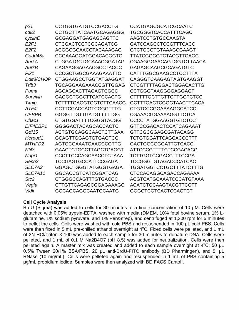
p21 CCTGGTGATGTCCGACCTG CCATGAGCGCATCGCAATC cdk2 CCTGCTTATCAATGCAGAGGG TGCGGGTCACCATTTCAGC cyclinE GCGAGGATGAGAGCAGTTC AAGTCCTGTGCCAAGTA E2F1 CTCGACTCCTCGCAGATCG GATCCAGCCTCCGTTTCACC E2F2 ACGGCGCAACCTACAAAGAG GTCTGCGTGTAAAGCGAAGT Gadd45a CCGAAAGGATGGACACGGTG TTATCGGGGTCTACGTTGAGC AurkA CTGGATGCTGCAAACGGATAG CGAAGGGAACAGTGGTCTTAACA AurkB CAGAAGGAGAACGCCTACCC GAGAGCAAGCGCAGATGTC Plk1 CCCGCTGGCGAAAGAAATTC CATTTGGCGAAGCCTCCTTTA Ddit3/CHOP CTGGAAGCCTGGTATGAGGAT CAGGGTCAAGAGTAGTGAAGGT Trib3 TGCAGGAAGAAACCGTTGGAG CTCGTTTTAGGACTGGACACTTG Puma AGCAGCACTTAGAGTCGCC CCTGGGTAAGGGGAGGAGT Survivin GAGGCTGGCTTCATCCACTG CTTTTTGCTTGTTGTTGGTCTCC Txnip TCTTTTGAGGTGGTCTTCAACG GCTTTGACTCGGGTAACTTCACA ATF4 CCTTCGACCAGTCGGGTTTG CTGTCCCGGAAAAGGCATCC CEBPB GGGGTTGTTGATGTTTTTGG CGAAACGGAAAAGGTTCTCA Chac1 CTGTGGATTTTCGGGTACGG CCCCTATGGAAGGTGTCTCC EIF4EBP1 GGGGACTACAGCACCACTC GTTCCGACACTCCATCAGAAAT Gdf15 ACTGTGCAGGCAACTCTTGAA GTTCGCGGAGCGATACAGG Herpud1 GCAGTTGGAGTGTGAGTCG TCTGTGGATTCAGCACCCTTT MTHFD2 AGTGCGAAATGAAGCCGTTG GACTGGCGGGATTGTCACC Nfil3 GAACTCTGCCTTAGCTGAGGT ATTCCCGTTTTCTCCGACACG Nupr1 CCCTTCCCAGCAACCTCTAAA TCTTGGTCCGACCTTTCCGA Sesn2 TCCGAGTGCCATTCCGAGAT TCCGGGTGTAGACCCATCAC SLC7A3 GGAGCTGGGTATGGGTGAGA TGGATGGTCCTGCTTTATCTTTG SLC7A11 GGCACCGTCATCGGATCAG CTCCACAGGCAGACCAGAAAA Stc2 CTGGGCCAGTTTGTGACCC ACGTCATGCAAATCCCATGTAAA Vegfa CTTGTTCAGAGCGGAGAAAGC ACATCTGCAAGTACGTTCGTT Vldlr GGCAGCAGGCAATGCAATG GGGCTCGTCACTCCAGTCT
Cell Cycle Analysis BrdU (Sigma) was added to cells for 30 minutes at a final concentration of 10 μM. Cells were detached with 0.05% trypsin-EDTA, washed with media (DMEM, 10% fetal bovine serum, 1% L-glutamine, 1% sodium pyruvate, and 1% Pen/Strep), and centrifuged at 1,200 rpm for 5 minutes to pellet the cells. Cells were washed with cold PBS and resuspended in 100 μL cold PBS. Cells were then fixed in 5 mL pre-chilled ethanol overnight at 4oC. Fixed cells were pelleted, and 1 mL of 2N HCl/Triton X-100 was added to each sample for 30 minutes to denature DNA. Cells were pelleted, and 1 mL of 0.1 M Na2B4O7 (pH 8.5) was added for neutralization. Cells were then pelleted again. A master mix was created and added to each sample overnight at 4oC: 50 μL 0.5% Tween 20/1% BSA/PBS, 20 μL anti-BrdU-FITC antibody (BD Pharmingen), and 5 μL RNase (10 mg/mL). Cells were pelleted again and resuspended in 1 mL of PBS containing 5 μg/mL propidium iodide. Samples were then analyzed with BD FACS CantoII.
Mitochondrial Membrane Potential Measurement and mPTP Activity Test TMRE mitochondrial membrane potential assay kit was purchased from Abcam (113852) and the assay was performed according to the manufacturer’s protocol. For measuring mitochondrial membrane potential by fluorescence microscopy, cells were pre-incubated with Gboxin (1 μM) for 18 hours or Fccp (100 nM) for 10 minutes and then incubated for an additional 20 minutes with TMRE (200 nM) before pictures were taken. For measuring of mitochondrial membrane potential with acute Gboxin treatment, HTS cells were pre-incubated with TMRE (200 nM) for 10 minutes followed by another 10 minutes incubation of Gboxin (1 μM), Fccp (1 μM), rotenone (1 μM), antimycin A (1 μM), and oligomycin A (1 μM) before analyzed with BD FACS CantoII. Opening of mPTP at basal culture condition or in the presence of H2O2 (Sigma) were measured as described previously55. In this assay, the final concentration used for H2O2 was 500 μM, and for CsA (Sigma) was 2 μM. Oxygen Consumption Rate (OCR) OCR was measured by Seahorse Bioscience instrument (XF24/XF96, Agilent) with 80-90% confluent cells according to the manufacturer’s protocol. Briefly, on the day following cell seeding, cells were equilibrated for 1 hour in a 37oC incubator lacking CO2. Oxygen concentration in media was measured at basal conditions and after sequential addition of compounds as indicated in corresponding figure legends. Concentration of compounds used were: Gboxin 1 μM or as described in specific figure legend; oligomycin A (1 μM); Fccp (1 μM); rotenone (1 μM); and a mixture of rotenone (500 nM) and antimycin A (500 nM). A minimum of three wells were utilized per condition to calculate OCR. Cell Viability Assay 1,000 to 4,000 cells/well were seeded in 96 well plates. Following overnight incubation, cells were treated with different reagents as indicated in the corresponding figure legends and cell viability assay was performed 4 days after treatment as per the protocol provided by manufacturer for Cell Titer Glo® (Promega). For viability assays with siRNA transfected cells, compounds were added to cells 2 to 3 days after siRNA transfection, and cell viability was measured by Cell Titer Glo® two days after compound treatments. Gboxin, B-Gboxin, C-Gboxin, and S-Gboxin Synthesis All reactions were carried out under nitrogen atmosphere with dry solvents under anhydrous conditions, unless otherwise noted. All solvents were of HPLC or ACS grade. Anhydrous solvents were obtained by passing them through commercially available alumina columns (Innovative technology, Inc., MA). All reagents were commercial compounds of the highest purity available. Analytical thin layer chromatography (TLC) was performed on aluminium plates with Merck Kieselgel 60F254 and visualized by UV irradiation (254 nm) or by staining with a solution of potassium permanganate. Flash column chromatography was carried out using Merck Kieselgel 60 (230– 400 mesh) under pressure. Optical rotations were measured on a Rudolph Research Analytical Autopol® IV polarimeter at 20 ºC. 1H-NMR spectra were recorded in CDCl3 at ambient temperature on a Varian Inova-400 spectrometer at 400 MHz with residual protic solvent as the internal reference (CDCl3, dH = 7.26 ppm); chemical shifts (δ) are given in parts per million (ppm), and coupling constants (J) are given in Hertz (Hz). The proton spectra are reported as follows: δ (multiplicity, coupling constant J, number of protons). The following abbreviations were used to explain the multiplicities: app = apparent, b = broad, d = doublet, dd = doublet of doublets, ddd = doublet of doublet of doublets, dddd = doublet of doublet of doublet of doublets, m = multiplet, s = singlet, t = triplet. 13C-NMR
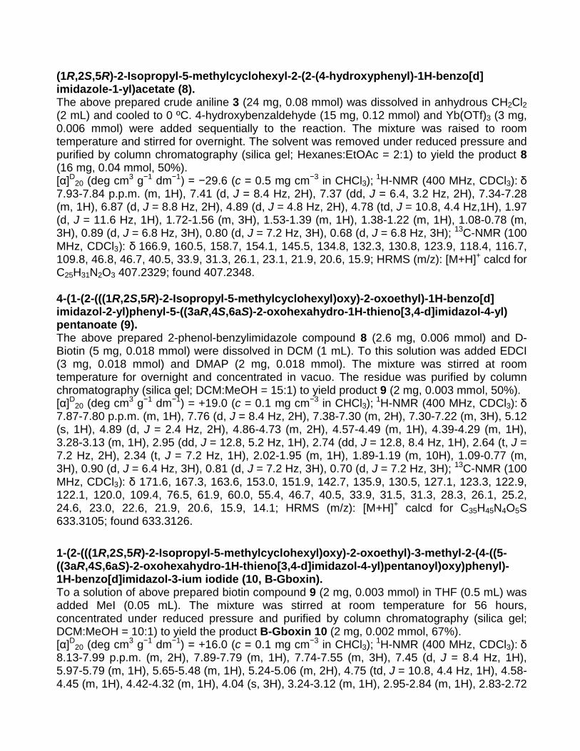
spectra were recorded in CDCl3 at ambient temperature on the same spectrometer at 100 MHz with the central peak of CDCl3 (dC = 77.0 ppm) as the internal reference. HRMS were performed on a Shimadzu IT-TOF. All synthetic compounds exhibited >95% purity as determined by1H-NMR and 13C-NMR or LC-MS analysis performed on an Agilent 1100 HPLC system using an Eclipse XDB-C18 column (4.6 Å~ 150 mm, 5 μm; Agilent) that was coupled to an Agilent G1956A (or 6120) ESI mass spectrometer run in the positive mode with a scan range of 100 to 800 (or 1000) m/z. Liquid chromatography was carried out at a flow rate of 0.5 mL/min at 30 °C with a 5 μL injection volume, using the gradient elution with aqueous acetonitrile containing 0.1% formic acid: 20–50% over 10 min followed by 50–90% for the next 10 min. DCC = N, N'-Dicyclohexylcarbodiimide, EDC = N-Ethylcarbodiimide hydrochloride, DMAP = 4-(Dimethylamino)pyridine, DMF = N,N-Dimethylformamide, DIPEA = N,N-Diisopropylethylamine, DIBAL-H = Diisobutylaluminium hydride, Py = Pyridine, THF = Tetrahydrofuran. Scheme 1. Synthesis of Gboxin, B-Gboxin, C-Gboxin, and S-Gboxin
F
NO2
a, b
Cl
O
ONHR
O
ON
N
O
ON
N
O
ON
N
ON3
O
ON
N
ON3
I
1 2: R=NO2
3: R=NH2
c
d
4
e
f
O
ON
N
F3C
O
ON
N
F3C
I
5 (Gboxin)
6 7 (S-Gboxin)
10 (B-Gboxin)
h
O OMe
OHN3
11
O OMe
ON3
TIPS
12
O H
ON3
TIPS
13 16 (C-Gboxin)
g
g3j k
O
ON
N
OH
O
ON
N
O Biotin
8 9
I
O
ON
N
O
O
S
NHHN
O
H
H
gi
R14: R=TIPS
15: R=Hm
3
l
Reagents and conditions: a) Glycine, NaOAc, MeOH, reflux; b) L-Menthol, DCC, DMAP, DCM, rt; c) H2, Pd/C, MeOH, rt; d) Propionaldehyde, Yb(OTf)3, DCM, rt; e) MeCl, THF, rt; f) 3-(Trifluoromethyl)benzaldehyde, Yb(OTf)3, DCM, rt; g) MeI, THF, rt; h) 4-Hydroxybenzaldehyde, Yb(OTf)3, DCM, rt; i) D-biotin Acid, EDCI, DMAP, DCM, rt; j) 3-Bromo-1-triisopropylsilanyl-prop-1-yne, K2CO3, DMF, rt; k) i) DIBAL-H, DCM, −78 ˚C, ii) Dess-Martin Periodinane, DCM, rt; l) Yb(OTf)3, DCM, rt; m) Bu4NF, THF, rt; (1R,2S,5R)-2-Isopropyl-5-methylcyclohexyl (2-nitrophenyl)glycinate (2). To a solution of 1-fluoro-2-nitrobenzene 1 (3.38 g, 24 mmol) in 30 mL of MeOH was added glycine (1.5 g, 20 mmol) and NaOAc (4.92 g, 60 mmol). This solution was stirred under reflux for overnight, cooled to room temperature. The solvent was concentrated under reduced pressure. The residue was adjusted to pH=1 using 1N HCl, extracted with EtOAc (3 x 30

mL), washed with brine (3 x 8 mL), dried over with anhydrous Na2SO4 and filtrated. The solvent was concentrated under reduced pressure to yield the crude product 2-((2-nitrophenyl)amino) acetic acid. To a solution of the above prepared acid (320mg, 1.6 mmol) and L-Menthol (382 mg, 2.4 mmol) in DCM (20 mL) was added DCC (494 mg, 2.4 mmol) and 4-(dimethylamino)pyridine (DMAP) (24 mg, 0.2 mmol) at room temperature. This solution was stirred at room temperature for overnight. The reaction mixture was diluted with EtOAc (80 mL), washed with brine (3 x 8 mL), dried over with Na2SO4 and filtered. The solvent was removed in vacuo. The residue was purified by column chromatography (silica gel; Hexanes:EtOAc = 4:1) yielding the compound 2 (450 mg, 1.34 mmol, 84%). [α]D20 (deg cm3 g−1 dm−1) = −92.9 (c = 1.3 mg cm−3 in CHCl3); 1H-NMR (400 MHz, CDCl3): δ 8.41 p.p.m. (t, J = 4.4 Hz,1H), 8.19 (dd, J = 8.8, 1.6 Hz, 1H), 7.51-7.36 (m, 1H), 6.81-6.61 (m, 2H), 4.80 (td, J = 11.2, 4.4 Hz, 1H), 4.07 (dd, J = 5.2, 0.8 Hz, 2H), 2.03-1.85 (m, 1H), 1.85-1.73 (m, 1H), 1.73-1.62 (m, 2H), 1.59-1.35 (m, 2H), 1.35-1.24 (m, 1H), 1.13-0.95 (m, 2H), 0.90 (d, J = 6.8 Hz, 3H), 0.87 (d, J = 7.2 Hz, 3H), 0.73 (d, J = 7.2 Hz, 3H); 13C-NMR (100 MHz, CDCl3): δ 168.7, 144.1, 136.1, 132.6, 126.93, 116.1, 113.6, 76.0, 46.9, 45.1, 40.7, 34.0, 31.3, 26.2, 23.3, 21.9, 20.7, 16.2; MS (m/z): [M+H]+ calcd for C18H28N2O4 335.1; found 335.1. (1R,2S,5R)-2-Isopropyl-5-methylcyclohexyl (2-aminophenyl)glycinate (3). To a solution of above prepared compound 2 (40 mg, 0.12 mmol) in 3 mL of MeOH was added 5 mg of 5% Pd/C. This solution was stirred at room temperature under H2 for 3h, filtered through a small column and concentrated in vacuo yielding the crude product 3 which was pure enough to be used in the following experiments. A small amount of compound was purified by column chromatography (silica gel; Hexanes:EtOAc = 4:1) for characterization. [α]D20 (deg cm3 g−1 dm−1) = −57.7 (c = 1.7 mg cm−3 in CHCl3); 1H-NMR (400 MHz, CDCl3): δ
6.81 p.p.m. (t, J = 7.2 Hz, 1H), 6.77-6.66 (m, 2H), 6.55 (d, J = 7.6 Hz, 1H), 4.79 (td, J = 10.8, 4.4 Hz, 1H), 3.89 (s, 2H), 3.81 (bs, 2H), 2.02 (d, J = 11.8 Hz, 1H), 1.88-1.76 (m, 1H), 1.76-1.59 (m, 2H), 1.59-1.44 (m, 1H), 1.40 (t, J = 12.0 Hz, 1H), 1.18-0.78 (m, 3H), 0.91 (d, J = 6.8 Hz, 3H), 0.88 (d, J = 7.2 Hz, 3H), 0.75 (d, J = 6.8 Hz, 3H); 13C-NMR (100 MHz, CDCl3): δ
171.0, 136.6, 134.1, 120.8, 119.4, 116.9, 112.3, 75.3, 46.9, 46.5, 40.8, 34.1, 31.3, 26.2, 23.4, 21.9, 20.7, 16.2; MS (m/z): [M+H]+ calcd for C18H29N2O2 305.1; found 305.2. (1R,2S,5R)-2-Isopropyl-5-methylcyclohexyl2-(2-ethyl-1H-benzoimidazole-1-yl)acetate (4). The above prepared crude aniline 3 (20 mg, 0.065 mmol) was dissolved in anhydrous CH2Cl2 (2 mL) and cooled to 0 ˚C. Propionaldehyde (7 mg, 0.13 mmol) and Yb(OTf)3 (3 mg, 0.006 mmol) were added sequentially to the reaction. The mixture was raised to room temperature and stirred for overnight. The solvent was removed under reduced pressure and purified by column chromatography (silica gel; Hexanes:EtOAc = 4:1) to yield the product 4 (14 mg, 0.04 mmol, 63%). [α]D20 (deg cm3 g−1 dm−1) = −29.8 (c = 0.4 mg cm−3 in CHCl3); 1H-NMR (400 MHz, CDCl3): δ 7.80-7.69 p.p.m. (m, 1H), 7.31-7.15 (m, 3H), 4.80 (s, 2H), 4.71 (td, J = 10.8, 4.4 Hz, 1H), 2.86 (q, J = 7.6 Hz, 2H), 1.96 (d, J = 12.0 Hz, 1H), 1.75-1.57 (m, 3H), 1.57-1.37 (m, 1H), 1.47 (t, J = 7.6 Hz, 3H), 1.29-1.18 (m, 2H), 1.07-0.78 (m, 2H), 0.88 (d, J = 6.4 Hz, 3H), 0.76 (d, J = 6.8 Hz, 3H), 0.64 (d, J = 6.8 Hz, 3H); 13C-NMR (100 MHz, CDCl3): δ 167.0, 156.1, 142.4, 135.2, 122.4, 122.1, 119.3, 108.5, 76.4, 46.7, 45.1, 40.5, 33.9, 31.3, 26.1, 23.1, 21.9, 20.6, 20.5, 16.0, 11.5; HRMS (m/z): [M+H]+ calcd for C21H31N2O2 343.2380; found 343.2393.

2-Ethyl-1-(2-(((1R,2S,5R)-2-isopropyl-5-methylcyclohexyl)oxy)-2-oxoethyl) -3-methyl-1H-benzo[d]imidazol-3-ium chloride (5, Gboxin). To a solution of above prepared compound 4 (10 mg, 0.03 mmol) in THF (1 mL) was added MeCl in THF (1 mL). This solution was stirred at room temperature for 48 hours, concentrated under reduced pressure and purified by column chromatography (silica gel; Hexanes:EtOAc = 2:1) to yield the product Gboxin 5 (6 mg, 0.015 mmol, 50%). [α]D20 (deg cm3 g−1 dm−1) = −26.0 (c = 0.2 mg cm−3 in CHCl3); 1H-NMR (400 MHz, CDCl3): δ 7.71-7.65 p.p.m. (m, 1H), 7.65-7.57 (m, 2H), 7.55-7.49 (m, 1H), 5.61 (d, J = 18.4 Hz, 1H), 5.53 (d, J = 18.4 Hz, 1H), 4.79 (dt, J = 11.2, 4.4 Hz, 1H), 4.14 (s, 3H), 3.77-3.55 (m, 2H), 2.03-1.87 (m, 1H), 1.86-1.52 (m, 3H), 1.52-1.22 (m, 2H), 1.39 (t, J = 7.6 Hz, 3H), 1.18-0.97 (m, 3H), 0.91 (d, J = 6.8 Hz, 3H), 0.90 (d, J = 7.2 Hz, 3H), 0.72 (d, J = 6.8 Hz, 3H); 13C-NMR (100 MHz, CDCl3): δ 166.0, 156.5, 131.5, 131.3, 127.1, 126.9, 112.3, 112.1, 77.7, 47.4, 46.7, 40.5, 33.8, 32.3, 31.4, 26.3, 23.1, 21.8, 20.7, 19.3, 16.0, 11.0; HRMS (m/z): [M-I]+ calcd for C22H33N2O2 357.2537, found 357.2564. (1R,2S,5R)-2-Isopropyl-5-methylcyclohexyl-2-(2-(3(trifluoromethyl)phenyl)-1H-benzo [d]imidazol-1-yl)acetate (6). The above prepared crude aniline 3 (20 mg, 0.065 mmol) was dissolved in anhydrous CH2Cl2 (2 mL) and cooled to 0 ºC. 3-(trifluoromethyl)benzaldehyde (23 mg, 0.13 mmol) and Yb(OTf)3 (3 mg, 0.006 mmol) were added sequentially to the reaction. The mixture was raised to room temperature and stirred for overnight. The solvent was removed under reduced pressure and purified by column chromatography (silica gel; Hexanes:EtOAc = 4:1) to yield the product 6 (16 mg, 0.03 mmol, 53%). [α]D20 (deg cm3 g−1 dm−1) = −29.9 (c = 0.3 mg cm−3 in CHCl3); 1H-NMR (400 MHz, CDCl3): δ
8.04 p.p.m. (s, 1H), 7.93 (d, J = 7.6 Hz, 1H), 7.89-7.82 (m, 1H), 7.79 (d, J = 8.0 Hz, 1H), 7.66 (t, J = 7.6 Hz, 1H), 7.40-7.33 (m, 2H), 7.33-7.29 (m, 1H), 4.87 (d, J = 2.4 Hz, 2H), 4.77 (td, J = 10.8, 4.4 Hz, 1H), 2.00-1.91 (m, 1H), 1.73-1.58 (m, 2H), 1.58-1.39 (m, 2H), 1.34-1.21 (m, 1H), 1.05-0.75 (m, 3H), 0.90 (d, J = 6.8 Hz, 3H), 0.77 (d, J = 7.2 Hz, 3H), 0.66 (d, J = 7.2 Hz, 3H); 13C-NMR (100 MHz, CDCl3): δ 167.0, 152.3, 142.8, 136.0, 132.5, 131.6, 131.3, 130.7, 129.4, 126.7, 126.3 (q, J = 4.2 Hz, 1C), 123.7, 123.2, 120.3, 109.5, 46.7, 46.7, 40.5, 33.9, 31.3, 26.1, 23.1, 21.8, 20.5, 15.9; MS (m/z): [M+H]+ calcd for C26H31F3N2O2 459.2; found 459.2. 1-(2-(((1R,2S,5R)-2-Isopropyl-5-methylcyclohexyl)oxy)-2-oxoethyl)-3-methyl-2-(3-(tri fluoromethyl)phenyl)-1H-benzo[d]imidazol-3-ium iodide (7, S-Gboxin). To a solution of above prepared compound 6 (16 mg, 0.035 mmol) in THF (1 mL) was added MeI (0.1 mL). This solution was stirred at room temperature for 48 hours, concentrated under reduced pressure and purified by column chromatography (silica gel; Hexanes:EtOAc = 2:1) to yield the product S-Gboxin 7 (12 mg, 0.02 mmol, 57%). [α]D20 (deg cm3 g−1 dm−1) = −38.6 (c = 0.3 mg cm−3 in CHCl3); 1H-NMR (400 MHz, CDCl3): δ
8.72-8.56 p.p.m. (m, 1H), 8.12-8.01 (m, 1H), 7.99 (d, J = 8.0 Hz, 1H), 7.94-7.82 (m, 2H), 7.74-7.57 (m, 3H), 5.25-4.91 (m, 2H), 4.71 (td, J = 10.8, 4.4 Hz, 1H), 3.99 (s, 3H), 1.93-1.78 (m, 1H), 1.70-1.48 (m, 3H), 1.48-1.26 (m, 2H), 1.06-0.90 (m, 2H), 0.90-0.80 (m, 1H), 0.87 (d, J = 6.8 Hz, 3H), 0.84-0.72 (m, 3H), 0.63 (d, J = 6.4 Hz, 3H); 13C-NMR (100 MHz, CDCl3): δ 165.4, 149.4, 135.8, 132.1 (d, J = 33.4 Hz, 1C), 131.7 (d, J = 33.4 Hz, 1C), 131.0, 130.3 (q, J = 3.3 Hz, 1C), 128.0 (d, J = 20.4 Hz, 1C), 127.3, 124.3, 121.5, 121.2, 113.9, 112.8, 77.9, 48.8, 46.5, 40.3, 34.4, 33.7, 31.3, 26.3, 23.1, 21.7, 20.5, 16.0.; HRMS (m/z): [M-I]+ calcd for C27H32F3N2O2 473.2410; found 473.2434.
(1R,2S,5R)-2-Isopropyl-5-methylcyclohexyl-2-(2-(4-hydroxyphenyl)-1H-benzo[d] imidazole-1-yl)acetate (8). The above prepared crude aniline 3 (24 mg, 0.08 mmol) was dissolved in anhydrous CH2Cl2 (2 mL) and cooled to 0 ºC. 4-hydroxybenzaldehyde (15 mg, 0.12 mmol) and Yb(OTf)3 (3 mg, 0.006 mmol) were added sequentially to the reaction. The mixture was raised to room temperature and stirred for overnight. The solvent was removed under reduced pressure and purified by column chromatography (silica gel; Hexanes:EtOAc = 2:1) to yield the product 8 (16 mg, 0.04 mmol, 50%). [α]D20 (deg cm3 g−1 dm−1) = −29.6 (c = 0.5 mg cm−3 in CHCl3); 1H-NMR (400 MHz, CDCl3): δ
7.93-7.84 p.p.m. (m, 1H), 7.41 (d, J = 8.4 Hz, 2H), 7.37 (dd, J = 6.4, 3.2 Hz, 2H), 7.34-7.28 (m, 1H), 6.87 (d, J = 8.8 Hz, 2H), 4.89 (d, J = 4.8 Hz, 2H), 4.78 (td, J = 10.8, 4.4 Hz,1H), 1.97 (d, J = 11.6 Hz, 1H), 1.72-1.56 (m, 3H), 1.53-1.39 (m, 1H), 1.38-1.22 (m, 1H), 1.08-0.78 (m, 3H), 0.89 (d, J = 6.8 Hz, 3H), 0.80 (d, J = 7.2 Hz, 3H), 0.68 (d, J = 6.8 Hz, 3H); 13C-NMR (100 MHz, CDCl3): δ 166.9, 160.5, 158.7, 154.1, 145.5, 134.8, 132.3, 130.8, 123.9, 118.4, 116.7, 109.8, 46.8, 46.7, 40.5, 33.9, 31.3, 26.1, 23.1, 21.9, 20.6, 15.9; HRMS (m/z): [M+H]+ calcd for C25H31N2O3 407.2329; found 407.2348. 4-(1-(2-(((1R,2S,5R)-2-Isopropyl-5-methylcyclohexyl)oxy)-2-oxoethyl)-1H-benzo[d] imidazol-2-yl)phenyl-5-((3aR,4S,6aS)-2-oxohexahydro-1H-thieno[3,4-d]imidazol-4-yl) pentanoate (9). The above prepared 2-phenol-benzylimidazole compound 8 (2.6 mg, 0.006 mmol) and D-Biotin (5 mg, 0.018 mmol) were dissolved in DCM (1 mL). To this solution was added EDCI (3 mg, 0.018 mmol) and DMAP (2 mg, 0.018 mmol). The mixture was stirred at room temperature for overnight and concentrated in vacuo. The residue was purified by column chromatography (silica gel; DCM:MeOH = 15:1) to yield product 9 (2 mg, 0.003 mmol, 50%). [α]D20 (deg cm3 g−1 dm−1) = +19.0 (c = 0.1 mg cm−3 in CHCl3); 1H-NMR (400 MHz, CDCl3): δ
7.87-7.80 p.p.m. (m, 1H), 7.76 (d, J = 8.4 Hz, 2H), 7.38-7.30 (m, 2H), 7.30-7.22 (m, 3H), 5.12 (s, 1H), 4.89 (d, J = 2.4 Hz, 2H), 4.86-4.73 (m, 2H), 4.57-4.49 (m, 1H), 4.39-4.29 (m, 1H), 3.28-3.13 (m, 1H), 2.95 (dd, J = 12.8, 5.2 Hz, 1H), 2.74 (dd, J = 12.8, 8.4 Hz, 1H), 2.64 (t, J = 7.2 Hz, 2H), 2.34 (t, J = 7.2 Hz, 1H), 2.02-1.95 (m, 1H), 1.89-1.19 (m, 10H), 1.09-0.77 (m, 3H), 0.90 (d, J = 6.4 Hz, 3H), 0.81 (d, J = 7.2 Hz, 3H), 0.70 (d, J = 7.2 Hz, 3H); 13C-NMR (100 MHz, CDCl3): δ 171.6, 167.3, 163.6, 153.0, 151.9, 142.7, 135.9, 130.5, 127.1, 123.3, 122.9, 122.1, 120.0, 109.4, 76.5, 61.9, 60.0, 55.4, 46.7, 40.5, 33.9, 31.5, 31.3, 28.3, 26.1, 25.2, 24.6, 23.0, 22.6, 21.9, 20.6, 15.9, 14.1; HRMS (m/z): [M+H]+ calcd for C35H45N4O5S 633.3105; found 633.3126. 1-(2-(((1R,2S,5R)-2-Isopropyl-5-methylcyclohexyl)oxy)-2-oxoethyl)-3-methyl-2-(4-((5-((3aR,4S,6aS)-2-oxohexahydro-1H-thieno[3,4-d]imidazol-4-yl)pentanoyl)oxy)phenyl)-1H-benzo[d]imidazol-3-ium iodide (10, B-Gboxin). To a solution of above prepared biotin compound 9 (2 mg, 0.003 mmol) in THF (0.5 mL) was added MeI (0.05 mL). The mixture was stirred at room temperature for 56 hours, concentrated under reduced pressure and purified by column chromatography (silica gel; DCM:MeOH = 10:1) to yield the product B-Gboxin 10 (2 mg, 0.002 mmol, 67%). [α]D20 (deg cm3 g−1 dm−1) = +16.0 (c = 0.1 mg cm−3 in CHCl3); 1H-NMR (400 MHz, CDCl3): δ
8.13-7.99 p.p.m. (m, 2H), 7.89-7.79 (m, 1H), 7.74-7.55 (m, 3H), 7.45 (d, J = 8.4 Hz, 1H), 5.97-5.79 (m, 1H), 5.65-5.48 (m, 1H), 5.24-5.06 (m, 2H), 4.75 (td, J = 10.8, 4.4 Hz, 1H), 4.58-4.45 (m, 1H), 4.42-4.32 (m, 1H), 4.04 (s, 3H), 3.24-3.12 (m, 1H), 2.95-2.84 (m, 1H), 2.83-2.72
(m, 1H), 2.66 (t, J = 7.6 Hz, 2H), 1.96-1.10 (m, 12H), 1.08-0.93 (m, 1H), 0.89 (d, J = 6.4 Hz, 3H), 0.84 (d, J = 6.8 Hz, 2H), 0.67 (d, J = 7.2 Hz, 3H); 13C-NMR (100 MHz, CDCl3): δ 171.1, 165.8, 154.6, 151.5, 150.7, 135.7, 132.9, 131.9, 131.7, 128.1, 127.7, 125.5, 123.4, 117.2, 113.3, 112.8, 77.2, 46.7, 46.1, 40.5, 34.2, 33.9, 33.8, 31.4, 30.3, 29.7, 28.4, 26.3, 24.5, 23.1, 21.8, 21.1, 20.6, 16.1, 8.5; HRMS (m/z): [M-I]+ calcd for C36H47N4O5S 647.3261; found 647.3280. Methyl 3-azido-4-((3-(triisopropylsilyl)prop-2-yn-1-yl)oxy)benzoate (12). To a solution of 3-azido-4-hydroxybenzoate 11 (300 mg, 1.55 mmol) in anhydrous DMF (3.1 mL) at room temperature was added K2CO3 (644 mg, 4.66 mmol) and TIPS-protected propargyl bromide (641 mg, 2.33 mmol). The resulting mixture was stirred at room temperature for 4 h. The reaction mixture was passed through a pad of celite and washed with EtOAc. The solvent was removed under reduced pressure, and the resulting residue was purified by flash chromatography (silica gel, gradient elution, 0-6% EtOAc in hexanes) to afford the product 12 (575 mg, 1.48 mmol, 96%). 1H-NMR (400 MHz, CDCl3): δ 7.74 p.p.m. (dd, J = 8.8, 2.4 Hz, 1H), 7.64 (d, J = 2.0 Hz, 1H), 7.08 (d, J = 8.8 Hz, 1H), 4.81 (s, 2H), 3.84 (s, 3H), 0.97 (s, 21H); 13C-NMR (100 MHz, CDCl3): δ 165.9, 153.4, 129.1, 127.2, 123.9, 121.7, 113.5, 100.2, 90.9, 57.6, 52.0, 18.4 (6C), 11.0 (3C); MS (m/z): [M+H]+ calcd for C20H31N3O3Si 388.2; found 388.2. 3-Azido-4-((3-(triisopropylsilyl)prop-2-yn-1-yl)oxy)benzaldehyde (13). To a solution of the above benzoate ester 12 (675 mg, 1.74 mmol) in anhydrous CH2Cl2 (5.8 mL) at −78 ºC was added Dibal-H (10 mL, 10 mmol) over the course of 1 h. The reaction mixture was slowly raised to room temperature overnight, then cooled to 0 ºC and quenched with excess MeOH. After stirring for 10 min, the mixture was poured into a flask containing a saturated aqueous Na/K-tartrate solution, and stirred vigorously at room temperature for 1 h. The mixture was extracted with EtOAc (3 x 10 mL) and the organic extract was washed with brine and dried over with anhydrous MgSO4. After filtration, the solvent was removed under reduced pressure, and the resulting residue was purified by flash chromatography on silica gel (silica gel, gradient elution, 0-25% EtOAc in hexanes) to afford the benzyl alcohol as a colorless gel (288 mg, 0.80 mmol, 46%). The crude alcohol was dissolved in anhydrous CH2Cl2 (2.67 mL) and added to a solution of Dess-Martin periodinane (510 mg, 1.2 mmol) in anhydrous CH2Cl2 (1.0 mL) at 0 ºC. The resulting mixture was raised to rt and stirred for 30 min. The reaction was quenched with a saturated aqueous NaHCO3 solution at 0 ºC and extracted with Et2O. The organic extract was washed with brine and dried over anhydrous MgSO4, and filtered. The solvent was removed under reduced pressure, and the resulting residue was purified by flash chromatography (silica gel, gradient elution, 0-15% EtOAc in hexanes) to afford the aldehyde 13 (205 mg, 0.57 mmol, 72%). 1H-NMR (500 MHz, CDCl3): δ 9.84 p.p.m. (s, 1H), 7.60 (dd, J = 8.4, 2.0 Hz, 1H), 7.52 (d, J = 2.0 Hz, 1H), 7.21 (d, J = 8.4 Hz, 1H), 4.88 (s, 2H), 1.00 (br, 21H); 13C-NMR (125 MHz, CDCl3): δ 190.1, 154.6, 130.8, 130.2, 128.3, 120.7, 113.9, 99.8, 91.4, 57.7, 18.4 (6C), 11.0 (3C); MS (m/z): [M+H]+ calcd for C19H29N3O2Si 358.2; found 358.2. (1R,2S,5R)-2-Isopropyl-5-methylcyclohexyl2-(2-(3-azido-4-((3-(triisopro- pylsilyl)prop-2-yn-1-yl)oxy)phenyl)-1H-benzo[d]imidazol-1-yl)acetate (14). To a solution of above prepared aniline 3 (28 mg, 0.09 mmol) in anhydrous CH2Cl2 (1.0 mL) at 0 ºC was added the above prepared benzaldehyde 13 (40 mg, 0.11 mmol) and Yb(OTf)3 (6 mg, 0.009 mmol). The temperature was raised to room temperature and the mixture was

stirred overnight, followed by removal of the solvent in vacuo. Purification of the residue by flash chromatography (silica gel, gradient elution, 0-30% EtOAc in hexanes) provided the benzimidazole product 14 (29.4 mg, 0.046 mmol, 50%). [α]D20 (deg cm3 g−1 dm−1) = −14.3 (c = 0.4 mg cm−3 in CHCl3); 1H-NMR (400 MHz, CDCl3): δ 7.86-7.80 p.p.m. (m, 1H), 7.44 (s, 1H), 7.47-7.41 (m, 1H), 7.37-7.30 (m, 2H), 7.30-7.25 (m, 1H), 7.23 (d, J = 8.4 Hz, 1H), 4.89 (s, 2H), 4.86 (d, J = 4.4 Hz, 2H), 4.79 (ddd, J = 11.6, 11.2, 4.4 Hz, 1H), 2.00 (d, J = 11.2 Hz, 1H), 1.69-1.64 (m, 3H), 1.59 (ddd, J = 7.2, 7.2, 2.0 Hz, 1H), 1.53-1.41 (m, 1H), 1.33-1.26 (m, 3H), 1.05 (s, 21H), 0.92 (d, J = 6.4 Hz, 3H), 0.81 (d, J = 7.2 Hz, 3H), 0.69 (d, J = 7.2 Hz, 3H); 13C-NMR (100 MHz, CDCl3): δ 167.4, 152.9, 151.3, 142.8, 136.0, 129.9, 126.2, 123.7, 123.3, 123.0, 121.7, 120.0, 114.5, 109.4, 100.4, 90.9, 76.6, 57.8, 46.8 (2C), 40.6, 34.0, 31.4, 26.1, 23.1, 21.9, 20.7, 18.5 (6C), 16.0, 11.0 (3C); MS (m/z): [M+H]+ calcd for C37H52N5O3Si 642.4; found 642.4. (1R,2S,5R)-2-Isopropyl-5-methylcyclohexyl-2-(2-(3-azido-4-(prop-2-yn-1-yloxy)phenyl) -1H-benzo[d]imidazol-1-yl)acetate (15). To a solution of the above prepared TIPS-protected alkyne 14 (23 mg, 0.036 mmol) in anhydrous THF (0.36 mL) at 0 ºC was added TBAF (0.05 mL, 1M solution in THF). The reaction was stirred for 2 h and slowly raised to rt. The reaction was quenched with saturated aqueous NaHCO3 solution (2 mL). The mixture was extracted with EtOAc (3 x 10 mL), washed with brine and dried over anhydrous MgSO4. After filtration, the solvent was removed under reduced pressure, and the resulting residue was purified by flash chromatography (silica gel, gradient elution, 0-35% EtOAc in hexanes) to afford the product 15 (9 mg, 0.02 mmol, 53%). [α]D20 (deg cm3 g−1 dm−1) = −29.4 (c = 0.8 mg cm−3 in CHCl3); 1H-NMR (400 MHz, CDCl3): δ 7.87-7.79 p.p.m. (m, 1H), 7.49-7.42 (m, 2H), 7.35-7.27 (m, 3H), 7.15 (d, J = 9.2 Hz, 1H), 4.87 (d, J = 3.6 Hz, 2H), 4.84 (d, J = 2.4 Hz, 2H), 4.78 (ddd, J = 11.2, 10.8, 4.8 Hz, 1H), 2.59 (t, J = 2.4 Hz, 1H), 1.96-1.87 (m, 1H), 1.72-1.53 (m, 4H), 1.53-1.40 (m, 1H), 1.35-1.22 (m, 2H), 1.07-0.93 (m, 1H), 0.90 (d, J = 6.4 Hz, 3H), 0.80 (d, J = 7.2 Hz, 3H), 0.68 (d, J = 7.2 Hz, 3H); 13C-NMR (100 MHz, CDCl3): δ 167.4, 152.7, 151.0, 142.8, 136.0, 129.9, 126.4, 124.0, 123.3, 123.0, 121.6, 120.1, 114.0, 109.4, 76.76, 76,75, 56.8, 46.80, 46.76, 40.6, 34.0, 31.4, 26.2, 23.1, 21.9, 20.7, 16.0; MS (m/z): [M+H]+ calcd for C28H32N5O3 486.2; found 486.0. 2-(3-Azido-4-(prop-2-yn-1-yloxy)phenyl)-1-(2-(((1R,2S,5R)-2-isopropyl-5-methylcyclo-hexyl)oxy)-2-oxoethyl)-3-methyl-1H-benzo[d]imidazol-3-ium iodide (16, C-Gboxin) A solution of the above benzylimidazole 15 (18 mg, 0.037 mmol) in MeI (1.0 mL) was heated at 50 ºC and stirred for 3 h. The excess MeI was removed in vacuo. The crude material was purified by preparative TLC (silica gel, 5% MeOH in CH2Cl2) to afford the benzimidazolinium salt C-Gboxin 16 as yellow amorphous solid (13.7 mg, 0.022 mmol, 60%). [α]D20 (deg cm3 g−1 dm−1) = −22.0 (c = 0.8 mg cm−3 in CHCl3); 1H-NMR (400 MHz, CDCl3): δ 8.13 p.p.m. (d, J = 8.8 Hz, 1H), 7.82-7.74 (m, 1H), 7.72-7.63 (m, 2H), 7.62-7.55 (m, 1H), 7.45-7.38 (m, 1H), 7.36 (d, J = 8.8 Hz, 1H), 5.14 (s, 2H), 4.90 (d, J = 2.4 Hz, 2H), 4.78 (dt, J = 10.8, 4.4 Hz, 1H), 4.02 (s, 3H), 2.66 (t, J = 2.4 Hz, 1H), 1.96-1.87 (m, 1H), 1.72-1.53 (m, 4H), 1.53-1.40 (m, 1H), 1.35-1.22 (m, 2H), 1.07-0.93 (m, 1H), 0.92 (d, J = 6.8 Hz, 3H), 0.87 (d, J = 7.2 Hz, 3H), 0.71 (d, J = 7.2 Hz, 3H); 13C-NMR (100 MHz, CDCl3): δ 166.1, 154.2, 150.6, 132.1, 131.9, 130.9, 130.1, 128.1, 127.9, 122.9, 114.8, 113.6, 113.5, 113.0, 78.0, 77.9, 76.7, 57.2, 49.0, 46.9, 40.8, 34.1, 34.0, 31.6, 26.7, 23.4, 22.1, 20.9, 16.3; HRMS (m/z): [M-I]+ calcd for C29H34N5O3 500.2656, found 500.2686.
B-Gboxin Pull-down Assay and Mass Spectrometry, For pulldown assays, cells were treated with Gboxin (1 μM) or B-Gboxin (10 μM) for 1 hour or as indicated in figure legends. In experiments for Fig. 3a, cells were treated with Gboxin (2 μM, 6 hours), B-Gboxin (10 μM, 6 hours), and Gboxin (30 μM, 1 hour) followed by B-Gboxin (10 μM, 6 hour). In experiments for Fig. 3b and Extended Data Fig. 3f, cells were treated with Gboxin (1 μM, 1 hour), B-Gboxin (10 μM, 1 hour), and Gboxin (30 μM, 30 minutes) followed by B-Gboxin (10 μM, 1 hour). After treatments, cells were washed once in cold PBS and lysed for 1 hour in the lysis buffer (Tris Hcl at pH 8.0 12 mM; Glycerol 10%; NaCl 137 mM; EDTA 1 mM) with protease inhibitor cocktail (Millipore) in the presence of 1% NP40 or other detergents as described in particular experiments. Cell lysates were then spun at 12,000 rpm for 10 minutes at 4 degrees. Supernatant was transferred to a new tube and protein concentration was measured. 1,000 μg of cell lysate at 1 μg/μL was then used to incubate with 40 μL Neutravidin Agarose (Thermo) for at least 4 hours. Agarose was then spun and washed for 4 times. Binding proteins were eluted by 2x LDS sample buffer (Thermo) with 5% beta-mercaptoethanol. An aliquot of the pulldown elution was used for protein SDS gel running and then stained with silver stain kit (Bio-Rad) or used for western blot. For protein identification with mass spectrometry, the whole elution of each pulldown sample was used. In some experiments, purified mitochondria from cells by mitochondrial isolation kit for cultured cells (Thermo fisher) was used for pulldown assay. In experiment for Extended Data Fig. 3h, HTS cell lysate at 10 μg/μL was treated with high salt (NaCl 300 mM), Urea (3 M) or SDS (0.2% and 0.5%) for 30 minutes. Treated lysate was diluted in 1% NP40 lysis buffer to 1 μg/μL and then the same steps for pulldown were followed. LC-MS/MS analysis service provided by UT southwestern Proteomics core was used for protein identification from pulldown assays. Immunopreciptation and Assay for PH Dependent B-Gboxin and Protein Interactions For immunopreciptation, cell lysates were prepared as described for B-Gboxin pulldown assay. Instead of incubating with Neutravidin Agarose, 1,000 μg cell lysate was incubated with 2.5 μg antibody overnight. Next day, 40 μL protein G sepharose (Thermo) was used for immunoprecipitation. Binding proteins were then eluted with 2x LDS sample buffer (Thermo). For pH dependent B-Gboxin and protein interactions, HTS cell lysate at 1 μg/µL was incubated with B-Gboxin (2 μM) in buffer (Tris Hcl 12mM; Glycerol 10%; NaCl 137 mM; EDTA 1 mM) with a range of pH for 1 hour at 370C. Buffer pH was adjusted with NaOH. Western blot assay was used for detecting B-Gboxin interacting proteins with avidin-HRP. Immunoflouscence and Assay for C-Gboxin mitochondrial colocalization Cells treated as indicated in corresponding figure (CsA at 1.5 μM was added 30 minutes before C-Gboxin treatment) were placed approximately 3-4 inches below the stratalinker and exposed to 10 minutes of UVC radiation. Cell were then washed once with PBS and fixed with 4% PFA for 10 minutes at room temperature. C-Gboxin was probed by an Azide Fluor 545 (sigma) using Click-ItTM Cell Reaction Buffer kit (Thermo Fisher) according to manufacturer protocol. Mitochondria were stained with a Sdha antibody (Proteintech) followed by an anti-Rabbit second antibody tagged with FITC (Jackson labs). Pharmacokinetic assays For S9 fraction assay, compound was incubated with male ICR/CD-1 mouse S9 fractions (Celsis/In Vitro Technologies) and Phase I (NADPH Regenerating System) cofactors for 0-240 minutes. Reactions were quenched with 1 mL (1:1) of MeOH/IS/0.2% formic acid mixture, vortexed for 15 seconds, incubated at room temperature (RT) for 10 minutes and spun for 5 minutes at 2,400 rpm. Supernatant (1 mL) was then transferred to an eppendorf tube and spun

in a table top, chilled centrifuge for 5 minutes at 13,200 rpm. Supernatant (800 μL) was transferred to an HPLC vial (OxPhos/out insert) and analyzed by HPLC/MS. For hepatocyte assays compound was incubated with murine hepatocyte cells for 0-240 minutes. Cells were quenched (2:1) with 200 μL of MeOH/IS/0.2% formic acid, vortexed for 15 seconds and incubated at RT for 10 minutes. The eppendorf tubes were then spun in a table-top, chilled centrifuge for 5 minutes at 13,200 rpm. Supernatant (180 μL) was transferred to a 1.5 mL HPLC vial (OxPhos/insert) and analyzed by HPLC/MS. For plasma stability assay, compound was incubated with murine plasma and saline for 0-1,440 minutes. Reactions were quenched with 200 μL (1:2) of methanol containing 0.1% formic acid and 200 ng/mL IS (IS final concentration = 100 ng/mL). Samples were vortexed for 15 seconds, incubated at RT for 10 minutes and spun for 5 minutes at 13,200 rpm. Supernatant (1 mL) was then transferred to an eppendorf tube and spun in a table top, chilled centrifuge for 5 minutes at 13,200 rpm. Supernatant (800 μL) was transferred to an HPLC vial (OxPhos/out insert). Samples were analyzed by Qtrap 3200 mass spectrometer. For in vivo plasma and brain pharmacokinetic analysis, 12 female CD-1 mice (6 weeks) were administered Gboxin or S-Gboxin intraperitoneally at 10 mg/kg, 0.2 mL/mouse formulated as 5% DMSO, 10% Cremophor EL, and 85% water. Plasma was processed from whole blood by centrifugation of the ACD treated blood for 10 minutes at 10,000 rpm in a standard centrifuge. Brains were weighed and snap frozen in liquid nitrogen. Brain homogenates were prepared by mincing the brain tissue and homogenizing in a 3-fold volume of PBS (total volume of homogenate in mL = 4X weight in gram). 100 μL plasma or brain homogenate was mixed with 200 μL of methanol containing 0.1% formic acid and 100 ng/μL IS (final concentration of IS = 100 ng/mL). The samples were vortexed for 15 seconds, incubated at RT for 10 minutes and spun for 2 minutes at 13,200 rpm in a standard microcentrifuge. The supernatant was then analyzed by LC/MS/MS. Buffer A: Water + 0.1% formic acid; Buffer B: MeOH + 0.1% formic acid; flow rate 1.5 mL/min.; column Agilent C18 XDB column, 5 micron packing 50 X 4.6 mm size; 0-1.5 min 97% A, 1.5-2.5 min gradient to 100% B; 2.5-3.5 min 100% B, 3.5-3.6 min gradient to 97% A; 3.6-4.5 min 97% A; IS N-benzylbenzamide (transition 212.1 to 91.1); Compound transition 358.2 to 220.2. In vivo Test of S-Gboxin 24 female nude mice (6 to 9 weeks) were implanted with 200,000 HTS cells subcutaneously on both the left and right shoulders. 24 days post-implantation, the mice were administered 10 mg/kg S-Gboxin intraperitoneally, 0.2 mL/mouse formulated as 5% DMSO, 10% Cremophor EL, and 85% water. Plasma was processed from whole blood by centrifugation of the ACD treated blood for 10 minutes at 10,000 rpm in a standard centrifuge. Tumors were weighed and snap frozen in liquid nitrogen. Tumor homogenates were prepared by mincing the tumor tissue and homogenizing in a 3-fold volume of PBS (total volume of homogenate in mL = 4X weight in gram.) 100 μL plasma or brain homogenate was mixed with 200 μL of acetonitrile containing formic acid and an internal standard (final concentration of formic acid = 0.1%, IS = 25 ng/mL). The samples were vortexed for 15 seconds, incubated at RT for 10 minutes and spun at 13,200 rpm for 2 minutes in a standard microcentrifuge. The supernatant was then analyzed by LC/MS/MS. Buffer A: Water + 0.1% formic acid; Buffer B: MeOH + 0.1% formic acid; flow rate 1.5 mL/min; column Agilent C18 XDB column, 5 micron packing 50 X 4.6 mm in size; 0-1.5 min 3% B, 1.5-2.5 min gradient to 100% B; 2.5-3.5 min 100% B, 3.5-3.6 min gradient to 3% B; 3.6-4.5 min 3% B; IS N-benzylbenzamide (transition 212.1 to 91.1); Compound transition 474.2 to 336.3.

Evaluation of S-Gboxin in GBM Models All mouse experiments were performed in accordance with protocols approved by the Institutional Animal Care and Use Committees (IACUC) of at University of Texas Southwestern Medical Center and Memorial Sloan Kettering Cancer Center, and maximal tumor measurements in all experiments were under limits (20 x 20 x 20 mm3) permitted by IACUCs. For subcutaneous GBM models, human GBM cells (2x10⁵) in matrigel (Corning) or mouse GBM cells (1x10⁵) were subcutaneously injected into flanks of nude mice (6-9 weeks). After the indicated time periods as described in figure legends, S-Gboxin (formulated as 5% DMSO, 10% Cremophor EL, and 85% water) or vehicle was intraperitoneally (IP) injected daily and tumor growth was measured by tumor volume assessed every 2 days (W x L x H). For orthotopic GBM models, 6-9 week-old female wildtype mice (for mouse tumors) or nu/nu mice (for PDX) were anesthetized and injected with mouse GBM cells #1663 (2x10⁴ cells in 2 μL) or minced fresh tumor tissue (PDX-170620, 3 μL) or dissociated single cells from fresh tumor tissue (PDX-170404, 2x10⁴ cells in 2 μL). Tumor tissue or cells were injected at coordinates: -0.5 mm AP, 1.8 mm ML, and 3 mm DV with respect to the bregma. Osmotic minipumps (ALZET®, model 2006) were implanted for direct delivery of vehicle or S-Gboxin (formulated as 5% DMSO, 10% Cremophor EL, and 85% water) to the injection site through brain infusion kit (ALZET®) on indicated days (see corresponding figure legend) after intracranial tumor cell injection. The two independent PDXs are namely PDX-170620 (Tumor 1) and PDX-170404 (Tumor 2), both treatment naive at the time of surgery (see Supplementary Table 4 for mutation statute and other information) as obtained from MSK IMPACT56. Histology and Immunohistochemistry Tissues were fixed in 4% paraformaldehyde and paraffin embedded. H&E and immunohistochemistry staining was performed in paraffin-embedded sections. For immunohistochemistry, sections were blocked in 10% normal donkey serum prepared in PBS and 0.3% TritonX-100 for 1 hour. Sections were incubated overnight at 4°C with the following primary antibodies: GFAP (1:400; DAKO; Z-033429-2); Ki67 (1:1000; Novus Biologicals; NB500-170); Olig2 (1:400; Millipore/Chemicon; AB9610); and Nestin (1:500; Novus Biologycals; NB100-1604). On the next day sections were incubated with biotinylated secondary antibodies, followed by signal amplification and visualization with the avidin–biotin complex (ABC) system and DAB substrate (Vector Laboratories). Olympus BX43 microscope was used for imaging. For examining S-Gboxin effects on intracranial transplanted tumors, histologic images comparing vehicle or S-Gboxin treated tumor were taken at the same distance from the tips though which vehicle or S-Gboxin was delivered by minipumps. Statistical Analysis Representative results for biological replicates are shown for every figure except where specified otherwised in the figure legends. Data are presented as mean ± SD or mean ± SEM as specified in the figure legend. Statistical analysis was performed using software GraphPad Prism 7.0a. Statistical significance with exact p value was determined with the method used as indicated in corresponding figure legend.

Supplementary references 51 Alberts, B., Wilson, J. H. & Hunt, T. Molecular biology of the cell. 5th edn, (Garland Science,
2008). 52 Kwon, C. H. et al. Pten haploinsufficiency accelerates formation of high-grade astrocytomas.
Cancer Res 68, 3286-3294, doi:10.1158/0008-54/2.Can-07-6867 (2008). 53 Vogel, K. S. et al. Mouse tumor model for neurofibromatosis type 1. Science 286, 2176-2179
(1999). 54 Cho, W. S. & Messing, A. Properties of astrocytes cultured from GFAP over-expressing and GFAP
mutant mice. Exp Cell Res 315, 1260-1272, doi:10.1016/j.yexcr.2008.12.012 (2009). 55 Bonora, M. et al. Comprehensive analysis of mitochondrial permeability transition pore activity in
living cells using fluorescence-imaging-based techniques. Nat Protoc 11, 1067-1080, doi:10.1038/nprot.2016.064 (2016).
56 Cheng, D. T. et al. Memorial Sloan Kettering-Integrated Mutation Profiling of Actionable Cancer Targets (MSK-IMPACT) A Hybridization Capture-Based Next-Generation Sequencing Clinical Assay for Solid Tumor Molecular Oncology. J Mol Diagn 17, 251-264, doi:10.1016/j.jmoldx.2014.12.006 (2015).