arthroscopy after total hip replacement...
TRANSCRIPT

5
Arthroscopy after Total Hip Replacement Surgery
Cuéllar Ricardo1, Ponte Juan2, Esnal Edorta3 and Tey Marc4 1University Hospital Donostia (San Sebastián)
2Quirón Hospital (San Sebastián) 3Alto Deba Hospital (Mondragón)
4Dexeus University Hospital (Barcelona) Spain
1. Introduction
Hip arthroscopy has been available for some years. Although arthroscopy has not been as widely adopted in the hip joint as compared to its use in other joints, it is currently in a phase of rapid development. This is mainly due to the description of femoroacetabular impingement syndrome, where hip arthroscopy has proven to be a precious diagnostic and therapeutic tool. The development and improvement of the technique together with the instrumentation has allowed broadening its indications. The hip joint, is more difficult to access than other joints such as the knee and shoulder. This is due to its tight congruency, the degree of coverage of the ball and socket articular surfaces – the acetabulum extends beyond the equator of the femoral head - the powerful surrounding muscles and the proximity of important vessels and nerves. These anatomical features restrict the manoeuvrability of the arthroscopic instruments making hip arthroscopy a more demanding technique. With the techniques for the knee and shoulder joints already well established, we have witnessed the development of arthroscopy for the diagnosis and treatment of hip disorders over the last decade (Johnston et al., 2008; Kelly et al., 2003; Larson et al., 2009; Lubowitz & Poehling, 2006). Arthroscopic techniques have improved, increasing the surgical indications and achieving better outcomes (Byrd, 2006; Byrd & Jones, 2009; Larson & Giveans, 2009; Philippon, 2007a, 2007b). This procedure is performed in children and in adults for both diagnostic and therapeutic purposes, (Kocher et al., 2005; McCarthy & Lee, 2006; Parisien, 1988; Philippon et al., 2007b; Roy et al., 2009; Sampson, 2006), the most commonly treated disorder being femoroacetabular impingement syndrome (Philippon et al., 2007b). The first publication concerning visualization of the hip joint was by Burman in 1931, who reported his experience in 20 hips using a 4-mm arthroscope and the use of water to achieve joint distension (Burman, 1931). He described the anterior peritrochanteric portal, concluding that this was the best option for visualizing the hip joint. He concluded that the hip joint was not suitable for arthroscopy due to the inability to access and visualize what nowadays is known as the central compartment. Although there were some other reports in the intervening years, it is considered that Gross, in 1977, was the first person to describe the
www.intechopen.com

Modern Arthroscopy
80
clinical application of this approach and its therapeutic effect in hip diseases in children (Legg-Calvé-Perthes disease, congenital dislocation and epiphysiolysis). He used a 2.2-mm arthroscope and manual distraction. The use of traction was first described by Eriksson who employed forces of between 300 to 400 Newtons to distract the hip (Eriksson et al., 1986). The first description of the use of traction in the supine position was made by Byrd 8Byrd, 1994); while Glick was the first to report the use of the lateral decubitus position (Glick et al., 1987). Monllau published results of a study demonstrating that hip arthroscopy required instruments with a minimum length of 16 cm (Monllau et al., 2003). It is well known that following total hip replacement THR, pain disappears in approximately 95-98% of the cases, usually between 3 to 6 months up to a year after surgery. However, between 1 and 2% of patients refer persistence of pain. Although prosthesis components loosening is responsible for these complications in more than 90% of the cases, there are other potential causes of pain such as heterotopic ossification, muscle and tendon pain around the prosthesis, impingement and radiating back pain. In 1% of the cases the cause of pain remains unknown (Witvoët, 2001). There is a clear similarity between the aforementioned sources of ongoing pain after hip replacement and those reported in relation to persistence of pain after knee replacement. Since 1989, several authors have reported the use of arthroscopy as a diagnostic and therapeutic tool in painful complications of knee implants, Wasilewski being the earliest (Bocell et al., 1991; Johnson et al., 1990; Lawrence & Kan, 1992; Lucas et al., 1999; Markel et al., 1996; Scranton, 2001; Tzagarakis et al., 2001; Wasilewski & Frankl, 1989a, 1989b). The use of arthroscopy in selected patients with hip implants represents a step forward in diagnosis and possible treatment of painful, apparently well implanted prostheses (Cuéllar et al., 2009; McCarthy et al., 2009; Bajwa & Villar, 2011). In our hospital environment we have established an arthroscopic protocol for hip implant monitoring, similar to the approach we use in knee replacement patients and indicate arthroscopic surgery in cases in which, despite the prosthesis being apparently well implanted, patients continue to experience pain (Cuéllar et al., 2009).
2. Differential diagnosis
The differential diagnosis of pain following THR is wide and includes intrinsic and extrinsic causes to the implant. Septic or aseptic loosening is the cause of intrinsic complications in more than 90% of cases, but there are other potential causes such as stress fractures, mechanical failure or elasticity of the implant itself, subluxation or impingement (Bozic & Rubash, 2004; Smith & Rorabeck, 1999). Additionally there are other extrinsic causes of pain including lumbar radicular pain, neurogenic or vascular claudication (Beck, 2009), heterotopic ossification, trochanteritis or trochanteric non-union (Brown & Callaghan, 2008), peripheral nerve lesions (Malik et al., 2007), tendon and muscle pain around the implant (adductor and iliopsoas tendinitis, arthrofibrosis) (Hyman et al., 1999), femoral or inguinal hernia, and, more rarely, concomitant malignant conditions (Merkel et al., 1985). Finally, in around 1% of cases the source of the pain is never found. The protocol that we routinely use for differential diagnosis includes a detailed medical history and a thorough clinical examination, laboratory tests (FBC, ESR, and CRP), Radiology investigations (X-rays, CT scan, and scintigraphy) and diagnostic nerve blocks. Of all the possible causes of pain (Bozic & Rubash, 2004; Smith & Rorabeck, 1999; Witvoët, 2001), we highlight those which we can be addressed using arthroscopy (Table 1): loosening (diagnostic value), tendon pain (iliopsoas, piriformis and plica syndromes or arthrofibrosis)
www.intechopen.com

Arthroscopy after Total Hip Replacement Surgery
81
(Bajwa & Villar, 2011; Beck, 2009; Smith & Rorabeck, 1999), trochanteritis, subluxation and femoroacetabular impingement syndrome (Bajwa & Villar, 2011; Beck, 2009; Bozic & Rubash, 2004; Brown & Callaghan, 2008; Malik et al., 2007; Smith & Rorabeck, 1999), acute and sub-acute infection (Hyman et al., 1999; McCarthy et al., 2009), and pain of unknown origin (Bozic & Rubash, 2004; Witvoët, 2001).
1. Pain associated with the site of surgical approach Neuralgia Calcification Trochanteritis
2. Pain due to the prosthesis itself Septic or aseptic loosening “Tip effect”
3. Tendon and muscle pain Piriformis syndrome Psoas syndrome Arthrofibrosis / plica
4. Neuropathic pain Algodystrophy
5. Referred pain Lumbar, vascular…
6. Femoroacetabular impingement Cam, pincer
7. Acute infection in total hip prosthesis Acute arthritis
8. Pain of unknown origin Differential diagnosis
Table 1. Causes of pain following total Hip Replacement and indications for arthroscopic surgery. In blue font: Indications for diagnostic hip arthroscopy; in red font: Indications for diagnostic and therapeutic hip; and in black: those conditions which cannot be addressed using arthroscopic surgery
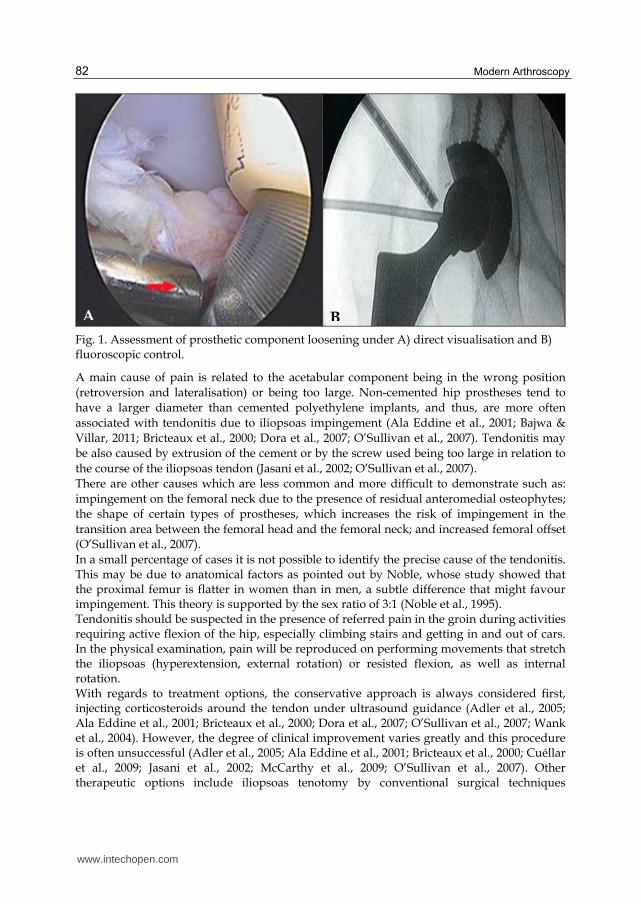
2.1 Prosthesis loosening A battery of complementary tests is available for its diagnosis. These include blood tests (CRP and ESR), X-rays and particularly scintigraphy. The combination of scintigraphy tests with Ga67, Tc99, and In111- labelled leukocytes has high sensitivity and specificity for the diagnosis of implant loosening and for distinguishing between septic and aseptic inflammation (Merkel et al., 1985, 1986; Rushton et al., 1982). We routinely perform arthocentesis and take samples of synovial tissue from three or four areas around the prosthetic joint, as a first stage at the beginning of the surgical procedure. Samples were sent to the Pathology Unit. None of the patients receive antibiotic therapy for at least 5 days prior to the intervention and prophylaxis is not initiated until after the samples were taken. The assessment of signs of loosening is completed by applying force with a blunt-ended instrument to all the prosthetic components in turn and carrying out movements causing leverage under arthroscopy and fluoroscopic control (Fig-1).
2.2 Tendon pain (iliopsoas tendonitis) Tendon inflammation around the implant is one of the typical causes of pain after THR. Amongst these, iliopsoas tendonitis secondary to hip replacement has a prevalence of up to 4.3% according to several authors (Ala Eddine et al., 2001; Bricteaux et al., 2000; Dora et al., 2007). Various factors can be responsible for iliopsoas inflammation, but particular attention should be drawn to changes in the course of the tendon due to the resection of the femoral head in hip replacement surgery. This modifies the course of the tendon bringing it closer to the medial edge of the prosthetic acetabulum and femoral neck, increasing the probability of impingement (O’Sullivan et al., 2007).
www.intechopen.com
Modern Arthroscopy
82
Fig. 1. Assessment of prosthetic component loosening under A) direct visualisation and B) fluoroscopic control.
A main cause of pain is related to the acetabular component being in the wrong position
(retroversion and lateralisation) or being too large. Non-cemented hip prostheses tend to
have a larger diameter than cemented polyethylene implants, and thus, are more often
associated with tendonitis due to iliopsoas impingement (Ala Eddine et al., 2001; Bajwa &
Villar, 2011; Bricteaux et al., 2000; Dora et al., 2007; O’Sullivan et al., 2007). Tendonitis may
be also caused by extrusion of the cement or by the screw used being too large in relation to
the course of the iliopsoas tendon (Jasani et al., 2002; O’Sullivan et al., 2007).
There are other causes which are less common and more difficult to demonstrate such as:
impingement on the femoral neck due to the presence of residual anteromedial osteophytes;
the shape of certain types of prostheses, which increases the risk of impingement in the
transition area between the femoral head and the femoral neck; and increased femoral offset
(O’Sullivan et al., 2007).
In a small percentage of cases it is not possible to identify the precise cause of the tendonitis. This may be due to anatomical factors as pointed out by Noble, whose study showed that the proximal femur is flatter in women than in men, a subtle difference that might favour impingement. This theory is supported by the sex ratio of 3:1 (Noble et al., 1995). Tendonitis should be suspected in the presence of referred pain in the groin during activities requiring active flexion of the hip, especially climbing stairs and getting in and out of cars. In the physical examination, pain will be reproduced on performing movements that stretch the iliopsoas (hyperextension, external rotation) or resisted flexion, as well as internal rotation. With regards to treatment options, the conservative approach is always considered first, injecting corticosteroids around the tendon under ultrasound guidance (Adler et al., 2005; Ala Eddine et al., 2001; Bricteaux et al., 2000; Dora et al., 2007; O’Sullivan et al., 2007; Wank et al., 2004). However, the degree of clinical improvement varies greatly and this procedure is often unsuccessful (Adler et al., 2005; Ala Eddine et al., 2001; Bricteaux et al., 2000; Cuéllar et al., 2009; Jasani et al., 2002; McCarthy et al., 2009; O’Sullivan et al., 2007). Other therapeutic options include iliopsoas tenotomy by conventional surgical techniques
A B
www.intechopen.com

Arthroscopy after Total Hip Replacement Surgery
83
techniques (Bricteaux et al., 2000; Della Valle et al., 2001; Heaton & Dorr, 2002; Taher & Power, 2003) and, more recently, using arthroscopic techniques (Cuéllar et al., 2009; McCarthy et al., 2009). Iliopsoas tendon lengthening has also been proposed (Trousdale et al., 1995). In some cases, the acetabular component needs to be revised.
2.3 Trochanteritis - Gluteal muscle tears
A frequent cause of pain following hip replacement surgery, commonly associated with Trendelenburg gait pattern. This is more common when a transgluteal approach has been used (Horwitz et al., 1993; Masonis & Bourne, 2002; Nolan et al., 1975; Obrant et al., 1989; Svensson et al., 1990). The pathological findings are very similar to those found in rotator cuff tendons in the shoulder, as has been previously described (Bunker et al., 1997; Kagan, 1999). These include bursitis, tendonitis and other tendon injuries, as well as muscle atrophy. The diagnosis of these conditions can be reached using modified MRI techniques to minimise the artefacts generated by the implants and performing the imaging using frequency-encoding gradient parallel to the long axis of the prosthesis (Pfirrmann et al., 2005; Twair et al., 2003; White et al., 2000). In most patients with hip implants some fluid accumulates around the trochanter. Pfirrmann Pfirrmann et al., 2005) reports in his paper that he found a volume than greater than 4 mls of fluid in those patients with pain and a limp. The same paper reports statistics concerning other complications in the trochanteric region related to hip replacement surgery: defects in the gluteus minimus and gluteus medius tendons were found in 56% and 62% symptomatic patients, respectively, compared to in just 8% and 16% of asymptomatic patients; while poor gait was associated with tears larger than 2.5 cm. MRI allows assessing muscle atrophy and fatty degeneration. As in the shoulder, these signs are a poor prognostic factor. These findings are associated with a Trendelenburg gait and are almost exclusively seen in patients with painful hips (Pfirrmann et al., 2005). Another cause of this gait pattern is a lesion in the superior gluteal nerve, which may result from hip surgery, in particular, when the lateral approach is used. (Ramesh et al., 1996). The approaches that entail greater trochanter osteotomy may cause pain due to non union, failure to remove loose bone fragments or breakage of the wires used in the procedure. Treatment in such cases often requires surgical intervention, although it is possible to remove loose bone fragments and wires using bursoscopy. (Cuéllar et al., 2009). The treatment of greater trochanter pain syndrome and trochanteric bursitis can also be achieved using bursoscopy (Weber & Berry, 2007). High-grade tears of gluteal tendons may need to be repaired by open surgery (Weber & Berry, 2007), but can also be addressed using arthroscopy.
2.4 Intra-articular adhesions: Arthrofibrosis
This is a common cause of pain following hip surgery (Beck, 2009; Krueger et al., 2007). Any adhesions within the joint capsule or around the femoral neck tend to cause impingement, producing pain and limiting mobility (Krueger et al., 2007). Indeed, such adhesions have been described as a potential cause of pain in relation to hip prostheses. (Bajwa & Villar, 2011; Cuéllar et al., 2009; McCarthy et al., 2009). We reported the presence of structured fibrous bands occupying the medial recess (Fig-2 A,B) (Cuéllar et al., 2009). We also found fibrous structures located between the acetabulum and the prosthetic neck and in wider areas across the new joint (Fig-3 A,B).
www.intechopen.com

Modern Arthroscopy
84
The symptoms are similar to those of iliopsoas tendonitis. Patients refer pain in the groin
radiating down the inner thigh during activities involving flexion of the hip, such as
climbing stairs, but also going down stairs and up or downhill, getting in and out of cars,
and turning over in bed . (Beck, 2009; Krueger et al., 2007). There are usually no signs of
iliopsoas tendonitis with ultrasound-guided injections, and the response to nerve block
tends to be non conclusive or negative.
A definitive diagnosis can be obtained by arthroscopy. Treatment consists of debridement
and removal of the adhesions, by the same arthroscopic portal (Cuéllar et al., 2009; Krueger
et al., 2007; McCarthy et al., 2009).
Fig. 2. Structured fibrous bands in the medial compartment: A) medial and B) infero-medial view (righ hip)
Fig. 3. Fibrosis: A) between the acetabulum and the prosthetic neck; and B) widespread arthrofibrosis
A B
A B
www.intechopen.com

Arthroscopy after Total Hip Replacement Surgery
85
2.5 Femoro-acetabular impingement - Subluxation-Prosthesis dislocation
The principles of impingement in the prosthetic hip are similar to those described by Ganz for the normal hip (Ganz et al., 2003). A cam type impingement is found in implants with small femoral heads that are poorly differentiated from the femoral neck, Pincer type impingement is caused in those hips where there’s been inadequate and insufficient removal of osteophytes. Finally, mixed cam-pincer impingement is caused by a combination of having a small femoral head, a ratio between head and neck of less than 2.0, over sizing of the acetabular component, and a polyethylene liner having sharp rather than rounded edges
(Malik et al., 2007).
Certain anatomical conditions may increase the risk of prosthetic impingement. It has been reported that very flexible patients have a greater risk of impingement at the extremes of the range of motion (Beaulé et al., 2002; Geller et al., 2006). There is a difference in tilt of the pelvis when the patient is supine on the operating table, and when they are active in movement. This difference tends to lead to an overly horizontal positioning of the acetabular component, which makes impingement more likely (Malik et al., 2007; McCollum & Gray, 1990). The short term clinical consequences of prosthetic impingement include pain, reduced
mobility, instability, subluxation and frank dislocation (Barrack et al., 2001; Barrack, 2003;
Brien et al., 1993; Brown & Callaghan, 2008; Cobb et al., 1996; Hedlundh & Carlsson, 1996;
Malik et al., 2007; McCollum & Gray, 1990; Padgett et al., 2006). In the longer term, excessive
friction between the prosthetic components results in the release of metallic particles and
wear of metallic edges which may lead to metallosis and osteolysis. These make early
loosening of the implant more likely.
It is not always easy to identify impingement on the basis of patient medical history, clinical
examination or radiographic studies, given that it is a dynamic process. Patients with pain
and subluxation require CT scans to identify the presence of osteophytes and the relative
position and orientation of the components. (Cuéllar et al., 2009; Cuéllar et al., 2010;
Pierchon et al, 1994). As we have indicated in previous studies (Cuéllar et al., 2009, 2010), the
best way to demonstrate the existence of instability is by Examination Under Anesthesia
(EUA) with X ray control (Fig- 4 A,B A,B).
Hip resurfacing implants, having larger femoral heads, offer a greater degree of mobility
and stability but the ideal ratio between femoral head and neck is hard to achieve and,
therefore, they involve a higher risk of impingement with associated instability (Fig-4 B).
(Bajwa & Villar, 2011; Cuéllar et al., 2009; Cuéllar et al., 2010; Khanduja & Villar, 2008).
2.6 Pain of unknown origin - Other causes of pain
In around 1% of the cases the cause of pain remains unknown.
Lumbar spine and radicular pain should be ruled out because of the well known association
between degenerative changes in the spine and hip joint. Pain in the gluteal area extending
beyond the popliteal region also suggests that it has its origin in the lumbar spine (Bozic &
Rubash, 2004; White, 1998). Patients with lumbar spine disorders may experience worsening
of radicular pain after hip replacement surgery due to increase in mobility and physical
activity (Bozic & Rubash, 2004; Bohl & Steffee, 1979).
Pain that begins when a patient starts walking is commonly associated with loosening, and
iliopsoas tendonitis, but it can also be derived from lumbar spine disorders (Bozic &
Rubash, 2004; Bohl & Steffee, 1979)
www.intechopen.com
Modern Arthroscopy
86
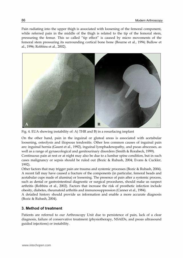
Pain radiating into the upper thigh is associated with loosening of the femoral component,
while referred pain in the middle of the thigh is related to the tip of the femoral stem,
pressuring the femur. This so called “tip effect” is caused by micro movements of the
femoral stem pressuring its surrounding cortical bone bone (Bourne et al., 1994; Bullow et
al., 1996; Robbins et al., 2002).
Fig. 4. EUA showing instability of: A) THR and B) in a resurfacing implant
On the other hand, pain in the inguinal or gluteal areas is associated with acetabular
loosening, osteolysis and iliopsoas tendonitis. Other less common causes of inguinal pain
are: inguinal hernia (Gaunt et al., 1992), inguinal lymphadenopathy, and psoas abscesses, as
well as a range of gynaecological and genitourinary disorders (Smith & Rorabeck, 1999).
Continuous pain at rest or at night may also be due to a lumbar spine condition, but in such
cases malignancy or sepsis should be ruled out (Bozic & Rubash, 2004; Evans & Cuckler,
1992).
Other factors that may trigger pain are trauma and systemic processes (Bozic & Rubash, 2004). A recent fall may have caused a fracture of the components (in particular, femoral heads and acetabular cups made of alumina) or loosening. The presence of pain after a systemic process, such as dental or gastrointestinal diagnostic or surgical procedures, should make us suspect arthritis (Robbins et al., 2002). Factors that increase the risk of prosthetic infection include obesity, diabetes, rheumatoid arthritis and immunosuppression (Canner et al., 1984).
A detailed history should provide us information and enable a more accurate diagnosis (Bozic & Rubash, 2004).
3. Method of treatment
Patients are referred to our Arthroscopy Unit due to persistence of pain, lack of a clear
diagnosis, failure of conservative treatment (physiotherapy, NSAIDs, and psoas ultrasound
guided injections) or instability.
A B
www.intechopen.com

Arthroscopy after Total Hip Replacement Surgery
87
In all cases, we complete a diagnostic protocol including a full clinical history, blood tests (FBC, ESR, CRP); imaging tests (X-rays, CT and MRI scans); scintigraphy (Tc, Ga, In labeled leukocytes); and ultrasound-guided psoas injections. In some cases scintigraphy and CT scans are repeated after a period of at least 3 months to rule out implant loosening. We perform arthroscopic surgery before indicating revision total hip replacement surgery in all cases of persistent pain where the cause of this has not been clearly identified. Patients fulfil an informed consent form.
3.1 Surgical procedure
The procedure lasts between 60 and 90 minutes. The anaesthetist selects the most appropriate anaesthetic technique in each case: spinal
anaesthesia, general anaesthesia or a combination of both.
The patient lies supine on a traction table, as this facilitates fluoroscopic control of the
procedure. In all cases, the procedure is preceded by examination under anaesthesia to
assess instability and the presence of “snapping”.
Joint distraction is required only in a few cases. We favour the anterolateral and the anterior arthroscopic portals. Depending on therequirements in each case, the posterior peritrochanteric or another distal anterior portal may be additionally used To gain access, progressive larger dilatators are slid into position through a nitinol
guidewire previously inserted under fluoroscopic control (Fig. 5 A, B).
We routinely follow a three steps protocol 1) collection of samples for culture 2) assessment
of the degree of loosening of the components; and 3) assessment and treatment of the
condition itself.
Fig. 5. Introduction of a “nitinol” guide wire under fluoroscopic control (A) Progresive larger dilators are slide into position (B).
3.2 Specific surgical procedures 3.2.1 Psoas tenotomy
The indications for psoas tenotomy are tendonitis or painful internal snapping hip syndrome that have not improved with conservative treatment, in particular with
www.intechopen.com

Modern Arthroscopy
88
ultrasound-guided steroid injections. We use the same concept and technique as those applied in cases of iliopsoas tendonitis in patients that have not had total hip replacement surgery. As standardised by Ilizaliturri, there are two ways to perform tenotomy (Ilizaliturri et al., 2009): 1) at its site of insertion on the lesser trochanter, and 2) along its course close to the joint. In the latter, the tendon can be partially seen behind the articular capsule or can be directly observed through an orifice that communicates the joint with the iliopectinea bursa.
Fig. 6. Psoas tenotomy: Tenotomy at the level of the acetabular rim (A). Release of the tendinous fibres up to the level of the muscle fibres (B)
Fig. 7. Gluteal muscle tears in a right hip, seen through the distal peritrochanteric portal: A) similarity to rotator cuff ttears, B) repair with suture anchors
The psoas tendon is divided close to the acetabular component in the cases in which there is evident acetabular involvement (Fig. 6 A, B A, B). If this is not clear, the tenotomy is performed near the lesser trochanter. In any case, only the tendinous fibres are releasd , stopping the intervention when the muscular fibres are reached (Fig. 6 B).
A B
A B
www.intechopen.com

Arthroscopy after Total Hip Replacement Surgery
89
Fig. 8. Impingement of the acetabular rim of the implant on the lesser trochanter (white asterisk) leading to dislocation of this right hip (white arrow)
3.2.2 Trochanterplasty - Gluteal muscle repair
Hip Bursitis and trochanteritis are treated by debridement in the same way as in non prosthetic hips. An early description of a bursectomy using bursoscopy was given by Bradley (Bradley & Dillingham, 1998). Problems related to the peritrochanteric space can be approached from the peripheral compartment or using the inside-outside technique, described by Ilizaliturri. We perform bursoscopy at a second stage following the arthroscopic examinatiooon of the hip joint. For this reason we tend to perform the technique from the peripheral compartment, reorienting the peritrochanteric portal. Aditionaly we use one or two portals, one distal and one proximal to the tip of the trochanter. A wide range of techniques can be used. These include debridement, trochanteric abrasion,
z-tenotomy of the fascia lata and suture using anchors, depending on the condition to be
treated. There is a great similarity between gluteus medius and minimus tears with shoulder
rotator cuff injuries (Fig. 7 A), It is suspected that that their prevalence is higher than
believed to date and that this condition may be responsible for many cases of pain in the
trochanter region after hip replacement surgery. Treatment is similar and involves repair by
placement of suture anchors (Cuéllar et al., 2010). (Fig. 7 B).
3.2.3 Plica resection for arthrofibrosis
Fibrous structures are a potential cause of pain related to hip replacement surgery (Bajwa & Villar, 2011; Cuéllar et al. 2009; McCarthy et al., 2009). The symptoms in such cases are similar to those of iliopsoas tendonitis: inguinal pain radiating down the inner thigh and pain during activities involving flexion of the hip like climbing stairs and slopes, getting in and out of cars, and turning over in bed among others (Beck, 2009; Krueger et al., 2007).
Often large longitudinal fan shaped fibrous adhesions, occupying the medial recess can be found (Cuéllar et al., 2009). (Fig-2). In these cases, mechanical debridement and thermo coagulation are performed, and any scar tissue around the joint the synovial plica is resected.
www.intechopen.com

Modern Arthroscopy
90
Fig. 9. Capsular plicature technique: A) Redundant capsular tissue in a case of an unstable THR; B) illustration of the technique used in cases of instability, by threading a double no. 2 suture through parallel incisions secured with a loop knot; C) Dissection of the capsular plane from the underlying muscle using a blunt dissector; D) once the capsular plane has been freed; E) two or three parallel incisions are made in the redundant capsule. A double no. 2 suture is threaded in and out through alternate incisions using a bird-beak passer; F) An end loop is left; G) one end of the suture is brought through the end loop and the two ends of the suture are knotted; H) the procedure can be repeated; I) until sufficient reduction of the redundant capsular volume is achieved.
A B C
D E F
G H I
www.intechopen.com

Arthroscopy after Total Hip Replacement Surgery
91
3.2.4 Abrasion for impingement
To date, we have only treated one case of impingement of the edge of the acetabular component on the lesser trochanter. This impingement caused dislocation of the prosthesis (Bajwa & Villar, 2011; Cuéllar et al., 2010). (Fig-8). Preoperative investigations (X-ray and CT scan) showed a short femoral neck leading to cam impingement with a head/neck ratio <2. Arthroscopy-guided exploration confirmed the presence of impingement between the lesser trochanter and the inferior edge of the acetabulum in external rotation which caused prosthesis dislocation and left redundant capsular tissue in the anterior and lateral recesses. The bone across all the contact area between the lesser trochanter and the prosthetic acetabulum was shaved away until the impingement disappeared. This was combined with capsulorraphy, as described below.
3.2.5 Capsular plicature
In cases of instability, plication of redundant capsular tissue is performed. This is achieved by threading a double no. 2 suture through parallel incisions in the capsule secured with a loop knot, in a similar manner to the technique described for the treatment of instability of a non-prosthetic hip (Shindle et al., 2006; Tibor & Sekiya, 2008). (Fig. 9). Surgery is indicated after failure of conservative treatment with Physiotherapy and muscle strength rehabilitation focussed in the trochanteric and pelvic region. A CT scan should be carried out to confirm that there is no significant acetabular anteversion or retroversion nor signs of prosthetic components loosening. Before proceeding with surgery, a EUA is carried out under X ray control looking for instability or snapping. The main operative finding often is the presence of a large capsular recess generally located in the lateral and posterior aspects of the hip joint (Fig. 9 A, B). Capsular plicature is preceded by dissecting the capsular plane from the underlying muscle (Fig. 9 C, D), using a blunt dissector. In the posterior recess, this is carried out through a posterior and lateral approach and supported in the muscular plane of the lateral rotator group of muscles (obturator externus, gemelli). The sciatic nerve runs laterally and is protected by this muscular plane. In the anterior plane, we dissect the capsular plane from the rectus femoris. Once the capsular plane to be plicated has been freed, we make two or three parallel incisions with a scalpel. Through these, we thread a double no. 2 suture in and out using a bird-beak passer leaving an end loop (Fig. 9 E). Subsequently, one end of the suture is brought through the end loop. Finally, the two ends of the suture are knotted, gathering up the redundant capsular volume, like a tobacco pouch” (Fig. 9 F, G). The procedure can be repeated in other regions of the capsule as many times as necessary to obtain sufficient reduction of the redundant capsular volume (Fig. 9 H, I). During this procedure laxity is checked until all signs of instability have disappeared. In the postoperative period no orthosis is usually required. Patients are, however, given guidance concerning how to avoid movements that might cause new dislocations and instructed to perform exercises to strengthen the glutei muscles. Patients are discharged 24 to 48 hours after surgery
4. Clinical evidence
In our experience, it was possible to gain access to the prosthetic joint with the arthroscopic instruments in all cases. This was technically more demanding in cases of arthrofibrosis
www.intechopen.com

Modern Arthroscopy
92
where the fibrous bands make the cavity difficult to visualize and therefore more time is required to perform the debridement procedure. At follow up, three months after surgery, patients that underwent psoas tenotomy had recovered the range of hip flexion to grade 4 and by 6 months all patients had regained grade 5 strength. We have found that patients with lumbar spine disorders experience more back and radiating leg pain after having their painful prosthetic hips treated. In all the cases of capsulorraphy the instability and the episodes of subluxation had disappeared. This was maintained at the 6-month and 1-year follow-ups. None of the patients had to undergo further surgery in relation to their hip replacement. They were given instructions to avoid hip flexion of more than 100º, especially together with external rotation and adduction. We have not observed any neurovascular complications.
5. Discussion
In 1% of the cases the reason for the persistence of pain following hip replacement surgery remains unknown (Witvoët, 2001). Despite this, the cause of pain should always be investigated and we should not rush in carrying out revision surgery (Witvoët, 2001). This is where arthroscopy plays an important role enabling a progress in the diagnosis and a potential treatment in certain patients whose prostheses, although apparently properly implanted, continue to cause pain (McCarthy et al., 2009). This is already being used as a diagnostic and therapeutic tool in some painful complications associated with total knee replacements (Bocell et al., 1991; Johnson et al., 1990; Lawrence & Kan, 1992; Lucas et al., 1999; Markel et al., 1996; Scranton, 2001; Tzagarakis et al., 2001; Wasilewski & Frankl, 1989a, 1989b). Access can be gained to the prosthetic joint using the arthroscopic technique and instruments. It is possible to apply this to resurfacing type prostheses, as indicated in the only paper that we found on this topic (Khanduja & Villar, 2008). We favour capsular plication using sutures rather than thermal methods. Regarding the use of ultrasound-guided steroid injections into the psoas (Adler et al., 2005; Ala Eddine et al., 2001; Bricteaux et al., 2000; Dora et al., 2007; O’Sullivan et al., 2007; Wank et al., 2004), we believe that this technique has few advantages: it is not easy to perform; and in our opinion doesn´t provide much information, even in cases in which it was clear intraoperatively that there was tendon involvement. The outcomes reported in the literature are very variable and it is often not successful (Adler et al., 2005; Ala Eddine et al., 2001; Bricteaux et al., 2000; Cuéllar et al., 2009; Jasani et al., 2002; McCarthy et al., 2009; O’Sullivan et al., 2007; Witvoët, 2001). For this reason, we recommend that this technique is not used systematically, but rather only in selected cases. It is possible to perform endoscopy-guided trochanteric bursoscopy and fasciotomy. Additionally, if necessary, gluteal muscle repair can be performed. To date we have not treated any patients with acute or subacute arthritis, but we believe that the arthroscopy technique could be used in such cases, similarly to when indicated in infected total knee replacements (Hyman et al., 1999; McCarthy et al., 2009). To avoid prosthetic dislocation in the immediate postoperative period, unnecessary wide capsulotomies should not be done, and the patents should be given clear instructions about postural training (Cuéllar et al., 2009).
www.intechopen.com
Arthroscopy after Total Hip Replacement Surgery
93
6. Conclusions
Arthroscopy can be successfully applied to the diagnosis and treatment of pain of unknown
origin after hip replacement surgery. This very often associated with lumbar spine
disorders, other medical conditions and old age. This association makes the differential
diagnosis difficult.
The technique has proven to be especially useful in the treatment of instability, muscular
and tendon pain and arthrofibrosis.
On the other hand, the technique has not been found to be reliable for identifying cases of
loosening of prosthetic components.
7. References
Adler, R.S.; Buly, R.; Ambrose, R. & Sculco, T. (2005). Diagnostic and therapeutic use of ultrasound-guided psoas peritendinous injections. American Journal of Roentgenology, Vol. 185, (2005), pp. (940-943), DOI:10.2214 AJR.04.1207.
Ala Eddine, T.; Remy, F.; Chantelot, C.; Giraud, F.; Migaud, H. & Duquennoy, A. (2001). Anterior iliopsoas impingement alter total hip arthroplasty: diagnosis and conservative treatment in 9 cases. Revue de Chirurgie Orthopédique et Reparatrice de L’Appareil Moteur, Vol.87, (2001), pp. (815-819), ISSN 0035-1040.
Bajwa, A.R. & Villar, S.N. (2011). Arthroscopy of the hip in patients following joint replacement. Journal of Bone and Joint Surgery Br, Vol.93, No.4, (2011), pp. (890-896), DOI 10.1302/0301-620X.93B7.24902.
Barrack, R.L., Butler, R.A. Laster, D.R. & Andrews, P. (2001). Stem desing and dislocation after revision total hip arthroplasty: clinical results and computer modeling. The Journal of Arthroplasty, Vol. 16, No. 8 SI, (2001), pp. (S8-S12), DOI 10.1054/ARTH 2001.28359
Barrack, R.L. (2003). Dislocation after total hip arthroplasty: implant design and orientation. Journal of American Academy of Orthopaedic Surgeons, Vol. 11, Nº 2, (2003), pp. (89-99), PMID 12670135.
Beaulé, P.E.; Schmalzriued, T.P.; Udomkiat, P. & Amstutz, H.C. (2002). Jumbo femoral head for the treatment of recurrent dislocation following total hip replacement. Journal of Bone and Joint Surgery Am, Vol. 84, Nº 2, (2002), pp. (256-263), PMID 11861732.
Beck, M. (2009). Groin pain alter open FAI surgery. Clinical Orthopaedics and Related Research, No.467, (2009), pp. (769-774), ISSN 0009-921X.
Bohl, W. & Steffee, A. (1979). Lumbar spinal stenosis: a cause of continued pain and disability in patients after total hip arthroplasty. Spine, Vol. 4, Nº 2, (1979), pp. (168-173), ISSN 0362-2436.
Bourne, R.B.; Rorabeck, C.H., Ghazal, M.E. & Lee, M.H. (1994). Pain in the thigh following total hip replacement with a porous-coated anatomic prosthesis for osteoarthrosis. Journal of Bone and Joint Surgery Am, Vol. 76, Nº 10, (1994), pp. (1464-1470), PMID 7929493.ñ¡.
Bocell, J.R.; Thorpe, C.D. & Tullos, H.S. (1991). Arthroscopic treatment of symptomatic total knee arthroplasty. Clinical Orthopaedics and Related Research, No.271, (October 1991), pp. (125-134), ISSN 0009-921X.
www.intechopen.com

Modern Arthroscopy
94
Bozic, K.J. & Rubash, H.E. (2004). The painful total hip replacement. Clinical Orthopaedics and Related Research, No.420, (2004), pp. (18-25), ISSN 0009-921X.
Bradley, D.M. & Dillingham, M.F. (1998). Bursoscopy of the trochanteric bursa. The Journal of Arthroscopy and Related Surgery, Vol. 14, Nº 8, (1998), pp. (884-887), ISSN 0749-8063/98/1408-1810.
Bricteaux, S.; Seutin, B.; Beguin, L.; Farizon, F. & Fessy, M.-H. (2000). Arthroplastie totale de hanche doloreuse; rechercher les conflicts avec le psoas: A propos de 10 cas. Revue de Chirurgie Orthopédique et Reparatrice de L’Appareil Moteur, Vol.86 (S-II), (2000), pp. (SII 84-85), ISSN 0035-1040.
Brien, W.W., Salvati, E.A.; Wright, T.M. & Burstein, A.H. (1993). Dislocation following THA: comparison of two acetabular component designs. Orthopedics, Vol. 16, Nº 8, (1993), pp. (869-872), PMID 8415270.
Brown, T. & Callaghan, J.J. (2008). Impingement in total hip replacement: mechanism and consequences. Current Orthopaedics, Vol.22, No.6, (December 2008), pp. (376-391), DOI:10.1016.
Bullow, J.U.;.Scheller, G.; Arnold, P.; Synastchke, M. & Jani, L. (1996). Uncemented total hip replacement and thigh pain. International Orthopaedics, Vol. 20, Nº 2, (1996), pp. (56-69) PMID 8739695.
Bunker, T.D.; Esler, C.N. & Leach, W.J. (1997). Rotator-cuff tear of the hip. Journal of Bone and Joint Surgery Br, Vol.79, No.4, (1997), pp. (618-620), ISSN 0301-620X/97/47033.
Burman, M.S. (1931). Arthroscopy or the direct visualization of joints. Journal of Bone and Joint Surgery, Vol.4, (1931), pp. (669-695).
Byrd, J.W.T. (1994). Hip arthroscopy utilizing the supine position. The Journal of Arthroscopy and Related Surgery, Vol.10, No.3, (June 1994), pp. (275-280), ISNN 0749-8063.
Byrd, J.W.T. (2006). Hip arthroscopy: Surgical indications. The Journal of Arthroscopy and Related Surgery, Vol.22, No.12, (December 2006), pp. (1260-1262), ISSN 0749-8063-062212-6404.
Byrd, J.W.T. & Jones, K.S. (2009). Hip arthroscopy for labral pathology: Prospective analysis with 10-year follow-up. The Journal of Arthroscopy and Related Surgery, Vol.25, No.4, (April 2009), pp. (365-368), ISSN 0749-8063-09.
Canner, G.C.; Steinberg, M.E.; Heppenstall, R.B. & Balderston, R. (1984). The infected hip after total hip arthroplasty. Journal of Bone and Joint Surgery Am, Vol. 66, Nº 9, (1984), pp. (1393-1399), ISSN 0021-9355.
Cuéllar, R.; Aguinaga, I.; Corcuera, I. & Baguer, A. (2009). Artroscopia en prótesis de cadera: resultados preliminares (en español original). Cuadernos de Artroscopia, Vol.16, No.2, (October 2009), pp. (35-42), ISNN 1134-7872.
Cuéllar, R., Aguinaga, I., Corcuera, I.; Ponte, J. & Usabiaga, J. (2010). Arthroscopic treatment of unstable Total Hip Replacement. The Journal of Arthroscopy and Related Surgery, Vol.26, No.6, (June 2010), pp. (861-865), ISNN 0749-8063-9564, DOI 10.1016.
Cobb, T.K.; Morrey, B.F. & Ilstrup, D.M. (1996). The elevated-rim acetabular liner in total hip arthroplasty: relationship to postoperative dislocation. Journal of Bone and Joint Surgery Am, Vol. 78, Nº 1, (1996), pp. (80-86), ISSN 0021-9355.
www.intechopen.com

Arthroscopy after Total Hip Replacement Surgery
95
Della Valle, C.J.; Rafii, M. & Jaffe, W.L. (2001). Iliopsoas tendonitis after total hip arthroplasty. The Journal of Arthroplasty, Vol.16, No.7, (2001), pp. (923-926), PMID 11607911.
Dora, C.; Houweling, M.; Koch, P. & Sierra, R.J. (2007). Iliopsoas impingement alter total hip replacement. The resuilts of non-operative Management, tenotomy or acetabular revision. Journal of Bone and Joint Surgery Br, Vol.89, No.8, (2007), pp. (1031-1035), ISSN 0301-620X.
Erikson, E.; Arvidsson, I. & Arvidsson, H. (1986). Diagnostic and operative arthroscopy of the hip. Orthopedics, Vol.9, No.2, (1986), pp. (169-176), PMID 3960759.
Evans, B.G. & Cuckler, J.M. (1992). Evaluation of the painful total hip arthroplasty. The Orthopedic Clinics of North America, Vol. 23, Nº 2, (1992), pp. (303-311), PMID 1570142.
Ganz, R.; Parvizi, J.; Beck, M., Leunig, M.; Notzil, H. & Siebenrock, K.A. (2003). Acetabular impingement: a cause for osteoarthritis of the hip. Clinical Orthopaedics and Related Research, No.417, (2003), pp. (112-120), ISSN 0009-921X..
Gaunt, M.; Tan, S. & Dias, J. (1992). Strangulated obturator hernia masquerading as pain from a total hip replacement. Journal of Bone and Joint Surgery Br, Vol.74, Nº 5, (1992), pp. (782-783), ISSN 0301-620X/92/5R61.
Geller, J.A.; Malchau, H.; Bragdon, C.; Greene, M. et al. (2006). Large diameter femoral heads on highly cross-linked poyethilene: minimum 3-year results. Clinical Orthopaedics and Related Research, No.447, (2006), pp. (53-59), ISSN 0009-921X.2006.
Glick, J.M.; Sampson, T.G.; Gordon, R.B.; Behr, J.T. & Schmidt, E. (1987). Hip arthroscopy by the lateral approach. The Journal of Arthroscopy and Related Surgery, Vol.3, (1987), pp. (4-12), ISNN 0749-8063.
Gross, R.H. (1977). Arthroscopy in hip disorders in children. Orthopaedic Review, Vol.6, (1977), pp. (43-49).
Heaton, K. & Dorr, L.D. (2002). Surgical release of iliopsoas tendon for groin pain after total hip arthroplasty. The Journal of Arthroplasty, Vol.17, No.6, (2002), pp. (779-781), DOI 10.1054/ARTH.2002.33570.
Hedlundh,U. & Carlsson, A.S. (1996). Increased risk of dislocation with collar reinforced modular heads of the Lubinus SP-2 hip prosthesis. Acta Orthopaedica Scandinavica, Vol. 67, Nº 2, (1996) pp. (204-205), PMID 8623583.
Horwitz, B.R.; Rockowitz, N.L.; Goll, S.R.; Booth, R.E. Jr.; Balderston, R.A.; Rothman, R.H. & Cohn, J.C. (1993). A prospective randomized comparison of two surgical approaches to total hip arthroplasty. Clinical Orthopaedics and Related Research, No.291, (1993), pp. (154-163), ISSN 0009-921X.
Hyman, J.L., Salvati, E.A., Laurencin, C.T.; Rogers, D.E.; Maynard, M. & Brause, B.D. (1999). The arthroscopic drainage, irrigation, and débridement of late, acute total hip arthroplasty infections. Journal of Arthroplasty; Vol.14, No.8, (December 1999), pp. (903-910), PMID 10614878.
Jasani, V.; Richards, P. & Wynn-Jones, C. (2002). Pain related to the psoas muscle after total hip replacement. Journal of Bone and Joint Surgery Am, Vol.84, No.8, (2002), pp. (991-993), ISSN 00219355.
www.intechopen.com

Modern Arthroscopy
96
Johnson, D.R.; Friedman, R.J.; McGinty, J.B.; Mason, J.L. & St Mary, E.W. (1990). The role of arthroscopy in the problema total knee replacement. The Journal of Arthroscopy and Related Surgery, Vol.6, No.1, (March 1990), pp. (31-32), ISNN 0749-8063.
Johnston, T.L.; Schenker, M.L.; Briggs, K.K. & Philippon, M.J. (2008). Relationship between offset angle alpha and hip chondral injury in femoroacetabular impingement. The Journal of Arthroscopy and Related Surgery, Vol.24, No.6, (June 2008), pp. (669-675), ISSN 0749-8063-08-2406-7302.
Kagan, A. (1999). 2nd rotator cuff tears of the hip. Clinical Orthopaedics and Related Research, No.368, (1999), pp. (135-140), ISSN 0009-921X.
Khanduja, V. & Villar, R.N. (2008). The role of arthroscopy in resurfacing arthroplasty of the hip. The Journal of Arthroscopy and Related Surgery, Vol.24, No.1, (January 2008), pp. (122e1), ISNN 0749-8063-08-2401-6501.
Kelly, B.T.; Williams, R.J. III & Philippon, M.J (2003). Hip Arthroscopy: Current Indications, Treatment Options, and Management Issues. The American Journal of Sports Medicine, Vol.31, No.6, (November/December 2003), pp. (1020-1037), ISSN 0363-5465-103-3131-1020.
Kocher, M.S.; Kim, Y.J.; Millis, M.B.; Mandiga, R.; Siparsky, P.; Micheli, L.J. & Kasser, J.R. (2005). Hip arthroscopy in children and adolescents. Journal of Pediatric Orthopaedics, Vol.25, No.5, (2005), pp. (680-686), PMID 16199955.
Krueger,A.; Leunig, M.; Siebenrock, K.A. & Beck, M. (2007). Hip arthroscopy after previous surgical hip dislocation for femoroacetabular impingement. The Journal of Arthroscopy and Related Surgery; Vol. 23, Nº 12, (2007), pp. (1285-1289), ISSN 0749-8063/07/2312-6610.
Ilizaliturri, V.M.; Chaidez, C.; Villegas, P.; Briceño, A. & Camacho-Galindo, J. (2009). Prospective randomized study of 2 different techniques for endoscopio iliopsoas tendon release in the treatment of internal snapping hip síndrome. The Journal of Arthroscopy and Related Surgery, Vol. 25, Nº 2, (2009), pp. (159-163), ISSN 0749-8063/09/2502-8171.
Ilizaliturri, V.M.; Martínez-Escalante, F.A.; Chaidez, P.A. & Camacho-Galindo, J. (2006). Endoscopio iliotibial band release for externa snapping hip síndrome. The Journal of Arthroscopy and Related Surgery, Vol. 22, Nº 5, (2006), pp. (505-510), ISSN 0749-8063/06/2205-5156.
Larson, C.M.; Guanche, C.A.; Kelly, B.T.; Clohisy, J.C. & Ranawat, A.S. (2009). Advanced techniques in hip arthroscopy. Journal of American Academy of Orthopaedic Surgeons, Instructional Course Lecture, Vol. 58, (2009), pp. (423-436), PMID 19385552.
Larson, C.M. & Giveans, M.R. (2009), Arthroscopic debridement versus refixation of the acetabular labrum associated with femoroacetabular impingement. The Journal of Arthroscopy and Related Surgery, Vol.25, No.4, (April 2009), pp. (369-376), ISSN 0749-8063-09-2504-8590.
Lawrence, S.J. & Kan, R.O. (1992). Arthroscopic lysis of adhesions after New Jersey LCS total knee arthroplasty. Orthopedics, Vol.15, No.8, (August 1992), pp. (943-944), PMID 1508769.
Lubowitz, J.H. & Poehling, G.G. (2006). Hip Arthroscopy: An Emerging Gold Standard. The Journal of Arthroscopy and Related Surgery, Vol. 22, No.12, (December 2006), pp. (1257-1259), ISSN 0749-8063-06-2212-1780.
www.intechopen.com

Arthroscopy after Total Hip Replacement Surgery
97
Lucas, T.S.; DeLuca, P.F.; Nazarian, D.G.; Bartolozzi, A.R. & Booth, R.E. Jr (1999). Arthoscopic treatment of patellar clunk. Clinical Orthopaedics and Related Research, No.367, (1999), pp. (226-229), ISSN 0009-921X.
Malik, A.; Maheshwari, A. & Dorr, L.D. (2007). Impingement with total hip replacement. Journal of Bone and Joint Surgery Am, Vol.89, No.8, (2007), pp. (1832-1842), ISSN 00219355.
Markel, D.C.; Luessenhop, C.P.; Windsor, R.E. & Sulco, T.A. (1996). Arthroscopic treatment of peripatellar fibrosis alter total knee arthroplasty. Journal of Arthroplasty, Vol.11, No.3, (April 1996), pp. (293-297), PMID 8713909.
Masonis, J.L. & Bourne, R.B. (2002). Surgical approach, abductor function, and total hip arthroplasty dislocation. Clinical Orthopaedics and Related Research, No.405, (2002), pp. (46-53), ISSN 0009-921X.
McCarthy, J.C., Jibodh, S.R. & Lee, J.A. (2009). The role of arthroscopy in evaluation of painful hip arthroplasty. Clinical Orthopaedics and Related Research, No.467, (2009), pp. (174-180), ISSN 0009-921X.
McCollum, D.E. & Gray, W.J. (1990). Dislocation after total hip arthroplasty. Causes and prevention. Clinical Orthopaedics and Related Research, No.261, (1990), pp. (159-170), ISSN 0009-921X.
Merkel, K.D.; Brown, M.L.; Dewanjee, M.K. & Jr Fitzgerald, R.H. (1985). Comparison of indium-labeled-leucocyte imaging with sequential technetium-gallium sacanning in the diagnosis of low-grade musculkoskeletal sepsis: A prospective study. Journal of Bone and Joint Surgery Am, Vol.67, No.3, (1985), pp. (465-476), ISSN 00219355.
Merkel, K.D.; Brown, M.L. & Jr Fitzgerald, R.H. (1986). Sequential technetium99m HMDP-gallium-67 citrate imaging for the evaluation of infection in the painful prosthesis. Journal of Nuclear Medicine, Vol. 27,(1986), pp. (1413-1417).McCarthy, J.C. & Lee, J.A. (2006). Hip arthroscopy: indications, outcomes and complications. American Academy of Orthopaedic Surgeons, Instructional Course Lecture, Vol.55, (2006), pp. (301-308), PMID16958465.
Monllau, J.C.; Solano, A.; León, A.; Hinarejos, P. & Ballester, J. (2003). Tomographic study of the arthroscopic approaches to the hip joint. The Journal of Arthroscopy and Related Surgery, Vol.19, No.4, (April 2003), pp. (368-372), ISNN 0749-8063-03-1904-3152.
Noble, Ph.C.; Box, G.; Kamaric, E.; Fink, M.J.; Alexander, J.W. & Tullos, H.S. (1995). The effect of aging on the shape of th proximal femur. Clinical Orthopaedics and Related Research, No.316, (1995), pp. (31-44), ISSN 0009-921X.
Nolan, D.R.; Fitzgerald, R.H.; Jr. Beckenbaugh, R.D. & Coventry, M.B. (1975). Complications of total hip arthroplasty treated by reoperation. Journal of Bone and Joint Surgery Am, Vol.57, (1975), pp. (977-981), ISSN 00219355.
Obrant, K.J., Ringsberg, K. & Sanzen, L. (1989). Decreased abduction strength after Charnley hip replacement without trochanteric osteotomy. Acta Orthopaedica Scandinavica, Vol. 60, Nº 3, pp. (305-307), PMID 2750505.
O’Sullivan,M.; Chin Tai, Ch.; Richards, S.; Skyrme, A.D.; Walter, W.L. & Walter W.K. (2007). Iliopsoas Tendonitis: A complication after total hip arthrosplasty. The Journal of Arthroplasty, Vol.22, No.2, (2007), pp. (166-170), ISSN 0883-5403-07-1906-0004.
www.intechopen.com

Modern Arthroscopy
98
Padgett, D.E.; Lipman, J., Robie, B. & Nestor, B.J. (2006). Influence of total hip design on dislocation: a computer model and clinical analysis. Clinical Orthopaedics and Related Research, No.447, (2006), pp. (48-52), ISSN 0009-921X.
Parisien, J.S. (1988). Arthroscopy Surgery of the hip, In: Arthroscopic Surgery, Parisien, J.S. (Ed.), pp. (283-292), McGraw-Hill, Inc., ISBN 0-07-048474-0, U.S.A..
Pfirrmann, Ch.W.A.; Notzli, H.P.; Dora, C.; Hodler, J. & Zanetti, M. (2005). Abductor tendons and muscles assessed at MR imaging after total hip arthroplasty in asymptomatic and symptomatic patients. Radiology, Vol.235, (2005), pp. (969-976), DOI 10.1148/radiol.2353040403.
Philippon, M.J.; Stubbs, A.J.; Schenker, M.L.; Maxwell, R.B.; Ganz, R. & Leunig, M. (2007). Arthroscopic management of femoroacetabular impingement: Osteoplasty technique and literature review. The American Journal of Sports Medicine, Vol. 35, No.9, (November/December 2007), pp. (1571-1580), ISNN 10-1177-0363546507300258.
Philippon, M.J.; Schenker, M.L.; Briggs, K. & Kuppersmith, D. (2007) Femoroacetabular impingement in 45 professional athletes: Associated pathologies and return to sport following arthroscopic decompresion. Knee Surgery, Sports Traumatology, Arthroscopy, Vol.15, No.7, (May 2007), pp. (908-914), DOI 10.1007/s00167-007-0332-x.
Pierchon, F.; Pasquier, G.; Cotton, A.; Fontaine, C. Clarisse, J. & Duquennoy, A. (1994). Causes of dislocation of total hip arthroplasty. CT study of component alignment. Journal of Bone and Joint Surgery Br, Vol.76, Nº 1, (1994), pp. (45-48), ISSN 0301-620X.
Ramesh, M., O’Byme, J.M.; McCarty, N.; Jarvis, A.; Mahalingham, K. & Cashman, W.F. (1996). Damage to the superior gluteal nerve after the Hardinge approach to the hip. Journal of Bone and Joint Surgery Br, Vol.78, No.6, (1996), pp. (903-906), ISSN 0301-620X/96/61289903-906.
Robbins, G.M., Masra, B.A.; Garbuz, D.S. & Duncan, C.P. (2002). Evaluation of pain in patients with apparently solidly fixed total hip arthroplasty components. Journal of American Academy of Orthopaedic Surgeons, Vol. 10, Nº 2, (2002), pp. (86-94), PMID 11929203.
Roy, D.R. (2009). Arthroscopy of the hip in children and adolescents. Journal of Children´s Orthopaedic, Vol.3, No.2, (April 2009), pp. (89-100), DOI 10.1007/s11832-008-0143-8.
Rushton, N.; Coakley, A.J.; Tudor, J. & Wraight, E. (1982). The value of technetium and gallium scanning in assessing pain alter total hip replacement. Journal of Bone and Joint Surgery Br, Vol.64, No.3, (1982), pp. (313-318), ISSN 0301-620X-82-3068-0313.
Sampson, T.G. (2006). Arthroscopic treatment of femoroacetabular impingement: a proposed technique with clinical experience. American Academy of Orthopaedic Surgeons, Instructional Course Lecture, Vol.55, (2006), pp. (337-346), PMID 16958469.
Scranton, P.E. Jr (2001). Management of knee pain after total knee arthroplasty. Journal of Arthroplasty; Vol.16, No.4, (June 2001), pp. (428-435), PMID 11402404.
Shifrin, L.Z. & Reis, N.D. (1980). Arthroscopy of a dislocated hip replacement: a case report. Clinical Orthopaedics and Related Research, No.146, (1980), pp. (213-214), ISSN 0009-921X.
www.intechopen.com

Arthroscopy after Total Hip Replacement Surgery
99
Shindle, M.K., Ranawat, A.S. & Kelly, D.T. (2006). Diagnosis and Management of traumatic and atraumatic instability the hip in the athletic patient. Clinics in Sports Medicine, Vol. 25, Nº 2, (2006), pp. (309-326), PMID 16638494.
Smart, L.R.; Oetgen, M.; Noonan, B. & Medvecky, M. (2007). Beginning hip arthroscopy: indications, positioning, portals, basic techniques and complications. The Journal of Arthroscopy and Related Surgery, Vol.23, No.12, (December 2007), pp. (1348-1353), ISSN 0749-8063-07-2312-7252.
Smith, P. & Rorabeck, C. (1999). Clinical evaluation of the symptomatic Total hip Arthrosplasty. In: Revision Total hip Arthroplasty, Steinberg, M. & Garino, J. (Eds.), pp. (109-120), Lippincott Williams & Wilkins, Philadelphia.
Svensson, O.; Skold, S. & Blomgren, G. (1990). Integrity of the gluteus medius after the transgluteal approach in total hip arthroplasty. The Journal of Arthroplasty, Vol.5, No.1, (1990), pp. (57-60), PMID 2319249.
Taher, R.T. & Power, R.A. (2003). Iliopsoas tendon dysfunction as a cause of pain after total hip arthroplasty relieved by surgical release. The Journal of Arthroplasty, Vol.18, No.3, (2003), pp. (387-388), PMID 12728436.
Tibor, L.M. & Sekiya, J.K. (2008). Differential diagnosis of pain around the hip joint. The Journal of Arthroscopy and Related Surgery; Vol. 24, Nº 12, (2008), pp. (1407-1421), ISSN 0749-8063/08/2412-8249.
Trousdale, R.T.; Cabanela, M.E. & Berry, D.J. (1995). Anterior iliopsoas impingement after total hip arthroplasty. The Journal of Arthroplasty, Vol.10, No.4, (1995), pp. (546-549), PMID 8523018.
Twair, A.; Ryan, M.; O’Connell, M.; Powel, T.; O’Byrne, J. & Eustace, S. (2003) MRI of failed total hip replacement caused by abductor mule avulsion. American Journal of Roentgenology; Vol. 181, (2003), pp. (1547-1550), DOI 10.2214.
Tzagarakis, G.P.; Papagelopoulos, P.J.; Kaseta, M.A.; Vlamis, J.A.; Makestas, M.A. & Nikolopoulos, K.E. (2001). The role of arthroscopic intervention for symtomatic total knee arthroplasty. Orthopedics, Vol.24, No.11, (November 2001), pp. (1090-1997), PMID 11727813.
Vernace, J.V.; Rothman, R.H.; Booth, R.E. Jr & Balderston, R.A. (1989). Arthroscopic Management of the patellar clunk syndrome following posterior stabilized total knee arthroplasty. Journal of Arthroplasty; Vol.4, (1989), pp. (179-182), PMID 2746250.
Wank, R.; Miller, T.T. & Shapito, J.F. (2004). Sonographically guided injection of anesthetic for iliopsoas tendinopathy after total hip arthroplasty. Journal of Clinical Ultrasound, Vol. 32, Nº 3, (2004), pp. (354-357), PMID 15293303.
Wasilewski, S.A. & Frankl, U. (1989). Arthroscopy of the painful dysfunctional total knee replacement. The Journal of Arthroscopy and Related Surgery, Vol.5, No.4, (December 1989), pp. (294-297), ISNN 0749-8063.
Wasilewski, S.A. & Frankl, U. (1989). Fracture of polyethylene of patellar component in total knee arthroplasty, diagnosed by arthroscopy. Journal of Arthroplasty, Vol.4(S), (1989), pp. (S19-S22), PMID 2584983.
Weber, M. & Berry, D.J. (2007) Abductor avulsion after primary total hip arthroplasty: results of the repair. The Journal of Arthroplasty, Vol.22, No.2, (2007), pp. (166-170)1997;12:202-206.
www.intechopen.com

Modern Arthroscopy
100
White, R. (1998). Evaluation of the painful total hip arthroplasty. In: The adult hip, Callagan, J.; Rosenberg, A. & Rubash, H. (Eds.), pp. (1377-1385), Lippincott-Raven Publishers, Philadelphia.
White, L.M.; Kim, J.K.; Mehta, M., Merchant, N.; Scheweitzer, M.E.; Hutchinson, C.R. & Gross, A.E. (2000). Complications of total hip arthroplasty: MR imaging-Initial experience. Radiology, Vol. 215, Nº 1, (2000), pp. (254-262), PMID 10751496.
Witvoët, J. (2001). Diagnostic et conduite à tenir devant une prosthèse totale de hanche douloureuse. In: Encyclopedie Medico Chirurgica, Appareil locomoteur, Editions Scientifiques et Médicales, pp. (14-316-A-10). Elsevier SAS, París.
www.intechopen.com
Modern ArthroscopyEdited by Dr Jason L. Dragoo
ISBN 978-953-307-771-0Hard cover, 302 pagesPublisher InTechPublished online 09, December, 2011Published in print edition December, 2011
InTech EuropeUniversity Campus STeP Ri Slavka Krautzeka 83/A 51000 Rijeka, Croatia Phone: +385 (51) 770 447 Fax: +385 (51) 686 166www.intechopen.com
InTech ChinaUnit 405, Office Block, Hotel Equatorial Shanghai No.65, Yan An Road (West), Shanghai, 200040, China
Phone: +86-21-62489820 Fax: +86-21-62489821
Modern Arthroscopy will assist practitioners to stay current in the rapidly changing field of arthroscopic surgery.The chapters in this book were written by a panel of international experts in the various disciplines ofarthroscopy. The goals of this text are to present the classical techniques and teachings in the fields ofOrthopaedics and Dentistry, but also to include new, cutting-edge applications of arthroscopy, such astemporomandibular arthroscopy and extra-articular arthroscopy of the knee, just to name a few. We hopeModern Arthroscopy becomes a core reference for your arthroscopic surgery practice.
How to referenceIn order to correctly reference this scholarly work, feel free to copy and paste the following:
Cue ́llar Ricardo, Ponte Juan, Esnal Edorta and Tey Marc (2011). Arthroscopy after Total Hip ReplacementSurgery, Modern Arthroscopy, Dr Jason L. Dragoo (Ed.), ISBN: 978-953-307-771-0, InTech, Available from:http://www.intechopen.com/books/modern-arthroscopy/arthroscopy-after-total-hip-replacement-surgery

© 2011 The Author(s). Licensee IntechOpen. This is an open access articledistributed under the terms of the Creative Commons Attribution 3.0License, which permits unrestricted use, distribution, and reproduction inany medium, provided the original work is properly cited.