arthrogryposis in infancy, multidisciplinary approach
TRANSCRIPT
Binkiewicz-Glinska et al. BMC Pediatrics 2013, 13:184http://www.biomedcentral.com/1471-2431/13/184
CASE REPORT Open Access
Arthrogryposis in infancy, multidisciplinaryapproach: case reportAnna Binkiewicz-Glinska1*, Agnieszka Sobierajska-Rek1, Stanislaw Bakula1, Jolanta Wierzba2, Konrad Drewek3,Ireneusz M Kowalski4 and Katarzyna Zaborowska-Sapeta4
Abstract
Background: Arthrogryposis multiplex congenita is an etiopathogenetically heterogeneous disorder characterisedby non-progressive multiple intra-articular contractures, which can be recognised at birth. The frequency is esti-mated at 1 in 3,000 newborns. Etiopathogenesis of arthrogryposis is multifactorial.
Case presentation: We report first 26 weeks of life of a boy with severe arthrogryposis. Owing to the integratedrehabilitation approach and orthopaedic treatment a visible improvement in the range of motion as well as thefunctionality of the child was achieved. This article proposes a cooperation of various specialists: paediatrician,orthopaedist, specialist of medical rehabilitation and physiotherapist.
Conclusions: Rehabilitation of a child with arthrogryposis should be early, comprehensive and multidisciplinary.Corrective treatment of knee and hip joints in infants with arthrogryposis should be preceded by the ultrasoundcontrol. There are no reports in the literature on the ultrasound imaging techniques which can be used prior to theplanned orthopaedic and rehabilitative treatment in infants with arthrogryposis. The experience of our teamindicates that such an approach allows to minimise the diagnostic errors and to maintain an effective treatmentwithout the risk of joint destabilisation.
Keywords: Infant, Arthrogryposis, Rehabilitation
BackgroundArthrogryposis multiplex congenita (AMC) is an etio-pathogenetically heterogeneous disorder, characterisedby non-progressive multiple intra-articular contractures,which can be recognised at birth [1]. The frequency isestimated at 1 in 3,000 newborns [2]. Etiopathogenesisof arthrogryposis is multifactorial.Symptoms of some forms of arthrogryposis can be
found in selected monogenic diseases (with the auto-somal recessive, autosomal dominant or X-linked in-heritance), chromosomal aberrations and syndromes ofmulti-organ congenital malformations.Arthrogryposis can also result from environmental
factors, affecting the mother and the foetus, such as:infections, medications, traumas, chronic illnesses,oligohydramnios or abnormal structure of the uterus[3]. These are, however, factors leading not only to
* Correspondence: [email protected] of Rehabilitation, Medical University of Gdansk, ul. Debinki 7,80-952 Gdańsk, PolandFull list of author information is available at the end of the article
© 2013 Binkiewicz-Glinska et al.; licensee BioMCreative Commons Attribution License (http:/distribution, and reproduction in any medium
arthrogryposis, but underlying about 7% of all congeni-tal malformations [4]. In the last months of pregnancyreduction of foetal movements is a common denomin-ator of these cases. Movement is essential for the nor-mal development of joints and the periarticular tissues.Lack, or limitation, of movements leads to excessivedevelopment of the periarticular connective tissue [5].Contractures secondary to foetal akinesia are usually moresevere in infants diagnosed in early pregnancy [6].Only few reports can be found in the literature on
the rehabilitation programmes for infants with AMC.Paediatricians are usually the primary care doctorsfor children with AMC, later, often at the time the chil-dren start to walk, they are usually referred to ortho-paedists [7].This study presents a multi-disciplinary rehabilitation
approach in a case of a child with full-blown form ofarthrogryposis, complicated by perinatal hypoxia. Wepropose a cooperation of specialists of various disciplines,initiated as early as the neonatal period.
ed Central Ltd. This is an open access article distributed under the terms of the/creativecommons.org/licenses/by/2.0), which permits unrestricted use,, provided the original work is properly cited.
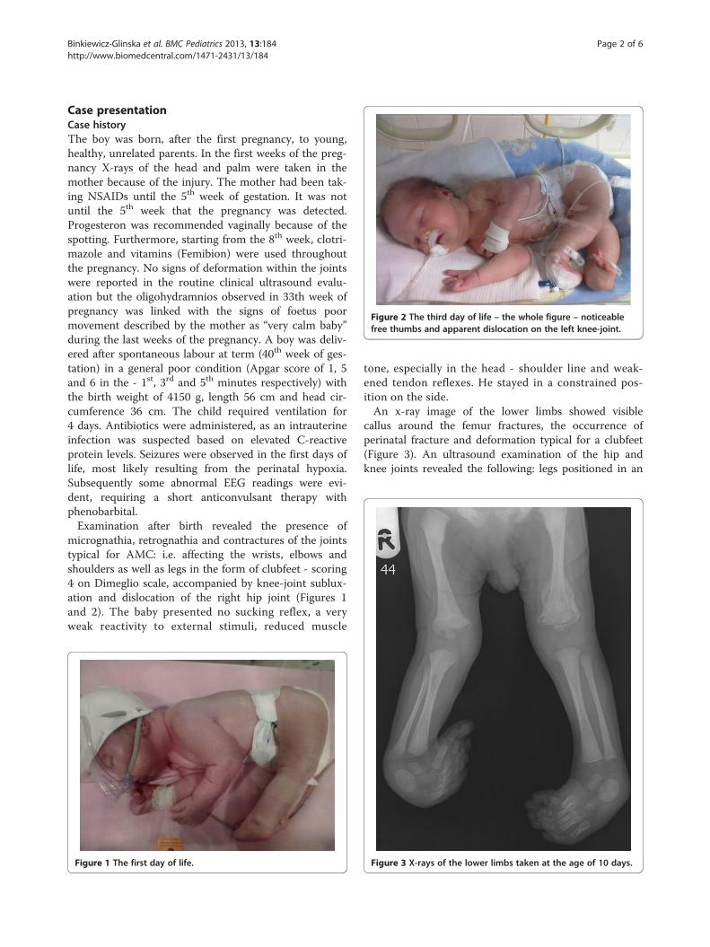
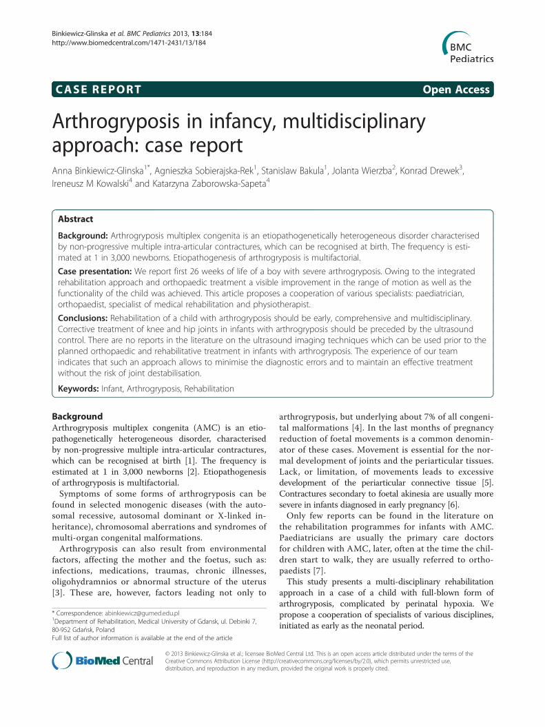
Figure 2 The third day of life – the whole figure – noticeablefree thumbs and apparent dislocation on the left knee-joint.
Binkiewicz-Glinska et al. BMC Pediatrics 2013, 13:184 Page 2 of 6http://www.biomedcentral.com/1471-2431/13/184
Case presentationCase historyThe boy was born, after the first pregnancy, to young,healthy, unrelated parents. In the first weeks of the preg-nancy X-rays of the head and palm were taken in themother because of the injury. The mother had been tak-ing NSAIDs until the 5th week of gestation. It was notuntil the 5th week that the pregnancy was detected.Progesteron was recommended vaginally because of thespotting. Furthermore, starting from the 8th week, clotri-mazole and vitamins (Femibion) were used throughoutthe pregnancy. No signs of deformation within the jointswere reported in the routine clinical ultrasound evalu-ation but the oligohydramnios observed in 33th week ofpregnancy was linked with the signs of foetus poormovement described by the mother as “very calm baby”during the last weeks of the pregnancy. A boy was deliv-ered after spontaneous labour at term (40th week of ges-tation) in a general poor condition (Apgar score of 1, 5and 6 in the - 1st, 3rd and 5th minutes respectively) withthe birth weight of 4150 g, length 56 cm and head cir-cumference 36 cm. The child required ventilation for4 days. Antibiotics were administered, as an intrauterineinfection was suspected based on elevated C-reactiveprotein levels. Seizures were observed in the first days oflife, most likely resulting from the perinatal hypoxia.Subsequently some abnormal EEG readings were evi-dent, requiring a short anticonvulsant therapy withphenobarbital.Examination after birth revealed the presence of
micrognathia, retrognathia and contractures of the jointstypical for AMC: i.e. affecting the wrists, elbows andshoulders as well as legs in the form of clubfeet - scoring4 on Dimeglio scale, accompanied by knee-joint sublux-ation and dislocation of the right hip joint (Figures 1and 2). The baby presented no sucking reflex, a veryweak reactivity to external stimuli, reduced muscle
Figure 1 The first day of life.
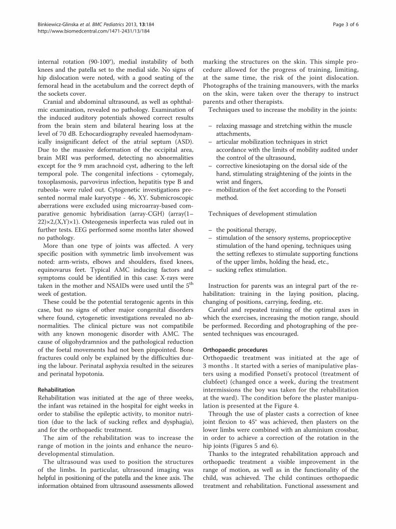
tone, especially in the head - shoulder line and weak-ened tendon reflexes. He stayed in a constrained pos-ition on the side.An x-ray image of the lower limbs showed visible
callus around the femur fractures, the occurrence ofperinatal fracture and deformation typical for a clubfeet(Figure 3). An ultrasound examination of the hip andknee joints revealed the following: legs positioned in an
Figure 3 X-rays of the lower limbs taken at the age of 10 days.
Binkiewicz-Glinska et al. BMC Pediatrics 2013, 13:184 Page 3 of 6http://www.biomedcentral.com/1471-2431/13/184
internal rotation (90-100°), medial instability of bothknees and the patella set to the medial side. No signs ofhip dislocation were noted, with a good seating of thefemoral head in the acetabulum and the correct depth ofthe sockets cover.Cranial and abdominal ultrasound, as well as ophthal-
mic examination, revealed no pathology. Examination ofthe induced auditory potentials showed correct resultsfrom the brain stem and bilateral hearing loss at thelevel of 70 dB. Echocardiography revealed haemodynam-ically insignificant defect of the atrial septum (ASD).Due to the massive deformation of the occipital area,brain MRI was performed, detecting no abnormalitiesexcept for the 9 mm arachnoid cyst, adhering to the lefttemporal pole. The congenital infections - cytomegaly,toxoplasmosis, parvovirus infection, hepatitis type B andrubeola- were ruled out. Cytogenetic investigations pre-sented normal male karyotype - 46, XY. Submicroscopicaberrations were excluded using microarray-based com-parative genomic hybridisation (array-CGH) (array(1–22)×2,(X,Y)×1). Osteogenesis inperfecta was ruled out infurther tests. EEG performed some months later showedno pathology.More than one type of joints was affected. A very
specific position with symmetric limb involvement wasnoted: arm-wrists, elbows and shoulders, fixed knees,equinovarus feet. Typical AMC inducing factors andsymptoms could be identified in this case: X-rays weretaken in the mother and NSAIDs were used until the 5th
week of gestation.These could be the potential teratogenic agents in this
case, but no signs of other major congenital disorderswhere found, cytogenetic investigations revealed no ab-normalities. The clinical picture was not compatibilewith any known monogenic disorder with AMC. Thecause of oligohydramnios and the pathological reductionof the foetal movements had not been pinpointed. Bonefractures could only be explained by the difficulties dur-ing the labour. Perinatal asphyxia resulted in the seizuresand perinatal hypotonia.
RehabilitationRehabilitation was initiated at the age of three weeks,the infant was retained in the hospital for eight weeks inorder to stabilise the epileptic activity, to monitor nutri-tion (due to the lack of sucking reflex and dysphagia),and for the orthopaedic treatment.The aim of the rehabilitation was to increase the
range of motion in the joints and enhance the neuro-developmental stimulation.The ultrasound was used to position the structures
of the limbs. In particular, ultrasound imaging washelpful in positioning of the patella and the knee axis. Theinformation obtained from ultrasound assessments allowed
marking the structures on the skin. This simple pro-cedure allowed for the progress of training, limiting,at the same time, the risk of the joint dislocation.Photographs of the training manouvers, with the markson the skin, were taken over the therapy to instructparents and other therapists.Techniques used to increase the mobility in the joints:
– relaxing massage and stretching within the muscleattachments,
– articular mobilization techniques in strictaccordance with the limits of mobility audited underthe control of the ultrasound,
– corrective kinesiotaping on the dorsal side of thehand, stimulating straightening of the joints in thewrist and fingers,
– mobilization of the feet according to the Ponsetimethod.
Techniques of development stimulation
– the positional therapy,– stimulation of the sensory systems, proprioceptive
stimulation of the hand opening, techniques usingthe setting reflexes to stimulate supporting functionsof the upper limbs, holding the head, etc.,
– sucking reflex stimulation.
Instruction for parents was an integral part of the re-habilitation: training in the laying position, placing,changing of positions, carrying, feeding, etc.Careful and repeated training of the optimal axes in
which the exercises, increasing the motion range, shouldbe performed. Recording and photographing of the pre-sented techniques was encouraged.
Orthopaedic proceduresOrthopaedic treatment was initiated at the age of3 months . It started with a series of manipulative plas-ters using a modified Ponseti's protocol (treatment ofclubfeet) (changed once a week, during the treatmentintermissions the boy was taken for the rehabilitationat the ward). The condition before the plaster manipu-lation is presented at the Figure 4.Through the use of plaster casts a correction of knee
joint flexion to 45° was achieved, then plasters on thelower limbs were combined with an aluminium crossbar,in order to achieve a correction of the rotation in thehip joints (Figures 5 and 6).Thanks to the integrated rehabilitation approach and
orthopaedic treatment a visible improvement in therange of motion, as well as in the functionality of thechild, was achieved. The child continues orthopaedictreatment and rehabilitation. Functional assessment and

Figure 4 Lower limbs at the age of 14 weeks. Panel A: Front view. Panel B: Right side view.
Binkiewicz-Glinska et al. BMC Pediatrics 2013, 13:184 Page 4 of 6http://www.biomedcentral.com/1471-2431/13/184
joint deformation assessment had been performed fourtimes since the child’s birth (Table 1).
DiscussionArthrogryposis in infancy is an interdisciplinary prob-lem. Children affected by this disease require the care ofpaediatricians, rehabilitation professionals as well asorthopaedists.Children with arthrogryposis can be subjected to
orthopaedic treatment with satisfactory results. Thecurrent technological advancements allow for an ortho-paedic interventions to minimise the limitations ineveryday activities [8]. In the case discussed here, wedealt with a number of joint deformities. Limb axis dis-tortion and significant contractures made it practicallyimpossible to determine the position of each anatomicalelement in clinical assessment of the infant. Clinical ob-servation suggested the presence of knee joint subluxa-tions as well as hip joint dislocations. We found noreports in the literature on the ultrasound imaging tech-niques which can be used prior to the planned ortho-paedic and rehabilitative treatment in infants witharthrogryposis. The experience of our team indicates
Figure 5 Manipulative casts – Improvement of knee flexionwith bar improving external rotation in the hip joints, the firstattempt to correct the foot by Ponseti – age 20 weeks.
that such an approach allows to minimise diagnostic er-rors and maintain an effective treatment without the riskof joint destabilisation.The ultrasound revealed features of the medial in-
stability of the knee joints resulting from the improperearlier rehabilitation by relatives during a short stay athome.At the moment it is not possible to determine what
level of motor and intellectual development the de-scribed child can reach [9]. Parallel occurrence ofarthrogryposis, micrognathia, retrognathia, and theconsequences of perinatal hypoxia make the prognosisdifficult and uncertian. Intensive rehabilitation startedearly, supported by daily supervised exercises con-ducted by the parents gives the child with arthrogrypo-sis an opportunity to improve the range of motion inthe joints and to reduce the need for subsequent rad-ical invasive corrections [10]. Treatment of clubfoot inarthrogryposis with Ponseti’s method is believed bymany researchers to be an effective method helping to
Figure 6 The improvement in the external rotation of the hipjoints – age 20 weeks.
Table 1 Range of the joint mobility before and after rehabilitation.
Limited motion direction Before rehabilitation(3 week of life)
After rehabilitation(26 week of life)
Improvement [°]
Shoulder joint L and R flexion 90° 170° 80°
extension 20° 100° 80°
Elbow joint L flexion 30° 120° 90°
Elbow joint R straightening −90° −60° 30°
Carpal joint L and R straightening - 20° stiffness 20°
extension −10° stiffness 10°
Hip joint L and R straightening −115° −45° 70°
extension 20° 45° 25°
external rotation −135° −115° 20°
Knee joint L and R straightening −15° −5° 10°
flexion 0° 45° 45°
Binkiewicz-Glinska et al. BMC Pediatrics 2013, 13:184 Page 5 of 6http://www.biomedcentral.com/1471-2431/13/184
avoid radical surgery [11]. But despite the intensiverehabilitation and corrective actions the surgery forarticular deformations may be unavoidable. In 76% ofpatients with arthrogryposis foot joints require surgicalcorrection, knee joints need intervention in 39% ofpatients, and the hips in 18% of cases [10]. Surgicalrelease of the soft tissues with the total release of thetendons is recommended before the child learns towalk. In the case described, a subtalar release treat-ment is planned as the use of plasters, and even releaseof the soft tissue in the case of arthrogryposis does notprovide lasting results [11]. The literature emphasisesthe importance of the earliest possible initiation of thecorrective action, with the best results obtainable inthe first months of life [12]. While focusing on correctingthe joint deformities, the child’s developmental stimulationshould not be forgotten. Due to the articular restrictions aninfant with arthrogryposis is less able to explore the world,thus the development of cognitive and motor function ishampered [13,14]. In case of infants with retrognathia thecontrol of feeding is an important issue. Due to the suckingand swallowing disturbances it is extremely important toadjust the appropriate feeding method to avoid problemssuch as aspiration pneumonia or malnutrition.Early rehabilitation of a child with arthrogryposis
requires the involvement of the parents/guardians.Unfortunately, in the face of serious illness of the childvery often there is a relationship crisis between theparents and even the disintegration of a relationship.Parents of a child burdened with severe congenital dis-ease often experience disappointment, frustration anda sense of losing the control. The care for a sick childusually imposes changes of the everyday life, changesof the long-term life plans, which may result in depres-sion and even in a post traumatic stress disorder [15].
Conclusion
1. Rehabilitation of a child with arthrogryposis shouldbe early, comprehensive and multidisciplinary.
2. Corrective treatment of knee and hip joints in thecase of infants with arthrogryposis should bepreceded by the ultrasound control.
ConsentWritten informed consent was obtained from the pa-tient’s parents for publication of this Case report andany accompanying images. A copy of the written consentis available for review by the Editor of this journal.
Competing interestsThe authors declare that they have neither financial nor non-financial com-peting interests.
Authors’ contributionsABG – originator of the project in its final form, exercised rehabilitationconsultations, commissioned rehabilitation activities, coordinatedmultidisciplinary team activities, obtained funds for the publication,translated the text from Polish and was responsible for drafting a coverletter. ASR – performed all rehabilitation activities, assessed the patient interms of functionality and was responsible for collecting data and draftingthe article. SB – substantial supervision. JW – was responsible for conductingthe paediatric proceedings and edited the introduction to the article. KD –performed all consultations and orthopaedic actions, was consulted on theorthopaedic aspect of the article. IMK – made the final adjustments to thetext and literature. KZS – introduced final modifications to the article andtable. All authors approved the final version of the article.
Authors’ informationABG - MD, specialist of medical rehabilitation, member of Polish Rehabilita-tion Society, assistant in the Department of Rehabilitation.ASR- Physiotherapist, PhD, assistant in the Department of Rehabilitation,member of Polish Physiotherapy Association.SB - MD, associate professor in Medical University of Gdansk, specialist ofinternal medicine and medical rehabilitation, head of the Department of Re-habilitation, member of Polish Society of internal Medicine, member of Polish
Binkiewicz-Glinska et al. BMC Pediatrics 2013, 13:184 Page 6 of 6http://www.biomedcentral.com/1471-2431/13/184
Rehabilitation Society, member of Rehabilitation, Physical Culture, Social Inte-gration Committee of Polish Academy of Science.JW - MD, PhD, specialist of paediatrics and clinical genetics, head of the De-partment of Infant Pathology, member of European and Polish Human Gen-etics Society, member of Polish Registry of Congenital MalformationsWorking Group, member of Advisory Council – Cornelia de Lange SyndromeAssociation.KD - MD, PhD, specialist Of Orthopaedics, assistant in Department of Ortho-paedics and Traumatology, member of Polish Orthopaedics and Traumatol-ogy Society, Polish Foot and Ankle Society and European Foot and AnkleSociety.IMK - MD, associate professor in University of Warmia and Mazury, specialistof paediatrics and medical rehabilitation, director of Rehabilitation Depart-ment, member of Polish Rehabilitation Society, voivodeship regional consult-ant of medical rehabilitation.KZS - MD, specialist of medical rehabilitation, assistant in Department of Re-habilitation, member of Polish Rehabilitation Society and Polish MedicalWriters Association.
AcknowledgementsWe thank the parents of the patient for their co-operation and support andfor providing their consent for the publication.We thank Dr Piotr Sieliwonczyk for professional ultrasound examination.
Author details1Department of Rehabilitation, Medical University of Gdansk, ul. Debinki 7,80-952 Gdańsk, Poland. 2Department of General Nursing University ofGdansk, 80-952 Gdańsk, Poland. 3Department of Orthopedics andTraumatology, Medical University of Gdansk, ul. Powstancow Warszawskich 1/ 2, 80-152 Gdansk, Poland. 4Department of Rehabilitation, University ofWarmia and Mazury in Olsztyn, ul. Oczapowskiego 2, 10-719 Olsztyn, Poland.
Received: 19 December 2012 Accepted: 31 October 2013Published: 11 November 2013
References1. Taricco LD, Aoki SS: Rehabilitation of an adult patient with arthrogryposis
multiplex congenital treated with an external fixator. Am J Phys MedRehabi l 2009, 88(Suppl 5):431–434.
2. Darin N, Kimber E, Kroksmark AK, Tulinius M: Multiple congenitalcontractures: birth prevalence, etiology, and outcome. J Pediatr 2002,140(Suppl 1):61–67.
3. Alves PV, Zhao L, Patel PK, Bolognese AM: Arthrogryposis: diagnosis andtherapeutic planning for patients seeking orthodontic treatment ororthognathic surgery. J Craniofac Surg 2007, 18(Suppl 4):838–843.
4. Kossakowska-Krajewska A: Analysis of the factors which mayhaveinfluenced the incidence of congenital malformations in children bornin the provinc of Warmia and Mazury between 1999 and 2000. Pol AnnMed 2009, 16(Suppl 1):78–93.
5. Bamshad M, Van Heest AE, Pleasure D: Arthrogryposis: a review andupdate. J Bone Joint Surg Am 2009, 91(Suppl 4):40–46.
6. Wierzba J, Piotrowski K, Limanowka M: Arthrogryposis: pre- and postnataldifferantiation, Clinical cause of the Pena-Shokeir syndrome. FMR 2011,5(Suppl 4):352–358.
7. Eriksson M, Gutierrez-Farewik E, Brostroom E: Gait in children with arthro-gryposis multiplex congenital. J Child Orthop 2010, 4:21–31.
8. Stahelli L, Hall J, Jaffe K: Physical and Occupational Therapy. InArthrogryposis. A text atlas. Cambridge University Press: Global Help;2008:87–97.
9. Hielkema T, Hamer E, Reinders-Messelink H: LEARN 2 MOVE 0–2 years: ef-fects of a new intervention program in infants at very high risk for cere-bral palsy; a randomized controlled trial. BMC Pediatr 2010, 10:Suppl 76.
10. Eidelmann M, Katzman A: Treatment of Arthrogryposis foot deformitieswith Taylor spatial frame. J Pediatr Orthop 2011, 4:429–434.
11. Kowalczyk B, Lejman T: Short-term experience with Ponseti casting andthe Achilles tenotomy method for clubfeet treatment in arthrogryposismultiplex congenita. J Child Orthop 2008, 2:365–371.
12. Ezaki M: An approach to the upper limb in arthrogryposis. J PediatrOrthop 2010, 30(Suppl 2):57–62.
13. Blauw-Hospers CH, De Graaf-Peters VB, Dirks T: Does early intervention ininfants at high risk for a developmental motor disorder improve motor
and cognitive development? Neuroscience & Biobehavioral Reviews 2007,31:1201–1212.
14. Oberg G, Campbell S, Girolami G: An early intervention program toimprove motor outcome in preterm infants: a randomized controlledtrial and a qualitative study of physiotherapy performance and parentalexperiences. BMC Pediatr 2012, 12:Suppl 15.
15. Singer G, Ethridge B, Aldana S: Primary and secondary effects of parentingand stress management interventions for parents of children withdevelopmental disabilities: A meta-analysis. MRDD Research Reviews 2007,13(Suppl 4):357–369.
doi:10.1186/1471-2431-13-184Cite this article as: Binkiewicz-Glinska et al.: Arthrogryposis in infancy,multidisciplinary approach: case report. BMC Pediatrics 2013 13:184.
Submit your next manuscript to BioMed Centraland take full advantage of:
• Convenient online submission
• Thorough peer review
• No space constraints or color figure charges
• Immediate publication on acceptance
• Inclusion in PubMed, CAS, Scopus and Google Scholar
• Research which is freely available for redistribution
Submit your manuscript at www.biomedcentral.com/submit