arthritis from 36,000 feet an overview paul f. howard md, facp, facr director, arthritis health 9097...
TRANSCRIPT

Arthritis from 36,000 feetAn Overview
Paul F. Howard MD, FACP, FACR
Director, Arthritis Health
9097 E. Desert Cove #100
Scottsdale, AZ, 85260 [email protected]

Close Up View =Individual Diagnoses

Overview from 36,000 ft

Overview
•Impact of Arthritis–Prevalence –Economic–Social
•Clinical Approach to Arthritis
-Monoarthritis -Polyarthritis-Gout
•Laboratory Tests

Arthritis - Scope of the Problem
• Increasing Incidence– Affects ~46 million Americans
• 15% of U.S. population has some form of arthritis or rheumatic condition• Estimated to increase to 18.2% by 2020
• Second leading cause of work disability– Arthritis is a more frequent cause of functional impairment
(activity limitation) than heart disease, cancer or diabetes
– Health-related quality of life measures are consistently worse for people with arthritis

Escalating Prevalence of Arthritis
0
10
20
30
40
50
60
1990 2020
Arthritis Prevalence
Arthritis Causing ActivityLimitation
This represents a 57% increasein 30 years

Economic Impact
Arthritis and musculoskeletal conditions account for 13% of all health care spending
• 315 million physician visits per year• 8,000,000 hospitalizations• 1.5 billion days of restricted activity per year
• #1 most common reason for doctor visits• #2 most common reason for hospitalization• #4 most common reason for surgery
Healthy People 2010; Yelin, Callahan. Arthritis Rheum; 38:1351-1362

Economic Impact
Arthritis and other rheumatic conditions
(AORC)
Direct Medical Costs $ 80.8 billion
Indirect Costs $ 47.0 billion
Total Costs $ 127.8 billion
The total cost of arthritis accounts for nearly 38% of all musculoskeletal conditions
Arthritis and Rheumatism 2007;56(5):1397-1407

Work Loss Due to Illness
47%
5%4%5%
4%
7%
5%
23%Musculoskeletal
Upper Respiratory
Headache/Migraines
Lower Respiratory
Upper GI
Cardiovascular
Ob/Gyn
Other
Musculoskeletal Illness is #1 Cause for Loss of Work

Quality of Care Issues
• 40% of US individuals reporting chronic joint symptoms were not diagnosed by a doctor
• Up to 50 % of those diagnosed are not receiving treatment
• 30-53% of patients diagnosed with arthritis do not know what kind they have
MMWR 1998; 47(17):345-350

Cause of Arthritis
• Traumatic• Mechanical / Degenerative • Metabolic-------------------------------------------• Infectious• Inflammatory • Crystal related-------------------------------------------• Malignancy 3 Non Inflammatory
3 Inflammatory1 other

Data Collection and Decision Making
Careful History and Physical Exam will yield the 1st set of decision in the differential diagnosis
Acute vs ChronicArticular vs Periarticular
Non inflammatory vs Inflammatory Cadence/pattern of involvement Additional - associated findings
Note - labs or x-rays are not initially required

Onset • Acute or chronic
– Extremely rapid (seconds to minutes)• Internal derangement, fracture, trauma, loose body
– Acute onset (several hours to 2 days)• Typical of most inflammatory arthritis, bacterial
infection or crystal arthritis
– Long standing problems - subacute or chronic• Acute on chronic problem (flare of OA or RA)• Second superimposed process (infection)

Localization
What site is involved? – Joint– Adjacent bone– Soft tissue
• Ligaments, tendons, bursae
– Referred pain• Nerve root impingement - sciatica• Entrapment neuropathy -carpal tunnel• Pathology in another joint
– hip arthritis → referred knee pain
– Subacromial Bursitis → referred upper arm pain
You must determine where the pain is coming from !

Character of the Arthritis
Inflammatory vs Mechanical– Inflammatory
• Waxing and waning disease activity• >1 hour of morning stiffness• Improvement with use• Systemic symptoms (fever or malaise)
– Mechanical • Pain after use• Improvement with rest• No systemic symptoms

Physical Examination
• Isolated vs Multiple Sites Mono
Oligo (Pauci)
Polyarthritis
• Symmetric / Asymmetric involvement

Pattern of Pain
Cadence and Pattern of Involvment
•Episodic
•Migratory
•Additive
•Progressive

Additional - Associated findings
•Demographic and historical information
•Family history
•Social and travel information
•Physical examination findings

Case #1 20 year old female student (accounting major) at ASU present with a swollen left knee x 1 day
Ski trip with friends
Woke this am with pain, stiffness and severe swelling left knee
Very stiff for hours this morning.
PMH +UTI 10 days ago treated with AB -x 2 days and symptoms resolved. Sexually active with new partner on ski trip.
ROS No trauma, rashes, or other pain. + fever, chills, nausea
Meds/Supplements None
Exam T 101.4 BP 132/90 P 124 R 20
Ill appearing, sweating, with swollen left knee

Knee EffusionCase Example

Building a Differential DX Data Collection - Decision Making
Careful History and Physical Exam will yield the 1st set of decision in the differential diagnosis
Acute vs Chronic
Articular vs Periarticular
Non inflammatory vs Inflammatory
Mono, Oligo or Poly(articular)
Cadence: Episodic, Migratory, Additive, Progressive
Additional - associated findings

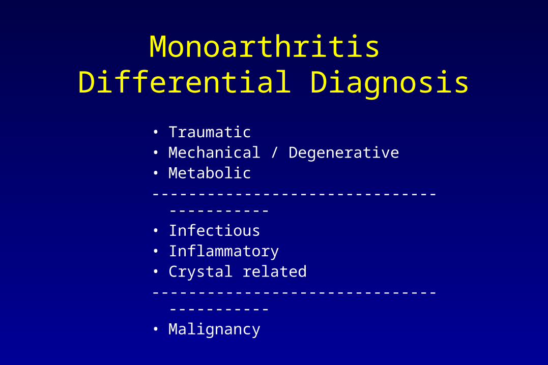
Monoarthritis Differential Diagnosis
• Traumatic• Mechanical / Degenerative • Metabolic------------------------------------------• Infectious• Inflammatory • Crystal related------------------------------------------• Malignancy

Diagnostic Studies
• CBC WBC 15,5000 with left shift
• CMP normal
• U/A normal
• ESR 25 mm/hr
• Arthrocentesis - Synovial fluid 58,000 WBC 1000 RBC
• Knee X-ray - no apparent fracture
• Pelvic exam and cultures, GC PCR,

Monoarthritis Differential Diagnosis
• Traumatic• Mechanical / Degenerative • Metabolic------------------------------------------• Infectious• Inflammatory • Crystal related------------------------------------------• Malignancy

Septic Arthritis
• First question: to hospitalize or not?• Intense local pain• Resistive to motion• Swelling, heat, redness• Persons at high risk
– Steroid therapy– Immunodeficiency/Immunosuppression– Diabetes– IV drug abuse– Other focus of infection (eg. UTI, pneumonia, etc.)
• Staph aureus = most common• Ortho Consult
– Repetitive arthrocentesis vs. open drainage in OR

Causes of Septic Arthritis
• Bacterial– Staph E. Coli Lyme Disease– Strep Pseudomonas Anaerobes
• Tuberculosis– TB vs atypical TB
• Fungal– Coccidiodomycosis (Valley Fever)– Candida– Histoplasmosis
Arthrocentesis - Arthrocentesis - Arthrocentesis

Septic Arthritis
Osteopenia
Joint spaceNarrowing
Erosions
Dissolutionof joint

Septic Arthritis Tuberculosis

Case #2 20 year old female student (accounting major) at ASU present with a swollen right knee x 2 day
Ski trip with friends
Woke this am with pain, stiffness and severe swelling right knee
PMH +UTI 10 days ago treated with AB -x 2 days and symptoms resolved. Sexually active with new partner on ski trip.
ROS No trauma or other pain. + fever, chills, nausea, rash, am stiff
Meds/Supplements None
Exam T 101.4 BP 132/90 P 124 R 20
Ill appearing, sweating, with swollen right knee
Rash on legs and arms

Knee EffusionCase Example

Associated Findings
Pustular/EncrustedSkin Lesions

Building a Differential DX Data Collection - Decision Making
Careful History and Physical Exam will yield the 1st set of decisions in the differential diagnosis
Acute vs Chronic
Articular vs Periarticular
Non inflammatory vs Inflammatory
Mono, Oligo or Poly(articular)
Cadence: Episodic, Migratory, Additive, Progressive
Additional - associated findings

Monoarthritis Differential Diagnosis
• Traumatic• Mechanical / Degenerative • Metabolic------------------------------------------• Infectious• Inflammatory • Crystal related------------------------------------------• Malignancy

Diagnostic Studies
• CBC WBC 15,5000 with left shift • CMP normal• U/A normal• ESR 25 mm/hr• Arthrocentesis - Synovial fluid 15,600 WBC
1000 RBC• Knee X-ray - no apparent fracture + effusion• Pelvic Exam – culture and GC PCR -

Monoarthritis Differential Diagnosis
• Traumatic• Mechanical / Degenerative • Metabolic------------------------------------------• Infectious• Inflammatory • Crystal related------------------------------------------• Malignancy

Gonococcal ArthritisAssociated Findings
Pustular/EncrustedSkin Lesions

Case #3 20 year old female student (accounting major) at ASU present with a swollen right knee x 2 day
Ski trip with friends
Woke yesterday am with pain, stiffness and severe swelling right knee
PMH +UTI 10 days ago treated with AB -x 2 days and symptoms resolved. Sexually active with new partner on ski trip.
ROS No trauma, rashes, or other pain.
Meds/Supplements None
Exam T 98.6 BP 132/90 P 88 R 16
Healthy appearing in obvious distress due to pain and swelling in the right knee

Knee EffusionCase Example

Building a Differential DX Data Collection - Decision Making
Careful History and Physical Exam will yield the 1st set of decisions in the differential diagnosis
Acute vs Chronic
Articular vs Periarticular
Non inflammatory vs Inflammatory
Mono, Oligo or Poly(articular)
Cadence: Episodic, Migratory, Additive, Progressive
Additional - associated findings
Monoarthritis Differential Diagnosis
• Traumatic• Mechanical / Degenerative • Metabolic------------------------------------------• Infectious• Inflammatory • Crystal related------------------------------------------• Malignancy

Diagnostic Studies
• CBC normal• CMP normal• U/A normal• ESR normal• Arthrocentesis - Synovial fluid 1,750,000 RBC
100 WBC• Knee X-ray - no apparent fracture

Monoarthritis Differential Diagnosis
• Traumatic• Mechanical / Degenerative • Metabolic------------------------------------------• Infectious• Inflammatory • Crystal related------------------------------------------• Malignancy

Hemarthrosis Differential DX
•Trauma
•Infection
•Malignancy –Lymphoma, sarcoma, PVS
•Metabolic - Bleeding Disorder –Primary coagulopathy–Medications–Platelet disorder–Malignancy

Case # 4 45 year old female accounting professor at ASU present with a swollen left knee x 2 day
Ski trip with friends
Woke yesterday am with pain, stiffness and swelling left knee
PMH +UTI 10 days ago treated with AB -x 2 days and symptoms resolved. + chronic pain in knee, flares x 2 in past three years related to activity
ROS No trauma, rashes, or other pain. No fever, chills, systemic sympt
Meds/Supplements None
Exam T 98.6 BP 132/90 P 88 R 16
Healthy appearing in obvious distress due to pain and swelling in the left knee, no increase in warmth of the knee, no tenderness

Knee Effusion

Building a Differential DX Data Collection - Decision Making
Careful History and Physical Exam will yield the 1st set of decisions in the differential diagnosis
Acute vs Chronic
Articular vs Periarticular
Non inflammatory vs Inflammatory
Mono, Oligo or Poly(articular)
Cadence: Episodic, Migratory, Additive, Progressive
Additional - associated findings

Monoarthritis Differential Diagnosis
• Traumatic• Mechanical / Degenerative • Metabolic------------------------------------------• Infectious• Inflammatory • Crystal related------------------------------------------• Malignancy

Diagnostic Studies
• CBC WBC normal
• CMP normal
• U/A normal
• ESR 5 mm/hr
• Arthrocentesis - Synovial fluid 1000 RBC 200
WBC
• Knee X-ray - no apparent fracture + joint space narrowing, sclerosis
and spur formation

Monoarthritis Differential Diagnosis
• Traumatic• Mechanical / Degenerative • Metabolic------------------------------------------• Infectious• Inflammatory • Crystal related------------------------------------------• Malignancy

Mechanical / Traumatic / Metabolic
• Osteoarthritis – Most common form of arthritis– Single or multiple joints– Asymmetric – Not inflammatory
– Hands, spine, hip, knee most common sites

Osteoarthritis

Osteoarthritis
Joint spacenarrowing
Sclerosis
Osteophytes
KellgrenGrading I - IV

Osteoarthritis
Grade IV
SevereJoint spaceNarrowing
Sclerosis
Osteophytosis

Osteoarthritis
Most Common Sites
SpineCervicalLumbar Thoracic
HandsKnees Hips Feet Other

Osteoarthritis

Treatment of Osteoarthritis
• Joint Protection • Conditioning around damaged joints • Weight reduction• Analgesics • MSM, Glucosamine• NSAID’s • Bracing • Surgery

Case # 5 45 year old male accounting professor at ASU present with a swollen left great toe and ankle x 2 days
Ski trip with friends
Woke yesterday am with pain, stiffness and swelling left ankle
PMH Occurred last June after a golf tournament - resolved in 7 days
ROS No trauma, rashes, or other pain. + fever, no chills or systemic symptoms
Meds/Supplements None
Exam T 98.6 BP 132/90 P 88 R 16
Healthy appearing in obvious distress due to pain and swelling
in the left ankle and great toe (1st MTP)


Building a Differential DX Data Collection - Decision Making
Careful History and Physical Exam will yield the 1st set of decisions in the differential diagnosis
Acute vs Chronic
Articular vs Periarticular
Non inflammatory vs Inflammatory
Mono, Oligo or Poly(articular)
Cadence: Episodic, Migratory, Additive, Progressive
Additional - associated findings

Monoarthritis Differential Diagnosis
• Traumatic• Mechanical / Degenerative • Metabolic------------------------------------------• Infectious• Inflammatory • Crystal related------------------------------------------• Malignancy

Diagnostic Studies
• CBC WBC 13,000 with left shift
• CMP normal
• U/A normal
• ESR 25 mm/hr
• Arthrocentesis - refused
• Knee X-ray - no apparent fracture soft tissue swelling about ankle and great toe

Monoarthritis Differential Diagnosis
• Traumatic• Mechanical / Degenerative • Metabolic------------------------------------------• Infectious• Inflammatory • Crystal related------------------------------------------• Malignancy

Podagra = ( most of the time)

Monoarthritis Differential Diagnosis
• Traumatic• Mechanical / Degenerative • Metabolic------------------------------------------• Infectious• Inflammatory • Crystal related------------------------------------------• Malignancy

Podagra = gout ( most of the time)
50% 1st episode
90% of all gout patients over time will have podagra

Gout Clinical Presentation
History of recurrent, self-limited (3-10 days) attack
Severe pain and inflammation extending into the local tissue
Abrupt onset of severe joint inflammation often at night
May have hyperuricemia
Monosodium urate crystals - + 95% time arthrocentesis
Podagra is characteristic (75% of cases) but not pathognomonic
Gradually increasing to become polyarticular and chronic (years)

Gout
•Impact of Gouty Arthritis–Prevalence –Impact Economic Social
•Clinical Presentations
•Diagnosis
•Treatment - Acute and Long Term Management

Clinical Presentations • Acute episodic inflammatory arthritis
–Lower extremity - esp great toe 50% 1st episode 90% cases–Men in middle life - obese, drink regular alcohol–Women - post menopause, on diuretics, heavy alcohol drinkers
–Upper extremities in chronic tophaceous gout - - esp DIP’s
–Associations with HTN, hypertriglyceridemia, renal insufficiency
–HPRT deficiency, PRPP synthetase overactivity, heritable renal disease should be suspected in young adults, adolescents or young adults with gout.

Clinical Presentations

Clinical Presentations

Diagnosis of Gout
Rome Criteria
• Serum urate > 7.0 men , >6.0 women
• Acute onset inflammatory arthritis, resolves 1-2 wk
• Presence of urate crystals in synovial fluid
• Presence of tophi

Gout = Urate Crystals
Needle shaped -pointed
Negatively Birefringent
YUP yellow - urate - parallel

What Is New in Gout Management
Old New
Episodic Arthritis Chronic Accumulation UA
Focus on Joints Focus on Serum Uric Acid
Episodic Tx Chronic Uric Acid lowering No prophylaxsis 6 months prophyaxsis Colchicine or low dose NSAID’s
Expanded drug options

Treatment - Acute Gout
Goal is to reduce inflammation
Key points It will improve usually over days -We can help it along
NSAID’s Indomethacin - or any Nsaid in full dose.
Caution in “older patients, or renal insufficiency, HTN, GI Hx
Steroids Intraarticular injection - triamcinalone, methylprednisilone
Intramuscular injection - triamcinalone, methylprednisilone
Oral burst of steroids - prednisone, methylprednisone
x 3-6 day
Colchicine Not preferred but can use 1.2mg followed by 0.6 mg x1 day

Long Term Management
• seek and correct contributing factors to hyperuricemia
–Regular alcohol intake ( esp beer)–High purine diet –Obesity–Diuretic therapy–Renal insufficiency–hypertension

Long term management of Gout
Begin a uric acid lowering agent - allopurinol or febuxostat
Target level of uric acid < 6.0mg
Ensure hitting target - increase meds as needed
Colchicine prophylaxis 1st six to twelve month
Once started, stay with stable dose of uric acid lowering agent irrespective of the occasional gout flares
Goal is to reduce uric acid - not prevent gout flares !!!

Other Crystal Induced Arthritis
• Pseudogout/CPPD
• Calcium Oxylate
• Basic Calcium Phosphate
• Cholesterol
• Steroids - iatrogenic

Calcium Pyrophosphate Crystals Pseudogout
Rods -blunt
+birefringent

Condrocalcinosis

Case # 6 20 year old female student (accounting major) at ASU present with a swollen right knee x 2 day
Ski trip with friends
Woke yesterday am with pain, stiffness and severe swelling right knee
PMH +UTI 28 days ago treated with AB -x 2 days and symptoms resolved. Sexually active with new partner on another ski trip one month ago.
ROS No trauma, rashes, or other pain. + conjunctivitis one week ago
Meds/Supplements None
Exam T 98.6 BP 132/90 P 88 R 16
Healthy appearing in obvious distress due to pain and swelling in the right knee which is warm, right eye injected with no discharge

Knee Effusion

Building a Differential DX Data Collection - Decision Making
Careful History and Physical Exam will yield the 1st set of decisions in the differential diagnosis
Acute vs Chronic
Articular vs Periarticular
Non inflammatory vs Inflammatory
Mono, Oligo or Poly(articular)
Cadence: Episodic, Migratory, Additive, Progressive
Additional - associated findings

Monoarthritis Differential Diagnosis
• Traumatic• Mechanical / Degenerative • Metabolic------------------------------------------• Infectious• Inflammatory • Crystal related------------------------------------------• Malignancy

Diagnostic Studies
• CBC WBC normal
• CMP normal
• U/A 2+ WBC, - nitrates, gram stain
• ESR 45 mm/hr
• Arthrocentesis - Synovial fluid 1000 RBC20,500 WBC
• Knee X-ray - no apparent fracture
+ effusion

Monoarthritis Differential Diagnosis
• Traumatic• Mechanical / Degenerative • Metabolic------------------------------------------• Infectious• Inflammatory • Crystal related------------------------------------------• Malignancy

Inflammatory Monoarthritis
• Reactive Arthritis (Post infectious)
• Psoriatic Arthritis
• Ankylosing Spondylitis
• Juvenile Inflammatory Arthritis JIA
• Onset of a chronic polyarthritis - ? rheumatoid arthritis

Malignancy
• Osteosarcoma
• Metastatic Tumor
• Pigmented Villonodular Synovitis
• Rare tumor of cartilage, bone and muscle

PolyarthritisDifferential Diagnosis
• Traumatic • Mechanical / Degenerative • Metabolic ---------------------------------------------• Infectious - extremely rare • Inflammatory• Crystal related----------------------------------------------• Malignancy

Metabolic Arthropathy
• Hematologic– Coagulopathies– Dialysis Arthropathy– Hemoglobinopathies
• Endocrine– Hypothyroidism – Hyperthyroidism – Adrenal syndromes

Inflammatory Polyarthritis
• Hypersensitivity - Serum Sickness Arthritis
• RA• SLE• Sjogren’s Syndrome• Scleroderma • Psoriatic Arthritis • Reiter’s Disease • Vasculitis

Additive Chronic Symmetric Inflammatory Polyarthritis

Additive Chronic Symmetric Inflammatory Polyarthritis

Clinical Features
Symmetric inflammatory progressive destructive polyarthritis

Characteristics of RA
Demographics
World wide
Female > male 3:1
Onset - any age 40-50 most common
+ Genetic predilection
Criteria based not laboratory based

RA Criteria ( ACR)
AM stiffness > 1 hour
Three or more joint areas
Symmetric
Involving hands
+RF
Joint erosions
Nodules ACR 1987

Stages of RAEarly Intermediate Late
Courtesy of J. Cush, 2002.

S
ever
ity
(arb
itra
ry u
nit
s)
0
Duration of Disease (years)
5 10 15 20 25 30
RA Progression RA Progression
Early RA Intermediate Late
Graph: Adapted from Kirwan JR. J Rheumatol. 2001;28:881-886.Photo: Copyright © American College of Rheumatology.
InflammationDisabilityRadiographs
© ACR

Treatment and Outcomes of RA
Aggressive control of inflammation NSAID’s -- Steroids -- Synthetic DMARD’s -- Biologic DMARD’s
Historically RA results in inexorable chronic pain Erosions begin within months and progressive destructive erosive change in multiple joints Within 10 years, nearly 50% disabled -- reduce QOL7-10 year reduced life expectance
Today, 80% of RA is able to be arrested =Arresting disease symptoms - joint damage - disability = Higher
QOL

Sjogren’s Syndrome
• Second most common autoimmune arthritis - 0.5% of pop • Immunologic disorder of B Cells
– Dry Eyes and Mouth– Polyarthritis - non erosive – Adenopathy and Glandular Hypertrophy– Multisystem lymphocytic infiltration
– Women predominate 9:1 Onset 30-50 years old– progressive but treatable and controllable
– MUST BE RECOGNIZED - THIS CONDITION IS OFTEN NOT CONSIDERED IN A DIF DX OF JOINT PAIN.

Features of Sjogren’s Syndrome

Systemic Lupus Erythematosus
Multisystem autoimmune disorder with immune complexes
Rashes Polyarthritis - non deformingGlomerulonephritis Pleuropericarditis
Central Nervous System Hematologic - low WBC, RBC, PlateletsSerologic
ACR SLE Criteria 4 of 11 findings needed for what?

Systemic Lupus Erythematosus
Malar Rash Sun sensitivityDiscoid LupusOral ulcersNon deforming polyarthritisRenal disease Pericarditis PleurisyNeurologic Hematologic Low counts + ANA+ dsDNA, low C3/C4 + SM Ab

Demographics of SLE
Worldwide distribution
Female > male 9:1
Age most common 13 - 30
Younger = more renal disease
Darker skin races have greater prevalence
+ genetic links Population studies, twins

Treatments for SLE
• Treatment is directed toward reducing inflammation to prevent damage
• NSAID’s• Antimalarial agents • Steroids • Cytotoxic - MTX, Myophenylate , Cyclophosphamide• Anti B cell tx - Rituximab
Treatments have resulted reduced mortality - 50% to <5%

Scleroderma

Scleroderma
Systemic disorder of fibrosis of skin and multiple organs
Joint - symmetric inflammatory polyarthritis Skin - sclerodactyl - scleroderma - telangectasia Lungs - interstitial fibrosisGI - esophageal dysmotilityRenal - afferent arteriole fibrosis - HTN - renal crisisNeuro - neuropathies

Psoriatic Arthritis

Psoriatic Arthritis
• Skin and/or nail involvement
• Oligoarthritis
• Classic - DIP
• Polyarthritis
• Arthritis Mutilans
• Spondyloarthropathy

Arthritis Type?

Reactive Arthritis
Asymmetric oligo-polyarthritisRash - lesions on hands, feet, penis Urethritis Iritis
Outcome - 50% remission 25% episodic 25% chronic
Cause -- Post infectious – Urethritis - Chlamydia– Dysentery - bacterial

Ankylosing Spondylitis
• Inflammatory Spinal Involvement - SI joints start
• Progressive Fusion
• Common peripheral joint involvement
• Male 3:1
• + HLA B 27 90%
• Treat stepwise and aggressively to control inflammation and preserve function
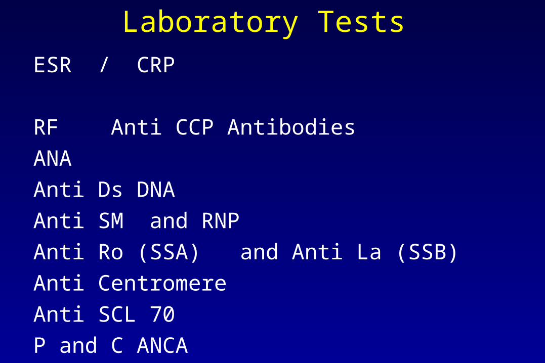
Laboratory Tests ESR / CRP
RF Anti CCP Antibodies
ANA
Anti Ds DNA
Anti SM and RNP
Anti Ro (SSA) and Anti La (SSB)
Anti Centromere
Anti SCL 70
P and C ANCA

ESR CRP
•Both tests are Acute Phase Reactants •Reflect ongoing inflammation•Monitoring levels helps to assess inflammatory disease activity
•FACTORS - INCREASE ACUTE PHASE REACTANTS
Infections
Trauma
Malignancy
Inflammmatory rheumatic diseases
Reactions to medications

ESR• ESR “sed rate” of RBC’s in glass column• RBC’s are held in suspension by their negative surface charge
from Sialic adic residues. • Indirect measure of positively charged proteins between the RBC’s
fibrinogen dominant protein
haptoglobin, alpha 1 antitrypsin, aerum amyloid protein, Ig M
IgG, ceruloplasm,
• Greater the agglutination or RBC’s , the faster the rate of descent • Normal < 20 mm/hr
Affected by Age, testing procedures, anemia

ESR- CRPCRP • Measurement of a single acute phase reactant • Can rise > 100 x baseline level in certain inflammatory conditions• Not affected by age, other acute phase reactants-------------------------------------------------------------------------------------------------------------
- ESR CRP
Affected by other factors Isolated valueHalf Life 10 days 2 days Normal Values Wide Range Narrow Range
20-48 mm/hr 0.2-1.0mg/dlCost $3 $35
Clinical knowledge of the patient is more important than any laboratory test result

ESR - CRP
Discrepancies between ESR and CRP occur commonly especially in
• SLE and Sjogren’s Syndrome• Waldenstrom’s macroglobulinemia • Hypergammaglobulinemic purpura
ESR high - CRP low Which one is “correct”?
These conditions all share high levels of agglutinating properties of IgG or IgM when found in excessively high levels or participate in high levels of immune complexes.

RF and anti-CCPRHEUMATOID FACTORS
Traditionally, autoantibodies directed against the FC fragment of IgG
All classes Ig produced a the synovium
IgM and IgA detected in the serum
All tests that measure it rely on agglutination of IgG covered particles
Tests reported as a titer - 1:2 1:80 1:320
OD <10 12 25 ……….

RF - anti CCPRheumatoid Factor
• Sensitivity 70-80% in fully established disease less earlier on in disease ( + 30-40%) may appear several years before disease develops
• Specificity 60-80% many other conditions SjS, MCTD, SLE, JIA,
Sarcoid chronic infections (hepatitis B and C , SBE, TB)

RF - anti CCPAnti CCP antibodies
• Autoantibodies directed to the citrullinated parts of proteins
result of diimination fo arginine residues during inflammation induced apoptosis
Sensitivity Early RA 50% to 85% in established disease
25% positive in sero -RF patients
Predicts erosive disease in sero - RF polyarthritis
Specificity 95% in many studies, recently questioned in AIM 2010
Can help to differential between future erosive disease

RF and anti CCP
Key point
RF and anti CCP do not make the diagnosis of rheumatoid arthritis
Higher the titer or level - more sensitive and specific
Higher the titer ----------- harder it is to ignore in the face of clinical setting
Predicts -- EROSIVE , DESTRUCTIVE DISEASE

ANAANTI NUCLEAR ANTIBODIES
ANA found in many autoimmune disorders %
SLE 97MCTD 100Sjogrens 60-90 Drug induced SLE 90Scleroderma 60-80RA 50JIA - Pauci 70Polymyositis 60Discoid lupus 15

ANA ANTI NUCLEAR ANTIBODIES Sensitivity
%
Ds DNA SLE Renal correlates with active disease 50Anti-Ro SjS 40-60
SLE 40-60 SCLE 100 Neonatal Lupus 100
Anti-La SjS 20 SLE with anti Ro/La - low renal involvment 20
RNP MCTD 100 SLE 40
Sm (Smith) SLE highly specific 20

ANA and other Labs Test Sensitivity
%
Anti Centromere CREST 80 Scleroderma high specificity 20
SCL-70 Scleroderma high specificity 30
ANCAp ANCA proteinase 3 Wegener’ granulomatosis 85
c ANCA myeloperoxidase microscopic polyangiitis 60 Necrotizing GN 60 Drug induced Lupus 90
C3/C4 SLE inverse association renal

Take Home about Labs Lab test are for
DiagnosisAssessment of disease activity
Prognosis
Try not to confuse the meaning of the test results
Remember - when in doubt -- lab tests are always trumped by
The patient

Overview from 36,000 ft

Conclusions
Properly identify classification of arthritis prognosis, treatment
Aggressive management to reduce inflammation reduces risk for permanent joint damage
Drugs are not the only answer !
Proper DX, and then diet, supplements, exercise, preventive measures along with proper use of medications
results in optimal control of arthritis