arrhythmia diagnosis and...
TRANSCRIPT

Arrhythmia Diagnosis and Management


Heart Physiology
Closed system Pressure driven Supply nutrients/O2 Remove metabolites

Heart Physiology
P - atria depolarization QRS - ventricle depolarization PR - conduction A-V T - ventricle repolarization QT - duration ventricle of repolarization

Arrhythmia Presentation
• Palpitation. • Dizziness. • Chest Pain. • Dyspnea. • Fainting. • Sudden cardiac death.

Etiology • Physiological • Pathological:
Valvular heart disease. Ischemic heart disease. Hypertensive heart diseases. Congenital heart disease. Cardiomyopathies. Carditis. RV dysplasia. Drug related. Pericarditis. Pulmonary diseases. Others.

Arrhythmia Assessment
• ECG • 24h Holter monitor • Echocardiogram • Stress test • Coronary angiography • Electrophysiology study

Mechanism of Arrhthmogensis
1. Disorder of impulse formation. a) Automaticity. b) Triggered Activity.
1) Early after depolarization. 2) Delayed after depolarization.
2. Disorder of impulse conduction. a) Block – Reentry. b) Reflection.
3. Combined disorder.


SINUS TACHYCARDIA • Rate: 101-160/min • P wave: sinus • QRS: normal • Conduction: normal • Rhythm: regular or slightly irregular • The clinical significance of this dysrhythmia depends on the
underlying cause. It may be normal. • Underlying causes include:
increased circulating catecholamines CHF hypoxia PE increased temperature stress response to pain
• Treatment includes identification of the underlying cause and correction.


SINUS BRADYCARDIA • Rate: 40-59 bpm • P wave: sinus • QRS: Normal (.06-.12) • Conduction: P-R normal or slightly prolonged at slower rates • Rhythm: regular or slightly irregular • This rhythm is often seen as a normal variation in athletes, during
sleep, or in response to a vagal maneuver. • Treatment includes:
treat the underlying cause, atropine, isuprel, or artificial pacing if patient is hemodynamically compromised.


SINUS ARRHYTHIMIA • Rate: 45-100/bpm • P wave: sinus • QRS: normal • Conduction: normal • Rhythm: regularly irregular • The rate usually increases with inspiration and decreases with
expiration. • This rhythm is most commonly seen with respiration due to
fluctuations in vagal tone. • Treatment is not usually required unless symptomatic bradycardia is
present.


PREAMATURE ATRIAL CONTRACTIONS
• Rate: normal or accelerated • P wave: usually have a different morphology than sinus P waves
because they originate from an ectopic pacemaker • QRS: normal • Conduction: normal, however the ectopic beats may have a different
P-R interval. • Rhythm: PAC's occur early in the cycle and they usually do not have
a complete compensatory pause. • PAC's occur normally in a non diseased heart. • However, if they occur frequently, they may lead to a more serious
atrial dysrhythmias. • They can also result from CHF, ischemia and COPD.


Paroxysmal Supraventricular Tachycardia (PSVT)
Note REGULAR rhythm in the tachycardia Rhythm usually begins
with PAC
Ectopic Foci and Beats

Fast Conduction Path Slow Recovery
Slow Conduction Path Fast Recovery
The Reentry Mechanism of Ectopic Beats & Rhythms
Electrical Impulse Cardiac
Conduction Tissue
Tissues with these type of circuits may exist:
• in the SA node, AV node, or any type of heart tissue • in a “macroscopic” structure such as an accessory pathway in WPW

Fast Conduction Path Slow Recovery
Slow Conduction Path Fast Recovery
Premature Beat Impulse Cardiac
Conduction Tissue
1. An arrhythmia is triggered by a premature beat
2. The beat cannot gain entry into the fast conducting pathway because of its long refractory period and therefore travels down the slow conducting pathway only
Repolarizing Tissue (long refractory period)
The Reentry Mechanism of Ectopic Beats & Rhythms

3. The wave of excitation from the premature beat arrives at the distal end of the fast conducting pathway, which has now recovered and therefore travels retrograde (backwards) up the fast pathway
Fast Conduction Path Slow Recovery
Slow Conduction Path Fast Recovery
Cardiac Conduction
Tissue
The Reentry Mechanism of Ectopic Beats & Rhythms

4. On arriving at the top of the fast pathway it finds the slow pathway has recovered and therefore the wave of excitation ‘re-enters’ the pathway and continues in a ‘circular’ movement. This creates the re-entry circuit
Fast Conduction Path Slow Recovery
Slow Conduction Path Fast Recovery
Cardiac Conduction
Tissue
The Reentry Mechanism of Ectopic Beats & Rhythms

Atrial Flutter
EKG Characteristics: Typical: “sawtooth” flutter waves at a rate of ~ 300 bpm
Flutter waves have constant amplitude, duration, and morphology through the cardiac cycle
There is usually either a 2:1 or 4:1 block at the AV node, resulting in ventricular rates of either 150 or 75 bpm
www.uptodate.com


ATRIAL FIBRILLATION • Rate: atrial rate usually between 400-650/bpm. • P wave: not present; wavy baseline is seen instead. • QRS: normal • Conduction: variable AV conduction; if untreated the ventricular
response is usually rapid. • Rhythm: irregularly irregular. (This is the hallmark of this
dysrhythmia). • Atrial fibrillation may occur paroxysmally, but it often becomes
chronic. It is usually associated with COPD, CHF or other heart disease.
• Treatment includes: Digoxin to slow the AV conduction rate. Cardioversion may also be necessary to terminate this rhythm.


FIRST DEGREE A-V HEART BLOCK
• Rate: variable • P wave: normal • QRS: normal • Conduction: impulse originates in the SA node but has prolonged
conduction in the AV junction; P-R interval is > 0.20 seconds. • Rhythm: regular • This is the most common conduction disturbance. It occurs in both
healthy and diseased hearts. • First degree AV block can be due to:
inferior MI, digitalis toxicity hyperkalemia increased vagal tone acute rheumatic fever myocarditis.
• Interventions include treating the underlying cause and observing for progression to a more advanced AV block.


SECOND DEGREE A-V BLOCK MOBITZ TYPE I (WENCKEBACK)
• Rate: variable • P wave: normal morphology with constant P-P interval • QRS: normal • Conduction: the P-R interval is progressively longer until one P wave
is blocked; the cycle begins again following the blocked P wave. • Rhythm: irregular • Second degree AV block type I occurs in the AV node above the
Bundle of His. • It is often transient and may be due to acute inferior MI or digitalis
toxicity. • Treatment is usually not indicated as this rhythm usually produces no
symptoms.


SECOND DEGREE A-V BLOCK MOBITZ TYPE II
• Rate: variable • P wave: normal with constant P-P intervals • QRS: usually widened because this is usually associated with a bundle
branch block. • Conduction: P-R interval may be normal or prolonged, but it is
constant until one P wave is not conducted to the ventricles. • Rhythm: usually regular when AV conduction ratios are constant • This block usually occurs below the Bundle of His and may progress
into a higher degree block. • It can occur after an acute anterior MI due to damage in the bifurcation
or the bundle branches. • It is more serious than the type I block. • Treatment is usually artificial pacing.


THIRD DEGREE (COMPLETE) A-V BLOCK
• Rate: atrial rate is usually normal; ventricular rate is usually less than 70/bpm. The atrial rate is always faster than the ventricular rate.
• P wave: normal with constant P-P intervals, but not "married" to the QRS complexes.
• QRS: may be normal or widened depending on where the escape pacemaker is located in the conduction system
• Conduction: atrial and ventricular activities are unrelated due to the complete blocking of the atrial impulses to the ventricles.
• Rhythm: irregular • Complete block of the atrial impulses occurs at the A-V junction, common
bundle or bilateral bundle branches. • Another pacemaker distal to the block takes over in order to activate the
ventricles or ventricular standstill will occur. • May be caused by:
digitalis toxicity acute infection MI and degeneration of the conductive tissue.
• Treatment modalities include: external pacing and atropine for acute, symptomatic episodes and permanent pacing for chronic complete heart block.

Normal Impulse Conduction Sinoatrial node
AV node
Bundle of His
Bundle Branches
Purkinje fibers

Bundle Branch Blocks So, depolarization of the Bundle Branches and Purkinje fibers are seen as the QRS complex on the ECG.
Therefore, a conduction block of the Bundle Branches would be reflected as a change in the QRS complex.
Right BBB


Right Bundle Branch Block
V1: RSR prime pattern with inverted T wave V6: Wide deep slurred S wave

LBBB

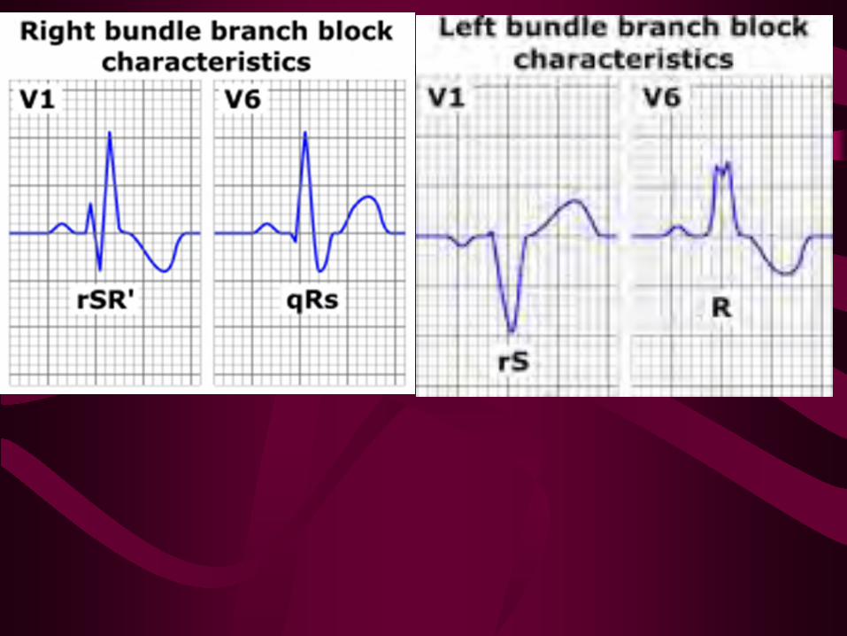


Left Bundle Branch Block
Monophasic R wave in I and V6, QRS > 0.12 sec Loss of R wave in precordial leads QRS T wave discordance I, V1, V6 Consider cardiac ischemia if a new finding


PVC BIGEMNY • Rate: variable • P wave: usually obscured by the QRS, PST or T wave of the PVC • QRS: wide > 0.12 seconds; morphology is bizarre with the ST segment and the T wave
opposite in polarity. May be multifocal and exhibit different morphologies. • Conduction: the impulse originates below the branching portion of the Bundle of His;
full compensatory pause is characteristic. • Rhythm: irregular. PVC's may occur in singles, couplets or triplets; or in bigeminy,
trigeminy or quadrigeminy. • PVCs can occur in healthy hearts. For example, an increase in circulating
catecholamines can cause PVCs. They also occur in diseased hearts and from drug (such as digitalis) toxicities.
• Treatment is required if they are: associated with an acute MI, occur as couplets, bigeminy or trigeminy, are multifocal, or are frequent (>6/min).
• Interventions include: lidocaine, pronestyl, or quinidine.

•a "retrograde” p-wave may sometimes be seen on the right hand side of beats that originate in the ventricles, indicating that depolarization has spread back up through the atria from the ventricles
QRS is wide and much different looking than the normal beats. This indicates that the beat originated somewhere in the ventricles.
Ventricular Escape Beats
“PVCs”
•no p wave, indicating that the beat did not originate anywhere in the atria
Ectopic Foci and Beats

VENTRICULAR TACHYCARDIA
• Rate: usually between 100 to 220/bpm, but can be as rapid as 250/bpm • P wave: obscured if present and are unrelated to the QRS complexes. • QRS: wide and bizarre morphology • Conduction: as with PVCs • Rhythm: three or more ventricular beats in a row; may be regular or irregular. • Ventricular tachycardia almost always occurs in diseased hearts. • Some common causes are:
CAD acute MI digitalis toxicity CHF ventricular aneurysms.
• Patients are often symptomatic with this dysrhythmia. • Ventricular tachycardia can quickly deteriorate into ventricular fibrillation.
Electrical countershock is the intervention of choice if the patient is symptomatic and rapidly deteriorating.
Some pharmacological interventions include lidocaine, pronestyl, and bretylium.


TORSADE DE POINTES • Rate: usually between 150 to 220/bpm, • P wave: obscured if present • QRS: wide and bizarre morphology • Conduction: as with PVCs • Rhythm: Irregular • Paroxysmal –starting and stopping suddenly • Hallmark of this rhythm is the upward and downward deflection of the QRS
complexes around the baseline. The term Torsade de Pointes means "twisting about the points."
• Consider it V-tach if it doesn’t respond to antiarrythmic therapy or treatments • Caused by:
drugs which lengthen the QT interval such as quinidine electrolyte imbalances, particularly hypokalemia myocardial ischemia
• Treatment: Synchronized cardioversion is indicated when the patient is unstable. IV magnesium IV Potassium to correct an electrolyte imbalance Overdrive pacing


VENTRICULAR FIBRILLATION
• Rate: unattainable • P wave: may be present, but obscured by ventricular waves • QRS: not apparent • Conduction: chaotic electrical activity • Rhythm: chaotic electrical activity • This dysrhythmia results in the absence of cardiac output. • Almost always occurs with serious heart disease, especially acute MI. • The course of treatment for ventricular fibrillation includes:
immediate defibrillation and ACLS protocols. Identification and treatment of the underlying cause is also needed.


IDIOVENTRICULAR RHYTHM • Rate: 20 to 40 beats per minute
• P wave: Absent • QRS: Widened • Conduction: Failure of primary pacemaker • Rhythm: Regular • Absent P wave
Widened QRS > 0.12 sec. Also called " dying heart" rhythm Pacemaker will most likely be needed to re-establish a normal heart rate.
• Causes: – Myocardial Infarction – Pacemaker Failure – Metabolic imbalance – Myoardial Ischemia
• Treatment goals include measures to improve cardiac output and establish a normal rhythm and rate.
• Options include: – Atropine – Pacing
• Caution: Supressing the ventricular rhythm is contraindicated because that rhythm protects the heart from complete standstill.

Nonsustained Monomorphic VT

Nonsustained LV VT

Sustained Monomorphic VT 72-year-old woman with CHD

Nonsustained Polymorphic VT

Sustained Polymorphic VT Exercise induced in patient with no structural heart disease

Ventricular Flutter Spontaneous conversion to NSR (12-lead ECG)

VF with Defibrillation (12-lead ECG)

Wide QRS Irregular Tachycardia: Atrial Fibrillation with antidromic conduction in patient
with accessory pathway – Not VT

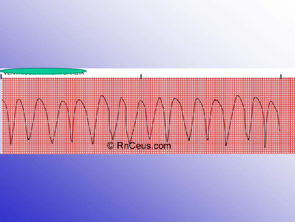
VENTRICULAR STANDSTILL (ASYSTOLE)
• Rate: none • P wave: may be seen, but there is no ventricular response • QRS: none • Conduction: none • Rhythm: none • Asystole occurs most commonly following the termination of atrial,
AV junctional or ventricular tachycardias. This pause is usually insignificant.
• Asystole of longer duration in the presence of acute MI and CAD is frequently fatal.
• Interventions include: CPR, artificial pacing, and atropine.


Thank You
Any Questions?