aricetc1/mariner-like element transposes in yeast
TRANSCRIPT

A Rice Tc1/Mariner-Like Element Transposes in Yeast
Guojun Yang,a Clifford F. Weil,b and Susan R. Wesslera,1
a Department of Plant Biology, University of Georgia, Athens, Georgia 30602b Department of Agronomy, Purdue University, West Lafayette, Indiana 47907
The Tc1/mariner transposable element superfamily is widely distributed in animal and plant genomes. However, no active
plant element has been previously identified. Nearly identical copies of a rice (Oryza sativa) Tc1/mariner element called
Osmar5 in the genome suggested potential activity. Previous studies revealed that Osmar5 encoded a protein that bound
specifically to its own ends. In this report, we show that Osmar5 is an active transposable element by demonstrating that
expression of its coding sequence in yeast promotes the excision of a nonautonomous Osmar5 element located in a re-
porter construct. Element excision produces transposon footprints, whereas element reinsertion occurs at TA dinucleotides
that were either tightly linked or unlinked to the excision site. Several site-directed mutations in the transposase abolished
activity, whereas mutations in the transposase binding site prevented transposition of the nonautonomous element from the
reporter construct. This report of an active plant Tc1/mariner in yeast will provide a foundation for future comparative
analyses of animal and plant elements in addition to making a new wide host range transposable element available for plant
gene tagging.
INTRODUCTION
The Tc1/mariner superfamily contains transposable elements
from diverse taxa, including fungi, flies, nematodes, fishes, and
mammals (Plasterk and van Luenen, 2002). These elements
share three characteristics: a target site duplication (TSD) of the
dinucleotide TA, a transposase with a DDE/D catalytic motif (the
active site where divalent cations bind), and short terminal
inverted repeats (TIRs) of related sequences. Variation in the
DDE/D signature led to the placement of Tc1/mariner elements
into six monophyletic groups: DD34E, DD34D, DD37D, DD37E,
DD31-33D, and DD35E (Doak et al., 1994; Capy et al., 1998;
Robertson et al., 1998; Plasterk et al., 1999; Shao and Tu, 2001).
Although two plant Tc1/mariner elements were identified from
soybean (Glycine max) (Soymar1) and rice (Oryza sativa) (later
named Osmar1), it was not until the design of plant-specific PCR
primers that related elements were found to be widespread in
plant genomes and to compose a seventh monophyletic group
(DD39D) (Jarvik and Lark, 1998; Tarchini et al., 2000; Feschotte
and Wessler, 2002; Feschotte et al., 2003; Jacobs et al., 2004).
Mutational analysis of various Tc1/mariner transposases con-
firmed the critical role of the DDE/D motif and has provided
evidence that an intact DNA binding domain (DBD) is also re-
quired for activity. Mutations in the DD34E motifs of Tc1 and Tc3
abolished transposase activity in vitro (van Luenen et al., 1994;
Vos and Plasterk, 1994). Furthermore, the crystal structure of
the Mos1 catalytic domain suggests an interaction between its
DD34D motif and divalent cations (Mg2þ or Mn2þ) (Richardson
et al., 2006). The Tc1/mariner transposases also contain helix-
turn-helix (HTH) motifs that are required for its binding to TIRs,
the first step of transposition (Lampe et al., 1996; van Pouderoyen
et al., 1997; Wang et al., 1999; Auge-Gouillou et al., 2001; Zhang
et al., 2001; Izsvak et al., 2002; Watkins et al., 2004).
To date, activity has been demonstrated for seven naturally oc-
curring Tc1/mariner elements: Tc1 and Tc3 from Caenorhabditis
elegans (Emmons et al., 1983; Collins et al., 1989); Minos, Mos1,
and Himar1 from flies (Bryan et al., 1990; Franz and Savakis,
1991; Robertson and Lampe, 1995), and Impala and Fot1 from
the fungus Fusarium oxysporum (Daboussi et al., 1992; Langin
et al., 1995). Although superfamily members are widespread in
vertebrate genomes, no active elements have been isolated to
date. Instead, two active transposases were phylogenetically
reconstructed from nonfunctional vertebrate elements: Sleeping
Beauty from eight fish species and Frog Prince from Rana pipiens
(frog) (Ivics et al., 1997; Miskey et al., 2003). Both reconstructed
elements transpose in a variety of vertebrates, including pri-
mates, and, as such, have been developed into valuable tools for
human gene discovery (Yant et al., 2000; Davidson et al., 2003;
Miskey et al., 2003; Ivics and Izsvak, 2004; Dupuy et al., 2005;
Starr and Largaespada, 2005).
The availability of sequence from most of the genomes of two
subspecies of rice, indica and japonica, facilitated a computer-
assisted survey that identified 34 Tc1/mariner elements belonging
to 25 subfamilies (Feschotte et al., 2003). Seven of the 34 ele-
ments (Osmar1A, Osmar5A, Osmar5Bi, Osmar9A, Osmar15Bi,
Osmar17A, and Osmar19) encode potentially functional trans-
posases with no interrupting stop codons. Among these, Osmar5
was chosen as the best candidate for an active element because
virtually identical copies were present in japonica (one copy) and
indica (two copies; one full length and one truncated) at different
genomic loci. In a previous study, binding of the Osmar5 trans-
posase to its TIRs was demonstrated in a yeast one-hybrid assay
1 To whom correspondence should be addressed. E-mail [email protected]; fax 706-542-1805.The author responsible for distribution of materials integral to thefindings presented in this article in accordance with the policy describedin the Instructions for Authors (www.plantcell.org) is: Susan R. Wessler([email protected]).www.plantcell.org/cgi/doi/10.1105/tpc.106.045906
The Plant Cell, Vol. 18, 2469–2478, October 2006, www.plantcell.org ª 2006 American Society of Plant Biologists
in which the protein bound specifically to copies of the TIR on a
reporter construct. Specific binding was also demonstrated in
vitro using a fusion protein synthesized in Escherichia coli and
DNA fragments from the ends of Osmar5. The first 206 residues
of Osmar5 transposase, which contain two HTH motifs (Figure 1),
were shown to bind specifically to two sequence motifs that
comprise a 17-bp region of the TIR (called Box1 and Box2; Figure
1). An additional copy of the 17-bp binding site adjacent to the 39
TIR also binds transposase (Feschotte et al., 2005).
In this study, we have again used a yeast assay, but here to test
for Osmar5 transposition, including excision and reinsertion. We
turned to a yeast assay for two reasons. First, previous studies
indicated that transposition of Tc1/mariner elements (e.g.,
Himar1, Mos1, and Tc1) could occur without host-specific factors
(Lampe et al., 1996; Vos et al., 1996; Tosi and Beverley, 2000). That
is, members of this superfamily transpose in organisms as diverse
as bacteria and human (Ivics et al., 1997; Rubin et al., 1999). The
secondreason for turning toyeast is that itwas shownpreviously to
support transposition of the maize Ac and Ds elements (Weil and
Kunze, 2000). Here, we report that the rice Osmar5 element
transposes in the budding yeast Saccharomyces cerevisiae. Anal-
ysis of transposon footprints at the excision site suggests a model
for how the transposase cleaves this site to promote element
transposition. In addition, new insertions of Osmar5 into TA dinu-
cleotides were detected in the vector and in yeast chromosomes.
Finally, transposition was reduced or prevented by mutation of the
DD39D catalytic domain and by either deletion of the transposase
DBD or mutation of the TIR binding site.
RESULTS
Yeast Transposition Assay
A yeast assay was devised to determine whether Osmar5 en-
coded an active transposase and, if so, the features of excision
and reinsertion. The assay involved two constructs, one encod-
ing the transposase source and the other a reporter for excision.
The transposase source, pOsm5Tp, has Osmar5 coding se-
quence (Figure 1) fused to the inducible gal1 promoter and con-
tains his3 as a selectable marker (Figure 2). The reporter
construct, pOsm5NA, contains a nonautonomous Osmar5 ele-
ment (Osmar5NA) (Figure 1) inserted in the 59 untranslated region
(59 UTR) of an ade2 reporter gene with ura3 as a selectable
marker (Figure 2). To prevent the repair of excision sites by the
very efficient yeast homologous recombination system, a hap-
loid yeast strain was used as recipient (DG2523; see Methods) in
addition to including ARS1/CEN4 in the plasmid reporter con-
struct (pOsm5NA), so that it was maintained as a single copy in
yeast (Falcon and Aris, 2003).
Transformants containing both plasmids were selected on
plates containing 2% galactose and 1% raffinose but lacking his-
tidine and uracil. Colonies were picked from plates containing the
double transformants, and ADE2 revertants were selected based
on growth on agar plates without adenine. Excision events were
confirmed by PCR amplification of the ade2 59 UTR and subse-
quent sequencing (Figure 2, see primer location). Finally, as a
control, we used plasmid pRS413, which is identical to pOsm5Tp
except that it lacks the Pgal1-Osmar5 transposase gene.
Excision of Osmar5NA
Double transformants containing pOsm5Tp (or control plasmid
pRS413) and pOsm5NA were streaked onto plates lacking ade-
nine to select for ADE2 revertants. Many ADE2 revertant colonies
were obtained for pOsm5Tp, but none were obtained for control
plasmid pRS413 (Figure 3A). Plasmid DNA was prepared from
ADE2 revertants, and excision of the Osmar5NA element from
the reporter construct was confirmed by PCR amplification using
primers flanking the element insertion site on pOsm5NA (Figure
3B). Sequencing of this locus from independent ADE2 revertants
revealed that excision of Osmar5NA was accompanied by the
formation of many and diverse transposon footprints (Figure 3C).
Compared with this locus in the original plasmid (Figure 3C,
pOsm5NA, boxed region), all but one of the plasmids from ADE2
revertant colonies had the TA duplication intact but also con-
tained between one and seven additional nucleotides that
appeared to be derived from the ends of Osmar5NA. For all of
these excision events, none had what would be equivalent to a
precise excision, that being the removal of the entire element and
one copy of the dinucleotide TA from the TSD (see Discussion).
Reinsertion of Osmar5NA
Transposition involves both excision and reinsertion of the
excised element into a new locus. To understand the fate of
the excised Osmar5NA, DNA extracted from eight independent
ADE2 revertants was used for DNA gel blot analysis. To this
end, the DNAs were digested with DraI (which does not cut in
Osmar5NA), and the resultant DNA gel blot was probed with
labeled Osmar5NA (Figure 4).
Compared with the plasmid control (Figure 4, pOsm5NA), new
bands were visualized in samples 1, 4, 5, and 8, suggesting
insertion of Osmar5NA at new loci. However, because samples
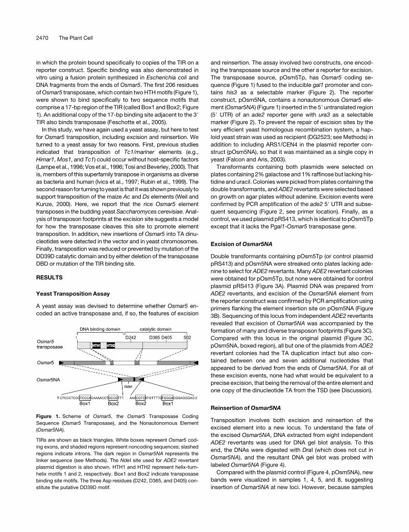
Figure 1. Scheme of Osmar5, the Osmar5 Transposase Coding
Sequence (Osmar5 Transposase), and the Nonautonomous Element
(Osmar5NA).
TIRs are shown as black triangles. White boxes represent Osmar5 cod-
ing exons, and shaded regions represent noncoding sequences; slashed
regions indicate introns. The dark region in Osmar5NA represents the
linker sequence (see Methods). The NdeI site used for ADE2 revertant
plasmid digestion is also shown. HTH1 and HTH2 represent helix-turn-
helix motifs 1 and 2, respectively. Box1 and Box2 indicate transposase
binding site motifs. The three Asp residues (D242, D365, and D405) con-
stitute the putative DD39D motif.
2470 The Plant Cell

2, 3, 6, and 7 contained a single band that comigrated with the
plasmid control, as does one of the two bands in sample 1, we
reexamined the presumptive excision sites in these strains. For
each strain, sequenced PCR products revealed a transposon
footprint in place of the Osmar5NA element (data not shown).
Based on these results, we hypothesized that in each strain, the
Osmar5NA element had transposed to new sites in the pOsm5NA
vector. To test this hypothesis, DNAs isolated from each strain
were used to transform E. coli and recover their plasmids. Be-
cause the DNA samples contained both pOsm5Tp and pOsm5NA,
PCR amplification of the ade2 59 UTRs of the recovered plasmids
was performed to screen for plasmids containing the ade2 gene
(in the plasmid derivatives of pOsm5NA) (Figure 5A).
Reinsertion sites of Osmar5NA in the excision derivatives of
pOsm5NA (called pOsm5NA-d) were analyzed by comparing
their restriction digestion patterns with those of control plasmids
after digestion with DraI (Figure 5B) and NdeI (Figure 5C). Four of
the eight plasmids (Figures 5B and 5C, lanes 1, 2, 5, and 6) have
an altered pattern from that of pWL89A (otherwise identical to
pOsm5NA except lacking Osmar5NA), suggesting that Osmar5NA
had reinserted into the plasmid after excision. The putative
insertion sites in pOsm5NA-d plasmids were approximated by
analysis of the restriction digests with DraI and NdeI (data not
shown). Once the approximate location of the reinserted element
was known, sequencing primers were designed to determine
precise insertion sites of Osmar5NA in the vector (Figure 5D). All
four had inserted at TA dinucleotides and generated TSDs upon
insertion (Figure 5E). The fact that all insertion sites were inter-
genic suggests that the majority of insertions may have been
eliminated by selection for plasmid functions.
The remaining four plasmids (Figures 5B and 5C, lanes 3, 4, 7,
and 8) have an identical pattern to that of pWL89A, indicating the
absence of Osmar5NA in the vector and the possibility that the
element had transposed into a yeast chromosome. For these
strains, insertion sites in the yeast genome were determined by
performing inverse PCR with primers located near the Osmar5NA
termini, with their 39 ends to be extended outward into presumed
flanking yeast genomic DNA (see Methods). PCR products were
successfully obtained for two samples (lanes 4 and 8 in Figure 4;
data not shown), and BLAST searches of the resultant sequences
Figure 2. Yeast Transposition Assay Constructs and Protocol.
The positions of primers used for PCR analysis in Figure 3B are shown as gray arrows. amp, ampicillin resistance gene; ARS1, autonomous replication
sequence1; ARS H4, autonomous replication sequence of the H4 gene; CEN6 and CEN4, centromere sequences of yeast chromosomes 6 and 4,
respectively; cyc1 ter, terminator of yeast cyclin gene cyc1; OriEC, E. coli replication origin; Pgal1, yeast gal1 promoter; pRS413, control vector like
pOsm5Tp but without the transposase. See Methods and text for details.
Plant Tc1/Mariner Transposition in Yeast 2471

led to the identification of insertion sites of Osmar5NA in the yeast
genome (Figure 5E).
Mutagenesis Analysis of Osmar5 Transposase
and Transposon TIRs
In a previous study, the putative transposase peptide sequences
for 34 Osmar elements were aligned with that of Soymar1 to
identify conserved residues (Feschotte et al., 2003). Highly
conserved sites include Met-220, which is located at the junction
of the DBD and the catalytic domain, and the predicted DD39D
motif (Asp-242, Asp-365, Asp-405). Interestingly, Asp-400, which
is 34 residues from Asp-365, is also well conserved (94% iden-
tity). To evaluate the importance of these conserved sites for
transposition, site-directed mutagenesis was performed. Muta-
tion of Met-220 to Ile and Asp-242, Asp-400, Asp-405 to His
abolished activity, as no ADE2 revertants were obtained in the
excision assay (Figure 6). However, mutation of Asp-365 to His
reduced the ADE2 revertant frequency to approximately
one-fourth (0.40 3 10�6/cell) of that of intact Osmar5 transpos-
ase (1.51 3 10�6/cell). These results suggest that the putative
DD39D motif, as well as the conserved Met-220 and Asp-400
motifs, are important for efficient transposition activity.
To test whether interaction between Osmar5 TIRs and trans-
posase DBDs is required for transposition, site-directed muta-
genesis of Osmar5NA was performed so that the TIRs contained
mutations in the strictly conserved (>99% identity among 34
Osmar elements) terminal sequence CTCCCTCC as well as in the
two previously identified motif boxes of the TIRs (Figure 6)
(Feschotte et al., 2005). When a derivative of pOsm5NA con-
taining mutated Osmar5NA TIRs was used in the excision assay
with pOsm5Tp, no ADE2 revertants were obtained, indicating
that transposition of Osmar5 requires correct TIR sequences.
Similarly, no ADE2 revertants were obtained when the DBD of
Osmar5 transposase was deleted (Figure 6). These results sug-
gest that both functional TIRs and transposase DBDs are re-
quired for transposition.
DISCUSSION
The Tc1/mariner superfamily is widespread and well characterized
in eukaryotic genomes. However, although it is also widespread
in the genomes of flowering plants, no active elements have been
reported. In this study, we demonstrate that the rice Osmar5
Figure 3. Osmar5NA Footprints.
(A) ADE2 revertants on medium lacking adenine. The left two sectors
show single colonies derived from two independent pOsm5Tp and
pOsm5NA double transformant colonies. Sectors at right are from
pRS413 and pOsm5NA double transformant colonies.
(B) Agarose gel of PCR products from the ade2 59 UTR of the ADE2
revertant plasmids. Expected band size is 1.4 or 0.4 kb (control), with or
without Osmar5NA, respectively.
(C) Sequences of excision sites of ADE2 revertants. Part of the sequence
of pOsm5NA before excision is shown at top, including the ends of
Osmar5NA (boxed) and flanking sequence. The dinucleotides TA that
flank Osmar5 in the donor vector and in each footprint are shown in red.
Figure 4. Genomic DNA Gel Blot Analysis of ADE2 Revertants.
Genomic DNA (from eight independent revertants, labeled 1 to 8) was
digested with DraI, and blots were probed with Osmar5NA. Controls are
untransformed yeast (DG2523) and pOsm5NA. Two minor bands in the
vector control and revertant lanes are attributable to nonspecific cleav-
age by DraI. DNA size markers at left are in kilobases.
2472 The Plant Cell

element encodes a transposase that catalyzes the excision and
reinsertion of a nonautonomous derivative element in yeast.
Because the catalytic domains of plant Tc1/mariner elements
form a distinct monophyletic clade, it was of interest to initiate a
comparative analysis of the catalytic properties of plant and
animal elements. In addition, as discussed in more detail below,
Tc1/mariner elements are thought to furnish the transposase for
the movement of the nonautonomous Stowaway miniature
inverted-repeat transposable elements (MITEs) (Feschotte and
Mouches, 2000; Feschotte et al., 2003). Stowaway MITEs are
present in thousands of copies in the genomes of many plant
species, where they are particularly enriched in the noncoding
regions of genes (Bureau and Wessler, 1994; Turcotte et al.,
2001; Schenke et al., 2003). To date, no actively transposing
Stowaway elements have been identified. As such, the availabil-
ity of an active plant Tc1/mariner element provides an opportu-
nity to analyze the amplification of Stowaway MITEs and their
contribution to the evolution of plant genomes.
Tc1/Mariner Element Transposition: Plants versus Animals
A transposition mechanism for Tc1/mariner elements was orig-
inally proposed based on in vivo and in vitro analysis of Tc3 from
C. elegans (van Luenen et al., 1994), whereby transposition
occurs in several steps: (1) transposase binds to the element TIR
through its bipartite DBD; (2) the catalytic domain mediates
element excision by cleavage at two sites, two nucleotides inside
the 59 ends and precisely at the 39 junction between the TSD and
Figure 5. Reintegration Sites of Osmar5NA.
(A) Scheme of plasmid rescue from ADE2 revertant genomic DNA. Yeast genomic DNA was extracted from ADE2 revertants and used to transform E.
coli (see text for details). The small gray and black circles represent pOsm5NA and pOsm5Tp, respectively.
(B) Agarose gel analysis of DraI digestion of the recovered pOsm5NA derivative plasmids from (A). DNA size markers are shown at left.
(C) NdeI digestion of the plasmids used for (B).
(D) Insertion sites in pOsm5NA derivatives (pOsm5NA-d); pWL89A lacks Osmar5NA. Note that Osmar5NA has a NdeI site but not a DraI site.
(E) Insertion sites of ADE2 revertants in either the plasmid vector or yeast genomic DNA. Accession numbers of yeast genomic DNA are shown at right.
Plant Tc1/Mariner Transposition in Yeast 2473
the element ends (Figure 7); cleavage results in excision sites
(and excised elements) with two-nucleotide protruding 39 ends;
(3) excised elements exist as free circular intermediates that
target TA dinucleotides for insertion; (4) the 39 hydroxyl group
initiates nucleophilic attack at a TA dinucleotide, producing a
staggered cut; (5) element integration is accompanied by DNA
synthesis, which repairs the gaps and generates the TSD; and (6)
host repair of the excision site, creating transposon footprints.
This model was also shown to hold for Tc1 and Himar1 (Radice
and Emmons, 1993; Lipkow et al., 2004).
Consistent with the transposition mechanism proposed for
Tc3, Tc1, and Himar1, Osmar5 transposase binds specifically to
its TIR through the N-terminal binding domain, as demonstrated
previously (Feschotte et al., 2005). An interaction between DBD
and TIRs is further supported in this study by the failure of TIR
mutations and a DBD deletion to mediate transposition in yeast.
The most significant contribution of this study with regard to
the mechanism of transposition of a plant Tc1/mariner element
comes from the analysis of the transposon footprints. As men-
tioned above, transposase endonuclease activity mediates
cleavage of the element from the donor site. Like animal Tc1/
mariner elements, Osmar5 transposase appears to cut several
nucleotides within the element’s 59 end. This view is supported
by the composition of footprints generated by Osmar5 excision
(Figures 3C and 7). Specifically, the nucleotides located between
the remaining TSDs are identical to nucleotides at the element
ends. By comparison with the Tc3 footprints and its deduced
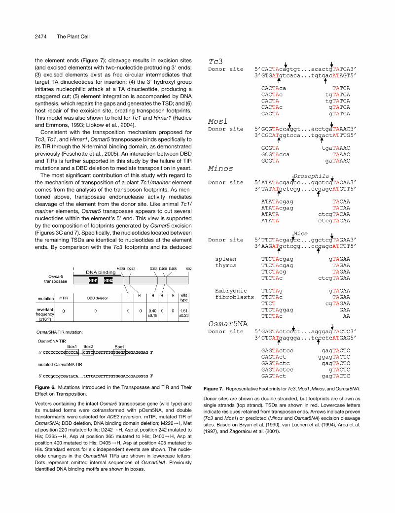
Figure 6. Mutations Introduced in the Transposase and TIR and Their
Effect on Transposition.
Vectors containing the intact Osmar5 transposase gene (wild type) and
its mutated forms were cotransformed with pOsm5NA, and double
transformants were selected for ADE2 reversion. mTIR, mutated TIR of
Osmar5NA; DBD deletion, DNA binding domain deletion; M220/I, Met
at position 220 mutated to Ile; D242/H, Asp at position 242 mutated to
His; D365/H, Asp at position 365 mutated to His; D400/H, Asp at
position 400 mutated to His; D405/H, Asp at position 405 mutated to
His. Standard errors for six independent events are shown. The nucle-
otide changes in the Osmar5NA TIRs are shown in lowercase letters.
Dots represent omitted internal sequences of Osmar5NA. Previously
identified DNA binding motifs are shown in boxes.
Figure 7. RepresentativeFootprints for Tc3, Mos1, Minos, andOsmar5NA.
Donor sites are shown as double stranded, but footprints are shown as
single strands (top strand). TSDs are shown in red. Lowercase letters
indicate residues retained from transposon ends. Arrows indicate proven
(Tc3 and Mos1) or predicted (Minos and Osmar5NA) excision cleavage
sites. Based on Bryan et al. (1990), van Luenen et al. (1994), Arca et al.
(1997), and Zagoraiou et al. (2001).
2474 The Plant Cell

mechanism, we propose that the Osmar5 transposase cleaves
four nucleotides within the element’s two 59 ends, and, at its 39
ends, precisely at the TSD/element junction. As such, both the
excised element and the excision site would contain 39 over-
hangs of four nucleotides, thus accounting for the number and
composition of nucleotides between the TSDs.
Variation in the 59 cleavage site has been observed for Tc1/
mariner transposases. For example, the transposases from
Mos1, Sleeping Beauty, and Frog Prince cleave three nucleo-
tides within the element ends (Dawson and Finnegan, 2003;
Miskey et al., 2003; Yant and Kay, 2003). Interestingly, the puta-
tive site of Osmar5 cleavage, four nucleotides from the element
ends, has also been observed for the Drosophila Minos element
(Figure 7), a distantly related member of the Tc1/mariner super-
family (belonging to the DD34E group) (Arca et al., 1997;
Zagoraiou et al., 2001).
Although our study provides evidence for the importance of
the Osmar5 DD39D motif in the transposition reaction (Figure 6),
we were surprised to find that mutation of the second Asp
residue (Asp-365) did not completely abolish transposition ac-
tivity. This could be explained by one of two possibilities: (1) the
Asp-365–to–His mutation does not completely disrupt the reac-
tion center, because His may act like a cation and the role of the
mutated Asp residue may be compensated by another nearby
Asp residue (Asp-375, present in all 34 Osmar elements in the
rice genome); (2) the DD39D motif may not accurately reflect the
reaction center of the plant elements, as its significance was
based on sequence conservation rather than functional criteria.
In fact, comparison of the rice transposases and that of Soymar1
revealed five conserved Asp residues (Asp-242, -365, -375, -400,
and -405) and two conserved Glu residues (Glu-243 and Glu-261)
in the presumed catalytic domain. The fact that mutation of Asp-
400, which is not part of the DD39D motif, completely abolished
transposition activity supports the view that the exact compo-
nents of the catalytic motif in plant transposases remain to be
defined further.
Although flowering plants are rich in Tc1/mariner elements, it is
not known whether they have a preference, like the maize Ac and
other hAT elements (Chen et al., 1987; Moreno et al., 1992; Tower
et al., 1993), to transpose into linked sites. Local transposition
has been demonstrated for other Tc1/mariner elements (e.g.,
Sleeping Beauty) (Luo et al., 1998; Fischer et al., 2001). In this
study, insertion of Osmar5NA was documented to both linked
(reporter pOsm5NA) and unlinked (yeast chromosome) sites.
Four of the eight excised Osmar5NA elements (independent
events) inserted into sites in the reporter plasmid. In addition, this
number is probably a considerable underestimate, as only in-
sertions between plasmid genes were recovered in this assay
because of a requirement for several plasmid functions. How-
ever, although these data strongly suggest a preference for local
transposition of Osmar5NA, target selection in the yeast assay
may have been influenced by the location of Osmar5NA on a
plasmid. This ambiguity can be addressed in future experiments
by analyzing transposition from an Osmar5NA reporter construct
that is integrated into the yeast chromosome.
The extreme evolutionary distances involved can also compli-
cate conclusions drawn from the analyses of plant transposases
in yeast. For example, it is important to understand whether the
observed events are attributable to the properties of the trans-
posase or to the yeast host, or both. In this regard, comparison of
the footprints generated by two plant transposases (Ac and
Osmar5) in yeast is informative. Footprints generated by Ac and
Osmar5 are markedly different (Weil and Kunze, 2000; Yu et al.,
2004). The Ac transposase, in either a yeast or a plant host,
generates footprints with deletions in the TSD and some that
extend into flanking sequences. In addition, nucleotides are not
retained from the element ends (Baran et al., 1992; Bancroft and
Dean, 1993; Rinehart et al., 1997; Weil and Kunze, 2000). By
contrast, the majority of footprints generated by Osmar5 (and
other Tc1/mariner elements) contain intact TSDs and nucleotides
from the element ends. This difference can be explained by the
different transposition mechanisms of Ac/Ds and Tc1/mariner
elements. The prevailing model for Ac transposition hypothe-
sizes that the transposase cleaves in the TSD and at the element
boundary and that the resultant repair of excision sites produces
footprints with inverted repeat structures (Peacock et al., 1984;
Kunze and Weil, 2002). By contrast, as discussed above, Tc1/
mariner elements have been shown to cleave within the element,
and the Osmar5 footprints in yeast are consistent with previ-
ously described mechanisms, although transposition activity of
Osmar5 in the rice genome has yet to be demonstrated. To-
gether, these data indicate that the very different plant trans-
posases require no host-specific factors, and as such, yeast is an
excellent system in which to study diverse transposition mech-
anisms.
Stowaway MITEs and Osmar Elements
In a previous study, computer-assisted analysis of rice genomic
sequence led to the identification of >34 Osmar elements and
>22,000 Stowaway MITEs (Feschotte et al., 2003). Several lines
of evidence had suggested that Tc1/mariner elements were the
source of transposase for the nonautonomous Stowaway ele-
ments (Feschotte and Mouches, 2000; Turcotte and Bureau,
2002; Feschotte et al., 2003). Specifically, they have related TIRs
and the same TA dinucleotide TSD. For this reason, it was
surprising that none of the Stowaway elements in the rice
genome were derived from the Osmar elements by deletion
(Feschotte et al., 2003). Thus, to understand Stowaway ampli-
fication in plant genomes, it will be necessary to establish
functional connections between Stowaway MITEs and plant
Tc1/mariner elements. As such, this study provides two impor-
tant starting points. First, it demonstrates that at least one Osmar
element, Osmar5, is active. Second, demonstration of Osmar
transposition in yeast provides a valuable assay system to
screen for functional partners between Osmar elements and
rice Stowaway elements. Without extensive sequence similarity
between presumed autonomous elements (the Osmar elements)
and nonautonomous partners (the Stowaway elements), it may
be necessary to test many, perhaps dozens, of combinations of
Osmar and Stowaway pairs to establish functional connections.
The assay system described in this study would be ideal for such
large-scale screening, with yeast serving as a living test tube in
which the relationships among Osmar and Stowaway elements
can be dissected to understand the spread of these important
elements throughout plant genomes.
Plant Tc1/Mariner Transposition in Yeast 2475
METHODS
Yeast Strain and Plasmid Construction
Excision assays were performed after transformation of the yeast haploid
strain DG2523 (MATalpha ura3-167 trp1-hisG leu2-hisG his3-del200
ade2-hisG) (obtained from David Garfinkel). The plasmid containing the
Osmar5 transposase, pOsm5Tp, was constructed from plasmid pRS416
(New England Biolabs) as follows. First, the gal1 promoter was inserted
between the SacI and NotI sites, and the cyc1 terminator was inserted
into the KpnI site (resulting in plasmid pRS416-gal1). Then, the fragment
between SacI and NaeI from pRS416-gal1 was cloned into the corre-
sponding sites in plasmid pRS413 (New England Biolabs), resulting in
plasmid pRS413-gal1. Finally, the coding sequence of the Osmar5 trans-
posase (previously described by Feschotte et al., 2005) was cloned
between the BamHI and EcoRI sites (downstream of the gal1 promoter)
of pRS413-gal1, resulting in plasmid pOsm5Tp. The reporter plasmid
containing the nonautonomous Osmar5 element, pOsm5NA, was con-
structed as follows. First, Osmar5NA was constructed using PCR and
rice (Oryza sativa) genomic DNA from cv Nipponbare to amplify se-
quences from the ends of Osmar5 (562 and 319 bp from the 59 and 39
ends, respectively) and joining the resultant PCR products with a linker
sequence (available upon request). The combined fragment of 950 bp
(including TA at both ends) was inserted into the XhoI site of pWL89A (Yu
et al., 2004), resulting in plasmid pOsm5NA. The orientation of Osmar5NA
insertion is opposite that of ade2 transcription (the other orientation
results in leaky expression of ADE2).
Yeast Transformation and ADE2 Revertant Selection
Transformation reactions (50 mL of competent cells, 5.8 mL of 5 mg/mL
denatured salmon sperm DNA, 1 mL [;200 ng] each of plasmids
pOsm5Tp and pOsm5NA, and 400 mL of 50% PEG-3500 buffer [Gietz
and Woods, 2002]) were incubated at 428C for 45 min. Cells were col-
lected and plated on plates containing complete supplement mixture
(CSM) (Q-BIOgene), 2% galactose, and 1% raffinose but lacking histidine
and uracil. Colonies appeared after 3 to 4 d of incubation at 308C and were
grown to saturation at room temperature (;10 d). ADE2 revertants were
selected from the double transformants by streaking colonies onto CSM
plates containing 2% galactose and 1% raffinose but lacking adenine. To
calculate excision frequency, colonies from plates lacking histidine and
uracil were picked into 50 mL of water, of which 49 mL was plated onto
CSM plates containing 2% galactose but lacking adenine and 1 mL was
used for 105 or 106 dilutions. Of the diluted yeast cell suspension, 49 mL
was plated on YPD (yeast extract/peptone/dextrose) or CSM plates
lacking histidine and uracil to calculate the total number of live yeast cells
in the cell suspension. The revertant frequency was calculated as the
number of ADE2 revertants per cell.
Footprint Analysis
ADE2 revertant colonies were cultured in YPD liquid medium overnight or
in CSM drop-out medium lacking adenine for 2 to 3 d. Plasmid DNA was
extracted using the E.Z.N.A. yeast plasmid kit (Bio-Tek). PCR primers
used to detect the excision of Osmar5NA on pOsm5NA were 59-CTGAC-
AAATGACTCTTGTTGCAGGGCTACGAAC-39 and 59-TGGAAAAGGAG-
CCATTAACGTGGTCATTGGAG-39.PCRproductsweresequenceddirectly.
Genomic DNA Gel Blot Analysis
Genomic DNA (100 ng) from ADE2 revertants was extracted using the
E.Z.N.A. yeast DNA kit (Bio-Tek), digested with DraI, and resolved on an
agarose gel (1%). DNA was blotted onto a Hybond Nþ nylon membrane
(Amersham Biosciences) using capillary transfer in 203 SSC (13 SSC is
0.15 M NaCl and 0.015 M sodium citrate). Probes were prepared with the
DECA prime II kit (Ambion) using Osmar5NA as template. Hybridization
and washing conditions were as described by the supplier (ULTRAhyb
ultrasensitive hybridization buffer; Ambion).
Plasmid Recovery from ADE2 Revertant Genomic DNA
Genomic DNA (30 to 100 ng) from ADE2 revertants was transformed into
Escherichia coli competent cells (Invitrogen), and transformants were
selected on Luria-Bertani plates with carbenicillin (50 mg/L). Plasmid DNA
was extracted from transformant colonies. Because there were two
plasmids in the genomic DNA samples, PCR amplification of the ade2 59
UTR was used to identify strains containing pOsm5NA-d.
Mutagenesis of Osmar5 Transposase and TIRs
To delete the DBD of Osmar5 transposase, a BamHI site was created
using site-directed mutagenesis at the junction of the DBD and the
catalytic domain (primer 59-AGGAAAGGCTGCAGTGGTGGATCCCTAT-
GCTAGATCCGCACACA-39) so that a BamHI fragment (with the DBD)
could be removed and the remaining plasmid could be self-ligated. Site-
directed mutagenesis of transposase sites Met-220, Asp-242, Asp-365,
Asp-400, and Asp-405 was performed with the QuikChange multi-site-
directed mutagenesis kit (Stratagene) using primers 59-GGCTGCAGTG-
GTGTGTTTCTATACTAGATCCGCACACATTGCCAA-39, 59-ATGGAAAA-
TATTATCCACATACATGAGAAATGGTTCAATGCATCA-39, 59-AAAACCA-
TATGGATTCAGCAGCATAATGCTAGAACTCATCATCCT-39, 59-CCTCCA-
AATTCCCCGCATATGAATTGTCTAGATCTTGGATTCTTT-39, and 59-CCA-
AATTCCCCGGATATGAATTGTCTACATCTTGGATTCTTTGCT-39, respec-
tively. Primers for mutagenesis of Osmar5NA TIRs were 59-AAAAACA-
AGAAAATCGGACCTCGAGTAGTCGCTGCGATACACAAAACCTGCCG-
TTTCACC-39 and 59-CCATACTTGATCTCGAGTACACCGTCGGTCCCA-
CAAAACATAAAATTTTAAGGTTAGCAG-39. Mutagenesis reactions of
25 mL contained 100 ng of template vector and 0.5 mL of Quik solution.
Site-directed mutagenesis was also performed of the Osmar5 TIRs using
pOsm5NA as template. All plasmids were sequenced to confirm the
presence of the targeted mutation. ADE2 revertant frequencies were
calculated for all mutant constructs.
Inverse PCR
Genomic DNA from ADE2 revertants (;100 ng) was digested with DraI,
purified (with a PCR purification kit [Qiagen]), and ligated with T4 DNA
ligase (in 35 mL at 258C for 3 h, then overnight at 68C). Ligation products
(5 mL) were amplified with primers (59-CGCACTTCTTTTTTCTGGTT-
CACCTCCACGTATAC-39 and 59-CTGGATGCATGTACAAATGCTGTAA-
ATGACAGC-39) and either Pfu DNA polymerase (Stratagene) or Phusion
DNA polymerase (New England Biolabs) using the same cycling condi-
tions for both enzymes (988C for 45 s; 35 cycles of 988C for 45 s, 588C for
45 s, and 728C for 2 min; and 728C for 10 min). PCR products were
sequenced directly, and the resultant sequences were used as queries for
BLAST searches to determine Osmar5NA insertion sites.
Accession Numbers
The GenBank accession numbers for Osmar5 used in this study are
AP008207 and AP003294.
ACKNOWLEDGMENTS
We thank David J. Garfinkel and Abram Gabriel for yeast strains, plas-
mids, and technical assistance. We also thank Ryan Peeler, Cedric
Feschotte, Mark Osterland, Tianle Chen, Nathan Hancock, Feng Zhang,
2476 The Plant Cell

and Dawn Holligan for technical assistance and helpful discussions. This
study was supported by grants from the National Institutes of Health and
the University of Georgia Research Foundation to S.R.W.
Received July 14, 2006; revised August 21, 2006; accepted September
22, 2006; published October 13, 2006.
REFERENCES
Arca, B., Zabalou, S., Loukeris, T.G., and Savakis, C. (1997). Mobi-
lization of a Minos transposon in Drosophila melanogaster chromo-
somes and chromatid repair by heteroduplex formation. Genetics
145, 267–279.
Auge-Gouillou, C., Hamelin, M.H., Demattei, M.V., Periquet, G., and
Bigot, Y. (2001). The ITR binding domain of the Mariner Mos-1 trans-
posase. Mol. Genet. Genomics 265, 58–65.
Bancroft, I., and Dean, C. (1993). Transposition pattern of the maize
element Ds in Arabidopsis thaliana. Genetics 134, 1221–1229.
Baran, G., Echt, C., Bureau, T., and Wessler, S. (1992). Molecular
analysis of the maize wx-B3 allele indicates that precise excision of
the transposable Ac element is rare. Genetics 130, 377–384.
Bryan, G., Garza, D., and Hartl, D. (1990). Insertion and excision of the
transposable element mariner in Drosophila. Genetics 125, 103–114.
Bureau, T.E., and Wessler, S.R. (1994). Stowaway: A new family of in-
verted repeat elements associated with the genes of both monocot-
yledonous and dicotyledonous plants. Plant Cell 6, 907–916.
Capy, P., Bazin, C., Higuet, D., and Langin, T. (1998). Dynamics and
Evolution of Transposable Elements. (Austin, TX: Springer).
Chen, J., Greenblatt, I.M., and Dellaporta, S.L. (1987). Transposition
of Ac from the P locus of maize into unreplicated chromosomal sites.
Genetics 117, 109–116.
Collins, J., Forbes, E., and Anderson, P. (1989). The Tc3 family of
transposable genetic elements in Caenorhabditis elegans. Genetics
121, 47–55.
Daboussi, M.J., Langin, T., and Brygoo, Y. (1992). Fot1, a new family
of fungal transposable elements. Mol. Gen. Genet. 232, 12–16.
Davidson, A.E., Balciunas, D., Mohn, D., Shaffer, J., Hermanson, S.,
Sivasubbu, S., Cliff, M.P., Hackett, P.B., and Ekker, S.C. (2003).
Efficient gene delivery and gene expression in zebrafish using the
Sleeping Beauty transposon. Dev. Biol. 263, 191–202.
Dawson, A., and Finnegan, D.J. (2003). Excision of the Drosophila
mariner transposon Mos1. Comparison with bacterial transposition
and V(D)J recombination. Mol. Cell 11, 225–235.
Doak, T.G., Doerder, F.P., Jahn, C.L., and Herrick, G. (1994). A pro-
posed superfamily of transposase genes: Transposon-like elements in
ciliated protozoa and a common ‘‘D35E’’ motif. Proc. Natl. Acad. Sci.
USA 91, 942–946.
Dupuy, A.J., Akagi, K., Largaespada, D.A., Copeland, N.G., and
Jenkins, N.A. (2005). Mammalian mutagenesis using a highly mobile
somatic Sleeping Beauty transposon system. Nature 436, 221–226.
Emmons, S.W., Yesner, L., Ruan, K.S., and Katzenberg, D. (1983).
Evidence for a transposon in Caenorhabditis elegans. Cell 32, 55–65.
Falcon, A.A., and Aris, J.P. (2003). Plasmid accumulation reduces life
span in Saccharomyces cerevisiae. J. Biol. Chem. 278, 41607–41617.
Feschotte, C., and Mouches, C. (2000). Evidence that a family of min-
iature inverted-repeat transposable elements (MITEs) from the Arabi-
dopsis thaliana genome has arisen from a pogo-like DNA transposon.
Mol. Biol. Evol. 17, 730–737.
Feschotte, C., Osterlund, M.T., Peeler, R., and Wessler, S.R. (2005).
DNA-binding specificity of rice mariner-like transposases and inter-
actions with Stowaway MITEs. Nucleic Acids Res. 33, 2153–2165.
Feschotte, C., Swamy, L., and Wessler, S.R. (2003). Genome-wide
analysis of mariner-like transposable elements in rice reveals complex
relationships with stowaway miniature inverted repeat transposable
elements (MITEs). Genetics 163, 747–758.
Feschotte, C., and Wessler, S.R. (2002). Mariner-like transposases are
widespread and diverse in flowering plants. Proc. Natl. Acad. Sci. USA
99, 280–285.
Fischer, S.E., Wienholds, E., and Plasterk, R.H. (2001). Regulated
transposition of a fish transposon in the mouse germ line. Proc. Natl.
Acad. Sci. USA 98, 6759–6764.
Franz, G., and Savakis, C. (1991). Minos, a new transposable element
from Drosophila hydei, is a member of the Tc1-like family of transpo-
sons. Nucleic Acids Res. 19, 6646.
Gietz, R.D., and Woods, R.A. (2002). Transformation of yeast by lithium
acetate/single-stranded carrier DNA/polyethylene glycol method.
Methods Enzymol. 350, 87–96.
Ivics, Z., Hackett, P.B., Plasterk, R.H., and Izsvak, Z. (1997). Molec-
ular reconstruction of Sleeping Beauty, a Tc1-like transposon from
fish, and its transposition in human cells. Cell 91, 501–510.
Ivics, Z., and Izsvak, Z. (2004). Transposable elements for transgenesis
and insertional mutagenesis in vertebrates: A contemporary review of
experimental strategies. Methods Mol. Biol. 260, 255–276.
Izsvak, Z., Khare, D., Behlke, J., Heinemann, U., Plasterk, R.H., and
Ivics, Z. (2002). Involvement of a bifunctional, paired-like DNA-binding
domain and a transpositional enhancer in Sleeping Beauty transpo-
sition. J. Biol. Chem. 277, 34581–34588.
Jacobs, G., Dechyeva, D., Menzel, G., Dombrowski, C., and
Schmidt, T. (2004). Molecular characterization of Vulmar1, a com-
plete mariner transposon of sugar beet and diversity of mariner- and
En/Spm-like sequences in the genus Beta. Genome 47, 1192–1201.
Jarvik, T., and Lark, K.G. (1998). Characterization of Soymar1, a
mariner element in soybean. Genetics 149, 1569–1574.
Kunze, R., and Weil, C.F. (2002). The hAT and CACTA superfamilies of
plant transposons. In Mobile DNA II, N.L. Craig, R. Gragie, M. Gellert,
and A.M. Lambowitz, eds (Washington, DC: American Society for
Microbiology), pp. 565–610.
Lampe, D.J., Churchill, M.E., and Robertson, H.M. (1996). A purified
mariner transposase is sufficient to mediate transposition in vitro.
EMBO J. 15, 5470–5479.
Langin, T., Capy, P., and Daboussi, M.J. (1995). The transposable
element impala, a fungal member of the Tc1-mariner superfamily. Mol.
Gen. Genet. 246, 19–28.
Lipkow, K., Buisine, N., Lampe, D.J., and Chalmers, R. (2004). Early
intermediates of mariner transposition: Catalysis without synapsis of
the transposon ends suggests a novel architecture of the synaptic
complex. Mol. Cell. Biol. 24, 8301–8311.
Luo, G., Ivics, Z., Izsvak, Z., and Bradley, A. (1998). Chromosomal
transposition of a Tc1/mariner-like element in mouse embryonic stem
cells. Proc. Natl. Acad. Sci. USA 95, 10769–10773.
Miskey, C., Izsvak, Z., Plasterk, R.H., and Ivics, Z. (2003). The Frog
Prince: A reconstructed transposon from Rana pipiens with high trans-
positional activity in vertebrate cells. Nucleic Acids Res. 31, 6873–6881.
Moreno, M.A., Chen, J., Greenblatt, I., and Dellaporta, S.L. (1992).
Reconstitutional mutagenesis of the maize P gene by short-range Ac
transpositions. Genetics 131, 939–956.
Peacock, W.J., Dennis, E.S., Gerlach, W.L., Sachs, M.M., and
Schwartz, D. (1984). Insertion and excision of Ds controlling elements
in maize. Cold Spring Harb. Symp. Quant. Biol. 49, 347–354.
Plasterk, R.H., Izsvak, Z., and Ivics, Z. (1999). Resident aliens: The
Tc1/mariner superfamily of transposable elements. Trends Genet. 15,
326–332.
Plasterk, R.H.A., and van Luenen, H.G.A.M. (2002). The Tc1/Mariner
family of transposable elements. In Mobile DNA II, N.L. Craig, R.
Plant Tc1/Mariner Transposition in Yeast 2477

Craigie, M. Geller, and A.M. Lambowitz, eds (Washington, DC: American
Society for Microbiology), pp. 519–532.
Radice, A.D., and Emmons, S.W. (1993). Extrachromosomal circular
copies of the transposon Tc1. Nucleic Acids Res. 21, 2663–2667.
Richardson, J.M., Dawson, A., O’Hagan, N., Taylor, P., Finnegan,
D.J., and Walkinshaw, M.D. (2006). Mechanism of Mos1 transposi-
tion: Insights from structural analysis. EMBO J. 25, 1324–1334.
Rinehart, T.A., Dean, C., and Weil, C.F. (1997). Comparative analysis
of non-random DNA repair following Ac transposon excision in maize
and Arabidopsis. Plant J. 12, 1419–1427.
Robertson, H., Soto-Adames, F., Walden, K., Avancini, R., and
Lampe, D. (1998). The mariner transposons of animals: Horizontally
jumping genes. In Horizontal Gene Transfer, M. Syvanen and C. Kido,
eds (London: Chapman & Hall), pp. 268–284.
Robertson, H.M., and Lampe, D.J. (1995). Recent horizontal transfer of
a mariner transposable element among and between Diptera and
Neuroptera. Mol. Biol. Evol. 12, 850–862.
Rubin, E.J., Akerley, B.J., Novik, V.N., Lampe, D.J., Husson, R.N.,
and Mekalanos, J.J. (1999). In vivo transposition of mariner-based
elements in enteric bacteria and mycobacteria. Proc. Natl. Acad. Sci.
USA 96, 1645–1650.
Schenke, D., Sasabe, M., Toyoda, K., Inagaki, Y.S., Shiraishi, T., and
Ichinose, Y. (2003). Genomic structure of the NtPDR1 gene, harbor-
ing the two miniature inverted-repeat transposable elements, NtToya1
and NtStowaway101. Genes Genet. Syst. 78, 409–418.
Shao, H., and Tu, Z. (2001). Expanding the diversity of the IS630-Tc1-
mariner superfamily: Discovery of a unique DD37E transposon and
reclassification of the DD37D and DD39D transposons. Genetics 159,
1103–1115.
Starr, T.K., and Largaespada, D.A. (2005). Cancer gene discovery
using the Sleeping Beauty transposon. Cell Cycle 4, 1744–1748.
Tarchini, R., Biddle, P., Wineland, R., Tingey, S., and Rafalski, A.
(2000). The complete sequence of 340 kb of DNA around the rice
Adh1-adh2 region reveals interrupted colinearity with maize chromo-
some 4. Plant Cell 12, 381–391.
Tosi, L.R., and Beverley, S.M. (2000). cis and trans factors affecting
Mos1 mariner evolution and transposition in vitro, and its potential for
functional genomics. Nucleic Acids Res. 28, 784–790.
Tower, J., Karpen, G.H., Craig, N., and Spradling, A.C. (1993).
Preferential transposition of Drosophila P elements to nearby chro-
mosomal sites. Genetics 133, 347–359.
Turcotte, K., and Bureau, T. (2002). Phylogenetic analysis reveals
stowaway-like elements may represent a fourth family of the IS630-
Tc1-mariner superfamily. Genome 45, 82–90.
Turcotte, K., Srinivasan, S., and Bureau, T. (2001). Survey of
transposable elements from rice genomic sequences. Plant J. 25,
169–179.
van Luenen, H.G., Colloms, S.D., and Plasterk, R.H. (1994). The
mechanism of transposition of Tc3 in C. elegans. Cell 79, 293–301.
van Pouderoyen, G., Ketting, R.F., Perrakis, A., Plasterk, R.H., and
Sixma, T.K. (1997). Crystal structure of the specific DNA-binding
domain of Tc3 transposase of C. elegans in complex with transposon
DNA. EMBO J. 16, 6044–6054.
Vos, J.C., De Baere, I., and Plasterk, R.H. (1996). Transposase is the
only nematode protein required for in vitro transposition of Tc1. Genes
Dev. 10, 755–761.
Vos, J.C., and Plasterk, R.H. (1994). Tc1 transposase of Caenorhab-
ditis elegans is an endonuclease with a bipartite DNA binding domain.
EMBO J. 13, 6125–6132.
Wang, H., Hartswood, E., and Finnegan, D.J. (1999). Pogo transpos-
ase contains a putative helix-turn-helix DNA binding domain that
recognises a 12 bp sequence within the terminal inverted repeats.
Nucleic Acids Res. 27, 455–461.
Watkins, S., van Pouderoyen, G., and Sixma, T.K. (2004). Structural
analysis of the bipartite DNA-binding domain of Tc3 transposase
bound to transposon DNA. Nucleic Acids Res. 32, 4306–4312.
Weil, C.F., and Kunze, R. (2000). Transposition of maize Ac/Ds trans-
posable elements in the yeast Saccharomyces cerevisiae. Nat. Genet.
26, 187–190.
Yant, S.R., and Kay, M.A. (2003). Nonhomologous-end-joining factors
regulate DNA repair fidelity during Sleeping Beauty element transpo-
sition in mammalian cells. Mol. Cell. Biol. 23, 8505–8518.
Yant, S.R., Meuse, L., Chiu, W., Ivics, Z., Izsvak, Z., and Kay, M.A.
(2000). Somatic integration and long-term transgene expression in
normal and haemophilic mice using a DNA transposon system. Nat.
Genet. 25, 35–41.
Yu, J., Marshall, K., Yamaguchi, M., Haber, J.E., and Weil, C.F.
(2004). Microhomology-dependent end joining and repair of transpo-
son-induced DNA hairpins by host factors in Saccharomyces cerevi-
siae. Mol. Cell. Biol. 24, 1351–1364.
Zagoraiou, L., Drabek, D., Alexaki, S., Guy, J.A., Klinakis, A.G.,
Langeveld, A., Skavdis, G., Mamalaki, C., Grosveld, F., and
Savakis, C. (2001). In vivo transposition of Minos, a Drosophila mo-
bile element, in mammalian tissues. Proc. Natl. Acad. Sci. USA 98,
11474–11478.
Zhang, L., Dawson, A., and Finnegan, D.J. (2001). DNA-binding
activity and subunit interaction of the mariner transposase. Nucleic
Acids Res. 29, 3566–3575.
2478 The Plant Cell