arabbit venous model of infusion …avasasm.org.au/images/structure/ppts/hidenoritanabe.pdf ·...
TRANSCRIPT
A RABBIT VENOUS MODEL OF INFUSION INFILTRATION TO
STUDY THE EFFECT OF A HYPEROSMOTIC SOLUTIONHidenori Tanabe1,2, Naoto Takemura2,
Ryoko Murayama1, Makoto Oe3, Hiromi Sanada4
The University of Tokyo
1 Advanced Nursing Technology, the University of Tokyo2 Terumo corporation3 Global Nursing Research Center, the University of Tokyo4 Department of Gerontological Nursing / Wound Care Management, the University of Tokyo

Conflict of interestHidenori Tanabe is a collaborative researcher of the University of Tokyo and has been employed by TERUMO corporation (Tokyo, Japan). Naoto Takemura is also a research associate at TERUMO corporation. The other authors have been determined to have no conflict of interest.
FundingThis research was a joint project with Terumo corporation, conducted with sponsorship from this organization.
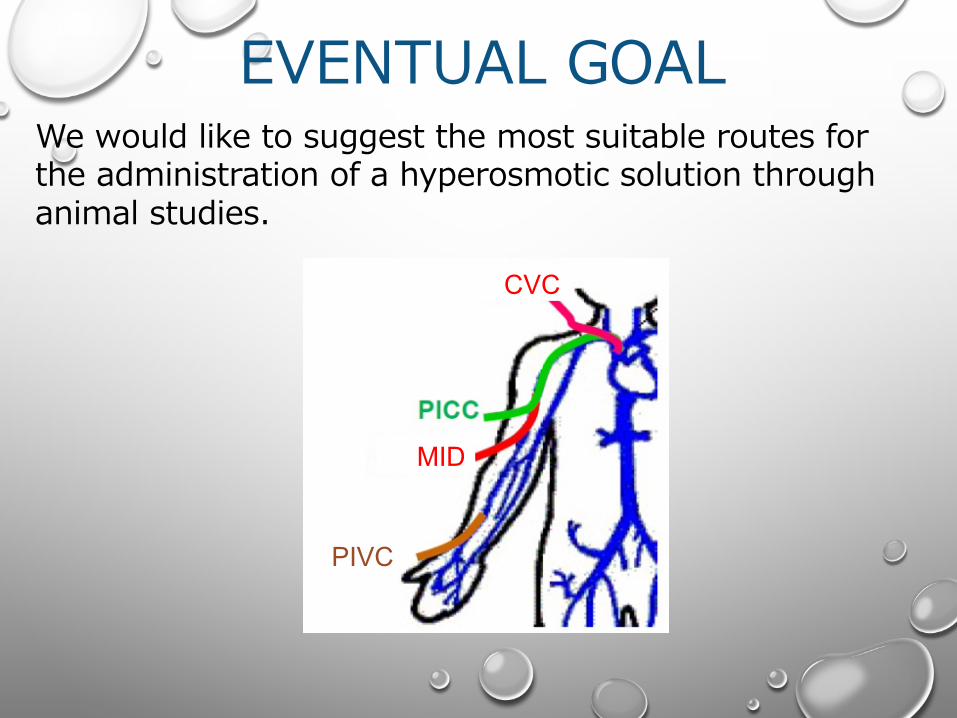
CVC
MID
PIVC
EVENTUAL GOALWe would like to suggest the most suitable routes for the administration of a hyperosmotic solution through animal studies.
OUR PREVIOUS STUDY
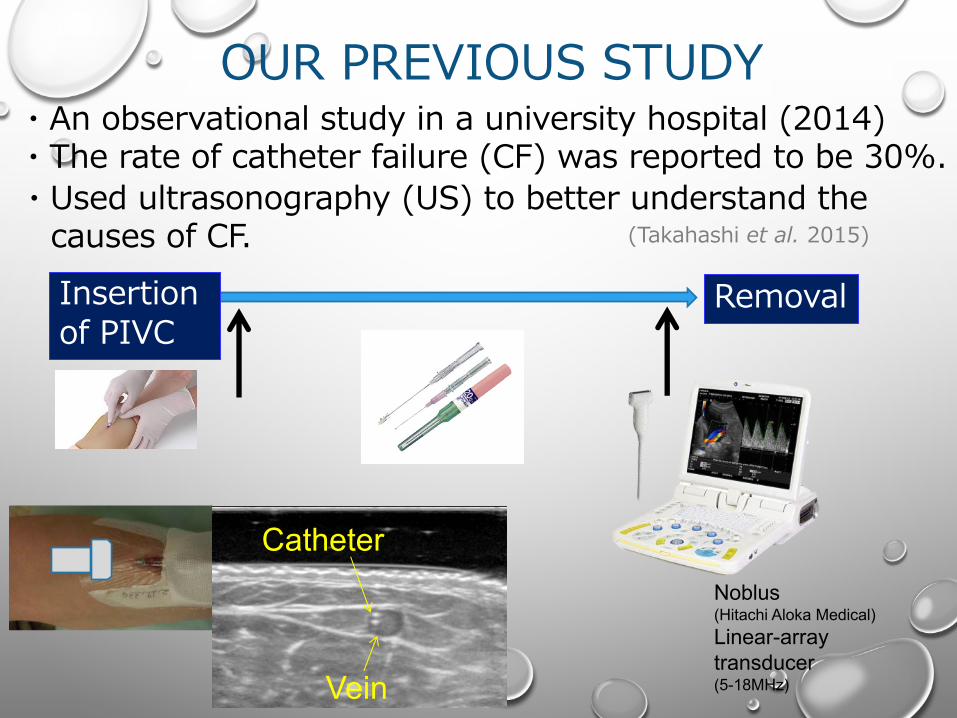
Insertion of PIVC
Removal
Noblus(Hitachi Aloka Medical)Linear-array transducer(5-18MHz)
・An observational study in a university hospital (2014)・The rate of catheter failure (CF) was reported to be 30%.・Used ultrasonography (US) to better understand the
causes of CF.
Vein
Catheter
(Takahashi et al. 2015)
OUR PREVIOUS STUDY
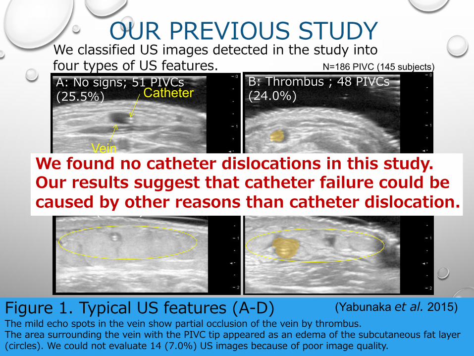
Figure 1. Typical US features (A-D)The mild echo spots in the vein show partial occlusion of the vein by thrombus.The area surrounding the vein with the PIVC tip appeared as an edema of the subcutaneous fat layer (circles). We could not evaluate 14 (7.0%) US images because of poor image quality.
A: No signs; 51 PIVCs (25.5%)
B: Thrombus ; 48 PIVCs (24.0%)
D: Thrombus with subcutaneous edema; 57 PIVCs (28.5%)
C: Subcutaneous edema; 30 PIVCs (15.0%)
N=186 PIVC (145 subjects)
(Yabunaka et al. 2015)
Vein
Catheter
We classified US images detected in the study into four types of US features.
We found no catheter dislocations in this study.Our results suggest that catheter failure could be caused by other reasons than catheter dislocation.
Rates of CF
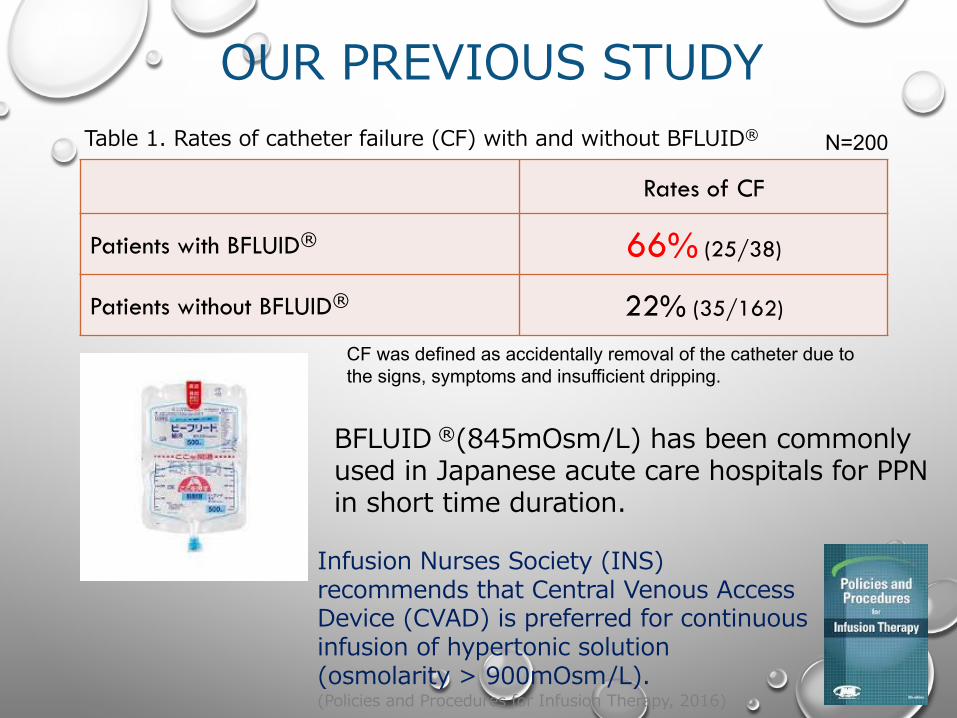
Patients with BFLUID® 66% (25/38)
Patients without BFLUID® 22% (35/162)
Table 1. Rates of catheter failure (CF) with and without BFLUID®
BFLUID ®(845mOsm/L) has been commonly used in Japanese acute care hospitals for PPN in short time duration.
N=200
CF was defined as accidentally removal of the catheter due to the signs, symptoms and insufficient dripping.
OUR PREVIOUS STUDY
Infusion Nurses Society (INS) recommends that Central Venous Access Device (CVAD) is preferred for continuous infusion of hypertonic solution (osmolarity > 900mOsm/L). (Policies and Procedures for Infusion Therapy, 2016)
Clinical presentation Rates (n/N)
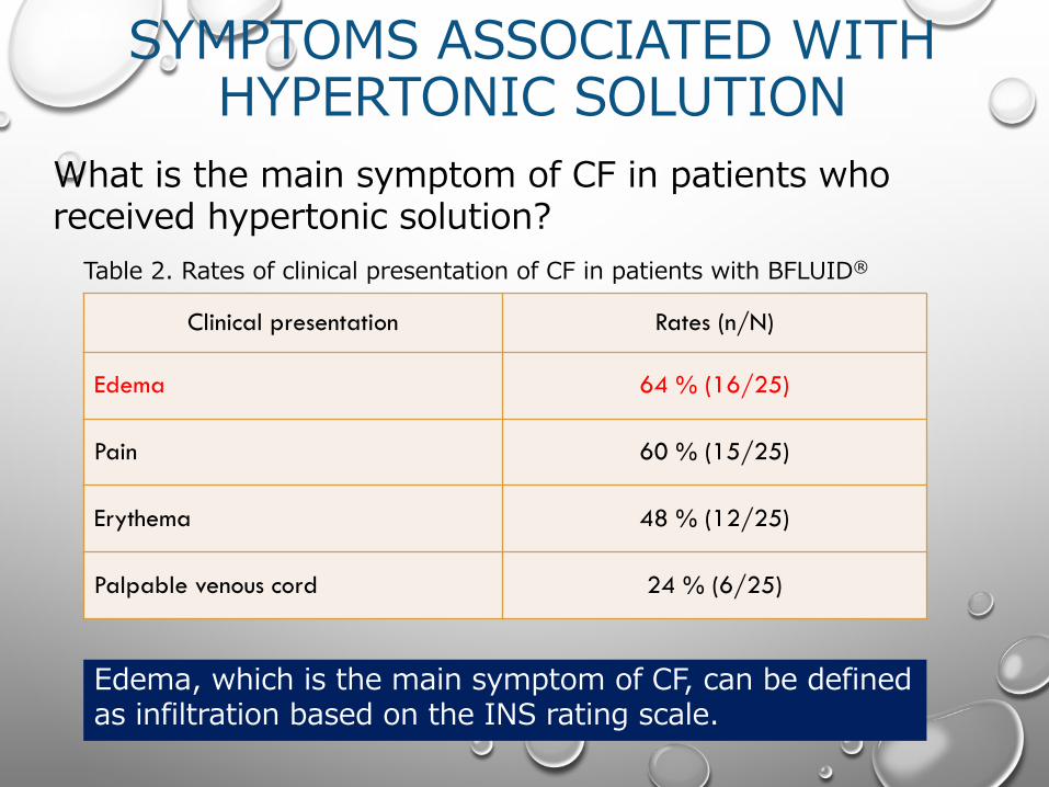
Edema 64 % (16/25)
Pain 60 % (15/25)
Erythema 48 % (12/25)
Palpable venous cord 24 % (6/25)
Table 2. Rates of clinical presentation of CF in patients with BFLUID®
Edema, which is the main symptom of CF, can be defined as infiltration based on the INS rating scale.
What is the main symptom of CF in patients who received hypertonic solution?
SYMPTOMS ASSOCIATED WITH HYPERTONIC SOLUTION

InfiltrationMean (SD), mm
n=5
No-infiltrationMean (SD), mm
n=6p
Vessel diameter 1.3 (0.29) 2.6 (0.81) .008
Short-axis sonographic image of a vein with inserted PIVC tip
Table 3. Vessel diameter with and without infiltration in patients receiving BFLUID®
Noblus(Hitachi Aloka Medical)Liner-array transducer(5-18MHz)
Vessel diameter
OUR PREVIOUS STUDY
To clarify whatʼs happening in the vein, experimental studies using animal models are needed.
To reveal any differences in mean diameter between veins with infiltration and those without infiltration, US was used.
HYPOTHESIS
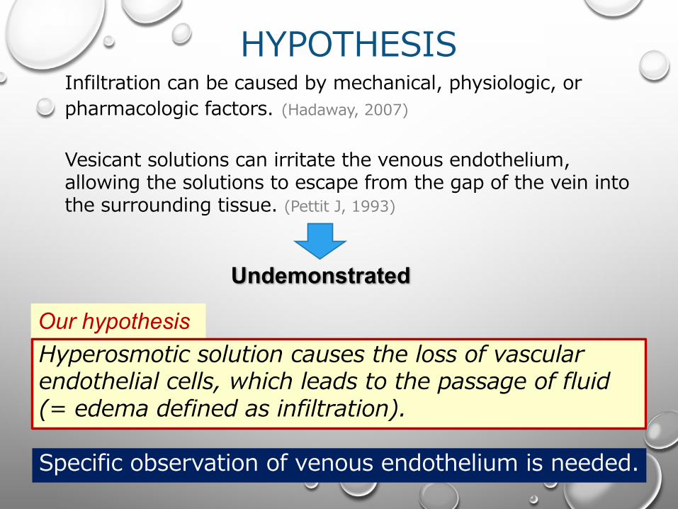
Specific observation of venous endothelium is needed.
Hyperosmotic solution causes the loss of vascular endothelial cells, which leads to the passage of fluid (= edema defined as infiltration).
Our hypothesis
Infiltration can be caused by mechanical, physiologic, or pharmacologic factors. (Hadaway, 2007)
Vesicant solutions can irritate the venous endothelium, allowing the solutions to escape from the gap of the vein into the surrounding tissue. (Pettit J, 1993)
Undemonstrated
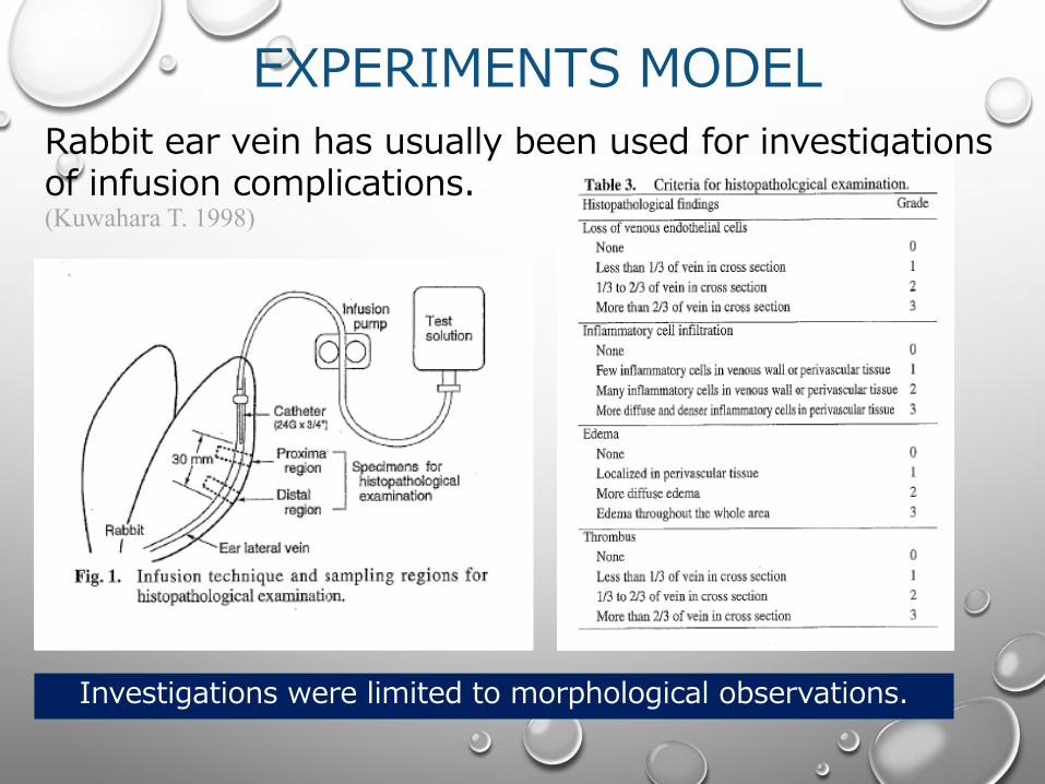
EXPERIMENTS MODELRabbit ear vein has usually been used for investigations of infusion complications.
Investigations were limited to morphological observations.
(Kuwahara T. 1998)

AIMTo establish an animal model of infiltration induced by a hyperosmotic solution, a rabbit ear vein was administered with such a solution and observed specifically using immunostaining method.
METHOD・Male Japanese white rabbit weighing from 2.5 to
3.5 kg was used.
・This study was approved by our Institutional Committee on the care and use of laboratory animals.
・Our facility (TERUMO corporation) has full accreditation by AAALAC International.

METHODHyperosmotic solution was administered via the rabbitʼs left ear vein and normal saline was infused via the rabbitʼs right ear vein using PIVCs (24G).
Rabbit ear vein
Syringe pump(24ml/h)
The total infusion duration was 1 hr; infusion comprised 10 cyclic infusions with 1-min infusion each, and the solution in the vessel was clipped by clamps for 5 mins.
clip
Glucose:36%Osmolality rate:2250 mOsm/L
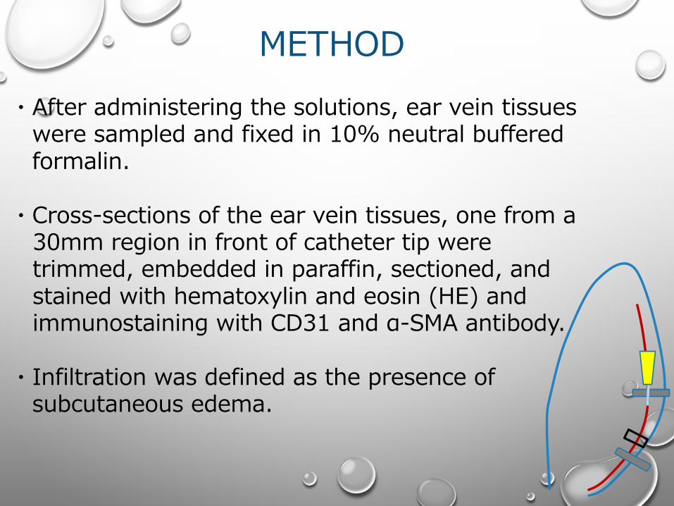
・After administering the solutions, ear vein tissues were sampled and fixed in 10% neutral buffered formalin.
・Cross-sections of the ear vein tissues, one from a 30mm region in front of catheter tip were trimmed, embedded in paraffin, sectioned, and stained with hematoxylin and eosin (HE) and immunostaining with CD31 and α-SMA antibody.
・Infiltration was defined as the presence of subcutaneous edema.
METHOD
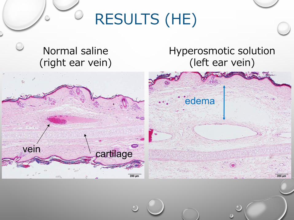
RESULTS (HE)
Hyperosmotic solution (left ear vein)
Normal saline (right ear vein)
vein cartilage
edema
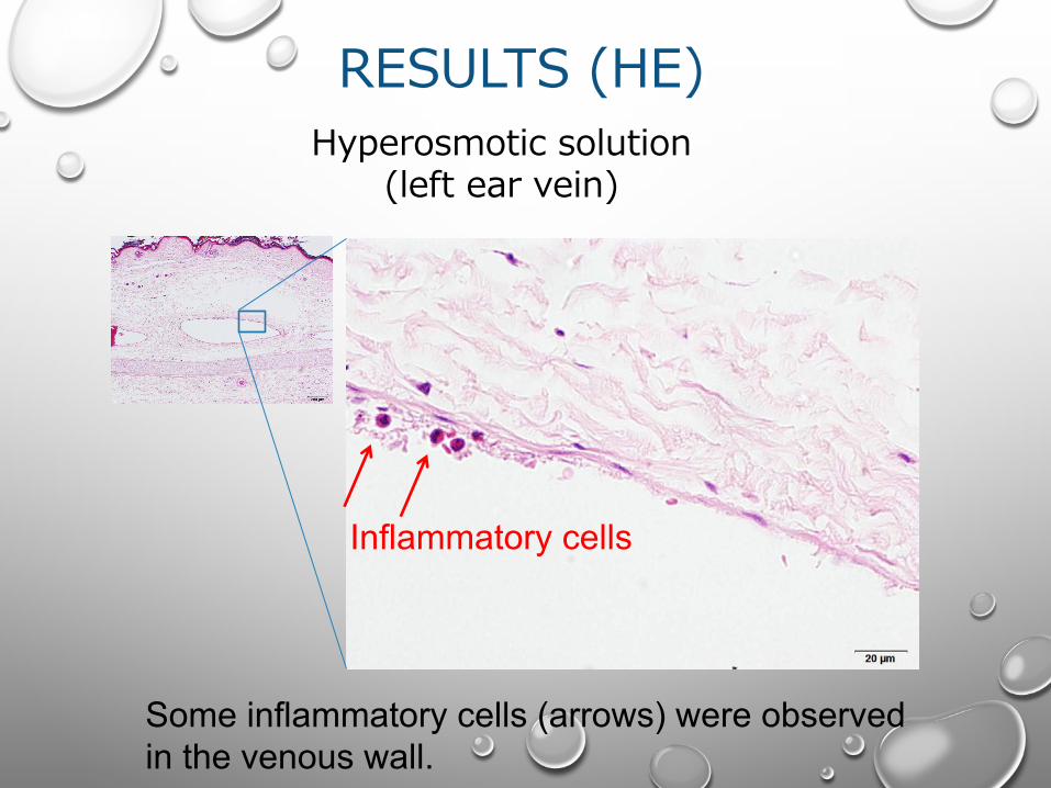
RESULTS (HE)
Some inflammatory cells (arrows) were observed in the venous wall.
Hyperosmotic solution (left ear vein)
Inflammatory cells
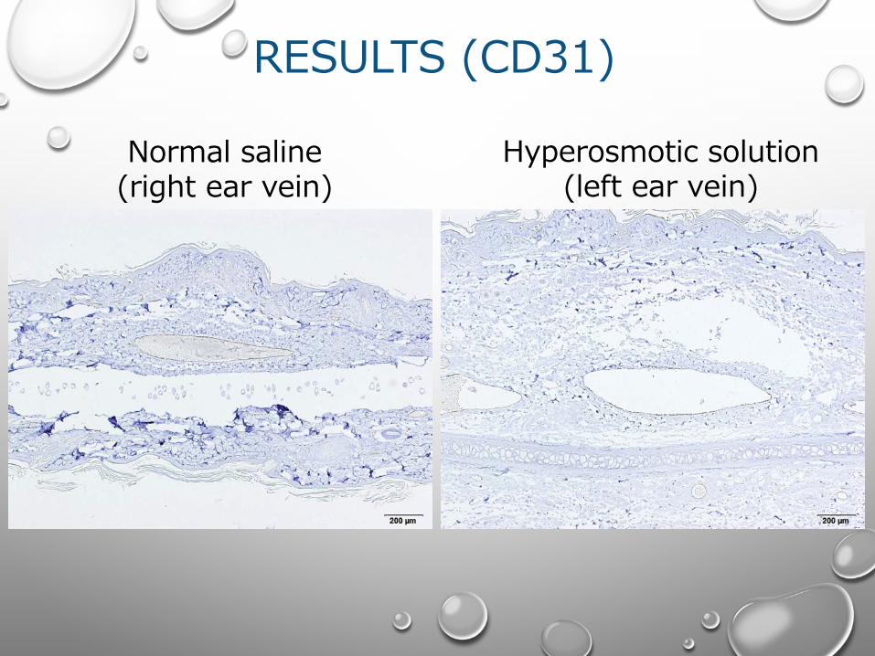
RESULTS (CD31)
Hyperosmotic solution (left ear vein)
Normal saline (right ear vein)
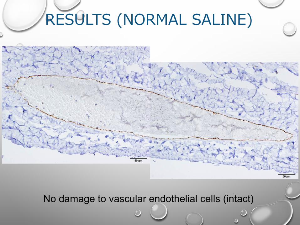
RESULTS (NORMAL SALINE)
No damage to vascular endothelial cells (intact)
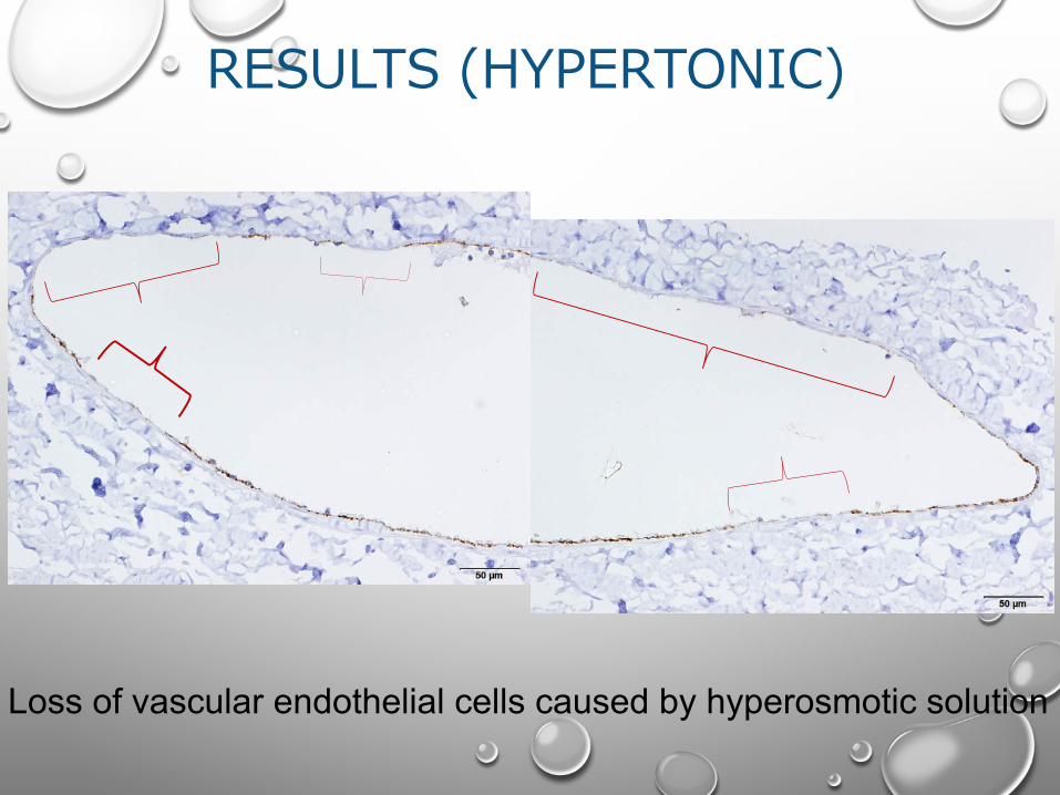
RESULTS (HYPERTONIC)
Loss of vascular endothelial cells caused by hyperosmotic solution
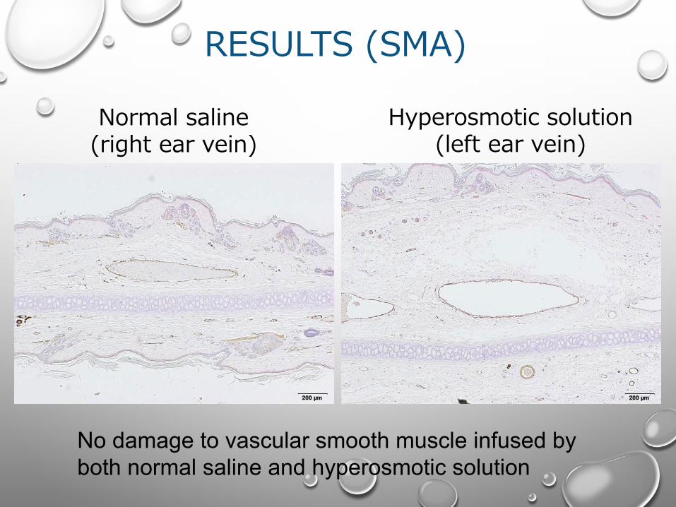
RESULTS (SMA)
Hyperosmotic solution (left ear vein)
Normal saline (right ear vein)
No damage to vascular smooth muscle infused by both normal saline and hyperosmotic solution
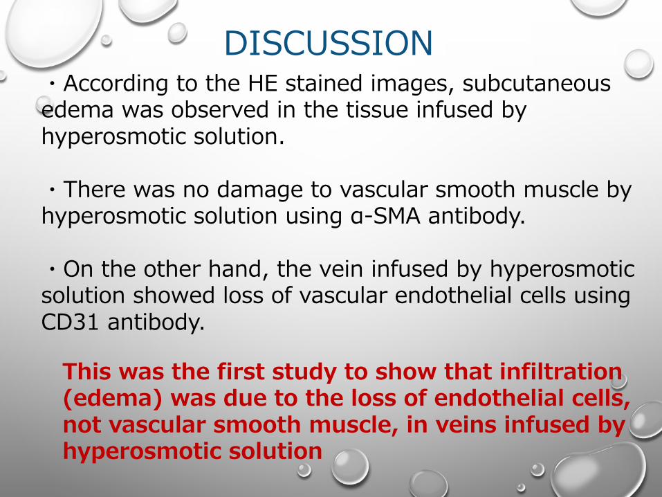
DISCUSSION
This was the first study to show that infiltration (edema) was due to the loss of endothelial cells, not vascular smooth muscle, in veins infused by hyperosmotic solution
・According to the HE stained images, subcutaneous edema was observed in the tissue infused by hyperosmotic solution.
・There was no damage to vascular smooth muscle by hyperosmotic solution using α-SMA antibody.
・On the other hand, the vein infused by hyperosmotic solution showed loss of vascular endothelial cells using CD31 antibody.
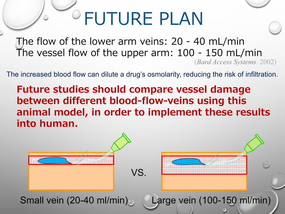
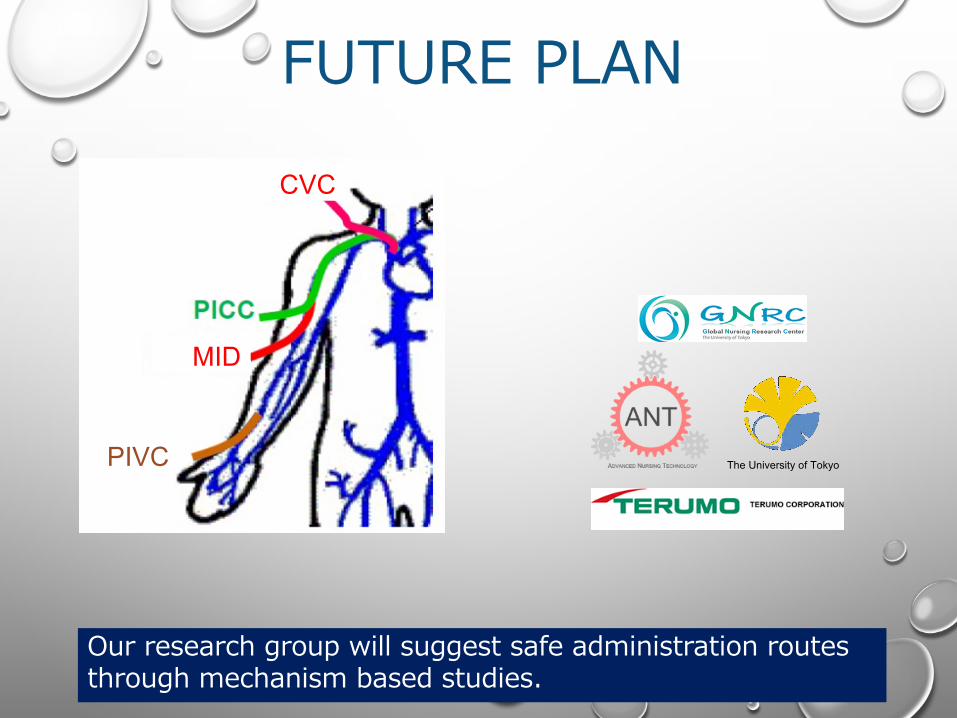
FUTURE PLAN
veinvein
The flow of the lower arm veins: 20 - 40 mL/minThe vessel flow of the upper arm: 100 - 150 mL/min
(Bard Access Systems. 2002)
Future studies should compare vessel damage between different blood-flow-veins using this animal model, in order to implement these results into human.
Small vein (20-40 ml/min) Large vein (100-150 ml/min)
The increased blood flow can dilute a drug’s osmolarity, reducing the risk of infiltration.
VS.
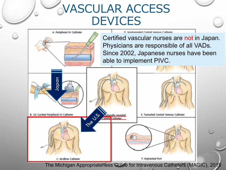
VASCULAR ACCESS DEVICES
The Michigan Appropriateness Guide for Intravenous Catheters (MAGIC), 2015
Japa
n
Certified vascular nurses are not in Japan.Physicians are responsible of all VADs.Since 2002, Japanese nurses have been able to implement PIVC.
CVC
MID
PIVC
Our research group will suggest safe administration routes through mechanism based studies.
The University of Tokyo
FUTURE PLAN

Limitation・It is still unclear whether the administered solution leaked from the vein, or whether tissue fluid increased in surrounding subcutaneous tissue due to inflammation reaction.
・Hyperosmotic solutions can lead to edema, defined as infiltration due to the loss of endothelial cells.
・This model can be useful in establishing safe administration routes and adequate PPN osmolality.
CONCLUSION
Conclusion