aquatic animal health exotic disease training...
TRANSCRIPT
AQUATIC ANIMAL HEALTH EXOTIC DISEASE TRAINING
MANUAL
Shane Raidal, Garry Cross, Stan Fenwick, Philip Nicholls, Barbara Nowak, Kevin Ellard & Frances Stephens

AQUATIC ANIMAL HEALTH: EXOTIC DISEASE TRAINING MANUAL
Shane Raidal, Garry Cross, Stan Fenwick, Philip Nicholls,
Barbara Nowak, Kevin Ellard & Frances Stephens
June 2004
FRDC Project 2002/645
Authors Shane Raidal, Garry Cross, Stan Fenwick, Philip Nicholls, Barbara Nowak, Kevin Ellard & Frances Stephens
Title AQUATIC ANIMAL HEALTH: EXOTIC DISEASE TRAINING
MANUAL
© Fisheries Research and Development Corporation and Murdoch University
This work is copyright. Except as permitted under the Copyright Act 1968 (Cth), no part of this publication may be reproduced by any process, electronic or otherwise, without the specific written permission of the copyright owners. Neither may information be stored electronically in any form whatsoever without such permission. The Fisheries Research and Development Corporation plans, invests in and manages fisheries research and development throughout Australia. It is a federal statutory authority jointly funded by the Australian Government and the fishing industry. Printed by Murdoch Print ISBN 0 86905 847 9
ii

Contributors
Garry Cross BVSc MVSc PhD
Associate Professor in Animal Health. Faculty of Veterinary Science, University of Sydney.
Stan Fenwick BVMS, MSc (Aquatic Vet Studies), MSc (Tropical Vet Med), PhD
Senior Lecturer in Veterinary Public Health, School of Veterinary Studies, Murdoch University. South Street Murdoch, WA, 6150
Philip Nicholls BSc, BVSc, PhD, MRCPath, MRCVS
Senior Lecturer in Veterinary Pathology, School of Veterinary Studies, Murdoch University. South Street, Murdoch, WA, 6150
Barbara Nowak MSc, PhD, DSc
Senior Lecturer Aquatic Animal Health, School of Aquaculture, University of Tasmania, Locked Bag 1370, Launceston 7250, Tasmania.
Kevin Ellard BSc BVMS Dip Agric MACVSc (Aquatic Animal Health)
Senior Veterinary Officer, Dept Primary Industry and Water and Environment, New Town Laboratories. 13 St Johns Avenue, New Town, Tasmania 7008
Shane Raidal BVSc PhD FACVSc
Senior Lecturer in Veterinary Pathology, School of Veterinary Studies, Murdoch University. South Street Murdoch, WA, 6150
Frances Stephens BVSc GradDip PhD
Fish Pathologist, Dept of Fisheries, Western Australia.
iii

Photograph and Review Acknowledgments
David Alderman
Jenni Bartholomew
Eva-Maria Bernoth
Susan Bower
David Bruno
Kurt Buchmann
Eugene Burreson
Angus Cameron
Patrick Caplazi
Serge Corbeil
Mark Crane
John Creeper
Diane Elliott
Tim Flegel
Carolyn Friedman
Andrew Goodwin
John Grizzle
Larry Hammell
Tor Håstein
Don Lightner
Ken McColl
Fred Meyer
Tor Atle Mo
Susan Noh
John Plumb
Tristan Renault
Jill Rolland
Motohiko Sano
Ron Stagg
Thomas Wellborn Jr
iv
Contents Contributors.............................................................................................................................................iii Photograph and Review Acknowledgments ............................................................................................iv Contents....................................................................................................................................................v CHAPTER ONE: INTRODUCTION ......................................................................................................1
List of exotic diseases that are reportable in Australia .........................................................................2 Other serious diseases not reportable in Australia................................................................................2 Methods of entry of new pathogens into Australia...............................................................................3 1.1.1. Stages in the investigation of animal health problems ..........................................................4 1.1.2. Recognising clinical signs and lesions ..................................................................................9 1.1.3. Making a diagnosis..............................................................................................................10 1.1.4. General references on diseases of aquatic animals..............................................................12
CHAPTER TWO: MANAGEMENT DURING A SUSPECTED OR DIAGNOSED OUTBREAK OF AN EXOTIC DISEASE .........................................................................................................................15
WHAT TO DO IF AN EXOTIC DISEASE IS SUSPECTED ...........................................................15 MANAGEMENT OF EXOTIC DISEASE ON AN AQUACULTURE FARM................................16
CHAPTER THREE: DISEASES ...........................................................................................................19 3.1. DISEASES OF FISH.......................................................................................................................19
3.1.1. INFECTIOUS HAEMATOPOIETIC NECROSIS (IHN)........................................................19 3.1.2. ONCORHYNCHUS MASOU VIRUS DISEASE (OMVD)....................................................23 3.1.3. INFECTIOUS SALMON ANAEMIA (ISA) ...........................................................................26 3.1.4. BACTERIAL KIDNEY DISEASE (BKD) ..............................................................................30 3.1.5. PISCIRICKETTSIOSIS ...........................................................................................................34 3.1.6. FURUNCULOSIS....................................................................................................................37 3.1.7. ENTERIC REDMOUTH DISEASE (ERM) ............................................................................40 3.1.8. WHIRLING DISEASE.............................................................................................................43 3.1.9. GYRODACTYLOSIS..............................................................................................................47 3.1.10. VIRAL HAEMORRHAGIC SEPTICAEMIA (VHS)............................................................51 3.1.11. INFECTIOUS PANCREATIC NECROSIS (IPN).................................................................56 3.1.12. RED SEA BREAM IRIDOVIRAL DISEASE (RSIVD) .......................................................61 3.1.13. CHANNEL CATFISH VIRUS DISEASE (CCVD)...............................................................64 3.1.14. ENTERIC SEPTICAEMIA OF CATFISH (ESC) .................................................................67 3.1.15. WHITE STURGEON IRIDOVIRAL DISEASE (WSIVD)...................................................73 3.1.16. KOI MASS MORTALITY.....................................................................................................75 3.1.17. SPRING VIRAEMIA OF CARP (SVC) ................................................................................78
3.2. DISEASES OF CRUSTACEA........................................................................................................81 3.2.1. YELLOW HEAD DISEASE (YHVD).....................................................................................81 3.2.2. WHITE SPOT DISEASE (WSD).............................................................................................85 3.2.3. TAURA SYNDROME.............................................................................................................90 3.2.4. BACULOVIRUS MIDGUT GLAND NECROSIS..................................................................94 3.2.5. TETRAHEDRAL BACULOVIROSIS ....................................................................................96 3.2.6. INFECTIOUS HYPODERMAL AND HAEMATOPOIETIC NECROSIS (IHHN)...............99 3.2.7. NECROTISING HEPATOPANCREATITIS.........................................................................103 3.2.8. CRAYFISH PLAGUE............................................................................................................106
3.3. DISEASES OF MOLLUSCS ........................................................................................................109 3.3.1. INFECTION WITH BONAMIA OSTREAE.........................................................................109 3.3.2. INFECTION WITH HAPLOSPORIDIUM NELSONI..........................................................112 3.3.3.INFECTION WITH HAPLOSPORIDIUM COSTALE.........................................................115 3.3.4. INFECTION WITH MARTEILIA REFRINGENS ...............................................................118 3.3.5. INFECTION WITH PERKINSUS MARINUS......................................................................121 3.3.6. INFECTION WITH MIKROCYTOS MACKINI ..................................................................125 3.3.7. IRIDOVIROSES ....................................................................................................................129 3.3.8. AKOYA OYSTER DISEASE................................................................................................132 3.3.9. INFECTION WITH CANDIDATUS XENOHALIOTIS CALIFORNICUS.........................135 APPENDIX ONE. LIST OF AQUATIC ANIMAL HEALTH DIAGNOSTIC LABORATORIES IN AUSTRALIA ...................................................................................................................................139 APPENDIX TWO. SELF ASSESSMENT TESTS..........................................................................141 APPENDIX THREE: COMMON NAMES OF AQUATIC ANIMALS .........................................153
v

vi
CHAPTER ONE: INTRODUCTION This exotic disease training manual has been prepared for students of Australian tertiary institutions studying veterinary science, aquatic health, fish biology and aquaculture. It is envisaged that it will be used by students and staff involved in teaching some aspects of diseases of aquatic animals and focuses on reportable diseases of aquatic animals that are exotic to Australia. The main aim of the manual is to provide information that is likely to be most relevant to veterinarians and aquatic health specialists. Epidemiological information and disease control methods are the main focus of the manual. Australia is fortunate to be free of many of the infectious diseases that occur in aquatic animals in other parts of the world. Infectious diseases that do not occur in Australia are termed exotic diseases and those of concern to Australia are listed on a national list of reportable diseases that is administered by the Australian Department of Agriculture, Fisheries and Forestry (DAFF)1. Such diseases are caused by well-defined infectious agents and are expected to have serious social, environmental and/or economic impacts if established in Australia. The national list of reportable diseases is routinely re-assessed by state, territory and Commonwealth government agencies in order to ensure that the list remains relevant to Australia’s interests. In addition to the national list of reportable diseases, each state and territory has its own list of notifiable or reportable diseases that include these exotic diseases. In the event of incursion by an exotic disease or disease agent, both the Commonwealth and state/territory government agencies together with industry or community groups may become involved in an emergency response, including control and eradication measures. Many of the diseases of aquatic animals that are reportable within Australia are also reportable internationally to the World Organisation for Animal Health (Office International des Epizooties or OIE)2. As a member of the OIE, Australia has an obligation to report outbreaks of OIE listed diseases. In addition there is a regional reporting scheme administered by the Network of Aquaculture Centres in Asia-Pacific (NACA)3. It must not be presumed that all exotic diseases of aquatic animals are listed on the national list of reportable diseases in Australia. There are many serious infectious diseases that occur overseas but have not been diagnosed in Australia. Such diseases are not currently considered to warrant inclusion on Australia’s list of reportable diseases. Similarly, not all diseases listed are exotic to Australia. For example, Epizootic Haematopoietic Necrosis Virus (EHNV) occurs in Victoria, New South Wales and South Australia and is reportable to the OIE and agencies within Australia.
1 www.daff.gov.au 2 www.oie.int 3 www.enaca.org
1

Australia’s quarantine regulations are designed to reduce to an acceptable level, the likelihood of exotic diseases and pests establishing in Australia. There have been a series of import risk analyses undertaken by Biosecurity Australia (formerly the policy wing of the Australian Quarantine Inspection Service) to identify significant diseases, agents or pests (termed ‘hazards’) that may be introduced via the importation of aquatic animal commodities and to determine their associated biosecurity risks. Determination of biosecurity risk takes into account the likelihood of hazard entry and establishment, as well as the likely consequences or impact of such establishment. This proactive approach has been adopted to facilitate safe trade in animals and plant-based commodities, whilst protecting Australia from exotic disease agents and pests. Several import risk assessments have been published by the Australian Quarantine and Inspection Service or Biosecurity Australia. They clearly outline the methodology and criteria that were used during the assessment process. Import risk analyses for Live Ornamental Finfish (1999) and Non-viable salmonids and non-salmonid Marine Finfish (1999) can be downloaded from the website of the Australian Department of Agriculture, Fisheries and Forestry’s website (www.daff.gov.au) under Market Access and Biosecurity.
List of exotic diseases that are reportable in Australia The list of reportable diseases is regularly reviewed and updated by the National Aquatic Animal Health Technical Working Group. The list is found under AQUAPLAN on DAFF’s website4. In addition, each state and territory has an equivalent list of reportable or notifiable diseases. These lists are updated from time-to-time and can be found by visiting the relevant state and territory websites.
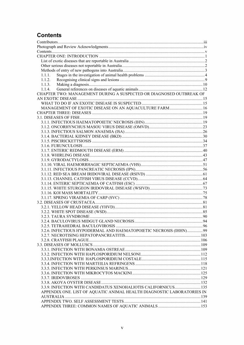
Other serious diseases not reportable in Australia As research into diseases of aquatic animals continues and new diseases emerge, it becomes increasingly evident that translocation of animals between regions has the potential to introduce previously unexposed animals to new pathogens. Many pathogens are identified following translocation of the original host when large-scale mortalities occur in the same or different hosts at the new location. Recent examples are the catastrophic spread of the prawn viruses White Spot Disease throughout Asia and America and Taura Syndrome in the Americas and, more recently to some Asian countries. Similarly, crayfish plague of freshwater crayfish (see Figure 1) and Gaffkaemia of marine lobster both spread from North America to Europe. The development of intensive aquaculture systems allows opportunity for previously undetected pathogens to proliferate and cause diseases in farmed stocks.
4 www.daff.gov.au
2
Fig 1. Map showing the suspected translocation events that have resulted in crayfish plague spreading throughout most of Europe. The disease agent, Aphanomyces astaci, was introduced on carrier North American crayfish, Pacifastacus sp., Procambarus sp. and Orconectes sp. Map courtesy of Dr David Alderman. Many disease agents, particularly some groups of viruses such as the Birnaviruses and Iridoviruses have highly pleomorphic regions in their genome. It appears that new strains of these viruses are frequently emerging. Virulence factors can also change in this process and the agent can become more virulent to the same or a different species. Viral Haemorrhagic Septicaemia and Infectious Pancreatic Necrosis are examples of viruses that have many strains, sub strains and a large host range that includes both marine and freshwater fish. Consequently ongoing research and surveillance will be required to identify and prevent the spread of newly emerging diseases globally.
Methods of entry of new pathogens into Australia Pathogens of aquatic animals that are not already present in Australian waters can enter Australia by a variety of routes. They can be inadvertently introduced with ornamental fish that are destined for the aquarium trade; in water such as ballast water or water holding aquarium fish; or in chilled or frozen seafood for bait or human consumption that contains viable infectious material. Some viruses pose a particular risk as they survive freezing eg. The viruses that cause White Spot Syndrome of prawns, Infectious Pancreatic Necrosis and Viral Haemorrhagic Septicaemia of finfish survive freezing and could enter Australia in imported whole, product. Fortunately many serious pathogens of aquatic animals do not survive normal cooking temperatures, thus reducing the risk of translocation of diseases in “ready to eat” seafood but still present a significant risk if used as bait or aquaculture feed. Nevertheless, Australia has strict controls on the introduction of product from certain seafood species from areas that are known to harbour pathogens that are not found in Australia.
3
Such control measures may include a requirement to partially process fish in order to remove body organs likely to harbour pathogens, inspection and grading practices, removal of heads and gills, removal of tail fins and skin and processing product to a consumer-ready state. For example, risk management measures identified as reducing the risk associated with the establishment of Infectious Pancreatic Necrosis Virus in Australia following the importation of salmon product include:
• A requirement that the fish are not juvenile salmonids • Inspections and grading prior to export in order to remove clinically
diseased fish • Removal of internal organs through thorough cleaning of internal
surfaces • Appropriate certification from a competent authority
The regulations of the Australian Quarantine and Inspection Service (AQIS) aim to reduce the risk of introduction of exotic pathogens to an acceptable level. Its policies are based on import risk analyses undertaken by Biosecurity Australia. AQIS also undertakes publicity campaigns to increase the level of awareness of the general public and interest groups to the risks of introducing exotic pathogens and pests into Australia. It is a well known fact that many unwanted pests have been inadvertently introduced to new regions overseas by thoughtless or illegal introductions of organic material or live plants or animals from other regions.
1.1.1. Stages in the investigation of animal health problems Many disease outbreaks have an underlying pre-disposing factor that has caused stress of the aquatic animal. Stress results in a series of events including increased cortisol production in teleosts and a decrease in lymphocyte production. Similarly, decreased immunocompetence occurs in invertebrate species such as crustacea and molluscs when they are stressed or exposed to poor water quality. Common causes of stress include: suboptimal water quality such as low dissolved oxygen, high suspended solids or high amounts of metabolic by-products; overcrowding; intra or interspecies aggression; sudden increases or decreases in water temperature or salinity; moulting or spawning; inadequate nutrition; handling and transport. In addition, very young and very old animals often are less immunocompetent than other animals in the population.
4
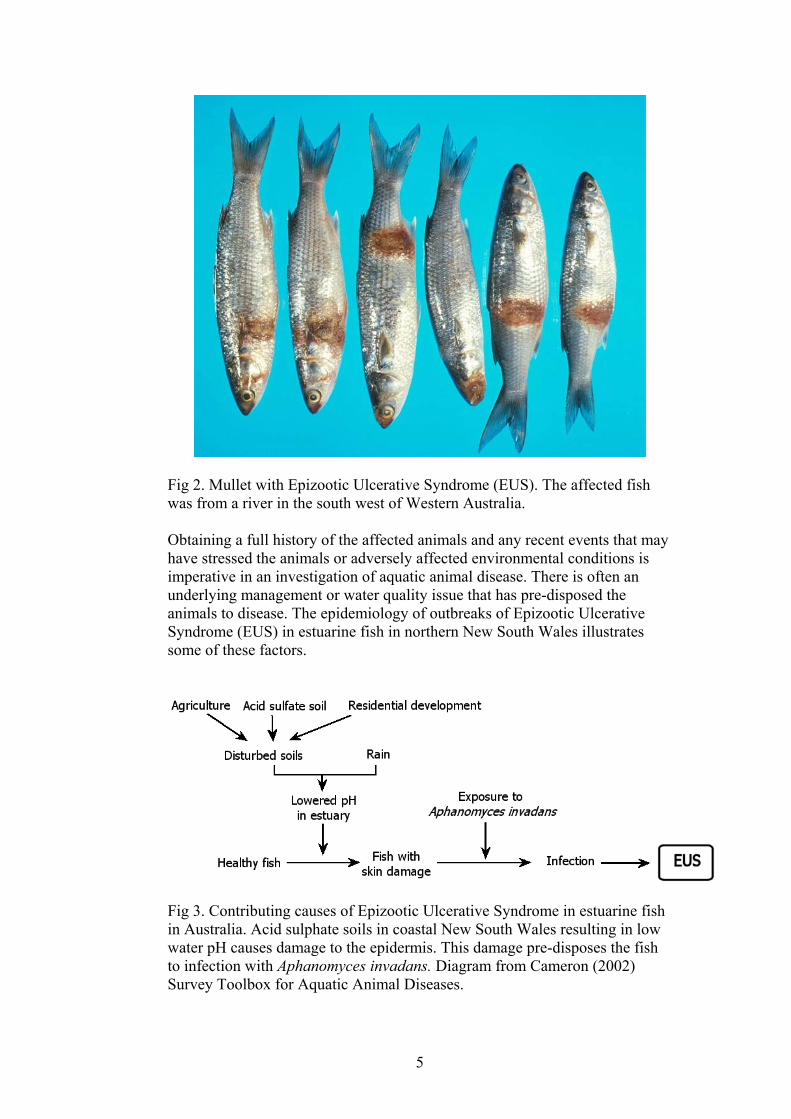
Fig 2. Mullet with Epizootic Ulcerative Syndrome (EUS). The affected fish was from a river in the south west of Western Australia. Obtaining a full history of the affected animals and any recent events that may have stressed the animals or adversely affected environmental conditions is imperative in an investigation of aquatic animal disease. There is often an underlying management or water quality issue that has pre-disposed the animals to disease. The epidemiology of outbreaks of Epizootic Ulcerative Syndrome (EUS) in estuarine fish in northern New South Wales illustrates some of these factors.
Fig 3. Contributing causes of Epizootic Ulcerative Syndrome in estuarine fish in Australia. Acid sulphate soils in coastal New South Wales resulting in low water pH causes damage to the epidermis. This damage pre-disposes the fish to infection with Aphanomyces invadans. Diagram from Cameron (2002) Survey Toolbox for Aquatic Animal Diseases.
5

Fig. 4. In many cases a diseased population does not display the clinical signs of overt disease, but rather has subclinical disease or ill-thrift which is characterised by decreased production. From Cameron (2002) Survey Toolbox for Aquatic Animal Disease. In many instances the first sign that the animals are diseased is a decrease in production. This is because a ‘disease pyramid’ (Fig. 4) is common and many animals in a population will be affected by decreased production or subclinical disease, with only a relatively few animals showing clinical disease. Epidemiology, the study of patterns of disease in a population, is an important aspect of understanding outbreaks of disease. The dynamics of host factors, environmental factors and pathogen factors frequently determine the effect of disease on a population of aquatic animals. The following model is appropriate for studying diseases in aquatic animals because aquatic animals are intimately bound to the aquatic environment and, especially in the case of animals in aquaculture facilities, are unable to leave a less than ideal environment in search of a more suitable habitat. In the event of large-scale sudden mortality, water toxicity or an outbreak of a disease caused by a highly virulent disease must be considered. Water samples must be collected as soon as possible as water quality may rapidly improve as toxins are flushed from the system. The cause of many mortality events of wild fish are never definitively diagnosed because the samples that are submitted for investigation and analysis are either taken too long after the event or are unsuitable for the diagnostic methods required. For example, animal tissues that have been frozen or undergone autolysis are not suitable for the majority of common diagnostic procedures. Water sampling containers may need to be made of certain materials, have air excluded, or contain additives if certain analyses are to be performed.
6
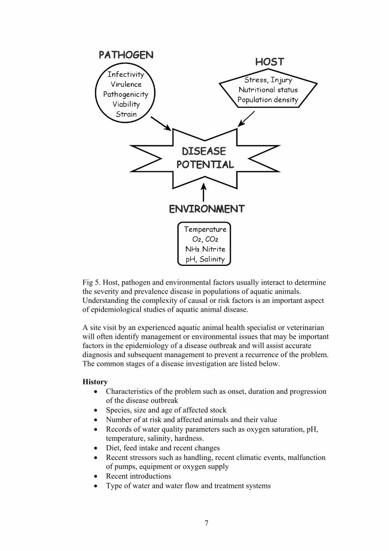
Fig 5. Host, pathogen and environmental factors usually interact to determine the severity and prevalence disease in populations of aquatic animals. Understanding the complexity of causal or risk factors is an important aspect of epidemiological studies of aquatic animal disease. A site visit by an experienced aquatic animal health specialist or veterinarian will often identify management or environmental issues that may be important factors in the epidemiology of a disease outbreak and will assist accurate diagnosis and subsequent management to prevent a recurrence of the problem. The common stages of a disease investigation are listed below. History
• Characteristics of the problem such as onset, duration and progression of the disease outbreak
• Species, size and age of affected stock • Number of at risk and affected animals and their value • Records of water quality parameters such as oxygen saturation, pH,
temperature, salinity, hardness. • Diet, feed intake and recent changes • Recent stressors such as handling, recent climatic events, malfunction
of pumps, equipment or oxygen supply • Recent introductions • Type of water and water flow and treatment systems
7

• Clinical signs noted, past health problems, preventative treatments that have been used
• Any recent management changes, any particular group of animals that are affected, any contact with other species that might act as vectors.
Clinical examination of aquatic animals Note schooling or other activity, position in the water, colour, response to feeding and the presence of any departures from normal behaviour or appearance of the species. The position and appearance of any external lesions should be noted. Examination of animals
Biopsy of gills and scraping of skin from fish is often one of the first stages of examination. A light microscope is used to identify relatively large pathogens such as metazoan or protozoan parasites or fungal agents in tissues of live, affected animals. These pathogens are often motile and can be identified by their pattern of motility, size and morphology. Many of these pathogens are removed during processing of histological sections and it is important that their morphology and intensity of infestation is determined in live or suitably preserved animals.
Post mortem examination • Description of lesions noted • Collection of suitable samples from live, affected animals together
with unaffected animals for comparison for submission to the laboratory.
Analysis of water Records of water pH, salinity, temperature, dissolved oxygen, ammonia, nitrite, nitrate, hardness, alkalinity, carbon dioxide content are very important in investigation of aquatic animal disease because less than optimal water conditions are common pre-disposing causes of disease. Water may need to be collected on more than one occasion. Dissolved oxygen in ponds needs to be taken very early in the morning before photosynthesis has impacted on the lowest overnight value. Each species has unique requirements and stress occurs when the water does not meet these requirements. Water quality in aquaculture premises must be carefully managed to prevent build up of animal and food metabolites that are toxic to the animals being cultured. Water may need to be collected for further examination in a laboratory for agricultural or industrial chemicals, heavy metal, hydrogen sulphide, bacteria, or phytoplankton such as diatoms and dinoflagellates if these are possible contributing factors to disease. Diagnostic tests in a laboratory. These may include histology, culture for bacterial and fungal pathogens, cell culture for virus isolation, electron microscopy and specific immunological or molecular diagnostic tests to identify the presence of antigens or DNA or RNA sequences from known pathogens. Biosecurity measures are undertaken if there is a suspicion of a reportable disease.
8
Reporting of findings to a competent authority. If the disease is reportable, the Chief Veterinary Officer or their delegate in the affected state is notified. Recommend control and management strategies. This may be the responsibility of the Chief Veterinary Officer or the Director of Fisheries if the disease is listed as exotic.
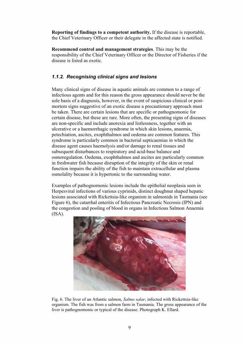
1.1.2. Recognising clinical signs and lesions Many clinical signs of disease in aquatic animals are common to a range of infectious agents and for this reason the gross appearance should never be the sole basis of a diagnosis, however, in the event of suspicious clinical or post-mortem signs suggestive of an exotic disease a precautionary approach must be taken. There are certain lesions that are specific or pathognomonic for certain disease, but these are rare. More often, the presenting signs of diseases are non-specific and include anorexia and listlessness, together with an ulcerative or a haemorrhagic syndrome in which skin lesions, anaemia, petechiation, ascites, exophthalmos and oedema are common features. This syndrome is particularly common in bacterial septicaemias in which the disease agent causes haemolysis and/or damage to renal tissues and subsequent disturbances to respiratory and acid-base balance and osmoregulation. Oedema, exophthalmos and ascites are particularly common in freshwater fish because disruption of the integrity of the skin or renal function impairs the ability of the fish to maintain extracellular and plasma osmolality because it is hypertonic to the surrounding water. Examples of pathognomonic lesions include the epithelial neoplasia seen in Herpesviral infections of various cyprinids, distinct doughnut shaped hepatic lesions associated with Rickettsia-like organism in salmonids in Tasmania (see Figure 6), the catarrhal enteritis of Infectious Pancreatic Necrosis (IPN) and the congestion and pooling of blood in organs in Infectious Salmon Anaemia (ISA).
Fig. 6. The liver of an Atlantic salmon, Salmo salar, infected with Rickettsia-like organism. The fish was from a salmon farm in Tasmania. The gross appearance of the liver is pathognomonic or typical of the disease. Photograph K. Ellard.
9

The clinical signs below are non-specific and common to diseases caused by many infectious and non infectious agents: anorexia; lethargy; affected animals separate from the normal population; lack of schooling activity; deviations from normal colour; abnormal posture or location in the water column (sick fish will often move to the corners of a pen or prawns to the side of the pond); increased volume and rate of opercular movements in fish; pale gills in fish; black gills in crustacean; necrotic, ulcerative, melanotic or haemorrhagic lesions of the skin or carapace; distended abdomen; exophthalmos; frayed fins; haemorrhage at the base of fins; protruding scales and excessive mucus production in fish. Necropsy. Check the following: size and colour of organs such as the gills, liver, kidney, spleen, heart; thymus, contents of stomach, gall bladder and intestines; amount of intrabdominal fat. Impression smears can be made from blood, liver, kidney and intestinal contents. Sterile samples should be collected for bacteriological, mycological and virological examination. An Australian Standard Diagnostic Technique for Aquatic Animal Health: Collection and Submission of Samples for Investigation of Diseases of Fin fish is available on the DAFF website under AQUAPLAN5.
1.1.3. Making a diagnosis A definitive diagnosis is made only after all information relevant to the outbreak is compiled and examined. The history, epidemiology and gross appearance of animals, together with water quality parameters form a vital part of this process. However this information is only part of that used to reach a diagnosis. Histological examination of suitably fixed, affected moribund animals is seldom sufficient to diagnose a specific causative agent of disease. More commonly, and especially in the case of exotic diseases, definitive diagnosis is only reached after isolation and identification of bacterial, fungal or viral agents on isolation media or in cell culture and these findings are assessed in combination with epidemiology, gross and histological signs.
5 www.daff.gov.au
10

Fig. 7. A series of photographs showing the instruments required and the technique of performing a necropsy on a fish and fixing tissues for histology. Photographs K. Ellard.
Electron microscopy can be useful in confirming the presence of virus particles during the acute phase of a disease when large numbers of virus particles are likely to be present. Electron microscopy also provides
11
information on the morphology of the virus and the family to which it may belong. Immunological or molecular techniques are often necessary to confirm the presence of specific pathogens, particularly those that have large numbers of strains and sub strains that may otherwise lead to a false-negative or false-positive diagnosis. Diagnosis should be based on examination of a number of replicate samples whenever possible, but especially when a reportable disease is suspected. It is possible to have false positive and false negative results from laboratory tests. The limitations of each test method need to be fully understood and more that one test method should be used to confirm a diagnosis wherever possible. The aquatic Consultative Committee on Emergency Animal Diseases (CCEAD) would normally require positives from at least two separate laboratory tests plus clinical manifestations in order to make a diagnosis of an exotic disease.
1.1.4. General references on diseases of aquatic animals There are several good, general texts listed here that are useful for providing an overview of diseases of aquatic animals and their diagnosis. Understanding water quality management is another important aspect of being able to predict pre-disposing factors that may contribute to disease outbreaks and for the ability to provide useful advice to aquaculturists on improvements in management that may reduce stress and pre-disposition of cultured aquatic species to disease. Bell, T.A., Lightner, D.V., 1988. A handbook of normal penaeid shrimp histology. World Aquaculture Society, Baton Rouge. This text summarises the normal histology of prawns and includes many black and white photographs. 114pp. ISBN 0935868372. Currently out of print. Bruno, D.W., Alderman, D.J. and Schlotfeldt, H.J., 1997. What should I do? A practical guide for the marine fish farmer. European Association of Fish Pathologists. Aberdeen, Scotland. (www.eafp.org). Cameron, A., 2002. Survey Toolbox for Aquatic Animal Diseases. A Practical Manual and Software Package. Australian Centre for International Agricultural Research (ACIAR) Monograph No. 94. 374pp. ISBN 1863203508. A guide to conducting surveys of disease in aquatic animals and the underlying principles of sampling, and conducting different types of surveys. Chanratchakool, P., Turnbull, J.F., Funge-Smith, S.J., MacRae, I.H., Limsuwan, C., 1998. Health Management in Shrimp Ponds. Aquatic Animal Health Research Institute, Bangkok. 152pp. ISBN 9757604515. This is a general text on the methods used in prawn aquaculture.
12
Elston, R.A., 1999. Health management, development and histology of seed oysters. World Aquaculture Society, Baton Rouge. 110pp. ISBN 1888807032. There are chapters and black and white photographs of life-stages, gross anatomy and histology of oysters and chapters on many of the diseases of oysters. FAO, 2001. Asian Diagnostic Guide to Aquatic Animal Diseases. FAO Fisheries Technical paper T402/2. FAO and ENACA. 191pp. ISBN 9251046204. Ferguson, H.W., 1989. Systemic pathology of fish. Iowa State University Press, Ames. There is a general chapter on necropsy techniques and general pathology of fish. This is followed by chapters on each organ system which describes the normal structure and function and pathology. There are many black and white plates of gross structures, histology and scanning and transmission electron microscopy of tissues and pathology. 263pp. ISBN 013801478. This book is currently out of print. Kent, M.L. and Poppe, T.T. 1998. Diseases of seawater netpen-reared salmonid fish. Fisheries and Oceans, Nanaimo Lightner, D.V., 1996. A handbook of shrimp pathology and diagnostic procedures for diseases of cultured penaeid shrimp. The World Aquaculture Society, Baton Rouge. This is a comprehensive guide to prawn diseases and their diagnosis. It includes many colour photographs of prawns affected by the specific diseases as well as the histological appearance of affected organs. Noga, E.J., 2000. Fish Disease: diagnosis and treatment. Iowa State
University Press, Ames. This text has sections on diagnostic techniques and treatments as well as colour photographs of many common disease agents. It is a particularly useful book for clinicians. 367pp. ISBN 081382558X. OIE publications including: OIE, 2003. International Aquatic Animal Health Code. ISBN 9290445807.
Available at URL: http://www.oie.int/eng/normes/fcode/A_summry.htm
OIE, 2003. Manual for Diagnostic Tests for Aquatic Animals. ISBN 9290445637. Available at URL:http://www.oie.int/eng/normes/fmanual/A_summry.htm
Roberts, R.J., 2001. Fish Pathology. W.B.Saunders, London. There are general chapters on water quality, anatomy, physiology and pathology of teleosts and laboratory methods. There are chapters on immunology, neoplasia, virology, bacteriology, parasitology, bacteriology, mycology, nutritional and non-infectious causes of disease. 472pp. ISBN 0702025631.
13
Schlotfeldt, H.J and Alderman, D.J., 1996. What should I do? A practical guide for the fresh water fish farmer. European Association of Fish Pathologists, Aberdeen, Scotland. (www.eafp.org). Stoskopf, M.K., 1993. Fish Medicine. W.B.Saunders Company,
Philadelphia. There are sections on anatomy, histology, physiology, necropsy, anaesthesia and surgery. The book summarises disease information on many different types of fish and has a chapter on chemotherapeutic agents and doses that have been used in some species. 882pp. ISBN 0721626297 Timmons, M.B., Ebeling, J.M., Wheaton, F.W., Summerfelt, S.T., Vinci, B.J., 2002. Recirculating aquaculture systems. Cayuga Aqua Ventures, Ithaca. A useful text that is available for a reasonable price from the World Aquaculture Society. There are useful, easy to read chapters on water quality management in recirculation systems, including water chemistry, pumps, filtration, gas transfer, ozonation and uv-irradiation. It also includes a chapter on aquaponics. 769pp. ISBN 0971264619. Treves-Brown, K.M., 2000. Applied Fish Pharmacology. Kluwer Academic Publishers, Dordrecht. A useful text with technical information about the groups of chemicals that are used in aquaculture. It gives an overview of registrations and registration practices for aquatic animal treatments in many countries. It describes treatments that are used for various groups of pathogens, and includes chapters on anaesthetics, sex control, breeding induction agents, immuno-stimulants, vaccines, osmoregulators and disinfectants. 309pp. ISBN 0412621800. Wildgoose, W.H. 2001. Manual of ornamental fish. 2nd Edition, British Small Animal Veterinary Association, Gloucester. A handbook for clinicians with many colour photographs and sections on water quality management, handling, anaesthesia, surgery, causes of disease and treatment. ISBN 0905214579.
14

CHAPTER TWO: MANAGEMENT DURING A SUSPECTED OR DIAGNOSED OUTBREAK OF AN EXOTIC DISEASE
WHAT TO DO IF AN EXOTIC DISEASE IS SUSPECTED The introduction of a disease agent to a naïve population of aquatic animals frequently results in high mortality and the shedding of large numbers of infectious agents into the environment. In open water bodies such as rivers or the ocean this can have devastating effects on entire populations of animals. Mortality may be identified at an early stage in the outbreak eg. Pilchard Herpesvirus in South Australia and Western Australia, however, at other times mortality may remain unnoticed for some time eg. sometimes crayfish plague outbreaks are not reported by the public in the UK, possibly because predators remove moribund or dead animals. Any large-scale mortality or ‘fish kill’ should be investigated. Although such outbreaks are often the result of water quality or toxicity problems, it is imperative that potentially important diseases are rapidly diagnosed so that movement restrictions can be put in place to prevent further spread of the pathogen. The following steps should be taken in the event of large-scale mortality. Further detail on these operations can be obtained from other AQUAVETPLAN manuals such as the Enterprise Manual that are available on the Australian Department of Agriculture, Forestry and Fisheries website (www.daff.gov.au).
Fig. 8. Some of the AQUAPLAN and AQUAVETPLAN manuals that are available from the Australian Department of Agriculture, Fisheries and Forestry.
15
1. Notify a private or government veterinarian or government agency
responsible for aquatic animal health, fisheries or rivers if large-scale mortality of aquatic animals is seen. (refer to Appendix 1).
2. Moribund animals and water samples should be collected as soon as possible and submitted to a laboratory for investigation. Animals must not be frozen but animals that have recently died can be placed in plastic bags in ice slurry. There are special requirements for analysis of water for many compounds and some states provide collection bottles for water samples following a large-scale mortality event in aquatic animals. In general, clean glass containers are required for toxicology whilst plastic containers can be used for algal samples. Always contact the appropriate government veterinary agency or laboratory for advice on appropriate sample preservation early in the investigation process.
3. If a reportable disease is suspected, the farm or water body may have quarantine and movement restrictions placed on it pending a definitive diagnosis.
4. Once the presence of a reportable disease is suspected, a response plan will be put in place and the following management practices will be implemented:
• Tracing to identify the source and extent of the outbreak • Declaration of the infected premises or area and a surrounding
restricted and control zone. Areas within this site will be subject to controls that may include destruction of animals, drying, disinfection and decontamination of equipment and ponds, and movement controls.
• Quarantine of premises and areas and restrictions on movement in and out of the areas that aim to prevent further spread of the infectious agent.
• Surveillance within and outside the control zone is used to monitor the progress of the disease control or eradication process.
MANAGEMENT OF EXOTIC DISEASE ON AN AQUACULTURE FARM The first priority in an outbreak of an exotic disease is to prevent further spread of the infectious agent. The other important priority is to eliminate infectious agent that is already present. Tracing the spread of infection and identifying stock and material that may have come in contact with the infectious agent are and important part of this process. The emergency response plan that is adopted will be determined by the type of enterprise. Examples of some types of aquaculture enterprise are shown in
16
Figures 9-12. Further details can be obtained from the AQUAVETPLAN manuals.
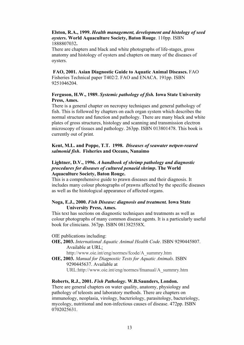
Fig. 9. An example of abalone aquaculture. The abalone are being grown in tanks and water flow can be controlled, an example of a closed aquaculture system. Photograph K. Ellard.
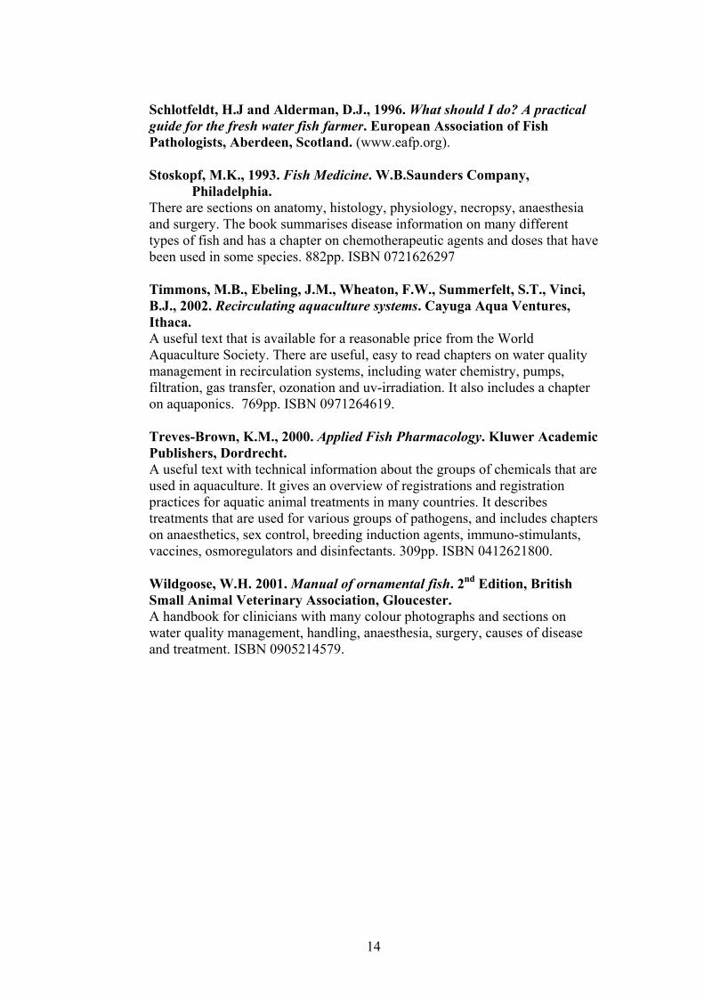
Fig. 10. Sea cages containing Atlantic salmon in Tasmania. This is an example of an open aquaculture system in which water flow cannot be controlled. Photograph K. Ellard.
17
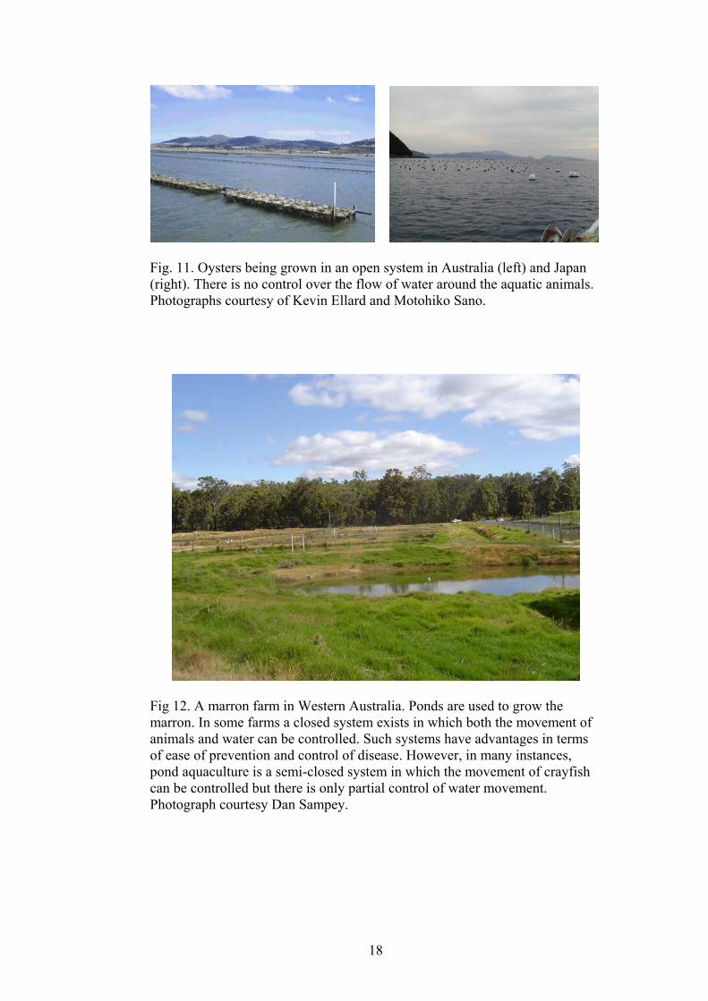
Fig. 11. Oysters being grown in an open system in Australia (left) and Japan (right). There is no control over the flow of water around the aquatic animals. Photographs courtesy of Kevin Ellard and Motohiko Sano.
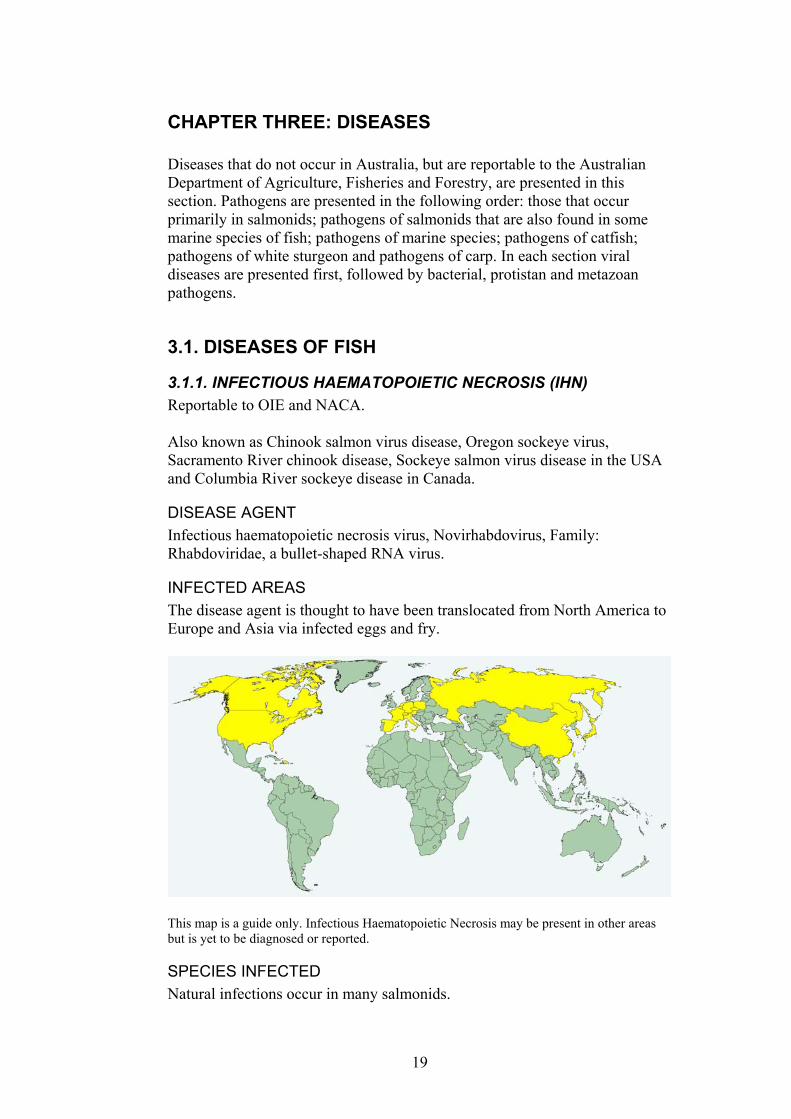
Fig 12. A marron farm in Western Australia. Ponds are used to grow the marron. In some farms a closed system exists in which both the movement of animals and water can be controlled. Such systems have advantages in terms of ease of prevention and control of disease. However, in many instances, pond aquaculture is a semi-closed system in which the movement of crayfish can be controlled but there is only partial control of water movement. Photograph courtesy Dan Sampey.
18
CHAPTER THREE: DISEASES Diseases that do not occur in Australia, but are reportable to the Australian Department of Agriculture, Fisheries and Forestry, are presented in this section. Pathogens are presented in the following order: those that occur primarily in salmonids; pathogens of salmonids that are also found in some marine species of fish; pathogens of marine species; pathogens of catfish; pathogens of white sturgeon and pathogens of carp. In each section viral diseases are presented first, followed by bacterial, protistan and metazoan pathogens.
3.1. DISEASES OF FISH
3.1.1. INFECTIOUS HAEMATOPOIETIC NECROSIS (IHN) Reportable to OIE and NACA. Also known as Chinook salmon virus disease, Oregon sockeye virus, Sacramento River chinook disease, Sockeye salmon virus disease in the USA and Columbia River sockeye disease in Canada.
DISEASE AGENT Infectious haematopoietic necrosis virus, Novirhabdovirus, Family: Rhabdoviridae, a bullet-shaped RNA virus.
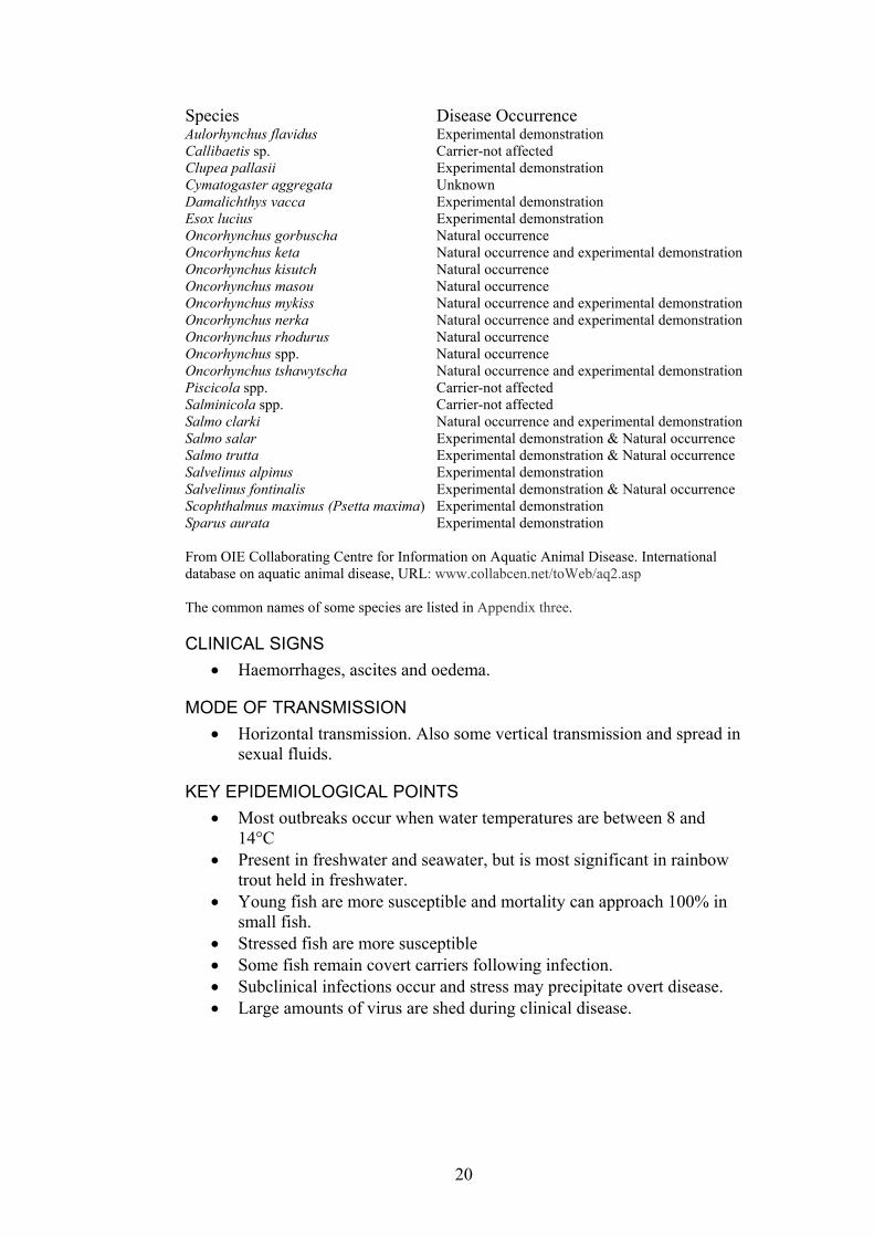
INFECTED AREAS The disease agent is thought to have been translocated from North America to Europe and Asia via infected eggs and fry.
This map is a guide only. Infectious Haematopoietic Necrosis may be present in other areas but is yet to be diagnosed or reported.
SPECIES INFECTED Natural infections occur in many salmonids.
19
Species Disease Occurrence Aulorhynchus flavidus Experimental demonstration Callibaetis sp. Carrier-not affected Clupea pallasii Experimental demonstration Cymatogaster aggregata Unknown Damalichthys vacca Experimental demonstration Esox lucius Experimental demonstration Oncorhynchus gorbuscha Natural occurrence Oncorhynchus keta Natural occurrence and experimental demonstration Oncorhynchus kisutch Natural occurrence Oncorhynchus masou Natural occurrence Oncorhynchus mykiss Natural occurrence and experimental demonstration Oncorhynchus nerka Natural occurrence and experimental demonstration Oncorhynchus rhodurus Natural occurrence Oncorhynchus spp. Natural occurrence Oncorhynchus tshawytscha Natural occurrence and experimental demonstration Piscicola spp. Carrier-not affected Salminicola spp. Carrier-not affected Salmo clarki Natural occurrence and experimental demonstration Salmo salar Experimental demonstration & Natural occurrence Salmo trutta Experimental demonstration & Natural occurrence Salvelinus alpinus Experimental demonstration Salvelinus fontinalis Experimental demonstration & Natural occurrence Scophthalmus maximus (Psetta maxima) Experimental demonstration Sparus aurata Experimental demonstration
From OIE Collaborating Centre for Information on Aquatic Animal Disease. International database on aquatic animal disease, URL: www.collabcen.net/toWeb/aq2.asp The common names of some species are listed in Appendix three.
CLINICAL SIGNS Haemorrhages, ascites and oedema. •
• MODE OF TRANSMISSION
Horizontal transmission. Also some vertical transmission and spread in sexual fluids.
KEY EPIDEMIOLOGICAL POINTS • Most outbreaks occur when water temperatures are between 8 and
14°C • Present in freshwater and seawater, but is most significant in rainbow
trout held in freshwater. • Young fish are more susceptible and mortality can approach 100% in
small fish. • Stressed fish are more susceptible • Some fish remain covert carriers following infection. • Subclinical infections occur and stress may precipitate overt disease. • Large amounts of virus are shed during clinical disease.
20
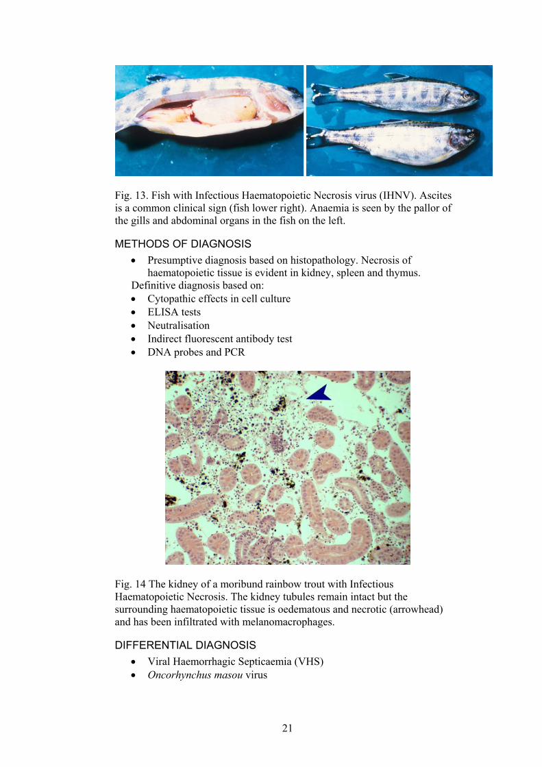
Fig. 13. Fish with Infectious Haematopoietic Necrosis virus (IHNV). Ascites is a common clinical sign (fish lower right). Anaemia is seen by the pallor of the gills and abdominal organs in the fish on the left.
METHODS OF DIAGNOSIS Presumptive diagnosis based on histopathology. Necrosis of haematopoietic tissue is evident in kidney, spleen and thymus.
•
• • • • •
Definitive diagnosis based on: Cytopathic effects in cell culture ELISA tests Neutralisation Indirect fluorescent antibody test DNA probes and PCR
Fig. 14 The kidney of a moribund rainbow trout with Infectious Haematopoietic Necrosis. The kidney tubules remain intact but the surrounding haematopoietic tissue is oedematous and necrotic (arrowhead) and has been infiltrated with melanomacrophages.
DIFFERENTIAL DIAGNOSIS Viral Haemorrhagic Septicaemia (VHS) •
• Oncorhynchus masou virus
21
CONTROL METHODS OVERSEAS Disinfection of eggs •
• •
Rearing fry and fingerlings in virus-free water. Certification, notification of infections and zoning of disease-free and infected areas
REFERENCES Bergmann, S.M., Fichtner, D., Skall, H.F., Schlotfeldt, H., Olesen, N.J. (2003)
Age- and weight-dependent susceptibiltiy of rainbow trout Oncorhynchus mykiss to isolates of infectious haematopoietic necrosis virus (IHHNV) of varying virulaence. Diseases of Aquatic Organisms 55, 205-210.
OIE (2003). International Aquatic Animal Health Code. ISBN 9290445807. Available at URL: http://www.oie.int/eng/normes/fcode/A_summry.htm ISBN 9290445807. OIE (2003).
OIE Manual for Diagnostic Tests for Aquatic Animals. 2003. ISBN 9290445637. Available at http://www.oie.int/eng/normes/fmanual/A_summry.htm
St-Hilaire, S., Ribble, C., Traxler, G., Davies, T., Kent, M.L. (2001) Evidence for a carrier state of infectious hematopoietic necrosis virus in chinook salmon Oncorhynchus tshawytscha. Diseases of Aquatic Organisms 46, 173-179.
Winton, J.R. (1991) Recent advances in detection and control of infectious hematopoietic necrosis virus in aquaculture. Annual Review of Fish Diseases 1, 83-93.
Winton, J.R. (2001) Fish Health Management. In: Fish Hatchery Management.(Ed. by. G. Wedemeyer), pp. 733, American Fisheries Society, Bethesda, Maryland.
Wolf, K., 1988. Fish viruses and fish viral diseases. Cornell University Press, Ithaca, pp. 476. ISBN 0801412595.
22

3.1.2. ONCORHYNCHUS MASOU VIRUS DISEASE (OMVD) OIE and NACA reportable disease. Also known as Nerka virus Towada Lake, Akita Prefecture (NeVTA), Niigata tumour virus (NTV), Yamame tumour virus (YTV), Coho Salmon tumor virus (CSTV), Oncorhynchus kisutch virus (OKV), Rainbow trout kidney virus (RKV) and Rainbow trout herpes virus (RHV).
DISEASE AGENT Herpesvirus: Oncorhynchus masou virus (Salmonid herpesvirus type 2) The virus is icosahedral, the nucleocapsid is 100-115 nm, enveloped virus is 220-240 nm
INFECTED AREAS The disease has been reported in Japan and rivers in the eastern Asian region that have salmonids as well as in Kuwait and the UK including Northern Ireland.
This map is a guide only. Oncorhynchus masou virus disease may be present in other areas but is yet to be diagnosed or reported.
SPECIES INFECTED Salmonids especially masu (Oncorhynchus masou) and coho salmon (Oncorhynchus kisutch). Kokanee and chum salmon are very susceptible and the disease also occurs in rainbow trout, but they are less susceptible. Species Disease Occurrence Oncorhynchus keta Natural occurrence and experimental demonstration Oncorhynchus kisutch Natural occurrence and experimental transmission Oncorhynchus masou Natural occurrence Oncorhynchus mykiss Natural occurrence and experimental demonstration Oncorhynchus nerka Natural occurrence and experimental transmission From OIE Collaborating Centre for Information on Aquatic Animal Disease. International database on aquatic animal disease. URL: www.collabcen.net/toWeb/aq2.asp
23

CLINICAL SIGNS 2 main syndromes occur:
• Neoplasia, found primarily around the mouth in older fish that survive epizootics. Skin ulcers are also common.
• Haemorrhagic septicaemia in young fish (30 -150 day old fish): oedema, exophthalmos, petechial haemorrhages on the underside of the fish. White spots on the liver or liver is white in colour. The fish are often dark in colour, lethargic and gather at water inlets.
Fig. 15. A fish with a tumour in the mouth caused by Oncorhynchus masou virus.
MODE OF TRANSMISSION Horizontal, via water and egg associated transmission on the surface of eggs
•
• • • • •
KEY EPIDEMIOLOGICAL POINTS • Young fish (one month old) are most susceptible • Covert carriers are common. • Most outbreaks occur when water temperatures are below 14°C.
METHODS OF DIAGNOSIS Cytopathic effects in tissue culture ELISA Indirect fluorescent antibody test Histology of tumours PCR
24
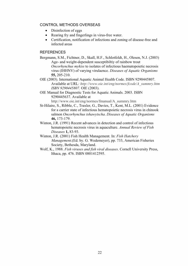
Fig. 16. Histological appearance of neoplastic tissue from fish infected with Oncorhynchus masou virus.
DIFFERENTIAL DIAGNOSIS Infectious Haemorrhagic Necrosis in young salmonids •
• •
CONTROL METHODS OVERSEAS Disinfection of eggs with iodophores Testing, certification and declaration of disease free areas
REFERENCES OIE (2003). International Aquatic Animal Health Code. ISBN 9290445807.
Available at URL: http://www.oie.int/eng/normes/fcode/A_summry.htm
OIE (2003). OIE Manual for Diagnostic Tests for Aquatic Animals. ISBN 9290445637. Available at URL: http://www.oie.int/eng/normes/fmanual/A_summry.htm
Winton, J.R. (2001) Fish Health Management. In: Fish Hatchery Management.(Ed. by. G. Wedemeyer), pp. 733, American Fisheries Society, Bethesda, Maryland. Wolf, K., 1988. Fish viruses and fish viral diseases. Cornell University Press,
Ithaca, pp. 476. Yoshimizu, M., Fukuda, H., Sano, T., Kimura, T. (1995) Salmonid
herpesvirus 2. Epizootiology and serological relationship. Veterinary Research 26, 486-492
25

3.1.3. INFECTIOUS SALMON ANAEMIA (ISA) Reportable to the OIE and NACA. Also known as Bremnes syndrome and salmon anaemia syndrome. The disease was first reported in Norway in 1984.
DISEASE AGENT Infectious salmon anaemia virus; a new genus within the Orthomyxoviridae, spherical, enveloped RNA virus, approximately 100 nm. The virus replicates in endothelial cells lining blood vessels.
INFECTED AREAS The disease occurs in Canada (Atlantic coast), Chile, Faroe Islands, Norway, United Kingdom (Scotland in 1998) and USA (Atlantic coast only).
This map is a guide only. Infectious salmon anaemia may be present in other areas but is yet to be diagnosed or reported.
SPECIES INFECTED Atlantic salmon, Salmo salar, is the only aquaculture species that develops overt disease. Species Disease Occurrence Clupea harengus Experimental demonstration Lepeophtheirus salmonis Carrier-not affected Oncorhynchus mykiss Experimental demonstration Salmo salar Natural occurrence and experimental transmission Salmo trutta Natural occurrence and experimental transmission and
non affected carrier From OIE Collaborating Centre for Information on Aquatic Animal Disease. International database on aquatic animal disease, URL: www.collabcen.net/toWeb/aq2.asp The common names of some species are listed in Appendix three.
26
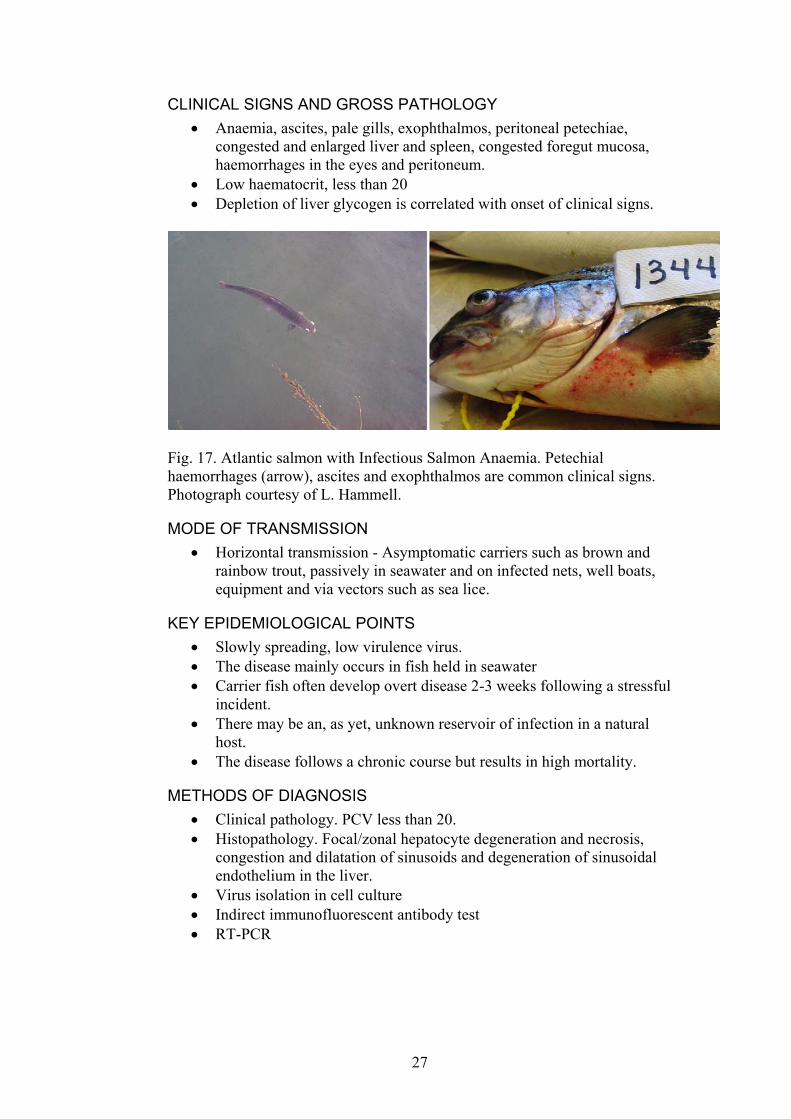
CLINICAL SIGNS AND GROSS PATHOLOGY Anaemia, ascites, pale gills, exophthalmos, peritoneal petechiae, congested and enlarged liver and spleen, congested foregut mucosa, haemorrhages in the eyes and peritoneum.
•
• •
Low haematocrit, less than 20 Depletion of liver glycogen is correlated with onset of clinical signs.
Fig. 17. Atlantic salmon with Infectious Salmon Anaemia. Petechial haemorrhages (arrow), ascites and exophthalmos are common clinical signs. Photograph courtesy of L. Hammell.
MODE OF TRANSMISSION Horizontal transmission - Asymptomatic carriers such as brown and rainbow trout, passively in seawater and on infected nets, well boats, equipment and via vectors such as sea lice.
•
• •
• • •
KEY EPIDEMIOLOGICAL POINTS • Slowly spreading, low virulence virus. • The disease mainly occurs in fish held in seawater • Carrier fish often develop overt disease 2-3 weeks following a stressful
incident. • There may be an, as yet, unknown reservoir of infection in a natural
host. • The disease follows a chronic course but results in high mortality.
METHODS OF DIAGNOSIS Clinical pathology. PCV less than 20. Histopathology. Focal/zonal hepatocyte degeneration and necrosis, congestion and dilatation of sinusoids and degeneration of sinusoidal endothelium in the liver. Virus isolation in cell culture Indirect immunofluorescent antibody test RT-PCR
27
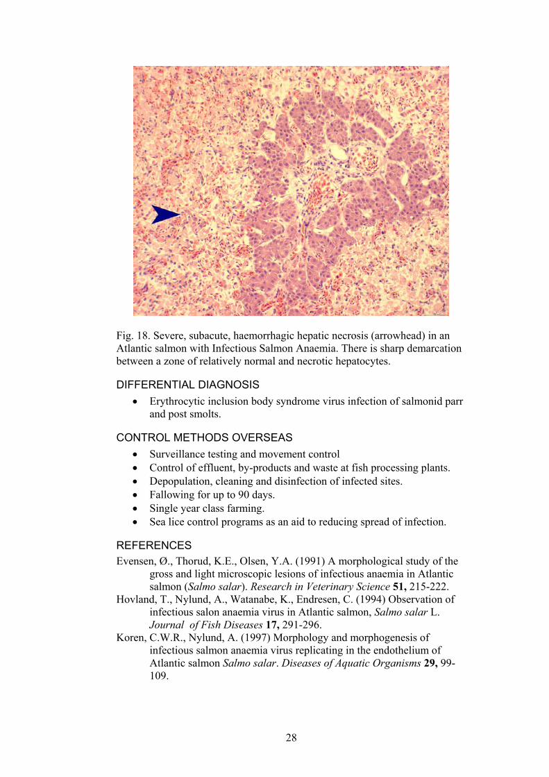
Fig. 18. Severe, subacute, haemorrhagic hepatic necrosis (arrowhead) in an Atlantic salmon with Infectious Salmon Anaemia. There is sharp demarcation between a zone of relatively normal and necrotic hepatocytes.
DIFFERENTIAL DIAGNOSIS Erythrocytic inclusion body syndrome virus infection of salmonid parr and post smolts.
•
• • • • • •
CONTROL METHODS OVERSEAS Surveillance testing and movement control Control of effluent, by-products and waste at fish processing plants. Depopulation, cleaning and disinfection of infected sites. Fallowing for up to 90 days. Single year class farming. Sea lice control programs as an aid to reducing spread of infection.
REFERENCES Evensen, Ø., Thorud, K.E., Olsen, Y.A. (1991) A morphological study of the
gross and light microscopic lesions of infectious anaemia in Atlantic salmon (Salmo salar). Research in Veterinary Science 51, 215-222.
Hovland, T., Nylund, A., Watanabe, K., Endresen, C. (1994) Observation of infectious salon anaemia virus in Atlantic salmon, Salmo salar L. Journal of Fish Diseases 17, 291-296.
Koren, C.W.R., Nylund, A. (1997) Morphology and morphogenesis of infectious salmon anaemia virus replicating in the endothelium of Atlantic salmon Salmo salar. Diseases of Aquatic Organisms 29, 99-109.
28
Nylund, A., Hovlund, T., Hodneland, K., Nilsen, F., Løvik, P. (1994) Mechanisms for transmission of infectious salmon anaemia (ISA). Diseases of Aquatic Organisms 19, 95-100.
Nylund, A., Krossoy, B., Watanabe, K., Holm, J.A. (1996) Target cells for the ISA virus in Atlantic salmon (Salmo salar L.). Bulletin of the European Association of Fish Pathologists 16, 68-72.
OIE (2003). International Aquatic Animal Health Code. ISBN 9290445807. Available at URL: http://www.oie.int/eng/normes/fcode/A_summry.htm
OIE (2003). OIE Manual for Diagnostic Tests for Aquatic Animals. ISBN 9290445637. Available at URL: http://www.oie.int/eng/normes/fmanual/A_summry.htm
ProMED (2003) Infectious salmon anemia-USA:OIE. Speilberg, L., Evensen, Ø., Dannevig, B.H. (1995) A sequential study of the
light and electron microscopic liver lesions of infectious anaemia in Atlantic salmon (Salmo salar L.). Veterinary Pathology 32, 456-478.
Thorud, K., Djupvik, H.O. (1988) Infectious anaemia in Atlantic salmon (Salmo salar L.). Bulletin of the European Association of Fish Pathologists 8, 109-111.
Winton, J.R. (2001) Fish Health Management. In: Fish Hatchery Management.(Ed. by. G. Wedemeyer), pp. 733, American Fisheries Society, Bethesda, Maryland.
29
3.1.4. BACTERIAL KIDNEY DISEASE (BKD) Reportable to OIE and NACA. Also known as corynebacterial kidney disease and salmonid kidney disease.
DISEASE AGENT Gram-positive bacterium: Renibacterium salmoninarum.
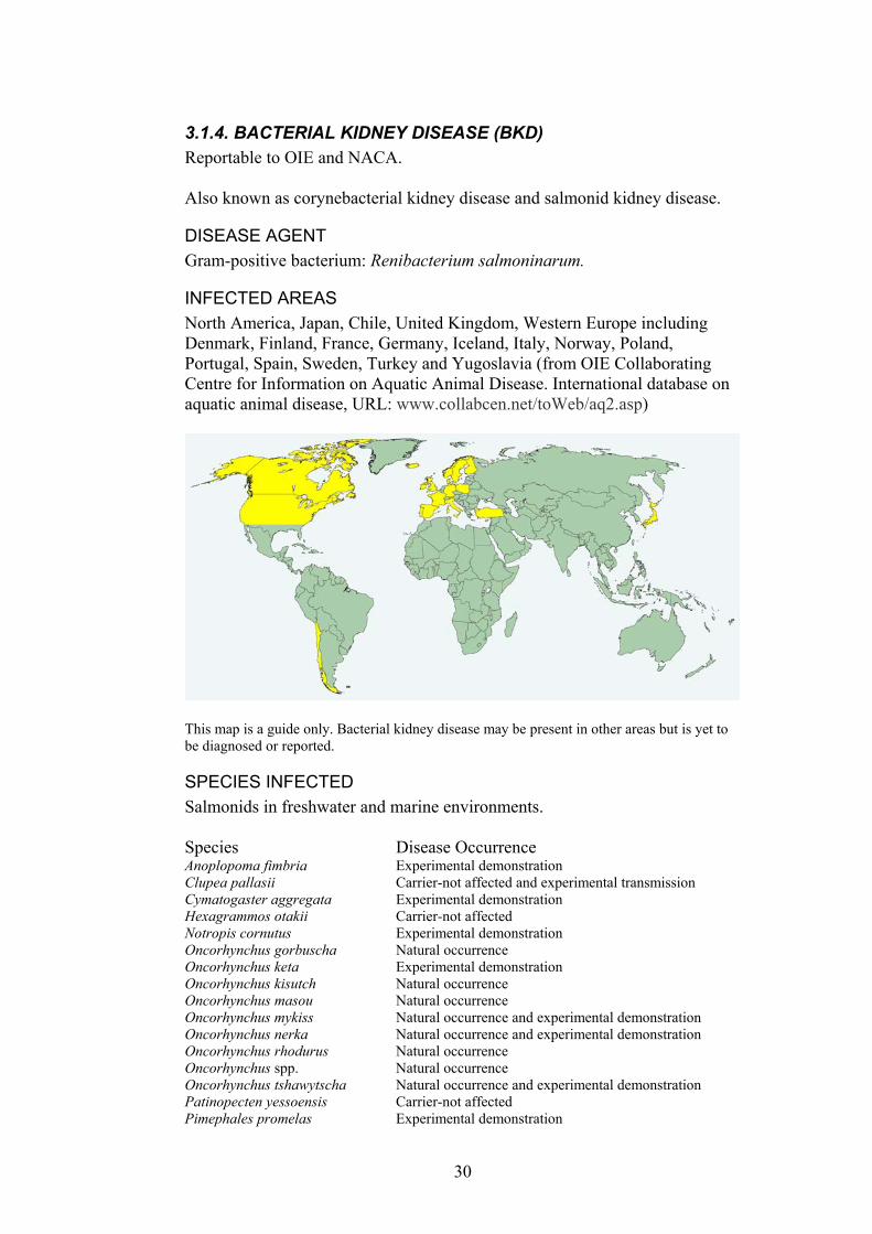
INFECTED AREAS North America, Japan, Chile, United Kingdom, Western Europe including Denmark, Finland, France, Germany, Iceland, Italy, Norway, Poland, Portugal, Spain, Sweden, Turkey and Yugoslavia (from OIE Collaborating Centre for Information on Aquatic Animal Disease. International database on aquatic animal disease, URL: www.collabcen.net/toWeb/aq2.asp)
This map is a guide only. Bacterial kidney disease may be present in other areas but is yet to be diagnosed or reported.
SPECIES INFECTED Salmonids in freshwater and marine environments. Species Disease Occurrence Anoplopoma fimbria Experimental demonstration Clupea pallasii Carrier-not affected and experimental transmission Cymatogaster aggregata Experimental demonstration Hexagrammos otakii Carrier-not affected Notropis cornutus Experimental demonstration Oncorhynchus gorbuscha Natural occurrence Oncorhynchus keta Experimental demonstration Oncorhynchus kisutch Natural occurrence Oncorhynchus masou Natural occurrence Oncorhynchus mykiss Natural occurrence and experimental demonstration Oncorhynchus nerka Natural occurrence and experimental demonstration Oncorhynchus rhodurus Natural occurrence Oncorhynchus spp. Natural occurrence Oncorhynchus tshawytscha Natural occurrence and experimental demonstration Patinopecten yessoensis Carrier-not affected Pimephales promelas Experimental demonstration
30
Platycephalus indicus Carrier-not affected Plecoglossus altivelis Experimental demonstration & Natural occurrence Salmo (Hucho) hucho Experimental demonstration & Natural occurrence Salmo clarki Natural occurrence Salmo salar Natural occurrence and experimental demonstration Salmo trutta Natural occurrence Salmonids Natural occurrence Salvelinus alpinus Natural occurrence Salvelinus fontinalis Natural occurrence Salvelinus namaycush Natural occurrence Thymallus thymallus Natural occurrence and experimental demonstration From OIE Collaborating Centre for Information on Aquatic Animal Disease. International database on aquatic animal disease, URL: www.collabcen.net/toWeb/aq2.asp The common names of some species are listed in Appendix three.
CLINICAL SIGNS Increasing mortalities over a long period of time. •
•
•
•
Exophthalmos and ascites resulting from poor osmoregulation as a result of kidney damage. Skin darkening, lethargy, anaemia and pale gills, skin blisters and haemorrhages, cystic cavities in skeletal muscle. Some fish do have a local rather than a systemic infection, with lesions in or around the eyes, brain or skin.
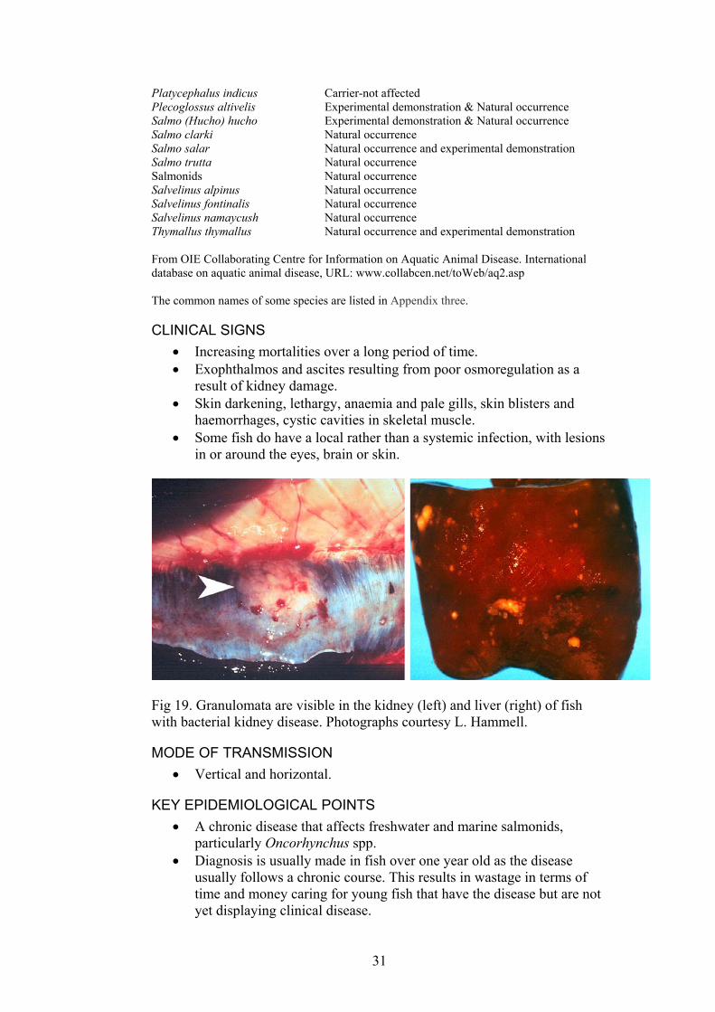
Fig 19. Granulomata are visible in the kidney (left) and liver (right) of fish with bacterial kidney disease. Photographs courtesy L. Hammell.
MODE OF TRANSMISSION Vertical and horizontal. •
KEY EPIDEMIOLOGICAL POINTS • A chronic disease that affects freshwater and marine salmonids,
particularly Oncorhynchus spp. • Diagnosis is usually made in fish over one year old as the disease
usually follows a chronic course. This results in wastage in terms of time and money caring for young fish that have the disease but are not yet displaying clinical disease.
31
METHODS OF DIAGNOSIS Gross examination. Large, grayish patches in the kidney and other organs.
•
•
•
• • •
Histopathology. Granulomata in the kidneys. Gram-positive bacteria in kidney sections, smears or prints. Bacterial culture. 6 to 19 weeks at 15°C. The isolation medium should contain cysteine and serum eg. KDM2 medium. The bacteria are slow growing both in fish and on isolation media. Direct and indirect fluorescent antibody tests. ELISA tests. PCR
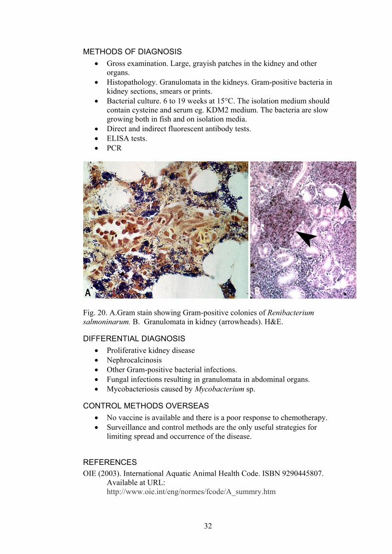
Fig. 20. A.Gram stain showing Gram-positive colonies of Renibacterium salmoninarum. B. Granulomata in kidney (arrowheads). H&E.
DIFFERENTIAL DIAGNOSIS Proliferative kidney disease •
• • • •
• •
Nephrocalcinosis Other Gram-positive bacterial infections. Fungal infections resulting in granulomata in abdominal organs. Mycobacteriosis caused by Mycobacterium sp.
CONTROL METHODS OVERSEAS No vaccine is available and there is a poor response to chemotherapy. Surveillance and control methods are the only useful strategies for limiting spread and occurrence of the disease.
REFERENCES OIE (2003). International Aquatic Animal Health Code. ISBN 9290445807.
Available at URL: http://www.oie.int/eng/normes/fcode/A_summry.htm
32
OIE (2003). OIE Manual for Diagnostic Tests for Aquatic Animals. ISBN 9290445637. Available at URL: http://www.oie.int/eng/nomes/fmanaul/A_summry.htm
Ortega, C., Sanz, F., Muzquiz, J.L., Ramos, P., Docando, J., Planas, E. (1993) Epidemiological study of BKD (Renibacterium salmoninarum) in Spain: role of temperature as a risk factor in a natural outbreak of the disease. Bulletin of the European Association of Fish Pathologists 13, 201-202.
Pascha, R.J., Elliott,D.G., Chase, D.M. (2002) Comparison of traditional and molecular methods for detection of Renibacterium salmoninarum. In: Cunningham, C.O. (ed.) Molecular diagnosis of salmonid diseases. Kluwer Academic Publishers, Dordrecht, The Netherlands. p. 157-209.
Winton, J.R. (2001) Fish Health Management. In: Fish Hatchery Management.(Ed. by. G. Wedemeyer), pp. 733, American Fisheries Society, Bethesda, Maryland.
33

3.1.5. PISCIRICKETTSIOSIS Reportable to OIE and NACA.
DISEASE AGENT Bacterial disease; Gram-negative, rod shaped, intracellular bacterium; Piscirickettsia salmonis
INFECTED AREAS Countries affected include Canada, Chile, Ireland and Norway.
This map is a guide only. Piscirickettsia salmonis may be present in other areas but is yet to be diagnosed or reported.
SPECIES INFECTED Species Disease Occurrence Oncorhynchus gorbuscha Natural occurrence Oncorhynchus kisutch Natural occurrence Oncorhynchus masou Natural occurrence Oncorhynchus mykiss Natural occurrence and experimental demonstration Oncorhynchus mykiss x O. kisutch Natural occurrence Oncorhynchus tshawytscha Natural occurrence and experimental demonstration Salmo salar Natural occurrence and experimental demonstration From OIE Collaborating Centre for Information on Aquatic Animal Disease. International database on aquatic animal disease, URL: www.collabcen.net/toWeb/aq2.asp The common names of some species are listed in Appendix three.
CLINICAL SIGNS Pale gills, dark colouration, inappetance, lethargy and swimming near the surface.
•
• •
•
Skin lesions, white patches which can progress to shallow ulcers. Swollen discoloured kidneys and enlarged spleen.
MODE OF TRANSMISSION Horizontal transmission
34
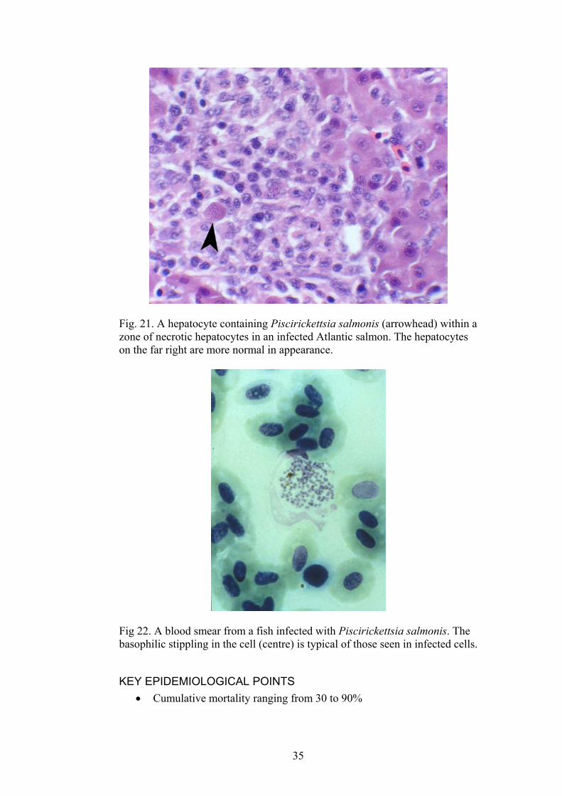
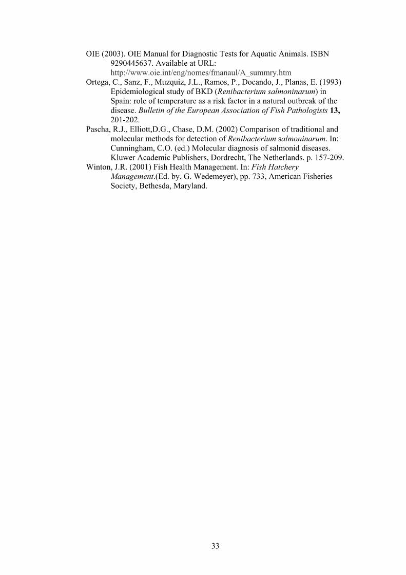
Fig. 21. A hepatocyte containing Piscirickettsia salmonis (arrowhead) within a zone of necrotic hepatocytes in an infected Atlantic salmon. The hepatocytes on the far right are more normal in appearance.
Fig 22. A blood smear from a fish infected with Piscirickettsia salmonis. The basophilic stippling in the cell (centre) is typical of those seen in infected cells.
KEY EPIDEMIOLOGICAL POINTS • Cumulative mortality ranging from 30 to 90%
35

• First signs of the disease commonly occur after fish are transferred from fresh water to sea water.
• The bacterium survives longer in salt water than in fresh water.
METHODS OF DIAGNOSIS Histopathology. Hepatic necrosis and the presence of intracellular inclusions with morphology typical of Piscirickettsia salmonis
•
• • • •
•
• • • • •
Culture in tissue culture Fluorescent antibody test Immunohistochemistry using specific antiserum PCR
DIFFERENTIAL DIAGNOSIS Other rickettsial and chlamydial infections.
CONTROL METHODS OVERSEAS Hygienic measures Holding only one age class of fish at any given site Rearing fish at lower densities. Treatment with antibacterial drugs is inconsistent and problematic. Vaccination trials have given promising results and may be a means of controlling this disease in the near future.
REFERENCES Almendras, F.E., Fuentealba, I.C. (1997) Salmonid rickettsial septicaemia caused by
Piscirickettsia salmonis: a review. Diseases of Aquatic Organisms 29, 137-144.
Almendras, F.E., Fuentealba, I.C., Jones, S.R.M., Markham, F., Spangler, E. (1997) Experimental infection and horizontal transmission of Piscirickettsia salmonis in freshwater-raised Atlantic salmon, Salmo salar L. Journal of Fish Diseases 20, 409-418.
Fryer, J.L., Hedrick, R.P. (2003) Piscirickettsia salmonis: a Gram-negative intracellular bacterial pathogen of fish. Journal of Fish Diseases 26, 251-262.
Fryer, J.L., Lannan, C.N. (1996) Rickettsial infections of fish. Annual Review of Fish Diseases 6, 3-13.
Fryer, J.L., Lannan, C.N., Garcés, L.H., Larenas, J.J., Smith, P.A. (1990) Isolation of a Rickettsia-like organism from diseased coho salmon (Oncorhynchus kisutch) in Chile. Fish Pathology 25, 107-114.
OIE (2003). International Aquatic Animal Health Code. ISBN 9290445807. Available at URL: http://www.oie.int/eng/normes/fcode/A_summry.htm
OIE (2003). OIE Manual for Diagnostic Tests for Aquatic Animals. ISBN 9290445637. Available at URL: http://www.oie.int/eng/nomes/fmanaul/A_summry.htm
Olsen, A.B., Melby, H.P., Speilberg, L., Evensen, Ø., Håstein, T. (1997) Piscirickettsia salmonis infection in Atlantic salmon Salmo salar in Norway-epidemiological, pathological and microbiological findings. Diseases of Aquatic Organisms 31, 35-48.
Winton, J.R. (2001) Fish Health Management. In: Fish Hatchery Management.(Ed. by. G. Wedemeyer), pp. 733, American Fisheries Society, Bethesda, Maryland.
36
3.1.6. FURUNCULOSIS
DISEASE AGENT Reportable in Australia, but not to the OIE or NACA. Aeromonas salmonicida subsp. salmonicida, a Gram-negative, non-motile bacterium. 0.8 x 1.3-2.0 µm
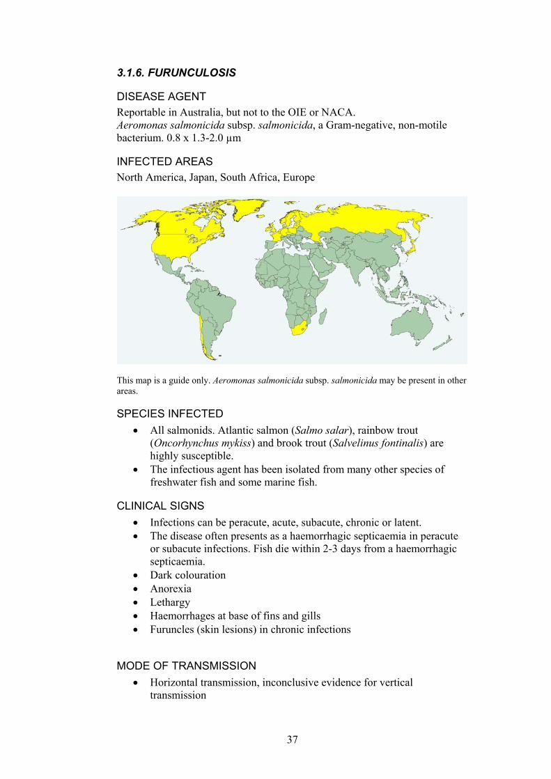
INFECTED AREAS North America, Japan, South Africa, Europe
This map is a guide only. Aeromonas salmonicida subsp. salmonicida may be present in other areas.
SPECIES INFECTED All salmonids. Atlantic salmon (Salmo salar), rainbow trout (Oncorhynchus mykiss) and brook trout (Salvelinus fontinalis) are highly susceptible.
•
•
• •
• • • • •
•
The infectious agent has been isolated from many other species of freshwater fish and some marine fish.
CLINICAL SIGNS Infections can be peracute, acute, subacute, chronic or latent. The disease often presents as a haemorrhagic septicaemia in peracute or subacute infections. Fish die within 2-3 days from a haemorrhagic septicaemia. Dark colouration Anorexia Lethargy Haemorrhages at base of fins and gills Furuncles (skin lesions) in chronic infections
MODE OF TRANSMISSION Horizontal transmission, inconclusive evidence for vertical transmission
37
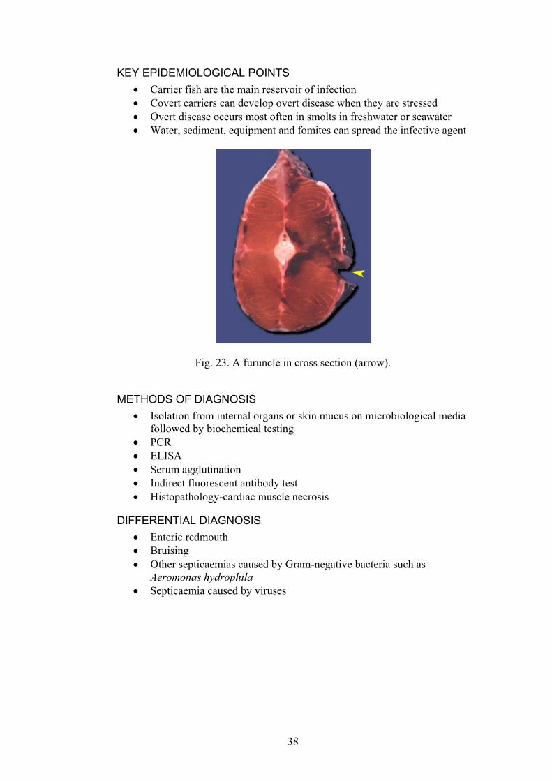
KEY EPIDEMIOLOGICAL POINTS • Carrier fish are the main reservoir of infection • Covert carriers can develop overt disease when they are stressed • Overt disease occurs most often in smolts in freshwater or seawater • Water, sediment, equipment and fomites can spread the infective agent
Fig. 23. A furuncle in cross section (arrow).
METHODS OF DIAGNOSIS Isolation from internal organs or skin mucus on microbiological media followed by biochemical testing
•
• • • • •
• • •
•
PCR ELISA Serum agglutination Indirect fluorescent antibody test Histopathology-cardiac muscle necrosis
DIFFERENTIAL DIAGNOSIS Enteric redmouth Bruising Other septicaemias caused by Gram-negative bacteria such as Aeromonas hydrophila Septicaemia caused by viruses
38
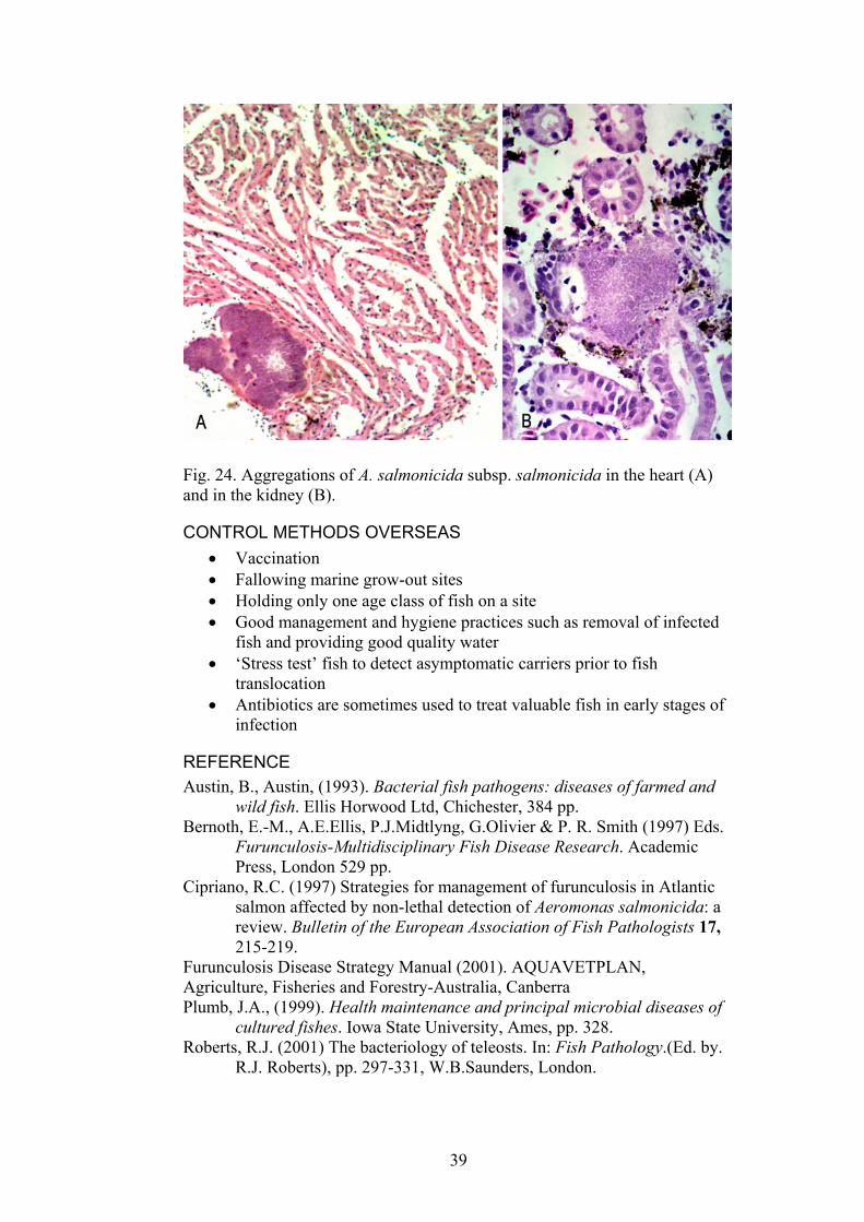
Fig. 24. Aggregations of A. salmonicida subsp. salmonicida in the heart (A) and in the kidney (B).
CONTROL METHODS OVERSEAS Vaccination •
• • •
•
•
Fallowing marine grow-out sites Holding only one age class of fish on a site Good management and hygiene practices such as removal of infected fish and providing good quality water ‘Stress test’ fish to detect asymptomatic carriers prior to fish translocation Antibiotics are sometimes used to treat valuable fish in early stages of infection
REFERENCE Austin, B., Austin, (1993). Bacterial fish pathogens: diseases of farmed and
wild fish. Ellis Horwood Ltd, Chichester, 384 pp. Bernoth, E.-M., A.E.Ellis, P.J.Midtlyng, G.Olivier & P. R. Smith (1997) Eds.
Furunculosis-Multidisciplinary Fish Disease Research. Academic Press, London 529 pp.
Cipriano, R.C. (1997) Strategies for management of furunculosis in Atlantic salmon affected by non-lethal detection of Aeromonas salmonicida: a review. Bulletin of the European Association of Fish Pathologists 17, 215-219.
Furunculosis Disease Strategy Manual (2001). AQUAVETPLAN, Agriculture, Fisheries and Forestry-Australia, Canberra Plumb, J.A., (1999). Health maintenance and principal microbial diseases of
cultured fishes. Iowa State University, Ames, pp. 328. Roberts, R.J. (2001) The bacteriology of teleosts. In: Fish Pathology.(Ed. by.
R.J. Roberts), pp. 297-331, W.B.Saunders, London.
39
3.1.7. ENTERIC REDMOUTH DISEASE (ERM) Reportable in Australia, but not to the OIE or NACA. Also known as Hagerman redmouth disease, yersiniosis or salmonid blood spot.
DISEASE AGENT The Hagerman strain serotype 01a, serotype 01 clonal group 5 of Yersinia ruckeri, a Gram-negative, motile, rod shaped bacterium. Rod-shaped, 0.5-0.8 x 1.0-3.0 µm.
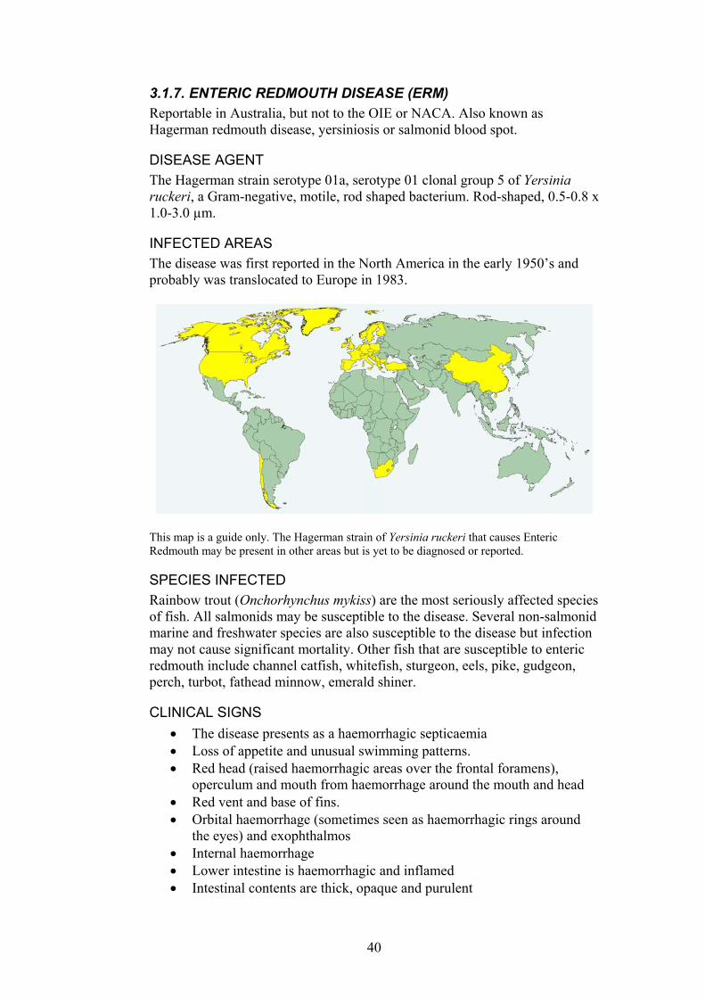
INFECTED AREAS The disease was first reported in the North America in the early 1950’s and probably was translocated to Europe in 1983.
This map is a guide only. The Hagerman strain of Yersinia ruckeri that causes Enteric Redmouth may be present in other areas but is yet to be diagnosed or reported.
SPECIES INFECTED Rainbow trout (Onchorhynchus mykiss) are the most seriously affected species of fish. All salmonids may be susceptible to the disease. Several non-salmonid marine and freshwater species are also susceptible to the disease but infection may not cause significant mortality. Other fish that are susceptible to enteric redmouth include channel catfish, whitefish, sturgeon, eels, pike, gudgeon, perch, turbot, fathead minnow, emerald shiner.
CLINICAL SIGNS The disease presents as a haemorrhagic septicaemia •
• •
• •
• • •
Loss of appetite and unusual swimming patterns. Red head (raised haemorrhagic areas over the frontal foramens), operculum and mouth from haemorrhage around the mouth and head Red vent and base of fins. Orbital haemorrhage (sometimes seen as haemorrhagic rings around the eyes) and exophthalmos Internal haemorrhage Lower intestine is haemorrhagic and inflamed Intestinal contents are thick, opaque and purulent
40
Distended abdomen and ascites • • •
Dark colouration Emaciation
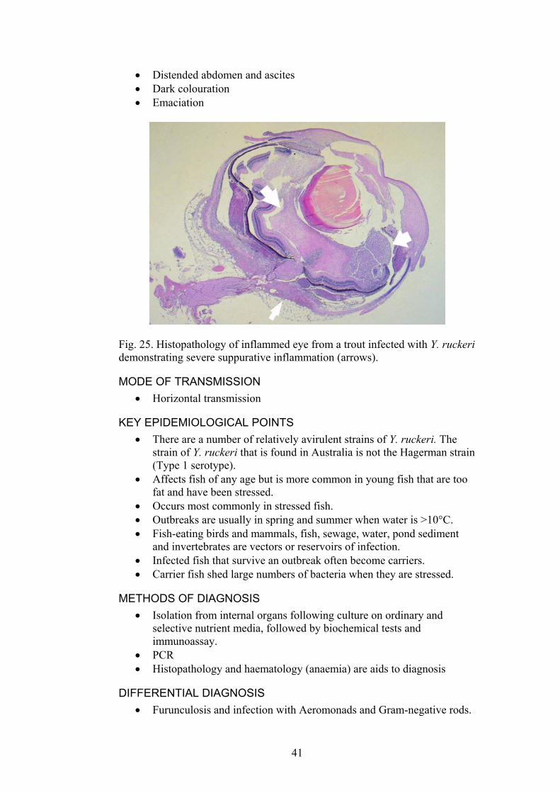
Fig. 25. Histopathology of inflammed eye from a trout infected with Y. ruckeri demonstrating severe suppurative inflammation (arrows).
MODE OF TRANSMISSION Horizontal transmission •
•
• •
•
KEY EPIDEMIOLOGICAL POINTS • There are a number of relatively avirulent strains of Y. ruckeri. The
strain of Y. ruckeri that is found in Australia is not the Hagerman strain (Type 1 serotype).
• Affects fish of any age but is more common in young fish that are too fat and have been stressed.
• Occurs most commonly in stressed fish. • Outbreaks are usually in spring and summer when water is >10°C. • Fish-eating birds and mammals, fish, sewage, water, pond sediment
and invertebrates are vectors or reservoirs of infection. • Infected fish that survive an outbreak often become carriers. • Carrier fish shed large numbers of bacteria when they are stressed.
METHODS OF DIAGNOSIS Isolation from internal organs following culture on ordinary and selective nutrient media, followed by biochemical tests and immunoassay. PCR Histopathology and haematology (anaemia) are aids to diagnosis
DIFFERENTIAL DIAGNOSIS Furunculosis and infection with Aeromonads and Gram-negative rods.
41
CONTROL METHODS OVERSEAS Vaccination •
• •
Provide good standards of hygiene and removal of dead and dying fish Remove stress and treatment with antibiotics
REFERENCES Austin, B., Austin, 1993. Bacterial fish pathogens: diseases of farmed and
wild fish. Ellis Horwood Ltd, Chichester, pp. 384. Danley, M.L., Goodwin, A.E., Killian, H.S. (1999) Epizootics in farm-raised
channel catfish, Ictalurus punctatus (Rafinesque), caused by the enteric redmouth bacterium Yersinia ruckeri. Journal of Fish Diseases 22, 451-456.
Horne, M.T. and Barnes, A.C. (1999) Enteric Redmouth Disease (Y. ruckeri) in Viral, Bacterial and Fungal Infections Ed. Woo and Bruno,D. Vol 3 ;455-477.
Plumb, J.A., (1999). Health maintenance and principal microbial diseases of cultured fishes. Iowa State University, Ames, pp. 328.
Roberts, R.J. (2001) The bacteriology of teleosts. In: Fish Pathology.(Ed. by. R.J. Roberts), pp. 297-331, W.B. Saunders, London.
42
3.1.8. WHIRLING DISEASE Reportable in Australia, but not to the OIE or NACA.
DISEASE AGENT Protozoan parasite, the myxosporean Myxobolus cerebralis. The disease agent probably originated in Europe and was translocated to the USA and other areas of the world in infected fish and fish products.
INFECTED AREAS Most of Europe is infected including Denmark, Finland, France, Czechoslovakia, Poland, Norway, Austria, Belgium, Hungary, Spain, Netherlands, parts of the former USSR, Italy, Germany, Yugoslavia, UK, Ireland, Bulgaria, Sweden, South Africa, New Zealand, USA, some parts of Asia, possibly Turkey, Morocco, Lebanon.
This map is a guide only. Myxobolus cerebralis may be present in other areas but is yet to be diagnosed or reported.
SPECIES INFECTED Salmonids, especially rainbow trout (Oncorhynchus mykiss). Brook trout are less severely affected and brown trout (Salmo trutta) are more resistant to clinical disease.
CLINICAL SIGNS AND GROSS PATHOLOGY Skeletal deformities: curvature of the spine (lordosis and scoliosis) and head deformities (skull depression, misshapen jaws, short operculae). Caused by the protozoans ability to damage the cartilage.
•
•
•
Abnormal swimming- ‘whirling’- a neuropathological consequence of lower brain stem and spinal cord constriction. Black tail- produced when the parasite infects cartilage in the posterior spinal cord.
43
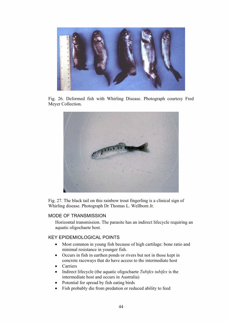
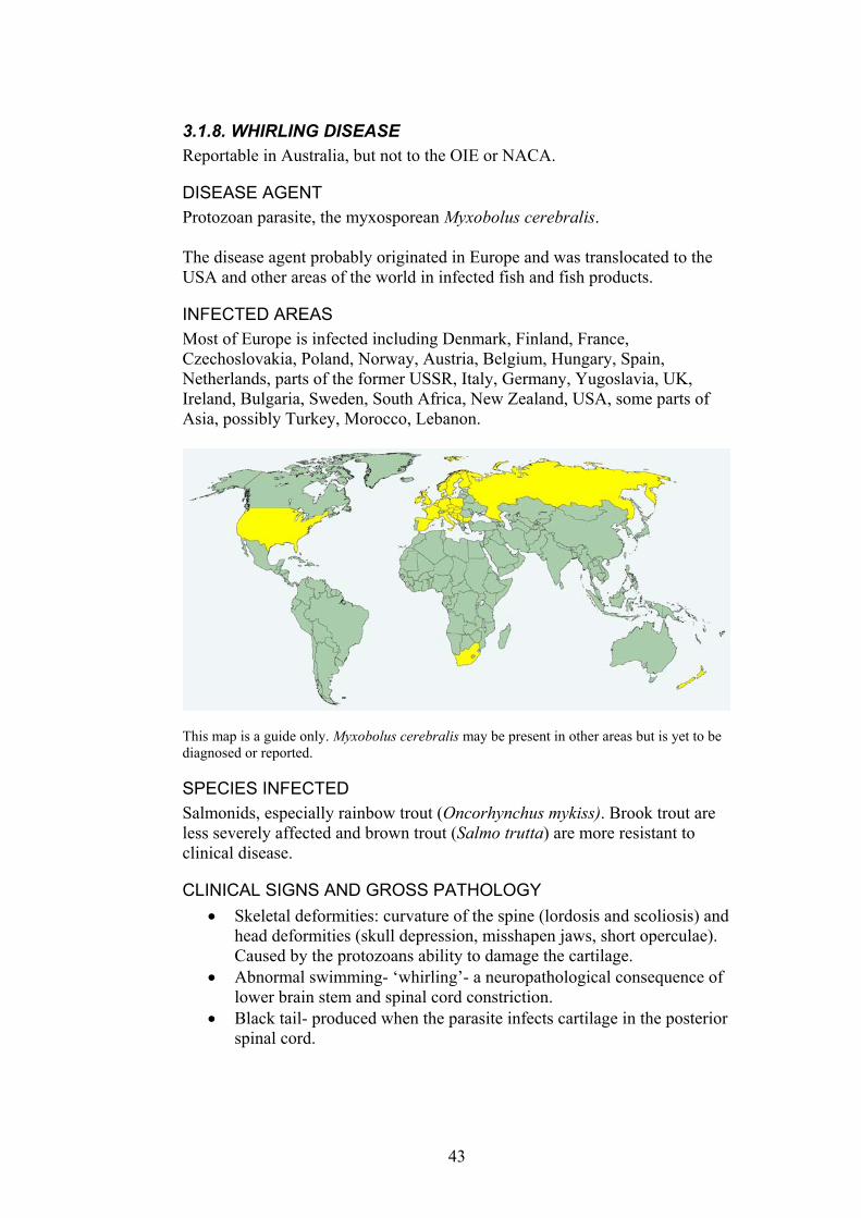
Fig. 26. Deformed fish with Whirling Disease. Photograph courtesy Fred Meyer Collection.
Fig. 27. The black tail on this rainbow trout fingerling is a clinical sign of Whirling disease. Photograph Dr Thomas L. Wellborn Jr.
MODE OF TRANSMISSION Horizontal transmission. The parasite has an indirect lifecycle requiring an aquatic oligochaete host.
KEY EPIDEMIOLOGICAL POINTS • Most common in young fish because of high cartilage: bone ratio and
minimal resistance in younger fish. • Occurs in fish in earthen ponds or rivers but not in those kept in
concrete raceways that do have access to the intermediate host • Carriers • Indirect lifecycle (the aquatic oligochaete Tubifex tubifex is the
intermediate host and occurs in Australia) • Potential for spread by fish eating birds • Fish probably die from predation or reduced ability to feed
44

• Environmental stress pre-disposes fish to infection • Fish can only be infected in fresh water environments • Spores are very resistant and survive freezing, drying and long periods
(years).
METHODS OF DIAGNOSIS Examination of enzyme-digested (pepsin-trypsin) cartilage for spores. Stain with methylene blue, Giemsa or malachite green.
•
•
•
• •
Histological demonstration of granulomatous inflammation of the head and spine, destruction of cartilage. The use of sentinel fish to detect presence of the parasite in a water body PCR for early detection In situ hybridisation
Fig. 28. Histopathology of a rainbow trout with Whirling Disease. The bone and cartilage has been invaded and distorted by granulomatous inflammation and myxospores (left). Mature myxospores and immature stages of the parasite can be seen in cartilage (right). Photograph courtesy Drs Patrick Caplazi and Susan Noh.
Fig. 29. A preparation of cartilage from the head of a rainbow trout stained with methylene blue. Myxobolus cerebralis spores can be seen (arrow).
45
DIFFERENTIAL DIAGNOSIS Other myxosporean parasites •
• • •
•
• •
• •
Vitamin C deficiency Viral Haemorrhagic Septicaemia, Oncorhynchus masou virus disease Yersinia ruckeri or other septicaemic conditions causing inflammation in the brain High incubation temperatures causing skeletal deformities.
CONTROL METHODS OVERSEAS Raise fingerlings to 6cm in length in spore-free water eg. groundwater Hold fish in lined or concrete ponds where Tubifex sp. worms do not occur Drain, clean and disinfect earthen ponds Toltrazuril and fumagillin may be useful chemotherapeutic agents.
REFERENCES Allen, M.B., Bergensen, E.P. (2002) Factors influencing the distribution of
Myxobolus cerebralis, the causative agent of whirling disease, in the Cache la Poudre River, Colorado. Diseases of Aquatic Organisms 49, 51-60.
El-Matbouli, M., Fischer-Scherl, T., Hoffman, R.W. (1992) Present knowledge on the life cycle, taxonomy, pathology, and therapy of some Myxosporea spp. important for freshwater fish. Annual Review of Fish Diseases, 367-402.
Garden, O. (1992) The Myxosporea of fish: a review. British Veterinary Journal 148, 223-239.
Halliday, M.M. (1976) The biology of Myxosoma cerebralis: the causative organism of whirling disease of salmonids. Journal of Fish Biology 9, 339-357.
Hiner, M., Moffitt, C.M. (2001) Variations in infections of Myxobolus cerebralis in field-exposed cuttroat and rainbow trout in Idaho. Journal of Aquatic Animal Health 13, 124-132.
Hoffman, G.L. (1990) Myxobolus cerebralis, a worldwide cause of salmonid Whirling disease. Journal of Aquatic Animal Health 2, 30-37.
Thompson, K.G., Nehring, R.B. (2000) A simple technique used to filter and quantify the actinospore of Myxobolus cerebralis and determine its seasonal abundance in the Colorado River. Journal of Aquatic Animal Health 12, 316-323.
www.whirling_disease.org/foundation.html
46
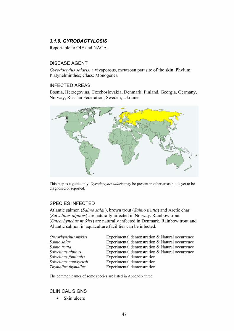
3.1.9. GYRODACTYLOSIS Reportable to OIE and NACA.
DISEASE AGENT Gyrodactylus salaris, a vivaporous, metazoan parasite of the skin. Phylum: Platyhelminthes; Class: Monogenea
INFECTED AREAS Bosnia, Herzegovina, Czechoslovakia, Denmark, Finland, Georgia, Germany, Norway, Russian Federation, Sweden, Ukraine
This map is a guide only. Gyrodactylus salaris may be present in other areas but is yet to be diagnosed or reported.
SPECIES INFECTED Atlantic salmon (Salmo salar), brown trout (Salmo trutta) and Arctic char (Salvelinus alpinus) are naturally infected in Norway. Rainbow trout (Oncorhynchus mykiss) are naturally infected in Denmark. Rainbow trout and Altantic salmon in aquaculture facilities can be infected. Oncorhynchus mykiss Experimental demonstration & Natural occurrence Salmo salar Experimental demonstration & Natural occurrence Salmo trutta Experimental demonstration & Natural occurrence Salvelinus alpinus Experimental demonstration & Natural occurrence Salvelinus fontinalis Experimental demonstration Salvelinus namaycush Experimental demonstration Thymallus thymallus Experimental demonstration The common names of some species are listed in Appendix three.
CLINICAL SIGNS Skin ulcers •
47
‘Flashing’ •
•
•
• •
•
MODE OF TRANSMISSION Horizontal transmission. The species is viviparous and has a direct life cycle.
KEY EPIDEMIOLOGICAL POINTS • High mortality in susceptible fish. • Some Baltic Sea strains of Atlantic salmon are resistant to clinical
disease. • Atlantic strains of Atlantic salmon are highly susceptible to clinical
disease. • Water quality affects susceptibility to clinical disease. • The parasite ‘browses’ on epithelial cells and the reasons for host
mortality are poorly understood, but is often associated with infection with opportunistic pathogens such as Saprolegnia spp.
• In experimental studies, parasite numbers increased more at 12°C than at 6°C and was negligible at 1.4°C and the number of parasites on infected, susceptible fish was reduced in salinities of >10 parts per thousand (ppt).
METHODS OF DIAGNOSIS Examination of fresh or ethanol fixed fish (specific collection procedures apply to prevent loss of parasites) using a dissecting microscope. The dorsal fin, followed by the anal and pectoral fins are the most heavily parasitised parts of the fish. Some fish have only one parasite. The parasite usually measures 250-350 µm but can be as small as 200 µm in summer months or as large as 500 µm. Morphology of hooks and bars in the opishaptor of Gyrodactylus spp. PCR
DIFFERENTIAL DIAGNOSIS Other gyrodactylids, especially G. teuchis and G. thymalli.
48

Fig. 30. Transmission electron micrograph of several Gyrodactylus salaris attached to the epidermis of Atlantic salmon. The parasite is loosely attached to the epithelium by 2 hooks (hamuli) and 16 hooklets on the opishaptor (arrowhead). Photograph courtesy Tor Atle Mo. Bar = 100 µm.
Fig. 31. Opishaptor and hooks of Gyrodactylus salaris. The morphology of these structures is used to morphologically distinguish this parasite from other gyrodactylids. Photograph courtesy of Tor Atle Mo.
49
CONTROL METHODS OVERSEAS Testing and certification of fish prior to translocation in Europe •
•
•
Rotenone treatment of infected rivers in Norway was used to remove entire fish populations Metals such as aluminium are toxic to the parasite but not fish
REFERENCES Ergens, R. (1983) Gyrodactylus from Eurasian freshwater Salmonidae and
Thymallidae. Folia Parasitologica 30, 15-26. Jensen, A.J., Johnsen, B.O. (1992) Site specificity of Gyrodactylus salaris
Malmberg, 1957 (Monogenea) on Atlantic salmon (Salmo salar) in the River Lakselva, northern Norway. Canadian Journal of Fisheries and Aquatic Science 70, 264-267.
OIE (2003). International Aquatic Animal Health Code. ISBN 9290445807. Available at URL: http://www.oie.int/eng/normes/fcode/A_summry.htm
OIE (2003). OIE Manual for Diagnostic Tests for Aquatic Animals. ISBN 9290445637. Available at URL: http://www.oie.int/eng/nomes/fmanaul/A_summry.htm
Mo, T.A. (1992) Seasonal variations in the prevalence and infestation intensity of Gyrodactylus salaris Malmberg, 1957 (Monogenea: Gyrodactylidae) on Atlantic salmon parr, Salmo salar L. in the River Batnfjordselva Norway. Journal of Fish Biology, 697-707.
Soleng, A., Bakke, T.A. (1997) Salinity tolerance of Gyrodactylus salaris (Platyhelminthes, Monogenea): laboratory studies. Canadian Journal of Fisheries and Aquatic Science 54, 1837-1845.
50
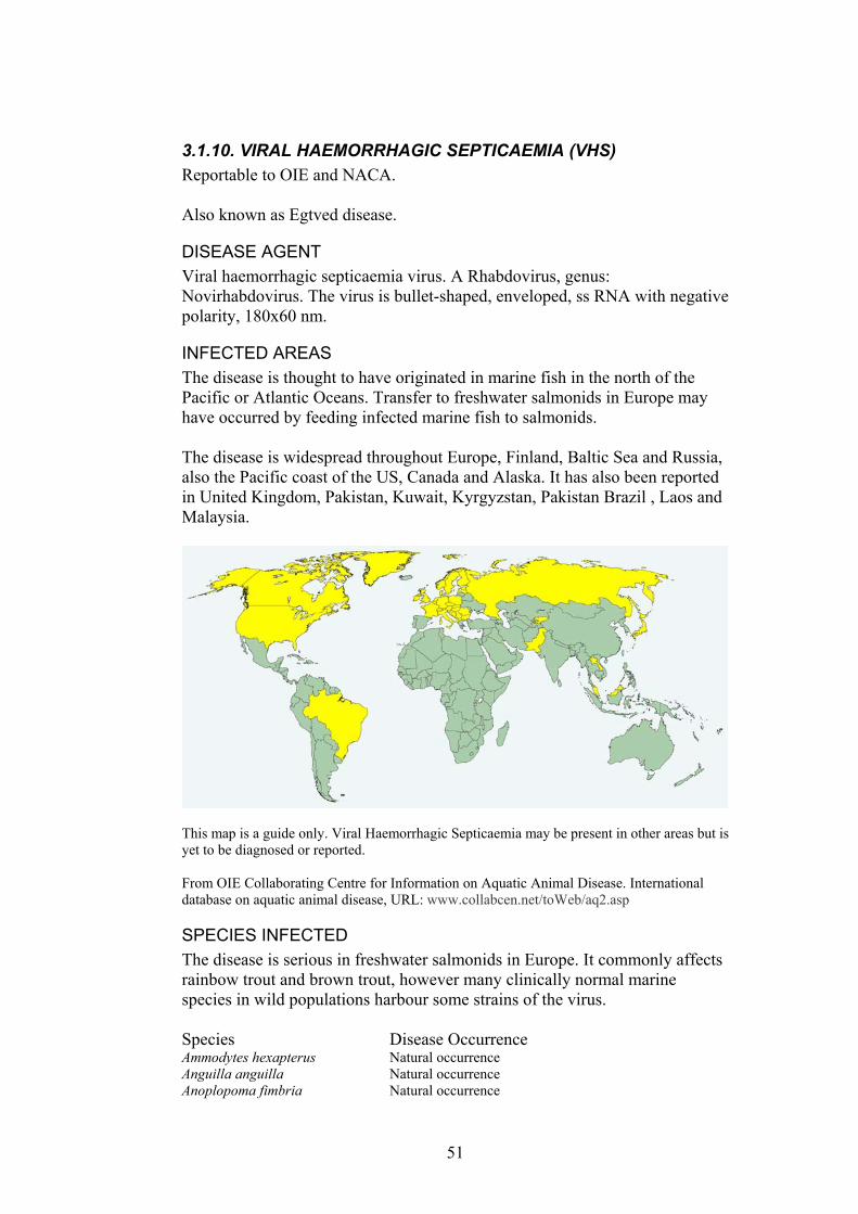
3.1.10. VIRAL HAEMORRHAGIC SEPTICAEMIA (VHS) Reportable to OIE and NACA. Also known as Egtved disease.
DISEASE AGENT Viral haemorrhagic septicaemia virus. A Rhabdovirus, genus: Novirhabdovirus. The virus is bullet-shaped, enveloped, ss RNA with negative polarity, 180x60 nm.
INFECTED AREAS The disease is thought to have originated in marine fish in the north of the Pacific or Atlantic Oceans. Transfer to freshwater salmonids in Europe may have occurred by feeding infected marine fish to salmonids. The disease is widespread throughout Europe, Finland, Baltic Sea and Russia, also the Pacific coast of the US, Canada and Alaska. It has also been reported in United Kingdom, Pakistan, Kuwait, Kyrgyzstan, Pakistan Brazil , Laos and Malaysia.
This map is a guide only. Viral Haemorrhagic Septicaemia may be present in other areas but is yet to be diagnosed or reported. From OIE Collaborating Centre for Information on Aquatic Animal Disease. International database on aquatic animal disease, URL: www.collabcen.net/toWeb/aq2.asp
SPECIES INFECTED The disease is serious in freshwater salmonids in Europe. It commonly affects rainbow trout and brown trout, however many clinically normal marine species in wild populations harbour some strains of the virus. Species Disease Occurrence Ammodytes hexapterus Natural occurrence Anguilla anguilla Natural occurrence Anoplopoma fimbria Natural occurrence
51
Ardea cinerea Carrier-not affected Argentina sphyraena Natural occurrence Barbus graellsi Natural occurrence Clupea harengus Carrier-not affected & Natural occurrence Clupea pallasii Experimental demonstration & Natural occurrence Coregonidae spp. Experimental demonstration & Natural occurrence Coregonus lavaretus Natural occurrence Cymatogaster aggregata Experimental demonstration & Natural occurrence Dicentrarchus labrax Experimental demonstration Esox lucius Experimental demonstration & Natural occurrence Gadus macrocephalus Natural occurrence Gadus morhua Carrier-not affected & Natural occurrence Limanda limanda Natural occurrence Melanogrammus aeglefinus Natural occurrence Merlangius merlangus Natural occurrence Merluccius productus Natural occurrence Micromesistius poutassou Natural occurrence Oncorhynchus kisutch Natural occurrence Oncorhynchus mykiss Experimental demonstration & Natural occurrence Oncorhynchus spp. Carrier-not affected & Natural occurrence Oncorhynchus tshawytscha Natural occurrence Paralichthys olivaceus Carrier-not affected, Experimental demonstration &
Natural occurrence Parophrys vetulus Natural occurrence Platichthys flesus Natural occurrence Pleuronectes platessa Natural occurrence Reinhardtius hippoglossoides Natural occurrence Rhinonemus cimbrius Natural occurrence Salmo aguabonita Experimental demonstration Salmo salar Experimental demonstration & Natural occurrence Salmo trutta Experimental demonstration & Natural occurrence Salvelinus fontinalis Experimental demonstration & Natural occurrence Salvelinus namaycush Experimental demonstration Sardinops sagax Natural occurrence Scophthalmus maximus (Psetta maxima) Experimental demonstration & Natural occurrence Sprattus( =Clupea ) sprattus Natural occurrence Theragra chalcogramma Natural occurrence Thymallus thymallus Experimental demonstration & Natural occurrence Trisopterus esmarki (Gadus esmarki) Natural occurrence Trisopterus minutus Natural occurrence From OIE Collaborating Centre for Information on Aquatic Animal Disease. International database on aquatic animal disease, URL: www.collabcen.net/toWeb/aq2.asp The common names of some species are listed in Appendix three.
CLINICAL SIGNS Mortality •
• •
• • • •
Lethargy or hyperactivity. Oedema and haemorrhage (exophthalmos, anaemia, ascites) due to impairment of the osmotic balance. Dark body colouration Enlarged spleen and kidney. Congested liver. The virus replicates in capillary endothelial cells, leukocytes, haematopoietic tissue and glomeruli.
52

MODE OF TRANSMISSION Horizontal transmission in faeces, urine and sexual fluids. •
•
• • • •
KEY EPIDEMIOLOGICAL POINTS • There are 3 major strains of the virus: North American (Pacific) strain,
European strain and a European marine strain that has 2 sub strains, a North Sea and a Baltic Sea strain. The European freshwater strain and the Baltic Sea marine strain are similar King et al 2001.
• The Pacific marine strain is pathogenic for Pacific herring Clupea harengus pallasi, Californian pilchards and mackerel but not to rainbow trout fry.
• The virus replicates rapidly in colder water (8-10°C) and not at all above 20°C.
• The European freshwater strain is the most virulent and there are acute, chronic and nervous forms.
• Younger fish are highly susceptible to disease. • Asymptomatic carriers act as reservoirs of infection, particularly in
wild, marine fish in the northern Atlantic and Pacific Oceans. • The disease in freshwater salmonids is more common in spring. • Clinical disease often occurs in spring and when water temperatures
are 4-14°C. At lower temperatures a high cumulative mortality may occur over a long period of time, but at higher temperatures, the disease takes a shorter course with lower total mortalities.
METHODS OF DIAGNOSIS Histopathology: congestion of liver, spleen, kidney and subcuticular tissue, increase in melanomacrophage centres in the liver and haematopoietic tissue, haemorrhage and acute necrosis of liver, heart, spleen and kidney. Cell culture ELISA Immunofluorescent antibody test (IFAT) PCR-based technology
53
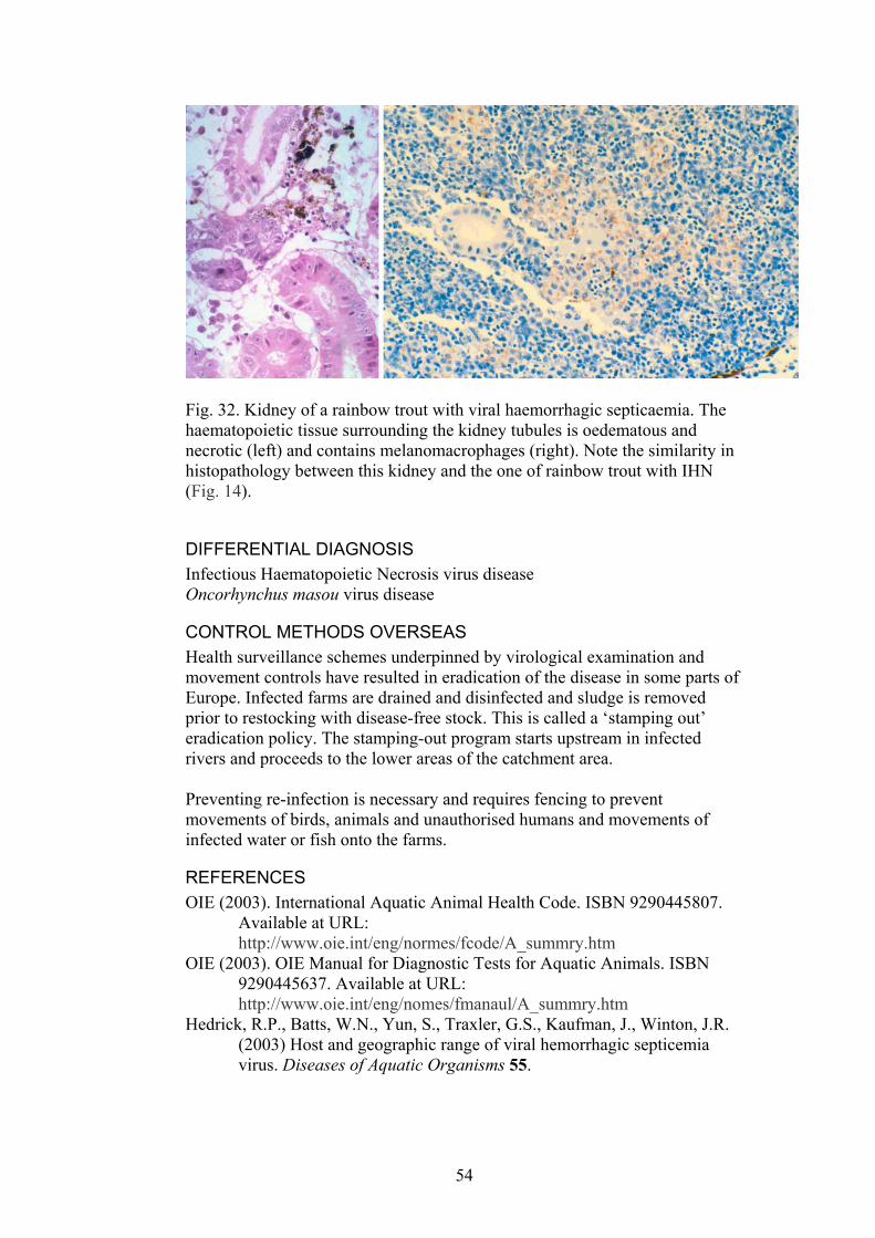
Fig. 32. Kidney of a rainbow trout with viral haemorrhagic septicaemia. The haematopoietic tissue surrounding the kidney tubules is oedematous and necrotic (left) and contains melanomacrophages (right). Note the similarity in histopathology between this kidney and the one of rainbow trout with IHN (Fig. 14).
DIFFERENTIAL DIAGNOSIS Infectious Haematopoietic Necrosis virus disease Oncorhynchus masou virus disease
CONTROL METHODS OVERSEAS Health surveillance schemes underpinned by virological examination and movement controls have resulted in eradication of the disease in some parts of Europe. Infected farms are drained and disinfected and sludge is removed prior to restocking with disease-free stock. This is called a ‘stamping out’ eradication policy. The stamping-out program starts upstream in infected rivers and proceeds to the lower areas of the catchment area. Preventing re-infection is necessary and requires fencing to prevent movements of birds, animals and unauthorised humans and movements of infected water or fish onto the farms.
REFERENCES OIE (2003). International Aquatic Animal Health Code. ISBN 9290445807.
Available at URL: http://www.oie.int/eng/normes/fcode/A_summry.htm
OIE (2003). OIE Manual for Diagnostic Tests for Aquatic Animals. ISBN 9290445637. Available at URL: http://www.oie.int/eng/nomes/fmanaul/A_summry.htm
Hedrick, R.P., Batts, W.N., Yun, S., Traxler, G.S., Kaufman, J., Winton, J.R. (2003) Host and geographic range of viral hemorrhagic septicemia virus. Diseases of Aquatic Organisms 55.
54
Isshiki, T., Nagano, T., Miyazaki, T. (2003) Susceptibility of various marine fish species to Viral Hemorrhagic Septicemia Virus isolated from Japanese flounder. Fish Pathology 38, 113-115.
Isshiki, T., Nishizawa, T., Kobayashi, T., Nagano, T., Miyazaki, T. (2001) An outbreak of VHSV (viral hemorrhagic septicemia virus) infection in farmed Japanese flounder Paralichthys olivaceus in Japan. Diseases of Aquatic Organisms 47, 87-99.
King, J.A., Snow, M., Skall, H.F., Raynard, R.S. (2001) Experimental susceptibility of Atlantic salmon Salmo salar and turbot Scophthalmus maximus to European freshwater and marine isolates of viral haemorrhagic septicaemia virus. Disease of Aquatic Organisms 47, 25-31.
Meyers, T.R., Winton, J.R. (1995) Viral hemorrhagic septicemia virus in North America. Annual Review of Fish Diseases 5, 3-24.
Olesen, N.J. (1998) Sanitation of viral haemorrhagic septicaemia (VHS). Journal of Applied Ichthyology 14, 173-177.
Winton, J.R. (2001) Fish Health Management. In: Fish Hatchery Management.(Ed. by. G. Wedemeyer), pp. 733, American Fisheries Society, Bethesda, Maryland.
Wolf, K., 1988. Fish viruses and fish viral diseases. Cornell University Press, Ithaca, pp. 476.
55

3.1.11. INFECTIOUS PANCREATIC NECROSIS (IPN) Reportable to the OIE and NACA. Also known as acute catarrhal enteritis (USA)
DISEASE AGENT Infectious pancreatic necrosis virus (Birnavirus): an icosahedral, non enveloped ds RNA virus, 60 nm in diameter. There are many strains or serotypes of the virus with varying pathogenicity.
INFECTED AREAS The disease has a wide geographical distribution, occurring in most, if not all, major salmonid-farming countries of North and South America, Europe, Russia, China and other parts of Asia. It has also been reported in South Africa.
This map is a guide only. Infectious Pancreatic Necrosis may be present in other areas but is yet to be diagnosed or reported. From OIE Collaborating Centre for Information on Aquatic Animal Disease. International database on aquatic animal disease, URL: www.collabcen.net/toWeb/aq2.asp
SPECIES INFECTED Infectious pancreatic necrosis was first described in 1941 in brook trout (Salvelinus fontinalis) in North America. Rainbow trout (O. mykiss) also highly susceptible to the disease, cutthroat trout are less susceptible and brown trout (Salmo trutta) are the least susceptible. The disease was traditionally a problem in young salmonids in freshwater but has emerged as a problem in Atlantic salmon post smolts in seawater. Species Disease Occurrence Abramis brama Carrier-not affected Anguilla anguilla Carrier-not affected & Natural occurrence Anguilla japonica Natural occurrence Anguillidae spp. Natural occurrence Atherinidae spp. Natural occurrence Barbus graellsi Carrier-not affected
56
Bothidae spp. Natural occurrence Brachydanio rerio Natural occurrence Brevoortia tyrannus Natural occurrence Carangidae spp. Natural occurrence Carassius auratus Carrier-not affected Carcinus maenas Carrier-not affected Catostomus commersoni Carrier-not affected Channa striatus Natural occurrence Chondrostoma toxostoma Natural occurrence Cichlidae spp. Experimental demonstration & Natural occurrence Clupeidae spp. Natural occurrence Cobitidae spp. Experimental demonstration Cobitidae spp. Natural occurrence Coregonidae spp. Natural occurrence Cotostomidae spp. Natural occurrence Cyprinidae spp. Experimental demonstration & Natural occurrence Cyprinus carpio Carrier-not affected Esocidae spp. Natural occurrence Esox lucius Experimental demonstration & Natural occurrence Hippoglossus hippoglossus Experimental demonstration & Natural occurrence Lampetra fluviatilis Carrier-not affected Misgurnus anguillicaudatus Natural occurrence Morone saxatilis Carrier-not affected Moronidae spp. Natural occurrence Oncorhynchus keta Natural occurrence Oncorhynchus kisutch Natural occurrence Oncorhynchus masou Natural occurrence Oncorhynchus mykiss Carrier-not affected & Natural occurrence Oncorhynchus nerka Experimental demonstration & Natural occurrence Oncorhynchus rhodurus Experimental demonstration & Natural occurrence Oncorhynchus spp. Natural occurrence Oncorhynchus tshawytscha Natural occurrence Paralichthydae spp. Natural occurrence Paralichthys dentatus Natural occurrence Paralichthys lethostigma Unknown Pecten maximus Natural occurrence Perca fluviatilis Carrier-not affected Percidae spp. Natural occurrence Petromyzontidae Natural occurrence Phoxinus phoxinus Carrier-not affected Pollachius virens Carrier-not affected Salmo (Hucho) hucho Natural occurrence Salmo clarki Natural occurrence Salmo salar Experimental demonstration & Natural occurrence Salmo trutta Natural occurrence Salmonids Natural occurrence Salvelinus alpinus Natural occurrence Salvelinus fontinalis Experimental demonstration & Natural occurrence Salvelinus fontinalis x Salvelinus namaycush Natural occurrence Salvelinus namaycush Natural occurrence Sciaenidae spp. Natural occurrence Scophthalmus maximus (Psetta maxima) Natural occurrence Seriola quinqueradiata Natural occurrence Soleidae spp. Natural occurrence Symphysodon discus Carrier-not affected Thymallus thymallus Natural occurrence From OIE Collaborating Centre for Information on Aquatic Animal Disease. International database on aquatic animal disease, URL: www.collabcen.net/toWeb/aq2.asp
57
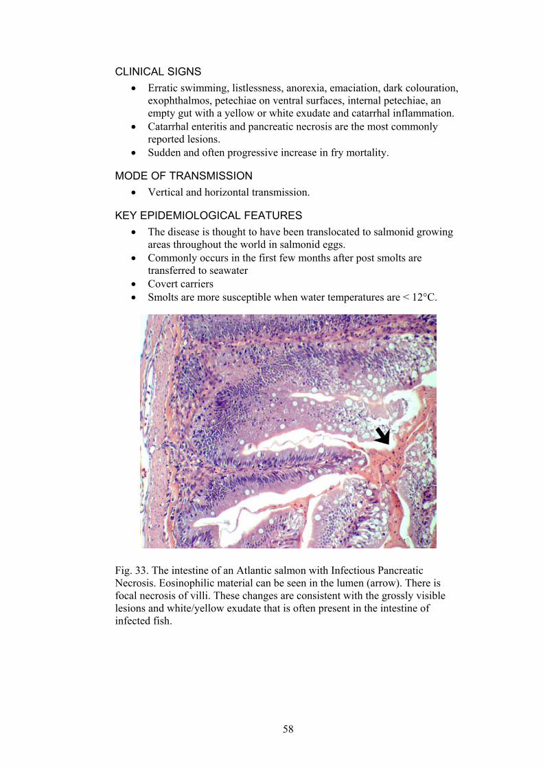
CLINICAL SIGNS Erratic swimming, listlessness, anorexia, emaciation, dark colouration, exophthalmos, petechiae on ventral surfaces, internal petechiae, an empty gut with a yellow or white exudate and catarrhal inflammation.
•
•
•
•
Catarrhal enteritis and pancreatic necrosis are the most commonly reported lesions. Sudden and often progressive increase in fry mortality.
MODE OF TRANSMISSION Vertical and horizontal transmission.
KEY EPIDEMIOLOGICAL FEATURES • The disease is thought to have been translocated to salmonid growing
areas throughout the world in salmonid eggs. • Commonly occurs in the first few months after post smolts are
transferred to seawater • Covert carriers • Smolts are more susceptible when water temperatures are < 12°C.
Fig. 33. The intestine of an Atlantic salmon with Infectious Pancreatic Necrosis. Eosinophilic material can be seen in the lumen (arrow). There is focal necrosis of villi. These changes are consistent with the grossly visible lesions and white/yellow exudate that is often present in the intestine of infected fish.
58

Fig. 34. Histopathology of the liver of an Atlantic salmon fingerling with Infectious Pancreatic Necrosis. There is acute, severe, diffuse necrosis of hepatocytes (arrowhead). Hepatic lesions are often not present in salmonids with IPN, but they are often present in other species with IPN. H&E.
METHODS OF DIAGNOSIS Histopathology. Focal enteritis, sloughing of mucosa and catarrhal enteritis. Multifocal to diffuse necrosis of pancreatic acinar cells, sometimes mild degeneration of renal haematopoietic tissue.
•
• •
•
• •
•
• • • • • • •
Virus isolation. Cytopathic effects on tissue culture. Immunohistochemistry . Monoclonal antibodies or Indirect fluorescent antibody test (IFAT). PCR
DIFFERENTIAL DIAGNOSIS Viral Haemorrhagic Septicaemia Infectious Haematopoietic Necrosis
CONTROL METHODS OVERSEAS Minimise stress when smolts are transferred to seawater and transfer fish when they have good tolerance to seawater and when water temperatures are suitable. Vaccination. Good husbandry. Surface disinfection of eggs. Screen 100% of broodstock. Cleaning and disinfection of infected sites. Prevent the feeding of contaminated tissues. Use of a protected water supply.
59

Lowering stocking density will reduce mortality rates during an outbreak.
•
REFERENCES FVS (2002) Infectious Pancreatic Necrosis (IPN) of Atlantic salmon. Fish
Veterinary Journal 6, 75-84. Laidler, L.A. (2002) Atlantic salmon broodstock and vertical transmission of
Infectious Pancreatic Necrosis virus. Fish Veterinary Journal 6, 71-74. OIE (2003). International Aquatic Animal Health Code. ISBN 9290445807.
Available at URL: http://www.oie.int/eng/normes/fcode/A_summry.htm
OIE (2003). OIE Manual for Diagnostic Tests for Aquatic Animals. ISBN 9290445637. Available at URL: http://www.oie.int/eng/nomes/fmanaul/A_summry.htm
McKnight, I.J., Roberts, R.J. (1976) The pathology of infectious pancreatic necrosis. I. The sequential histopathology of the naturally occurring condition. British Veterinary Journal 132, 76-85.
Smail, D.A., Munro, A.L.S. (2001) The virology of teleosts. In: Fish Pathology.(Ed. by. R.J. Roberts), pp. 169-253, WB Saunders, London.
Winton, J.R. (2001) Fish Health Management. In: Fish Hatchery Management.(Ed. by. G. Wedemeyer), pp. 733, American Fisheries Society, Bethesda, Maryland.
Wolf, K., 1988. Fish viruses and fish viral diseases. Cornell University Press, Ithaca, pp. 476.
60
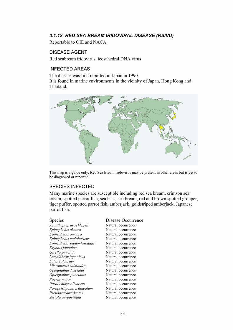
3.1.12. RED SEA BREAM IRIDOVIRAL DISEASE (RSIVD) Reportable to OIE and NACA.
DISEASE AGENT Red seabream iridovirus, icosahedral DNA virus
INFECTED AREAS The disease was first reported in Japan in 1990. It is found in marine environments in the vicinity of Japan, Hong Kong and Thailand.
This map is a guide only. Red Sea Bream Iridovirus may be present in other areas but is yet to be diagnosed or reported.
SPECIES INFECTED Many marine species are susceptible including red sea bream, crimson sea bream, spotted parrot fish, sea bass, sea bream, red and brown spotted grouper, tiger puffer, spotted parrot fish, amberjack, goldstriped amberjack, Japanese parrot fish. Species Disease Occurrence Acanthopagrus schlegeli Natural occurrence Epinephelus akaara Natural occurrence Epinephelus awoara Natural occurrence Epinephelus malabaricus Natural occurrence Epinephelus septemfasciatus Natural occurrence Evynnis japonica Natural occurrence Girella punctata Natural occurrence Lateolabrax japonicus Natural occurrence Lates calcarifer Natural occurrence Micropterus salmoides Natural occurrence Oplegnathus fasciatus Natural occurrence Oplegnathus punctatus Natural occurrence Pagrus major Natural occurrence Paralichthys olivaceus Natural occurrence Parapristipoma trilineatum Natural occurrence Pseudocaranx dentex Natural occurrence Seriola aureovittata Natural occurrence
61
Seriola dumerili Natural occurrence Seriola quinqueradiata Natural occurrence Takifugu (Fugu) rubripes Natural occurrence Thunnus thynnus Natural occurrence Trachinotus blochii Natural occurrence Trachurus japonicus Natural occurrence From OIE Collaborating Centre for Information on Aquatic Animal Disease. International database on aquatic animal disease, URL: www.collabcen.net/toWeb/aq2.asp
Fig. 35. A red sea bream with Red Sea Bream Iridoviral Disease. The enlarged spleen can be seen in the abdominal cavity and is characteristic of fish with the disease. Photograph courtesy of Motohiko Sano.
Fig. 36. Enlarged basophilic cells in the heart (left, stained with haematoxylin and eosin) and a spleen (right, stained with Giemsa) of fish with Red Sea Bream Iridoviral disease. Photographs courtesy Ken McColl and Motohiko Sano.
CLINICAL SIGNS Lethargy, •
• • • • •
Pale gills, kidney and liver. Anaemia Petechial haemorrhages of the gill Enlarged spleen. Pale or dark colouration of the body.
62
MODE OF TRANSMISSION Horizontal transmission •
• •
• • • •
•
• • •
KEY EPIDEMIOLOGICAL POINTS The epidemiology of the disease is not well understood but most outbreaks occur in summer.
METHODS OF DIAGNOSIS Stained impression smears. Histopathology: characteristic enlarged, basophilic cells (9x11-14x20 µm) are found in the spleen, kidney, liver, heart, endothelial cells and gill. Inflammatory cells are often present in the skeletal muscle, lamina propria, pancreas, choroid of the eye, gills and heart. Necrosis of the spleen and kidney. Cell culture for virus isolation. ELISA (monoclonal antibodies). Indirect fluorescent antibody test PCR
DIFFERENTIAL DIAGNOSIS Other viral and bacterial septicaemias.
CONTROL METHODS OVERSEAS On farm hygiene Reduce fish stressors A vaccine is now available in Japan
REFERENCES Jung, S., Miyazaki, T., Miyata, M., Danayadol, Y., Tanaka, S. (1997)
Pathogenicity of iridovirus from Japan and Thailand for the red sea bream Pagrus major in Japan, and histopathology of experimentally infected fish. Fisheries Science 63, 735-740.
Kawakami, H., Nakajima, K. (2002) Cultured fish species affected by red sea bream iridoviral disease from 1996 to 2000. Fish Physiology 37,45-47.
OIE (2003). International Aquatic Animal Health Code. ISBN 9290445807. Available at URL: http://www.oie.int/eng/normes/fcode/A_summry.htm
OIE (2003). OIE Manual for Diagnostic Tests for Aquatic Animals. ISBN 9290445637. Available at URL: http://www.oie.int/eng/nomes/fmanaul/A_summry.htm
Smail, D.A., Munro, A.L.S. (2001) The virology of teleosts. In: Fish Pathology.(Ed. by. R.J. Roberts), pp. 169-253, WB Saunders, London.
63
3.1.13. CHANNEL CATFISH VIRUS DISEASE (CCVD) Reportable to OIE and NACA.
DISEASE AGENT Ictalurid herpesvirus 1 or Channel catfish virus. An enveloped, icosahedral DNA virus. The nucleocapsid is 95-105 nm and the enveloped virus is 175 to 200 nm.
INFECTED AREAS
The disease occurs in the United States of America and has not yet been reported from other areas. From OIE Collaborating Centre for Information on Aquatic Animal Disease. International database on aquatic animal disease, URL: www.collabcen.net/toWeb/aq2.asp
SPECIES INFECTED Channel catfish (Ictaluris punctatus) and occasionally blue catfish (Ictalurus furcatus) Ictalurus catus Experimental demonstration Ictalurus furcatus Natural occurrence Ictalurus punctatus Natural occurrence From OIE Collaborating Centre for Information on Aquatic Animal Disease. International database on aquatic animal disease, URL: www.collabcen.net/toWeb/aq2.asp
CLINICAL SIGNS The disease presents as a haemorrhagic septicaemia. •
•
•
Exophthalmos, ascites, haemorrhages at the base of the fins and in muscle. Reddened belly, pale gills, erratic swimming. High mortality rates in populations of fry and juvenile catfish.
64

Fig. 37. A juvenile channel catfish (Ictalurus punctatus) infected with channel catfish virus. The fish has severe exophthalmos and abdominal distension. Photograph courtesy J. Plumb and J. Grizzle.
Fig. 38. Kidney of a channel catfish with channel catfish virus disease. The haematopoietic tissue is oedematous and necrotic. Photograph courtesy of J. Plumb and J. Grizzle.
MODE OF TRANSMISSION Horizontal (direct or vectorial) and probably vertical transmission •
65
KEY EPIDEMIOLOGICAL POINTS • High host specificity • Disease is most prevalent in fingerlings. • Fish that survive the infection become carriers but do not produce virus
making screening difficult. • Mortalities are high in summer when water temperatures are >27°C
and do not occur below 18°C. • Seawater and drying inactivate the virus but it is resistant to freezing. • Mortality in outbreaks can be 100% and although the overall impact of
the disease on the culture of channel catfish is not high, there are outbreaks every summer and the impact of the disease on affected farms is very severe..
METHODS OF DIAGNOSIS Histopathology: severe, diffuse necrosis of the renal tubules and interstitial tissue.
•
•
• • •
•
• •
•
Cytopathic effects in cell culture. This is the easiest method of diagnosis because cytopathic effects are pronounced with spectacular syncytia formation ELISA PCR Serology to detect neutralising antibody within a fish population
DIFFERENTIAL DIAGNOSIS Enteric septicaemia of catfish (but this usually occurs in cooler water of 21-27°C). The monogenean parasite Bulbophorus sp. Other bacterial diseases.
CONTROL METHODS OVERSEAS Avoid stressors such as handling of young fish in summer
REFERENCES OIE (2003). International Aquatic Animal Health Code. ISBN 9290445807.
Available at URL: http://www.oie.int/eng/normes/fcode/A_summry.htm
OIE (2003). OIE Manual for Diagnostic Tests for Aquatic Animals. ISBN 9290445637. Available at URL: http://www.oie.int/eng/nomes/fmanaul/A_summry.htm
Plumb, J.A., (1999). Health maintenance and principal microbial diseases of cultured fishes. Iowa State University, Ames, pp. 328.
Smail, D.A., Munro, A.L.S. (2001) The virology of teleosts. In: Fish Pathology.(Ed. by. R.J. Roberts), pp. 169-253, WB Saunders, London.
Winton, J.R. (2001) Fish Health Management. In: Fish Hatchery Management.(Ed. by. G. Wedemeyer), pp. 733, American Fisheries Society, Bethesda, Maryland.
Wolf, K., 1988. Fish viruses and fish viral diseases. Cornell University Press, Ithaca, pp. 476.
66
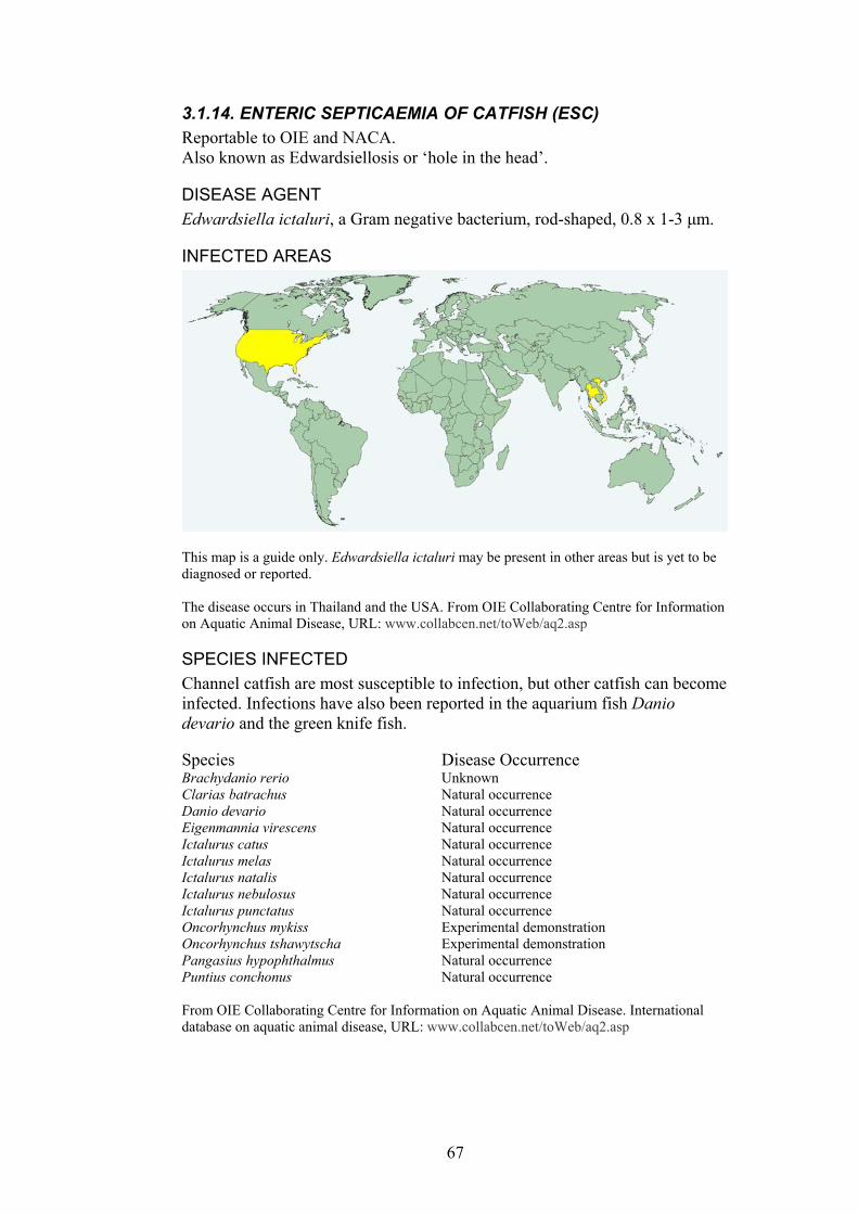
3.1.14. ENTERIC SEPTICAEMIA OF CATFISH (ESC) Reportable to OIE and NACA. Also known as Edwardsiellosis or ‘hole in the head’.
DISEASE AGENT Edwardsiella ictaluri, a Gram negative bacterium, rod-shaped, 0.8 x 1-3 µm.
INFECTED AREAS
This map is a guide only. Edwardsiella ictaluri may be present in other areas but is yet to be diagnosed or reported. The disease occurs in Thailand and the USA. From OIE Collaborating Centre for Information on Aquatic Animal Disease, URL: www.collabcen.net/toWeb/aq2.asp
SPECIES INFECTED Channel catfish are most susceptible to infection, but other catfish can become infected. Infections have also been reported in the aquarium fish Danio devario and the green knife fish. Species Disease Occurrence Brachydanio rerio Unknown Clarias batrachus Natural occurrence Danio devario Natural occurrence Eigenmannia virescens Natural occurrence Ictalurus catus Natural occurrence Ictalurus melas Natural occurrence Ictalurus natalis Natural occurrence Ictalurus nebulosus Natural occurrence Ictalurus punctatus Natural occurrence Oncorhynchus mykiss Experimental demonstration Oncorhynchus tshawytscha Experimental demonstration Pangasius hypophthalmus Natural occurrence Puntius conchonus Natural occurrence From OIE Collaborating Centre for Information on Aquatic Animal Disease. International database on aquatic animal disease, URL: www.collabcen.net/toWeb/aq2.asp
67

CLINICAL SIGNS There are several forms of the disease from hyper-acute to chronic. Usually each presents with only one of the following disease signs:
• Ascites and exophthalmos • Petechial haemorrhages of the skin, especially on the ventral surface • 2-5 mm red patches on the skin that eventually turn into ulcers • meningitis that results in the ‘hole in the head’ lesion when the skin
over the cranial foramen becomes perforated
MODE OF TRANSMISSION Horizontal (direct or vectorial) and probably vertical transmission
•
•
•
• • •
KEY EPIDEMIOLOGICAL POINTS • High host specificity • Disease is most prevalent in fingerlings. • Fish that survive the infection become carriers but do not produce virus
making screening difficult. • Mortalities are high in summer when water temperatures are >27°C
and do not occur below 18°C. • Seawater and drying inactivate the virus but it is resistant to freezing. • Mortality in outbreaks can be 100% and although the overall impact of
the disease on the culture of channel catfish is not high, there are outbreaks every summer and the impact of the disease on affected farms is very severe.
METHODS OF DIAGNOSIS Histopathology: severe, diffuse necrosis of the renal tubules and interstitial tissue. Cytopathic effects in cell culture. This is the easiest method of diagnosis because cytopathic effects are pronounced with spectacular syncytia formation ELISA PCR Serology to detect neutralising antibody within a fish population
68

Fig. 39. Channel catfish with enteric septicaemia. Depigmented areas can be seen along the body of the upper fish. The middle fish has petechial haemorrhages. The lower fish is not infected. Photograph courtesy of J. Plumb and J. Grizzle.
Fig. 40. Channel catfish with enteric septicaemia. The fish on the left has the ‘buckshot’ skin lesions that are often present on infected fish. The fish on the right has rash-like generalised petechial haemorrhages. Photographs courtesy A. Goodwin.
69
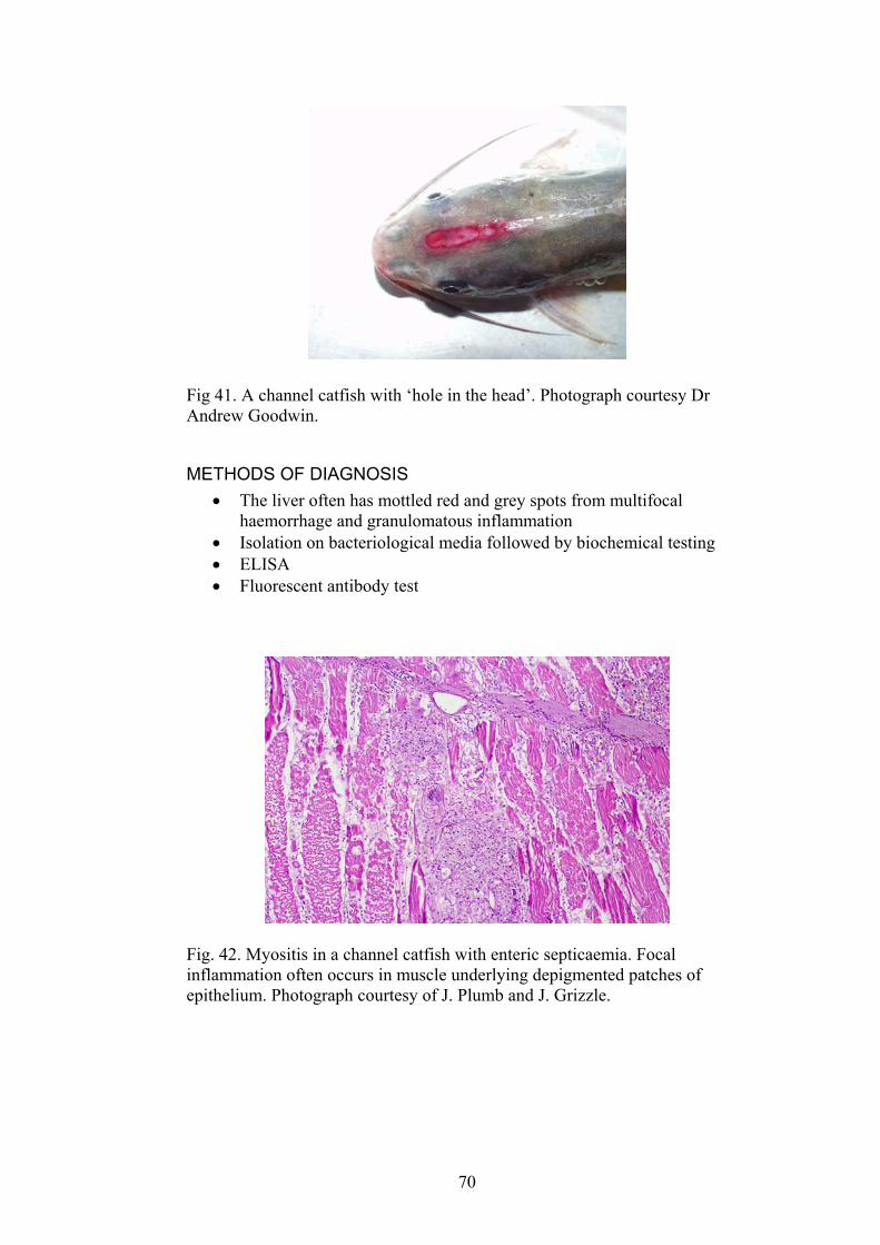
Fig 41. A channel catfish with ‘hole in the head’. Photograph courtesy Dr Andrew Goodwin.
METHODS OF DIAGNOSIS The liver often has mottled red and grey spots from multifocal haemorrhage and granulomatous inflammation
•
• • •
Isolation on bacteriological media followed by biochemical testing ELISA Fluorescent antibody test
Fig. 42. Myositis in a channel catfish with enteric septicaemia. Focal inflammation often occurs in muscle underlying depigmented patches of epithelium. Photograph courtesy of J. Plumb and J. Grizzle.
70
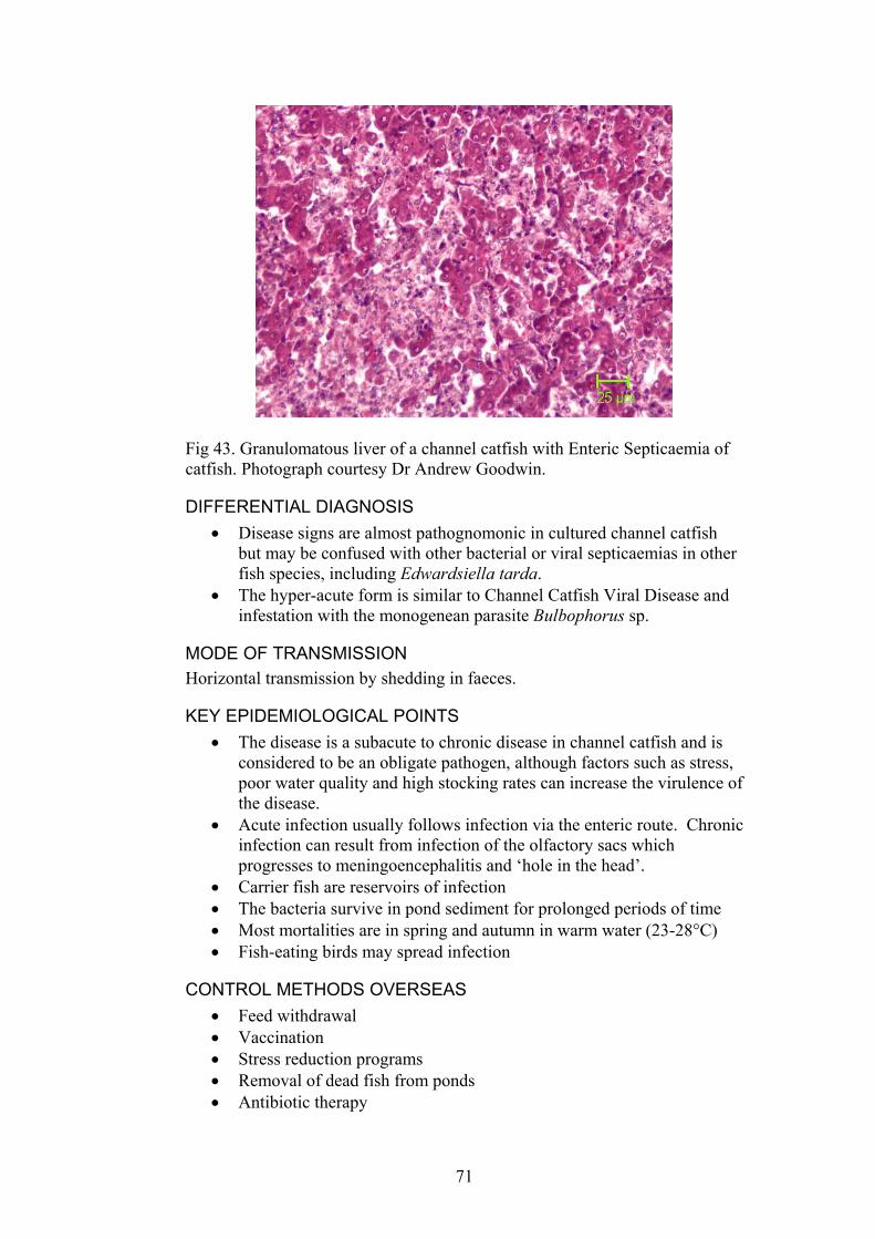
Fig 43. Granulomatous liver of a channel catfish with Enteric Septicaemia of catfish. Photograph courtesy Dr Andrew Goodwin.
DIFFERENTIAL DIAGNOSIS Disease signs are almost pathognomonic in cultured channel catfish but may be confused with other bacterial or viral septicaemias in other fish species, including Edwardsiella tarda.
•
•
• • • • •
The hyper-acute form is similar to Channel Catfish Viral Disease and infestation with the monogenean parasite Bulbophorus sp.
MODE OF TRANSMISSION Horizontal transmission by shedding in faeces.
KEY EPIDEMIOLOGICAL POINTS • The disease is a subacute to chronic disease in channel catfish and is
considered to be an obligate pathogen, although factors such as stress, poor water quality and high stocking rates can increase the virulence of the disease.
• Acute infection usually follows infection via the enteric route. Chronic infection can result from infection of the olfactory sacs which progresses to meningoencephalitis and ‘hole in the head’.
• Carrier fish are reservoirs of infection • The bacteria survive in pond sediment for prolonged periods of time • Most mortalities are in spring and autumn in warm water (23-28°C) • Fish-eating birds may spread infection
CONTROL METHODS OVERSEAS Feed withdrawal Vaccination Stress reduction programs Removal of dead fish from ponds Antibiotic therapy
71
REFERENCES OIE (2003). International Aquatic Animal Health Code. ISBN 9290445807.
Available at URL: http://www.oie.int/eng/normes/fcode/A_summry.htm
OIE (2003). OIE Manual for Diagnostic Tests for Aquatic Animals. ISBN 9290445637. Available at URL: http://www.oie.int/eng/nomes/fmanaul/A_summry.htm
Plumb, J.A. (1999) Edwardsiella septicaemias. In: Fish diseases and disorders: viral, bacterial and fungal infections,Vol. 3.(Ed. by. P.T.K. Woo, D.W. Bruno), pp. 479-521, CABI Publishing, Oxon.
Plumb, J.A., (1999). Health maintenance and principal microbial diseases of cultured fishes. Iowa State University, Ames, pp. 328.
Winton, J.R. (2001) Fish Health Management. In: Fish Hatchery Management.(Ed. by. G. Wedemeyer), pp. 733, American Fisheries Society, Bethesda, Maryland.
72
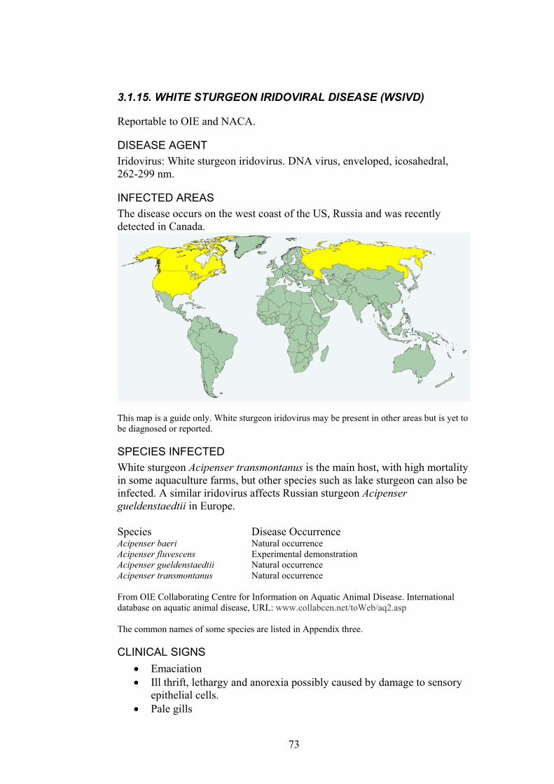
3.1.15. WHITE STURGEON IRIDOVIRAL DISEASE (WSIVD)
Reportable to OIE and NACA.
DISEASE AGENT Iridovirus: White sturgeon iridovirus. DNA virus, enveloped, icosahedral, 262-299 nm.
INFECTED AREAS The disease occurs on the west coast of the US, Russia and was recently detected in Canada.
This map is a guide only. White sturgeon iridovirus may be present in other areas but is yet to be diagnosed or reported.
SPECIES INFECTED White sturgeon Acipenser transmontanus is the main host, with high mortality in some aquaculture farms, but other species such as lake sturgeon can also be infected. A similar iridovirus affects Russian sturgeon Acipenser gueldenstaedtii in Europe. Species Disease Occurrence Acipenser baeri Natural occurrence Acipenser fluvescens Experimental demonstration Acipenser gueldenstaedtii Natural occurrence Acipenser transmontanus Natural occurrence From OIE Collaborating Centre for Information on Aquatic Animal Disease. International database on aquatic animal disease, URL: www.collabcen.net/toWeb/aq2.asp The common names of some species are listed in Appendix three.
CLINICAL SIGNS Emaciation •
•
•
Ill thrift, lethargy and anorexia possibly caused by damage to sensory epithelial cells. Pale gills
73
MODE OF TRANSMISSION Horizontal transmission and possibly vertical transmission although this has not been demonstrated.
•
• •
•
•
•
•
•
• •
KEY EPIDEMIOLOGICAL POINTS • Similar epidemiology to lymphocystis disease virus-both replicate
slowly, produce limited virus and host cells survive for a prolonged period.
• Mortality is high, up to 95% in naïve populations of white sturgeon • Survivors remain carriers.
METHODS OF DIAGNOSIS Gross examination. Scant body fat, pale liver, Histopathology: epithelial cells of the skin, gills and upper alimentary tract are affected. There is hypertrophy of epithelial cells of the gill and skin sometimes containing crystalline rod shaped bodies. Hyperplasia of the gill epithelium, necrosis of pillar cells and associated haemorrhages. Virology. Cytopathic effects in fresh white sturgeon cell lines at 10-20°C. Transmission electron microscopy of infected tissues showing typical virions. Polyclonal antisera and monoclonal antibodies used in indirect fluorescent antibody tests and immunohistochemical staining of tissue sections or infected cell lines.
DIFFERENTIAL DIAGNOSIS Other causes of epithelial hyperplasia such as lymphocystis, water quality/toxicity. Other viral and bacterial diseases causing high mortality.
CONTROL METHODS OVERSEAS Obtain disease-free stock. Grow fish at 25°C as the virus grows at 10-20°C.
REFERENCES OIE (2003). International Aquatic Animal Health Code. ISBN 9290445807.
Available at URL: http://www.oie.int/eng/normes/fcode/A_summry.htm
OIE (2003). OIE Manual for Diagnostic Tests for Aquatic Animals. ISBN 9290445637. Available at URL: http://www.oie.int/eng/nomes/fmanaul/A_summry.htm
Plumb, J.A., (1999). Health maintenance and principal microbial diseases of cultured fishes. Iowa State University, Ames, pp. 328.
Smail, D.A., Munro, A.L.S. (2001) The virology of teleosts. In: Fish Pathology.(Ed. by. R.J. Roberts), pp. 169-253, WB Saunders, London.
74
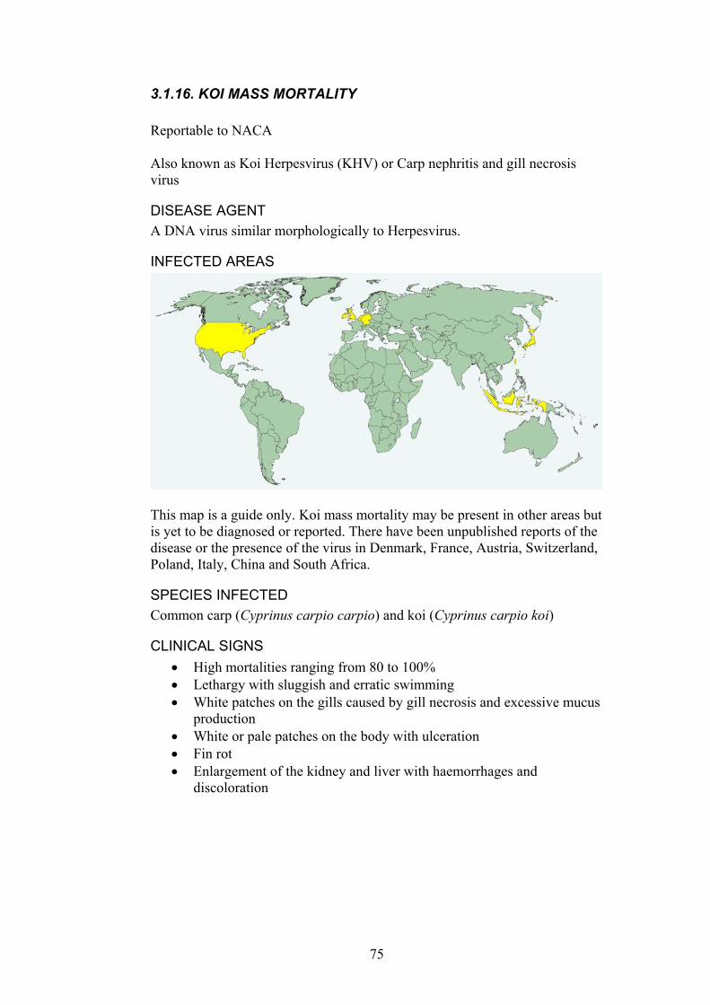
3.1.16. KOI MASS MORTALITY Reportable to NACA Also known as Koi Herpesvirus (KHV) or Carp nephritis and gill necrosis virus
DISEASE AGENT A DNA virus similar morphologically to Herpesvirus.
INFECTED AREAS
This map is a guide only. Koi mass mortality may be present in other areas but is yet to be diagnosed or reported. There have been unpublished reports of the disease or the presence of the virus in Denmark, France, Austria, Switzerland, Poland, Italy, China and South Africa.
SPECIES INFECTED Common carp (Cyprinus carpio carpio) and koi (Cyprinus carpio koi)
CLINICAL SIGNS High mortalities ranging from 80 to 100% •
• •
• • •
Lethargy with sluggish and erratic swimming White patches on the gills caused by gill necrosis and excessive mucus production White or pale patches on the body with ulceration Fin rot Enlargement of the kidney and liver with haemorrhages and discoloration
75

Fig. 44. Carp with koi mass mortality. Necrosis of the gill and enlarged kidneys can be seen (arrowheads). Tail rot can be also seen on the caudal fin of one fish. Photograph courtesy of Dr Angus Cameron.
MODE OF TRANSMISSION Horizontal transmission •
• • • •
• •
• • •
KEY EPIDEMIOLOGICAL POINTS • Water temperature is the principal environmental factor that influences
the onset and severity of this disease. Temperatures ranging between 18 and 25 C are optimal for this disease (permissive temperatures).
• Highly contagious and rapidly becoming widespread. • Survivors are resistant to subsequent virus exposures.
METHODS OF DIAGNOSIS Electron microscopy Transmission trials Cell culture for virus isolation PCR
DIFFERENTIAL DIAGNOSIS Other cyprinid herpesviral infections Bacterial septicaemias caused by organisms such as Aeromonas spp.
CONTROL METHODS OVERSEAS Water temperature control Vaccination with attenuated non-pathogenic viruses Transport restrictions from known infected areas
76

REFERENCES Hedrick, R.P., Gilad, O., Yun, S., Spangenberg, J.V., Marty, G.D.,
Nordhausen, R.W., Kebus, M.J., Bercovier, H., Eldar, A. (2000) A Herpesvirus associated with mass mortality of juvenile and adult koi, a strain of common carp. Journal of Aquatic Animal Health 12, 44-57.
Oh, M.-J., Jung, S.-J., Choi, T.-J., Kim, H.-R., Rajendran, K.V., Kim, Y.-J., Park, M.-A., Chun, S.-K. (2001) A viral disease occurring in cultured carp Cyprinus carpio in Korea. Fish Pathology 36, 147-151.
www.enaca.org
77

3.1.17. SPRING VIRAEMIA OF CARP (SVC) Reportable to OIE and NACA. Can also be referred to as infectious dropsy of carp.
DISEASE AGENT Rhabdovirus carpio: bullet-shaped, RNA virus, 70x180 nm.
INFECTED AREAS The disease has been reported in Austria, Belarus, Bosnia & Herzegov, China, Croatia, Czechoslovakia, Denmark, France, Germany, Hungary, Italy, Kuwait, Lithuania, Macedonia (Former Yugoslav Republic of), Moldavia, Rep, Netherlands, Poland, Romania, Russian Fed, Serbia & Montenegro, Slovakia, Slovenia, Spain, Switzerland, Ukraine, UK, USA, Vanuatu, Yugoslavia
This map is a guide only. Spring Viraemia of Carp may be present in other areas but is yet to be diagnosed or reported.
SPECIES INFECTED The disease primarily occurs in carp and other cyprinids including goldfish, but other fish can be infected. Common carp/koi are the most susceptible species. Species Disease Occurrence Aristichthys nobilis Natural occurrence and experimental demonstration Brachydanio rerio Experimental infection. Carassius auratus Natural occurrence Carassius carassius Natural occurrence Ctenopharyngodon idellus Experimental demonstration & natural occurrence Cyprinus carpio Natural occurrence Esox lucius Experimental demonstration & natural occurrence Hypophthalmichthys molitrix Experimental demonstration & natural occurrence Lebistes reticulata Experimental demonstration Leuciscus idus Natural occurrence Piscicola spp. Carrier-not affected & natural occurrence Poecilia reticulate Experimental demonstration
78

Rutilus rutilus Experimental demonstration Silurus glanis Experimental demonstration & natural occurrence Tinca tinca Natural occurrence From OIE Collaborating Centre for Information on Aquatic Animal Disease. International database on aquatic animal disease, URL: www.collabcen.net/toWeb/aq2.asp The common names of some species are listed in Appendix three.
CLINICAL SIGNS Haemorrhagic septicaemia. Disease signs include dark pigmentation, enlarged abdomen, exophthalmos, pale gills with petechiae, ascites, oedema, petechial haemorrhage of internal organs. Mucus sometimes with blood trailing from the vent.
Fig. 45. A European carp with Spring Viraemia of Carp. Abdominal distension, exophthalmos and petechial haemorrhages are common clinical signs of disease, as in these fish. Photograph courtesy Dr D Alderman.
Fig. 46. Petechial haemorrhages on the swimbladder of a European carp with spring viraemia of carp. Photograph courtesy Dr D Alderman.
MODE OF TRANSMISSION Horizontal transmission. Vectors such as fish lice, leeches and nematodes (Philometra geometra) may be important.
•
• Ovarian fluid contains virus particles in a small number of female fish in spring in endemic areas.
79
KEY EPIDEMIOLOGICAL POINTS • More common in cooler water temperatures and areas with cold water
temperatures in winter. Clinical disease usually occurs in spring when water temperatures rise to 10-18°C.
• Often a subacute to chronic disease. • Survivors have prolonged and strong resistance to the disease. • Mortalities in epizootics often approach 70% in yearlings. • Sporadic disease outbreaks are common in infected areas. • Incubation period 6 to 60 days. • Asymptomatic carriers occur and can develop overt disease when
stressed. • Egg-associated transmission. • Virus remains viable in water and mud for up to 42 days.
METHODS OF DIAGNOSIS PCR •
• • • •
• •
•
Virus isolation and cytopathic effects in cell culture Virus neutralization using specific antiserum ELISA Indirect fluorescent antibody test
DIFFERENTIAL DIAGNOSIS Other causes of haemorrhagic septicaemia and ascites such as bacterial septicaemia caused by Aeromonas hydrophila or atypical Aeromonas salmonicida. Secondary infections with bacteria are common in fish with Spring Viraemia of Carp.
CONTROL METHODS OVERSEAS Hygiene controls. The virus is inactivated by heating to 45°C, formalin, sodium hydroxide and chlorine. Certification and declaration of disease-free zones
REFERENCES Ahne, W., Bjorklund, H.V., Essbauer, S., Fijan, N., Kurath, G., Winton, J.R.
(2002) Spring viremia of carp (SVC). Diseases of Aquatic Organisms 52, 261-272.
OIE (2003). International Aquatic Animal Health Code. ISBN 9290445807. Available at URL: http://www.oie.int/eng/normes/fcode/A_summry.htm
OIE (2003). OIE Manual for Diagnostic Tests for Aquatic Animals. ISBN 9290445637. Available at URL: http://www.oie.int/eng/nomes/fmanaul/A_summry.htm
Winton, J.R. (2001) Fish Health Management. In: Fish Hatchery Management.(Ed. by. G. Wedemeyer), pp. 733, American Fisheries Society, Bethesda, Maryland.
Wolf, K., 1988. Fish viruses and fish viral diseases. Cornell University Press, Ithaca, pp. 476.
80
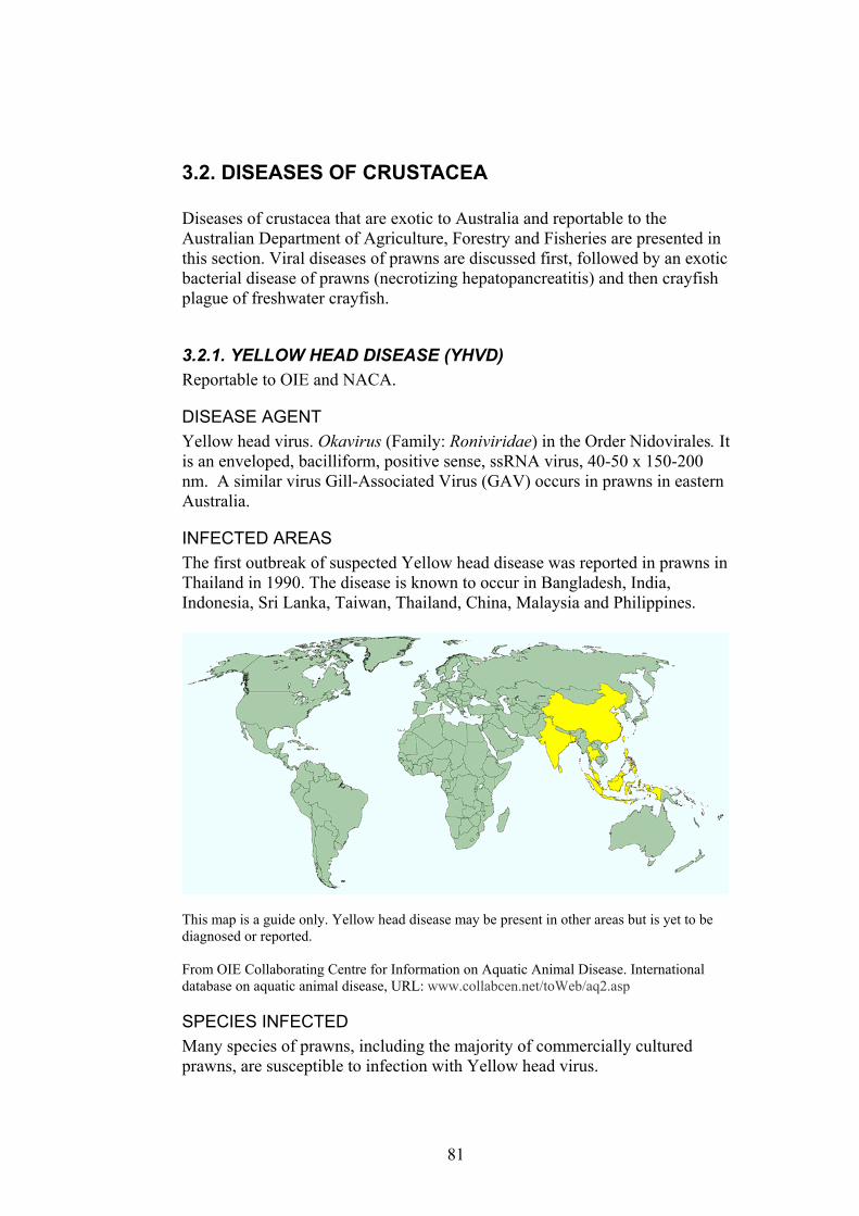
3.2. DISEASES OF CRUSTACEA Diseases of crustacea that are exotic to Australia and reportable to the Australian Department of Agriculture, Forestry and Fisheries are presented in this section. Viral diseases of prawns are discussed first, followed by an exotic bacterial disease of prawns (necrotizing hepatopancreatitis) and then crayfish plague of freshwater crayfish.
3.2.1. YELLOW HEAD DISEASE (YHVD) Reportable to OIE and NACA.
DISEASE AGENT Yellow head virus. Okavirus (Family: Roniviridae) in the Order Nidovirales. It is an enveloped, bacilliform, positive sense, ssRNA virus, 40-50 x 150-200 nm. A similar virus Gill-Associated Virus (GAV) occurs in prawns in eastern Australia.
INFECTED AREAS The first outbreak of suspected Yellow head disease was reported in prawns in Thailand in 1990. The disease is known to occur in Bangladesh, India, Indonesia, Sri Lanka, Taiwan, Thailand, China, Malaysia and Philippines.
This map is a guide only. Yellow head disease may be present in other areas but is yet to be diagnosed or reported. From OIE Collaborating Centre for Information on Aquatic Animal Disease. International database on aquatic animal disease, URL: www.collabcen.net/toWeb/aq2.asp
SPECIES INFECTED Many species of prawns, including the majority of commercially cultured prawns, are susceptible to infection with Yellow head virus.
81
Species Disease Occurrence Farantopenaeus aztecus Experimental demonstration Litopenaeus setiferus Experimental demonstration Litopenaeus stylirostris Experimental demonstration Litopenaeus vannamei Experimental demonstration Penaeus duorarum Experimental demonstration Penaeus indicus Natural occurrence Penaeus merguiensis Natural occurrence Penaeus monodon Experimental demonstration and natural occurrence Penaeus spp. Natural occurrence Metapenaeus ensis Natural occurrence The common names of some species are listed in Appendix three.
CLINICAL SIGNS Prior to the appearance of gross signs there is a short period of increased feeding followed by complete cessation of feeding.
•
• • •
•
•
Pale body Yellowish gills and hepatopancreas. Moribund prawns congregate at the edges of ponds and swim slowly near the surface.
MODE OF TRANSMISSION Horizontal transmission Vertical transmission can occur via supporting tissues of ovaries and testes.
KEY EPIDEMIOLOGICAL POINTS • The viral diseases of Australian prawns, lymphoid organ virus (LOV)
and gill-associated virus (GAV) are similar to Yellow head virus. • The disease often occurs when environmental conditions are poor and
in areas with high densities of prawn farms. • The virus may enter the ponds in incoming water or on wild crustacea
that act as carriers. • Mortality <100% within 3 days of first clinical signs. • Mortality is usually more than 20 days post-stocking. • Severe disease outbreaks in endemic are not as common as they were
in the early 1990’s.
METHODS OF DIAGNOSIS Histopathology: the lymphoid organ is enlarged and necrotic. There is necrosis of cells of ectodermal and mesodermal origin with intensely staining basophilic, perinuclear, intracytoplasmic inclusions, especially in the lymphoid organ, gill and stomach subcuticular tissue. Affected prawns have large amounts of fat in the hepatopancreas tubule cells, possibly from disruption of normal fat metabolism. Cytoplasmic inclusions in sub-cuticular epithelial cells can be distinguished from those of Taura syndrome since the latter always show accompanying eosinophilic to basophilic inclusions. These are lacking with YHV that shows only intensely basophilic inclusions. Stained tissue squashes of the gills and subcuticular tissues.
82

Haemolymph smears. Prawns with early infection have pyknotic and karyorrhectic nuclei.
•
• • • • •
Bioassay RT-PCR Western blot analysis In situ hybridisation Transmission electronmicroscopy
Fig. 47. Three Penaeus monodon with Yellow head disease (left). Note the yellow cephalothorax. Photograph courtesy D.V. Lightner.
Fig. 48. Diffuse necrosis of gill lamellae in a prawn with Yellowhead disease. Pyknotic and karyorrhectic nuclei (arrows) can be seen. Similar lesions occur in the lymphoid organ. Photograph courtesy D.V. Lightner.
DIFFERENTIAL DIAGNOSIS Gill-Associated virus (GAV). RT-PCR is required. •
• •
White spot virus Vibriosis
83

Ill thrift from poor water quality •
• • •
• • • • •
CONTROL METHODS OVERSEAS Prevention of translocation of infected animals. Surveillance and certification of broodstock and fry. Disposal of infected water and prawns in such a way that the disease agent is not spread to other farms. Emergency harvest early in outbreaks. Formalin stress test to identify infected post larvae. Maintenance of good water quality. Prevent entry in incoming water or wild crustacea. Clean and disinfect equipment prior to use on another pond.
REFERENCES Bonami, J.-R. (1997) Crustacean viral disease: recent developments. Bulletin
of the European Association of Fish Pathologists 17, 188-190. Chantanachookin, C., Boonyaratpalin, S., Kasornchandra, J., Direkbusarakom,
S., Ekpanithanpong, U., Supamataya, K., Sriurairatana, S., Flegel, T.W. (1993) Histology and unltrastructure reveal a new granulosis-like virus in Penaeus monodon affected by yellow-head disease. Disease of Aquatic Organisms 17, 145-157.
Lightner, D.V., (1996). A handbook of shrimp pathology and diagnostic procedures for diseases of cultured penaeid shrimp. The World Aquaculture Society, Baton Rouge.
Lu, Y., Tapay, L.M., Brock, J.A., Loh, P.C. (1994) Infection of the yellow head baculo-like virus (YBV) in two species of Litopenaeus stylirostris (Stimpson) and Litopenaeus vannamei (Boone). Journal of Fish Diseases 17, 649-656.
Mohan, C.V., Shankar, K.M., Kulkarni, S., Sudha, P.M. (1998) Histopathology of cultured shrimp showing gross signs of yellow head syndrome and white spot syndrome during 1994 Indian epizootics. Diseases of Aquatic Organisms 34, 9-12.
OIE (2003). International Aquatic Animal Health Code. ISBN 9290445807. Available at http://www.oie.int/eng/normes/fcode/A_summry.htm
OIE (2003). OIE Manual for Diagnostic Tests for Aquatic Animals. ISBN 9290445637. Available at http://www.oie.int/eng/nomes/fmanaul/A_summry.htm
Pantoja, C.R., Lightner, D.V. (2003) Similarity between the histopathology of White Spot Syndrome Virus and Yellow Head Syndrome Virus and its relevance to diagnosis of YHV disease in the Americas. Aquaculture 218, 47-54.
Pantoja, C.R., Lightner, D.V. (2001) Necrosis due to WSSV can mimic YHV lesions in lymphoid organ of penaeid shrimp. The Advocate 4 (3):15.
84

3.2.2. WHITE SPOT DISEASE (WSD) Reportable to OIE and NACA. Also known as red body or red disease, Systemic Ectodermal and Mesodermal Baculovirus (SEMBA)
DISEASE AGENT White spot virus. Whispovirus (Family: Nimaviridae). An enveloped, dsDNA virus with circular DNA. Rod-shaped to elliptical, nucleocapsid 58-67 x 250-350 nm.
INFECTED AREAS The disease is believed to have emerged in Taiwan in 1991 or 1992. It then spread to mainland China, then to other parts of Asia and America via infected prawns.
This map is a guide only. White spot virus may be present in other areas but is yet to be diagnosed or reported. From OIE Collaborating Centre for Information on Aquatic Animal Disease. International database on aquatic animal disease, URL: www.collabcen.net/toWeb/aq2.asp
SPECIES INFECTED A large number of crustacean species are susceptible including prawns, lobster, crabs and freshwater crayfish. Many species are asymptomatic carriers. Species Disease Occurrence Acetes sp. Experimental demonstration Alpheus brevicristatus Natural occurrence Alpheus lobidens Natural occurrence Astacus leptodactylus Experimental demonstration Calappa lophos Experimental demonstration Cancer pagurus Experimental demonstration Charybdis feriata Experimental demonstration
85
Charybdis granulata Experimental demonstration Charybdis japonica Natural occurrence Cherax quadricarinatus Experimental demonstration Exopalaemon orientalis Experimental demonstration and natural occurrence Farantopenaeus aztecus Experimental demonstration Helice tridens Natural occurrence Hemigrapsus sanguineus Natural occurrence Liocarcinus depurator Experimental demonstration Liocarcinus puber Experimental demonstration and natural occurrence Litopenaeus setiferus Experimental demonstration Litopenaeus stylirostris Experimental demonstration Litopenaeus vannamei Natural occurrence and experimental demonstration Macrobrachium rosenbergii Experimental demonstration Macrobrachium spp. Experimental demonstration Metapenaeus brevicornis Natural occurrence Metapenaeus dobsoni Experimental demonstration Metapenaeus ensis Experimental demonstration and natural occurrence Metapenaeus lysianassa Natural occurrence Metapenaeus monoceros Experimental demonstration and natural occurrence Metapograpsus sp. Experimental demonstration Ocypode stimpsoni Natural occurrence Orconectes limosus Experimental demonstration Pacifastacus leniusculus Experimental demonstration Palaemon adspersus Experimental demonstration Palaemon serrifer Natural occurrence Palaemon styliferus Natural occurrence Panulirus homarus Experimental demonstration Panulirus longipes Experimental demonstration Panulirus ornatus Experimental demonstration Panulirus penicillatus Experimental demonstration Panulirus polyphagus Experimental demonstration Panulirus versicolor Experimental demonstration Parathelphusa hydrodomous Experimental demonstration Parathelphusa pulvinata Experimental demonstration Penaeus chinensis Natural occurrence Penaeus duorarum Experimental demonstration Penaeus indicus Experimental demonstration and natural occurrence Penaeus japonicus Natural occurrence Penaeus merguiensis Natural occurrence Penaeus monodon Natural occurrence Penaeus penicillatus Natural occurrence Penaeus semisulcatus Carrier-not affected, natural occurrence and experimental
demonstration Penaeus spp. Natural occurrence Petrolisthes japonicus Natural occurrence Portunus pelagicus Natural occurrence and experimental demonstration Portunus sanguinolentus Natural occurrence and experimental demonstration Portunus trituberculatus Natural occurrence Procambarus clarkii Experimental demonstration Pseudograpsus intermedius Natural occurrence Scylla serrata Natural occurrence and experimental demonstration Scylla tranquebarica Experimental demonstration Scyllarus arctus Experimental demonstration Sesarma spp. Experimental demonstration Trachypenaeus curvirostris Experimental demonstration Uca pugilator Experimental demonstration Upogebia major Natural occurrence From OIE Collaborating Centre for Information on Aquatic Animal Disease. International database on aquatic animal disease, URL: www.collabcen.net/toWeb/aq2.asp
86
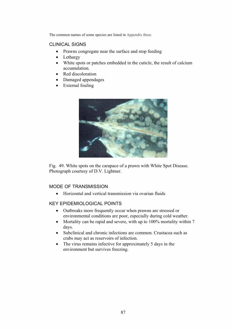
The common names of some species are listed in Appendix three.
CLINICAL SIGNS Prawns congregate near the surface and stop feeding •
• •
• • •
Lethargy White spots or patches embedded in the cuticle, the result of calcium accumulation. Red discoloration Damaged appendages External fouling
Fig. 49. White spots on the carapace of a prawn with White Spot Disease. Photograph courtesy of D.V. Lightner.
MODE OF TRANSMISSION Horizontal and vertical transmission via ovarian fluids •
KEY EPIDEMIOLOGICAL POINTS • Outbreaks more frequently occur when prawns are stressed or
environmental conditions are poor, especially during cold weather. • Mortality can be rapid and severe, with up to 100% mortality within 7
days. • Subclinical and chronic infections are common. Crustacea such as
crabs may act as reservoirs of infection. • The virus remains infective for approximately 5 days in the
environment but survives freezing.
87

Fig. 50. A haematopoietic nodule of a prawn with White Spot Disease. Necrotic cells, mitotic figures (blue arrowhead) and inclusion bodies (white arrowhead) can be seen.
Fig. 51. Subcuticular epithelium and connective tissue of the stomach of a prawn with White Spot Disease. Intranuclear inclusion bodies (arrows) can be seen in nuclei with marginated chromatin. Photograph courtesy D.V. Lightner.
METHODS OF DIAGNOSIS Histopathology: Hypertrophied nuclei with eosinophilic-basophilic inclusion bodies that begin as Cowdry type A inclusions in gills, subcuticular epithelium and connective tissue (cells of ectodermal and mesodermal origin). Necrosis of the lymphoid organ may occur, associated with intranuclear inclusion bodies in connective tissue cells of the lymphoid organ.
•
• Tissue squashes from gills, subcuticular tissue of stomach or cephalothorax showing hypertrophied nuclei in vacuolated cells.
88

Bioassay • • • • •
•
• •
• •
• • • • • •
PCR Western blot analysis In situ hybridisation Transmission electron microscopy
DIFFERENTIAL DIAGNOSIS Prawns reared in high water alkalinity water can have white spots on the exoskeleton that can be confused with those caused by white spot syndrome virus.
CONTROL METHODS OVERSEAS Prevent translocation of infected animals. Stock prawns at a low stocking density and maintain optimal pH and ammonia levels in culture water. Surveillance and certification of broodstock and post-larvae. Prevent spread of the virus in infected water and prawns from farms, processing plants or by scavenging birds in refuse. Emergency harvest early in outbreaks. Formalin stress test to identify infected post-larvae. Maintenance of good water quality. Prevent entry in incoming water or wild crustacea. Clean and disinfect equipment prior to use on another pond. Ensure that raw prawns infected with the virus are not used for prawn food or bait.
REFERENCES Bonami, J.-R. (1997) Crustacean viral disease: recent developments. Bulletin of the
European Association of Fish Pathologists 17, 188-190. Hill, B. (2002) National and international impacts of white spot disease of shrimp.
Bulletin of the European Association of Fish Pathologists 22, 58-65. Lightner, D.V., (1996). A handbook of shrimp pathology and diagnostic procedures for
diseases of cultured penaeid shrimp. The World Aquaculture Society, Baton Rouge.
Mohan, C.V., Shankar, K.M., Kulkarni, S., Sudha, P.M. (1998) Histopathology of cultured shrimp showing gross signs of yellow head syndrome and white spot syndrome during 1994 Indian epizootics. Diseases of Aquatic Organisms 34, 9-12.
OIE (2003). International Aquatic Animal Health Code. ISBN 9290445807. Available at http://www.oie.int/eng/normes/fcode/A_summry.htm
OIE (2003). OIE Manual for Diagnostic Tests for Aquatic Animals. ISBN 9290445637. Available at URL: http://www.oie.int/eng/nomes/fmanaul/A_summry.htm
Pantoja, C.R., Lightner, D.V. (2003) Similarity between the histopathology of White Spot Syndrome Virus and Yellow Head Syndrome Virus and its relevance to diagnosis of YHV disease in the Americas. Aquaculture 218, 47-54.
Supamattaya, K., Hoffman, R.W., Boonyaratpalin, S., Kanchanaphum, P. (1998) Experimental transmission of white spot syndrome virus (WSSV) from black tiger shrimp Penaeus monodon to the sand crab Portunus pelagicus, mud crab Scylla serrataand krill Acetes sp. Disease of Aquatic Organisms 32, 79-85.
Wang, Y.-C., Lo, C.-F., Chang, P.-S., Kou, G.-H. (1998) Experimental infection of white spot baculovirus in some cultured and wild decapods in Taiwan. Aquaculture 164, 221-231.
89
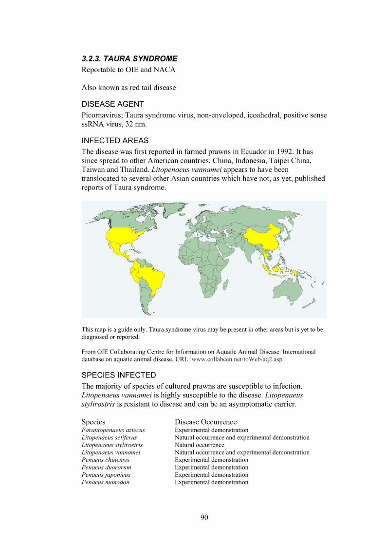
3.2.3. TAURA SYNDROME Reportable to OIE and NACA Also known as red tail disease
DISEASE AGENT Picornavirus; Taura syndrome virus, non-enveloped, icoahedral, positive sense ssRNA virus, 32 nm.
INFECTED AREAS The disease was first reported in farmed prawns in Ecuador in 1992. It has since spread to other American countries, China, Indonesia, Taipei China, Taiwan and Thailand. Litopenaeus vannamei appears to have been translocated to several other Asian countries which have not, as yet, published reports of Taura syndrome.
This map is a guide only. Taura syndrome virus may be present in other areas but is yet to be diagnosed or reported. From OIE Collaborating Centre for Information on Aquatic Animal Disease. International database on aquatic animal disease, URL: www.collabcen.net/toWeb/aq2.asp
SPECIES INFECTED The majority of species of cultured prawns are susceptible to infection. Litopenaeus vannamei is highly susceptible to the disease. Litopenaeus stylirostris is resistant to disease and can be an asymptomatic carrier. Species Disease Occurrence Farantopenaeus aztecus Experimental demonstration Litopenaeus setiferus Natural occurrence and experimental demonstration Litopenaeus stylirostris Natural occurrence Litopenaeus vannamei Natural occurrence and experimental demonstration Penaeus chinensis Experimental demonstration Penaeus duorarum Experimental demonstration Penaeus japonicus Experimental demonstration Penaeus monodon Experimental demonstration
90
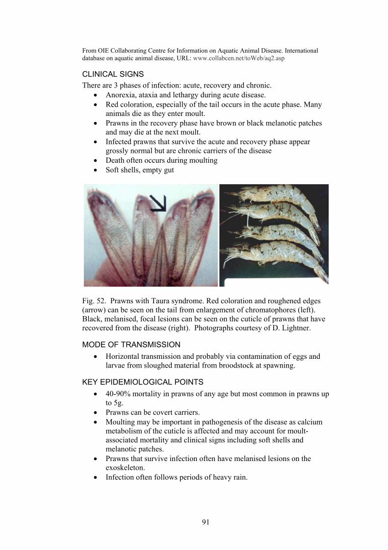
From OIE Collaborating Centre for Information on Aquatic Animal Disease. International database on aquatic animal disease, URL: www.collabcen.net/toWeb/aq2.asp
CLINICAL SIGNS There are 3 phases of infection: acute, recovery and chronic.
Anorexia, ataxia and lethargy during acute disease. • •
•
•
• •
Red coloration, especially of the tail occurs in the acute phase. Many animals die as they enter moult. Prawns in the recovery phase have brown or black melanotic patches and may die at the next moult. Infected prawns that survive the acute and recovery phase appear grossly normal but are chronic carriers of the disease Death often occurs during moulting Soft shells, empty gut
Fig. 52. Prawns with Taura syndrome. Red coloration and roughened edges (arrow) can be seen on the tail from enlargement of chromatophores (left). Black, melanised, focal lesions can be seen on the cuticle of prawns that have recovered from the disease (right). Photographs courtesy of D. Lightner.
MODE OF TRANSMISSION Horizontal transmission and probably via contamination of eggs and larvae from sloughed material from broodstock at spawning.
•
KEY EPIDEMIOLOGICAL POINTS • 40-90% mortality in prawns of any age but most common in prawns up
to 5g. • Prawns can be covert carriers. • Moulting may be important in pathogenesis of the disease as calcium
metabolism of the cuticle is affected and may account for moult-associated mortality and clinical signs including soft shells and melanotic patches.
• Prawns that survive infection often have melanised lesions on the exoskeleton.
• Infection often follows periods of heavy rain.
91
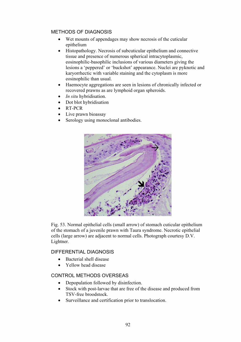
METHODS OF DIAGNOSIS Wet mounts of appendages may show necrosis of the cuticular epithelium
•
•
•
• • • • •
Histopathology. Necrosis of subcuticular epithelium and connective tissue and presence of numerous spherical intracytoplasmic, eosinophilic-basophilic inclusions of various diameters giving the lesions a ‘peppered’ or ‘buckshot’ appearance. Nuclei are pyknotic and karyorrhectic with variable staining and the cytoplasm is more eosinophilic than usual. Haemocyte aggregations are seen in lesions of chronically infected or recovered prawns as are lymphoid organ spheroids. In situ hybridisation. Dot blot hybridisation RT-PCR Live prawn bioassay Serology using monoclonal antibodies.
Fig. 53. Normal epithelial cells (small arrow) of stomach cuticular epithelium of the stomach of a juvenile prawn with Taura syndrome. Necrotic epithelial cells (large arrow) are adjacent to normal cells. Photograph courtesy D.V. Lightner.
DIFFERENTIAL DIAGNOSIS Bacterial shell disease •
•
• •
•
Yellow head disease
CONTROL METHODS OVERSEAS Depopulation followed by disinfection. Stock with post-larvae that are free of the disease and produced from TSV-free broodstock. Surveillance and certification prior to translocation.
92

REFERENCES Bonami, J.-R. (1997) Crustacean viral disease: recent developments. Bulletin
of the European Association of Fish Pathologists 17, 188-190. Hasson, K.W., Lightner, D.V., Mohney, L.L., Redman, R.M., Poulos, B.T.,
White, B.M. (1999) Taura syndrome virus (TSV) lesion development and the disease cycle in the Pacific white shrimp Litopenaeus vannamei. Diseases of Aquatic Organisms 36, 81-93.
Lightner, D.V., Redman, R.M., Hasson, K.W., Pantoja, C.R. (1995) Taura syndrome in Litopenaeus vannamei (Crustacea: Decapoda): gross signs, histopathology and ultrastructure. Diseases of Aquatic Organisms 21, 53-59.
Lightner, D.V., (1996). A handbook of shrimp pathology and diagnostic procedures for diseases of cultured penaeid shrimp. The World Aquaculture Society, Baton Rouge.
OIE (2003). International Aquatic Animal Health Code. ISBN 9290445807. Available at http://www.oie.int/eng/normes/fcode/A_summry.htm
OIE (2003). OIE Manual for Diagnostic Tests for Aquatic Animals. ISBN 9290445637. Available at URL: http://www.oie.int/eng/nomes/fmanaul/A_summry.htm
www.enaca.org
93
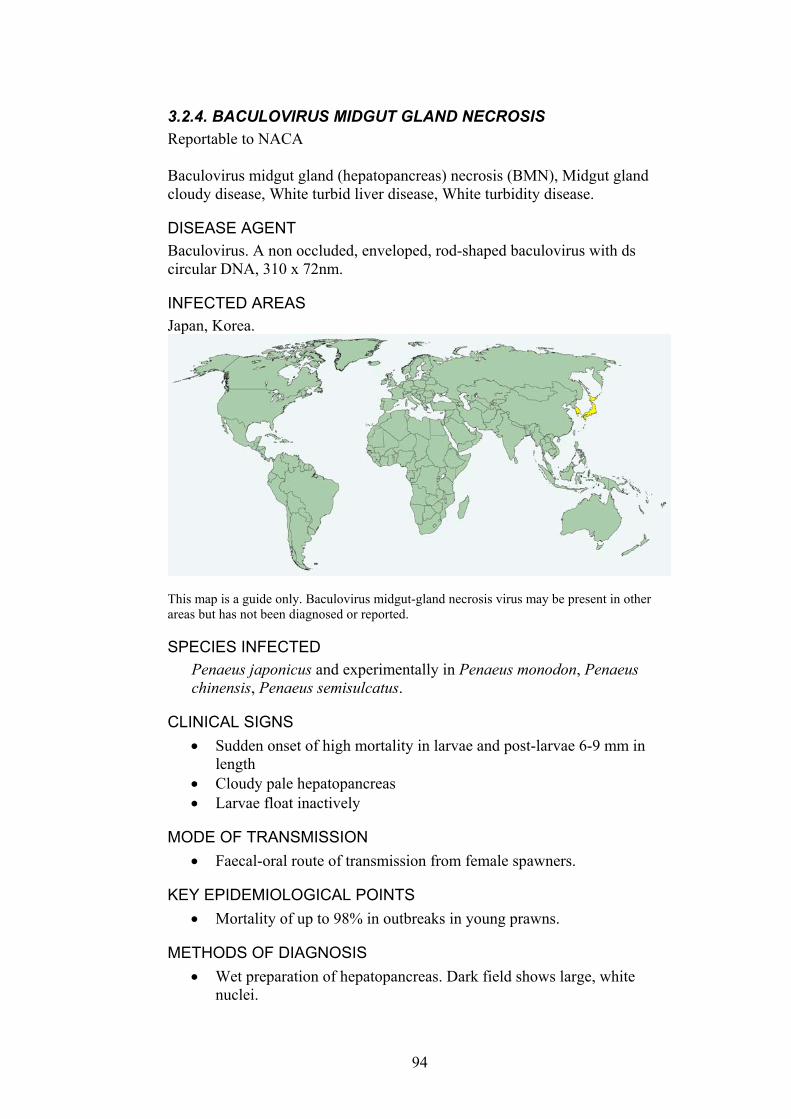
3.2.4. BACULOVIRUS MIDGUT GLAND NECROSIS Reportable to NACA Baculovirus midgut gland (hepatopancreas) necrosis (BMN), Midgut gland cloudy disease, White turbid liver disease, White turbidity disease.
DISEASE AGENT Baculovirus. A non occluded, enveloped, rod-shaped baculovirus with ds circular DNA, 310 x 72nm.
INFECTED AREAS Japan, Korea.
This map is a guide only. Baculovirus midgut-gland necrosis virus may be present in other areas but has not been diagnosed or reported.
SPECIES INFECTED Penaeus japonicus and experimentally in Penaeus monodon, Penaeus chinensis, Penaeus semisulcatus.
CLINICAL SIGNS Sudden onset of high mortality in larvae and post-larvae 6-9 mm in length
•
• •
•
•
•
Cloudy pale hepatopancreas Larvae float inactively
MODE OF TRANSMISSION Faecal-oral route of transmission from female spawners.
KEY EPIDEMIOLOGICAL POINTS Mortality of up to 98% in outbreaks in young prawns.
METHODS OF DIAGNOSIS Wet preparation of hepatopancreas. Dark field shows large, white nuclei.
94
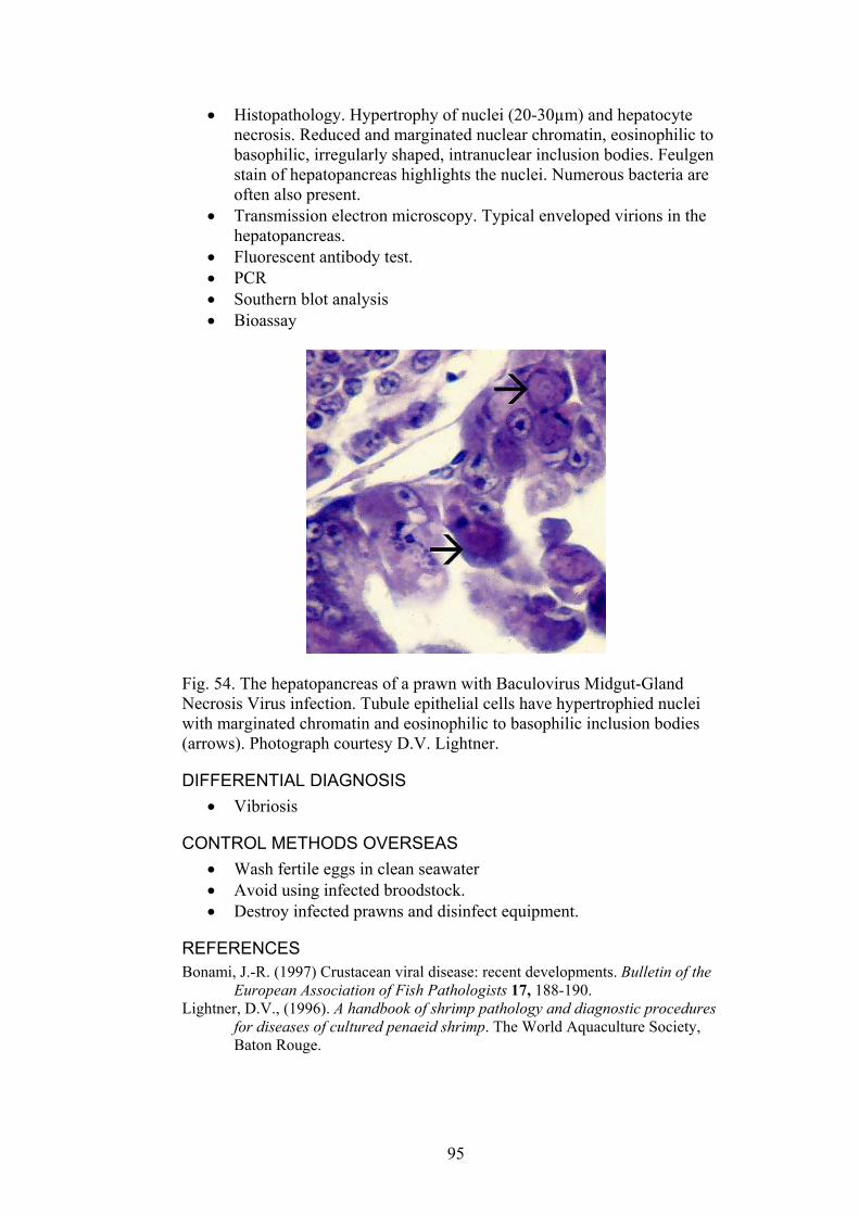
Histopathology. Hypertrophy of nuclei (20-30µm) and hepatocyte necrosis. Reduced and marginated nuclear chromatin, eosinophilic to basophilic, irregularly shaped, intranuclear inclusion bodies. Feulgen stain of hepatopancreas highlights the nuclei. Numerous bacteria are often also present.
•
•
• • • •
Transmission electron microscopy. Typical enveloped virions in the hepatopancreas. Fluorescent antibody test. PCR Southern blot analysis Bioassay
Fig. 54. The hepatopancreas of a prawn with Baculovirus Midgut-Gland Necrosis Virus infection. Tubule epithelial cells have hypertrophied nuclei with marginated chromatin and eosinophilic to basophilic inclusion bodies (arrows). Photograph courtesy D.V. Lightner.
DIFFERENTIAL DIAGNOSIS Vibriosis •
• • •
CONTROL METHODS OVERSEAS Wash fertile eggs in clean seawater Avoid using infected broodstock. Destroy infected prawns and disinfect equipment.
REFERENCES Bonami, J.-R. (1997) Crustacean viral disease: recent developments. Bulletin of the
European Association of Fish Pathologists 17, 188-190. Lightner, D.V., (1996). A handbook of shrimp pathology and diagnostic procedures
for diseases of cultured penaeid shrimp. The World Aquaculture Society, Baton Rouge.
95
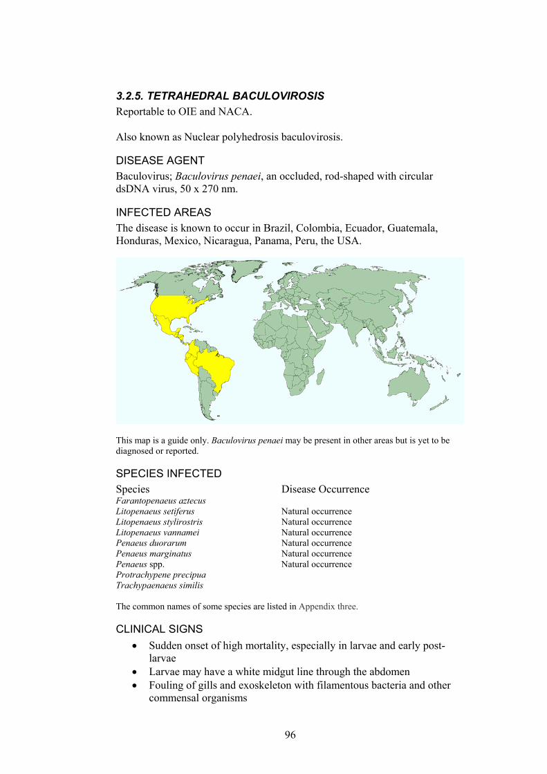
3.2.5. TETRAHEDRAL BACULOVIROSIS Reportable to OIE and NACA. Also known as Nuclear polyhedrosis baculovirosis.
DISEASE AGENT Baculovirus; Baculovirus penaei, an occluded, rod-shaped with circular dsDNA virus, 50 x 270 nm.
INFECTED AREAS The disease is known to occur in Brazil, Colombia, Ecuador, Guatemala, Honduras, Mexico, Nicaragua, Panama, Peru, the USA.
This map is a guide only. Baculovirus penaei may be present in other areas but is yet to be diagnosed or reported.
SPECIES INFECTED Species Disease Occurrence Farantopenaeus aztecus Litopenaeus setiferus Natural occurrence Litopenaeus stylirostris Natural occurrence Litopenaeus vannamei Natural occurrence Penaeus duorarum Natural occurrence Penaeus marginatus Natural occurrence Penaeus spp. Natural occurrence Protrachypene precipua Trachypaenaeus similis The common names of some species are listed in Appendix three.
CLINICAL SIGNS Sudden onset of high mortality, especially in larvae and early post-larvae
•
• •
Larvae may have a white midgut line through the abdomen Fouling of gills and exoskeleton with filamentous bacteria and other commensal organisms
96
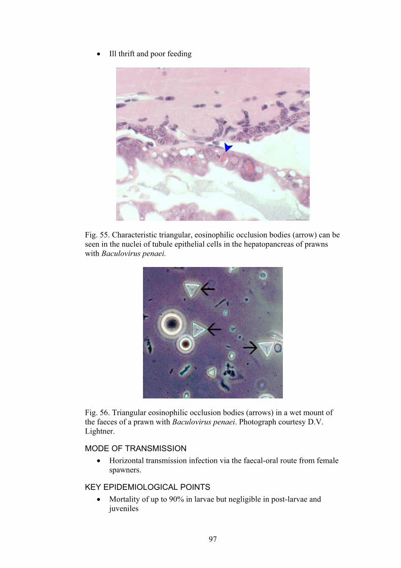
Ill thrift and poor feeding •
Fig. 55. Characteristic triangular, eosinophilic occlusion bodies (arrow) can be seen in the nuclei of tubule epithelial cells in the hepatopancreas of prawns with Baculovirus penaei.
Fig. 56. Triangular eosinophilic occlusion bodies (arrows) in a wet mount of the faeces of a prawn with Baculovirus penaei. Photograph courtesy D.V. Lightner.
MODE OF TRANSMISSION Horizontal transmission infection via the faecal-oral route from female spawners.
•
KEY EPIDEMIOLOGICAL POINTS • Mortality of up to 90% in larvae but negligible in post-larvae and
juveniles
97
• Multiple strains of the virus are known to exist. • Cannibalism increases transmission • Environmental stress and high stocking density increase the risk of
disease outbreaks.
METHODS OF DIAGNOSIS Histopathology. Triangular eosinophilic occlusion bodies in epithelial cells of the hepatopancreas tubules and midgut and in faeces. Hypertrophy of nuclei with marginated chromatin. Vacuolated cytoplasm at some stages of infection. Inclusion bodies stained with Phloxine B fluoresce when observed by a fluorescent microscope or an epifluorescent microscope.
•
•
• • • • • •
•
•
•
Wet mounts of whole larvae, hepatopancreas or faeces. Characteristic occlusion bodies and hypertrophied nuclei can be seen in heavily infected animals. In situ hybridization. Dot blot assay PCR Transmission electron microscopy Bioassay ELISA
DIFFERENTIAL DIAGNOSIS MBV (Spherical or Monodon-type baculovirus)
CONTROL METHODS OVERSEAS Disinfection of spawned eggs to prevent infection of eggs by faecal contamination. Drying and disinfection of tanks and equipment.
REFERENCES Bonami, J.-R. (1997) Crustacean viral disease: recent developments. Bulletin
of the European Association of Fish Pathologists 17, 188-190. Lightner, D.V., (1996). A handbook of shrimp pathology and diagnostic
procedures for diseases of cultured penaeid shrimp. The World Aquaculture Society, Baton Rouge.
OIE (2003). International Aquatic Animal Health Code. ISBN 9290445807. Available at http://www.oie.int/eng/normes/fcode/A_summry.htm
OIE (2003). OIE Manual for Diagnostic Tests for Aquatic Animals. ISBN 9290445637. Available at URL: http://www.oie.int/eng/nomes/fmanaul/A_summry.htm
Thurman, R.B., Bell, T.A., Lightner, D.V. (1990) Unique physicochemical properties of the occluded penaeid shrimp Baculoviruses and their use in diagnosis of infections. Journal of Aquatic Animal Health 2, 129-131.
98

3.2.6. INFECTIOUS HYPODERMAL AND HAEMATOPOIETIC NECROSIS (IHHN) Reportable to OIE and NACA Runt-deformity syndrome (RDS) of Litopenaus vannamei.
DISEASE AGENT Parvovirus: Infectious hypodermal and haematopoietic necrosis virus. A non enveloped, icosahedral, ssDNA virus, 22 nm.
INFECTED AREAS The virus may have been introduced to the Americas in IHHNV-infected asymptomatic Penaeus monodon that were introduced from the Philippines. The disease occurs in Belize, Brazil, China, Colombia, Costa Rica, Ecuador, El Salvador, French Polynesia, Guam, Guatemala, Honduras, India, Indonesia, Madagascar, Malaysia, Mexico, New Caledonia, Panama, Peru, Philippines, Singapore, Taiwan, Tanzania, Thailand, the USA, and Venezuela. There have been no reports of the disease on mainland USA and most areas of Hawaii for more than 10 years.
This map is a guide only. Infectious hypodermal and haematopoietic necrosis virus may be present in other areas but is yet to be diagnosed or reported.
SPECIES INFECTED Many species of commercially cultured prawns are susceptible to the disease. Litopenaeus stylirostris is the most severely affected species. Litopenaeus vannamei and Penaeus monodon may have a chronic infection and ill thrift. Species Disease Occurrence Farantopanaeus aztecus Experimental demonstration Farantopenaeus californiensis Natural occurrence Penaeus duorarum Experimental demonstration Penaeus japonicus Natural occurrence Penaeus monodon Natural occurrence
99

Penaeus semisulcatus Natural occurrence Litopenaeus setiferus Experimental demonstration Litopenaeus stylirostris Natural occurrence Litopenaeus vannamei Natural occurrence Adapted from OIE Collaborating Centre for Information on Aquatic Animal Disease. International database on aquatic animal disease, URL: www.collabcen.net/toWeb/aq2.asp
CLINICAL SIGNS Reduced feeding •
• •
•
Affected prawns often slowly rise and sink in the water column Whitish spots or a bluish coloration from necrosis of the cuticular epithelium Reduced growth and cuticle deformities in chronic infections (Litopenaeus vannamei often have chronic infection and runt deformity syndrome).
Fig. 57. A juvenile Litopenaeus stylirostris with acute IHHNV. White lesions can be seen (arrows, left photograph). Bent rostrums in prawns with runt deformity syndrome (photograph on right). Photograph courtesy D.V. Lightner.
MODE OF TRANSMISSION Vertical transmission in ovarian fluids and horizontal transmission, usually via ingestion of dead individuals.
•
KEY EPIDEMIOLOGICAL POINTS • IHHNV infections in Asia and Africa appear to be non pathogenic. • Prawns that survive infection often become asymptomatic carriers,
therefore nested PCR is important in helping to identify virus-free females for use as broodstock.
• Mortality up to 90% in highly susceptible species such as Litopenaeus stylirostris.
• Young juveniles and older post-larvae are most severely affected. • There are 3 main phylogenetic lineages of the virus (a Mexican group,
a Central/South American group and a Thailand/Tanzania group).
100
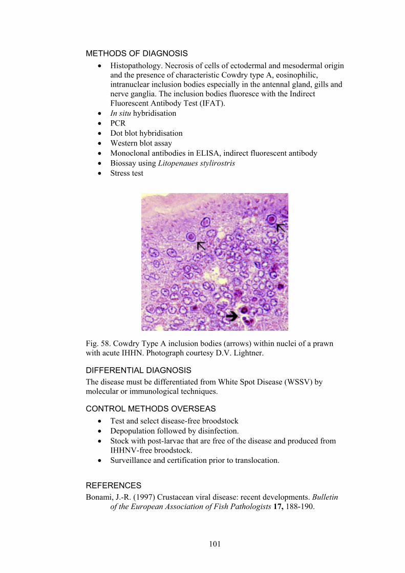
METHODS OF DIAGNOSIS Histopathology. Necrosis of cells of ectodermal and mesodermal origin and the presence of characteristic Cowdry type A, eosinophilic, intranuclear inclusion bodies especially in the antennal gland, gills and nerve ganglia. The inclusion bodies fluoresce with the Indirect Fluorescent Antibody Test (IFAT).
•
• • • • • • •
In situ hybridisation PCR Dot blot hybridisation Western blot assay Monoclonal antibodies in ELISA, indirect fluorescent antibody Biossay using Litopenaues stylirostris Stress test
Fig. 58. Cowdry Type A inclusion bodies (arrows) within nuclei of a prawn with acute IHHN. Photograph courtesy D.V. Lightner.
DIFFERENTIAL DIAGNOSIS The disease must be differentiated from White Spot Disease (WSSV) by molecular or immunological techniques.
CONTROL METHODS OVERSEAS Test and select disease-free broodstock •
• •
•
Depopulation followed by disinfection. Stock with post-larvae that are free of the disease and produced from IHHNV-free broodstock. Surveillance and certification prior to translocation.
REFERENCES Bonami, J.-R. (1997) Crustacean viral disease: recent developments. Bulletin
of the European Association of Fish Pathologists 17, 188-190.
101

Lightner, D.V., (1996). A handbook of shrimp pathology and diagnostic procedures for diseases of cultured penaeid shrimp. The World Aquaculture Society, Baton Rouge.
OIE (2003). International Aquatic Animal Health Code. ISBN 9290445807. Available at http://www.oie.int/eng/normes/fcode/A_summry.htm
OIE (2003). OIE Manual for Diagnostic Tests for Aquatic Animals. ISBN 9290445637. Available at URL: http://www.oie.int/eng/nomes/fmanaul/A_summry.htm
Tang, K.F.J., Poulos, B.T., Wang, J., Redman, R.M., Shih, H.-H., Lightner, D.V. (2003) Geographic variations among infectious hypodermal and hematopoietic necrosis virus (IHHNV) isolates and characteristics of their infection. Diseases of Aquatic Organisms 53, 91-99.
Thurman, R.B., Bell, T.A., Lightner, D.V. (1990) Unique physicochemical properties of the occluded penaeid shrimp Baculoviruses and their use in diagnosis of infections. Journal of Aquatic Animal Health 2, 129-131.
102
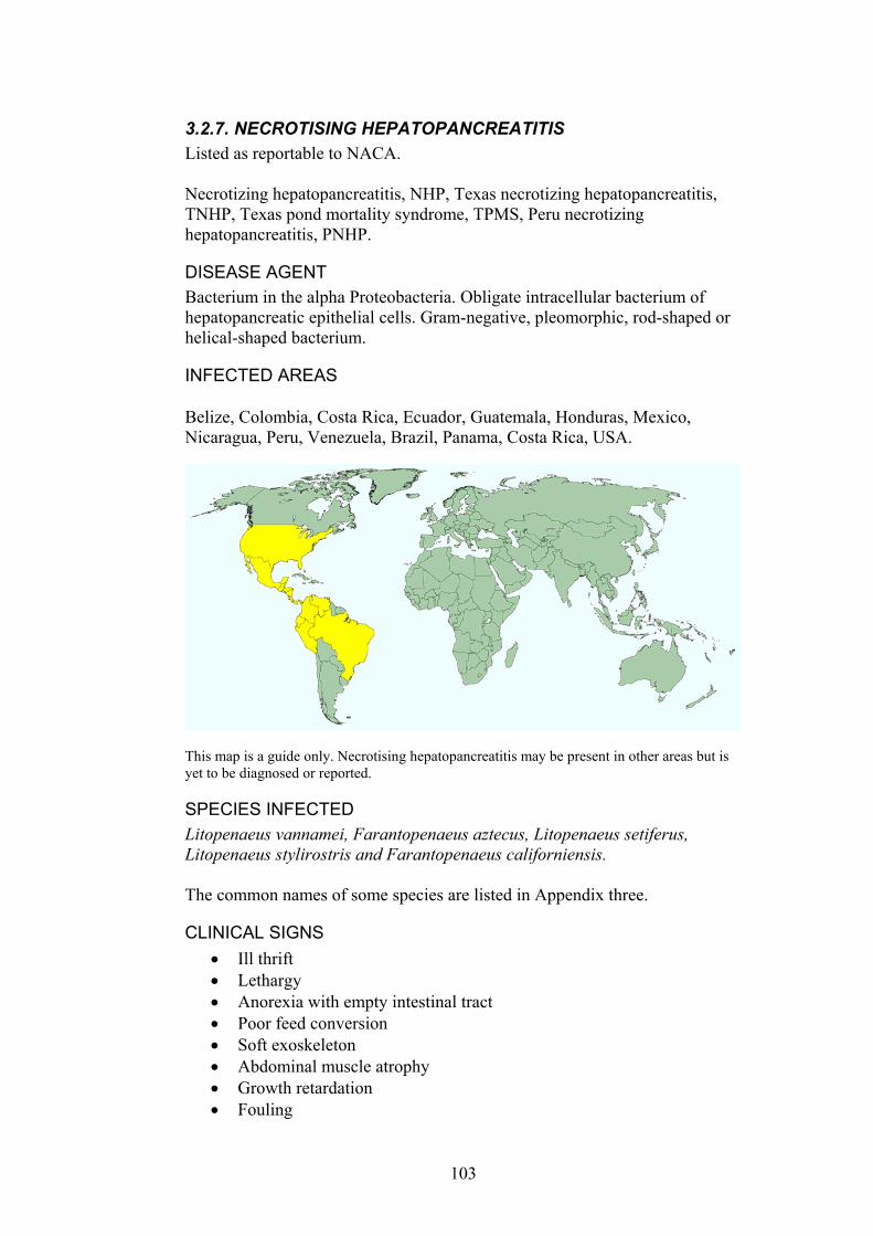
3.2.7. NECROTISING HEPATOPANCREATITIS Listed as reportable to NACA. Necrotizing hepatopancreatitis, NHP, Texas necrotizing hepatopancreatitis, TNHP, Texas pond mortality syndrome, TPMS, Peru necrotizing hepatopancreatitis, PNHP.
DISEASE AGENT Bacterium in the alpha Proteobacteria. Obligate intracellular bacterium of hepatopancreatic epithelial cells. Gram-negative, pleomorphic, rod-shaped or helical-shaped bacterium.
INFECTED AREAS Belize, Colombia, Costa Rica, Ecuador, Guatemala, Honduras, Mexico, Nicaragua, Peru, Venezuela, Brazil, Panama, Costa Rica, USA.
This map is a guide only. Necrotising hepatopancreatitis may be present in other areas but is yet to be diagnosed or reported.
SPECIES INFECTED Litopenaeus vannamei, Farantopenaeus aztecus, Litopenaeus setiferus, Litopenaeus stylirostris and Farantopenaeus californiensis. The common names of some species are listed in Appendix three.
CLINICAL SIGNS Ill thrift •
• • • • • • •
Lethargy Anorexia with empty intestinal tract Poor feed conversion Soft exoskeleton Abdominal muscle atrophy Growth retardation Fouling
103
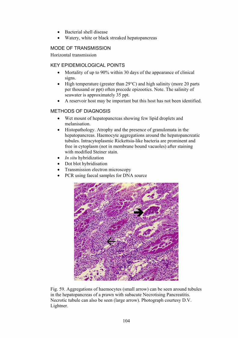
Bacterial shell disease • •
•
•
• • • •
Watery, white or black streaked hepatopancreas
MODE OF TRANSMISSION Horizontal transmission
KEY EPIDEMIOLOGICAL POINTS • Mortality of up to 90% within 30 days of the appearance of clinical
signs. • High temperature (greater than 29°C) and high salinity (more 20 parts
per thousand or ppt) often precede epizootics. Note. The salinity of seawater is approximately 35 ppt.
• A reservoir host may be important but this host has not been identified.
METHODS OF DIAGNOSIS Wet mount of hepatopancreas showing few lipid droplets and melanisation. Histopathology. Atrophy and the presence of granulomata in the hepatopancreas. Haemocyte aggregations around the hepatopancreatic tubules. Intracytoplasmic Rickettsia-like bacteria are prominent and free in cytoplasm (not in membrane bound vacuoles) after staining with modified Steiner stain. In situ hybridization Dot blot hybridisation Transmission electron microscopy PCR using faecal samples for DNA source
Fig. 59. Aggregations of haemocytes (small arrow) can be seen around tubules in the hepatopancreas of a prawn with subacute Necrotising Pancreatitis. Necrotic tubule can also be seen (large arrow). Photograph courtesy D.V. Lightner.
104
DIFFERENTIAL DIAGNOSIS Bacterial shell disease •
• • •
• •
Vibriosis Aflatoxicosis Taura syndrome
CONTROL METHODS OVERSEAS Maintain water temperature and salinity within the optimal range. Treatment with antibiotics
REFERENCES Lightner, D.V., (1996). A handbook of shrimp pathology and diagnostic
procedures for diseases of cultured penaeid shrimp. The World Aquaculture Society, Baton Rouge.
Lightner, D.V., Redman, R.M., Bonami,J.R. (1992). Morphological evidence for a single bacterial etiology in Texas necrotizing hepatopancreatitis in Litopenaeus vannamei (Curstacea: Decapoda). Diseases of Aquatic Organisms 13:235-239.
Pantoja, C.R. and Lightner,D.V. (2003). Necrotizing Hepatopancreatitis: Diagnosis, distribution in shrimp. Global Aquaculture Advocate 6(4):18
105
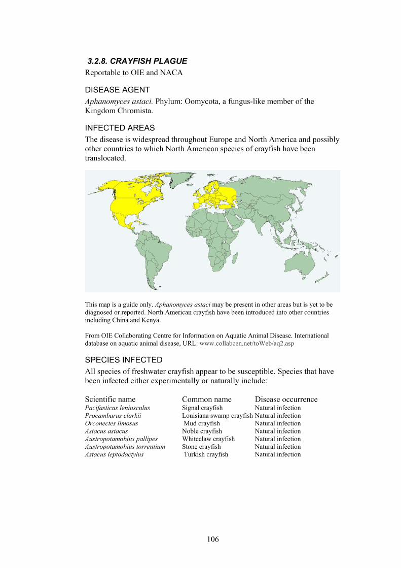
3.2.8. CRAYFISH PLAGUE Reportable to OIE and NACA
DISEASE AGENT Aphanomyces astaci. Phylum: Oomycota, a fungus-like member of the Kingdom Chromista.
INFECTED AREAS The disease is widespread throughout Europe and North America and possibly other countries to which North American species of crayfish have been translocated.
This map is a guide only. Aphanomyces astaci may be present in other areas but is yet to be diagnosed or reported. North American crayfish have been introduced into other countries including China and Kenya. From OIE Collaborating Centre for Information on Aquatic Animal Disease. International database on aquatic animal disease, URL: www.collabcen.net/toWeb/aq2.asp
SPECIES INFECTED All species of freshwater crayfish appear to be susceptible. Species that have been infected either experimentally or naturally include: Scientific name Common name Disease occurrence Pacifasticus leniusculus Signal crayfish Natural infection Procambarus clarkii Louisiana swamp crayfish Natural infection Orconectes limosus Mud crayfish Natural infection Astacus astacus Noble crayfish Natural infection Austropotamobius pallipes Whiteclaw crayfish Natural infection Austropotamobius torrentium Stone crayfish Natural infection Astacus leptodactylus Turkish crayfish Natural infection
106
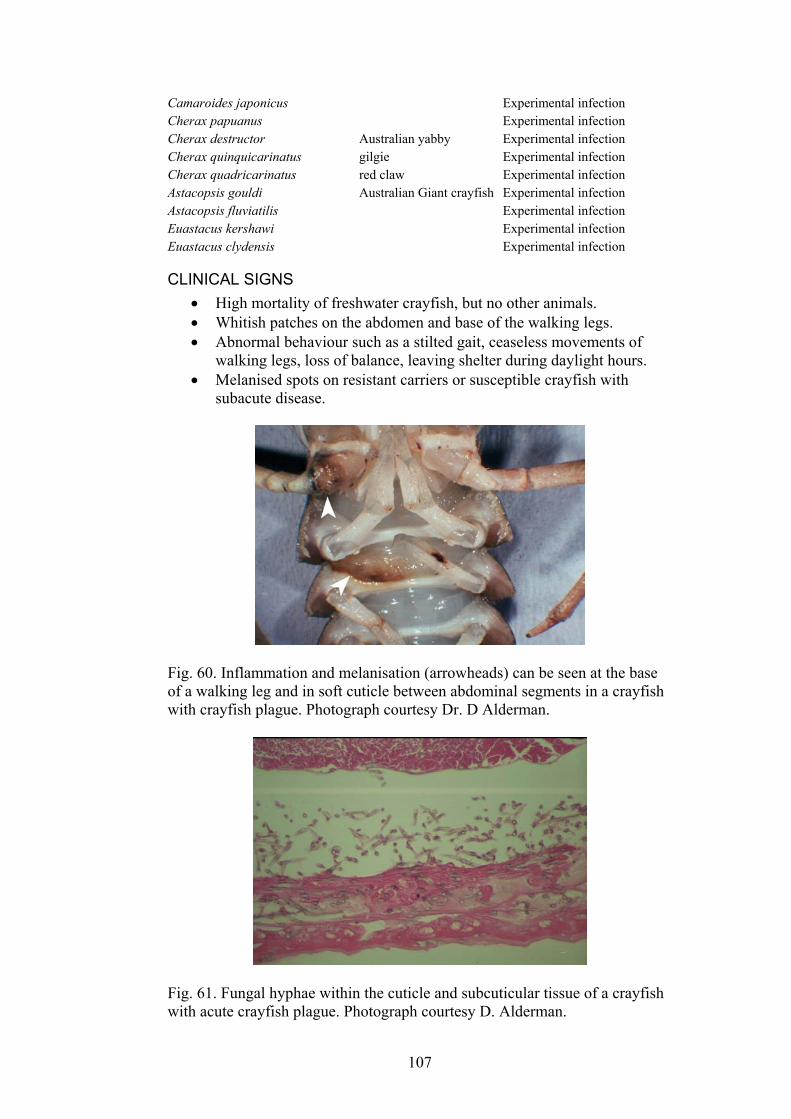
Camaroides japonicus Experimental infection Cherax papuanus Experimental infection Cherax destructor Australian yabby Experimental infection Cherax quinquicarinatus gilgie Experimental infection Cherax quadricarinatus red claw Experimental infection Astacopsis gouldi Australian Giant crayfish Experimental infection Astacopsis fluviatilis Experimental infection Euastacus kershawi Experimental infection Euastacus clydensis Experimental infection
CLINICAL SIGNS High mortality of freshwater crayfish, but no other animals. •
• •
•
Whitish patches on the abdomen and base of the walking legs. Abnormal behaviour such as a stilted gait, ceaseless movements of walking legs, loss of balance, leaving shelter during daylight hours. Melanised spots on resistant carriers or susceptible crayfish with subacute disease.
Fig. 60. Inflammation and melanisation (arrowheads) can be seen at the base of a walking leg and in soft cuticle between abdominal segments in a crayfish with crayfish plague. Photograph courtesy Dr. D Alderman.
Fig. 61. Fungal hyphae within the cuticle and subcuticular tissue of a crayfish with acute crayfish plague. Photograph courtesy D. Alderman.
107
MODE OF TRANSMISSION Horizontal transmission by motile zoospores in the water. •
•
•
• •
•
• • • •
• •
• •
•
KEY EPIDEMIOLOGICAL POINTS Aphanomyces astaci is a primary pathogen and does not survive in the environment for more than 3 weeks without a crayfish host. Northern American species of freshwater crayfish are resistant carriers and usually are the source of new outbreaks of disease. The parasite is encapsulated in a melanin sheath in resistant crayfish species where it causes no or little apparent harm to the host unless they become immunocompromised by stress such as moulting or poor water quality. Zoospores spread the disease in water. Wet equipment or vehicles can spread zoospores from one water body to another. The pathogen is readily killed by boiling (for one minute), freezing, drying and halogen disinfectants.
METHODS OF DIAGNOSIS Wet mounts of cuticle and subcuticular tissue Histopathology. Characteristic fungal hyphae Culture on isolation media. PCR and in situ hybridisation
DIFFERENTIAL DIAGNOSIS Environmental toxins or water quality problems Thelohania spp.
CONTROL METHODS OVERSEAS Movement restrictions on live crayfish and dead, uncooked crayfish Disinfection required for fishing equipment and boats prior to movement Controls on movement of water between water bodies
REFERENCES Alderman, D. (1993) Crayfish plague in Britain, the first twelve years. Freshwater Crayfish 9, 266-
272. Diéguez-Uribeondo, J., Söderhäll, K. (1999) RAPD evidence for the origin of an outbreak of
crayfish plague in Spain. Freshwater Crayfish 12, 313-318. Huang, T., Cerenius, L., Söderhäll, K. (1994) Analysis of genetic diversity in the crayfish plague
fungus, Aphanomyces astaci, by random amplification of polymorphic DNA. Aquaculture 126, 1-10.
Oidtmann, B., Heitz, E., Rogers, D., Hoffman, R.W. (2002) Transmission of crayfish plague. Diseases of Aquatic Organisms 52, 159-167.
OIE (2003). International Aquatic Animal Health Code. ISBN 9290445807. Available at URL: http://www.oie.int/eng/normes/fcode/A_summry.htm
OIE (2003). OIE Manual for Diagnostic Tests for Aquatic Animals. ISBN 9290445637. Available at URL: http://www.oie.int/eng/nomes/fmanaul/A_summry.htm
Söderhäll, K., Cerenius, L. (1999) The crayfish plague fungus: history and recent advances. Freshwater Crayfish 12, 11-35.
Unestam, T. (1975) Defence reactions in and susceptibility of Australian and New Guinean freshwater crayfish to European-crayfish-plague fungus. Australian Journal of Experimental Biology and Medical Science 53, 349-359.
108
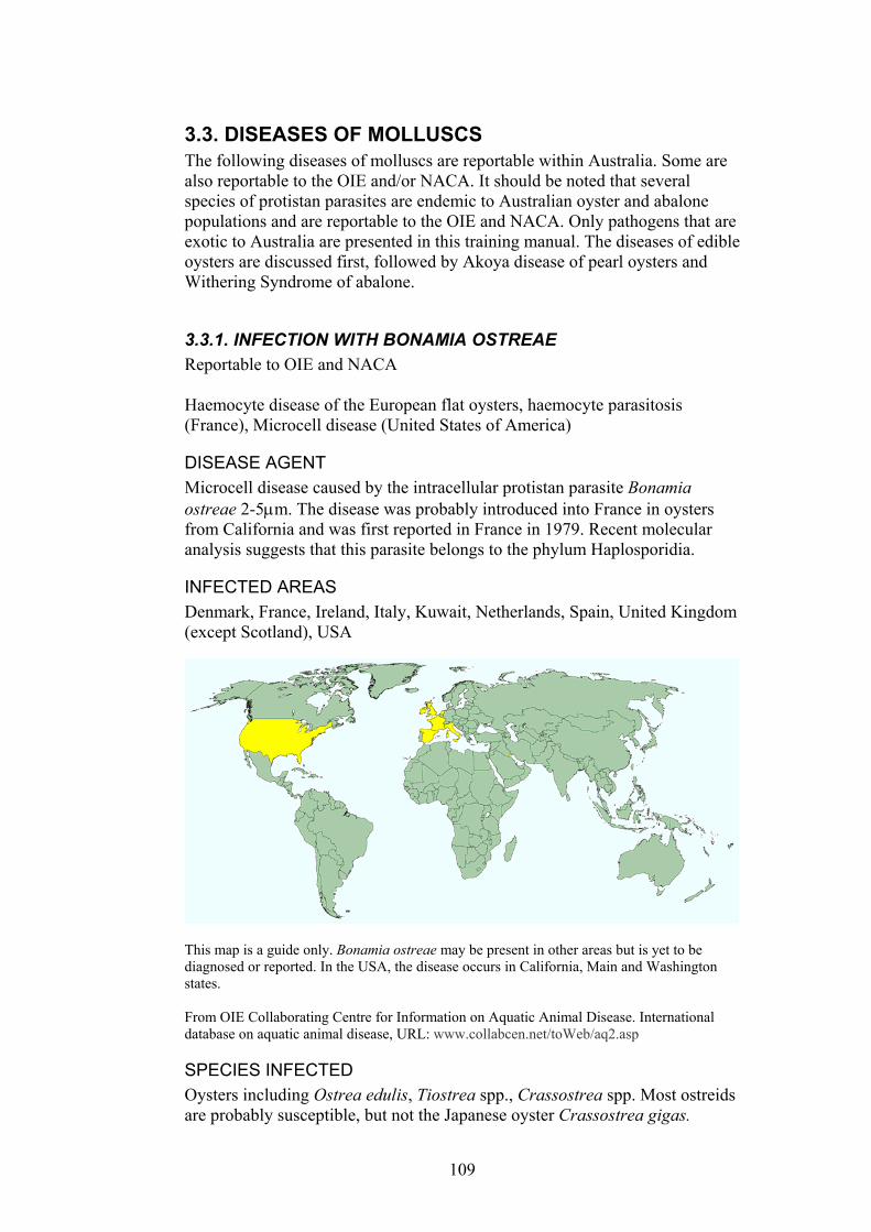
3.3. DISEASES OF MOLLUSCS The following diseases of molluscs are reportable within Australia. Some are also reportable to the OIE and/or NACA. It should be noted that several species of protistan parasites are endemic to Australian oyster and abalone populations and are reportable to the OIE and NACA. Only pathogens that are exotic to Australia are presented in this training manual. The diseases of edible oysters are discussed first, followed by Akoya disease of pearl oysters and Withering Syndrome of abalone.
3.3.1. INFECTION WITH BONAMIA OSTREAE Reportable to OIE and NACA Haemocyte disease of the European flat oysters, haemocyte parasitosis (France), Microcell disease (United States of America)
DISEASE AGENT Microcell disease caused by the intracellular protistan parasite Bonamia ostreae 2-5µm. The disease was probably introduced into France in oysters from California and was first reported in France in 1979. Recent molecular analysis suggests that this parasite belongs to the phylum Haplosporidia.
INFECTED AREAS Denmark, France, Ireland, Italy, Kuwait, Netherlands, Spain, United Kingdom (except Scotland), USA
This map is a guide only. Bonamia ostreae may be present in other areas but is yet to be diagnosed or reported. In the USA, the disease occurs in California, Main and Washington states. From OIE Collaborating Centre for Information on Aquatic Animal Disease. International database on aquatic animal disease, URL: www.collabcen.net/toWeb/aq2.asp
SPECIES INFECTED Oysters including Ostrea edulis, Tiostrea spp., Crassostrea spp. Most ostreids are probably susceptible, but not the Japanese oyster Crassostrea gigas.
109
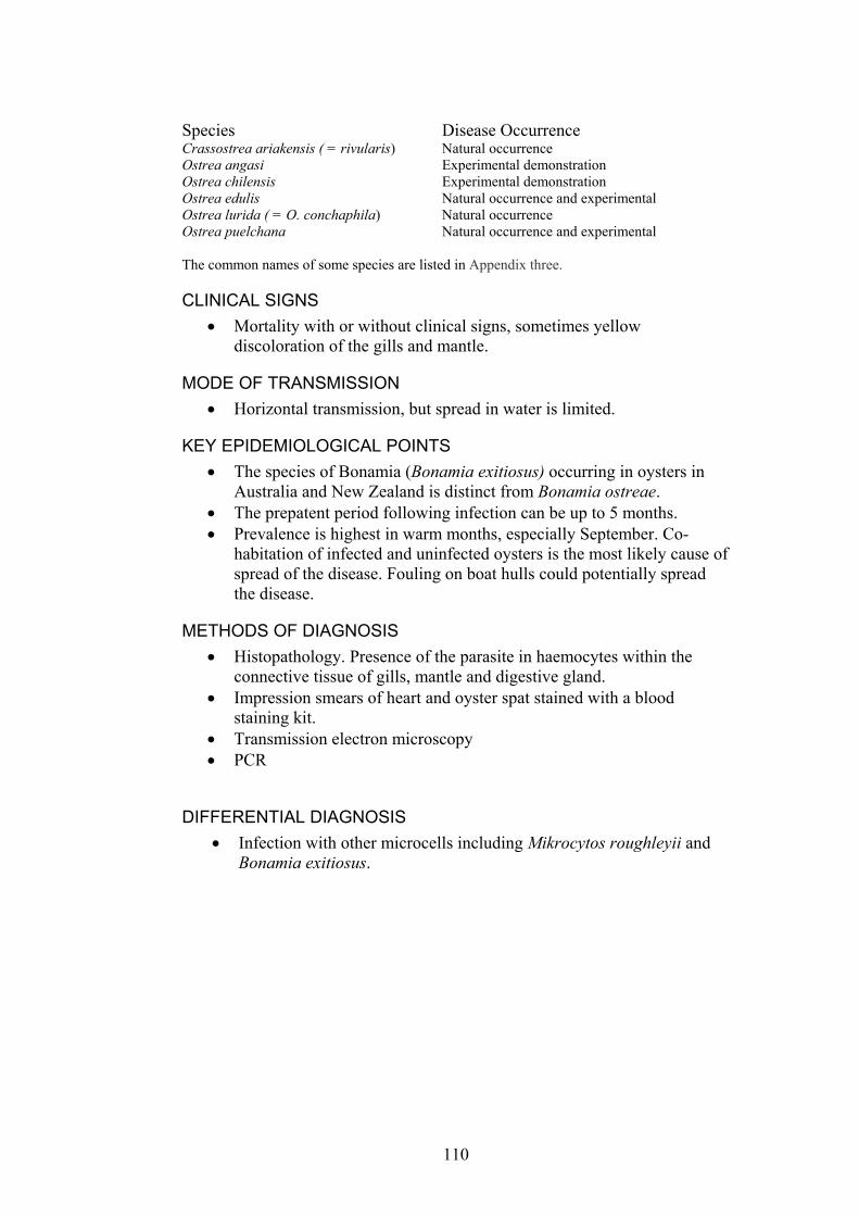
Species Disease Occurrence Crassostrea ariakensis ( = rivularis) Natural occurrence Ostrea angasi Experimental demonstration Ostrea chilensis Experimental demonstration Ostrea edulis Natural occurrence and experimental Ostrea lurida ( = O. conchaphila) Natural occurrence Ostrea puelchana Natural occurrence and experimental The common names of some species are listed in Appendix three.
CLINICAL SIGNS Mortality with or without clinical signs, sometimes yellow discoloration of the gills and mantle.
•
•
•
•
• •
•
MODE OF TRANSMISSION Horizontal transmission, but spread in water is limited.
KEY EPIDEMIOLOGICAL POINTS • The species of Bonamia (Bonamia exitiosus) occurring in oysters in
Australia and New Zealand is distinct from Bonamia ostreae. • The prepatent period following infection can be up to 5 months. • Prevalence is highest in warm months, especially September. Co-
habitation of infected and uninfected oysters is the most likely cause of spread of the disease. Fouling on boat hulls could potentially spread the disease.
METHODS OF DIAGNOSIS Histopathology. Presence of the parasite in haemocytes within the connective tissue of gills, mantle and digestive gland. Impression smears of heart and oyster spat stained with a blood staining kit. Transmission electron microscopy PCR
DIFFERENTIAL DIAGNOSIS Infection with other microcells including Mikrocytos roughleyii and Bonamia exitiosus.
110
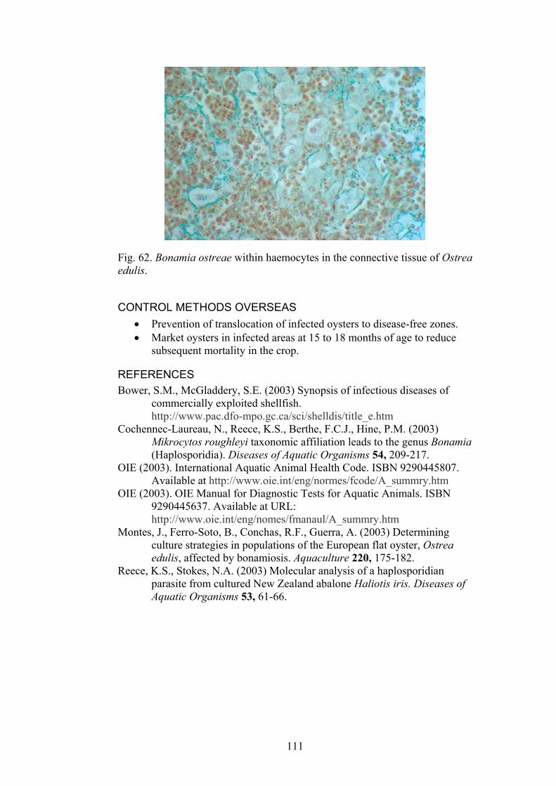
Fig. 62. Bonamia ostreae within haemocytes in the connective tissue of Ostrea edulis.
CONTROL METHODS OVERSEAS Prevention of translocation of infected oysters to disease-free zones. •
• Market oysters in infected areas at 15 to 18 months of age to reduce subsequent mortality in the crop.
REFERENCES Bower, S.M., McGladdery, S.E. (2003) Synopsis of infectious diseases of
commercially exploited shellfish. http://www.pac.dfo-mpo.gc.ca/sci/shelldis/title_e.htm
Cochennec-Laureau, N., Reece, K.S., Berthe, F.C.J., Hine, P.M. (2003) Mikrocytos roughleyi taxonomic affiliation leads to the genus Bonamia (Haplosporidia). Diseases of Aquatic Organisms 54, 209-217.
OIE (2003). International Aquatic Animal Health Code. ISBN 9290445807. Available at http://www.oie.int/eng/normes/fcode/A_summry.htm
OIE (2003). OIE Manual for Diagnostic Tests for Aquatic Animals. ISBN 9290445637. Available at URL: http://www.oie.int/eng/nomes/fmanaul/A_summry.htm
Montes, J., Ferro-Soto, B., Conchas, R.F., Guerra, A. (2003) Determining culture strategies in populations of the European flat oyster, Ostrea edulis, affected by bonamiosis. Aquaculture 220, 175-182.
Reece, K.S., Stokes, N.A. (2003) Molecular analysis of a haplosporidian parasite from cultured New Zealand abalone Haliotis iris. Diseases of Aquatic Organisms 53, 61-66.
111
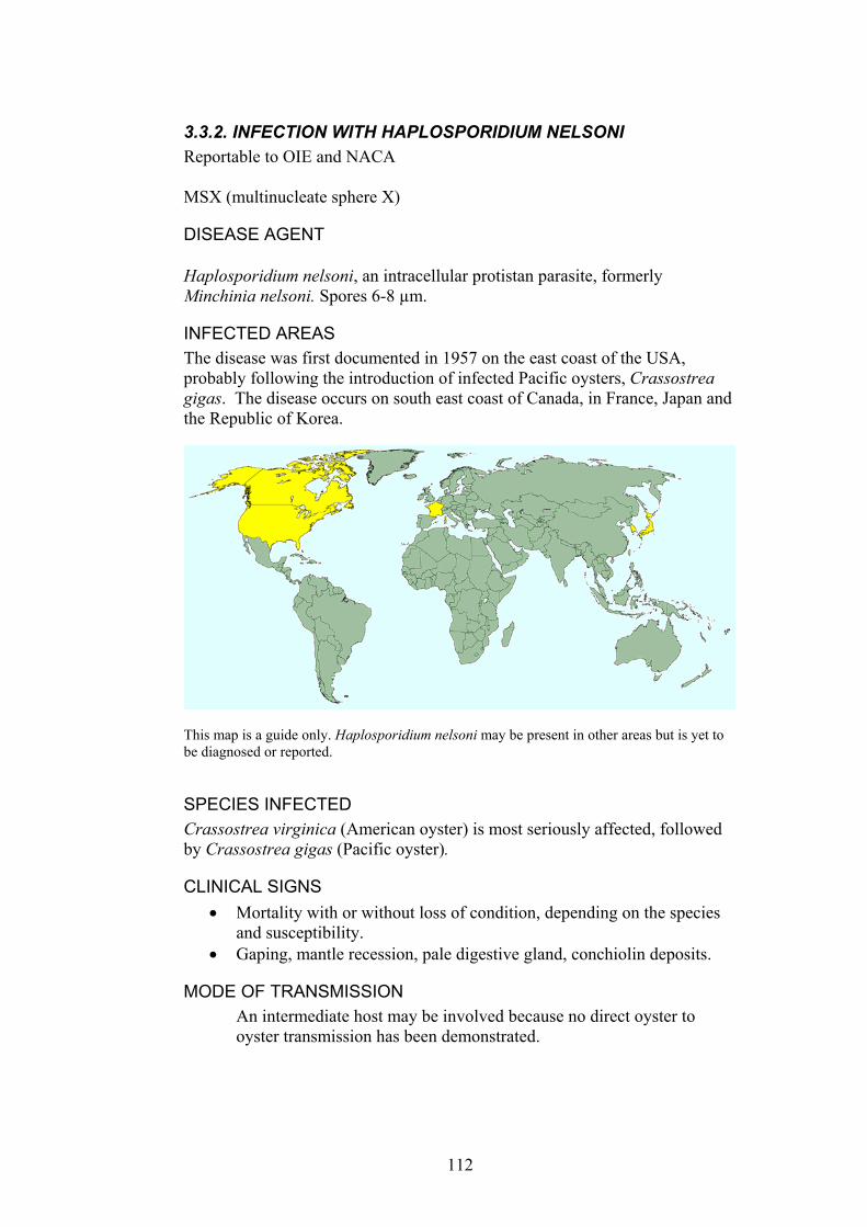
3.3.2. INFECTION WITH HAPLOSPORIDIUM NELSONI Reportable to OIE and NACA MSX (multinucleate sphere X)
DISEASE AGENT Haplosporidium nelsoni, an intracellular protistan parasite, formerly Minchinia nelsoni. Spores 6-8 µm.
INFECTED AREAS The disease was first documented in 1957 on the east coast of the USA, probably following the introduction of infected Pacific oysters, Crassostrea gigas. The disease occurs on south east coast of Canada, in France, Japan and the Republic of Korea.
This map is a guide only. Haplosporidium nelsoni may be present in other areas but is yet to be diagnosed or reported.
SPECIES INFECTED Crassostrea virginica (American oyster) is most seriously affected, followed by Crassostrea gigas (Pacific oyster).
CLINICAL SIGNS Mortality with or without loss of condition, depending on the species and susceptibility.
•
• Gaping, mantle recession, pale digestive gland, conchiolin deposits.
MODE OF TRANSMISSION An intermediate host may be involved because no direct oyster to oyster transmission has been demonstrated.
112

KEY EPIDEMIOLOGICAL POINTS • The life cycle may be indirect but the identity of the intermediate host
and its geographical distribution is unknown. • The parasite prefers a salinity of approximately 15ppt (parts per
thousand). Mortality is high at salinities of 20 ppt. Note. The salinity of seawater is approximately 35 ppt.
• Sporulation occurs in warmer months, spring to autumn, depending on the location.
• The parasite prefers temperatures of approximately 20°C. • Mortality in epizootics can reach 90%. • Epizootics often occur in cycles, commonly every 6 to 8 years.
Fig. 63. Haplosporidium nelsoni spores in digestive tubule epithelium of Crassostrea virginica. Photograph courtesy of E. Burreson.
Fig. 64. Haplosporidium nelsoni plasmodia in gut epithelium of Crassostrea virginica. Photograph courtesy of E. Burreson.
113

METHODS OF DIAGNOSIS Histopathology. Spores (6-8µm) are found only within epithelial cells of digestive tubules and sporulation is more common in juvenile oysters. Plasmodia (5-25 µm) are found within cells in connective tissue and epithelium of the gills, digestive and reproductive tract, perivascular cuffing with haemocytes in the mantle, a massive haemocyte response may occur, pyknosis and necrosis of infected cells.
•
• •
•
•
DNA probes and PCR primers. In situ hybridization
DIFFERENTIAL DIAGNOSIS On the east coast of the USA the following diseases must be differentiated from infection with Haplosporidium costale. In other areas the disease must be distinguished from infection with other diseases such as Perkinsus marinus and Haplosporidium costale.
CONTROL METHODS OVERSEAS Hold oysters at a low salinity (< 10 ppt) or cooler water (<20°C) for 2 weeks, especially in the summer infection period. Move to high salinity areas for growth and conditioning in non infective periods. Use disease resistant oyster strains.
REFERENCES Bower, S.M., McGladdery, S.E. (2003) Synopsis of infectious diseases of
commercially expoited shellfish. http://www.pac.dfo-mpo.gc.ca/sci/shelldis/title_e.htm
Chu, F.-L.E. (1997) Trends in research on Crassostrea virginica and its two protozoan parasites, Perkinsus marinus and Haplosporidium nelsoni. Bulletin of the European Association of Fish Pathologists 17, 184-187.
Elston, R.A. (1993) Infectious diseases of the Pacific oyster, Crassostrea gigas. Annual Review of Fish Diseases 3, 259-276.
OIE (2003). International Aquatic Animal Health Code. ISBN 9290445807. Available at http://www.oie.int/eng/normes/fcode/A_summry.htm
OIE (2003). OIE Manual for Diagnostic Tests for Aquatic Animals. ISBN 9290445637. Available at URL: http://www.oie.int/eng/nomes/fmanaul/A_summry.htm
Reece, K.S., Stokes, N.A. (2003) Molecular analysis of a haplosporidian parasite from cultured New Zealand abalone Haliotis iris. Diseases of Aquatic Organisms 53, 61-66.
VIMS OIE website (http://www.vims.edu/env/research/shellfish/oie/index.html)
114

3.3.3.INFECTION WITH HAPLOSPORIDIUM COSTALE Reportable to OIE and NACA. SSO (Seaside organism)
DISEASE AGENT Haplosporidium costale, a protistan parasite; Phylum Haplosporidia, spores 3-5 µm (formerly known as Minchinia costalis).
INFECTED AREAS The disease occurs on the east coast of Canada and the USA.
This map is a guide only. Haplosporidium costale may be present in other areas but is yet to be diagnosed or reported.
SPECIES INFECTED The disease occurs in Crassostrea virginica, the American oyster.
CLINICAL SIGNS Gaping, emaciation, mantle recession •
MODE OF TRANSMISSION An intermediate host may be involved because no direct oyster to oyster transmission has been demonstrated.
KEY EPIDEMIOLOGICAL POINTS • Sporulation and mortality peak in May to June, infection is sub clinical
at other times of the year. • Sporulation occurs within the connective tissue of moribund oysters
and probably causes mortality. • The presence of the intermediate host may restrict distribution of the
disease. • The disease only occurs where salinity is greater than 25ppt.
115
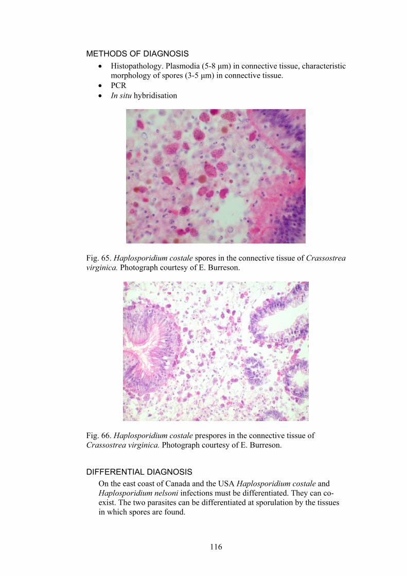
METHODS OF DIAGNOSIS Histopathology. Plasmodia (5-8 µm) in connective tissue, characteristic morphology of spores (3-5 µm) in connective tissue.
•
• •
PCR In situ hybridisation
Fig. 65. Haplosporidium costale spores in the connective tissue of Crassostrea virginica. Photograph courtesy of E. Burreson.
Fig. 66. Haplosporidium costale prespores in the connective tissue of Crassostrea virginica. Photograph courtesy of E. Burreson.
DIFFERENTIAL DIAGNOSIS On the east coast of Canada and the USA Haplosporidium costale and Haplosporidium nelsoni infections must be differentiated. They can co-exist. The two parasites can be differentiated at sporulation by the tissues in which spores are found.
116

CONTROL METHODS OVERSEAS There are no known control methods but harvesting oysters before 18-24 months of age or transferring infected oysters to low salinity water may help to reduce losses.
•
REFERENCES Bower, S.M., McGladdery, S.E. (2003) Synopsis of infectious diseases of
commercially expoited shellfish. http://www.pac.dfo-mpo.gc.ca/sci/shelldis/title_e.htm
OIE (2003). International Aquatic Animal Health Code. ISBN 9290445807. Available at http://www.oie.int/eng/normes/fcode/A_summry.htm
OIE (2003). OIE Manual for Diagnostic Tests for Aquatic Animals. ISBN 9290445637. Available at URL: http://www.oie.int/eng/nomes/fmanaul/A_summry.htm
Reece, K.S., Stokes, N.A. (2003) Molecular analysis of a haplosporidian parasite from cultured New Zealand abalone Haliotis iris. Diseases of Aquatic Organisms 53, 61-66.
VIMS OIE website (http://www.vims.edu/env/research/shellfish/oie/index.html)
117

3.3.4. INFECTION WITH MARTEILIA REFRINGENS Reportable to OIE and NACA. Also known as Aber disease and Digestive gland disease (France)
DISEASE AGENT Protistan parasite (Phylum: Paramyxea).
INFECTED AREAS The disease was first reported in France in 1968 and it is known to occur in Greece, Italy, Morocco, Portugal and Spain.
This map is a guide only. Marteilia refringens may be present in other areas but is yet to be diagnosed or reported. From OIE Collaborating Centre for Information on Aquatic Animal Disease. International database on aquatic animal disease, URL: www.collabcen.net/toWeb/aq2.asp
SPECIES INFECTED The European flat oyster, Ostrea edulis, is the most seriously affected bivalve. Species Disease Occurrence Cardium edule Possible natural occurrence Ostrea angasi Possible experimental demonstration Ostrea chilensis Possible natural and experimental demonstration Ostrea edulis Natural occurrence Ostrea puelchana Possible experimental demonstration The common names of some species are listed in Appendix three.
CLINICAL SIGNS Cessation of growth •
• •
Poor condition Emaciation
118

Discoloured digestive gland • • • •
Completely resorbed gonads Depletion of glycogen reserves Mortality
MODE OF TRANSMISSION An intermediate host may be involved because no direct oyster to oyster transmission has been demonstrated.
KEY EPIDEMIOLOGICAL FEATURES • Infections occur in spring and summer when water temperatures are
>17°C. • High salinity limits development of the parasite. • Sporangia are released into water from the lumen of the intestine.
Fig. 67. Proposed lifecycle of Marteilia refringens from Bower and McGladdery (2003).
METHODS OF DIAGNOSIS Histopathology. Typical protistan parasites in the epithelium of palps, stomach, digestive gland and gills. The parasite sporulates in the digestive gland epithelium and internal cleavage occurs to produce cells within cells.
•
119
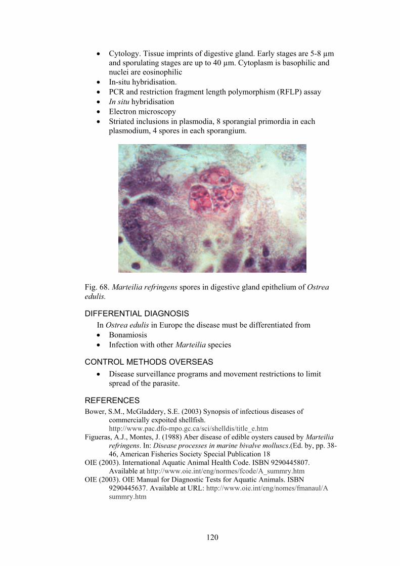
Cytology. Tissue imprints of digestive gland. Early stages are 5-8 µm and sporulating stages are up to 40 µm. Cytoplasm is basophilic and nuclei are eosinophilic
•
• • • • •
In-situ hybridisation. PCR and restriction fragment length polymorphism (RFLP) assay In situ hybridisation Electron microscopy Striated inclusions in plasmodia, 8 sporangial primordia in each plasmodium, 4 spores in each sporangium.
Fig. 68. Marteilia refringens spores in digestive gland epithelium of Ostrea edulis.
DIFFERENTIAL DIAGNOSIS In Ostrea edulis in Europe the disease must be differentiated from
Bonamiosis • •
•
Infection with other Marteilia species
CONTROL METHODS OVERSEAS Disease surveillance programs and movement restrictions to limit spread of the parasite.
REFERENCES Bower, S.M., McGladdery, S.E. (2003) Synopsis of infectious diseases of
commercially expoited shellfish. http://www.pac.dfo-mpo.gc.ca/sci/shelldis/title_e.htm
Figueras, A.J., Montes, J. (1988) Aber disease of edible oysters caused by Marteilia refringens. In: Disease processes in marine bivalve molluscs.(Ed. by, pp. 38-46, American Fisheries Society Special Publication 18
OIE (2003). International Aquatic Animal Health Code. ISBN 9290445807. Available at http://www.oie.int/eng/normes/fcode/A_summry.htm
OIE (2003). OIE Manual for Diagnostic Tests for Aquatic Animals. ISBN 9290445637. Available at URL: http://www.oie.int/eng/nomes/fmanaul/A summry.htm
120
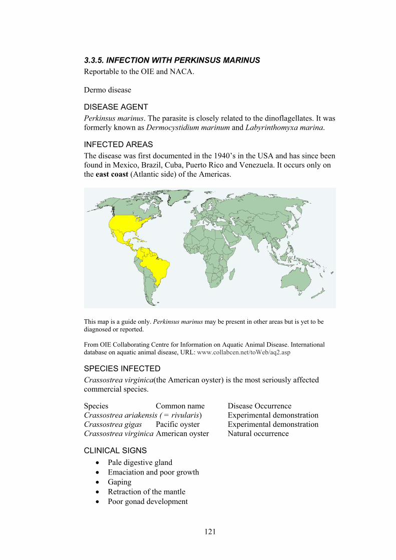
3.3.5. INFECTION WITH PERKINSUS MARINUS Reportable to the OIE and NACA. Dermo disease
DISEASE AGENT Perkinsus marinus. The parasite is closely related to the dinoflagellates. It was formerly known as Dermocystidium marinum and Labyrinthomyxa marina.
INFECTED AREAS The disease was first documented in the 1940’s in the USA and has since been found in Mexico, Brazil, Cuba, Puerto Rico and Venezuela. It occurs only on the east coast (Atlantic side) of the Americas.
This map is a guide only. Perkinsus marinus may be present in other areas but is yet to be diagnosed or reported. From OIE Collaborating Centre for Information on Aquatic Animal Disease. International database on aquatic animal disease, URL: www.collabcen.net/toWeb/aq2.asp
SPECIES INFECTED Crassostrea virginica(the American oyster) is the most seriously affected commercial species. Species Common name Disease Occurrence Crassostrea ariakensis ( = rivularis) Experimental demonstration Crassostrea gigas Pacific oyster Experimental demonstration Crassostrea virginica American oyster Natural occurrence
CLINICAL SIGNS Pale digestive gland •
• • • •
Emaciation and poor growth Gaping Retraction of the mantle Poor gonad development
121
Sometimes abscesses are present. •
•
• •
•
• •
•
MODE OF TRANSMISSION Horizontal transmission.
KEY EPIDEMIOLOGICAL POINTS • Mortality often occurs in the second summer after infection. Most
mortality occurs in summer months in oysters more than one year old. • Perkinsus olseni (occurring in abalone in Australia and clams in
eastern Asia and coastal southern Europe) is distinct from P. marinus. • Mortality can be greater than 95%. • Mortality is most common in summer when water temperature is
greater than 20°C but the infective agent can survive at 4°C. • Prefers salinities of greater than 12-15 ppt but can survive at 3-9 ppt. • Clinical disease only occurs when trophozoites are present in tissues. • Water pollution increases infection by causing stress and suppressing
some host defence mechanisms. • Haemocytes recognise and phagocytose the parasite but are unable to
destroy it and contain the infection.
METHODS OF DIAGNOSIS Histopathology. Typical schizonts and trophozoites (2-10 µm). Hypnospores stain with iodine following incubation in Ray’s thioglycollate broth. DNA probes and PCR primers
DIFFERENTIAL DIAGNOSIS Infection with other species of Perkinsus.
CONTROL METHODS OVERSEAS Hold oysters at <9 ppt salinity. Fallow the 'grow out' areas for 2 years before restocking with seed stocks. Do not move infected oysters to uninfected areas.
122
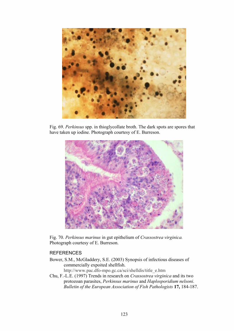
Fig. 69. Perkinsus spp. in thioglycollate broth. The dark spots are spores that have taken up iodine. Photograph courtesy of E. Burreson.
Fig. 70. Perkinsus marinus in gut epithelium of Crassostrea virginica. Photograph courtesy of E. Burreson.
REFERENCES Bower, S.M., McGladdery, S.E. (2003) Synopsis of infectious diseases of
commercially expoited shellfish. http://www.pac.dfo-mpo.gc.ca/sci/shelldis/title_e.htm
Chu, F.-L.E. (1997) Trends in research on Crassostrea virginica and its two protozoan parasites, Perkinsus marinus and Haplosporidium nelsoni. Bulletin of the European Association of Fish Pathologists 17, 184-187.
123

Cochennec-Laureau, N., Reece, K.S., Berthe, F.C.J., Hine, P.M. (2003) Mikrocytos roughleyi taxonomic affiliation leads to the genus Bonamia (Haplosporidia). Diseases of Aquatic Organisms 54, 209-217.
OIE (2003). International Aquatic Animal Health Code. ISBN 9290445807. Available at http://www.oie.int/eng/normes/fcode/A_summry.htm
OIE (2003). OIE Manual for Diagnostic Tests for Aquatic Animals. ISBN 9290445637. Available at URL: http://www.oie.int/eng/nomes/fmanaul/A_summry.htm
Paynter, K.T. (1996) The effects of Perkinsus marinus infection on physiological processes in the eastern oyster, Crassostrea virginica. Journal of Shellfish Research 15, 119-125.
VIMS OIE website (http://www.vims.edu/env/research/shellfish/oie/index.html)
124

3.3.6. INFECTION WITH MIKROCYTOS MACKINI Reportable to the OIE and NACA. Denman Island disease (Canada), Microcell disease of Pacific oyster.
DISEASE AGENT Mikrocytos mackini. A haplosporidian protistan parasite.
INFECTED AREAS The disease occurs on the west coast of the USA and Canada.
This map is a guide only. Mikrocytos mackini may be present in other areas but is yet to be diagnosed or reported. From OIE Collaborating Centre for Information on Aquatic Animal Disease. International database on aquatic animal disease, URL: www.collabcen.net/toWeb/aq2.asp
SPECIES INFECTED Several species of oyster are infected. Natural infections have been reported in Crassostrea gigas, Ostrea conchaphila and Ostrea edulis. Crassostrea virginica has been experimentally infected. C. gigas appears to be the most resistant species. Species Disease Occurrence Crassostrea gigas Natural occurrence and experimental
demonstration Crassostrea virginica Experimental demonstration Ostrea edulis Natural occurrence and experimental
demonstration Ostrea lurida ( = O. conchaphila) Natural occurrence and experimental
demonstration From OIE Collaborating Centre for Information on Aquatic Animal Disease. International database on aquatic animal disease, URL: www.collabcen.net/toWeb/aq2.asp
125
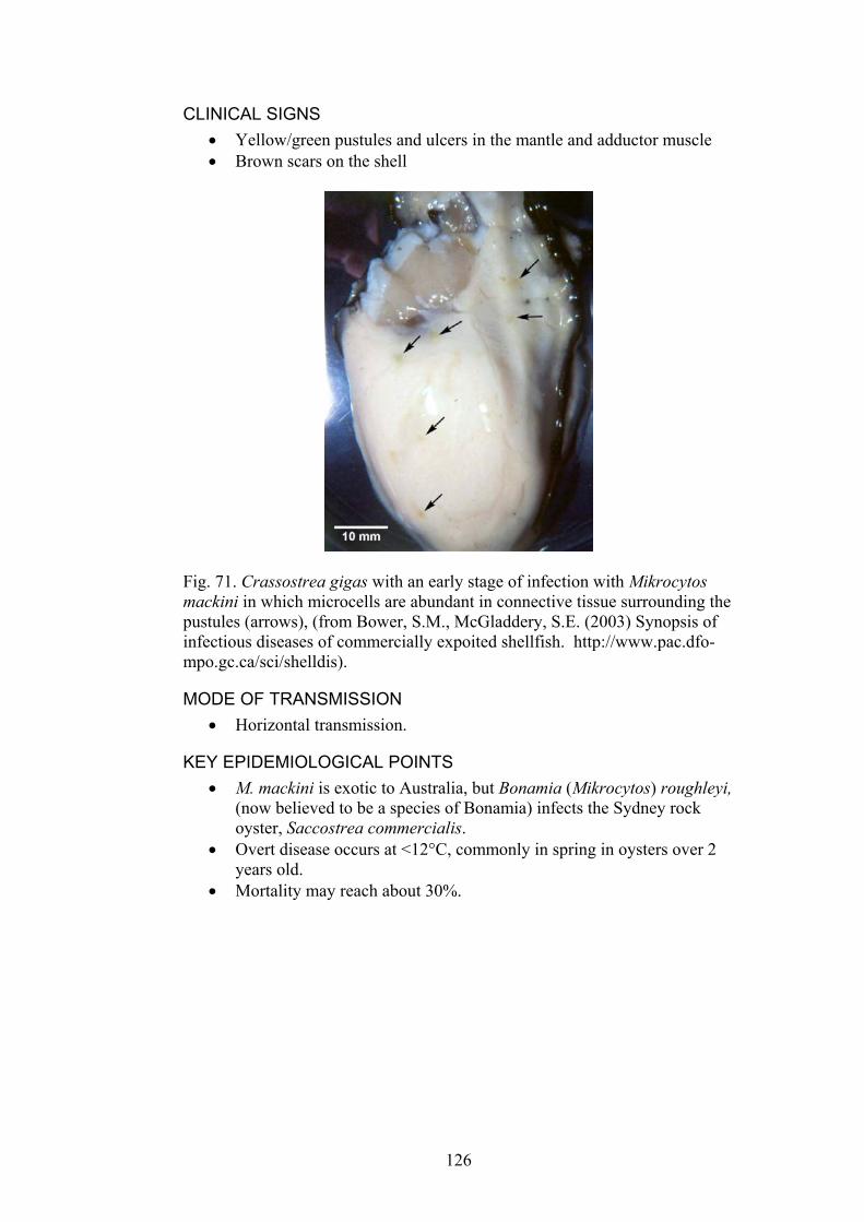
CLINICAL SIGNS Yellow/green pustules and ulcers in the mantle and adductor muscle •
• Brown scars on the shell
Fig. 71. Crassostrea gigas with an early stage of infection with Mikrocytos mackini in which microcells are abundant in connective tissue surrounding the pustules (arrows), (from Bower, S.M., McGladdery, S.E. (2003) Synopsis of infectious diseases of commercially expoited shellfish. http://www.pac.dfo-mpo.gc.ca/sci/shelldis).
MODE OF TRANSMISSION Horizontal transmission. •
KEY EPIDEMIOLOGICAL POINTS • M. mackini is exotic to Australia, but Bonamia (Mikrocytos) roughleyi,
(now believed to be a species of Bonamia) infects the Sydney rock oyster, Saccostrea commercialis.
• Overt disease occurs at <12°C, commonly in spring in oysters over 2 years old.
• Mortality may reach about 30%.
126
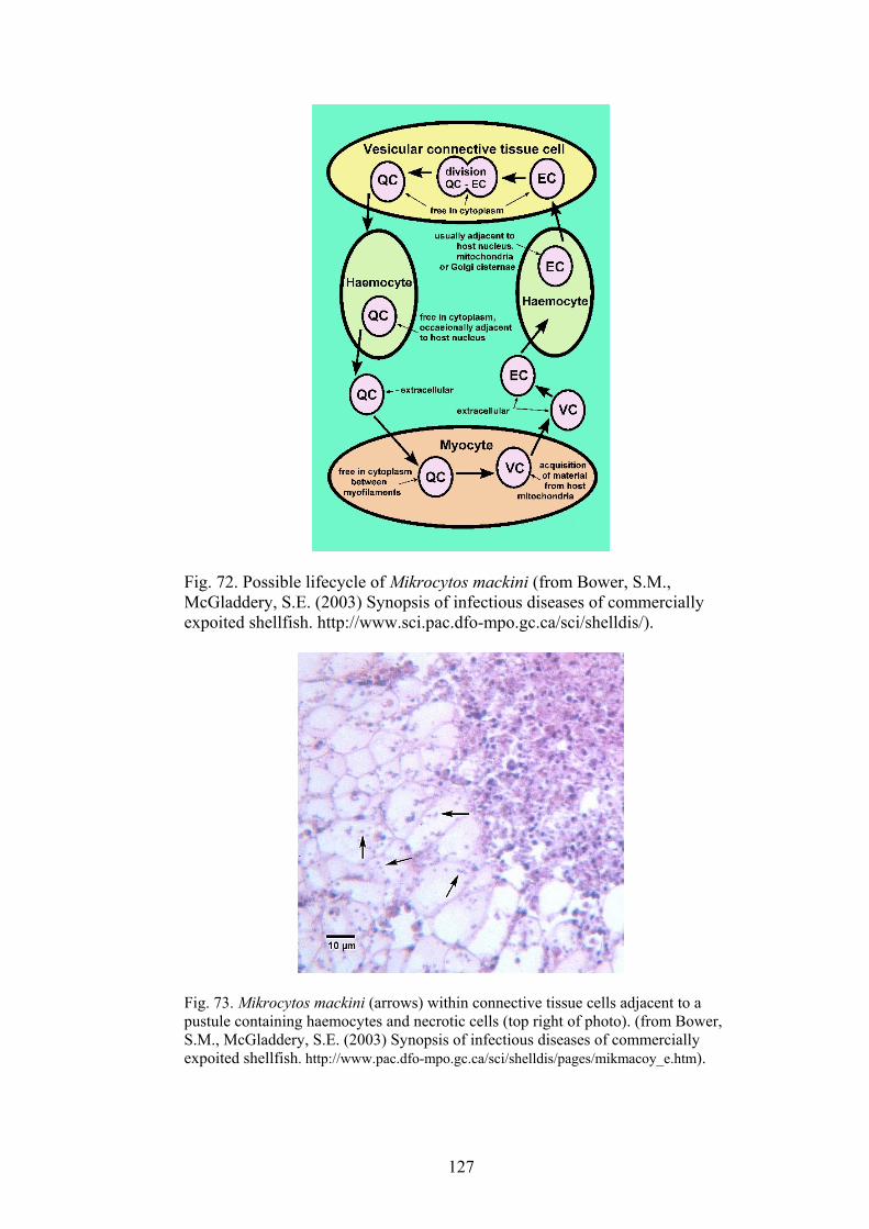
Fig. 72. Possible lifecycle of Mikrocytos mackini (from Bower, S.M., McGladdery, S.E. (2003) Synopsis of infectious diseases of commercially expoited shellfish. http://www.sci.pac.dfo-mpo.gc.ca/sci/shelldis/).
Fig. 73. Mikrocytos mackini (arrows) within connective tissue cells adjacent to a pustule containing haemocytes and necrotic cells (top right of photo). (from Bower, S.M., McGladdery, S.E. (2003) Synopsis of infectious diseases of commercially expoited shellfish. http://www.pac.dfo-mpo.gc.ca/sci/shelldis/pages/mikmacoy_e.htm).
127
METHODS OF DIAGNOSIS Histopathology. Intracellular parasites (1-3 µm diameter) are present in connective tissue cells and sometimes muscle cells and haemocytes at the periphery of abscess-like lesions. Severe focal haemocyte infiltration causes abscess formation.
•
•
•
•
•
•
Tissue imprints. The microcells (M. mackini) can be seen using oil immersion in imprints of pustules that have been fixed and stained. Transmission electron microscopy for morphology to differentiate M. mackini from other microcells.
DIFFERENTIAL DIAGNOSIS Disease in susceptible oysters such as Crassostrea gigas must be differentiated from other micro cell diseases, especially Bonamia spp. including Bonamia (Mikrocytos) roughleyi.
CONTROL METHODS OVERSEAS Prevent translocation of oysters from infected areas to uninfected areas. Harvest large oysters or move them to areas high in the intertidal zone before the end of winter. Do not place oysters in the lower intertidal zone until summer.
REFERENCES Bower, S.M., McGladdery, S.E. (2003) Synopsis of infectious diseases of
commercially expoited shellfish. http://www.sci.pac.dfo-mpo.gc.ca/sci/shelldis/.
Elston, R.A. (1993) Infectious diseases of the Pacific oyster, Crassostrea gigas. Annual Review of Fish Diseases 3, 259-276.
OIE (2003). International Aquatic Animal Health Code. ISBN 9290445807. Available at http://www.oie.int/eng/normes/fcode/A_summry.htm
OIE (2003). OIE Manual for Diagnostic Tests for Aquatic Animals. ISBN 9290445637. Available at URL: http://www.oie.int/eng/nomes/fmanaul/A_summry.htm
128
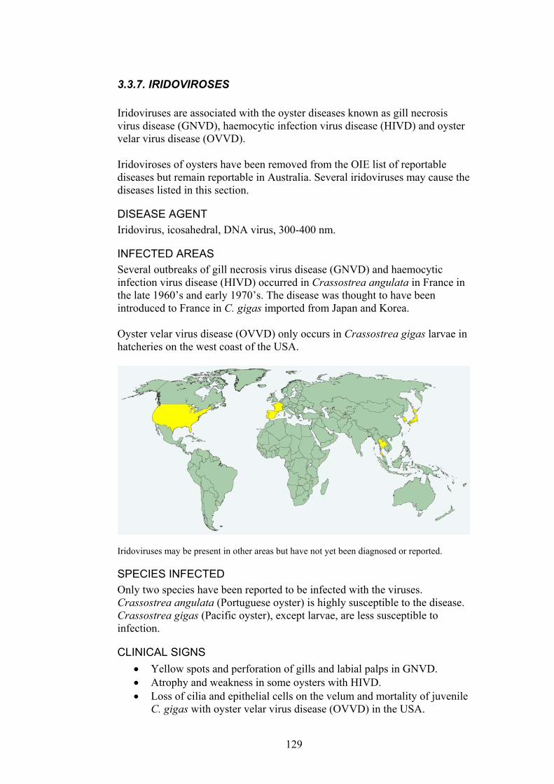
3.3.7. IRIDOVIROSES Iridoviruses are associated with the oyster diseases known as gill necrosis virus disease (GNVD), haemocytic infection virus disease (HIVD) and oyster velar virus disease (OVVD). Iridoviroses of oysters have been removed from the OIE list of reportable diseases but remain reportable in Australia. Several iridoviruses may cause the diseases listed in this section.
DISEASE AGENT Iridovirus, icosahedral, DNA virus, 300-400 nm.
INFECTED AREAS Several outbreaks of gill necrosis virus disease (GNVD) and haemocytic infection virus disease (HIVD) occurred in Crassostrea angulata in France in the late 1960’s and early 1970’s. The disease was thought to have been introduced to France in C. gigas imported from Japan and Korea. Oyster velar virus disease (OVVD) only occurs in Crassostrea gigas larvae in hatcheries on the west coast of the USA.
Iridoviruses may be present in other areas but have not yet been diagnosed or reported.
SPECIES INFECTED Only two species have been reported to be infected with the viruses. Crassostrea angulata (Portuguese oyster) is highly susceptible to the disease. Crassostrea gigas (Pacific oyster), except larvae, are less susceptible to infection.
CLINICAL SIGNS Yellow spots and perforation of gills and labial palps in GNVD. •
• •
Atrophy and weakness in some oysters with HIVD. Loss of cilia and epithelial cells on the velum and mortality of juvenile C. gigas with oyster velar virus disease (OVVD) in the USA.
129
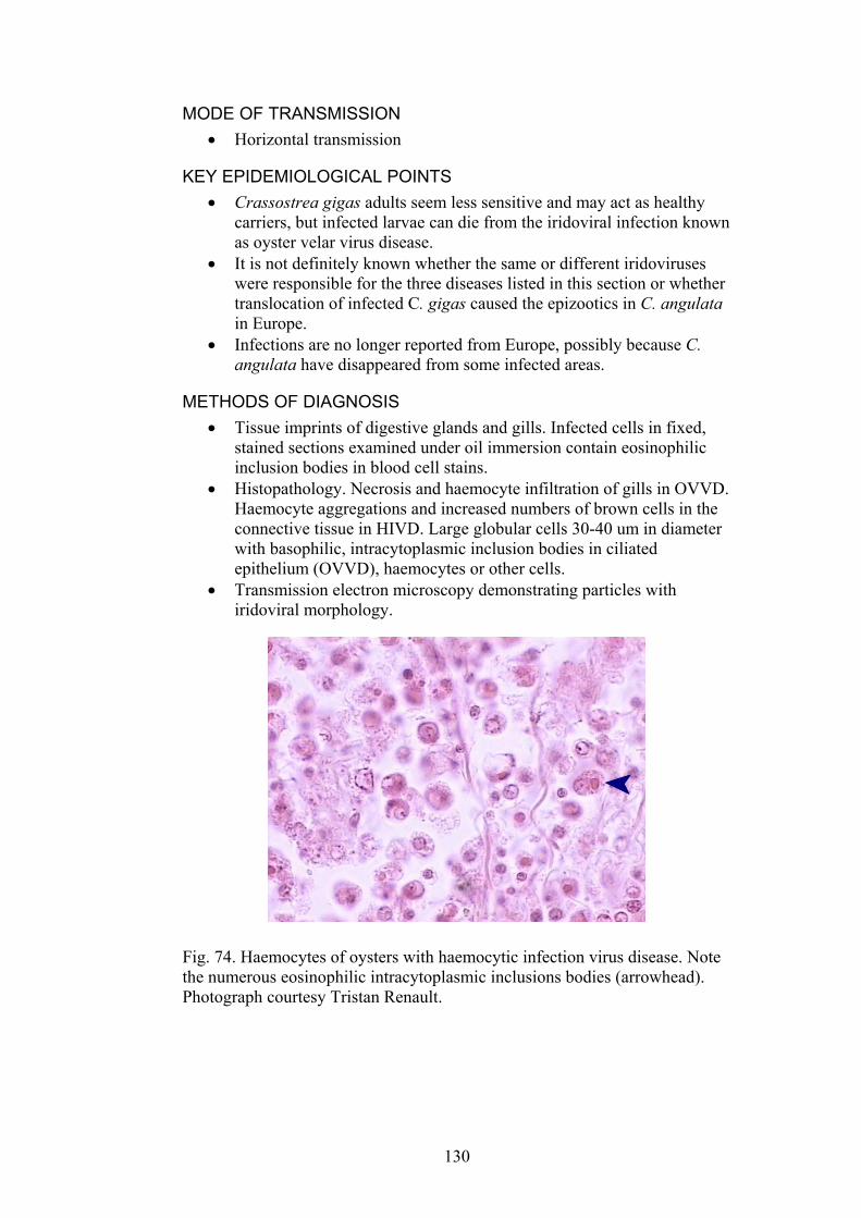
MODE OF TRANSMISSION Horizontal transmission •
•
•
•
KEY EPIDEMIOLOGICAL POINTS • Crassostrea gigas adults seem less sensitive and may act as healthy
carriers, but infected larvae can die from the iridoviral infection known as oyster velar virus disease.
• It is not definitely known whether the same or different iridoviruses were responsible for the three diseases listed in this section or whether translocation of infected C. gigas caused the epizootics in C. angulata in Europe.
• Infections are no longer reported from Europe, possibly because C. angulata have disappeared from some infected areas.
METHODS OF DIAGNOSIS Tissue imprints of digestive glands and gills. Infected cells in fixed, stained sections examined under oil immersion contain eosinophilic inclusion bodies in blood cell stains. Histopathology. Necrosis and haemocyte infiltration of gills in OVVD. Haemocyte aggregations and increased numbers of brown cells in the connective tissue in HIVD. Large globular cells 30-40 um in diameter with basophilic, intracytoplasmic inclusion bodies in ciliated epithelium (OVVD), haemocytes or other cells. Transmission electron microscopy demonstrating particles with iridoviral morphology.
Fig. 74. Haemocytes of oysters with haemocytic infection virus disease. Note the numerous eosinophilic intracytoplasmic inclusions bodies (arrowhead). Photograph courtesy Tristan Renault.
130
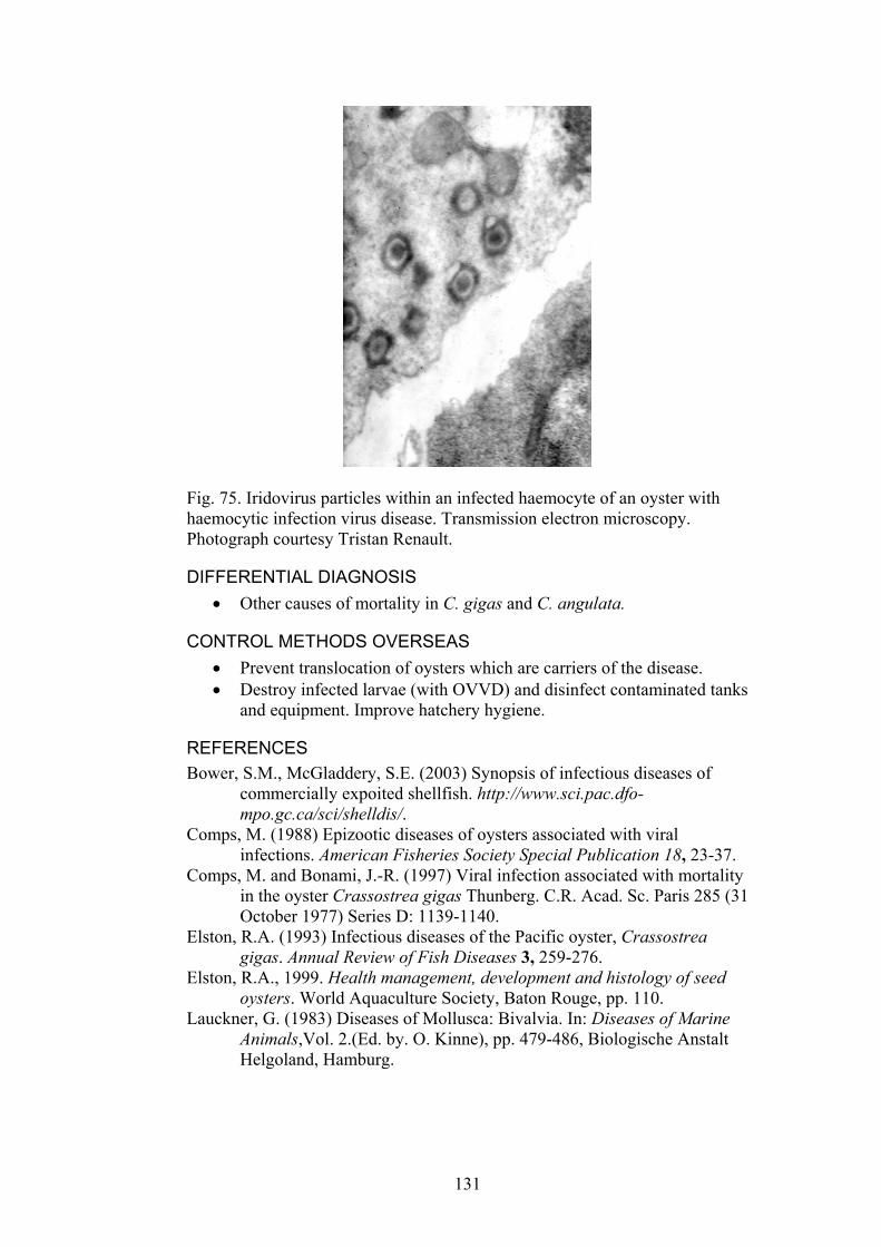
Fig. 75. Iridovirus particles within an infected haemocyte of an oyster with haemocytic infection virus disease. Transmission electron microscopy. Photograph courtesy Tristan Renault.
DIFFERENTIAL DIAGNOSIS Other causes of mortality in C. gigas and C. angulata. •
• •
CONTROL METHODS OVERSEAS Prevent translocation of oysters which are carriers of the disease. Destroy infected larvae (with OVVD) and disinfect contaminated tanks and equipment. Improve hatchery hygiene.
REFERENCES Bower, S.M., McGladdery, S.E. (2003) Synopsis of infectious diseases of
commercially expoited shellfish. http://www.sci.pac.dfo-mpo.gc.ca/sci/shelldis/.
Comps, M. (1988) Epizootic diseases of oysters associated with viral infections. American Fisheries Society Special Publication 18, 23-37.
Comps, M. and Bonami, J.-R. (1997) Viral infection associated with mortality in the oyster Crassostrea gigas Thunberg. C.R. Acad. Sc. Paris 285 (31 October 1977) Series D: 1139-1140.
Elston, R.A. (1993) Infectious diseases of the Pacific oyster, Crassostrea gigas. Annual Review of Fish Diseases 3, 259-276.
Elston, R.A., 1999. Health management, development and histology of seed oysters. World Aquaculture Society, Baton Rouge, pp. 110.
Lauckner, G. (1983) Diseases of Mollusca: Bivalvia. In: Diseases of Marine Animals,Vol. 2.(Ed. by. O. Kinne), pp. 479-486, Biologische Anstalt Helgoland, Hamburg.
131
3.3.8. AKOYA OYSTER DISEASE Reportable to NACA.
DISEASE AGENT Unknown, but possibly a small virus, size <45 nm.
INFECTED AREAS The disease was first observed in Japan in 1994 and has since caused marked economic losses in akoya oyster culture in southwest Japan. The same disease agent might also cause Syndrome 85 in Pinctada margaritifera in French Polynesia.
This map is a guide only. The infectious agent that causes Akoya oyster disease may be present in other areas but is yet to be diagnosed or reported.
SPECIES INFECTED The Akoya pearl oyster, Pinctada fucata martensii. The susceptibility of other molluscs such as Crassostrea gigas (the Pacific oyster) and Chlamys nobilis is unknown.
CLINICAL SIGNS Reddish brown adductor muscle •
• •
•
•
•
Stunted oysters with atrophy of all tissues Gaping or sluggish closure of valves
MODE OF TRANSMISSION Horizontal transmission
KEY EPIDEMIOLOGICAL POINTS Mass mortality followed importation of Chinese pearl oysters into Japanese waters. The hybrid akoya/Chinese oyster is resistant to the disease. Mortality of 50 to 80% in akoya oysters over one year old.
132
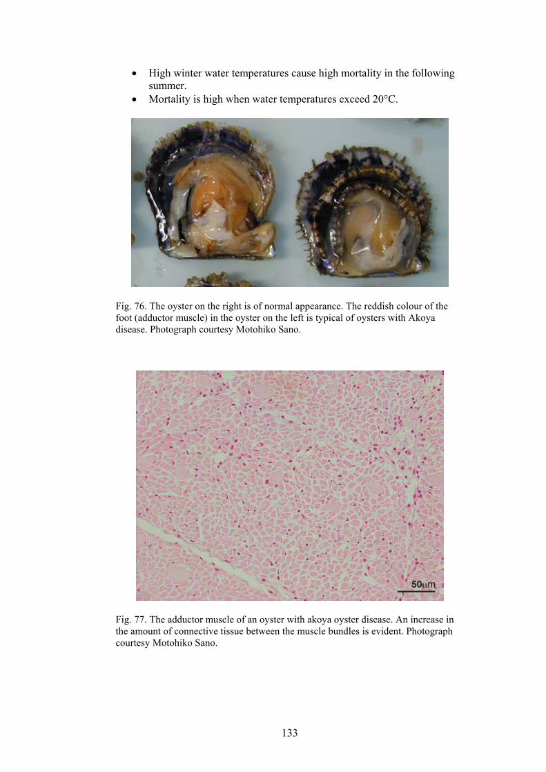
High winter water temperatures cause high mortality in the following summer.
•
• Mortality is high when water temperatures exceed 20°C.
Fig. 76. The oyster on the right is of normal appearance. The reddish colour of the foot (adductor muscle) in the oyster on the left is typical of oysters with Akoya disease. Photograph courtesy Motohiko Sano.
Fig. 77. The adductor muscle of an oyster with akoya oyster disease. An increase in the amount of connective tissue between the muscle bundles is evident. Photograph courtesy Motohiko Sano.
133
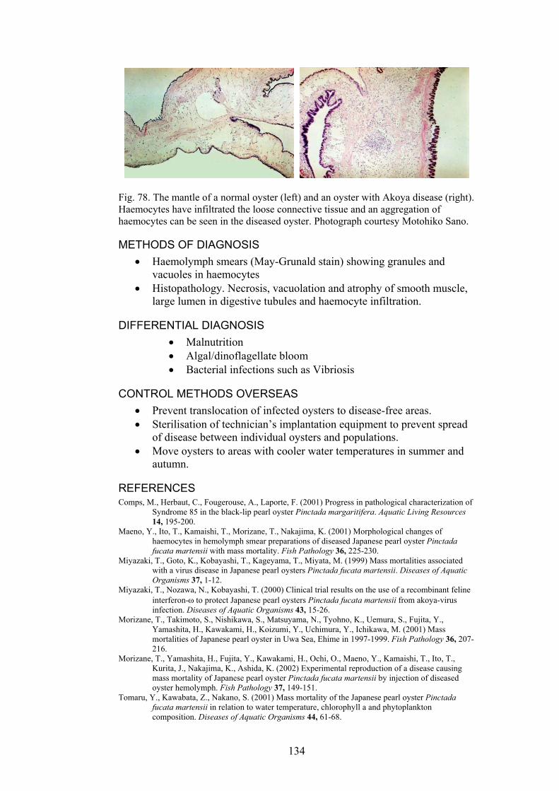
Fig. 78. The mantle of a normal oyster (left) and an oyster with Akoya disease (right). Haemocytes have infiltrated the loose connective tissue and an aggregation of haemocytes can be seen in the diseased oyster. Photograph courtesy Motohiko Sano.
METHODS OF DIAGNOSIS •
•
•
• •
•
Haemolymph smears (May-Grunald stain) showing granules and vacuoles in haemocytes Histopathology. Necrosis, vacuolation and atrophy of smooth muscle, large lumen in digestive tubules and haemocyte infiltration.
DIFFERENTIAL DIAGNOSIS Malnutrition
• Algal/dinoflagellate bloom • Bacterial infections such as Vibriosis
CONTROL METHODS OVERSEAS Prevent translocation of infected oysters to disease-free areas. Sterilisation of technician’s implantation equipment to prevent spread of disease between individual oysters and populations. Move oysters to areas with cooler water temperatures in summer and autumn.
REFERENCES Comps, M., Herbaut, C., Fougerouse, A., Laporte, F. (2001) Progress in pathological characterization of
Syndrome 85 in the black-lip pearl oyster Pinctada margaritifera. Aquatic Living Resources 14, 195-200.
Maeno, Y., Ito, T., Kamaishi, T., Morizane, T., Nakajima, K. (2001) Morphological changes of haemocytes in hemolymph smear preparations of diseased Japanese pearl oyster Pinctada fucata martensii with mass mortality. Fish Pathology 36, 225-230.
Miyazaki, T., Goto, K., Kobayashi, T., Kageyama, T., Miyata, M. (1999) Mass mortalities associated with a virus disease in Japanese pearl oysters Pinctada fucata martensii. Diseases of Aquatic Organisms 37, 1-12.
Miyazaki, T., Nozawa, N., Kobayashi, T. (2000) Clinical trial results on the use of a recombinant feline interferon-ω to protect Japanese pearl oysters Pinctada fucata martensii from akoya-virus infection. Diseases of Aquatic Organisms 43, 15-26.
Morizane, T., Takimoto, S., Nishikawa, S., Matsuyama, N., Tyohno, K., Uemura, S., Fujita, Y., Yamashita, H., Kawakami, H., Koizumi, Y., Uchimura, Y., Ichikawa, M. (2001) Mass mortalities of Japanese pearl oyster in Uwa Sea, Ehime in 1997-1999. Fish Pathology 36, 207-216.
Morizane, T., Yamashita, H., Fujita, Y., Kawakami, H., Ochi, O., Maeno, Y., Kamaishi, T., Ito, T., Kurita, J., Nakajima, K., Ashida, K. (2002) Experimental reproduction of a disease causing mass mortality of Japanese pearl oyster Pinctada fucata martensii by injection of diseased oyster hemolymph. Fish Pathology 37, 149-151.
Tomaru, Y., Kawabata, Z., Nakano, S. (2001) Mass mortality of the Japanese pearl oyster Pinctada fucata martensii in relation to water temperature, chlorophyll a and phytoplankton composition. Diseases of Aquatic Organisms 44, 61-68.
134
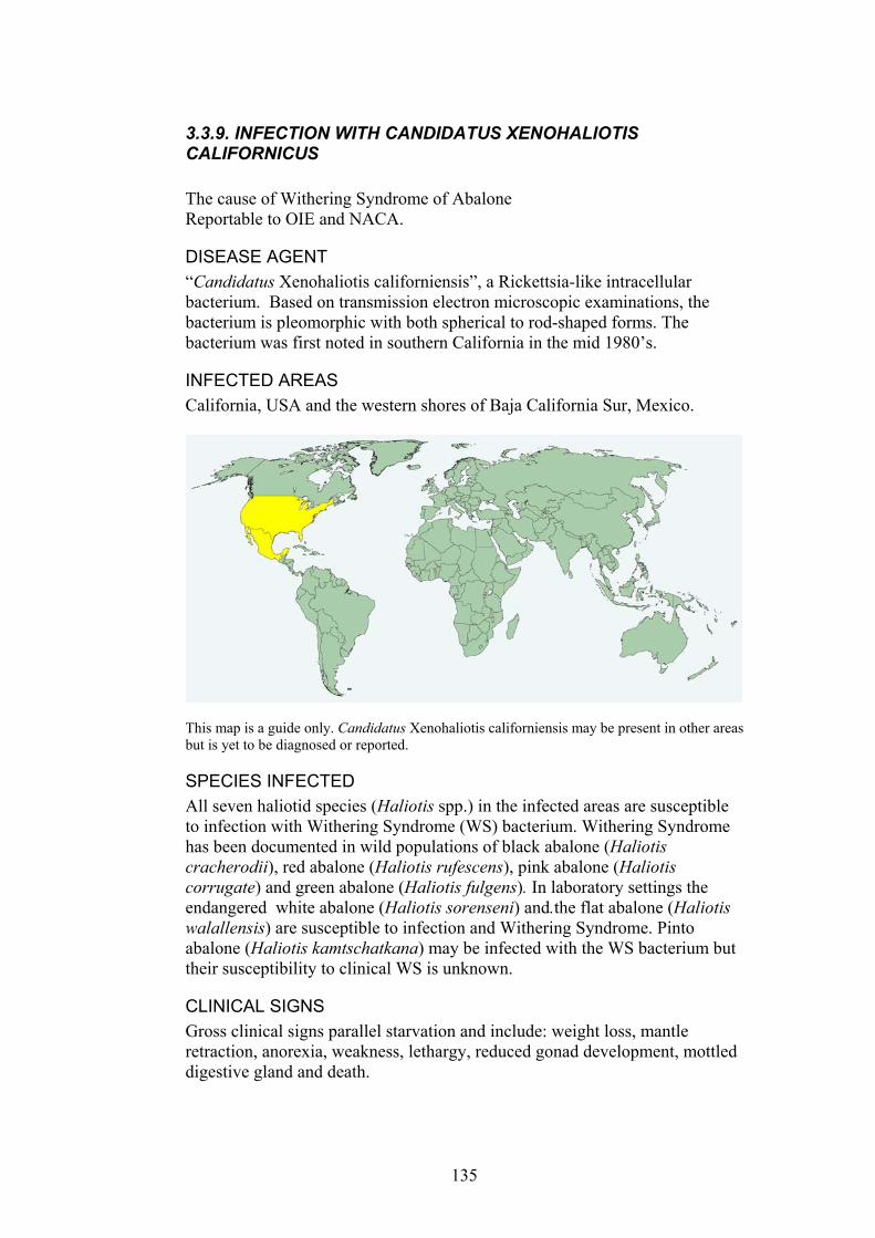
3.3.9. INFECTION WITH CANDIDATUS XENOHALIOTIS CALIFORNICUS The cause of Withering Syndrome of Abalone Reportable to OIE and NACA.
DISEASE AGENT “Candidatus Xenohaliotis californiensis”, a Rickettsia-like intracellular bacterium. Based on transmission electron microscopic examinations, the bacterium is pleomorphic with both spherical to rod-shaped forms. The bacterium was first noted in southern California in the mid 1980’s.
INFECTED AREAS California, USA and the western shores of Baja California Sur, Mexico.
This map is a guide only. Candidatus Xenohaliotis californiensis may be present in other areas but is yet to be diagnosed or reported.
SPECIES INFECTED All seven haliotid species (Haliotis spp.) in the infected areas are susceptible to infection with Withering Syndrome (WS) bacterium. Withering Syndrome has been documented in wild populations of black abalone (Haliotis cracherodii), red abalone (Haliotis rufescens), pink abalone (Haliotis corrugate) and green abalone (Haliotis fulgens). In laboratory settings the endangered white abalone (Haliotis sorenseni) and.the flat abalone (Haliotis walallensis) are susceptible to infection and Withering Syndrome. Pinto abalone (Haliotis kamtschatkana) may be infected with the WS bacterium but their susceptibility to clinical WS is unknown.
CLINICAL SIGNS Gross clinical signs parallel starvation and include: weight loss, mantle retraction, anorexia, weakness, lethargy, reduced gonad development, mottled digestive gland and death.
135
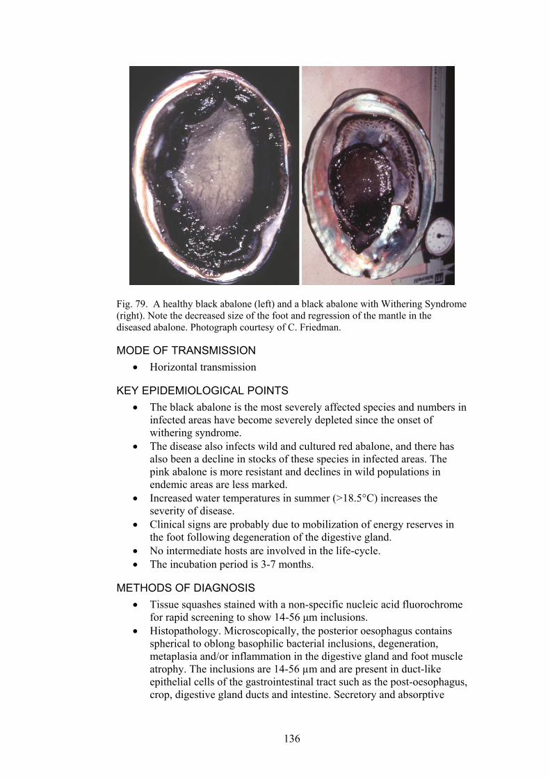
Fig. 79. A healthy black abalone (left) and a black abalone with Withering Syndrome (right). Note the decreased size of the foot and regression of the mantle in the diseased abalone. Photograph courtesy of C. Friedman.
MODE OF TRANSMISSION Horizontal transmission •
•
•
KEY EPIDEMIOLOGICAL POINTS • The black abalone is the most severely affected species and numbers in
infected areas have become severely depleted since the onset of withering syndrome.
• The disease also infects wild and cultured red abalone, and there has also been a decline in stocks of these species in infected areas. The pink abalone is more resistant and declines in wild populations in endemic areas are less marked.
• Increased water temperatures in summer (>18.5°C) increases the severity of disease.
• Clinical signs are probably due to mobilization of energy reserves in the foot following degeneration of the digestive gland.
• No intermediate hosts are involved in the life-cycle. • The incubation period is 3-7 months.
METHODS OF DIAGNOSIS Tissue squashes stained with a non-specific nucleic acid fluorochrome for rapid screening to show 14-56 µm inclusions. Histopathology. Microscopically, the posterior oesophagus contains spherical to oblong basophilic bacterial inclusions, degeneration, metaplasia and/or inflammation in the digestive gland and foot muscle atrophy. The inclusions are 14-56 µm and are present in duct-like epithelial cells of the gastrointestinal tract such as the post-oesophagus, crop, digestive gland ducts and intestine. Secretory and absorptive
136
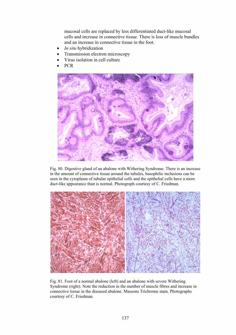
mucosal cells are replaced by less differentiated duct-like mucosal cells and increase in connective tissue. There is loss of muscle bundles and an increase in connective tissue in the foot. In situ hybridization •
• • •
Transmission electron microscopy Virus isolation in cell culture PCR
Fig. 80. Digestive gland of an abalone with Withering Syndrome. There is an increase in the amount of connective tissue around the tubules, basophilic inclusions can be seen in the cytoplasm of tubular epithelial cells and the epithelial cells have a more duct-like appearance than is normal. Photograph courtesy of C. Friedman.
Fig. 81. Foot of a normal abalone (left) and an abalone with severe Withering Syndrome (right). Note the reduction in the number of muscle fibres and increase in connective tissue in the diseased abalone. Massons Trichrome stain. Photographs courtesy of C. Friedman.
137

DIFFERENTIAL DIAGNOSIS Poor food supply •
• •
CONTROL METHODS USED OVERSEAS Reduce stocking density of abalone Medicate feed with oxytetracycline
REFERENCES Andree, K.B., Friedman, C.S., Moore, J.D., Hedrick, R.P. (2000) A
polymerase chain reaction assay for the detection of genomic DNA of a Rickettsiales-like prokaryote associated with Withering Syndrome in California abalone. Journal of Shellfish Research 19, 213-218.
OIE (2003). International Aquatic Animal Health Code. ISBN 9290445807. Available at URL: http://www.oie.int/eng/normes/fcode/A_summry.htm
OIE (2003). OIE Manual for Diagnostic Tests for Aquatic Animals. ISBN 9290445637. Available at URL: http://www.oie.int/eng/nomes/fmanaul/A_summry.htm
Moore, J.D., Cherr, G.N., Friedman, C.S. (2001) Detection of 'Candidatus Xenohaliotis californiensis' (Rickettsiales-like prokaryote) inclusions in tissue squashes of abalone (Haliotis spp.) gastrointestinal epithelium using a nucleic acid fluorochrome. Diseases of Aquatic Organisms 46, 147-152.
Moore, J.D., Robbins, T.T., Friedman, C.S. (2000) Withering syndrome in farmed red abalone Haliotis rufescens: thermal induction and association with a gastrointestinal Rickettsia-like prokaryote. Journal of Aquatic Animal Health 12, 26-34.
Moore, J.D., Robbins, T.T., Hedrick, R.P., Friedman, C.S. (2001) Transmission of the Rickettsiales-like Procaryote "Candidatus Xenohaliotis californiensis" and its role in Withering Syndrome of California abalone, Haliotis spp. Journal of Shellfish Research 20, 867-874.
www.enaca.org
138
APPENDIX ONE. LIST OF AQUATIC ANIMAL HEALTH DIAGNOSTIC LABORATORIES IN AUSTRALIA
New South Wales NSW Fisheries Veterinary Officer (Aquatic Animal Health) Regional Veterinary Laboratory Bruxner Highway Wollongbar NSW 2477 Telephone: (02) 66 261 293 Mob 0428 698 112 Fax: (02) 66 261 276
Northern Territory Department of Business, Industry and Resource Development Berrimah Veterinary Laboratory Berrimah Farm Strath Rd Berrimah NT Telephone: (08) 8999 2249 Fax: (08) 8999 2024
Queensland Telephone 13 25 23 Diagnostic laboratories are situated in Brisbane and Townsville: Oonoonba Veterinary Laboratory PO Box 1085 Townsville QLD 4810 Yeerongpilly Veterinary Laboratory 6645 Fairfield Road Yeerongpilly QLD 4105
South Australia PIRSA Aquaculture Aquatic Animal Health Unit 14th Floor, 25 Grenfell St. Adelaide SA GPO Box 1625 Adelaide SA 5001 Telephone: (08) 8226 0314 Fax: (08) 8226 0330 Idexx Laboratory 33 Flemington Street Glenside SA 5065 Telephone:08 8372 3700 Fax:08 8372 3777
139
Tasmania Fish Health Unit Animal Health Laboratories Mt Pleasant Laboratories Kings Meadows, Tasmania 7249 Telephone; (03) 6336 5389 Fax: (03) 6344 3085 Victoria Aquatic Animal Health Laboratory Animal Health Laboratory CSIRO Geelong Victoria Department of Veterinary Investigations Victorian Institute of Animal Science 475 Mickleham Road Attwood Vic 3049 Telephone (03) 9217 4200 Fax (03) 9217 4399 Western Australia Fish Health Laboratory C/o Animal Health Laboratories Department of Agriculture 3 Baron-Hay Court South Perth WA Telephone: (08) 9368 3351 Fax: (08) 9474 1881
140
APPENDIX TWO. SELF ASSESSMENT TESTS Questions 1-20. Select the most correct answer or answers. 1. Exotic diseases of aquatic animals in Australia are: a) diseases that have very unusual clinical signs b) diseases that are hard to diagnose c) diseases that do not occur in Australia d) all diseases that are included on the Australian list of reportable diseases e) all of the above 2. Diagnosis of reportable diseases in Australia that result in large-scale deaths of
aquatic animals in aquaculture facilities: a) are difficult to diagnose b) are difficult to differentiate from deaths resulting from water quality problems c) indicate that animals have been illegally imported into the aquaculture facility d) are most easily diagnosed if clinically affected, live animals are submitted for
diagnosis e) is always followed by total destocking and disinfection of the facility 3. When a large-scale mortality of aquatic animals occurs in a river or aquaculture
facility: a) a reportable disease will be the cause of death of the animals b) samples of water and animals must be collected and frozen as soon as possible c) an appropriate government department or aquatic animal health specialist should
be consulted d) an infectious disease will be the reason for death of the animals e) the death of animals will be the result of a toxic agricultural or industrial
contaminating the water 4. The aetiological agent of the majority of reportable diseases of crustacea in
Australia is: a) bacterial b) fungal c) a metazoan parasite d) a protistan parasite e) viral 5. Reportable diseases of aquatic animals in Australia are: a) all diseases of economic importance to individual states or the nation b) all endemic diseases of indigenous species c) all exotic diseases that do not occur in Australia d) all included on the OIE list of significant diseases of aquatic animals e) none of the above 6. When a reportable disease is suspected on a farm: a) all animals must be immediately destroyed and the premises disinfected b) the appropriate government department must be informed c) the owner is arrested for having introduced the disease to his farm d) the owner must quickly sell his stock and move them from his farm e) none of the above
141
7. Definitive diagnosis of exotic diseases of aquatic animals in Australia may include:
a) matching clinical disease signs and the affected species of animal with those of a disease that occurs in the same species overseas
b) a complete water analysis to eliminate other causes of death c) bacterial and fungal culture d) submission of tissue samples to a reference laboratory e) all of the above 8. The following properties of an exotic disease or disease agent increase the risk of
the disease becoming established in Australia: a) the disease usually occurs in areas with similar climate and geography to
Australia b) the disease agent survives freezing c) the disease occurs in species that are commonly found in Australia d) infected animals are covert carriers e) all of the above 9. What do VHS, IHN, spring viraemia of carp and ISA have in common: a) ascites is a common clinical sign b) they only affect salmonids c) they can all be diagnosed by histology d) affected fish recover if they are treated with antibiotics e) they can be all be prevented by vaccination 10. The following event may precipitate an outbreak of disease in fish in an aquaculture facility that holds fish in ponds: a) fish have recently been transported to the facility b) the water temperature has increased c) the electricity supply to the aquaculture facility ceased for several hours d) there was recently a severe storm that caused heavy, torrential rain and runoff
into ponds e) all of the above 11. White spot disease virus (WSD) and yellowhead virus (YHV): a) are both bacterial diseases b) both can cause lesions in the lymphoid organ c) both can often have lesions in the hepatopancreas d) both have characteristics inclusion bodies in histological sections e) both are endemic in wild populations of Australian prawns 12. The aetiological agent of the majority of reportable, exotic diseases of molluscs
in Australia is: a) viral b) bacterial c) fungal d) a protistan parasite e) a metazoan parasite 13. The purpose of OIE listing is to: a) list all serious diseases b) list diseases that only occur in aquaculture species c) list diseases of importance in Europe d) identify disease agents that may introduce serious disease to new regions e) all of the above
142

14. Gaping in bivalves, fouling in crustacea and separation of individuals from a school of fish may indicate that the animal is: a) lethargic and anorexic b) in good health c) being held in water that is of optimal quality for the species d) about to spawn e) none of the above 15. Tissue squashes are most useful for diagnosing: a) Gyrodactylus salaris b) Myxobolus cerebralis c) Red sea bream iridovirus d) Bonamia ostrea e) White spot disease 16. The most effective and efficient strategy to prevent the spread of reportable diseases in aquatic animals is: a) to vaccinate young animals b) to identify and destroy all infected animals c) to treat infected animals d) to control the movement of infected animals e) to use culture water that has been treated with ultraviolet light or ozone 17. Asymptomatic carriers of disease: a) can infect their offspring by vertical transmission b) can be difficult to identify by non lethal testing c) have recovered from clinical disease and are no longer a potential source of
infection d) can shed large numbers of pathogens e) a,b and d 18. Crayfish plague: a) can infect all freshwater crayfish b) can only cause mortality in redclaw in Australia c) is a bacterial disease d) can infect both marine lobsters and freshwater crayfish e) is rendered non transmissible by freezing 19. Bonamiosis, Haplosporidiosis, Marteiliosis, Mikrocytosis and Perkinsosis are: a) all serious exotic diseases of molluscs b) serious diseases of molluscs within Australia and overseas c) are readily differentiated from each other by morphology on histology sections d) have pathognomonic clinical signs e) all bacterial diseases 20. Targeted surveys to detect the presence of specific viral disease agents in prawns most often use the following technique for indicating the presence of the agent: a) electron microscopy b) histology c) history of disease outbreaks in an area d) tissue culture e) immunological or molecular tools
143

Answers to Questions 1-20. 1. c 2. d is the most accurate answer. Not all reportable diseases are exotic to Australia. Some are reportable to the OIE or NACA but are endemic in certain areas of Australia and do not always require quarantine, destocking and movement controls. 3. c Sometimes options a, b, d and e are also true 4. e 5. a or e 6. b 7. e 8. e 9. a 10. e 11. b The inclusion body morphology cannot be used to differentiate the two viruses 12. d 13. d is the most correct answer, although some diseases that are not listed by the OIE have serious, economic significance to some regions such as Australia. 14. a 15. b 16. d 17. e 18. a 19. b 20. e
144
Questions 21-40. Select the most correct answer. 21. Reportable diseases. Which of the following statements is correct with respect
to reportable diseases? a) Reportable diseases must be notified within 12 hours of diagnosis. b) Reportable diseases must be notified within 12 hours of diagnosis or suspicion. c) Reportable diseases must be notified within 24 hours of diagnosis. d) Reportable diseases must be notified within 24 hours of diagnosis or suspicion. e) Reportable diseases must be notified within 12 hours of diagnosis or 24 hours of
suspicion. 22. The OIE. Which of the following statements about the OIE is correct? a) OIE stands for the Organisation Intercontinentale d’Epidemiologie. b) OIE stands for the Office International des Epizooties. c) OIE stands for the Office International d’Epidemiologie. d) OIE stands for the Office for Investigative Epidemiology. e) OIE stands for the Oficina Internacionale de Epidemiologia. 23. Actions after notification. Which one of the following statements about actions
after notifying the suspicion of an exotic disease is correct? a) Notifiable diseases are subject to mandatory slaughter after confirmation of
diagnosis. b) After notification, there is mandatory quarantine until the diagnosis is confirmed,
after which there is mandatory slaughter. c) The veterinarian may leave the property as long as s/he does not visit another
animal facility within 12 hours. d) The minimum action after reporting a notifiable disease is mandatory quarantine,
which can be lifted if the diagnosis is not confirmed. e) After reporting a notifiable disease, the veterinarian should remain on the
property until advised otherwise by a government veterinary officer. 24. AUSVETPLAN. From the AUSVETPLAN enterprise manual on veterinary
practice, which of the following best explains the powers of the veterinarian with respect to quarantine?
a) The veterinarian may not impose quarantine. Movement of animals from a property where animals are known to be suffering from an exotic disease is only an offence after quarantine has been imposed officially.
b) The veterinarian may not impose quarantine, but movement of animals from a property where animals are suspected of suffering from an exotic disease is an offence.
c) Quarantine may be imposed by a stock inspector, a veterinarian, or a government veterinary officer.
d) Quarantine may be imposed only under the direct authority of the Chief Veterinary Officer.
e) Upon suspicion of a notifiable disease, a veterinarian may impose quarantine for 24 hours, which automatically expires at the end of the 24 hour period, unless the diagnosis is either confirmed or the Chief Veterinary Officer elects to uphold the quarantine pending further investigations.
25. Types of aquaculture facility. AQUAVETPLAN’s enterprise manual defines
various types of production system, and this affects management procedures in a disease outbreak. Which is the best definition of a semi-closed system?
a) A system where there is no control of either host movement or water flow. b) A system where there is control of host movement but no control of water flow.
145
c) A system where there is control of host movement and some control of water flow
d) A system where there is good control of both host movement and water flow. e) A system where there is poor control of host movement and good control of water
flow. 26. In freshwater fish, oedema, exophthalmos and ascites are common signs of
disease because a) The key primary pathogens have tropisms for cutaneous, ocular and peritoneal
tissues. b) Skin or renal disease hinders osmoregulation, resulting in swelling due to osmotic
water influx. c) Freshwater fish have tissues that are hypotonic with respect to the surrounding
water, making them vulnerable to water influx if osmotic barriers are disrupted. d) Generalised septicaemia results in accumulation of inflammatory exudates which
distend body cavities and other tissues, causing multifocal swellings. e) Diffuse disease in fish often results in systemic hypertension, resulting in fluid
effusions and multi-organ swellings. 27. Which of these is the least useful sample collection in a disease outbreak? a) Sterile swabs and fresh tissue samples for bacteriological examinations. b) Impression smears from blood, liver, kidney and intestinal contents. c) Sterile samples of fresh tissue for mycological examinations. d) Sterile samples of fresh tissue for virological examinations. e) Frozen samples of tissues for histological examinations. 28. Infectious Haematopoietic Virus can be diagnosed by cytopathic effects in cell
culture, followed by confirmation with PCR, a neutralisation test or fluorescent antibody tests. Which of the following is the best sample to collect in a disease outbreak for cultivating virus in cell culture?
a) Aseptically collected, fresh, chilled samples of multiple organs (e.g. spleen, kidney, encephalon) from a moribund fish.
b) Aseptically collected, fresh, chilled samples of multiple organs (e.g. spleen, kidney, encephalon) from ten moribund fish.
c) Aseptically collected, fresh, frozen samples of organs multiple organs (e.g. spleen, kidney, encephalon) from a moribund fish.
d) Aseptically collected, fresh, frozen samples of organs multiple organs (e.g. spleen, kidney, encephalon) from ten moribund fish.
e) Formalin-fixed samples of multiple organs (e.g. spleen, kidney, encephalon) from ten moribund fish.
29. If you were screening an apparently healthy population for the presence of an
exotic disease, which of the following statements is most accurate, if you wish to have a statistically significant statement relating to freedom from disease?
a) A negative finding after sampling ten individuals is generally a reliable technique, irrespective of population size.
b) The number of individuals sampled depends upon the size of the population being assessed, and is independent of disease prevalence.
c) The number of individuals sampled depends upon the disease prevalence, and is independent of the size of the population being assessed.
d) The number of individuals sampled depends upon both the size of the population being assessed and the disease prevalence.
e) In a population of a 1000 fish, sampling 20 fish would enable one to detect, with 95% confidence, disease with a prevalence of 2%
146
30. The prophenoloxidase cascade is part of the immune defence mechanism of crayfish, prawns and lobsters. Which is the most correct statement? It is:
a) a specific immune response that results in the production of antibodies b) a cell mediated response causing abscess formation in infected tissues c) an enzyme system found in cells in the hepatopancreas d) an enzyme system found in haematopoietic tissue e) causes brown pigment to form at sites of injury or infection 31. The presence of many infectious agents can be detected in tissues using molecular
techniques. Which is the best method of storing samples for molecular testing? a) 10% neutral buffered formalin b) 10% neutral buffered saline c) ethanol d) Lugol’s iodine e) freezing at -20°C 32. Many marine aquaculture facilities are in isolated locations making it difficult to
submit live animals for diagnosis. Which of the following is the least useful method of sample submission?
a) dead, chilled animals b) small pieces of tissue 10% neutral buffered formalin c) small pieces of tissue in 10% formalin diluted with sea water d) sterile, chilled swabs in transport medium e) small pieces of tissue in ethanol 33. Rapid circling in fish may indicate: a) Whirling disease b) betanodavirus infection c) high ammonia content of water d) all of the above e) none of the above 34. Which of the following mammalian cells are most similar in function to the
haemocytes of invertebrates: a) leucocytes b) erythrocytes c) lymphocytes d) platelets e) hepatocytes 35. Which of the following is the most correct? Carriers of disease: a) are weak and lethargic b) have poor feed conversion rates c) do not show clinical signs of disease d) act as reservoirs of infection e) carry the disease agent on their skin, shell or exoskeleton 36. ‘Fouling’ of the gills, mantle or exoskeleton of invertebrate aquatic animals is
least likely when: a) animals have a viral infection b) water quality is suboptimal c) nutrition is poor d) animals have a bacterial infection e) animals are about to spawn
147
37. Exotic viral disease in Australian aquaculture facilities are most likely to be
managed by the following method: a) treatment with antibiotics b) treatment with formalin c) destruction and burial of all animals d) frequent collection and burial of dead animals e) a 100% water exchange 38. Liming of ponds may be done after an outbreak of an exotic disease because: a) it increases pH and acts as a sediment disinfectant b) it acts as a source of calcium for the next batch of animals c) it ensures adequate alkalinity and hardness for the next batch of animals d) all sources of lime are suitable for this use e) limestone is a suitable source of lime for this purpose 39. Which is the most correct answer? Free available chlorine is often used to
disinfect aquaculture water and equipment because: a) it is not toxic to mammals b) it is effective against most infectious agents c) it can be neutralised by sodium thiosulphate d) it is cheap and easy to obtain e) it is not affected by the presence of organic matter 40. Large aquatic animals are sometimes more likely to die from some diseases and
water quality problems because: a) they are more likely to be old and immunocompromised b) they are more susceptible to high ammonia and nitrite levels in the water c) they are more susceptible to low dissolved oxygen d) they have fewer granulocytes and haemocytes e) they are more likely to be anaemic
148

Correct answer: 21. d 22. b OIE also known as World Organisation for Animal Health 23. e 24. b 25. c as in race culture 26. b freshwater fish are hypertonic and damage to skin or impaired renal function prevents maintenance of plasma and extracellular osmolality 27. e Freezing often causes ice crystal damage to tissues, making histology less useful. Fix tissues in formalin for histology. 28. b Generally, multiple sick animals should be sampled in a disease outbreak. Fresh tissues are required for virus isolation. Samples can be combined to form pools of a maximum of five fish each. The amount of material should be approximately 1.5 g/pool of material from five fish. Entire moribund fish can be sent, as an alternative, refrigerated or on ice, but not frozen. 29. d Population size and disease prevalence are key factors in determining sampling size for screening purposes in clinically normal populations. For example, 140 individuals would need to be sampled from a population of 1000, to be 95% certain of detecting disease at 2% prevalence. Find out more on this and related sampling issues from the introductory section of the OIE Manual of Diagnostic Tests for Aquatic Animals at URL: http://www.oie.int/eng/normes/fmanual/A_00017.htm 30. e 31. c 32. a Dead animals are rapidly autolysed and pathogenic bacteria may be rapidly overgrown by non-pathogenic bacteria making diagnosis difficult 33. d 34. a 35. d 36. e 37. c 38. a There are many sources of lime that are used in ponds but only a few result in a significant increase in pH 39. b is the most correct answer. c and d are also often true but are not the main reason for the choice of chlorine as a disinfectant. 40. c Large fish have a smaller gill surface area to body mass ratio which makes them more susceptible to anoxia.
149

Questions 41-60. Select the most correct answer. 41. Egg disinfection is important to control diseases when a) pathogen is transmitted inside the egg b) virus is the causative agent c) external surface of the egg may be contaminated with the pathogen d) all the time for all species e) bacteria are the disease agents 42. Specific pathogen free (SPF) stock is very useful a) in areas where the pathogen is endemic b) any time c) in areas where the pathogen is exotic d) only for viral diseases e) only for bacterial diseases 43. Histopathology was traditionally used for diagnosis of viral diseases of crustaceans, because a) there are no cell lines for crustaceans b) it is faster than CPE c) it is easier than other diagnostic methods d) crustacean viruses would not cause CPE e) crustacean viruses were discovered by histopathologists 44. Bacterial Kidney Disease affects a) only kidney b) it is a systemic disease c) kidney and spleen d) kidney and gills e) all lymphoid organs 45. The only virus affecting multiple species of crustaceans and causing an economically important disease in penaeid prawns is a) YHV b) IHHN c) TSV d) WSSV e) BMNV 46. Temperature can be a risk factor in many infectious diseases when a) it increases b) it decreases c) never d) both increase or decrease of temperature can increase the risk e) only for bacterial and protozoan diseases 47. Most disease signs are a) nonspecific b) specific c) specific but only for pathogen group d) specific but only for viral pathogens e) specific but only for bacterial pathogens
150

48. Vaccines are available for a) most aquatic animal diseases b) only viral diseases of crustaceans c) mostly bacterial diseases of fish d) only viral diseases of fish e) only parasitic diseases of fish 49. Presence of granuloma in fish kidney means that a) the fish has Bacterial Kidney Disease b) chronic inflammatory response is present in kidney c) the fish suffers from nephrocalcinosis d) there are too many granulocytes in kidney e) the fish has parasites in kidney 50. Rickettsia and chlamydia are a) groups of bacteria affecting only fish b) intracellular bacteria pathogenic to different groups of animals c) causative agent of Bacterial Kidney Disease d) only involved in infection of broodstock e) groups of viruses causing viral diseases of crustaceans 51. Freezing a) kills only some pathogens b) kills all pathogens c) kills all viral pathogens d) kills all bacterial pathogens e) kills all viral pathogens of penaeid prawns 52. Occlusion bodies are a) formed by all viruses b) present in nuclei of cells infected by ISAV c) formed only by viruses pathogenic to penaeid prawns d) formed by some baculoviruses e) made only of viral particles 53. Yersinia ruckeri is a) present in Australia b) exotic to Australia c) present only in Victoria d) present only in native fish in Australia e) present only in tropical fish 54. Myxobolus cerebralis damages a) cartilage b) nervous tissue c) epithelium d) blood cells e) bones
151
55. Rotenone treatment is used in Norwegian rivers to control a) ISA b) furunculosis c) IPN d) IHN e) Gyrodactylus salaris 56. VHSV is present a) in freshwater and marine fish b) only in freshwater salmonids c) only in freshwater fish d) only in marine fish e) only in tropical fish 57. Feeding imported frozen seafood to aquaculture animals can a) result in immunosuppression b) cause spread of exotic pathogens c) control spread of diseases as freezing kills all pathogens d) cause inflammation due to low temperature of the food entering digestive tract e) control spread of all exotic viral diseases 58. If a fish does not show any sign of disease it a) does not carry pathogens b) is safe to translocate c) is free of viral pathogens d) can be used as broodstock e) could carry pathogens 59. Increase in the melanomacrophage numbers a) is a sign of disease b) is a sign of VHS c) can occur during or after infection but also during ageing or exposure to pollutants d) is always related to the presence of parasites e) occurs only in viral diseases 60. Most diseases are a) vertically transmitted b) transmitted both vertically and horizontally c) horizontally transmitted d) transmitted through feed e) transmitted vertically if the pathogen is a virus and horizontally if it is a bacteria Correct answers: 41. c 42. c 43. a 44. b 45. d 46. d 47. a 48. c 49. b 50. b
51. a 52. d 53. a 54. a 55. e 56. a 57. b 58. e 59. c 60. c
152
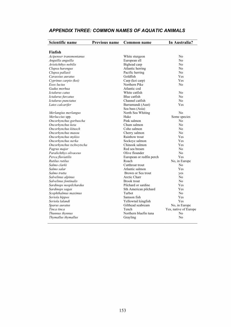
APPENDIX THREE: COMMON NAMES OF AQUATIC ANIMALS Scientific name Previous name Common name In Australia? Finfish Acipenser transmontanus White sturgeon No Anguilla anguilla European ell No Aristichthys nobilis Bighead carp No Clupea harengus Atlantic herring No Clupea pallasii Pacific herring No Carassius auratus Goldfish Yes Cyprinus carpio (koi) Carp (koi carp) Yes Esox lucius Northern Pike No Gadus morhua Atlantic cod Ictalurus catus White catfish No Ictalurus furcatus Blue catfish No Ictalurus punctatus Channel catfish No Lates calcarifer Barramundi (Aust)
Sea bass (Asia) Yes
Merlangius merlangus North Sea Whiting No Merluccius spp. Hake Some species Oncorhynchus gorbuscha Pink salmon No Oncorhynchus keta Chum salmon No Oncorhynchus kitusch Coho salmon No Oncorhynchus masou Cherry salmon No Oncorhynchus mykiss Rainbow trout Yes Oncorhynchus nerka Sockeye salmon Yes Oncorhynchus tschwytscha Chinook salmon Yes Pagrus major Red sea bream No Paralichthys olivaceus Olive flounder No Perca fluviatilis European or redfin perch Yes Rutilus rutilus Roach No, in Europe Salmo clarki Cutthroat trout No Salmo salar Atlantic salmon Yes Salmo trutta Brown or Sea trout yes Salvelinus alpinus Arctic Charr No Salvelinus fontinalis Brook trout No Sardinops neopilchardus Pilchard or sardine Yes Sardinops sagax Sth American pilchard Yes Scophthalmus maximus Turbot No Seriola hippos Samson fish Yes Seriola lalandi Yellowtail kingfish Yes Sparus auratus Gilthead seabream No, in Europe Tinca tinca Tench Yes, native of EuropeThunnus thynnus Northern bluefin tuna No Thymallus thymallus Grayling No
153
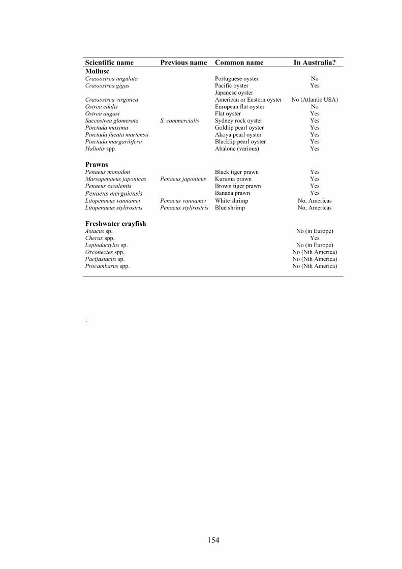
Scientific name Previous name Common name In Australia? Mollusc Crassostrea angulata Portuguese oyster No Crassostrea gigas Pacific oyster
Japanese oyster Yes
Crassostrea virginica American or Eastern oyster No (Atlantic USA) Ostrea edulis European flat oyster No Ostrea angasi Flat oyster Yes Saccostrea glomerata S. commercialis Sydney rock oyster Yes Pinctada maxima Goldlip pearl oyster Yes Pinctada fucata martensii Akoya pearl oyster Yes Pinctada margaritifera Blacklip pearl oyster Yes Haliotis spp. Abalone (various) Yes Prawns Penaeus monodon Black tiger prawn Yes Marsupenaeus japonicas Penaeus japonicus Kuruma prawn Yes Penaeus esculentis Brown tiger prawn Yes Penaeus merguiensis Banana prawn Yes Litopenaeus vannamei Penaeus vannamei White shrimp No, Americas Litopenaeus stylirostris Penaeus stylirostris Blue shrimp No, Americas Freshwater crayfish Astacus sp. No (in Europe) Cherax spp. Yes Leptodactylus sp. No (in Europe) Orconectes spp. No (Nth America) Pacifastacus sp. No (Nth America) Procambarus spp. No (Nth America) .
154