aquaporins in brain edema and neuropathological conditions
TRANSCRIPT

Aquaporins in Brain Edema andNeuropathological Conditions
The Harvard community has made thisarticle openly available. Please share howthis access benefits you. Your story matters
Citation Filippidis, Aristotelis S., Richard B. Carozza, and Harold L.Rekate. 2016. “Aquaporins in Brain Edema and NeuropathologicalConditions.” International Journal of Molecular Sciences 18 (1): 55.doi:10.3390/ijms18010055. http://dx.doi.org/10.3390/ijms18010055.
Published Version doi:10.3390/ijms18010055
Citable link http://nrs.harvard.edu/urn-3:HUL.InstRepos:31731803
Terms of Use This article was downloaded from Harvard University’s DASHrepository, and is made available under the terms and conditionsapplicable to Other Posted Material, as set forth at http://nrs.harvard.edu/urn-3:HUL.InstRepos:dash.current.terms-of-use#LAA

International Journal of
Molecular Sciences
Review
Aquaporins in Brain Edema andNeuropathological Conditions
Aristotelis S. Filippidis 1,2,*, Richard B. Carozza 3 and Harold L. Rekate 4
1 Division of Neurosurgery, Beth Israel Deaconess Medical School, Harvard Medical School, Boston,MA 02115, USA
2 Department of Neurosurgery, Boston Medical Center, Boston University, Boston, MA 02215, USA3 Boston University School of Medicine, Boston, MA 02118, USA; [email protected] Department of Neurosurgery, The Chiari Institute, Hofstra Northwell School of Medicine, Hempstead,
NY 11549, USA; [email protected]* Correspondence: [email protected]; Tel.: +1-617-632-9795
Academic Editor: Kenichi IshibashiReceived: 19 October 2016; Accepted: 20 December 2016; Published: 28 December 2016
Abstract: The aquaporin (AQP) family of water channels are a group of small, membrane-spanningproteins that are vital for the rapid transport of water across the plasma membrane. These proteinsare widely expressed, from tissues such as the renal epithelium and erythrocytes to the various cellsof the central nervous system. This review will elucidate the basic structure and distribution ofaquaporins and discuss the role of aquaporins in various neuropathologies. AQP1 and AQP4, the twoprimary aquaporin molecules of the central nervous system, regulate brain water and CSF movementand contribute to cytotoxic and vasogenic edema, where they control the size of the intracellularand extracellular fluid volumes, respectively. AQP4 expression is vital to the cellular migration andangiogenesis at the heart of tumor growth; AQP4 is central to dysfunctions in glutamate metabolism,synaptogenesis, and memory consolidation; and AQP1 and AQP4 adaptations have been seen inobstructive and non-obstructive hydrocephalus and may be therapeutic targets.
Keywords: aquaporin 1 (AQP1); AQP4; aquaporins; neuroscience
1. Introduction
Since the discovery of the aquaporin family of proteins, integral membrane pores facilitatingdiffusion of water molecules, researchers have finally been able to uncover the true nature ofphysiological osmotic balance. We now have a clearer understanding of the structure, function,and distribution of aquaporins, and begin to discover their contributions to many disease states.Aquaporin molecules are basically involved in water movement in tissues as well as cellularmigration and angiogenesis in tumor formation [1], development and resolution of cytotoxic andvasogenic edema [2,3], synaptogenesis and memory formation [4], cerebrovascular disease [5,6],neuroimmunology [7,8], and support of neurostructures of sensory organs [9]. The ubiquity ofaquaporin channels in the nervous system might provide many targets for novel therapies for diseasesfrom brain ischemia to traumatic brain injury to Alzheimer’s disease. The contributions of aquaporinsto various pathophysiologies is predicated on their ability to mediate the entry and exit of waterfrom the central nervous system. The permeability of the central nervous system is dependent on thedistribution, number, and permeability of channels; any potential pharmacological interventions willlikely focus on the permeability of select populations.
Int. J. Mol. Sci. 2017, 18, 55; doi:10.3390/ijms18010055 www.mdpi.com/journal/ijms

Int. J. Mol. Sci. 2017, 18, 55 2 of 13
2. Structure and Function of Aquaporins
All mammalian plasma membranes are permeable to water, but with great variability. Those thatare only marginally permeable to water are believed to only experience diffusion of water moleculesacross the lipid bilayer. Those tissues that are more permeable—such as the renal epithelium anderythrocytes—are granted this property by aquaporins, homotetrameric proteins present in essentiallyall organisms, with 13 discrete forms in mammals. Aquaporins are members of a family of integralmembrane proteins, which form pores that allow for the passage of water through membranes, whileblocking passage of ions and charged solutes. A subset, termed aquaglyceroporins, can transportsmall, uncharged molecules as well, such as glycerol and ammonia. While movement of water acrossthe plasma membrane occurs by both passive diffusion and channel-mediated transport, the velocityof flow is much faster through aquaporins [10]. Thus, aquaporins regulate the temporal profile ofwater movement allowing for more water molecules to be transferred per unit of time, compared todiffusion alone, and obey the rules of osmosis.
Aquaporins were discovered coincidentally as a contaminant while attempting to isolate Rhpolypeptides. This contaminant, initially named CHIP28, which stands for channel-forming integralprotein of 28 kDa, was found to be a tetrameric integral membrane protein, expressed in highconcentrations in erythrocytes and cells of the renal proximal tubule. The presence of a water channelin erythrocytes was suggested by Benga et al. in 1986 [11]. Experiments in Xenopus oocytes revealedthat cells expressing CHIP28 swelled in response to a hypotonic buffer, lysed when placed in distilledwater, but saw no detectable movement of any solute. These results strongly suggested CHIP28functioned as a water channel [12]. The discovery and subsequent research on aquaporins earned Agreand colleagues the Nobel Prize in Chemistry in 2003.
Aquaporins contain six transmembrane alpha helices, with two highly conserved loops, each witha characteristic motif, asparagine-proline-alanine (NPA). These NPA loops—cytoplasmic portion Band extracellular portion E—are oriented at 180 degrees with respect to one another [13]. Experimentssuggest that the B and E loops form an “hourglass” structure, with the two chambers connecting toform the aqueous pore [14]. The shape of the aquaporin channel core allows only the passage of onewater molecule per time, acting as a filter.
Many initial discoveries about the function of aquaporins were made in the kidney, where theyare vital for water reabsorption. Immunohistochemical studies have localized AQP1 in the apicaland basolateral membranes of the proximal tubule and descending thin limbs of Henle [15] and thedescending vasa recta [16]. The AQP1 was identified at the plasma membrane, but not any intracellularlocations, nor was it located in the collecting duct. Nielsen and colleagues suggested that water istransported across the renal epithelium through apical and basolateral plasma membranes, driven bythe standing osmotic gradients established by movement of solutes through these membranes [17].AQP1 has also been identified in other secretory tissues, including cholangiocytes, non-pigmentedepithelium in the anterior compartment of the eye, and the choroid plexus [18].
Aquaporin Distribution in the Central Nervous System (CNS)
There are 8 aquaporins expressed in the CNS: AQP1, AQP3, AQP4, AQP5, AQP7, AQP8, AQP9,AQP11, with AQP1 and especially AQP4 expressed in the highest concentrations [19–22]. AQP1 andAQP4 are structurally and functionally similar, except that unlike AQP1, AQP4 is not sensitive tomercurials [23]. This property is derived from the absence of a cysteine preceding the NPA motif ofloop E [24].
AQP1 is expressed at the apical and basolateral surfaces of the choroid plexus and functions inthe production of cerebrospinal fluid (CSF). Its greater presence on the apical membrane underliesits role in transcellular movement of water for the production of CSF [18,25]. AQP3 was found inthe piriform cortex, hippocampus, dorsal thalamus, globus pallidus (GP), and choroid plexus andalso at the border region of ischemic stroke in rats [26]. AQP4 is pervasive throughout the brain andretina, most prominently in astroglia at brain-liquid interfaces. End feet membranes adjacent to the

Int. J. Mol. Sci. 2017, 18, 55 3 of 13
ventricles, capillaries, and subarachnoid space contain 10–15 times the number of AQP4 proteinscompared to non-end feet membranes, with microvilli expressing no AQP4 at all [19]. AQP4 is alsofound in the hippocampal dentate gyrus and Cornu Ammonis (CA) areas CA-1 and CA-3, nucleus ofstria terminalis, medial habenular nucleus, neocortex, cerebellum, and supraoptic and suprachiasmaticnucleus of the hypothalamus [27]. AQP5 was localized on the cytoplasmic membrane and in thecytoplasm of astrocytes and was found to be expressed when the brain is exposed to metabolicstress such as ischemic or traumatic injuries [28,29]. AQP8 was also found in the pyriform cortex,hippocampus, dorsal thalamus and globus pallidus and it’s expression was correlated with the grade ofastrocytomas [30]. AQP9 belongs to the aquaglyceroporin family, and is permeable to water as well asneutral solutes such as glycerol, lactate, and urea. AQP9 is expressed in astrocytes, cerebellar neurons,pial vessel endothelium, glia limitans, hypothalamic tanycytes, and CA2 of the hippocampus [20,27].The physiological relevance of AQP7 and AQP11 has not yet been elucidated, although recent literatureshows that AQP11 is localized at the epithelium of the choroid plexus and at the endothelium ofthe brain capillary, suggesting potential involvement in water transport at the choroid plexus andblood-brain barrier (BBB) in the brain. Brains of AQP11-deficient mice, however, did not show anymorphological abnormalities and the function of the BBB was intact. AQP11 was expressed mainlyin the pia matter with limited expression in the capillary at early postnatal stages. It is possible thatAQP11 is needed by the developing brain, to support the water flow [31].
3. Role of Aquaporins in Cerebral Edema
Cerebral edema is associated with many neurological disorders, including ischemic injury,traumatic brain injury (TBI), and brain tumor, ultimately leading to increased intracranial pressure andits associated comorbidities, such as ischemia, brain herniation, and death. Under normal conditions,water moves bidirectionally across the BBB, obeying the rules of osmosis, into the CNS and awayvia the CSF and venous circulation. Three types of brain edema have been described per Klatzoand modified by Fishman [32]: cytotoxic (cellular), vasogenic and interstitial (or hydrocephalic).In cytotoxic brain edema, perturbation of cellular metabolism promotes excess fluid movementacross an intact BBB and contributes to an enlarged intracellular space; in vasogenic brain edema,a dysfunctional BBB allows for macromolecules and water to accumulate in an enlarged extracellularspace [3]. Interstitial edema occurs in obstructive hydrocephalus, resulting in increased movement ofbrain water and Na+ across the periventricular walls [32]. Aquaporins line the periventricular wall.Therefore, edema can be viewed as a dysfunction of aquaporins to properly prevent or facilitate watermovement. Different types of AQP4 expression can be found in Table 1.
Table 1. Expression of AQP4 in different pathologies.
Process Species AQPs Studied Results
Cytotoxic edema [33] Mice AQP4 UpregulatedVasogenic edema [34] Mice AQP4 Upregulated
Traumatic brain injury [35] Rats AQP4 UpregulatedEdematous brain tumor [36] Human AQP4 Upregulated
Hydrocephalus [37,38] Human AQP1 DownregulatedRat AQP4 Upregulated
Cellular migration [39,40] Mice AQP1 UpregulatedMice AQP4 Upregulated
Prolonged status epilepticus [4] Mice AQP4 Downregulated
Experiments in mice have shown improved survival and neurological outcomes in response tocytotoxic edema following pharmacological inhibition of AQP4 [2]. Reduced intracranial pressurewas also observed in AQP4-null mice in cytotoxic edema mimicking meningitis [34]. AQP4 inhibitionperturbs its ability to mediate water influx into the astrocytic end-feet and across the BBB [33],disrupting the cascade of events and leads to increased cerebral fluid volume, elevated intracranial
Int. J. Mol. Sci. 2017, 18, 55 4 of 13
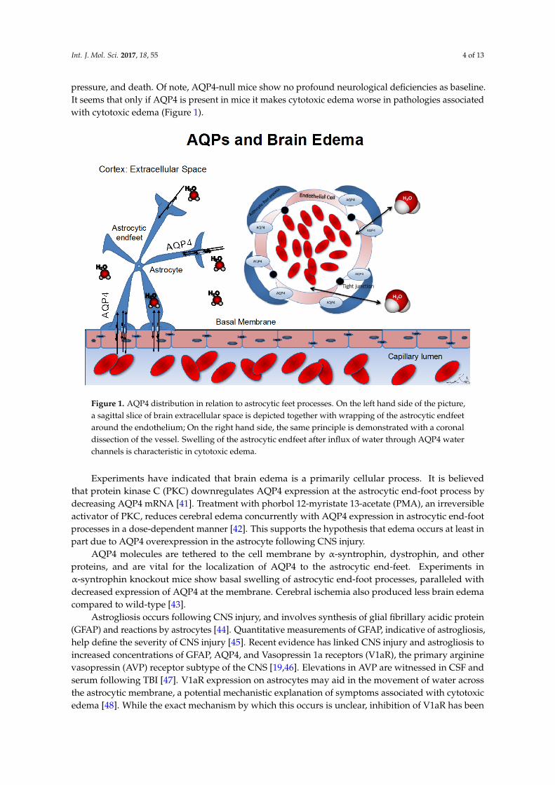
pressure, and death. Of note, AQP4-null mice show no profound neurological deficiencies as baseline.It seems that only if AQP4 is present in mice it makes cytotoxic edema worse in pathologies associatedwith cytotoxic edema (Figure 1).
Int. J. Mol. Sci. 2017, 18, 55 4 of 13
baseline. It seems that only if AQP4 is present in mice it makes cytotoxic edema worse in pathologies associated with cytotoxic edema (Figure 1).
Figure 1. AQP4 distribution in relation to astrocytic feet processes. On the left hand side of the picture, a sagittal slice of brain extracellular space is depicted together with wrapping of the astrocytic endfeet around the endothelium; On the right hand side, the same principle is demonstrated with a coronal dissection of the vessel. Swelling of the astrocytic endfeet after influx of water through AQP4 water channels is characteristic in cytotoxic edema.
Experiments have indicated that brain edema is a primarily cellular process. It is believed that protein kinase C (PKC) downregulates AQP4 expression at the astrocytic end-foot process by decreasing AQP4 mRNA [41]. Treatment with phorbol 12-myristate 13-acetate (PMA), an irreversible activator of PKC, reduces cerebral edema concurrently with AQP4 expression in astrocytic end-foot processes in a dose-dependent manner [42]. This supports the hypothesis that edema occurs at least in part due to AQP4 overexpression in the astrocyte following CNS injury.
AQP4 molecules are tethered to the cell membrane by α-syntrophin, dystrophin, and other proteins, and are vital for the localization of AQP4 to the astrocytic end-feet. Experiments in α-syntrophin knockout mice show basal swelling of astrocytic end-foot processes, paralleled with decreased expression of AQP4 at the membrane. Cerebral ischemia also produced less brain edema compared to wild-type [43].
Astrogliosis occurs following CNS injury, and involves synthesis of glial fibrillary acidic protein (GFAP) and reactions by astrocytes [44]. Quantitative measurements of GFAP, indicative of astrogliosis, help define the severity of CNS injury [45]. Recent evidence has linked CNS injury and astrogliosis to increased concentrations of GFAP, AQP4, and Vasopressin 1a receptors (V1aR), the primary arginine vasopressin (AVP) receptor subtype of the CNS [19,46]. Elevations in AVP are witnessed in CSF and serum following TBI [47]. V1aR expression on astrocytes may aid in the movement of water across the astrocytic membrane, a potential mechanistic explanation of symptoms associated with cytotoxic edema [48]. While the exact mechanism by which this occurs is unclear, inhibition of V1aR has been shown to both reduce brain edema [48] and modulate astrocyte swelling and the expression of GFAP and AQP4 [49–51], strongly suggesting that V1aR and AQP4 expression mediate cytotoxic edema.
Figure 1. AQP4 distribution in relation to astrocytic feet processes. On the left hand side of the picture,a sagittal slice of brain extracellular space is depicted together with wrapping of the astrocytic endfeetaround the endothelium; On the right hand side, the same principle is demonstrated with a coronaldissection of the vessel. Swelling of the astrocytic endfeet after influx of water through AQP4 waterchannels is characteristic in cytotoxic edema.
Experiments have indicated that brain edema is a primarily cellular process. It is believedthat protein kinase C (PKC) downregulates AQP4 expression at the astrocytic end-foot process bydecreasing AQP4 mRNA [41]. Treatment with phorbol 12-myristate 13-acetate (PMA), an irreversibleactivator of PKC, reduces cerebral edema concurrently with AQP4 expression in astrocytic end-footprocesses in a dose-dependent manner [42]. This supports the hypothesis that edema occurs at least inpart due to AQP4 overexpression in the astrocyte following CNS injury.
AQP4 molecules are tethered to the cell membrane by α-syntrophin, dystrophin, and otherproteins, and are vital for the localization of AQP4 to the astrocytic end-feet. Experiments inα-syntrophin knockout mice show basal swelling of astrocytic end-foot processes, paralleled withdecreased expression of AQP4 at the membrane. Cerebral ischemia also produced less brain edemacompared to wild-type [43].
Astrogliosis occurs following CNS injury, and involves synthesis of glial fibrillary acidic protein(GFAP) and reactions by astrocytes [44]. Quantitative measurements of GFAP, indicative of astrogliosis,help define the severity of CNS injury [45]. Recent evidence has linked CNS injury and astrogliosis toincreased concentrations of GFAP, AQP4, and Vasopressin 1a receptors (V1aR), the primary argininevasopressin (AVP) receptor subtype of the CNS [19,46]. Elevations in AVP are witnessed in CSF andserum following TBI [47]. V1aR expression on astrocytes may aid in the movement of water acrossthe astrocytic membrane, a potential mechanistic explanation of symptoms associated with cytotoxicedema [48]. While the exact mechanism by which this occurs is unclear, inhibition of V1aR has been

Int. J. Mol. Sci. 2017, 18, 55 5 of 13
shown to both reduce brain edema [48] and modulate astrocyte swelling and the expression of GFAPand AQP4 [49–51], strongly suggesting that V1aR and AQP4 expression mediate cytotoxic edema.
Despite these discoveries, hyperosmotic therapy or surgical decompression remain the twoprimary treatment options for brain edema in traumatic brain injury, with few pharmacological optionsavailable [52,53]. However, inhibition of AVP action in the brain following cytotoxic edema maypresent itself as an effective clinical tool for decreasing brain edema [50].
Yet, while AQP4-null mice respond better to cytotoxic edema, where the pathology is predicatedon water movement through AQP4, they are more susceptible to complications following vasogenicedema. Astrocytic end-feet are pivotal components of the BBB, and regulation of water movementacross the BBB is thus heavily dependent on AQP4. In vasogenic edema, where water accumulatesin the brain due to leaky vessels, evidence supports AQP4 expression contributes to the enlargedextracellular fluid space [3]. This suggests an additional role for AQP4, as extracellular fluid wasbelieved to move out of the CNS by bulk flow independently of intracellular transport mechanisms.
3.1. Hypoxia-Induced Changes in Aquaporin Expression
During early ischemia, anaerobic glycolysis and leakage of lactic acid from necrotic tissue causesacidosis, where protons are exchanged for extracellular Na+ by Na+/H+. Cl−/HCO3
− antiportersplayed also significant role [54]. Increased glutamate release from necrotic tissues is also transportedvia the Na+ gradient [55]. To counteract the influx of osmoles, glial cells concurrently intake water tocompensate for the increased intracellular osmolarity, a process primarily mediated by AQP4.
The exact role of AQP4 in cases of ischemic injury is unclear. In hypoxia-induced brain edema,cytotoxic edema occurs first, followed by vasogenic edema. AQP4 expression has been shown to bothdecrease in the 48 h following hypoxia [56,57] and increase, although on a slower time scale [58,59].Taniguchi and colleagues described a biphasic expression pattern of AQP4, peaking at hours 1 and48h following ischemic injury. These two time points may correspond to focal ischemia and vasogenicedema, respectively. These results are consistent with others suggesting that the hypoxia induciblefactor (HIF)-binding motif is found within the AQP4 promoter, and that HIF-1α upregulates AQP4expression following hypoxic injury [58].
3.2. Traumatic Brain Injury
Brain edema is a common complication of TBI, with vasogenic edema occurring rapidly afterthe injury, primarily in the center of the lesion, while cytotoxic edema has a later onset with a morepervasive effect [35,60]. In traumatic brain injury, in humans, Marmarou et al. demonstrated withMRI studies, that cytotoxic (cellular) brain edema is predominant [61]. Based on this information wecould possibly explain why most of the studies in TBI demonstrate an increase in AQP4 expressionpost-injury, and also why studies or treatments that result in AQP4 reduction/inhibition in thesesettings could possibly reduce brain edema. Expression levels of AQP4 mRNA have been shown tobe elevated in the injured area compared to other brain areas, and the degree of expression correlatesto the severity of cerebral edema as measured by MRI. However, tissues immediately adjacent to theaffected areas showed downregulation of AQP4, potentially as a protective mechanism [62,63].
3.3. Edematous Brain Tumors
Both benign and malignant brain tumors produce brain edema which could be due to defectivetight junctions between endothelial cells of the BBB, and increased angiogenesis within the tumoritself [64]. While production of brain edema in tumors is independent of AQP4, expression isupregulated in edematous brain tumors and does not exclusively localize to astrocytic end-feetin these tumors [36]. AQP4-null mice with edematous brain tumors showed increased intracranialpressure and more neurological complications compared to wild-type, indicating AQP4 may havea protective effect in this case [36]. However, this contrasts with studies showing that AQP1 and AQP4have been shown to promote cancer metastasis through their role in endothelial cell migration and

Int. J. Mol. Sci. 2017, 18, 55 6 of 13
angiogenesis [39]. Reports also imply that upregulation of the AQ4 M1 orthogonal array structuremight be responsible for the loss of the normal orthogonal array structure, a structure necessary forAQP4 to maintain it’s function [65]. AQP contribution in gliomas is multiparametric as described byDubois et al. and Maugeri et al. [66,67]. We mentioned previously that AQP8 in human astrocytomasis associated with the pathological grade, with higher expression in higher grades. AQP9, is highlyexpressed in tumor stem cells, with resistance to treatment [66,67].
4. Hydrocephalus
Hydrocephalus was described by the Rekate model as the disease state that evolves afterobstruction of CSF circulation. The points of obstruction could be in different sites and areas inthe macro-world (e.g., aqueduct of Sylvius) or micro-world (e.g., channels) [68]. Given the role ofaquaporins in both production (via AQP1) and absorption of CSF (via AQP4), they are a potentialtherapeutic target. AQP1-null mice had osmotic water permeability reduced by a factor of fivecompared to wild-type, reduced CSF production by 20%–25%, and intracranial pressure by 56%.While much of this effect was due to decreased central venous pressure due to effects of AQP1deficiency in the kidney, the results suggest reducing AQP1 function decreases both production of CSFand development of nonobstructive hydrocephalus [69,70].
Smith and colleagues describe the case of a 15-month-old girl with choroid plexus hyperplasia,leading to CSF overproduction and nonobstructive hydrocephalus. Immunohistochemical analysisshowed decreased AQP1 expression in samples taken in the patient compared to controls, whichis indicative of adaptive downregulation [37]. However, further studies have indicated a moreheterogenous expression of AQP1 in cases of hydrocephalus, but still supporting an adaptivemechanism that decreases AQP1 expression [71,72].
AQP4 appears to have a protective role in cases of hydrocephalus, given its role in CSF absorptionin cerebral vasculature. AQP4-null mice showed significant ventriculomegaly after kaolin injection toreproduce obstructive hydrocephalus, increasing intracranial pressure by 2%–3%. After 5 days, themortality of AQP4-deficient mice was 34% compared to 16% of wild-type [73]. After kaolin-inducedhydrocephalus in wild-type mice, AQP4 expression was increased 3–4 weeks post-injection, withhighest levels in the perivascular areas, parietal cerebrum and hippocampus, ependymal lining, andglia limitans [74]. Similar results were seen in expression patterns at the blood-CSF barrier and BBB [38].
5. Role of Aquaporins in Cellular Migration
In response to trauma, reactive glial cells produce scar tissue; scar formation facilitates repair ofthe BBB, minimizes neuronal death, and prevents migration of inflammatory cells to the site of injury.However, scar formation limits the brain’s capacity for axonal regeneration. This process is believed tobe mediated at least in part by AQP4. Astrocyte migration and formation of scar tissue were delayedin AQP4-null mice, and blockade of AQP4 may be a novel method of promoting synaptogenesisand axonal sprouting following CNS injury [40,75]. Saadoun and colleagues propose that AQP4mediates an influx of water to dilute the accumulation of ions and depolymerized actin at the leadingedge of astrocytes. The membrane of the leading edge is stretched by the increased intracellularhydrostatic pressure in the direction of the leading edge. The increased space provided permits actinrepolymerization, thereby promoting cellular migration.
Proliferating microvessels of malignant tumors have been shown to highly express AQP1, aswell as some tumor cells. Blockade of AQP1 expression has been shown to decrease angiogenesisand induce tumor necrosis [1,39,76]. AQP1 expression is vital for cellular migration in endothelialcells: AQP1-null endothelial cells show slowed, impaired migration towards chemoattractantscompared to wild-type [77]. Both AQP1 and nitric oxide (NO) levels are elevated during brainedema. NO production by nitric oxide synthase (NOS) confers a protective function, promotingvasodilation of ischemic areas. Experiments blocking endothelial NOS (eNOS) also caused a paralleldecrease in AQP1 expression, indicating that the two are linked by a common signaling pathway [78].
Int. J. Mol. Sci. 2017, 18, 55 7 of 13
AQP1 and AQP4 are both potential targets to modulate migration: when expressed in CHOor FRT epithelial cells, both aquaporins lead to enhanced cellular migration compared to AQP-null.Video microscopy studies have shown that AQP1 facilitates the growth of more lamellipodia anda decreased mean residence time, indicating AQP1 assists in the rapid turnover of lamellipodia at theleading edge [77].
6. Epilepsy, Memory Consolidation, and Aquaporins
Epilepsy is a medical condition characterized by unpredictable onset of seizures. Approximately30% of patients who take antiepileptic drugs do not see a complete reversal of symptoms, and currentantiepileptic drugs are accompanied by a host of developmental, behavioral, and cognitive side effects,since they target neurons directly [79]. While neuronal excitability is pivotal to the hyperexcitabilityof epilepsy, evidence supporting astrocytic involvement is growing and could be an avenue forpotential therapeutics.
Glial cells are of paramount importance to electrophysiological function of neurons, providingenergy and nutrition, recycling neurotransmitters, regulating ionic homeostasis, and modulatingsynaptogenesis, and are believed to be integrally involved in many cases of epilepsy [43,80].Hypoosmolarity and decreased extracellular fluid volume in the brain induces hyperexcitability andseizures, and the extracellular environment is modulated primarily by astrocytes [81]. AQP4-null micehave a decreased extracellular space (ECS) compared to wild-type; this corresponds to less seizureactivity after administration of pentylenetetrazol (PTZ), a GABAA antagonist [82]. They do, however,exhibit a longer seizure duration than that of wild-type [83].
Both AQP4 and Kir4.1, an inwardly rectifying potassium channel, are colocalized to theperisynaptic region of the astrocytic endfoot [84]. Increased neuronal activity increases extracellular[K+] substantially, most of which is taken up by neighboring astrocytes, mediated in part by Kir4.1;inactivation of Kir4.1 hampers regulation of extracellular K+, and thus disrupts potassium siphoning,a significant mechanism of extracellular potassium clearance [84]. Levels of K+ concentration, [K+],after baseline and moderate activity were not drastically affected by AQP4 deficiency, with heavystimulation-induced elevations showing slower time kinetics compared to wild-type. However, K+
kinetics after stimulation are delayed in AQP4-null mice, both in rise and decay, indicating a roleof AQP4 in regulating K+ homeostasis and neuronal activity [83]. These results are consistent withprevious studies showing a longer duration of seizure in AQP4-null mice, as prolonged depolarizationwould block seizure termination.
AQP4 is tethered to the membrane by α-syntrophin. Deficiencies in α-syntrophin disrupt AQP4expression at the perivascular and subpial membranes [43], and dystrophin deficiencies produce lowerlevels of expression at astrocytic end-foot processes, the polarized expression of AQP4 is impaired.despite normal AQP4 protein levels [85]. Research in mice has shown α-syntrophin-deficient miceexhibit greater seizure intensity in response to hyperthermia, coupled to a decrease in extracellularK+ clearance, due to decreased potassium siphoning [43]. While it is clear that AQP4, Kir4.1, andassociated anchoring proteins form a multifunctional unit involved in K+ homeostasis, the exactmechanism behind their activity remains unclear [86].
Glutamate Metabolism and Synaptic Plasticity
Low extracellular glutamate levels must be maintained in the ECS to ensure proper neuronalexcitability, mediated by glutamate transporters, most prominently glutamate transporter-1 (GLT1) [87].GLT1 deficiencies produce spontaneously epileptic activity due to overabundance of glutamate, whileoverexpression of GLT1 reduces seizure frequency [88,89].
Prolonged status epilepticus (SE) induces a downregulation of AQP4 in mouse hippocampalastrocytes at day 1 post SE with a prolonged period of recovery, and concurrent upregulation of GLT1at day 1 with significant decrease in expression levels at days 4 and 7 post SE. These results werepresent even in the absence of cell death or sclerosis, indicating that profound seizures regulate AQP4,

Int. J. Mol. Sci. 2017, 18, 55 8 of 13
GLT1 expression [90]. Downregulation of AQP4 may lead to both hyperactivity and dysfunctionalsynaptic plasticity and cognition, both of which are significant comorbidities of epilepsy.
The hypothesized role of AQP4 is consistent with results showing dysfunctional synaptic plasticity.AQP4-null mice exhibited deficiencies in long term potentiation (LTP) and long term depression (LTD)of the hippocampus, without significantly affecting synaptic transmutation or short-term plasticity [91].These findings were supported by others showing that AQP4-null mice exhibited impaired memoryconsolidation as measured by the Morris water maze (MWM) [92]. Similar experiments in theamygdala of AQP4-null mice showed impaired LTP and decreased associative fear memory, aswell as downregulated GLT1 expression, further suggesting that AQP4 has a pivotal role in memoryconsolidation [93].
The role of GLT1 as the mediator of AQP4-dependent memory deficiencies was supported byexperiments showing that LTP and memory formation were rescued in AQP4-null mice when treatedwith ceftriaxone, a stimulator of GLT1 [94]. Accumulations of glutamate, resultant of reduced GLT1expression in AQP4-deficient mice, causes strong activation of NMDA receptor (NMDAR) [95], andincreased NMDAR-mediated currents were seen in mice lacking AQP4. Pharmacological inhibition ofNMDAR rescued LTP dysfunction in AQP4-deficient mice, suggesting excess glutamate in the synapticcleft perturbs NMDAR-mediated currents and LTP in AQP4-deficient mice [93].
7. Aquaporins and the Glymphatic System
Aside from recent discoveries of lymphatic vessels in the dural sinuses [96], the brain is believedto lack the traditional lymphatic structure of the rest of the body and removes interstitial fluid (ISF)differently. Instead, the brain utilizes the glymphatic system, a microscopic fluid clearance systemformed by astrocytic perivascular tunnels that remove fluid, proteins, and waste molecules, anddistributes compounds such as nutrients, growth factors, and neuromodulators. In this system, the ISFand CSF constantly exchange, with movement of CSF into the brain parenchyma facilitated by AQP4in the astrocytic end-feet [97].
Iliff and colleagues used fluorescent tracers to show CSF enters the brain parenchyma via periarterialpathway surrounding the smooth muscle cells adjacent to astrocytic endfeet, and exited the brain viacentral deep veins and the lateral-ventral caudal rhinal veins. In many neurodegenerative diseases, thereis an accumulation of proteins, contributing to disease pathology. It was discovered that amyloid-β wascleared along the glymphatic pathway, and AQP4-null mice showed a reduction in CSF flux of 65%, anda reduction in amyloid-β clearance of 55%, suggesting a role of AQP4 in neurodegenerative diseasessuch as Alzheimer’s [97,98]. Antoine Louveau et al. and Aspelund et al. [96], while searching for T-cellgateways in the brain, pinpointed the presence of functional lymphatic vessels lining the dural sinusand draining to the deep cervical lymph node complex. This pathway, which was not brought tolight until recently, might be responsible for an alternative approach to current neuroimmunology anddogma that govern the immune response of the brain to disease insults. This dural lymphatic vascularsystem is capable of draining brain interstitial fluid and macromolecules as both Louveau et al. andAspelund et al. report [96].
8. Looking Forward
Aquaporins definitively represent a subset of channels that significantly contribute to thephysiology of cells. Their ability to facilitate water movement both in and out of the CNS contributesgreatly to brain water homeostasis. To be able to utilize their contributions therapeutically, we need toidentify non-toxic molecules to act as AQP inhibitors or amplifiers based on the expected benefit ineach case. A discovery, by error, shows a vast potential to alter the “roux” of history for the future ofthe theraupeutic modulation of brain edema. Our mission is to more deeply understand the physiologyof aquaporins in neuroscience and identify their modulators.
Int. J. Mol. Sci. 2017, 18, 55 9 of 13
Author Contributions: Aristotelis S. Filippidis conceived the idea; Aristotelis S. Filippidis and Richard B. Carozzadid the literature search; Aristotelis S. Filippidis and Richard B. Carozza wrote and revised the paper. Harold L. Rekatesupervised, revised and participated in the discussion of the paper.
Conflicts of Interest: The authors declare no conflict of interest.
References
1. Saadoun, S.; Papadopoulos, M.C.; Davies, D.C.; Bell, B.A.; Krishna, S. Increased aquaporin 1 water channelexpression in human brain tumours. Br. J. Cancer 2002, 87, 621–623. [CrossRef] [PubMed]
2. Manley, G.T.; Fujimura, M.; Ma, T.; Noshita, N.; Filiz, F.; Bollen, A.W.; Chan, P.; Verkman, A.S. Aquaporin-4deletion in mice reduces brain edema after acute water intoxication and ischemic stroke. Nat. Med. 2000, 6,159–163. [CrossRef] [PubMed]
3. Papadopoulos, M.C.; Manley, G.T.; Krishna, S.; Verkman, A.S. Aquaporin-4 facilitates reabsorption of excessfluid in vasogenic brain edema. FASEB J. 2004, 18, 1291–1293. [CrossRef] [PubMed]
4. Hubbard, J.A.; Szu, J.I.; Yonan, J.M.; Binder, D.K. Regulation of astrocyte glutamate transporter-1 (GLT1) andaquaporin-4 (AQP4) expression in a model of epilepsy. Exp. Neurol. 2016, 283, 85–96. [CrossRef] [PubMed]
5. Chu, H.; Huang, C.; Ding, H.; Dong, J.; Gao, Z.; Yang, X.; Tang, Y.; Dong, Q. Aquaporin-4 and CerebrovascularDiseases. Int. J. Mol. Sci. 2016, 17, 1249. [CrossRef] [PubMed]
6. Previch, L.E.; Ma, L.; Wright, J.C.; Singh, S.; Geng, X.; Ding, Y. Progress in AQP research and new developmentsin therapeutic approaches to ischemic and hemorrhagic stroke. Int. J. Mol. Sci. 2016, 17, 1146. [CrossRef][PubMed]
7. Ikeshima-Kataoka, H. Neuroimmunological Implications of AQP4 in Astrocytes. Int. J. Mol. Sci. 2016, 17,1306. [CrossRef] [PubMed]
8. Sánchez Gomar, I.; Díaz Sánchez, M.; Uclés Sánchez, A.J.; Casado Chocán, J.L.; Suárez-Luna, N.;Ramírez-Lorca, R.; Villadiego, J.; Toledo-Aral, J.J.; Echevarría, M. Comparative analysis for the presence ofIgG anti-Aquaporin-1 in patients with NMO-spectrum disorders. Int. J. Mol. Sci. 2016, 17, 1195. [CrossRef][PubMed]
9. Gleiser, C.; Wagner, A.; Fallier-Becker, P.; Wolburg, H.; Hirt, B.; Mack, A.F. Aquaporin-4 in astroglial cells inthe CNS and supporting cells of sensory organs-A comparative perspective. Int. J. Mol. Sci. 2016, 17, 1411.[CrossRef] [PubMed]
10. Agre, P.; King, L.S.; Yasui, M.; Guggino, W.B.; Ottersen, O.P.; Fujiyoshi, Y.; Engel, A.; Nielsen, S. Aquaporin waterchannels—From atomic structure to clinical medicine. J. Physiol. 2002, 542, 3–16. [CrossRef] [PubMed]
11. Benga, G.; Popescu, O.; Borza, V.; Pop, V.I.; Muresan, A.; Mocsy, I.; Brain, A.; Wrigglesworth, J.M.Water permeability in human erythrocytes: Identification of membrane proteins involved in water transport.Eur. J. Cell Biol. 1986, 41, 252–262. [PubMed]
12. Agre, P.; Preston, G.M.; Smith, B.L.; Jung, J.S.; Raina, S.; Moon, C.; Guggino, W.B.; Nielsen, S. Aquaporin CHIP:The archetypal molecular water channel. Am. J. Physiol. 1993, 265, F463–F476. [PubMed]
13. Gonen, T.; Walz, T. The structure of aquaporins. Q. Rev. Biophys. 2006, 39, 361–396. [CrossRef] [PubMed]14. Jung, J.S.; Preston, G.M.; Smith, B.L.; Guggino, W.B.; Agre, P. Molecular structure of the water channel
through aquaporin CHIP. The hourglass model. J. Biol. Chem. 1994, 269, 14648–14654. [PubMed]15. Maunsbach, A.B.; Marples, D.; Chin, E.; Ning, G.; Bondy, C.; Agre, P.; Nielsen, S. Aquaporin-1 water channel
expression in human kidney. J. Am. Soc. Nephrol. 1997, 8, 1–14. [PubMed]16. Pallone, T.L.; Kishore, B.K.; Nielsen, S.; Agre, P.; Knepper, M.A. Evidence that aquaporin-1 mediates
NaCl-induced water flux across descending vasa recta. Am. J. Physiol. 1997, 272, F587–F596. [PubMed]17. Nielsen, S.; Chou, C.L.; Marples, D.; Christensen, E.I.; Kishore, B.K.; Knepper, M.A. Vasopressin increases
water permeability of kidney collecting duct by inducing translocation of aquaporin-CD water channels toplasma membrane. Proc. Natl. Acad. Sci. USA 1995, 92, 1013–1017. [CrossRef] [PubMed]
18. Nielsen, S.; Smith, B.L.; Christensen, E.I.; Agre, P. Distribution of the aquaporin CHIP in secretory andresorptive epithelia and capillary endothelia. Proc. Natl. Acad. Sci. USA 1993, 90, 7275–7279. [CrossRef][PubMed]
19. Verkman, A.S. Aquaporins: Translating bench research to human disease. J. Exp. Biol. 2009, 212, 1707–1715.[CrossRef] [PubMed]

Int. J. Mol. Sci. 2017, 18, 55 10 of 13
20. Badaut, J.; Regli, L. Distribution and possible roles of aquaporin 9 in the brain. Neuroscience 2003, 129,971–981. [CrossRef] [PubMed]
21. Gorelick, D.A.; Praetorius, J.; Tsunenari, T.; Nielsen, S.; Agre, P. Aquaporin-11: A channel protein lackingapparent transport function expressed in brain. BMC Biochem. 2006, 7, 14. [CrossRef] [PubMed]
22. Shin, I.; Kim, H.J.; Lee, J.E.; Gye, M.C. Aquaporin 7 expression during perinatal development of mouse brain.Neurosci. Lett. 2006, 409, 106–111. [CrossRef] [PubMed]
23. Hasegawa, H.; Ma, T.; Skach, W.; Matthay, M.A.; Verkman, A.S. Molecular cloning of a mercurial-insensitivewater channel expressed in selected water-transporting tissues. J. Biol. Chem. 1994, 269, 5497–5500. [PubMed]
24. Preston, G.M.; Jung, J.S.; Guggino, W.B.; Agre, P. The mercury-sensitive residue at cysteine 189 in the CHIP28water channel. J. Biol. Chem. 1993, 268, 17–20. [PubMed]
25. Mobasheri, A.; Marples, D. Expression of the AQP-1 water channel in normal human tissues: A semiquantitativestudy using tissue microarray technology. Am. J. Physiol. Cell Physiol. 2004, 286, C529–C537. [CrossRef] [PubMed]
26. Yang, M.; Gao, F.; Liu, H.; Yu, W.H.; Sun, S.Q. Temporal changes in expression of aquaporin-3, -4, -5 and -8 inrat brains after permanent focal cerebral ischemia. Brain Res. 2009, 1290, 121–132. [CrossRef] [PubMed]
27. Badaut, J.; Lasbennes, F.; Magistretti, P.J.; Regli, L. Aquaporins in brain: Distribution, physiology, andpathophysiology. J. Cereb. Blood Flow Metab. 2002, 22, 367–378. [CrossRef] [PubMed]
28. Lambertz, N.; Hindy, N.E.; Adler, C.; Rump, K.; Adamzik, M.; Keyvani, K.; Bankfalvi, A.; Siffert, W.;Erol Sandalcioglu, I.; Bachmann, H.S. Expression of aquaporin 5 and the AQP5 polymorphism A(-1364)Cin association with peritumoral brain edema in meningioma patients. J. Neurooncol. 2013, 112, 297–305.[CrossRef] [PubMed]
29. Chai, R.C.; Jiang, J.H.; Wong, A.Y.K.; Jiang, F.; Gao, K.; Vatcher, G.; Hoi Yu, A.C. AQP5 is differentiallyregulated in astrocytes during metabolic and traumatic injuries. Glia 2013, 61, 1748–1765. [CrossRef] [PubMed]
30. Zhu, S.-J.; Wang, K.-J.; Gan, S.-W.; Xu, J.; Xu, S.-Y.; Sun, S.Q. Expression of aquaporin8 in human astrocytomas:Correlation with pathologic grade. Biochem. Biophys. Res. Commun. 2013, 440, 168–172. [CrossRef] [PubMed]
31. Koike, S.; Tanaka, Y.; Matsuzaki, T.; Morishita, Y.; Ishibashi, K. Aquaporin-11 (AQP11) Expression in theMouse Brain. Int. J. Mol. Sci. 2016, 17, 861. [CrossRef] [PubMed]
32. Fishman, R.A. Brain edema. N. Engl. J. Med. 1975, 293, 706–711. [CrossRef] [PubMed]33. Solenov, E.; Watanabe, H.; Manley, G.T.; Verkman, A.S. Sevenfold-reduced osmotic water permeability in
primary astrocyte cultures from AQP-4-deficient mice, measured by a fluorescence quenching method. Am. J.Physiol. Cell Physiol. 2004, 286, C426–C432. [CrossRef] [PubMed]
34. Papadopoulos, M.C.; Verkman, A.S. Aquaporin-4 gene disruption in mice reduces brain swelling and mortalityin pneumococcal meningitis. J. Biol. Chem. 2005, 280, 13906–13912. [CrossRef] [PubMed]
35. Barzó, P.; Marmarou, A.; Fatouros, P.; Hayasaki, K.; Corwin, F. Biphasic pathophysiological response ofvasogenic and cellular edema in traumatic brain swelling. Acta Neurochir. Suppl. 1997, 70, 119–122. [PubMed]
36. Saadoun, S.; Papadopoulos, M.C.; Davies, D.C.; Krishna, S.; Bell, B.A. Aquaporin-4 expression is increased inoedematous human brain tumours. J. Neurol. Neurosurg. Psychiatr. 2002, 72, 262–265. [CrossRef] [PubMed]
37. Smith, Z.A.; Moftakhar, P.; Malkasian, D.; Xiong, Z.; Vinters, H.V.; Lazareff, J.A. Choroid plexus hyperplasia:Surgical treatment and immunohistochemical results. Case report. J. Neurosurg. 2007, 107, 255–262. [CrossRef][PubMed]
38. Tourdias, T.; Dragonu, I.; Fushimi, Y.; Deloire, M.S.A.; Boiziau, C.; Brochet, B.; Moonen, C.; Petry, K.G.;Dousset, V. Aquaporin 4 correlates with apparent diffusion coefficient and hydrocephalus severity in the ratbrain: A combined MRI-histological study. Neuroimage 2009, 47, 659–666. [CrossRef] [PubMed]
39. Saadoun, S.; Papadopoulos, M.C.; Hara-Chikuma, M.; Verkman, A.S. Impairment of angiogenesis and cellmigration by targeted aquaporin-1 gene disruption. Nature 2005, 434, 786–792. [CrossRef] [PubMed]
40. Saadoun, S.; Papadopoulos, M.C.; Watanabe, H.; Yan, D.; Manley, G.T.; Verkman, A.S. Involvement ofaquaporin-4 in astroglial cell migration and glial scar formation. J. Cell Sci. 2005, 118, 5691–5698. [CrossRef][PubMed]
41. Nakahama, K.; Nagano, M.; Fujioka, A.; Shinoda, K.; Sasaki, H. Effect of TPA on aquaporin 4 mRNAexpression in cultured rat astrocytes. Glia 1999, 25, 240–246. [CrossRef]
42. Fazzina, G.; Amorini, A.M.; Marmarou, C.R.; Fukui, S.; Okuno, K.; Dunbar, J.G.; Glisson, R.; Marmarou, A.;Kleindienst, A. The protein kinase C activator phorbol myristate acetate decreases brain edema by aquaporin4 downregulation after middle cerebral artery occlusion in the rat. J. Neurotrauma 2010, 27, 453–461.[CrossRef] [PubMed]

Int. J. Mol. Sci. 2017, 18, 55 11 of 13
43. Amiry-Moghaddam, M.; Otsuka, T.; Hurn, P.D.; Traystman, R.J.; Haug, F.-M.; Froehner, S.C.; Adams, M.E.;Neely, J.D.; Agre, P.; Ottersen, O.P.; et al. An α-syntrophin-dependent pool of AQP4 in astroglial end-feetconfers bidirectional water flow between blood and brain. Proc. Natl. Acad. Sci. USA 2003, 100, 2106–2111.[CrossRef] [PubMed]
44. Eng, L.F.; Ghirnikar, R.S.; Lee, Y.L. Glial fibrillary acidic protein: GFAP-thirty-one years (1969–2000).Neurochem. Res. 2000, 25, 1439–1451. [CrossRef] [PubMed]
45. Bullock, R.; Maxwell, W.L.; Graham, D.I.; Teasdale, G.M.; Adams, J.H. Glial swelling following humancerebral contusion: An ultrastructural study. J. Neurol. Neurosurg. Psychiatr. 1991, 54, 427–434. [CrossRef][PubMed]
46. Hernando, F.; Schoots, O.; Lolait, S.J.; Burbach, J.P. Immunohistochemical localization of the vasopressin V1breceptor in the rat brain and pituitary gland: Anatomical support for its involvement in the central effectsof vasopressin. Endocrinology 2001, 142, 1659–1668. [CrossRef] [PubMed]
47. Huang, W.D.; Pan, J.; Xu, M.; Su, W.; Lu, Y.Q.; Chen, Z.J.; Jiang, T.Y.; Yang, Y.M. Changes and effects ofplasma arginine vasopressin in traumatic brain injury. J. Endocrinol. Investig. 2008, 31, 996–1000. [CrossRef][PubMed]
48. Bemana, I.; Nagao, S. Treatment of brain edema with a nonpeptide arginine vasopressin V1 receptorantagonist OPC-21268 in rats. Neurosurgery 1999, 44, 148–154, discussion 154–155. [CrossRef] [PubMed]
49. Serradeil-Le Gal, C.; Wagnon, J.; Garcia, C.; Lacour, C.; Guiraudou, P.; Christophe, B.; Villanova, G.; Nisato, D.;Maffrand, J.P.; Le Fur, G. Biochemical and pharmacological properties of SR 49059, a new, potent, nonpeptideantagonist of rat and human vasopressin V1a receptors. J. Clin. Investig. 1993, 92, 224–231. [CrossRef] [PubMed]
50. Marmarou, C.R.; Liang, X.; Abidi, N.H.; Parveen, S.; Taya, K.; Henderson, S.C.; Young, H.F.; Filippidis, A.S.;Baumgarten, C.M. Selective vasopressin-1a receptor antagonist prevents brain edema, reduces astrocyticcell swelling and GFAP, V1aR and AQP4 expression after focal traumatic brain injury. Brain Res. 2014, 1581,89–102. [CrossRef] [PubMed]
51. Filippidis, A.S.; Liang, X.; Wang, W.; Parveen, S.; Baumgarten, C.M.; Marmarou, C.R. Real-time monitoring ofchanges in brain extracellular sodium and potassium concentrations and intracranial pressure after selectivevasopressin-1a receptor inhibition following focal traumatic brain injury in rats. J. Neurotrauma 2014, 31,1258–1267. [CrossRef] [PubMed]
52. Diaz-Arrastia, R.; Kochanek, P.M.; Bergold, P.; Kenney, K.; Marx, C.E.; Grimes, C.J.; Loh, L.T.; Adam, L.T.;Oskvig, D.; Curley, K.C.; et al. Pharmacotherapy of traumatic brain injury: State of the science and the roadforward: Report of the Department of Defense Neurotrauma Pharmacology Workgroup. J. Neurotrauma2014, 31, 135–158. [CrossRef] [PubMed]
53. Marmarou, A. A review of progress in understanding the pathophysiology and treatment of brain edema.Neurosurg. Focus 2007, 22, E1. [CrossRef] [PubMed]
54. Staub, F.; Baethmann, A.; Peters, J.; Weigt, H.; Kempski, O. Effects of lactacidosis on glial cell volumeand viability. J. Cereb. Blood Flow Metab. 1990, 10, 866–876. [CrossRef] [PubMed]
55. Bouvier, M.; Szatkowski, M.; Amato, A.; Attwell, D. The glial cell glutamate uptake carrier countertransportspH-changing anions. Nature 1992, 360, 471–474. [CrossRef] [PubMed]
56. Sato, S.; Umenishi, F.; Inamasu, G.; Sato, M.; Ishikawa, M.; Nishizawa, M.; Oizumi, T. Expression of waterchannel mRNA following cerebral ischemia. Acta Neurochir. Suppl. 2000, 76, 239–241. [PubMed]
57. Yamamoto, N.; Yoneda, K.; Asai, K.; Sobue, K.; Tada, T.; Fujita, Y.; Katsuya, H.; Fujita, M.; Aihara, N.;Mase, M.; et al. Alterations in the expression of the AQP family in cultured rat astrocytes during hypoxiaand reoxygenation. Brain Res. Mol. Brain Res. 2001, 90, 26–38. [CrossRef]
58. Taniguchi, M.; Yamashita, T.; Kumura, E.; Tamatani, M.; Kobayashi, A.; Yokawa, T.; Maruno, M.; Kato, A.;Ohnishi, T.; Kohmura, E.; et al. Induction of aquaporin-4 water channel mRNA after focal cerebral ischemiain rat. Brain Res. Mol. Brain Res. 2000, 78, 131–137. [CrossRef]
59. Kaur, C.; Sivakumar, V.; Zhang, Y.; Ling, E.A. Hypoxia-induced astrocytic reaction and increased vascularpermeability in the rat cerebellum. Glia 2006, 54, 826–839. [CrossRef] [PubMed]
60. Kawamata, T.; Katayama, Y.; Aoyama, N.; Mori, T. Heterogeneous mechanisms of early edema formation incerebral contusion: Diffusion MRI and ADC mapping study. Acta Neurochir. Suppl. 2000, 76, 9–12. [PubMed]
61. Marmarou, A.; Signoretti, S.; Fatouros, P.P.; Portella, G.; Aygok, G.A.; Bullock, M.R. Predominance of cellularedema in traumatic brain swelling in patients with severe head injuries. J. Neurosurg. 2006, 104, 720–730.[CrossRef] [PubMed]

Int. J. Mol. Sci. 2017, 18, 55 12 of 13
62. Sun, M.-C.; Honey, C.R.; Berk, C.; Wong, N.L.M.; Tsui, J.K.C. Regulation of aquaporin-4 in a traumatic braininjury model in rats. J. Neurosurg. 2003, 98, 565–569. [CrossRef] [PubMed]
63. Vizuete, M.L.; Venero, J.L.; Vargas, C.; Ilundáin, A.A.; Echevarría, M.; Machado, A.; Cano, J. Differentialupregulation of aquaporin-4 mRNA expression in reactive astrocytes after brain injury: Potential role inbrain edema. Neurobiol. Dis. 1999, 6, 245–258. [CrossRef] [PubMed]
64. Papadopoulos, M.C.; Saadoun, S.; Binder, D.K.; Manley, G.T.; Krishna, S.; Verkman, A.S. Molecular mechanismsof brain tumor edema. Neuroscience 2003, 129, 1011–1020. [CrossRef] [PubMed]
65. Fallier-Becker, P.; Nieser, M.; Wenzel, U.; Ritz, R.; Noell, S. Is upregulation of Aquaporin 4-M1 isoformresponsible for the loss of typical orthogonal arrays of particles in astrocytomas? Int. J. Mol. Sci. 2016, 17,1230. [CrossRef] [PubMed]
66. Dubois, L.G.; Campanati, L.; Righy, C.; D’Andrea-Meira, I.; Spohr, T.C.; Porto-Carreiro, I.; Pereira, C.M.;Balça-Silva, J.; Kahn, S.A.; DosSantos, M.F.; et al. Gliomas and the vascular fragility of the blood brain barrier.Front. Cell. Neurosci. 2014, 8, 418. [CrossRef] [PubMed]
67. Maugeri, R.; Schiera, G.; di Liegro, C.M.; Fricano, A.; Iacopino, D.G.; di Liegro, I. Aquaporins andbrain tumors. Int. J. Mol. Sci. 2016, 17, 1029. [CrossRef] [PubMed]
68. Rekate, H.L. The definition and classification of hydrocephalus: A personal recommendation to stimulate debate.Cerebrospinal Fluid Res. 2008, 5, 2. [CrossRef] [PubMed]
69. Oshio, K.; Song, Y.; Verkman, A.S.; Manley, G.T. Aquaporin-1 deletion reduces osmotic water permeabilityand cerebrospinal fluid production. Acta Neurochir. Suppl. 2003, 86, 525–528. [PubMed]
70. Oshio, K.; Watanabe, H.; Song, Y.; Verkman, A.S.; Manley, G.T. Reduced cerebrospinal fluid production andintracranial pressure in mice lacking choroid plexus water channel Aquaporin-1. FASEB J. 2005, 19, 76–78.[CrossRef] [PubMed]
71. Longatti, P.; Basaldella, L.; Orvieto, E.; Dei Tos, A.; Martinuzzi, A. Aquaporin(s) expression in choroidplexus tumours. Pediatr. Neurosurg. 2005, 42, 228–233. [CrossRef] [PubMed]
72. Leena, P. The altered expression of aquaporin 1 and 4 in choroid plexus of congenital hydrocephalus.Cerebrospinal Fluid Res. 2009, 6, S7.
73. Bloch, O.; Auguste, K.I.; Manley, G.T.; Verkman, A.S. Accelerated progression of kaolin-induced hydrocephalusin aquaporin-4-deficient mice. J. Cereb. Blood Flow Metab. 2006, 26, 1527–1537. [CrossRef] [PubMed]
74. Mao, X.; Enno, T.L.; del Bigio, M.R. Aquaporin 4 changes in rat brain with severe hydrocephalus. Eur. J. Neurosci.2006, 23, 2929–2936. [CrossRef] [PubMed]
75. Auguste, K.I.; Jin, S.; Uchida, K.; Yan, D.; Manley, G.T.; Papadopoulos, M.C.; Verkman, A.S. Greatly impairedmigration of implanted aquaporin-4-deficient astroglial cells in mouse brain toward a site of injury. FASEB J.2007, 21, 108–116. [CrossRef] [PubMed]
76. Vacca, A.; Frigeri, A.; Ribatti, D.; Nicchia, G.P.; Nico, B.; Ria, R.; Svelto, M.; Dammacco, F. Microvesseloverexpression of aquaporin 1 parallels bone marrow angiogenesis in patients with active multiple myeloma.Br. J. Haematol. 2001, 113, 415–421. [CrossRef] [PubMed]
77. Verkman, A.S. More than just water channels: Unexpected cellular roles of aquaporins. J. Cell Sci. 2005, 118,3225–3232. [CrossRef] [PubMed]
78. Mohammadi, M.T.; Dehghani, G.A. Nitric oxide as a regulatory factor for aquaporin-1 and 4 gene expressionfollowing brain ischemia/reperfusion injury in rat. Pathol. Res. Pract. 2015, 211, 43–49. [CrossRef] [PubMed]
79. Hesdorffer, D.C.; Logroscino, G.; Benn, E.K.T.; Katri, N.; Cascino, G.; Hauser, W.A. Estimating risk fordeveloping epilepsy: A population-based study in Rochester, Minnesota. Neurology 2011, 76, 23–27.[CrossRef] [PubMed]
80. Ransom, B.; Behar, T.; Nedergaard, M. New roles for astrocytes (stars at last). Trends Neurosci. 2003, 26,520–522. [CrossRef] [PubMed]
81. Chebabo, S.R.; Hester, M.A.; Aitken, P.G.; Somjen, G.G. Hypotonic exposure enhances synaptic transmissionand triggers spreading depression in rat hippocampal tissue slices. Brain Res. 1995, 695, 203–216. [CrossRef]
82. Binder, D.K.; Oshio, K.; Ma, T.; Verkman, A.S.; Manley, G.T. Increased seizure threshold in mice lackingaquaporin-4 water channels. Neuroreport 2004, 15, 259–262. [CrossRef] [PubMed]
83. Binder, D.K.; Yao, X.; Zador, Z.; Sick, T.J.; Verkman, A.S.; Manley, G.T. Increased seizure duration and slowedpotassium kinetics in mice lacking aquaporin-4 water channels. Glia 2006, 53, 631–636. [CrossRef] [PubMed]
84. Nagelhus, E.A.; Mathiisen, T.M.; Ottersen, O.P. Aquaporin-4 in the central nervous system: Cellular andsubcellular distribution and coexpression with KIR4.1. Neuroscience 2004, 129, 905–913. [CrossRef] [PubMed]

Int. J. Mol. Sci. 2017, 18, 55 13 of 13
85. Vajda, Z.; Pedersen, M.; Füchtbauer, E.-M.; Wertz, K.; Stødkilde-Jørgensen, H.; Sulyok, E.; Dóczi, T.;Neely, J.D.; Agre, P.; Frøkiaer, J.; et al. Delayed onset of brain edema and mislocalization of aquaporin-4 indystrophin-null transgenic mice. Proc. Natl. Acad. Sci. USA 2002, 99, 13131–13136. [CrossRef] [PubMed]
86. Binder, D.K.; Nagelhus, E.A.; Ottersen, O.P. Aquaporin-4 and epilepsy. Glia 2012, 60, 1203–1214. [CrossRef][PubMed]
87. Danbolt, N.C. Glutamate uptake. Prog. Neurobiol. 2001, 65, 1–105. [CrossRef]88. Tanaka, K.; Watase, K.; Manabe, T.; Yamada, K.; Watanabe, M.; Takahashi, K.; Iwama, H.; Nishikawa, T.;
Ichihara, N.; Kikuchi, T.; et al. Epilepsy and exacerbation of brain injury in mice lacking the glutamatetransporter GLT-1. Science 1997, 276, 1699–1702. [CrossRef] [PubMed]
89. Kong, Q.; Takahashi, K.; Schulte, D.; Stouffer, N.; Lin, Y.; Lin, C.-L.G. Increased glial glutamate transporterEAAT2 expression reduces epileptogenic processes following pilocarpine-induced status epilepticus.Neurobiol. Dis. 2012, 47, 145–154. [CrossRef] [PubMed]
90. Hubbard, J.A.; Hsu, M.S.; Seldin, M.M.; Binder, D.K. Expression of the astrocyte water channel Aquaporin-4in the mouse brain. ASN Neuro 2015, 7. [CrossRef] [PubMed]
91. Skucas, V.A.; Mathews, I.B.; Yang, J.; Cheng, Q.; Treister, A.; Duffy, A.M.; Verkman, A.S.; Hempstead, B.L.;Wood, M.A.; Binder, D.K.; et al. Impairment of select forms of spatial memory and neurotrophin-dependentsynaptic plasticity by deletion of glial aquaporin-4. J. Neurosci. 2011, 31, 6392–6397. [CrossRef] [PubMed]
92. Fan, Y.; Liu, M.; Wu, X.; Wang, F.; Ding, J.; Chen, J.; Hu, G. Aquaporin-4 promotes memory consolidation inMorris water maze. Brain Struct. Funct. 2013, 218, 39–50. [CrossRef] [PubMed]
93. Li, Y.-K.; Wang, F.; Wang, W.; Luo, Y.; Wu, P.-F.; Xiao, J.-L.; Hu, Z.-L.; Jin, Y.; Hu, G.; Chen, J.-G. Aquaporin-4deficiency impairs synaptic plasticity and associative fear memory in the lateral amygdala: Involvementof downregulation of glutamate transporter-1 expression. Neuropsychopharmacology 2012, 37, 1867–1878.[CrossRef] [PubMed]
94. Yang, J.; Li, M.-X.; Luo, Y.; Chen, T.; Liu, J.; Fang, P.; Jiang, B.; Hu, Z.-L.; Jin, Y.; Chen, J.-G.; et al.Chronic ceftriaxone treatment rescues hippocampal memory deficit in AQP4 knockout mice via activationof GLT-1. Neuropharmacology 2013, 75, 213–222. [CrossRef] [PubMed]
95. Katagiri, H.; Tanaka, K.; Manabe, T. Requirement of appropriate glutamate concentrations in the synapticcleft for hippocampal LTP induction. Eur. J. Neurosci. 2001, 14, 547–553. [CrossRef] [PubMed]
96. Louveau, A.; Smirnov, I.; Keyes, T.J.; Eccles, J.D.; Rouhani, S.J.; Peske, J.D.; Derecki, N.C.; Castle, D.;Mandell, J.W.; Lee, K.S.; et al. Structural and functional features of central nervous system lymphatic vessels.Nature 2015, 523, 337–341. [CrossRef] [PubMed]
97. Iliff, J.J.; Wang, M.; Liao, Y.; Plogg, B.A.; Peng, W.; Gundersen, G.A.; Benveniste, H.; Vates, G.E.; Deane, R.;Goldman, S.A.; et al. A paravascular pathway facilitates CSF flow through the brain parenchyma and theclearance of interstitial solutes, including amyloid β. Sci. Transl. Med. 2012, 4, 147ra111. [CrossRef] [PubMed]
98. Kress, B.T.; Iliff, J.J.; Xia, M.; Wang, M.; Wei, H.S.; Zeppenfeld, D.; Xie, L.; Kang, H.; Xu, Q.; Liew, J.A.; et al.Impairment of paravascular clearance pathways in the aging brain. Ann. Neurol. 2014, 76, 845–861. [CrossRef][PubMed]
© 2016 by the authors; licensee MDPI, Basel, Switzerland. This article is an open accessarticle distributed under the terms and conditions of the Creative Commons Attribution(CC-BY) license (http://creativecommons.org/licenses/by/4.0/).