aquaporin-6 expression in the cochlear sensory … · the mechanism underlying salicylate-induced...
TRANSCRIPT

Hindawi Publishing CorporationJournal of Biomedicine and BiotechnologyVolume 2010, Article ID 264704, 8 pagesdoi:10.1155/2010/264704
Research Article
Aquaporin-6 Expression in the Cochlear Sensory Epithelium IsDownregulated by Salicylates
Paola Perin,1 Simona Tritto,1 Laura Botta,1 Jacopo Maria Fontana,2 Giulia Gastaldi,2
Sergio Masetto,1 Marisa Tosco,3 and Umberto Laforenza2
1 Department of Physiology, Section of General Physiology, University of Pavia, 27100 Pavia, Italy2 Department of Physiology, Section of Human Physiology, University of Pavia, Via Forlanini, 6, 27100 Pavia, Italy3 Department of Biomolecular Sciences and Biotechnologies, University of Milan, 20133 Milan, Italy
Correspondence should be addressed to Umberto Laforenza, [email protected]
Received 1 July 2009; Revised 21 September 2009; Accepted 22 October 2009
Academic Editor: Kenichiro Kitamura
Copyright © 2010 Paola Perin et al. This is an open access article distributed under the Creative Commons Attribution License,which permits unrestricted use, distribution, and reproduction in any medium, provided the original work is properly cited.
We characterize the expression pattern of aquaporin-6 in the mouse inner ear by RT-PCR and immunohistochemistry. Our datashow that in the inner ear aquaporin-6 is expressed, in both vestibular and acoustic sensory epithelia, by the supporting cellsdirectly contacting hair cells. In particular, in the Organ of Corti, expression was strongest in Deiters’ cells, which provide botha mechanical link between outer hair cells (OHCs) and the Organ of Corti, and an entry point for ion recycle pathways. Sinceaquaporin-6 is permeable to both water and anions, these results suggest its possible involvement in regulating OHC motility,directly through modulation of water and chloride flow or by changing mechanical compliance in Deiters’ cells. In further supportof this role, treating mice with salicylates, which impair OHC electromotility, dramatically reduced aquaporin-6 expression in theinner ear epithelia but not in control tissues, suggesting a role for this protein in modulating OHCs’ responses.
1. Introduction
Salicylates are a class of nonsteroidal anti-inflammatorydrugs (NSAIDs) with analgesic, antipyretic, andanti-inflammatory effects that are known to inducereversible hearing loss and tinnitus [1]. A rationale forsalicylate-induced hearing loss is the block of prestin,the molecular motor of outer hair cells (OHC), throughcompetition with intracellular Cl− [2–4]. On the other hand,the mechanism underlying salicylate-induced tinnitus is stillunclear, since in the cochlea these drugs, besides prestin [2],also target other systems, among which several players inthe stress/inflammation pathways [1]. The stress responseaffects a large number of targets, among which AQPs [5], theproteins forming water channels in biological membranes[6–8]. Several AQPs have been found in the inner ear, eachwith its distinct distribution [9–12], but so far only AQP4is known to affect hearing [13]. The distribution and roleof AQP6, however, are less clearly characterized, and maybe interesting, given the low permeability to water and highpermeability to anions (both increased by low pH) of this
isoform [14]. Therefore, the function of AQP6 could be thatof regulating the flow of anions rather than water, possiblyaffecting the behavior of outer hair cells (OHCs), which arevery sensitive to Cl− modulation [15]. Previous findingsshow that AQP6 is expressed in human and rat inner ear,but the protein localization is still controversial [9, 11, 12],possibly due to species differences, which have been observedfor other aquaporins [16, 17]. Whereas in vestibular epitheliaAQP6 was found in the apical part of supporting cells bothin human [9] and rat [11] in the cochlea AQP6 has beendetected in the human Organ of Corti [9] but not inthe rat Organ of Corti [11]. In agreement with AQP6localization in other tissues, AQP6 immunoreactivity inthe rat ear appeared limited to intracellular structures,with a negligible labeling in the plasma membranes[11].
In the present paper, we studied AQP6 mRNA expressionin the mouse inner ear and its regulation by aspirin, by per-forming semiquantitative reverse transcription/polymerasechain reaction (RT-PCR), real time RT-PCR (qRT-PCR),immunoblotting and immunohistochemistry.

2 Journal of Biomedicine and Biotechnology
Table 1: Primer sequences used for semiquantitative and real time reverse transcription/polymerase chain reaction (RT-PCR).
Gene Primer sequences Size (bp) References Accession number
β-actin∗Forward 5′-CAGATCATGTTTGAGACCTT-3′ 509 [18] NM 007393
Reverse 5′-CGGATGTCMACGTCACACTT-3′
AQP1Forward 5′-TGCGTTCTGGCCACCACTGAC-3′ 327 [19] NM 007472
Reverse 5′-GATGTCGTCAGCATCCAGGTC-3′
AQP2Forward 5′-GTTGCCATGTCTCCTTCCTT-3′ 441 This study NM 009699
Reverse 5′-GGAACAGCAGGTAGTTGTAG-3′
AQP4Forward 5′-TTTCAAAGGAGTCTGGACTC-3′ 383 [20] NM 009700
Reverse 5′-TTCCATGAACCGTGGTGACT-3′
AQP6Forward 5′-CTGCCATGATTGGAACCTCT-3′ 309 This study NM 175087
Reverse 5′-CTGTGTCCTCAGAGTTTGTC-3′ DQ 826418
AQP6a-bForward 5′-ACTGGCTGTTCCATGAACCC-3′ 727 (a), [14] NM 175087
Reverse 5′-AGGAAGTGGCCAGGAGGTACT-3′ 364 (b) DQ 826418
AQP7 Forward 5′-TTTGCAGGCGGAGACCTGTT-3′ 437 [21] NM 007473
Reverse 5′-CTCTGAGGATCCTGTGGTAT-3′∗In real time RT-PCR experiments β-actin primers were Mm Actb 2 SG, QuantiTect Primer Assay QT01136772, Qiagen. AQP6a-b, aquaporin-6 variants a
and b. (a) and (b): AQP6 variant a and variant b.
2. Methods
2.1. Animals. C57BL6 mice (20–25 days old) were housed atthe animal facility of the Department of Physiology, sectionof General Physiology, University of Pavia, cared for andsacrificed according to the current European legal AnimalPractice requirements.
2.2. Salicylate Treatment. After treatment with saline (i.p.,for 3 days) (controls) or aspirin (200 mg/kg/day for 3 days,i.p.) at least 4 animals for each condition in each experiment,the mice were sacrificed 3 hours (treatment) or 24 hours(recovery) after the last injection and processed for RNAisolation or immunolocalization.
2.3. RNA Isolation and Semiquantitative RT-PCR. Total RNAwas extracted from the inner ear of at least 4 mice foreach condition (saline or aspirin) using the QIAzol LysisReagent (QIAGEN, Italy). Single cDNA was synthesized fromRNA (1 μg) using random hexamers and M-MLV ReverseTranscriptase (Invitrogen S.R.L., Italy). Specific primers formouse β-actin and for AQPs were synthesized based on theirpublished sequences (Table 1).
cDNA amplification was performed by GoTaq Flexi DNAPolymerase (Promega, Italy), as previously described [20].The primers used for amplification are listed in the Table 1.The PCR protocol consisted of an initial denaturation of5 min at 96◦C followed by 35 cycles of denaturation at 96◦Cfor 30 s annealing at 60◦C for 30 s, and extension at 72◦Cfor 30 s. Reverse transcription was always performed both in
the presence (positive) or in the absence (negative control)of reverse transcriptase enzyme. The RT-PCR reactions werenormalized using β-actin as an internal standard. First,sequences of the AQP bands were checked by using theBig dye terminator cycle sequencing kit (Applied Biosystem,PE, USA). PCR products were separated on a 3% Nusieve(2 : 1) gel agarose, stained with ethidium bromide, andacquired with the Image Master VDS (Amersham Bio-sciences Europe). Densitometric analysis of the bands wasperformed by the Total Lab V 1.11 computer program(Amersham) and the results were expressed as a percentage ofthe AQP/β-actin densitometric ratio. The molecular weightof the PCR products was compared with the DNA molecularweight marker VIII (Roche Molecular Biochemicals, Italy).
2.4. qRT-PCR. qPCR was performed in triplicate using 1 μgcDNA, obtained as above indicated, and specific primers(Table 1). MESA GREEN qPCR MasterMix Plus (Eurogen-tec) was used according to the manufacturer instructionand qPCR performed using Rotor Gene 6000 (Corbett).The conditions were as follows: initial denaturation at95◦C for 5 min, 40 cycles of denaturation at 95◦C for 30 s;annealing at 60◦C for 30 s, and elongation at 72◦C for40 s. The qRT-PCR reactions were normalized using β-actinas housekeeping gene (Mm Actb 2 SG, QuantiTect PrimerAssay QT01136772, Qiagen). Melting curves were generatedto detect the melting temperatures of specific productsimmediately after the PCR run. Relative mRNA levels weredetermined by comparative quantitation (Corbett) and theresults expressed as fold change.
Journal of Biomedicine and Biotechnology 3
2.5. Immunoblotting. Immunoblotting was performed aspreviously described [22]. Mouse kidneys were homogenizedwith an ULTRA-TURRAX homogenizer (IKA) in a solutioncontaining; 100 mM NaCl, 1 mM EDTA, 10 mM Tris-HCl,pH 6.8, and 0.1 mg/mL PMSF, and treated as previouslydescribed [23]. Lanes were loaded with 30 μg proteins,subjected to 12.5% SDS-polyacrilamide gel electrophoresisand transferred to Hybond ECL nitrocellulose membranes(Amersham) by electroelution. After 3 hrs blocking withTris buffered saline (TBS) containing 5% nonfat dry milkand 1% Triton X-100 (blocking solution), membranes wereincubated overnight with affinity purified antibody to AQP6,diluted 1 : 800 in blocking solution. Antibodies directedagainst the C-terminal peptide of rat AQP6 with an addi-tional N-terminal cysteine were prepared according to Yasuiet al. [24]. Membranes were washed and incubated for 1 hwith peroxidase-conjugated goat antirabbit immunoglobulinG (1 : 120000 in blocking solution) (Amersham BiosciencesEurope, Italy). The bands were detected with an ECLAdvance western blotting detection system (AmershamBiosciences Europe, Italy). Control experiments (not shown)were performed, by preadsorbing the antibody with a 20-fold molar excess of the immunizing peptide and by incu-bating the blots with pre-immune rabbit serum. ChemiBlotMolecular Weight Markers were used to accurately estimatethe molecular weight and as a positive control for theimmunoblot (Chemicon International, Inc., CA, USA).
2.6. Immunohistochemistry. C57BL6 mice were anesthetizedwith 2-bromo-2-chloro-1,1,1-trifluoroethane and intracar-dially perfused with acetate-buffered 4% (w/v) formalin.Temporal bones were rapidly removed, postfixed for 1 h,decalcified for at least 1 week in EDTA 4 M and successivelyprocessed into paraffin. Serial paraffin sections (5 μm) werebrought to water and then washed with PBS. Sections weresuccessively treated with 3% (w/v) H2O2 for 20 min at roomtemperature (22◦C) to block endogenous peroxidases. Afterwashing for 5 min with PBS, sections were blocked with 3%(w/v) BSA in PBS for 60 min at room temperature. Sectionswere incubated overnight at 4◦C with rabbit anti-AQP6 [22]diluted at 1 : 500 in PBS containing 1% BSA. After three 5-minute washes with PBS containing 1% BSA, sections weretreated according to the Labeled Streptavidin Biotin (LSAB)Method (Universal LSAB kit, Dako Italia S.p.A., Italy) andwashed three times with PBS containing 1% BSA. Thereaction was visualized by incubation with DakoCytomationLiquid DAB (DakoCytomation, Milan, Italy). Sections werecounterstained with haematoxylin and mounted on DPX(Merck Eurolab, Milan, Italy). Control experiments wereperformed simultaneously using pre-immune serum oromitting the primary antibody.
Immunostained slides were examined by light microsco-py using an Olympus BX41 and the digital images capturedwith the Nikon DS-Fi1 digital camera using Nis Element FImaging Software.
2.7. Statistics. All data are expressed as means ± SEM. Thesignificance of the differences of the means was evaluatedwith one-way ANOVA followed by Newman-Keuls’s Q test,
AQP1 327 bp
AQP2 441 bp
AQP4 383 bp
AQP6 309 bp
AQP7 437 bp
β−actin 509 bp
+ − + −
Saline Aspirin
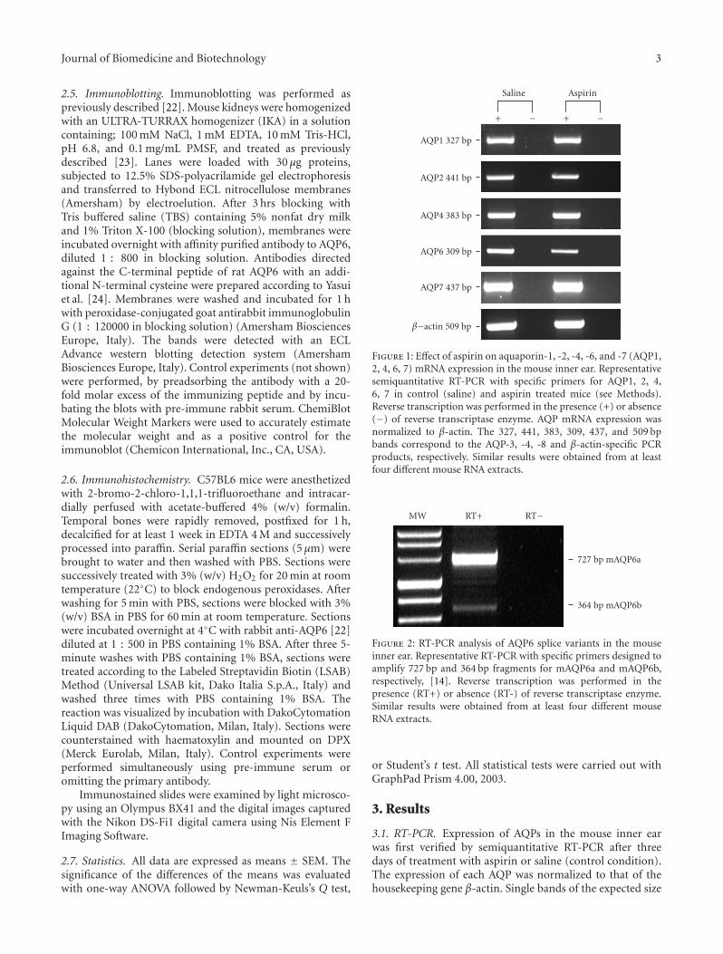
Figure 1: Effect of aspirin on aquaporin-1, -2, -4, -6, and -7 (AQP1,2, 4, 6, 7) mRNA expression in the mouse inner ear. Representativesemiquantitative RT-PCR with specific primers for AQP1, 2, 4,6, 7 in control (saline) and aspirin treated mice (see Methods).Reverse transcription was performed in the presence (+) or absence(−) of reverse transcriptase enzyme. AQP mRNA expression wasnormalized to β-actin. The 327, 441, 383, 309, 437, and 509 bpbands correspond to the AQP-3, -4, -8 and β-actin-specific PCRproducts, respectively. Similar results were obtained from at leastfour different mouse RNA extracts.
MW RT+ RT−
727 bp mAQP6a
364 bp mAQP6b
Figure 2: RT-PCR analysis of AQP6 splice variants in the mouseinner ear. Representative RT-PCR with specific primers designed toamplify 727 bp and 364 bp fragments for mAQP6a and mAQP6b,respectively, [14]. Reverse transcription was performed in thepresence (RT+) or absence (RT-) of reverse transcriptase enzyme.Similar results were obtained from at least four different mouseRNA extracts.
or Student’s t test. All statistical tests were carried out withGraphPad Prism 4.00, 2003.
3. Results
3.1. RT-PCR. Expression of AQPs in the mouse inner earwas first verified by semiquantitative RT-PCR after threedays of treatment with aspirin or saline (control condition).The expression of each AQP was normalized to that of thehousekeeping gene β-actin. Single bands of the expected size

4 Journal of Biomedicine and Biotechnology
0
1
2
Fold
chan
ge
Saline Aspirin
AQP1
(a)
0
0.25
0.5
0.75
1
1.25
Fold
chan
ge
Saline Aspirin
AQP2
(b)
0
1
2
Fold
chan
ge
Saline Aspirin
AQP4
(c)
0
0.5
1
1.5
2
2.5
Fold
chan
ge
Saline Aspirin
AQP7
(d)
0
0.25
0.5
0.75
1
1.25
Fold
chan
ge
Saline Aspirin Recovery
AQP6
∗
(e)
Figure 3: Effect of aspirin treatment on aquaporin (AQP1, 2, 4, 6, 7) mRNA expression in mouse inner ear. Quantitative real-time RT-PCRof total RNA was performed in mouse inner ear mRNA extract under three different conditions: saline (controls), aspirin (mice treated for3 days and sacrificed 3 hours after the last injection) recovery (treated for 3 days and sacrificed 24 hours after the last injection). Specificprimers for AQP1, 2, 4, 6, 7 were used and qRT-PCR performed as indicated (see Methods). The relative mRNA levels were determinedby comparative quantitation (Corbett), the values obtained were normalized to the corresponding β-actin level and the results expressed asfold change. Results indicate that mRNA expression level of AQP6 was significantly lower in aspirin treated mice. Bars represent the mean ±S.E.M. of 4–8 different experiments each from different RNA extracts. ∗, P < .05 versus saline and recovery (one-way ANOVA followed byNewman-Keuls’s Q test).

Journal of Biomedicine and Biotechnology 5
of cDNA fragments were amplified (327, 441, 383, 309, 437and 509 bp for AQP1, 2, 4, 6, 7 and β-actin, resp.). The resultsof agarose gel electrophoresis of representative PCR reactionproducts are shown in Figure 1. Negative controls wereperformed by omitting the reverse transcriptase. Only AQP6mRNA expression was clearly affected by aspirin treatment,while other AQPs were unchanged (Figure 1). Densitometricanalysis of the bands showed that AQP6 transcript wassignificantly reduced (means ± S.E.M. of AQP6/β-actindensitometric ratio): saline, 161 ± 15%; aspirin, 111 ± 13%(∗P < .043 Student’s t test).
Recently, Yasui and coworkers [14] found two AQP6splice variants in the mouse kidney: AQP6a (similar to therat AQP6), and AQP6b, found also in the mouse cerebellum.We performed RT-PCR using specific primers designed todifferentiate between the two variants as in [14] and foundboth variants expressed in the ear (Figure 2).
3.2. qRT-PCR. The effect of aspirin treatment on the expres-sion of AQP mRNAs in the mouse inner ear was then inves-tigated by quantitative real time RT-PCR. Results showedthat aspirin treatment significantly decreases AQP6 mRNAexpression (about 70%, Figure 3), while no significantchanges were observed for AQP1, 2, 4 and 7 transcripts underthis condition. Moreover, the results showed a significantrecovery of mRNA AQP6 expression 24 hours after thelast treatment with aspirin (Figure 3). To investigate thespecificity of aspirin effect, we checked AQP6 mRNA levelsin the kidney and in the small intestine. In these organs,no significant changes of AQP6 transcript expression wereobserved: small intestine, 0.883± 0.001 (saline) and 0.791±0.125 (aspirin); kidney, 1.01 ± 0.03 (saline) and 0.96 ± 0.09(aspirin) (fold change; P > .5, Student’s t test).
3.3. Immunoblotting. Immunoblots of mouse kidneyhomogenates have been performed to test the antibodyused for immunohistochemistry. The results revealed theexpected bands (Figure 4), consistent with published data[14].
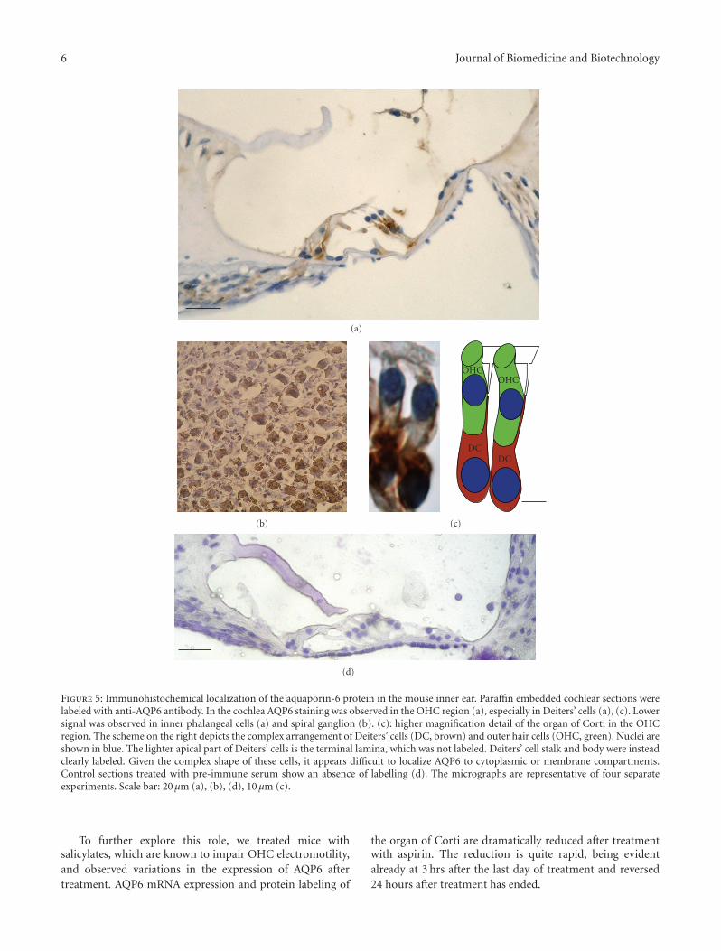
3.4. Immunohistochemistry. Cellular localization of AQP6was investigated with immunohistochemistry using affinity-purified antibodies against mouse and rat AQP6. AQP6 wasexpressed in both cochlear (Figures 5(a), 5(c) and 6(a)) andvestibular (Figure 6(b)) epithelia, and in neuronal somatain the spiral ganglion (Figure 5(b)); faint or no labellingwas noted in the stria vascularis (not shown). In the Organof Corti (Figure 5(a), 5(c)), labelling was most intense andconsistent in Deiters’ cells, which were labelled throughouttheir length (Figure 5(a), 5(c)), and also present at innerphalangeal cells and pillars of Corti (Figure 5(a)). Controls inwhich the primary antibody was replaced by the pre-immuneserum show an absence of labelling (Figure 5(d)). No clearlabelling was observed in hair cells (Figure 5(a) and 5(c)).In vestibular epithelia (Figure 6(b)), labelling was evident,mainly in supporting cells.
After treatment with aspirin (see Methods), labellingwas strongly reduced or lost in both the Organ of Corti(Figure 6(c)) and vestibular epithelia (Figure 6(d)). Reduc-tion in ganglion cells was less evident (not shown).
14
28
42
55
68
82
(kDa) MW Kidney
Figure 4: AQP6 protein expression in mouse kidney homogenate.Immunoblot of mouse kidney total homogenate was performed asdescribed (see Methods). Two bands of about 55 and 36 kDa wereobserved. Similar results were obtained from at least four separateexperiments. MW, molecular weight markers.
4. Discussion
In the present paper, we characterize for the first timethe expression pattern of AQP6 in the mouse ear sensoryepithelia and its regulation by salicylates.
Our data agree with previous studies in human [9] andrat [11] showing strong AQP6 expression in the supportingcells of vestibular epithelia. In the cochlea, on the other hand,our results disagree with previous localization in the rat,where labelling was seen in the stria vascularis but not inthe organ of Corti. Although some discrepancies could beascribed to differences in species and antibodies used, it isalso possible that the different labelling observed in the organof Corti results from variations in the modulation set pointof expression, as our salicylate experiments show.
Our data show that AQP6 is expressed in the organof Corti, and particularly in Deiters’ cells. This pattern ofexpression suggests a possible role for AQP6 in regulatingOHC electromotility, which involves both water and chlorideflow, or in modulating Deiters’ cell stiffness, which wouldaffect OHC action on basilar membrane movement [15].In facts, AQP6 displays interesting properties, and it isstill debated whether its main function is that of an anionchannel, a water channel, or both [25]. Moreover, althoughoriginally thought to be only intracellular [26], recentdata show that AQP6 can bind calmodulin with its N-terminal domain [27], which is responsible for its subcellularlocalization [26]. It is therefore possible that stimuli thatincrease cytoplasmic Ca2+ (such as purinergic signallingin Deiters’ cells [28]), induce AQP6 traslocation to theplasma membrane, similar to what vasopressin does to AQP2[29].
6 Journal of Biomedicine and Biotechnology
(a)
(b) (c)
(d)
OHCOHC
DCDC
Figure 5: Immunohistochemical localization of the aquaporin-6 protein in the mouse inner ear. Paraffin embedded cochlear sections werelabeled with anti-AQP6 antibody. In the cochlea AQP6 staining was observed in the OHC region (a), especially in Deiters’ cells (a), (c). Lowersignal was observed in inner phalangeal cells (a) and spiral ganglion (b). (c): higher magnification detail of the organ of Corti in the OHCregion. The scheme on the right depicts the complex arrangement of Deiters’ cells (DC, brown) and outer hair cells (OHC, green). Nuclei areshown in blue. The lighter apical part of Deiters’ cells is the terminal lamina, which was not labeled. Deiters’ cell stalk and body were insteadclearly labeled. Given the complex shape of these cells, it appears difficult to localize AQP6 to cytoplasmic or membrane compartments.Control sections treated with pre-immune serum show an absence of labelling (d). The micrographs are representative of four separateexperiments. Scale bar: 20 μm (a), (b), (d), 10 μm (c).
To further explore this role, we treated mice withsalicylates, which are known to impair OHC electromotility,and observed variations in the expression of AQP6 aftertreatment. AQP6 mRNA expression and protein labeling of
the organ of Corti are dramatically reduced after treatmentwith aspirin. The reduction is quite rapid, being evidentalready at 3 hrs after the last day of treatment and reversed24 hours after treatment has ended.

Journal of Biomedicine and Biotechnology 7
(a) (b)
(c) (d)
Figure 6: Effect of aspirin treatment on aquaporin-6 protein in the mouse inner ear. Mice were treated with saline (a), (b) and salicylate (c),(d) as described in Methods. Same conditions as Figure 5. In both the organ of Corti (a), (c) and vestibular epithelia (b), (d), AQP6 labelingwas lost or strongly attenuated. In vestibular epithelia of saline treated mice, AQP6 was present, possibly in supporting cells. Inset in (b)shows a 2.5×magnification of the region in the box displaying labeling of the apical part of the hair cell (above the nucleus in the inset) andof the thin supporting cells (whose nuclei are deeper in the epithelium and therefore not in the inset). The micrographs are representative offour separate experiments. Scale bar: 20 μm ((a), (c)), 10 μm ((b), (d)).
These results appear interesting because salicylatesare well known for their reversible ototoxic effect butthe pathways through which they act are still largely unclear.Since prestin and AQP6 appear to be expressed by differentcell types, the link between their modulations may becomplex.
One possible link between OHC block and AQP6 down-regulation in Deiters’ cells is COX-2. In fact, this enzyme hasbeen found to be involved in the downregulation of AQP2in the kidney [30], and is expressed in Deiters’ cells, whereit is modulated by sound conditioning, possibly throughmechanically sensitive pathways [31]. This enzyme couldbe modulated by salicylates (and by unknown physiologicalprocesses) in vestibular epithelia as well (where neitherprestin nor motility are present), thus explaining AQP6downregulation in these organs. Moreover, it is interestingto note the absence of downregulation in small intestine andin the kidney epithelia, suggesting the presence of differentmodulation mechanisms in different organs.
5. Conclusions
In the present paper, we characterize the expression patternof AQP6 in the mouse inner ear sensory epithelia. Our datashow that AQP6 is expressed in the organ of Corti, andin particular in Deiters’ cells. This suggests a possible rolefor AQP6 in regulating OHC motility, through modulationof water and chloride flow or of Deiters’ cell mechanicalcompliance. To further explore this role, we treated mice with
salicylates, which impair OHC electromotility, and observedvariations in the expression of AQP6 after treatment. AQP6mRNA expression and protein labeling of the organ ofCorti are dramatically reduced already at 3 hrs after thelast day of treatment with aspirin. Similar reduction isobserved in vestibular epithelia, but not in control tissues(small intestine and kidney). These results suggest that AQP6downregulation is not due to motility/prestin-dependentpathways, but may affect OHCs’ ionic environment and/ortheir mechanical response through changes in coupling tothe Organ of Corti.
Acknowledgments
This research was funded in part by AIT (AssociazioneItaliana Tinnitus) ONLUS.
References
[1] Y. Cazals, “Auditory sensori-neural alterations induced bysalicylate,” Progress in Neurobiology, vol. 62, no. 6, pp. 583–631,2000.
[2] D. Oliver, D. Z. Z. He, N. Klocker, et al., “Intracellular anionsas the voltage sensor of prestin, the outer hair cell motorprotein,” Science, vol. 292, no. 5525, pp. 2340–2343, 2001.
[3] S. Kakehata and J. Santos-Sacchi, “Effects of salicylate andlanthanides on outer hair cell motility and associated gatingcharge,” The Journal of Neuroscience, vol. 16, no. 16, pp. 4881–4889, 1996.
8 Journal of Biomedicine and Biotechnology
[4] J. Zheng, W. Shen, D. Z. Z. He, K. B. Long, L. D. Madison, andP. Dallos, “Prestin is the motor protein of cochlear outer haircells,” Nature, vol. 405, no. 6783, pp. 149–155, 2000.
[5] M. Fukushima, T. Kitahara, Y. Fuse, Y. Uno, K. Doi, and T.Kubo, “Changes in aquaporin expression in the inner ear ofthe rat after i.p. injection of steroids,” Acta Oto-Laryngologica,vol. 124, no. 553s, pp. 13–18, 2004.
[6] P. Agre, L. S. King, M. Yasui, et al., “Aquaporin waterchannels—from atomic structure to clinical medicine,” Jour-nal of Physiology, vol. 542, no. 1, pp. 3–16, 2002.
[7] A. S. Verkman, “Mammalian aquaporins: diverse physiologicalroles and potential clinical significance,” Expert Reviews inMolecular Medicine, vol. 10, article e13, 2008.
[8] K. Ishibashi, “New members of mammalian aquaporins:AQP10-AQP12,” Handbook of Experimental Pharmacology, no.190, pp. 251–262, 2009.
[9] I. A. Lopez, G. Ishiyama, M. Lee, R. W. Baloh, and A.Ishiyama, “Immunohistochemical localization of aquaporinsin the human inner ear,” Cell and Tissue Research, vol. 328, no.3, pp. 453–460, 2007.
[10] D. Huang, P. Chen, S. Chen, M. Nagura, D. J. Lim, andX. Lin, “Expression patterns of aquaporins in the inner ear:evidence for concerted actions of multiple types of aquaporinsto facilitate water transport in the cochlea,” Hearing Research,vol. 165, no. 1-2, pp. 85–95, 2002.
[11] D. Taguchi, T. Takeda, A. Kakigi, T. Okada, R. Nishioka, and H.Kitano, “Expression and immunolocalization of aquaporin-6(Aqp6) in the rat inner ear,” Acta Oto-Laryngologica, vol. 128,no. 8, pp. 832–840, 2008.
[12] M. Fukushima, T. Kitahara, Y. Uno, Y. Fuse, K. Doi, andT. Kubo, “Effects of intratympanic injection of steroids onchanges in rat inner ear aquaporin expression,” Acta Oto-Laryngologica, vol. 122, no. 6, pp. 600–606, 2002.
[13] J. Li and A. S. Verkman, “Impaired hearing in mice lackingaquaporin-4 water channels,” The Journal of Biological Chem-istry, vol. 276, no. 33, pp. 31233–31237, 2001.
[14] H. Nagase, J. Agren, A. Saito, et al., “Molecular cloningand characterization of mouse aquaporin 6,” Biochemical andBiophysical Research Communications, vol. 352, no. 1, pp. 12–16, 2007.
[15] G. I. Frolenkov, “Regulation of electromotility in the cochlearouter hair cell,” Journal of Physiology, vol. 576, no. 1, pp. 43–48,2006.
[16] Y. Miyabe, T. Kikuchi, and T. Kobayashi, “Compara-tive immunohistochemical localizations of aquaporin-1 andaquaporine-4 in the cochleae of three different species ofrodents,” Tohoku Journal of Experimental Medicine, vol. 196,no. 4, pp. 247–257, 2002.
[17] J. Loffing, D. Loffing-Cueni, A. Macher, et al., “Localization ofepithelial sodium channel and aquaporin-2 in rabbit kidneycortex,” American Journal of Physiology, vol. 278, no. 4, pp.F530–F539, 2000.
[18] G. Calamita, A. Mazzone, A. Bizzoca, et al., “Expression andimmunolocalization of the aquaporin-8 water channel in ratgastrointestinal tract,” European Journal of Cell Biology, vol. 80,no. 11, pp. 711–719, 2001.
[19] K. Oshio, D. K. Binder, B. Yang, S. Schecter, A. S. Verkman,and G. T. Manley, “Expression of aquaporin water channels inmouse spinal cord,” Neuroscience, vol. 127, no. 3, pp. 685–693,2004.
[20] U. Laforenza, E. Cova, G. Gastaldi, et al., “Aquaporin-8 isinvolved in water transport in isolated superficial colonocytesfrom rat proximal colon,” The Journal of Nutrition, vol. 135,no. 10, pp. 2329–2336, 2005.
[21] U. Laforenza, G. Gastaldi, M. Grazioli, et al., “Expressionand immunolocalization of aquaporin-7 in rat gastrointestinaltract,” The Biology of the Cell, vol. 97, no. 8, pp. 605–613, 2005.
[22] U. Laforenza, G. Gastaldi, M. Polimeni, et al., “Aquaporin-6 isexpressed along the rat gastrointestinal tract and upregulatedby feeding in the small intestine,” BMC Physiology, vol. 9,article 18, pp. 1–12, 2009.
[23] U. K. Laemmli, “Cleavage of structural proteins during theassembly of the head of bacteriophage T4,” Nature, vol. 227,no. 5259, pp. 680–685, 1970.
[24] M. Yasui, T.-H. Kwon, M. A. Knepper, S. Nielsen, and P. Agre,“Aquaporin-6: an intracellular vesicle water channel protein inrenal epithelia,” Proceedings of the National Academy of Sciencesof the United States of America, vol. 96, no. 10, pp. 5808–5813,1999.
[25] M. Yasui, “pH regulated anion permeability of aquaporin-6,”Handbook of Experimental Pharmacology, vol. 190, pp. 299–308, 2009.
[26] E. Beitz, K. Liu, M. Ikeda, W. B. Guggino, P. Agre, and M.Yasui, “Determinants of AQP6 trafficking to intracellular sitesversus the plasma membrane in transfected mammalian cells,”The Biology of the Cell, vol. 98, no. 2, pp. 101–109, 2006.
[27] N. E. Rabaud, L. Song, Y. Wang, P. Agre, M. Yasui, and J.M. Carbrey, “Aquaporin 6 binds calmodulin in a calcium-dependent manner,” Biochemical and Biophysical ResearchCommunications, vol. 383, no. 1, pp. 54–57, 2009.
[28] L. Lagostena and F. Mammano, “Intracellular calcium dynam-ics and membrane conductance changes evoked by Deiters’cell purinoceptor activation in the organ of Corti,” CellCalcium, vol. 29, no. 3, pp. 191–198, 2001.
[29] S. Nielsen, C.-L. Chou, D. Marples, E. I. Christensen, B. K.Kishore, and M. A. Knepper, “Vasopressin increases waterpermeability of kidney collecting duct by inducing transloca-tion of aquaporin-CD water channels to plasma membrane,”Proceedings of the National Academy of Sciences of the UnitedStates of America, vol. 92, no. 4, pp. 1013–1017, 1995.
[30] R. Nørregaard, B. L. Jensen, C. Li, et al., “COX-2 inhibitionprevents downregulation of key renal water and sodium trans-port proteins in response to bilateral ureteral obstruction,”American Journal of Physiology, vol. 289, no. 2, pp. F322–F333,2005.
[31] U.-R. Heinrich, J. Brieger, O. Selivanova, et al., “COX-2expression in the guinea pig cochlea is partly altered bymoderate sound exposure,” Neuroscience Letters, vol. 394, no.2, pp. 121–126, 2006.