aptameric molecular switch for cascade signal amplification
TRANSCRIPT

Aptameric Molecular Switch for CascadeSignal Amplification
Cuiping Ma,1 Chunhui Zhao,1 Yujie Ge,1 and Chao Shi1*
BACKGROUND: High sensitivity of analysis is constantlyin demand in biomedical research and clinical diagno-sis. In recent years aptamer-based analytical methodshave been developed for protein detection. We devel-oped a cascade signal amplification strategy based onmolecular switches and aptamers to improve proteindetection.
METHODS: Our cascade signal amplification strategybased on molecular switches and aptamers consisted of2 steps, including the recognition and the triggering ofa polymerase reaction. The procedure was designed tosimplify the analysis by detecting trace amounts of tar-get isothermally, in real time, and in a homogeneoussolution. We applied this method to measure thrombinin human serum samples.
RESULTS: This cascade signal amplification strategy ex-hibited a linear response in thrombin concentrationfrom 0.3 to 10 nmol/L, with a detection limit of 1.7 �10�10 mol/L within 60 min. Results of the analysis ofthrombin in human serum diluted 1:1 appeared to belinear, as was observed in buffer, in the tested concen-tration range of 0.3–10 nmol/L.
CONCLUSIONS: The aptameric sensor provides promis-ing potential for detecting and screening trace concen-trations of biomarkers in complex matrices for clinicalapplications.© 2011 American Association for Clinical Chemistry
High sensitivity of analysis is constantly in demand forclinical diagnosis. Thus, the development of assay sig-nal amplification paths has become important, andseveral amplification techniques have been developed,including catalytic nanoparticles (1, 2 ), PCR (3, 4 ),rolling-circle amplification (5, 6 ), enzyme-triggeredstrand displacement amplification (7–10 ), entropy-driven signal amplification (11, 12 ), and cascade signalamplification (13, 14 ). The PCR is currently the most
widely used thermal-cycling technique for DNA ampli-fication. However, thermal-cycling techniques requirethe use of special instruments, and the conventionalPCR product typically requires characterization at theend of the reaction (13 ). These disadvantages limit thistechnique. Isothermal amplification of DNA hasemerged as an alternative amplification technique(6, 8, 15 ). The reaction is performed at a constant tem-perature, so the time required for DNA amplification isless than that of thermal-cycling techniques. In addi-tion, isothermal amplification can be performed with-out specialized instrumentation and has the potentialfor use in point-of-care testing. The basis of this tech-nique is continuous strand displacement, in whichpolymerase extension uses 1 strand of the DNA chainas a template (16 ). He et al. (8 ) described an analyti-cally sensitive method for cocaine detection based onisothermal strand displacement. Shlyahovsky et al. (7 )provided an automated technique based on aptamersthat was activated by polymerase, nicking enzyme, anda molecular beacon for amplifying the detection of co-caine. The advantage of these fluorescent biosensorswas an enhanced limit of detection of the assay,whereas the linear signal amplification procedureseemed to be disadvantageous.
Aptamers have been integrated as recognition ele-ments in optical or electrochemical sensors (17, 18 ).Aptamers are nucleic acids selected from a rich nucleicacid library by means of the SELEX (systematic evolu-tion of ligands by exponential enrichment) procedure.Aptamers possess specific recognition properties towardlow molecular weight substrates or macromolecules suchas proteins (19). As novel elements for molecular recog-nition, aptamers offer many advantages over protein an-tibodies, such as high affinity and specificity for targetproteins, good stability, and ease of synthesis and modifi-cation (20). Aptamer-based analytical methods have beendeveloped for protein-detection methods that use electro-chemistry (21, 22), fluorescence (23, 24), and binding-induced label-free detection (25).
1 State Key Laboratory Base of Eco-Chemical Engineering, College of Chemistryand Molecular Engineering, Qingdao University of Science and Technology,Qingdao, P. R. China.
* Address correspondence to this author at: College of Chemistry and Mo-lecular Engineering, Qingdao University of Science and Technology, Zheng-
zhou Rd. 53, Qingdao, China 266042. Fax �86-532-84023927; [email protected].
Received July 26, 2011; accepted November 1, 2011.Previously published online at DOI: 10.1373/clinchem.2011.173195
Clinical Chemistry 58:2384–390 (2012)
Automation and Analytical Techniques
384
Dow
nloaded from https://academ
ic.oup.com/clinchem
/article/58/2/384/5620565 by guest on 15 October 2021
Thrombin is a serine protease that plays an impor-tant role in the coagulation cascade, thrombosis, andhemostasis. The 15mer single-strand DNA of thethrombin aptamer can bind with thrombin stronglyand selectively and is the first aptamer selected in vitro(26 ). The goal of our study was to develop a highlyselective and sensitive aptamer-based sensor to detectthrombin in human serum. We have introduced theparadigm of a signal amplification platform that iso-thermally and nonlinearly amplifies the recognitionevent between the aptamer and its substrate. The read-out of the analysis triggered by binding of the aptamerand substrate was have accomplished by a fluorescencesignal. We have developed a cascade signal amplifica-tion strategy that is isothermal and homogeneous.
Materials and Methods
All oligonucleotides used in this work were designedby using the mFold server (http://mfold.rna.albany.edu) and synthesized by Takara Biotechnology, asshown in Table 1.
Klenow fragment polymerase (exo�), Nt.BbvCIenzyme, and a mixture of deoxynucleoside triphos-phates (dNTPs)2 were purchased from New EnglandBiolabs. Human thrombin (10 U/mg) was purchasedfrom DingGuo. BSA was obtained from Westang.Trypsin and bovine thrombin were purchased fromSigma-Aldrich. Human serum was provided byQingdao Municipal Hospital in China. All chemicalswere of analytical grade and were used without furtherpurification. All solutions were prepared with doublydistilled water.
Fig. 1 outlines the principles of signal cascade am-plification based on aptamer recognition of thrombinas an example. Nucleic acid 1 consists of 4 regions (Fig.
1A). Region I is the sequence of thrombin aptamer. The7 bases at the 5�terminus excluded from the stem ofnucleic acid 1 are for a better switching of the DNAstructure. Region II consists of a sequence that is thesame as a molecular beacon. Thus, the complementarystrand of region II can act as the “product” and hybrid-ize with the molecular beacon when it is replicated anddisplaced, which leads to the signal transduction of thethrombin-binding event. Region III includes a nickingsite for Nt.BbvCI when the duplex of this region isformed. Region IV is located in the 3�terminus of nu-cleic acid 1, which can hybridize with region I for 10bases or for 7 bases with itself. Therefore, nucleic acid 1has 2 kinds of configurations in solution. Owing to thehybridization of 10 bases by nucleic acid 1 with higheraffinity than the hybridization of 7 bases, most of nu-cleic acid 1 exists in the state of region I hybridizingwith region IV in the absence of thrombin. This pre-vents uncontrolled folding of nucleic acid 1 from trig-gering the polymerase reaction. In addition, regionIV has 2-base extensions at the 3� terminus to avoidundesired replication reactions under experimentalconditions.
In the presence of thrombin, nucleic acid 1 acts asa molecular switch, folding the aptamer into its stableaptamer–thrombin complex (Fig. 1B) and forming a7-base duplex structure with itself that can initiate rep-lication. The 15mer thrombin aptamer appears to havea dissociation constant (Kd) from 25 to 200 nmol/L forthrombin (26 ), whereas the Klenow fragment poly-merase has a Kd of 7.9 nmol/L for DNA (27 ). Thethrombin aptamer has a greater Kd for thrombin thanthe polymerase for DNA, so the polymerase is able todisplace thrombin from the furled-up pocket on the 5�terminus. The polymerase replication is activated withthe Klenow fragment (exo�) and dNTPs and can dis-place thrombin to another nucleic acid 1. Simultane-ously, the replication of a single strand yields the du-plex that includes the nicking site for Nt.BbvCI.Scission of the replicated strand results in a new repli-cation site for polymerase and the concomitant dis-
2 Nonstandard abbreviations: dNTPs, deoxynucleoside triphosphates; Kd, dissoci-ation constant; DNA-MB, DNA molecular beacon; FAM, 6-carboxyfluorescein;DABCYL, 4-(dimethylaminoazo)benzene-4-carboxylic acid; �F, change in fluo-rescence signal.
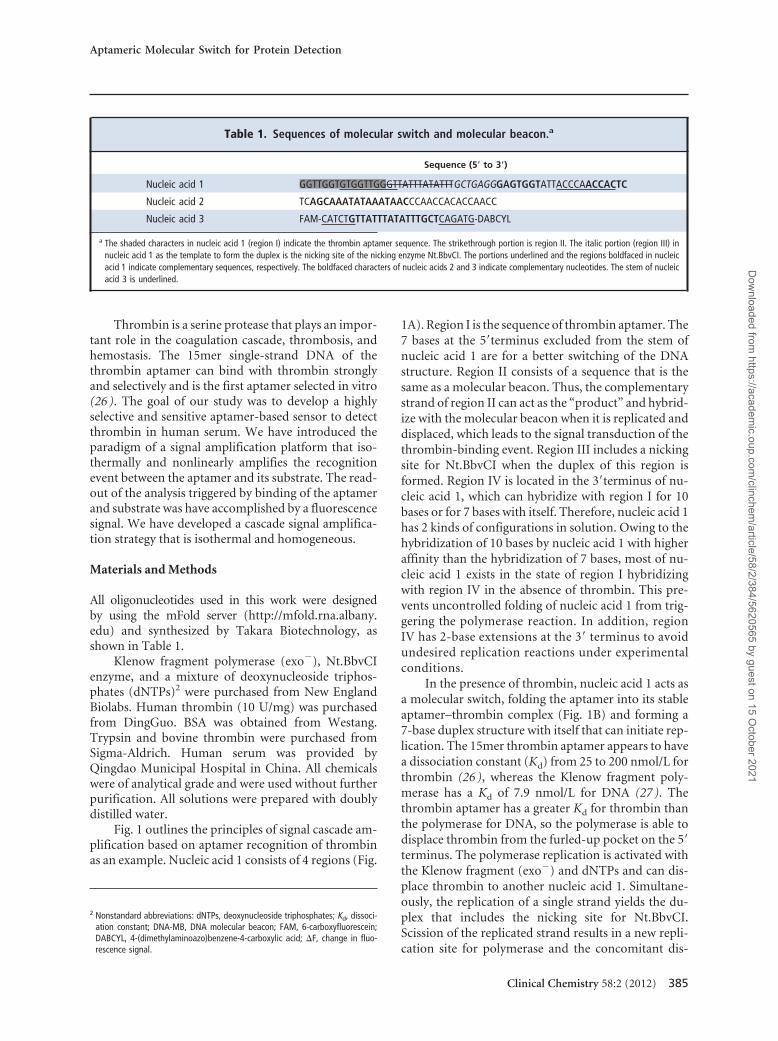
Table 1. Sequences of molecular switch and molecular beacon.a
Sequence (5� to 3�)
Nucleic acid 1 GGTTGGTGTGGTTGGGTTATTTATATTTGCTGAGGGAGTGGTATTACCCAACCACTC
Nucleic acid 2 TCAGCAAATATAAATAACCCAACCACACCAACC
Nucleic acid 3 FAM-CATCTGTTATTTATATTTGCTCAGATG-DABCYL
a The shaded characters in nucleic acid 1 (region I) indicate the thrombin aptamer sequence. The strikethrough portion is region II. The italic portion (region III) innucleic acid 1 as the template to form the duplex is the nicking site of the nicking enzyme Nt.BbvCI. The portions underlined and the regions boldfaced in nucleicacid 1 indicate complementary sequences, respectively. The boldfaced characters of nucleic acids 2 and 3 indicate complementary nucleotides. The stem of nucleicacid 3 is underlined.
Aptameric Molecular Switch for Protein Detection
Clinical Chemistry 58:2 (2012) 385
Dow
nloaded from https://academ
ic.oup.com/clinchem
/article/58/2/384/5620565 by guest on 15 October 2021
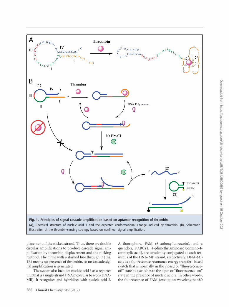
placement of the nicked strand. Thus, there are doublecircular amplifications to produce cascade signal am-plification by thrombin displacement and the nickingmethod. The circle with a slashed line through it (Fig.1B) means no presence of thrombin, so no cascade sig-nal amplification is generated.
The system also includes nucleic acid 3 as a reporterunit that is a single-strand DNA molecular beacon (DNA-MB). It recognizes and hybridizes with nucleic acid 2.
A fluorophore, FAM (6-carboxyfluorescein), and aquencher, DABCYL [4-(dimethylaminoazo)benzene-4-carboxylic acid], are covalently conjugated at each ter-minus of the DNA-MB strand, respectively. DNA-MBacts as a fluorescence resonance energy transfer– basedswitch that is normally in the closed or “fluorescence-off” state but switches to the open or “fluorescence-on”state in the presence of nucleic acid 2. In other words,the fluorescence of FAM (excitation wavelength: 480
Fig. 1. Principles of signal cascade amplification based on aptamer recognition of thrombin.
(A), Chemical structure of nucleic acid 1 and the expected conformational change induced by thrombin. (B), Schematicillustration of the thrombin-sensing strategy based on nonlinear signal amplification.
386 Clinical Chemistry 58:2 (2012)
Dow
nloaded from https://academ
ic.oup.com/clinchem
/article/58/2/384/5620565 by guest on 15 October 2021
nm) has only a residually minute emission in the ab-sence of nucleic acid 2. In contrast, the fluorescence ofFAM shows a large increase in the presence of nucleicacid 2.
TIME-DEPENDENT SENSING PROCEDURE
For real-time monitoring of the reaction process, thereaction was performed by using an ABI StepOne real-time PCR instrument (Applied Biosystems). Nucleicacid 1 was denatured at 95 °C for 10 min and cooled toroom temperature in 1� NEB buffer 4 (New EnglandBiolabs) [20 mmol/L Tris-acetate (pH 7.9), 50 mmol/Lpotassium acetate, 10 mmol/L magnesium acetate, and1 mmol/L dithiothreitol]. Each reaction mixture con-tained the following reagents in a final volume of 25�L: 1.25 U polymerase, 2.5 U Nt.BbvCI, 0.2 mmol/LdNTPs, 0.4 �mol/L nucleic acid 1, and 0.5 �mol/L mo-lecular beacon. Reaction buffer was 1� NEB buffer 4. A5-�L specific concentration of thrombin was added totrigger the polymerase reaction. The reactions were in-cubated at 37 °C for 60 min to detect fluorescence in-tensities. Fluorescence curves were recorded at 30-s in-tervals. All the reactions were run in triplicate, and thesame reaction mixtures without thrombin were used asnegative controls.
APPLICATION OF THE APTAMER-BASED SYSTEM IN HUMAN
SERUM
Thrombin was further tested in human serum withoutany pretreatment to illustrate the feasibility of the ap-proach. Serum, diluted 2 times, was tested alone orspiked with serially diluted thrombin at concentrationsranging from 0.3 to 300 nmol/L. Furthermore, the finalconcentrations of 3.0 � 10�6 mol/L BSA, 10�7 mol/Ltrypsin, and 10�7 mol/L bovine thrombin were testedin the selectivity experiments. The assay procedures forBSA, trypsin, and bovine thrombin were the same asthose for human thrombin in buffer, except for the useof BSA, trypsin, and bovine thrombin instead of hu-man thrombin, respectively.
Results and Discussion
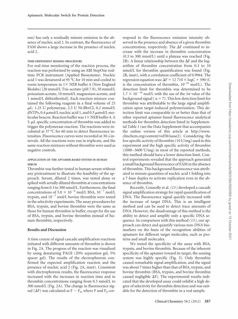
A time course of signal cascade amplification reactionsinitiated with different amounts of thrombin is shownin Fig. 2A. The progress of the reaction was visualizedby using denaturing PAGE (20% separation gel, 5%spacer gel). The results of the electrophoresis con-firmed the expected amplification reaction and thepresence of nucleic acid 2 (Fig. 2A, inset). Consistentwith electrophoresis results, the fluorescence responseincreased with the increases in reaction time and inthrombin concentrations ranging from 0.3 nmol/L to300 nmol/L (Fig. 2A). The change in fluorescence sig-nal (�F) was calculated as F � F0, where F and F0 cor-
respond to the fluorescence emission intensity ob-served in the presence and absence of a given thrombinconcentration, respectively. The �F continued to in-crease with the increase in thrombin concentration(0.3 to 300 nmol/L) until a plateau was reached (Fig.2B). A linear relationship between the �F and the log-arithm of thrombin concentration from 0.3 to 10nmol/L for thrombin quantification was found (Fig.2B, inset), with a correlation coefficient of 0.9964. Theregression equation was: �F � 12 710 � logC � 590 (Cis the concentration of thrombin, 10–10 mol/L). Thedetection limit for thrombin was determined to be1.7 � 10�10 mol/L with the use of the 3� value of thebackground signal ( n � 7). This low detection limit forthrombin was attributable to the large signal amplifi-cation upon target-induced polymerization. This de-tection limit was comparable to or better than that ofother reported aptamer-based fluorescence analyticalmethods for thrombin detection listed in Supplemen-tal Table 1 (see the Data Supplement that accompaniesthe online version of this article at http://www.clinchem.org/content/vol58/issue2). Considering thelow specific activity of thrombin (10 U/mg) used in thisexperiment and the high specific activity of thrombin(1000 –3600 U/mg) in most of the reported methods,this method should have a lower detection limit. Con-trol experiments revealed that the approach generateda small background fluorescence of FAM in the absenceof thrombin. This background fluorescence was attrib-uted to minute quantities of nucleic acid 1 folding intoa 7-base duplex to activate replication even in the ab-sence of thrombin.
Recently, Connolly et al. (13 ) developed a cascadesignal amplification strategy for rapid quantification ofDNA. The fluorescence signal quickly increased withthe increase of target DNA. This is an intelligentmethod and can be used to detect trace amounts ofDNA. However, the disadvantage of this method is theability to detect and amplify only a specific DNA se-quence. In comparison with this method (13 ), our ap-proach can detect and quantify various non-DNA bio-markers on the basis of the recognition abilities ofaptamers for different target molecules, such as pro-teins and small molecules.
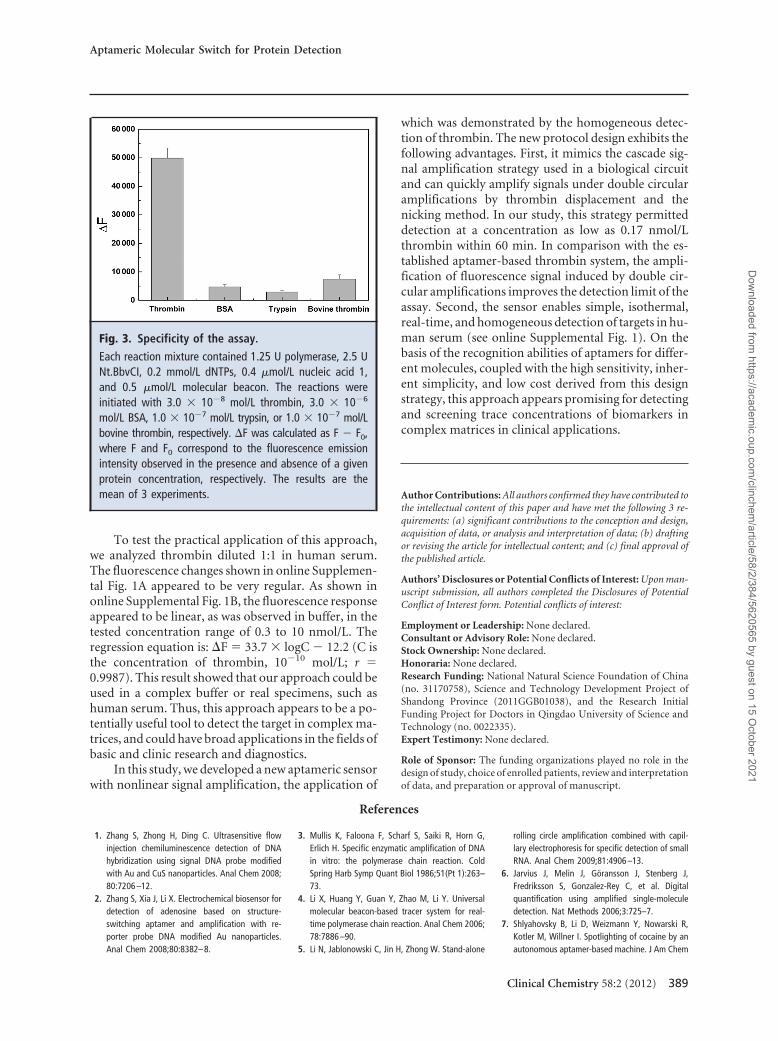
We tested the specificity of the assay with BSA,trypsin, and bovine thrombin. Because of the inherentspecificity of the aptamer toward its target, the sensingsystem was highly specific (Fig. 3). Only thrombincaused remarkable signal amplification, and the signalwas about 7 times higher than that of BSA, trypsin, andbovine thrombin (BSA, trypsin, and bovine thrombincaused negligible �F). The experimental results indi-cated that the developed assay could exhibit a high de-gree of selectivity for thrombin detection and was suit-able for the detection of thrombin in a real sample.
Aptameric Molecular Switch for Protein Detection
Clinical Chemistry 58:2 (2012) 387
Dow
nloaded from https://academ
ic.oup.com/clinchem
/article/58/2/384/5620565 by guest on 15 October 2021
Fig. 2. Signal cascade amplification reactions initiated with different amounts of thrombin.
(A), Time course of fluorescence experiments performed with an ABI StepOne real-time PCR instrument (thrombin concentra-tions: a, no thrombin; b, 0.3 nmol/L; c, 1 nmol/L; d, 3 nmol/L; e, 10 nmol/L, f, 30 nmol/L; g, 100 nmol/L; h, 300 nmol/L). Inset:Denaturing PAGE of the amplification reactions containing 0.4 �mol/L nucleic acid 1. L: 20-bp DNA ladder; 1: reactioncontaining nucleic acid 1 and DNA polymerase; 2: amplification reaction containing nucleic acid 1, 3 � 10�7 mol/L thrombin,and DNA polymerase; 3–5: amplification reactions containing both DNA polymerase and Nt.BbvCI endonuclease were initiatedwith different concentrations of thrombin (3, 0.3 �mol/L; 4, 30 nmol/L; 5, 3 nmol/L). nt, nucleotides. (B), �F values for differentconcentrations of thrombin. Reaction time was 60 min. Each value is the mean of the results of 3 experiments.
388 Clinical Chemistry 58:2 (2012)
Dow
nloaded from https://academ
ic.oup.com/clinchem
/article/58/2/384/5620565 by guest on 15 October 2021
To test the practical application of this approach,we analyzed thrombin diluted 1:1 in human serum.The fluorescence changes shown in online Supplemen-tal Fig. 1A appeared to be very regular. As shown inonline Supplemental Fig. 1B, the fluorescence responseappeared to be linear, as was observed in buffer, in thetested concentration range of 0.3 to 10 nmol/L. Theregression equation is: �F � 33.7 � logC � 12.2 (C isthe concentration of thrombin, 10�10 mol/L; r �0.9987). This result showed that our approach could beused in a complex buffer or real specimens, such ashuman serum. Thus, this approach appears to be a po-tentially useful tool to detect the target in complex ma-trices, and could have broad applications in the fields ofbasic and clinic research and diagnostics.
In this study, we developed a new aptameric sensorwith nonlinear signal amplification, the application of
which was demonstrated by the homogeneous detec-tion of thrombin. The new protocol design exhibits thefollowing advantages. First, it mimics the cascade sig-nal amplification strategy used in a biological circuitand can quickly amplify signals under double circularamplifications by thrombin displacement and thenicking method. In our study, this strategy permitteddetection at a concentration as low as 0.17 nmol/Lthrombin within 60 min. In comparison with the es-tablished aptamer-based thrombin system, the ampli-fication of fluorescence signal induced by double cir-cular amplifications improves the detection limit of theassay. Second, the sensor enables simple, isothermal,real-time, and homogeneous detection of targets in hu-man serum (see online Supplemental Fig. 1). On thebasis of the recognition abilities of aptamers for differ-ent molecules, coupled with the high sensitivity, inher-ent simplicity, and low cost derived from this designstrategy, this approach appears promising for detectingand screening trace concentrations of biomarkers incomplex matrices in clinical applications.
Author Contributions: All authors confirmed they have contributed tothe intellectual content of this paper and have met the following 3 re-quirements: (a) significant contributions to the conception and design,acquisition of data, or analysis and interpretation of data; (b) draftingor revising the article for intellectual content; and (c) final approval ofthe published article.
Authors’ Disclosures or Potential Conflicts of Interest: Upon man-uscript submission, all authors completed the Disclosures of PotentialConflict of Interest form. Potential conflicts of interest:
Employment or Leadership: None declared.Consultant or Advisory Role: None declared.Stock Ownership: None declared.Honoraria: None declared.Research Funding: National Natural Science Foundation of China(no. 31170758), Science and Technology Development Project ofShandong Province (2011GGB01038), and the Research InitialFunding Project for Doctors in Qingdao University of Science andTechnology (no. 0022335).Expert Testimony: None declared.
Role of Sponsor: The funding organizations played no role in thedesign of study, choice of enrolled patients, review and interpretationof data, and preparation or approval of manuscript.
References
1. Zhang S, Zhong H, Ding C. Ultrasensitive flowinjection chemiluminescence detection of DNAhybridization using signal DNA probe modifiedwith Au and CuS nanoparticles. Anal Chem 2008;80:7206–12.
2. Zhang S, Xia J, Li X. Electrochemical biosensor fordetection of adenosine based on structure-switching aptamer and amplification with re-porter probe DNA modified Au nanoparticles.Anal Chem 2008;80:8382–8.
3. Mullis K, Faloona F, Scharf S, Saiki R, Horn G,Erlich H. Specific enzymatic amplification of DNAin vitro: the polymerase chain reaction. ColdSpring Harb Symp Quant Biol 1986;51(Pt 1):263–73.
4. Li X, Huang Y, Guan Y, Zhao M, Li Y. Universalmolecular beacon-based tracer system for real-time polymerase chain reaction. Anal Chem 2006;78:7886–90.
5. Li N, Jablonowski C, Jin H, Zhong W. Stand-alone
rolling circle amplification combined with capil-lary electrophoresis for specific detection of smallRNA. Anal Chem 2009;81:4906–13.
6. Jarvius J, Melin J, Göransson J, Stenberg J,Fredriksson S, Gonzalez-Rey C, et al. Digitalquantification using amplified single-moleculedetection. Nat Methods 2006;3:725–7.
7. Shlyahovsky B, Li D, Weizmann Y, Nowarski R,Kotler M, Willner I. Spotlighting of cocaine by anautonomous aptamer-based machine. J Am Chem
Fig. 3. Specificity of the assay.
Each reaction mixture contained 1.25 U polymerase, 2.5 UNt.BbvCI, 0.2 mmol/L dNTPs, 0.4 �mol/L nucleic acid 1,and 0.5 �mol/L molecular beacon. The reactions wereinitiated with 3.0 � 10�8 mol/L thrombin, 3.0 � 10�6
mol/L BSA, 1.0 � 10�7 mol/L trypsin, or 1.0 � 10�7 mol/Lbovine thrombin, respectively. �F was calculated as F � F0,where F and F0 correspond to the fluorescence emissionintensity observed in the presence and absence of a givenprotein concentration, respectively. The results are themean of 3 experiments.
Aptameric Molecular Switch for Protein Detection
Clinical Chemistry 58:2 (2012) 389
Dow
nloaded from https://academ
ic.oup.com/clinchem
/article/58/2/384/5620565 by guest on 15 October 2021
Soc 2007;129:3814–5.8. He JL, Wu ZS, Zhou H, Wang HQ, Jiang JH, Shen
GL, Yu RQ. Fluorescence aptameric sensor forstrand displacement amplification detection ofcocaine. Anal Chem 2010;82:1358–64.
9. Bi S, Zhang J, Zhang S. Ultrasensitive and selec-tive DNA detection based on nicking endonu-clease assisted signal amplification and its appli-cation in cancer cell detection. Chem Commun2010;46:5509–11.
10. Ding C, Li X, Ge Y, Zhang S. Fluorescence detec-tion of telomerase activity in cancer cells basedon isothermal circular strand-displacement po-lymerization reaction. Anal Chem 2010;82:2850–5.
11. Zhang DY, Turberfield AJ, Yurke B, Winfree E.Engineering entropy-driven reactions and net-works catalyzed by DNA. Science 2007;318:1121–5.
12. Niu S, Jiang Y, Zhang S. Fluorescence detectionfor DNA using hybridization chain reaction withenzyme-amplification. Chem Commun 2010;46:3089–91.
13. Connolly AR, Trau M. Isothermal detection ofDNA by beacon-assisted detection amplification.Angew Chem Int Ed 2010;49:2720–3.
14. Cheng W, Yan F, Ding L, Ju H, Yin Y. Cascade
signal amplification strategy for subattomolarprotein detection by rolling circle amplificationand quantum dots tagging. Anal Chem 2010;82:3337–42.
15. Notomi T, Okayama H, Masubuchi H, YonekawaT, Watanabe K, Amino N, Hase T. Loop-mediatedisothermal amplification of DNA. Nucleic AcidsRes 2000;28:63e.
16. Guo Q, Yang X, Wang K, Tan W, Li W, Tang H, LiH. Sensitive fluorescence detection of nucleic ac-ids based on isothermal circular strand-displacement polymerization reaction. NucleicAcids Res 2009;37:20–5.
17. Hermann T, Patel DJ. Adaptive recognition bynucleic acid aptamers. Science 2000;287:820–5.
18. Xiang Y, Zhang Y, Qian X, Chai Y, Wang J, YuanR. Ultrasensitive aptamer-based protein detectionvia a dual amplified biocatalytic strategy. BiosensBioelectron 2010;25:2539–42.
19. Stoltenburg R, Reinemann C, Strehlitz B. SELEX: a(r)evolutionary method to generate high-affinitynucleic acid ligands. Biomol Eng 2007;24:381–403.
20. Jayasena SD. Aptamers: an emerging class ofmolecules that rival antibodies in diagnostics.Clin Chem 1999;45:1628–50.
21. Zhao Q, Li XF, Le XC. Aptamer-modified mono-
lithic capillary chromatography for protein sepa-ration and detection. Anal Chem 2008;80:3915–20.
22. Lu Y, Li X, Zhang L, Yu P, Su L, Mao L. Aptamer-based electrochemical sensors with aptamer-complementary DNA oligonucleotides as probe.Anal Chem 2008;80:1883–90.
23. Yang H, Liu H, Kang H, Tan W. Engineeringtarget-responsive hydrogels based on aptamer-target interactions. J Am Chem Soc 2008;130:6320–1.
24. Dittmer WU, Reuter A, Simmel FC. A DNA-basedmachine that can cyclically bind and releasethrombin. Angew Chem Int Ed 2004;43:3550–3.
25. Du Y, Li B, Wei H, Wang Y, Wang E. Multifunc-tional label-free electrochemical biosensor basedon an integrated aptamer. Anal Chem 2008;80:5110–7.
26. Bock LC, Griffin LC, Latham JA, Vermaas EH,Toole JJ. Selection of single-stranded DNA mole-cules that bind and inhibit human thrombin. Na-ture 1992;355:564–6.
27. Guest CR, Hochstrasser RA, Dupuy CG, Allen DJ,Benkovic SJ, Millar DP. Interaction of DNA withthe Klenow fragment of DNA polymerase I stud-ied by time-resolved fluorescence spectroscopy.Biochemistry 1991;30:8759–70.
390 Clinical Chemistry 58:2 (2012)
Dow
nloaded from https://academ
ic.oup.com/clinchem
/article/58/2/384/5620565 by guest on 15 October 2021