approaches to the small bowel biopsy - unispital basel · approaches to the small bowel biopsy...
TRANSCRIPT

APPROACHES TO THE SMALL BOWEL
BIOPSY Basel Seminars in Paediatric Pathology
and Genetics
Pierre Russo, MD
Director, Division of Anatomic Pathology
The Children’s Hospital of Philadelphia
Professor, Department of Pathology and Laboratory
Medicine
Perelman School of Medicine at the University of
Pennsylvania

Approaches to the Small Bowel Biopsy
Outline
• Overview of the intestinal biopsy in paediatrics –
indications, normal histology, artifacts
• Histologic patterns of the small bowel biopsy in
chronic diarrheal disorders of infancy
– Preserved villous morphology
– Villous atrophy
– Lymphangiectasia
– Metabolic and infiltrative disorders

Indications of
Esophagogastroduodenoscopy
(EGD) in children
Neonates and infants Toddlers Older children and teens
Vomiting Abdominal pain Abdominal pain
Hematemesis, melena Hematemesis, melena Dyspepsia
Apnea Vomiting Hematemesis, melena
Failure to thrive Dysphagia, odynophagia Weight loss
Chronic diarrhea Foreign body Chronic reflux
Irritability Caustic ingestion Chronic diarrhea
Chronic diarrhea Iron deficiency anemia
Failure to thrive Caustic ingestion
Suspected polyp Suspected IBD
Chronic constipation Cancer surveillance and
polyps
From Schaeppi, Mougenot, Belli, Pediatric Gastrointestinal Disease, 2004
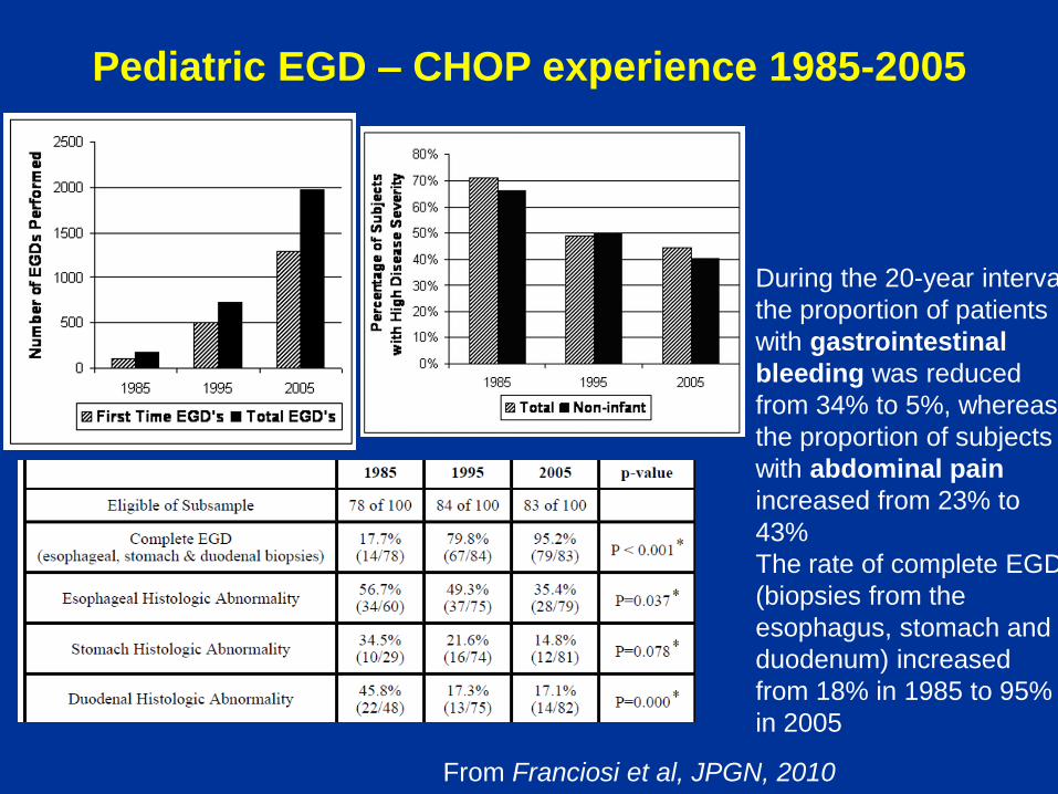
Pediatric EGD – CHOP experience 1985-2005
From Franciosi et al, JPGN, 2010
During the 20-year interval
the proportion of patients
with gastrointestinal
bleeding was reduced
from 34% to 5%, whereas
the proportion of subjects
with abdominal pain
increased from 23% to
43%
The rate of complete EGD
(biopsies from the
esophagus, stomach and
duodenum) increased
from 18% in 1985 to 95%
in 2005

Systematic evaluation of the intestinal
biopsy
• Villous architecture and villous to crypt ratio – Normal villous height/crypt
depth = 3:1 to 4:1
• Surface epithelium and brush border
• Intraepithelial lymphocytes (IEL) or other inflammatory cells – normal IEL’s average 20-
25/100 epithelial cells
• Presence of Paneth, goblet and endocrine cells
• Inflammatory population of lamina propria
• Presence of organisms (Giardia)
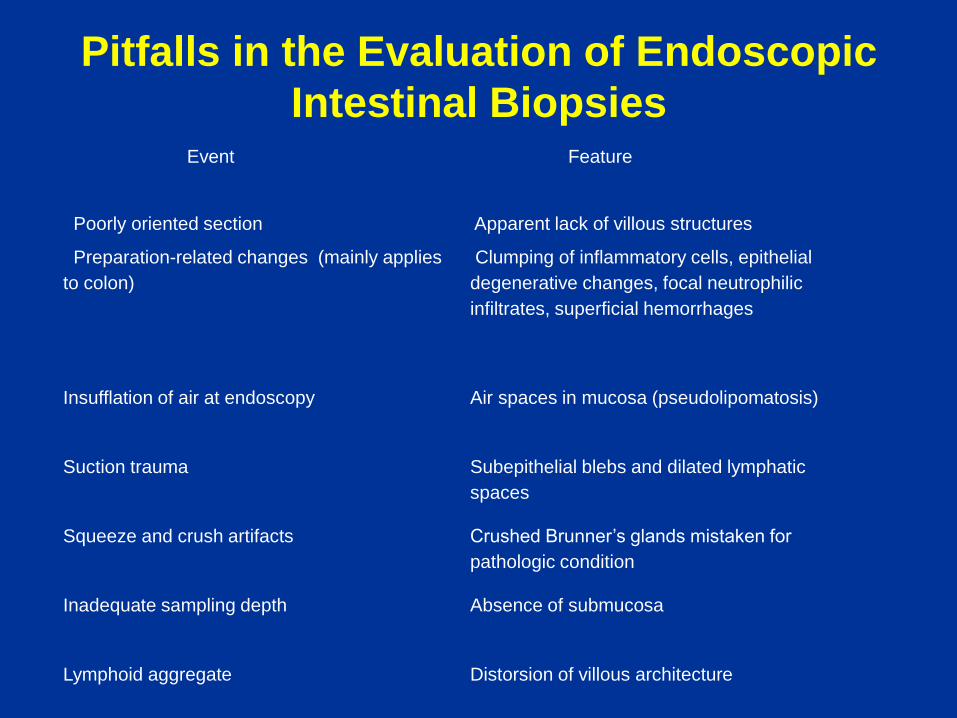
Pitfalls in the Evaluation of Endoscopic
Intestinal Biopsies Event Feature
Poorly oriented section Apparent lack of villous structures
Preparation-related changes (mainly applies
to colon)
Clumping of inflammatory cells, epithelial
degenerative changes, focal neutrophilic
infiltrates, superficial hemorrhages
Insufflation of air at endoscopy Air spaces in mucosa (pseudolipomatosis)
Suction trauma Subepithelial blebs and dilated lymphatic
spaces
Squeeze and crush artifacts Crushed Brunner’s glands mistaken for
pathologic condition
Inadequate sampling depth
Absence of submucosa
Lymphoid aggregate Distorsion of villous architecture
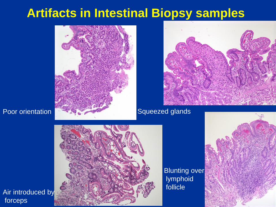
Artifacts in Intestinal Biopsy samples
Poor orientation
Air introduced by
forceps
Squeezed glands
Blunting over
lymphoid
follicle
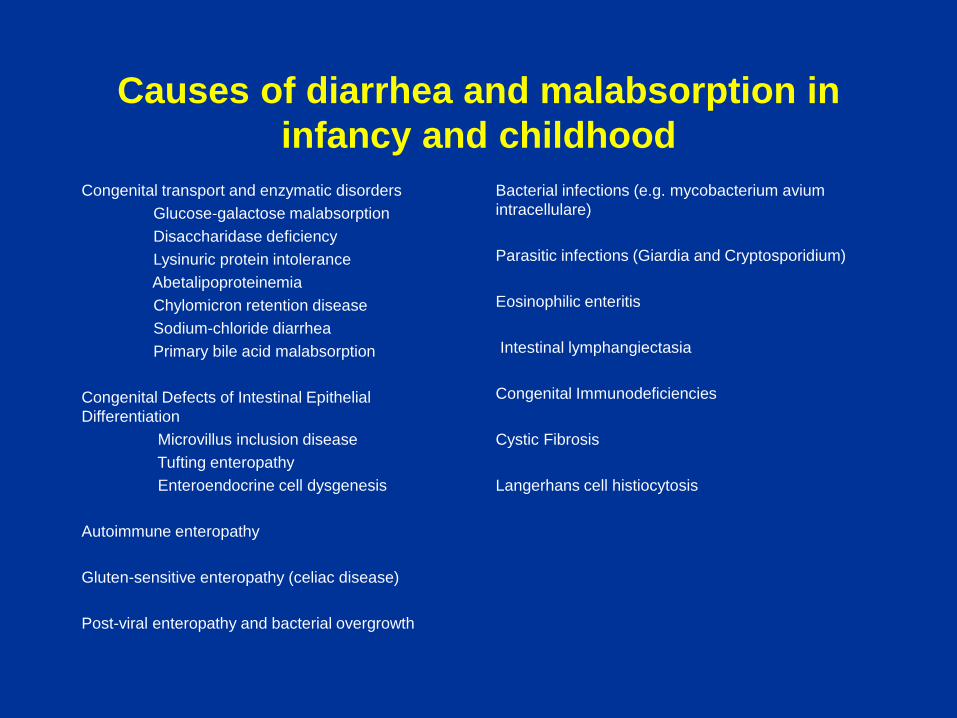
Causes of diarrhea and malabsorption in
infancy and childhood
Congenital transport and enzymatic disorders
Glucose-galactose malabsorption
Disaccharidase deficiency
Lysinuric protein intolerance
Abetalipoproteinemia
Chylomicron retention disease
Sodium-chloride diarrhea
Primary bile acid malabsorption
Congenital Defects of Intestinal Epithelial
Differentiation
Microvillus inclusion disease
Tufting enteropathy
Enteroendocrine cell dysgenesis
Autoimmune enteropathy
Gluten-sensitive enteropathy (celiac disease)
Post-viral enteropathy and bacterial overgrowth
Bacterial infections (e.g. mycobacterium avium
intracellulare)
Parasitic infections (Giardia and Cryptosporidium)
Eosinophilic enteritis
Intestinal lymphangiectasia
Congenital Immunodeficiencies
Cystic Fibrosis
Langerhans cell histiocytosis

Intestinal Biopsy Findings in
Enteropathies • Normal villous morphology
• congenital chloride diarrhea
• carbohydrate malabsorption
• sucrose isomaltase deficiency
• Villous atrophy +/- inflammation • Celiac disease
• Autoimmune enteropathy and IPEX
• Microvillous inclusion disease
• Epithelial dysplasia (“tufting”)
• Eosinophilic gastroenteritis and dietary protein-induced enteropathy
• Congenital immunodeficiency disorders
• Specific or characteristic features • Fat-filled enterocytes (abetalipoproteinemia, chylomicron retention)
• Infectious agents (i.e. Giardia)
• Absence of plasma cells – immunodeficiency
• Lymphangiectasia
• Metabolic disorders
• Infiltrative disorders

Diarrheal disorders - a pattern-based
approach
• Disorders with preserved or minimally abnormal
villous morphology
• Severe diarrhea of infancy or early childhood with
villous atrophy
• Lymphangiectasia
• Infiltrative disorders

Diarrheal disorders with preserved villous
morphology
• Congenital transport and enzymatic disorders
– Glucose-galactose malabsorption
– COH intolerance
• Lipid transport disorders (abetalipoproteinemia)
• Some infections (Giardia)
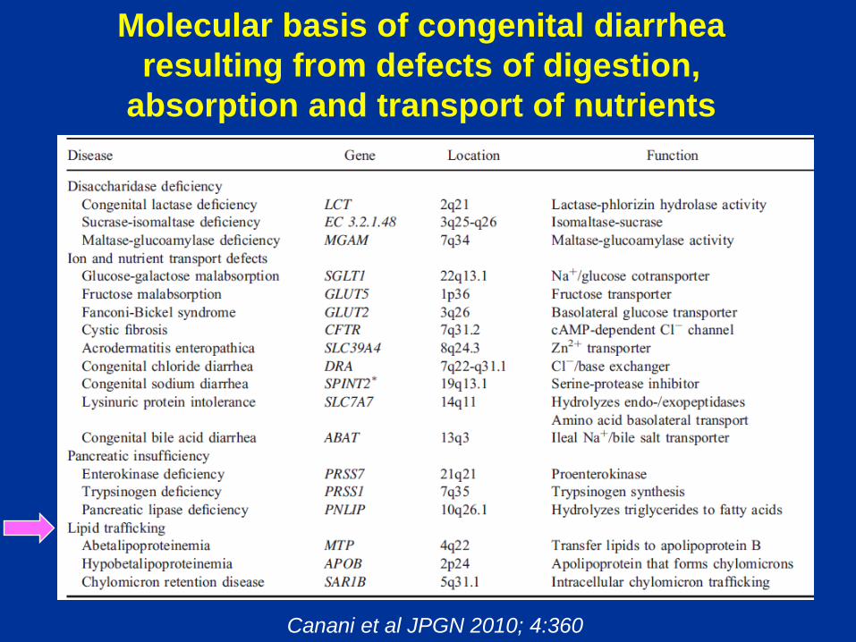
Molecular basis of congenital diarrhea
resulting from defects of digestion,
absorption and transport of nutrients
Canani et al JPGN 2010; 4:360

Abetalipoproteinemia
• AR
• Deficiency of Microsomal
Triglyceride Transfer Protein,
4q22
• Absence of apo-B lipoproteins
• Fat malabsorption with
neurologic and visual symptoms
• Low serum cholesterol and
triglycerides
• Irregular non-membrane bound
vacuoles on EM (absence of fat
in lacteals)
Acanthocytosis

Chylomicron Retention Disease (Anderson
disease)
Secretion Associated, Ras
Related GTPase 1B (SAR1B)
gene; 5q31
Encodes protein carrier GTPase
involved in transport from ER to
Golgi
Neurologic manifestations less
severe than abetalipoproteinemia

Key Features of Lipid Transport
Disorders
• Infantile diarrhea and fat malabsorption
• Low fasting serum cholesterol and triglycerides
• Deficiencies in fat-soluble vitamins
• Neurologic and visual manifestations
• Acanthocytosis
• Lipid-filled small bowel epithelium with preserved villous architecture
• Caution: overinterpretation of epithelial fat in normal patients after ingestion of lipids

An initially mysterious case with severe
CHO intolerance
• 9 month-old boy referrred from OSH for early-onset diarrhea, severe CHO intolerance, adrenal and exocrine pancreatic insufficiency
• Parental consanguinity
• Normal or non-specific histological features; PAS and IHC for chromogranin, CD10, MOC 31 normal

Prohormone convertase 1/3 deficiency
• Whole exome sequencing
identified homozygosity for
mutations in PCSK1
• Proprotein Convertase 1/3 -
endoprotease which converts
prohormones to active forms
normal patient
Chr A
PC1
Bandsma, J Clin Gastro 2013; 47:10

Preserved villous morphology - Giardia
• Flagellated protozoan
• Occurs worldwide
• Easily overlooked in
biopsies
• Immunocompetent and
immunocompromised
individuals (X-linked
agammaglobulinemia, IgA
deficiency)
– Look for plasma cells
– Immunostain for IgA

Diarrheal disorders with villous atrophy
• Congenital disorders of epithelial differentiation
– Microvillous inclusion disease
– Tufting enteropathy
– Neuroendocrine dysgenesis
• TPN dependent
• Small bowel transplantation only cure in many cases
• Autoimmune enteropathies
• Immunosuppression
• Celiac disease
• Gluten withdrawal

EVOLVING ETIOLOGIES OF SEVERE PROTRACTED
DIARRHEA IN CHILDREN IN ITALY
1977-1993 (N=38) 1993-1996 (N=32) 1997-2001 (N=61)
ETIOLOGY n(%) n(%) n(%)
Enteric infection 18 (48) 4 (12) 2 (3)
Food intolerance 8 (22) 3 (10) 10 (17)
Autoimmune enteropathy 2 (5) 8 (25) 7 (12)
Structural enterocyte defect 2 (5) 7 (22) 16 (26)
Celiac disease 1 (2.5) 0 (0) 0 (0)
Eosinophilic enteropathy 1 (2.5) 1 (3) 0 (0)
Lymphangiectasia 1 (2.5) 1 (3) 2 (3)
Motility disorders 2 (5) 3 (9) 16 (26)
Munchausen syndrome 0 (0) 0 (0) 1 (15)
Unknown 3 (7.5) 5 (16) 7 (11.5)
From: Guarino A and DeMarco G. Persistent Diarrhea, chapter 10, in Pediatric Gastrointestinal Disease, 4th edition (2004)

Microvillus Inclusion Disease (MVID)
Severe secretory diarrhea during 1st week
of life
Villous atrophy without significant
inflammation
Abnormal mucosal staining by PAS,
CD10, villin
Abnormal mucosal ultrastructure
MYO5B gene; 18q21
Encodes myosin Vb
Regulates intracellular trafficking
Intestinal transplantation only
cure for most patients
20% -25% of patients have low
GGT cholestasis before or after Itx
PAS, case normal

Microvillous inclusion disease
normal Microvillous Inclusion Disease
Proportion of enterocytes with typical inclusions is variable

“Atypical” microvillous inclusion disease
• Some patients have an
atypical presentation
with later onset of
symptoms, milder
disease and even
spontaneous cures
3 yr-old boy from UAE – symptoms
began in first few months
Mutation in the gene coding for
Syntaxin 3, chr 11q21 (WES)
MVID is likely genetically and clinically
heterogeneous

Congenital Tufting Enteropathy (CTE)
• Diarrhea during first weeks of life
• Higher frequency in families from the Gulf states
• No diagnostic ultrastructural features (abnormal desmosomes)
• Genetically heterogeneous
– EpCAM mutations (2p21)
• Isolated GI disease
– SPINT 2 mutations (19q13)
• Associated anomalies (keratitis, choanal atresia)

Congenital Tufting Enteropathy
2 months
4 months
6 months
Characteristic features evolve with time

Congenital Tufting Enteropathy –
Immunostain for EpCAM
EpCAm MOC 31 + control
EpCAm MOC 31 - case
IHC for MOC 31 normal in pts with
SPINT 2 mutations

Case Description:
23 day old female infant presented with a
history of diarrhea since birth.
Pregnancy was complicated by gestational
diabetes.
Infectious/immune work-up negative
Diarrhea resolved when enteral feeds were
held, but recurred with feedings.
Hyperglycemia first presented at age 5 1/2
months requiring insulin therapy.
Patient’s clinical condition deteriorated and she
passed away at 11 months of age secondary to
TPN liver disease
Duodenal biopsy, 5 months
…initially a mystery….

Normal duodenum (chromogranin) Current case
Chromogranin immunostain
No enteroendocrine cells per IHC
Neurogenin-3 -/- mice lack endocrine cells in pancreas and intestine,
and die in the first days of life

Enteroendocrine cell dysgenesis
NEUROG 3 mutation (10q21.3) Congenital diarrhea and eventual type I diabetes TPN-dependent; bowel transplantation No immune deficiency or autoimmunity; no autoimmune ab’s Non-specific changes on H+E No enteroendocrine cells per IHC for chromogranin NEUROG 3 is a protein involved in gut and pancreatic endocrine development Pts with autoimmune polyglandular syndrome have been described with diarrhea due to transient loss of enteroendocrine cells

Congenital disorders of
Immunomodulation – autoimmune
enteropathies
• Immune dysfunction, polyendocrinopathy, enteropathy,
X-linked (IPEX)
• IPEX-like syndomes
• Autoimmune polyendocrinopathy-candidiasis-ectodermal
dystrophy (APECED), autoimmune polyglandular
syndrome type I (APS 1)

Autoimmune Enteropathy
• Most common severe enteropathy of infancy
• Rarely observed in adults, but may account for some
cases of refractory celiac disease
• Heterogenous entity
• Severe early-onset diarrhea, male preponderance
• Concomitant colitis and gastritis present in majority
• Circulating gut-autoantibodies
• Autoimmune phenomena (thyroiditis, glomerulonephritis,
type I diabetes) in majority of cases
• Favorable response to immunosuppression (Tacrolimus)
• BMT attempted in some cases

Autoimmune Enteropathy
• Severe villus atrophy
• Marked inflammatory destruction of crypts
• Concomitant colitis and gastritis
• Few surface intraepithelial lymphocytes
• Marked decrease in Paneth and goblet cells
• Increased apoptosis
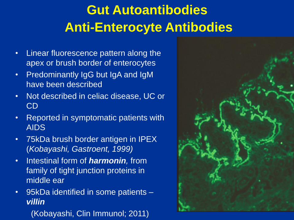
Gut Autoantibodies
Anti-Enterocyte Antibodies
• Linear fluorescence pattern along the
apex or brush border of enterocytes
• Predominantly IgG but IgA and IgM
have been described
• Not described in celiac disease, UC or
CD
• Reported in symptomatic patients with
AIDS
• 75kDa brush border antigen in IPEX
(Kobayashi, Gastroent, 1999)
• Intestinal form of harmonin, from
family of tight junction proteins in
middle ear
• 95kDa identified in some patients –
villin
(Kobayashi, Clin Immunol; 2011)

Autoimmune enteropathy (AE) –
associated clinical conditions • IPEX
Immunodysregulation / polyendocrinopathy / enteropathy / X-linked.
– Mutation in FOXP3 gene, Xp11.23-q13.3
– FOXP3 codes for a protein called Scurfin which is predominately expressed in CD4+/CD25+ regulator T-cells
5 month-old boy with IPEX

Autoimmune enteropathy (AE) –
associated clinical conditions
• “IPEX-like” – normal
FOXP3 but mutations in
– IL2RA, CD25, ITCH
– STAT1, STAT5b
(signal transducer
and activator of
transcription)
• Disorders of modulation
of inflammation
duodenum
colon

Autoimmune-polyendocrine-candidiasis
ectodermal dystrophy (APECED),
Autoimmune polyglandular syndrome,
type I (APLS I)
Duodenum, loss of endocrine cells, IHC for chromogranin
AR
AIRE (Autoimmune Regulator)gene, 21q22
Multiple endocrine manifestations
Autoimmune gastritis with B12 deficiency
Autoimmune hepatitis
Autoimmune enteropathy with loss of enteroendocrine
cells
gastritis

Autoimmune enteropathy in adults
• Protracted diarrhea, weight
loss, malnutrition
• Absence of response to
gluten-free diet or absence
of HLA genotypes
• AEA + variety of
autoantibodies
• T-cell rearrangement studies
neg
• Good response to
immunosuppression
Akram S, et al. Adult autoimmune
enteropathy: Mayo Clinic Rochester
experience. Clin Gastroenterol Hepatol
2007

Autoimmune enteropathy in adults
18 yr old female with thyroiditis
and protein-losing enteropathy 36 year old male with watery diarrhea
Anti-goblet antibodies
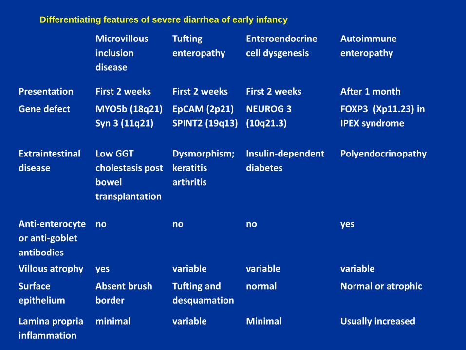
Microvillous
inclusion
disease
Tufting
enteropathy
Enteroendocrine
cell dysgenesis
Autoimmune
enteropathy
Presentation First 2 weeks First 2 weeks First 2 weeks After 1 month
Gene defect MYO5b (18q21)
Syn 3 (11q21)
EpCAM (2p21)
SPINT2 (19q13)
NEUROG 3
(10q21.3)
FOXP3 (Xp11.23) in
IPEX syndrome
Extraintestinal
disease
Low GGT
cholestasis post
bowel
transplantation
Dysmorphism;
keratitis
arthritis
Insulin-dependent
diabetes
Polyendocrinopathy
Anti-enterocyte
or anti-goblet
antibodies
no no no yes
Villous atrophy yes variable variable variable
Surface
epithelium
Absent brush
border
Tufting and
desquamation
normal Normal or atrophic
Lamina propria
inflammation
minimal variable Minimal Usually increased
Differentiating features of severe diarrhea of early infancy

Criteria for Diagnosis of Celiac
Disease
3 biopsies
• Characteristic morphologic
abnormalities in small
bowel of patient ingesting
gluten.
• Improvement/normalization
of biopsy findings on
gluten-free diet.
• Deterioration of mucosal
morphology following
gluten challenge.
1 biopsy
• Characteristic morphologic
abnormalities in small
bowel of patient ingesting
gluten + anti-TTG, anti-
endomysial (EMA) or anti-
gliadin antibodies.
• Clinical and serologic
remission while on gluten-
free diet.
Original ESPGAN criteria (1970) Revised ESPGAN criteria (1990)

Celiac disease (CD)
• Major clinical presentations – “Classic” – malabsorption,
diarrhea, wasted buttocks.
– Vomiting, abdominal distension – especially in children < 1 year of age
– Failure to thrive
– Iron resistant anemia
– Rickets
– Short stature
– Pubertal delay
– Personality problems
– Dermatitis herpetiformis
– Dental enamel hypoplasia
– Epilepsy with cerebellar calcifications
• Conditions associated with an increased prevalence of CD – Type I Diabetes mellitus
– Down Syndrome
– Selective IgA deficiency
– Juvenile chronic arthritis
– Autoimmune liver disease
– Autoimmune thyroiditis
– Turner Syndrome
– Williams Syndrome

Celiac Disease
• Increased IEL’s
(> 40/100 epithelial
cells )
• Villus atrophy
• Crypt hyperplasia
• Enterocyte damage
• Increase in
mononuclear cells,
eosinophils and
variable numbers of
neutrophils in the
lamina propria

Celiac disease – minor histological
changes
3 yo girl + tTG and EMA
CD 8 CD 4

Celiac Disease
Histological Changes
• Great variability (essentially normal villi to severe atrophy)
• Entire bowel involved, including stomach and colon
• Damage most severe proximally, in some cases limited to the proximal duodenum (up to 25 % of pediatric patients, Bonamico, 2004) distal proximal

Celiac Disease
Gluten diet Gluten-free diet

Celiac disease – differential diagnosis
Autoimmune enteropathy (IPEX and
IPEX-like)
Crypt-destructive inflammatory process with loss of goblet and
Paneth cells
Crypt apoptosis
No significantly increased intraepithelial lymphocytes
Circulating anti-enterocyte antibodies
Non-gluten food protein intolerance Often associated with increased eosinophils
Variable villous atrophy
Positive allergy testing
Kwashiokor History of severe protein malnutrition
No increased IELs
Primary Immunodeficiency Absence or paucity of plasma cells in the biopsy
Nodular lymphoid hyperplasia
Graft versus host disease Crypt apoptosis with minimal inflammatory infiltrate
Helicobacter pylori infection Usually in association with active gastritis due to Helicobacter
pylori rather than lymphocytic gastritis
Changes rarely extend beyond the duodenal bulb
Viral enteritis Surface epithelial degenerative and regenerative changes
Some infections may have increased IELs
Histologic changes rapidly revert as symptoms abate

Celiac disease – differential diagnosis
12 yo boy Kwashiorkor
7 yo boy IgA deficiency
17 month female
collagenous
enteritis

Duodenal Lymphocytosis with Normal
Villi
Kakar13 (n=43) Mahadeva14 (n=14) Hammer16 (n=100 Shmidt36 (n=48) Aziz15 (n=100)
Celiac disease 9% 21% 18% 19% 16%
Tropical sprue 1% — 1% — —
H. pylori gastritis — — 6% 6% 14%
Bacterial overgrowth 5% — 3% — —
NSAIDs 14% — 8% 20% 21%
IBD 12% — 8% 11% 2%
Autoimmune
conditions 14% — 6% — 4%
Unexplained 7% 21% 26% 33% 34%
IBS 9% 14% 20% — —
Other 28% 43% 4% 11% 9%
From Lauwers, Modern Path, 2015

Villous atrophy and negative celiac
serology
From DeGaetani et al, Am J Gastroenterol 2013

Intestinal lymphangiectasia
Key features:
• Protein losing enteropathy with malabsorption, hypoalbuminemia, hypogammaglobulinemia, and lymphopenia with secondary immunodeficiency
• May primarily affect gastrointestinal tract or involve multiple organs
• Secondary lymphangiectasia can result from congenital heart disease, lymphatic obstruction (e.g. malrotation), or vascular malformation
• Blunted villi with dilated lacteals
Pitfalls in diagnosis:
• Limited biopsies may not show features
• Separation and tearing artefacts may resemble the true lesion
• Occasional villi may normally contain dilated lacteals
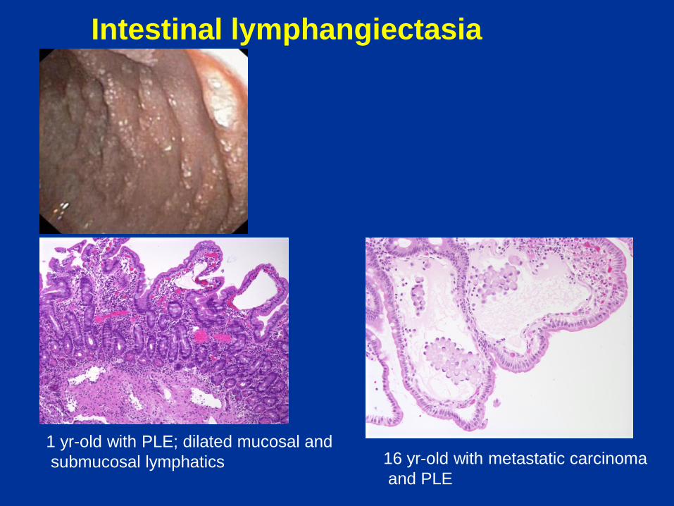
Intestinal lymphangiectasia
1 yr-old with PLE; dilated mucosal and
submucosal lymphatics 16 yr-old with metastatic carcinoma
and PLE

Infiltrative disorders of the GI tract that may
present with early-onset diarrhea
• Langerhans cell histiocytosis
• Myofibromatosis
• Infantile systemic hyalinosis

Langerhans cell histiocytosis
Cd1a

Myofibromatosis

Infantile systemic hyalinosis
Autosomal recessive
Painful joint contractures
Widespread deposition of glycoprotein-like
substance in skin, GI tract, heart, muscle
CMG2 (Capillary morphogenesis protein gene 2)
gene mutations on chr. 4q21

Approaches to the small bowel biopsy
Conclusions 1.
• Many disorders causing severe enteropathy have
been delineated only in the last few decades and
many rare monogenic disorders are being identified
by modern genetic techniques (WES)
• Morphologic findings on H&E in many severe
enteropathies may be inapparent, subtle or non-
specific
• The work-up of intestinal biopsies in severe infantile
enteropathies should include use of special stains
(PAS), immunohistochemistry (CD10, villin, EpCAM,
chromogranin) and EM; serum for indirect IF may be
useful to screen for anti-enterocyte antibodies

Approaches to the small bowel biopsy
Conclusions 2.
• Celiac disease may have a variable clinical and
histologic presentation
• Typical changes in celiac disease in children may be
confined to the proximal duodenum
• True lymphangiectasia may be difficult to distinguish
from artifact and needs good clinical-endoscopic
correlation
• Infiltrative disorders may cause severe diarrhea in
infancy
