approach to hemolysis - ksumscksumsc.com/download_center/2nd/2) gnt block/teams... · haemolysis:...
TRANSCRIPT

Objectives:
References:436 girls & boys’ slides435 teamwork slides Editing file
Do you have any suggestions? Please contact us!
@haematology436 E-mail: [email protected]
or simply use this form
Approach to Hemolysis
Important.Extra.Notes
To know the function of platelets and the relationship between the platelet count in peripheral blood and the extent of abnormal bleeding.
• To know about the diseases associated with 1) a failure of platelet production 2) a shortened platelet lifespan, especially immune thrombocytopenic purpura (ITP).
• To know the principles of investigation of patient suspected of having a haemostatic defect.
To understand the role of platelets, blood vessel wall and coagulation factors in normal haemostasis.
• To know the classification of haemostatic defects.
• To know the platelet morphology and life span.
• To know the platelet function and diseases due to platelet function disorders.
• To know the causes of thrombocytopenic purpura and non-thrombocytopenic purpura.

Haemolysis:It is a Premature destruction of RBCs.
Hemolysis is due to:
• Defect in the RBCs (intra-corpuscular) as in congenital hemolytic Anaemia.
• Defect in the surrounding environment (extracorpuscular) as in acquired Anaemia
Haemolytic Anaemias:• Haemolysis is the shortening of the lifespan of a mature red blood cell.
• haemolysis will result in anaemia more readily
• increased red cell output from the bone marrow stimulated by erythropoietin
• This mechanism compensates the loss of RBCs, and this requires an adequately function bone marrow and effective erythropoiesis
• More marked reductions in red blood cell life span 5-10 days from the usual 120 days
• will result in haemolytic anaemia
• a suboptimal marrow response is seen
• Pallor, lethargy• Jaundice• Splenomegaly• Gall stones (Pigment – bilirubin)• Dark urine (urobilinogen)• Bone deformity (In some types of • haemolytic anaemia) especially in congenital
• Leg ulcers (in some types of haemolytic• anaemia). Especially in sickle cell
Clinical Features of Hemolysis:
Intravascular haemolysis: extravascular haemolysis:
It is the process of breakdown of red cells directly in the circulation.
It is the excessive removal of red cells by cells of RE
system in the spleen and liver. main laboratory features of intravascular
haemolysis:• Haemoglobinaemia (free Hemoglobin in blood) and
Haemoglobinuria.• Haemosiderinuria (Iron storage protein in the spun
deposit of urine).
HEMATOLOGY TEAM 436
2
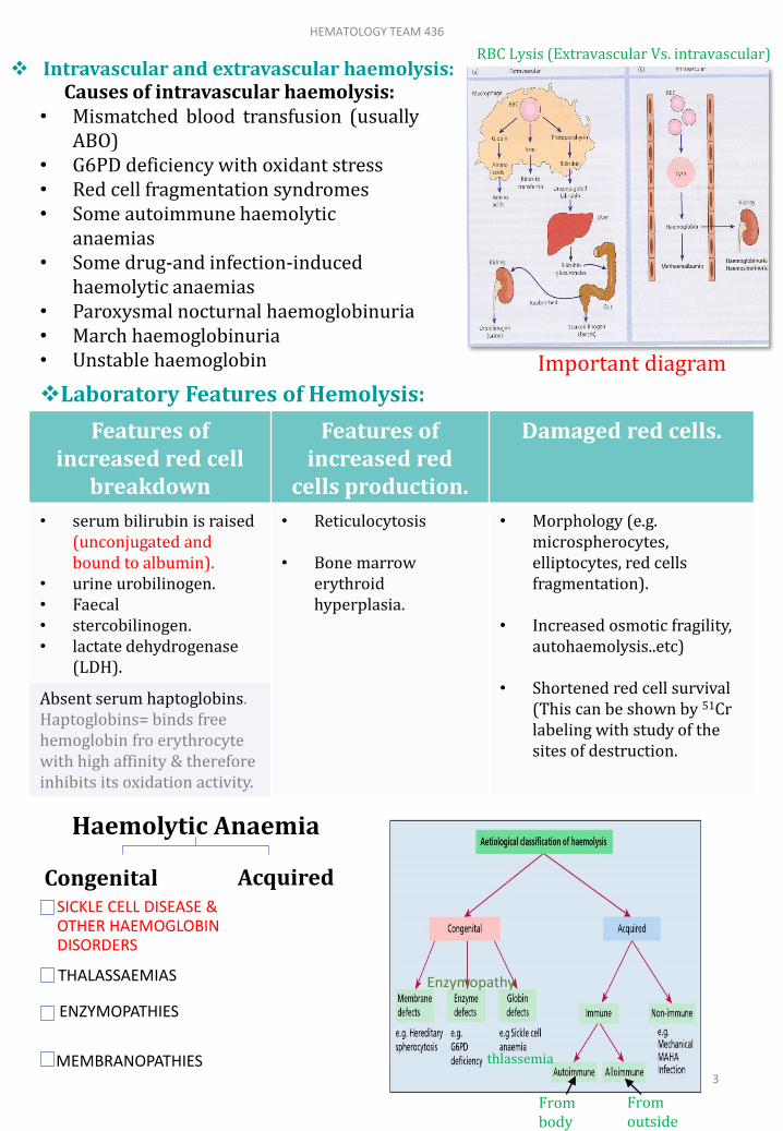
Laboratory Features of Hemolysis:
Intravascular and extravascular haemolysis:
Features of increased red cell
breakdown
Features of increased red
cells production.
Damaged red cells.
• serum bilirubin is raised (unconjugated and bound to albumin).
• urine urobilinogen.• Faecal• stercobilinogen.• lactate dehydrogenase
(LDH).
• Reticulocytosis
• Bone marrow erythroid hyperplasia.
• Morphology (e.g. microspherocytes, elliptocytes, red cells fragmentation).
• Increased osmotic fragility, autohaemolysis..etc)
• Shortened red cell survival (This can be shown by 51Cr labeling with study of the sites of destruction.
Absent serum haptoglobins. Haptoglobins= binds free hemoglobin fro erythrocyte with high affinity & therefore inhibits its oxidation activity.
Causes of intravascular haemolysis:• Mismatched blood transfusion (usually
ABO)• G6PD deficiency with oxidant stress• Red cell fragmentation syndromes• Some autoimmune haemolytic
anaemias• Some drug-and infection-induced
haemolytic anaemias• Paroxysmal nocturnal haemoglobinuria• March haemoglobinuria• Unstable haemoglobin
Haemolytic Anaemia
CongenitalSICKLE CELL DISEASE & OTHER HAEMOGLOBIN DISORDERS
THALASSAEMIAS
ENZYMOPATHIES
MEMBRANOPATHIES
Acquired
Enzymopathy
thlassemia
Frombody
Fromoutside
Important diagram
HEMATOLOGY TEAM 436
3
RBC Lysis (Extravascular Vs. intravascular)

Target cells + sickle shape
Name Substitution
Hb. S α2 β2 - 6 GLU VAL
Hb. C α2 β2 - 6 GLU LYS
1910: 1st published report of sickle cell anaemia (Herrick)
1949: Pauling et al : chemical difference between HbA and HbS
1956: Ingram: Fingerprinting
β glu val
Abnormal Haemoglobins
(Haemoglobinopathies):
• Some Known Haemoglobin Mutants : (all in beta chain)
DNA Coding for the Amino-Acid in the sixth position in the β-chain
• Normal:-5 6 7
Amino Acid pro glu gluDNA Base Composition CCT G A G G A G
• Sickle:-
DNA Base Composition CCT G T G G A GAmino Acid pro val glu
5 6 7
In position 6 it is supposed to be the amino acid glutamic acid but it transformed into Valine
HEMATOLOGY TEAM 436
4
Sickle Cell Anemia
• It is important to Know that the HbS is Due to abnormal Beta-Chain in Amino Acid number 6 wich transforms from glutamate to valine due to mutation!

• Traits heredity:عشان كذا نسوي فحص الزواج
AA: NormalAS: trait – carrier (could transmit the disease)SS: Diseased
AC: Trait (carrier)AS: Trait (carrier) for another disease.CS: Diseased
AS: Sicle cell traitAF-Thal: thalassemia traitSF-Thal: diseased
HEMATOLOGY TEAM 436
5

Sickle cell disease:
1. The sickle cell trait (AS) -
2. Homozygous sickle cell disease (ss) Sickle cell anemia
3. Doubly heterozygous sickle cell disease (2 diseases together)
sickle cell / hemoglobin C disease
sickle cell / thalassemia
Properties of Hb S :1. Solubility decrease 2. Conformational changes-
“tactoid formation” when exposed to O2
3. Sickled cells 4. Irreversibly sickled
cells 5. Increase mechanical
fragility hemolysis 6. Increase viscosity
organ infraction
Factors affecting sickling 1. Oxygen tension:
50-60 mm Hg for SS 20-30 mm Hg for AS
2. pH : inhibited at alkaline
pH (Alkalosis blood decrease
sickling attacks)
Exacerbated by acidification
3. Concentration of Hb S4. Presence of other
hemoglobins Polymerisation: S > D
> C > J=A > F
Sickling at low O2 tension
Increased PH Low O2 tension
Viscosity
Slow blood flow
HEMATOLOGY TEAM 436
6

Factors Precipitating Crises In Sickle Cell Disease :
Infections (Especially Malaria)
Pyrexia fever
Exposure To Cold
Dehydration (the most important factor with pregnancy)
Pregnancy
Crises in sickle cell disease:
Hyperhaemolytic.
Aregenerative or aplastic.
Small vessel occlusion.
Clinical manifestations of sickle cell disease:
Haemolytic anemia
Tissue infraction
Clinical Manifestations in Sickle Anaemia
Pallor (Anaemia)
Jaundice & Dark Urine
Apathy & Anorexia
Hand-Foot Syndrome (Young Children) ( one of earliest signs of sickle cells anemia)
Splenic sequestration (Young children) Hepatic Sequestration
Bones and Joints Pain very sever (the patient may scream from pain).
Abdominal Pain
Hand foot syndrome: swelling-pain-
inflamed small bones
HEMATOLOGY TEAM 436
7

Clinical Manifestations in Sickle Anaemia
Recurrent Infections & Chest Symptoms (Acute Chest Syndrome)
Hepato-Splenomegaly but more significant with thalassemia
(Early Childhood)
(Association with Thalassaemias)
CNS Presentations
Leg Ulceration characteristic for sickle cell patients
Skeletal Deformity
Laboratory Diagnosis of Sickle Cell Disease:
CBC Blood Film Sickle Solubility Test Hb Electrophoresis (Most accurate)
Genetic Study
Indications for Blood Transfusion in Sickle Cell Anaemia: Not important
Splenic sequestration (Stuck RBCs-spleen enlargment-dysphanction) Hepatic sequestration Aplastic crisis Overwhelming infections Elective or emergency surgical operation Severe painful crisis associated with severe haemolysis Pregnancy
Indications for exchange transfusion: Not important Strokes Pulmonary infarcts with
infection Pregnancy (Severe persistent
painful crisis) Priapism prolonged erection of
the penis Preparation for major surgery
We put the blood in a media
Right: normal (translucent).
Left: abnormal (turbid due to RBCs
breakdown).
HEMATOLOGY TEAM 436
8
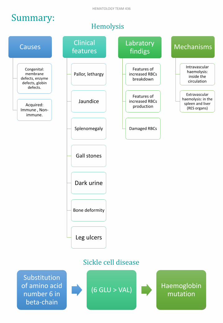
Causes
Congenital: membrane
defects, enzyme defects, globin
defects.
Acquired: Immune , Non-
immune.
Clinical features
Pallor, lethargy
Jaundice
Splenomegaly
Gall stones
Dark urine
Bone deformity
Leg ulcers
Labratoryfindigs
Features of increased RBCs
breakdown
Features of increased RBCs
production
Damaged RBCs
Mechanisms
Intravascular haemolysis: inside the circulation
Extravascularhaemolysis: in the
spleen and liver (RES organs)
Substitutionof amino acid number 6 in beta-chain
(6 GLU > VAL)Haemoglobin
mutation
Hemolysis
Sickle cell disease
Summary:HEMATOLOGY TEAM 436
9

MCQs:1-A patient came to the ER complaining of dark urine. On examination, they found jaundice in his sclera, splenomegaly and haemoglobinuria. What is the most common cause of these manifestations?A-Extravascular haemolysis.B-Intravascular haemolysis.C-Both A&B.
2-What is the substitution mutation in Hb.S?A-α2 β2 - 6 GLU > VAL B-α2 β2 - 6 GLU > LYSC-α2 β2 - 26 GLU > VAL
3-Haemolytic anemia is associated congenitally with which of the following:A-Sickle cell disease.B-Thalassemia.C-Both A&B.
4-In Which of the following we will find sickle cell disease & Alpha-Thalassemia in the patient at the same time?A-The sickle cell trait (AS)B-Homozygous sickle cell disease (ss)C-Doubly heterozygous sickle cell disease .
ANSWERS:1-B2-A 3-C4-C
Team membersHeba AlnasserHanin BashaikhShrooq AlsomaliShahd AlsowaidanFahad Al-Askar
Team LeadersSafa Al-OsaimiAbdulaziz Al-Hussainy
Good Luck!
HEMATOLOGY TEAM 436
10