approach to anemia | dr abdelhadi by hashem al-dujaily
TRANSCRIPT

Approach To Anemia | Dr Abdelhadi
By Hashem Al-Dujaily
_________________________________________________________
Case: A 10-year-old male previously healthy, presented to JUH since his parents
noticed yellowish discoloration in his face of 5 days duration, and shortness of
breath upon climbing a few stairs of 10 days duration.
First complaint possible interpretation:
Yellowish discoloration of the face ➔ It could be jaundice or pallor.
Since it started as yellowish discoloration of the sclera ➔ it is jaundice.
When did it start, where, and progression? It started in the sclera and progressed
to involve the face.
Second complaint possible interpretation:
Easily fatigued.
What are symptoms of anemia?
1. Dyspnea.
2. Dizziness.
3. Easy fatigability.
4. Palpitations.
5. Headache
What are the symptoms of anemia that can be seen in a 2-month-old child?
1. Poor sucking or poor feeding.
2. Poor activity.
3. Pallor.
Poor feeding and poor activity can be an early sign of:
1. Sepsis.
2. Anemia.
3. Heart failure.

Why not
sickle cell
anemia?
There are some classic presentations in medicine, such examples are:
o Headache, vomiting, and fever ➔ Meningitis.
o Fever, cough, and tachypnea ➔ Pneumonia.
o Fever, and diarrhea ➔ Gastroenteritis.
o Fever, and RLQ pain ➔ Appendicitis.
o Jaundice, and pallor ➔ Hemolytic Anemia (OUR CASE )
What things can be asked in history of presenting illness that go with hemolysis?
1) Urine!!
➢ Dark urine can be seen in hemolysis.
➢ Dark urine (and pale stools) can be seen in obstructive disease too.
2) Triggering factors:
Example: If patient went to a farm of fava beans, and ate some of it, and then
developed symptoms. ➔ G6PD Deficiency (Favism)
Triggering factors of G6PD that can induce hemolysis:
1. Infection.
2. Sulfa Drugs.
3. Naphthalene balls.
4. Henna.
3) Family History:
If this patient’s father had a medical condition that got better with splenectomy.
What do you think? What is the hematologic disease that gets better with
splenectomy?
Hereditary spherocytosis.
4) Past Medical History.
▪ If had transfusion before.
➢ If had regular transfusions ➔ thalassemic.
See Next Page

In sickle cell disease, you do not do splenectomy!!
Since the patient’s spleen, from the start becomes atrophied ➔ hypersplenism.
Clarification:
In sickle cell, the RBCs are more sticky, fragile, and have short life span, causing
Vaso-occlusion of vessels, if occlusion of vessels occurred:
o In brain ➔ stroke.
o In eyes ➔ blindness and retinopathies.
o In lung ➔ acute chest syndrome.
o In renal ➔ sickle cell nephropathy.
o In hips ➔ avascular necrosis of the femur heads.
o In spleen ➔ spleen atrophy.
That is why in “sicklers”, one of the supportive care measures, as soon as you
diagnose someone with sickle cell anemia, you must start them on:
• Hydroxyurea (to prevent painful attacks of pain crisis).
• Transfusion has indication in sickle cell disease, those with stroke, to
decrease hemoglobin, or in acute chest syndrome.
• Recurrent painful attacks, due to ischemia, mainly bone pain ➔ need
hydration.
While in thalassemia major, we may tend to splenectomy to reduce
the frequency of transfusions.
What is the importance of spleen?
1. Destruction of hematopoietic cell lines. 2. Activity against encapsulated organisms; H-influenza, Neisseria meningitis,
and streptococcus pneumonia.
❖ That is why someone with splenectomy, you must give vaccines against them.
❖ We do not try to do splenectomy before the age of five years to avoid having
overwhelming sepsis, since most common cause is streptococcus pneumonia.
But the problem with “sicklers”, splenic function is already diminished!
o They should receive the extra vaccines. For instance, streptococcus pneumonia
is not included in the national vaccines program and thus should be included.
o Sicklers are like splenectomized patients, need special precautions if presented
to the ER ➔ need to order CBC and may need admission!

Physical Examination
Vital Signs:
o Heart Rate: Increased.
o Respiratory Rate: Increased.
o Blood Pressure Decreased.
o Temperature
o Oxygen Saturation: could be very low.
General Examination:
Jaundice is seen in the upper part of the sclera, while pallor in conjunctiva,
mucous membranes, or in hand creases.
Examine Liver and Spleen.
Our patient findings:
Tachycardic, a bit tachypneic, spleen 4 cm below costal margin, no hepatomegaly.
___________________________________________________________________
Jaundice, Pallor, and Splenomegaly what labs do you do?
✓ CBC
✓ LFT
For example: if Liver function test results are as follows; What do you think?
o Total bilirubin 3mg/dl.
o Direct bilirubin 1mg/dl.
You define direct hyperbilirubinemia if d/T (direct/total) is more than 20%, as it is
wrong to subtract them and say 3-1 equals 2!
Direct/Total ➔ 1/3 equals 30% ➔ Direct hyperbilirubinemia.
This indicates a hepatic or post-hepatic cause. However, how other LFT can help
in narrowing down my differential diagnosis?
ALT and AST ➔ Hepatocellular injury.
Alkaline Phosphates and GGT ➔ Obstructive causes.
What is more specific to the liver? ALT or AST?
✓ ALT (L for Liver).
What are the synthetic liver functions?
1. Coagulation factors. ➔ Coagulation profile (PT and PTT).
2. Albumin.
3. Glucose.
Simple radiology to know whether it is a hepatic or post hepatic cause?
o Abdominal Ultrasound to see the bile duct ➔ However, it is operator
dependent.
o Golden Standard: ERCP or MRCP.
➢ Keep in mind that ERCP is both diagnostic and therapeutic just like cardiac
catheterization.
A patient 50 years old that has stricture in common bile duct, MCC is:
✓ Cholangiocarcinoma.
How to diagnose malignancies in general?
1. Histopathology (like bone marrow biopsy).
2. Fluid Cytology (to diagnose cholangiocarcinoma since there is no mass).
Major causes of hepatic injury?
1. Infectious ➔ Hepatitis.
2. Toxic ➔ Drug toxicity.
3. Autoimmune ➔ Autoimmune hepatitis.
4. Genetics ➔ Dubin Johnson syndrome.
5. Metabolic ➔ Wilson.
If nothing found ➔ need something invasive to diagnose liver disease: biopsy.

Our Patient Lab Results:
Hemoglobin 7.2
Total Bilirubin 4
Direct Bilirubin 0.4
Direct/Total ➔ 0.4/4 equals 10% ➔ Indirect hyperbilirubinemia.
What are other laboratory markers of hemolysis other than indirect bilirubin?
1. LDH ➔ High.
2. Haptoglobin ➔ Low.
But what is the most important marker?
Reticulocyte Count.
Normally, reticulocytes, which are immature RBCs, represent 1-2% of peripheral
blood. However, this percentage is increased as compensatory mechanism from
the bone marrow in anemia.
Reticulocytosis is seen in Two Hs: Hemolysis, and Hemorrhage.
Low reticulocyte count: in Bone marrow failure or invasion.
In general, in hematology, the bone marrow makes the cell types, so either the
problem is in the bone marrow; aka the bone marrow isn’t producing a
particular cell line, or the problem is in the peripheral blood.
If we look at the CBC, what cell types we look at?
1. Hemoglobin.
2. White blood cells.
3. Platelets.
Low WBC ➔ Leukopenia.
Low neutrophils ➔ Neutropenia; defined as absolute neutrophilic count
(ANC) less than 1500, calculated by multiplying percentage of neutrophils
by the white blood cells count.
Low platelets ➔ Thrombocytopenia.
High platelet count ➔ Thrombocytosis.
Low RBCs ➔ Anemia.

Definition of anemia varies according to age:
Newborn’s hemoglobin is nearly 20 and at 2 months of age it starts to go down to
9 (physiological anemia) and then goes up again.
Up to 7th month of age: up to 11 we consider it as anemia.
___________________________________________________________________
High White Blood Cell Count ➔ You think of infectious.
But if WBC count is in the 70-80 thousand ➔ Leukemia.
___________________________________________________________________
High Neutrophils, High WBC count ➔ Bacterial Infection.
High Lymphocytes, High WBC count ➔ Viral Infection.
High Eosinophils, High WBC count ➔ Allergic or Parasitic infections.
High Monocytes, High WBC count ➔ Fungal or Malignancy.
High Basophils, High WBC count ➔ Malignancy.
___________________________________________________________________
WBCs high, in 60-70 thousand, does it differ if you find it in an elderly or a young
patient?
Platelets are acute phase reactants; goes up with anything; stress, burn, surgery…
Secondary or reactive thrombocytosis.
Primary thrombocytosis underlying cause is malignant condition called
myeloproliferative disorder.
So, we fear thrombocytosis more in an elderly, he needs a very long workup.
➢ Bone marrow biopsy, genetic studies, etc.
However, if platelet count is high in children, there is a huge differential such as
iron deficiency anemia, Kawasaki disease, or even infectious.
o Bone marrow biopsy in children is invasive, it is not done bedside like in
adults! You need to consult surgery, and there is risk of anesthesia, you do
not do such invasive procedures without a real need.

❖ 1 year old iron deficiency anemia, pagophagia, what do you think?
Malnutrition.
❖ 50-year-old male iron deficiency anemia, what do you think? Colon cancer
due to ongoing blood loss.
❖ Teenage females with iron deficiency anemia, what do you think? Heavy
menses.
___________________________________________________________________
What are the mechanisms of peripheral destruction of cell lines?
1) Autoimmune:
o Immune thrombocytopenic purpura: autoantibodies against platelets.
o Autoimmune hemolytic anemia: autoantibodies against RBCs.
o Autoimmune neutropenia: autoantibodies against neutrophils.
2) Mechanical/endothelial injury that affects platelets and RBCs:
o Microangiopathic hemolytic anemia (MAHA): schistocytes, fragmented
RBC, or helmet cells
o Hemolytic Uremic Syndrome (HUS): Triad of hemolytic anemia, renal
failure, and thrombocytopenia.
o Thrombotic Thrombocytopenic Purpura: Pentad ➔ HUS + Fever +
Neurological (altered mental status).
o Disseminated Intravascular Coagulation (DIC).
o Mechanical Valve Replacement.
o HELLP Syndrome.
o Big Hemangiomas.
3) Hypersplenism
Most common cause of acquired pancytopenia.
If a patient has pancytopenia, and we did bone marrow biopsy to know whether
the problem is peripheral or not:
We look at the precursors: megakaryocytes, myeloid, and erythroid cells:
If high ➔ Peripheral Destruction.
If low ➔ Aplastic Anemia (all cell lines affected in BM).

Back to our patient, he had:
o Hemoglobin 7.2 ➔ Low.
o WBC normal.
o Platelets normal.
Rule: If more than one cell line affected, you should think of problem in bone
marrow.
A patient came to use with platelet count very low (4000), and high WBC count ➔
he had leukemia. So, even if Hemoglobin was normal you should think of a
problem in the bone marrow.
Indications of bone marrow biopsy:
1. Two cell lines affected.
2. Reticulocytopenia.
3. Pancytopenia.
4. Strange cell line (WBC count 70,000).
➔ Our patient has anemia, what is the next step to classify anemias?
We look at the MCV:
If high ➔ Macrocytic anemia: B12 Deficiency, Folate Deficiency, Liver Disease,
Alcoholics, Hypothyroidism, Bone marrow failure syndromes, anti-convulsant.
If low ➔ Microcytic anemia: IDA, thalassemia trait, sideroblastic anemia, anemia
of chronic disease (in late stages), and lead poisoning.
➔ Our patient had MCV 83, so we will start talking about normocytic
anemia:
We need Reticulocyte count to guide us
Reticulocyte count in this patient is 18% ➔ that means he has hemolytic anemia
What about Reticulocyte index? we do not really have to do it now, we only do it
when the reticulocyte count is borderline.

Why do we not need to correct the reticulocyte count in this patient?
Normally we have around 5 million RBCs, so if 2% of them are reticulocytes ➔
that’s 100,000. So, if hemoglobin went down due to anemia, RBCs will be low,
let’s say 2.5 million, thus reticulocyte percentage in this case would be 4%.
That’s why reticulocyte count of 4% in someone with hemoglobin 7 is not
actually Reticulocytosis.
That leads us to another term called absolute reticulocyte count, because the
reticulocyte count is expressed as a percentage of total RBCs, it must be
corrected according to the extent of anemia.
Let us correct it for our patient:
Corrected Reticulocyte count: Reticulocyte % × (Patient Hct/Normal Hct) =
18% x 20/40 = 9% (still high, that is why we did not need to do it since
reticulocyte count was so high anyway, that even with correction it would
remain high).
Low reticulocyte count ➔ indicates a problem in the bone marrow.
High reticulocyte count ➔ indicates hemolysis or hemorrhage.
A test that directs you towards the management of hemolytic anemia:
Direct Coombs test: it helps differentiate between immune hemolytic
anemia (positive) and non-immune hemolytic anemia (negative).
In our patient: direct Coomb’s test showed +4 reaction of IgG.
Immune hemolytic anemia of IgG type ➔ warm immune hemolytic anemia.
We finally diagnosed the patient!!
Most common virus that causes cold hemolytic anemia:
EBV and CMV, in addition to mycoplasma.
How do we treat immune hemolysis?
Immunosuppression, and maybe IVIG. Of course, not by transfusion as the
antibodies are already present and will lead to hemolysis of the new RBCs.
Non-immune causes of hemolysis ➔ 3 main categories:
1) Enzymopathies: diagnosis by G6PD level, etc.
2) Membranopathies: by blood film, osmotic fragility, or flow cytometry.
3) Hemoglobinopathies: diagnosis by hemoglobin electrophoresis.
Can you diagnose spherocytosis from CBC alone?? Yes, high MCHC.
What do you see in hemoglobin electrophoresis?
There are certain types of hemoglobin, the following is each tetramer
composition in addition to their percentage in a normal person:
1) Hemoglobin A or adult hemoglobin: (alpha2beta2 chains)
2) Hemoglobin A2 or trace hemoglobin: (alpha2delta2 chains) <3.5%
3) Hemoglobin F or Fetal hemoglobin: (alpha2gamma2 chains) <1%
Why cannot we survive with fetal hemoglobin? Due to its high affinity
to oxygen.
Beta Thalassemia Types:
1) Beta thalassemia major or Cooley’s anemia.
Beta chains are absent B0 ➔ in electrophoresis, since there are no beta chains,
we will find the major hemoglobin type HbF, while HbA2 you find it high in
carriers.
2) Beta thalassemia intermediate.
Beta chains are not absent but reduced B+ ➔ in electrophoresis, you find 20%
HbA and rest are HbF.
3) Beta Thalassemia minor or beta thalassemia trait.
Asymptomatic, mild microcytic anemia (low MCV) ➔ in electrophoresis ➔
diagnostic criteria for it is having hemoglobin A2 >3.5%.

Extra: The doctor had a 5-year-old patient that presented with femoral DVT
which is unusual to have at such a young age ➔ abdominal compression by a big
mass leading to compression of the femoral veins causing DVT ➔
rhabdomyosarcoma.
Extra: 2-year-old, poor weight gain, poor eating, and eats ice, hemoglobin 7,
MCV 60, platelet count 600k ➔ iron deficiency anemia.
Note: The patient could have Thalassemia trait and iron deficiency anemia ➔
MCV in this case would be in the 40s while in thalassemia trait alone it would be
60, and in Iron deficiency without anemia it would be in the 70s.
Marriage test: MCV ➔ if low ➔ Microcytic anemia, needs further
investigation.
We have a lot of beta thalassemia minor/trait in the region, so most
important thing when we see microcytic anemia is to differentiate
between thalassemia and iron deficiency anemia:
1) By RDW
High RDW ➔ Iron deficiency anemia.
Normal RDW ➔ thalassemia trait.
2) By CBC (Mentzer index)
Calculated by dividing MCV/RBC count.
In thalassemia, RBC count tend to be high, while the opposite is in IDA.
o 60/2-3 million ➔ >13 ➔ Iron deficiency anemia.
o 60/6-7 million ➔ <13 ➔ Thalassemia.
3) By Iron studies (ferritin TIBC, serum iron, etc.)
Ferritin very high ➔ could be falsely elevated since it is an acute phase reactant.
➢ Other acute phase reactants: Platelets, ESR, CRP, and calcitonin.
➢ Ferritin high or ESR very high: anemia of chronic disease (liver or renal
disease)
4) Hemoglobin Electrophoresis
➢ If ferritin is normal not high.
5) Genetic Studies
If thalassemia trait was not found through electrophoresis, it could be something
rare like alpha thalassemia trait, which is diagnosed by genetic studies.
6) Bone Marrow Biopsy
If genetic studies were negative too, in this case it could be Sideroblastic anemia,
which is can be seen by blood film, or by doing bone marrow biopsy.
➔ Notice how we always keep the most invasive test as the
last option!!
Summary:
MCV ➔ RDW ➔ Mentzer Index ➔ Iron studies ➔ If normal, electrophoresis ➔ if
negative suspect alpha thalassemia and do genetic studies ➔ if nothing found do
bone marrow biopsy.
True or False: Don’t give iron for a patient with Beta thalassemia trait
The statement if said like this, is wrong, because it doesn’t mean if the
patient has beta thalassemia trait, he will not have iron deficiency
anemia, but he is less likely to develop it because such patients absorb
more iron, so ferritin is always high. However, it is possible to have
mixed beta thalassemia trait with iron deficiency anemia.
Doctor’s approach: If someone is known to have beta thalassemia trait,
I give iron if ferritin low but in a normal person, guidelines state that
there is no need to order ferritin just give iron trial and an increment in
hemoglobin of more than 1 mg in one month establishes the diagnosis
of iron deficiency anemia.

We talked about auto-immune hemolytic anemia, but what if the
hemolysis was due to antibodies from outside the body?
Alloimmune Hemolytic Anemia
Occurs by Blood transfusion (we give RBCs not whole blood), or by vertical
transmission (the mother attacks her baby)
Blood grouping incompatibility
1. ABO incompatibility; mostly occurs in blood group O mothers.
2. RH incompatibility.
ABO incompatibility in children
Mother of O blood group has antibodies to both A and B, so if she had a blood
group A baby, she would attack him, and if she had a blood group B baby, she
would attack him too. As a result, hemolytic anemia occurs, and indirect bilirubin
levels would rise in the baby’s blood. Is it dangerous? Yes! It is something very
serious as the bilirubin could go to the brain and result in kernicterus.
Is there prevention for abo incompatibility? No
Does it occur in first or 2nd pregnancy? First pregnancy.
Rh incompatibility
Hemolysis occurs when mother is Rh negative and had a Rh-positive baby.
At first pregnancy sensitization occurs, and in 2nd pregnancy hemolysis occurs.
Prevention: Anti-D antibodies.
Consequences: same thing, bilirubin deposition in the brain causing kernicterus.
However, Rh incompatibility is more severe than ABO incompatibility.

Blood Blank
1) RBCs are stored in the refrigerator ➔ half-life of RBCs are 120 days in the
human body, but in the blood bank it lasts for one month as if kept further than
that, hemolysis occurs, and it can lead to hyperkalemia if given to the recipient.
2) Plasma ➔ fresh frozen plasma (FFP) ➔ when thouroughed, we get
cryoprecipitate which is very important as it is rich in:
Factor 8, vwf, fibrinogen, factor 1, and factor 13.
3) Platelets ➔ stored in room temperature ➔ half-life of 7-10 days ➔ usually
kept 5 days in blood bank, because beyond that, they are no longer efficient.
___________________________________________________________________
Neonatal alloimmune thrombocytopenia (NAT)
Platelet antigens are common between humans. However, in this rare case, the
baby inherits antigens from the father that are not present in the mother
(uncommon scenario, rare) and thus mother sees it as foreign and attacks it.
Very severe pathology ➔ such patient has high risk of intracranial hemorrhage.
Treatment: Antiplatelet antibodies. Immunosuppression. IVIG.
___________________________________________________________________
Differential of Delayed Separation of Umbilical Stump
1. Factor 13 deficiency.
2. Leukocyte Adhesion Deficiency (LAD).
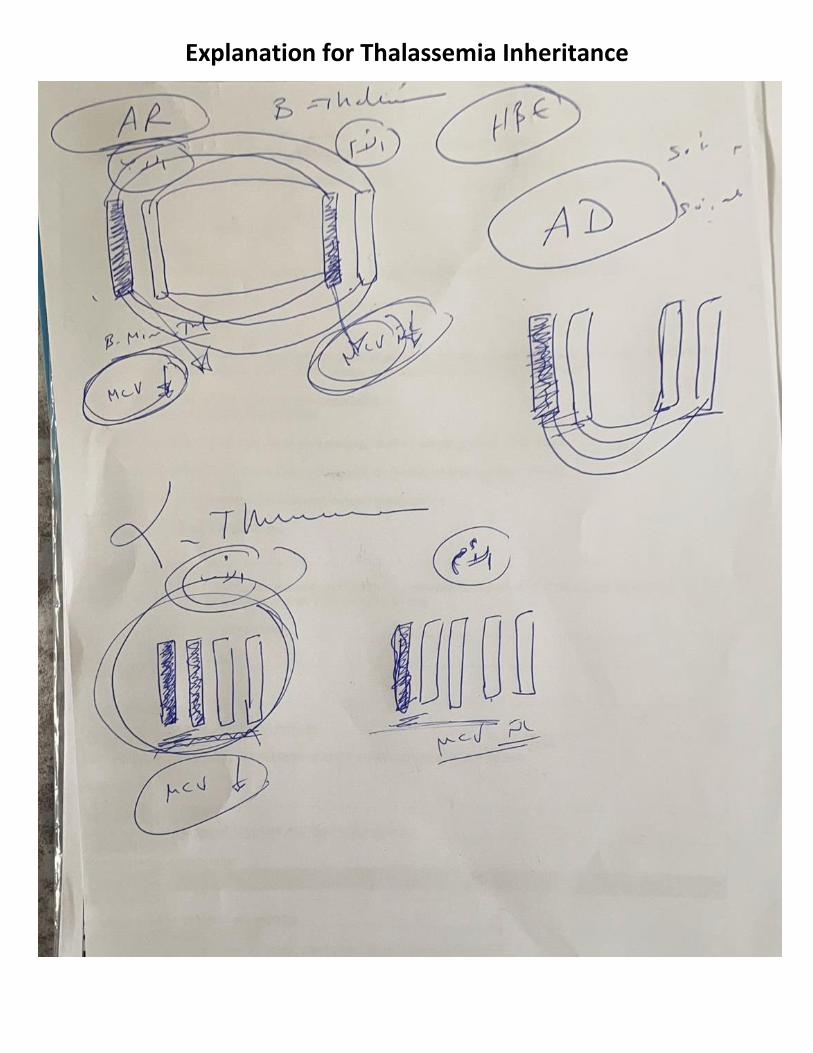
Explanation for Thalassemia Inheritance

Beta Thalassemia Marriage test: MCV, if they both had low
MCV, and we did electrophoresis:
Scenario (1): One has beta thalassemia trait, and the other partner is healthy ➔
we tell them to go on with the marriage as 50% will be carriers and 50% will be
healthy.
Scenario (2): Both have beta thalassemia trait (HbA2 more than 3.5%) ➔ since
it is an autosomal recessive disorder:
50% of offspring will be carriers, 25% will be affected, and 25% will be healthy ➔
we tell them not to go on with the marriage.
Alpha Thalassemia
Alpha thalassemia Inheritance is on four genes, not two genes.
o One gene affected: silent carrier with normal CBC.
o Two genes affected: alpha thalassemia minor or trait or carrier, they have mild
microcytic anemia, diagnosed by genetic testing.
o Three genes affected: Hemoglobin H disease, they have hemolysis and need
transfusions, but not as severe as beta thalassemia major.
o Four genes affected: Hemoglobin Barts; they die in vitro.
Alpha thalassemia is not common, that is why we do not test for it in
Jordan, and that is why the role of counseling is very crucial in this
case!
If someone had MCV low, and by genetic studies, we diagnosed him with alpha
thalassemia trait, if he married a silent carrier, things would be disastrous!
Clarification: by law normal MCV and low MCV can marry each other, so if the
patient married a silent carrier, they could have an affected alpha thalassemia
baby!
What is the role of counseling here? If there is a known alpha thalassemia trait
patient, we tell him to do genetic testing for his future wife to make sure she is
not a silent carrier (has totally normal 4 genes).
Other option for them to marry and still have normal babies: In-vitro Fertilization.