approach to acute stroke in the emergency department · pdf file ·...
TRANSCRIPT
Approach to Acute Stroke in
the Emergency Department
Dr Julia Hopyan
Objectives
Types of stroke
Differentiating hemorrhagic and ischemic strokes: Clinically and radiologically
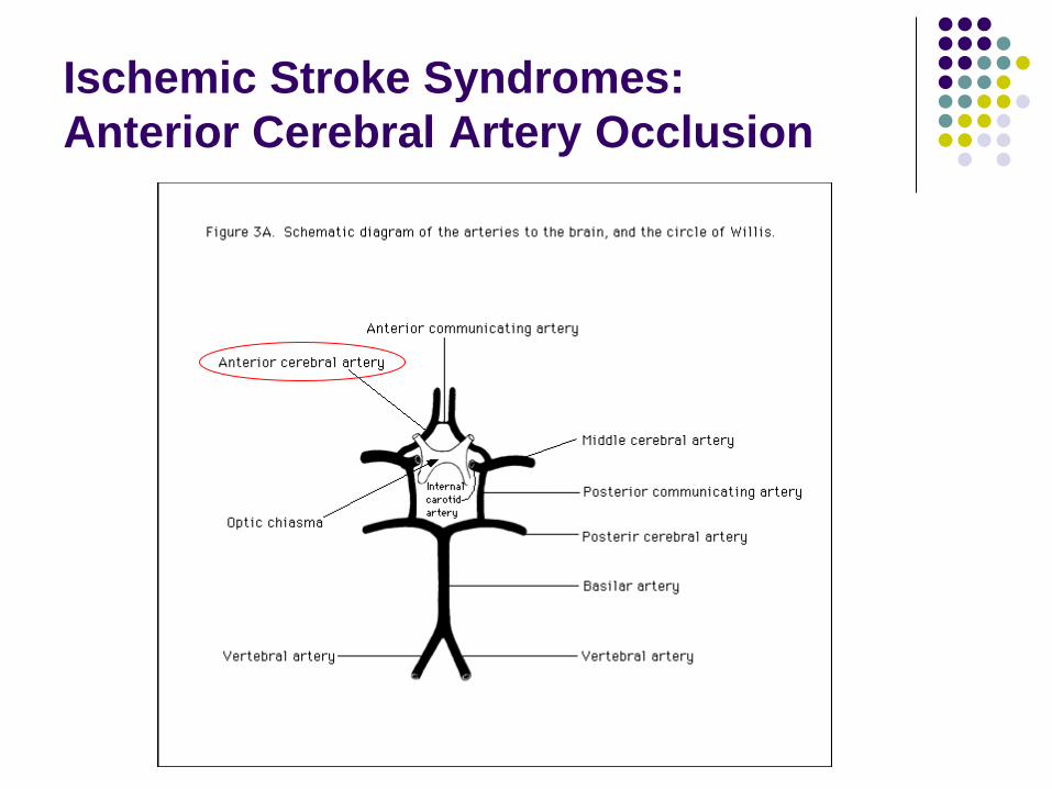
Stroke syndromes Anterior circulation:
Middle cerebral artery
Anterior cerebral artery
Posterior circulation:
Posterior cerebral artery
Basilar artery
Case examples
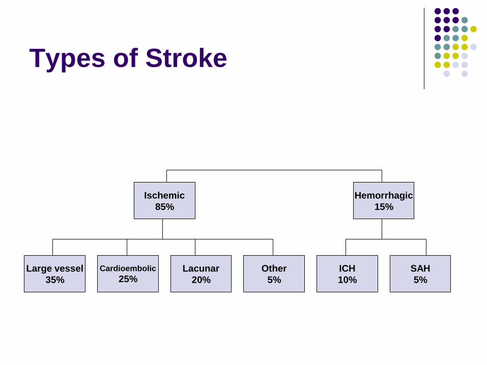
Types of Stroke
Cardioembolic
25%
Hemorrhagic
15%
Large vessel
35%
Lacunar
20%
ICH
10%
Other
5%
Ischemic
85%
SAH
5%
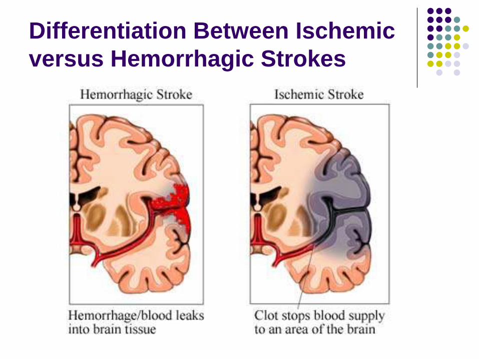
Differentiation Between Ischemic
versus Hemorrhagic Strokes
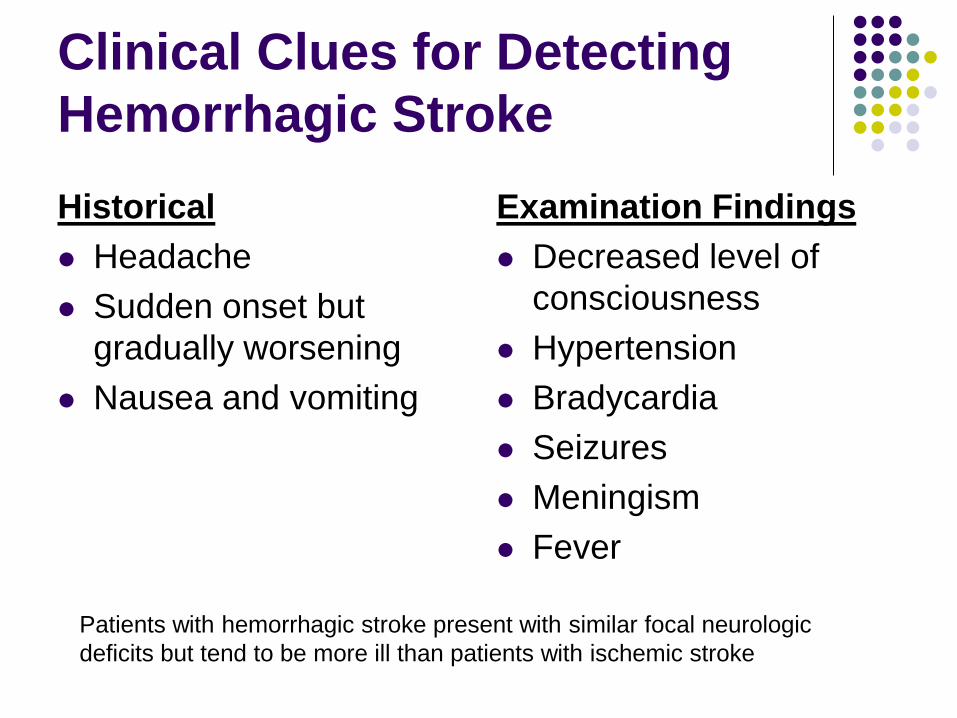
Clinical Clues for Detecting
Hemorrhagic Stroke
Historical
Headache
Sudden onset but
gradually worsening
Nausea and vomiting
Examination Findings
Decreased level of
consciousness
Hypertension
Bradycardia
Seizures
Meningism
Fever
Patients with hemorrhagic stroke present with similar focal neurologic
deficits but tend to be more ill than patients with ischemic stroke
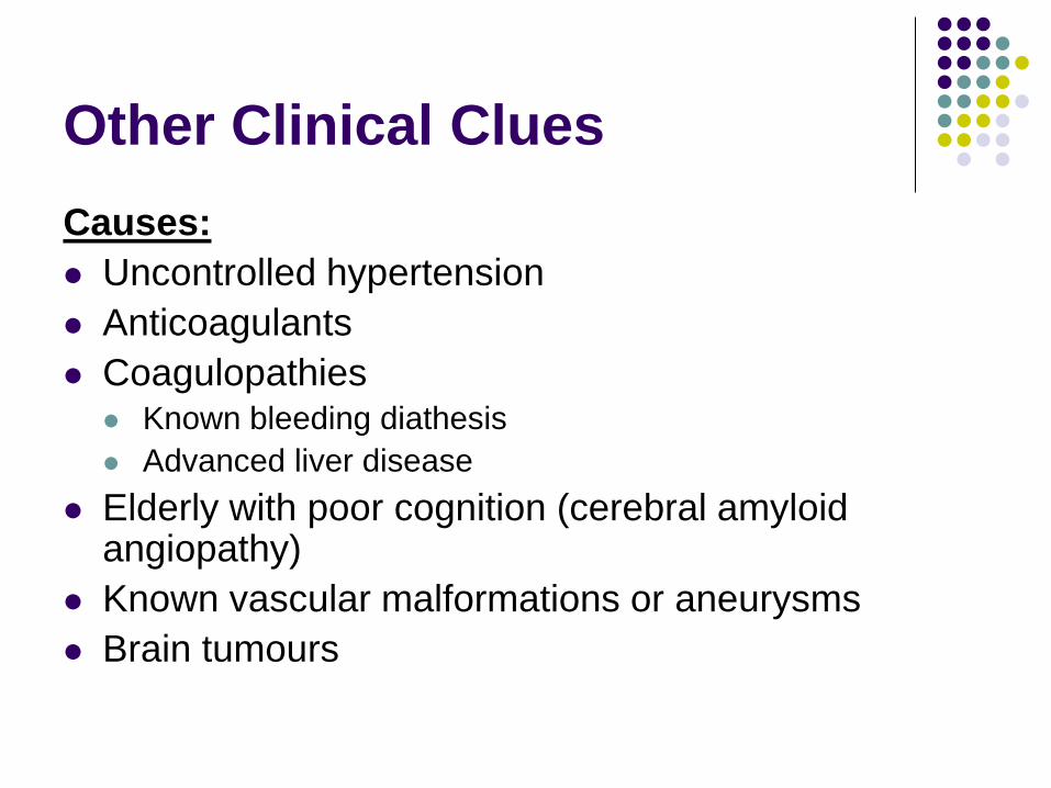
Other Clinical Clues
Causes:
Uncontrolled hypertension
Anticoagulants
Coagulopathies Known bleeding diathesis
Advanced liver disease
Elderly with poor cognition (cerebral amyloid angiopathy)
Known vascular malformations or aneurysms
Brain tumours
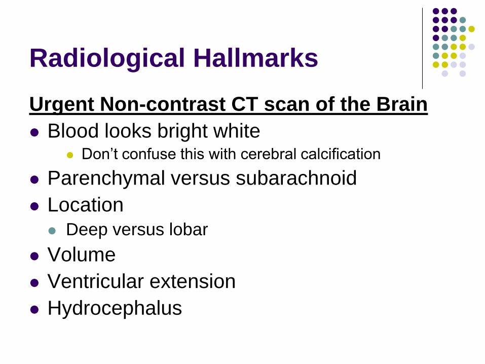
Radiological Hallmarks
Urgent Non-contrast CT scan of the Brain
Blood looks bright white Don’t confuse this with cerebral calcification
Parenchymal versus subarachnoid
Location
Deep versus lobar
Volume
Ventricular extension
Hydrocephalus

Location of Bleeds
Deep 50%
Lobar 35%
Cerebellum 10%
Brainstem 5%
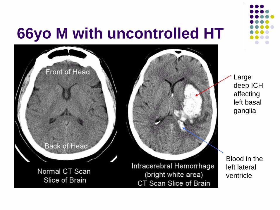
66yo M with uncontrolled HT
Large
deep ICH
affecting
left basal
ganglia
Blood in the
left lateral
ventricle
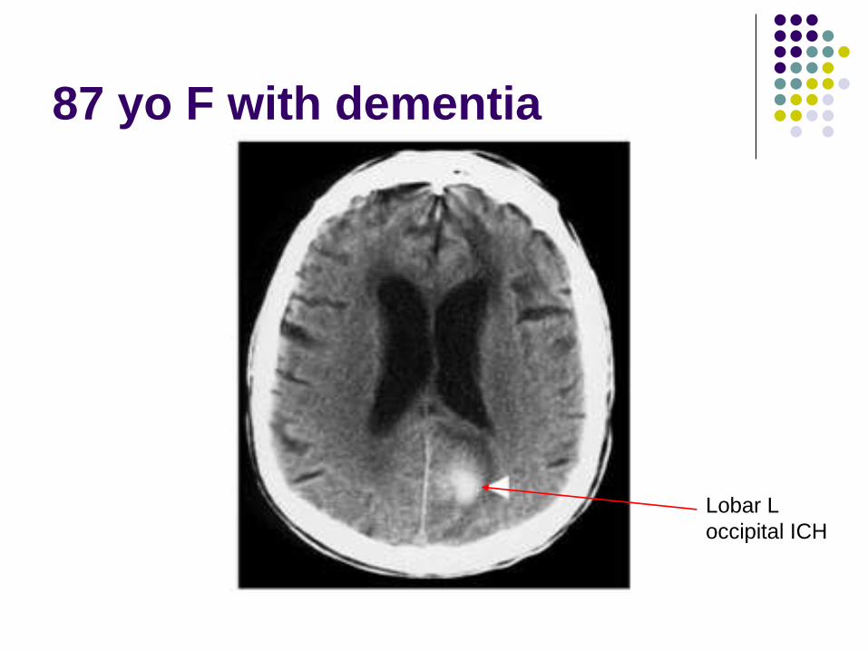
87 yo F with dementia
Lobar L
occipital ICH

Prognostic Factors in ICH
Up to 50% mortality rates at 1 year
Volume of hemorrhage predicts 30 day
mortality
Poor functional outcomes >30ml ICH
>70ml ICH is usually fatal
Pontine hemorrhage, >5ml is usually fatal
Cerebellar hemorrhage, >30ml is usually fatal
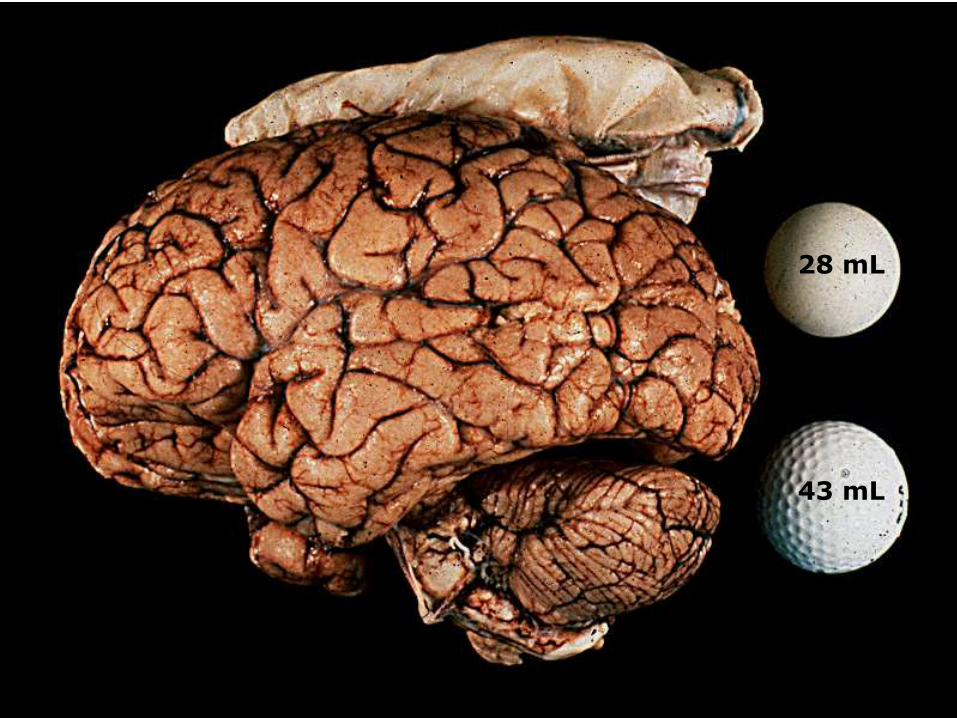
28 mL
43 mL
(Image courtesy T. Brott, MD)
Prognostic Factors in ICH
Hematoma Expansion
40% of hematomas expand by >1/3rd of their
volume
More than 2/3rd of hematomas grow in the
first hour
Hematoma expansion correlates with poor
functional outcome
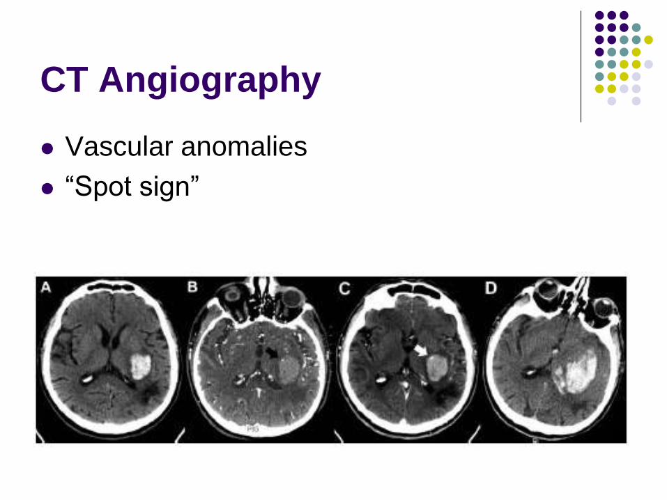
CT Angiography
Vascular anomalies
“Spot sign”
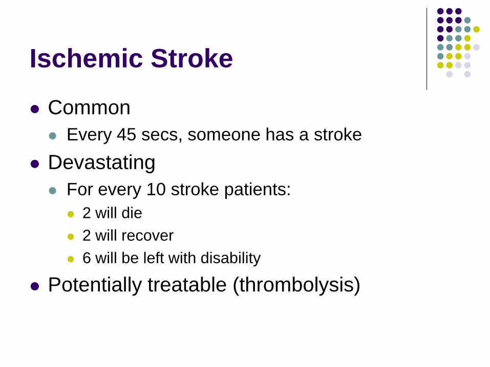
Ischemic Stroke
Common
Every 45 secs, someone has a stroke
Devastating
For every 10 stroke patients:
2 will die
2 will recover
6 will be left with disability
Potentially treatable (thrombolysis)
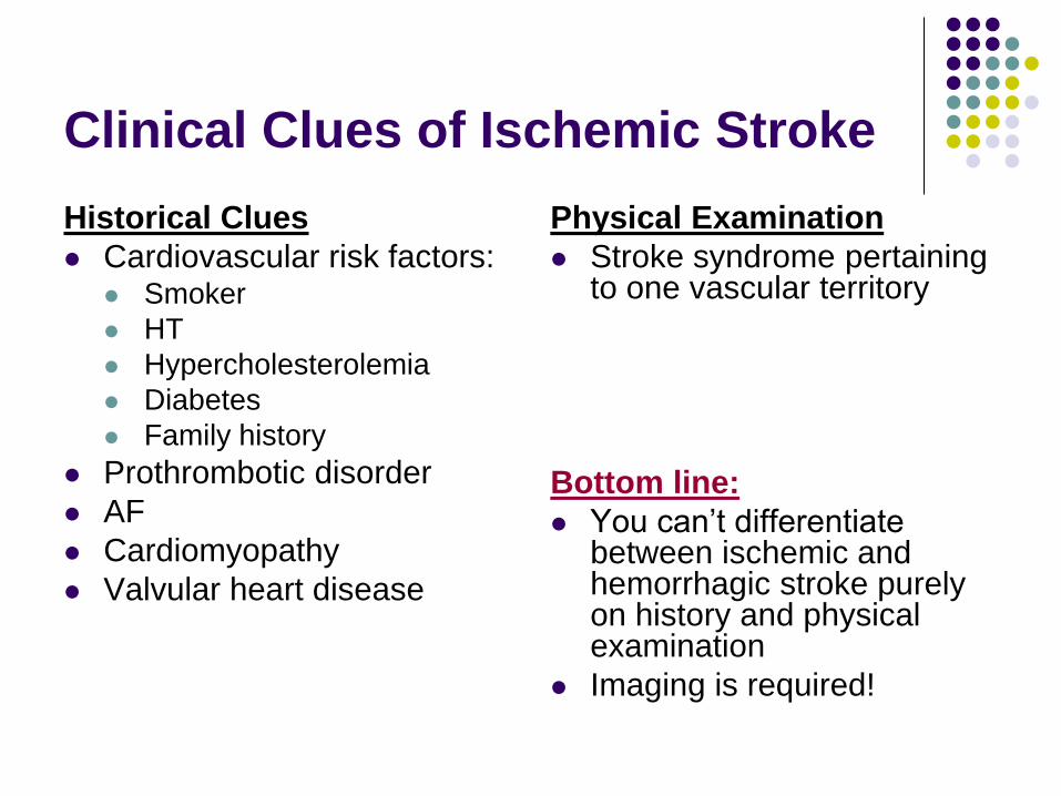
Clinical Clues of Ischemic Stroke
Historical Clues
Cardiovascular risk factors: Smoker
HT
Hypercholesterolemia
Diabetes
Family history
Prothrombotic disorder
AF
Cardiomyopathy
Valvular heart disease
Physical Examination
Stroke syndrome pertaining to one vascular territory
Bottom line:
You can’t differentiate between ischemic and hemorrhagic stroke purely on history and physical examination
Imaging is required!
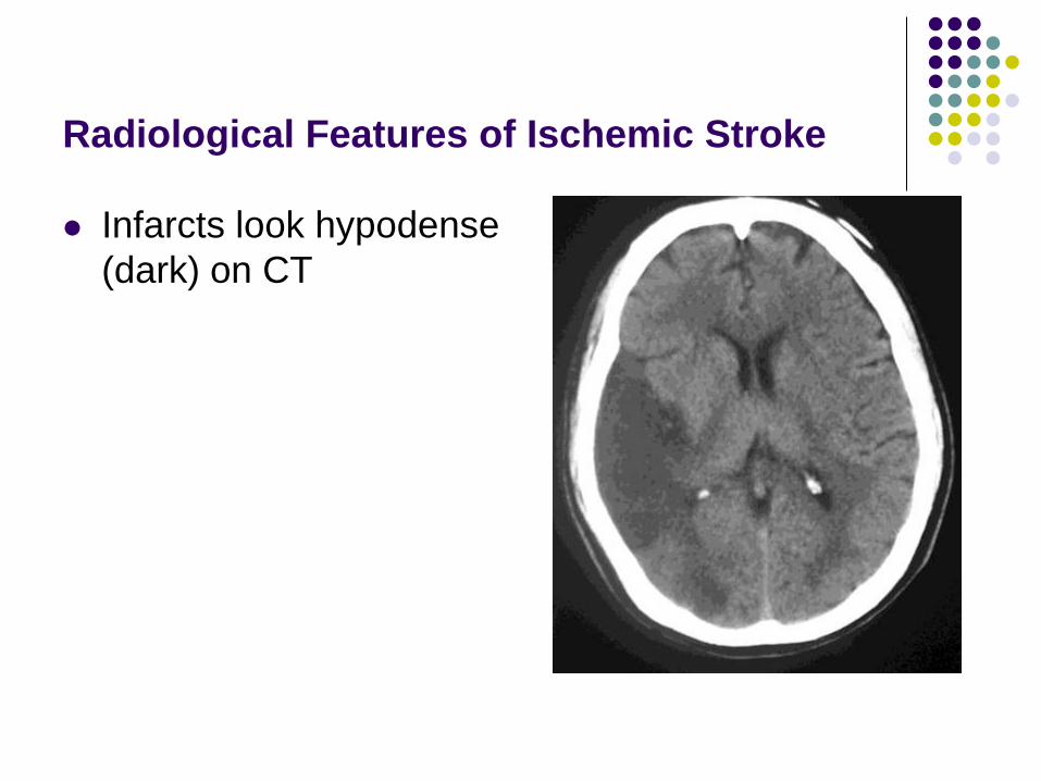
Radiological Features of Ischemic Stroke
Infarcts look hypodense
(dark) on CT
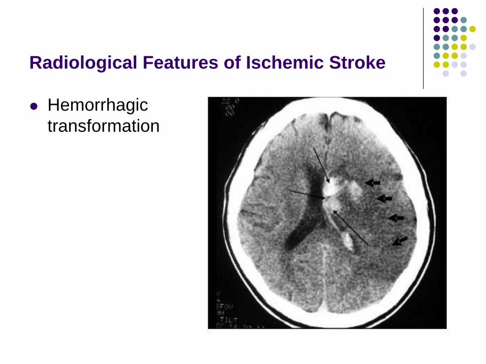
Radiological Features of Ischemic Stroke
Hemorrhagic
transformation
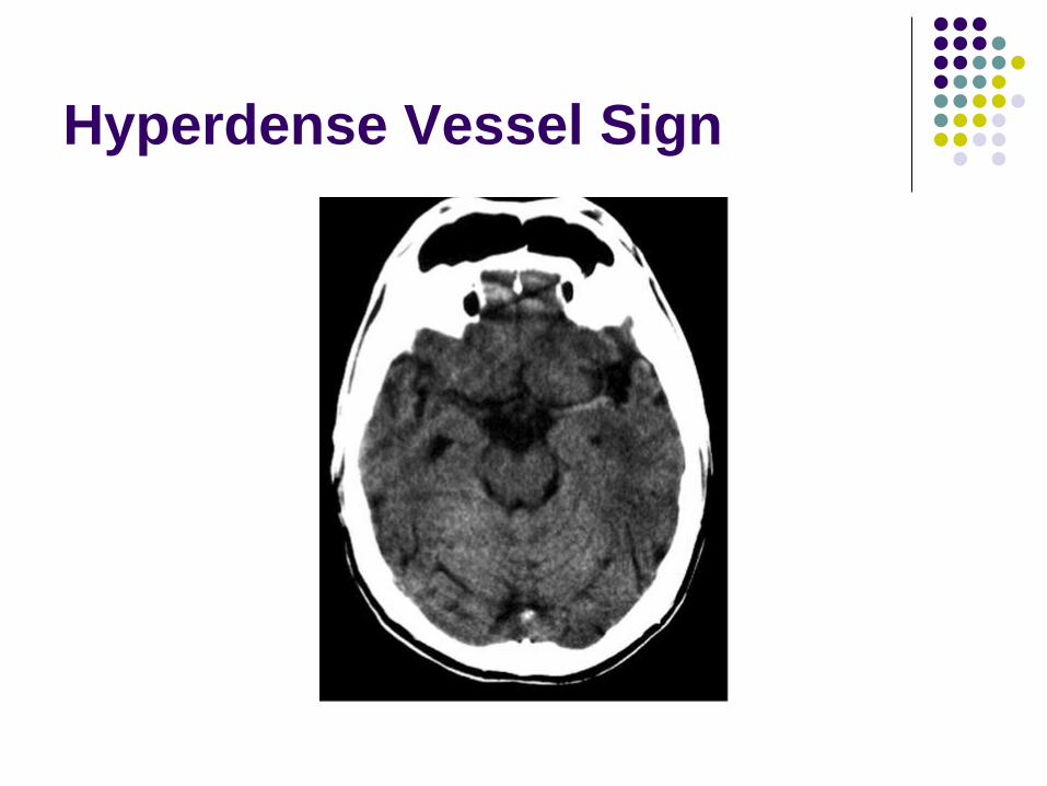
Hyperdense Vessel Sign
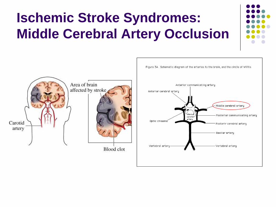
Ischemic Stroke Syndromes:
Middle Cerebral Artery Occlusion
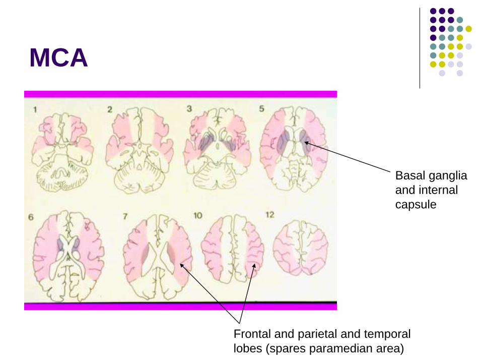
MCA
Frontal and parietal and temporal
lobes (spares paramedian area)
Basal ganglia
and internal
capsule
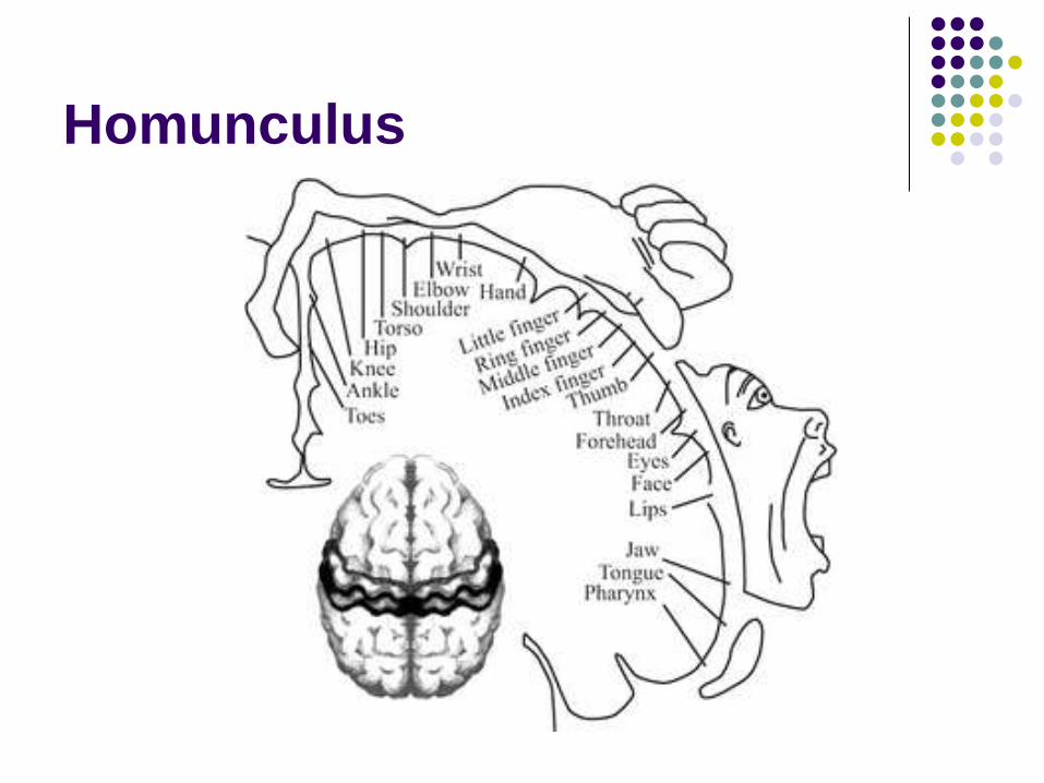
Homunculus
Middle Cerebral Artery
Contralateral hemiparesis (face, arm>leg)
Contralateral hemisensory impairment
Contralateral homonymous hemianopia
Specific hemispheric signs: Left (dominant hemisphere)
Aphasia (expressive, receptive, global)
Right Dysarthria
Neglect
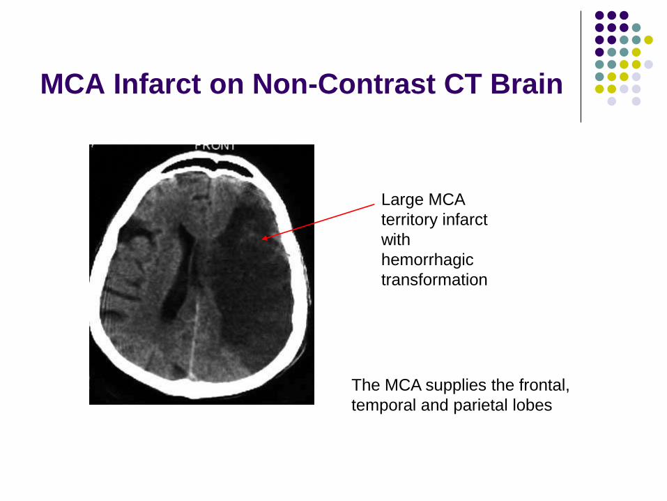
MCA Infarct on Non-Contrast CT Brain
Large MCA
territory infarct
with
hemorrhagic
transformation
The MCA supplies the frontal,
temporal and parietal lobes
Ischemic Stroke Syndromes:
Anterior Cerebral Artery Occlusion
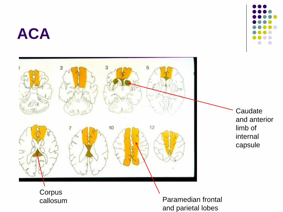
ACA
Corpus
callosum Paramedian frontal
and parietal lobes
Caudate
and anterior
limb of
internal
capsule
Anterior Cerebral Artery
Contralateral weakness of leg >> arm
Contralateral hemisensory impairment in the
same distribution
Mood and cognition disturbance:
Depression
Agitated confusion
Emotional lability
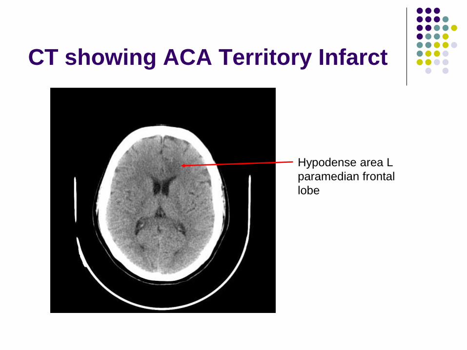
CT showing ACA Territory Infarct
Hypodense area L
paramedian frontal
lobe
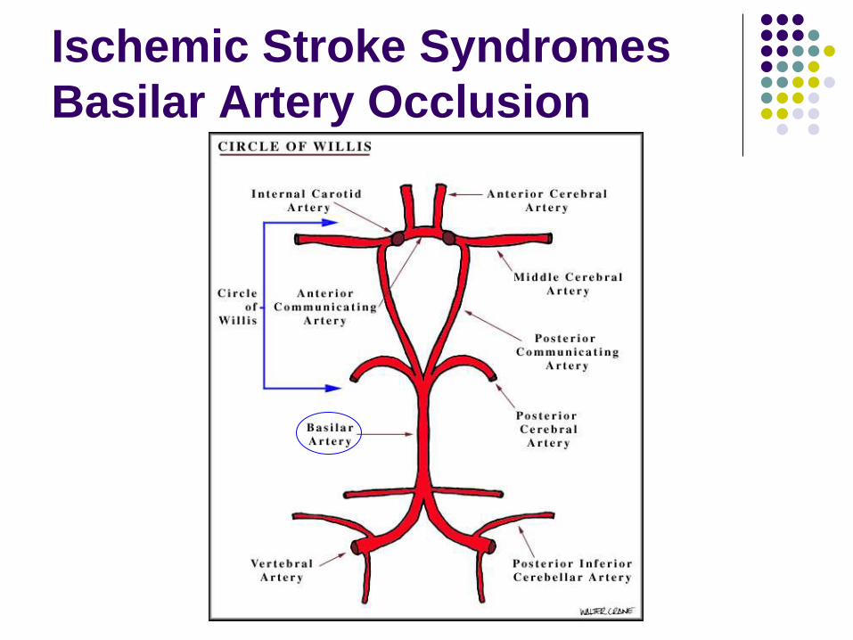
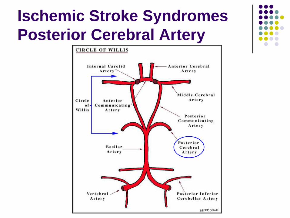
Ischemic Stroke Syndromes
Basilar Artery Occlusion
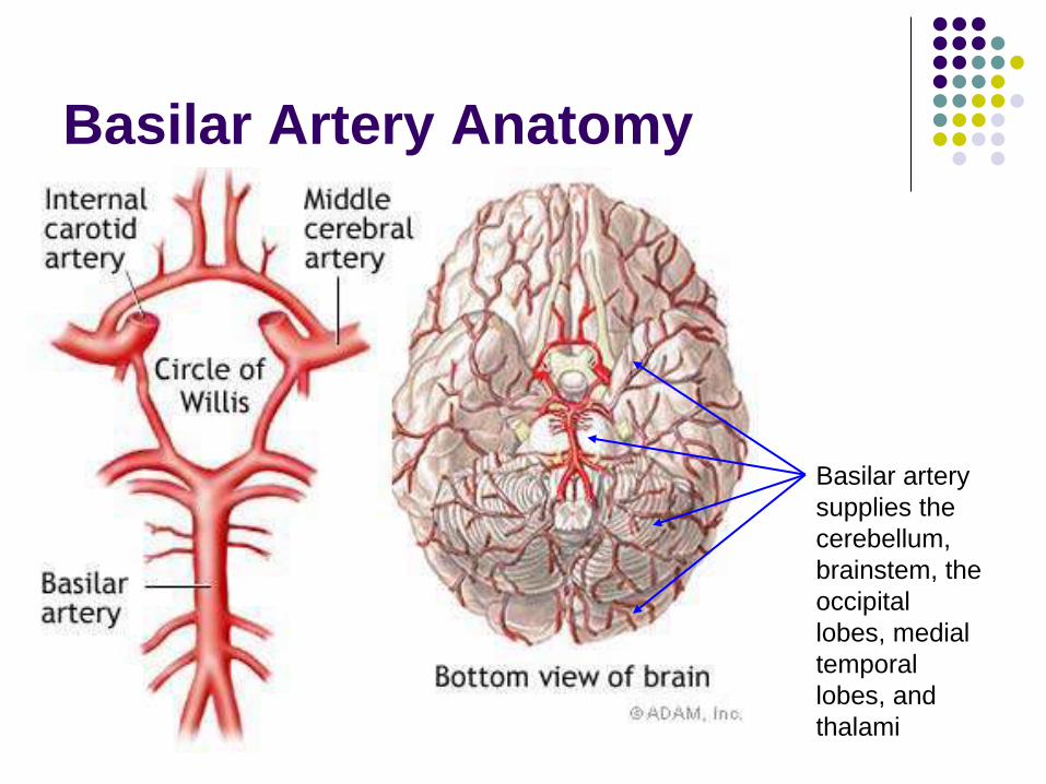
Basilar Artery Anatomy
Basilar artery
supplies the
cerebellum,
brainstem, the
occipital
lobes, medial
temporal
lobes, and
thalami
Symptoms associated with
Posterior Circulation Strokes
Slurred speech
Trouble swallowing
Double vision
Vertigo
Contralateral weakness
Crossed sensory signs
Cranial nerve palsies
Ipsilateral incoordination
Unsteady gait
Fluctuating level of consciousness
Hearing loss
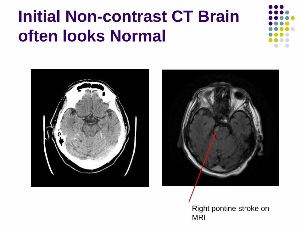
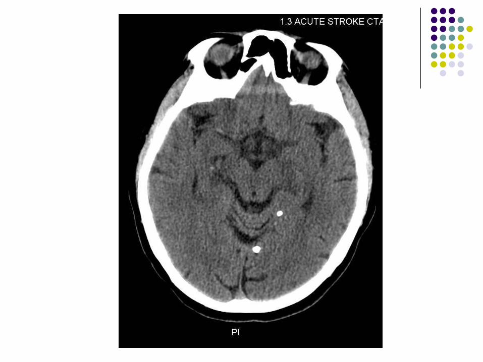
Initial Non-contrast CT Brain
often looks Normal
Right pontine stroke on
MRI
Ischemic Stroke Syndromes
Posterior Cerebral Artery
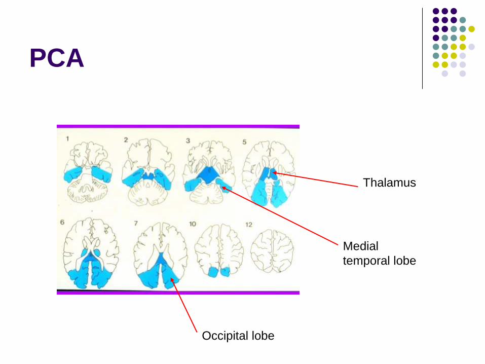
PCA
Occipital lobe
Medial
temporal lobe
Thalamus
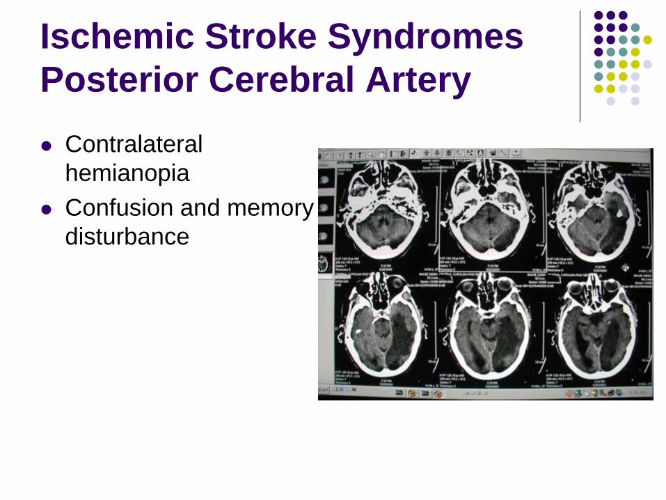
Ischemic Stroke Syndromes
Posterior Cerebral Artery
Contralateral
hemianopia
Confusion and memory
disturbance

Case Example 1
76 yo R handed M
Sudden onset of
Difficulty speaking (non-
fluent, unable to read or
name)
Right weakness (arm>leg)
Right sensory impairment
90 mins duration
No headache
History of CAD / HT / chol
On aspirin
Does this presentation
conform to a particular
stroke syndrome?
Which hemisphere is
affected?
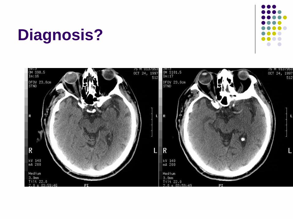
Diagnosis?
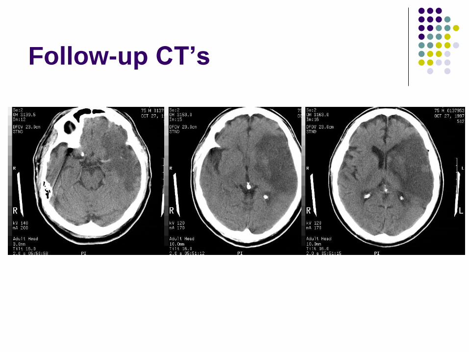
Follow-up CT’s
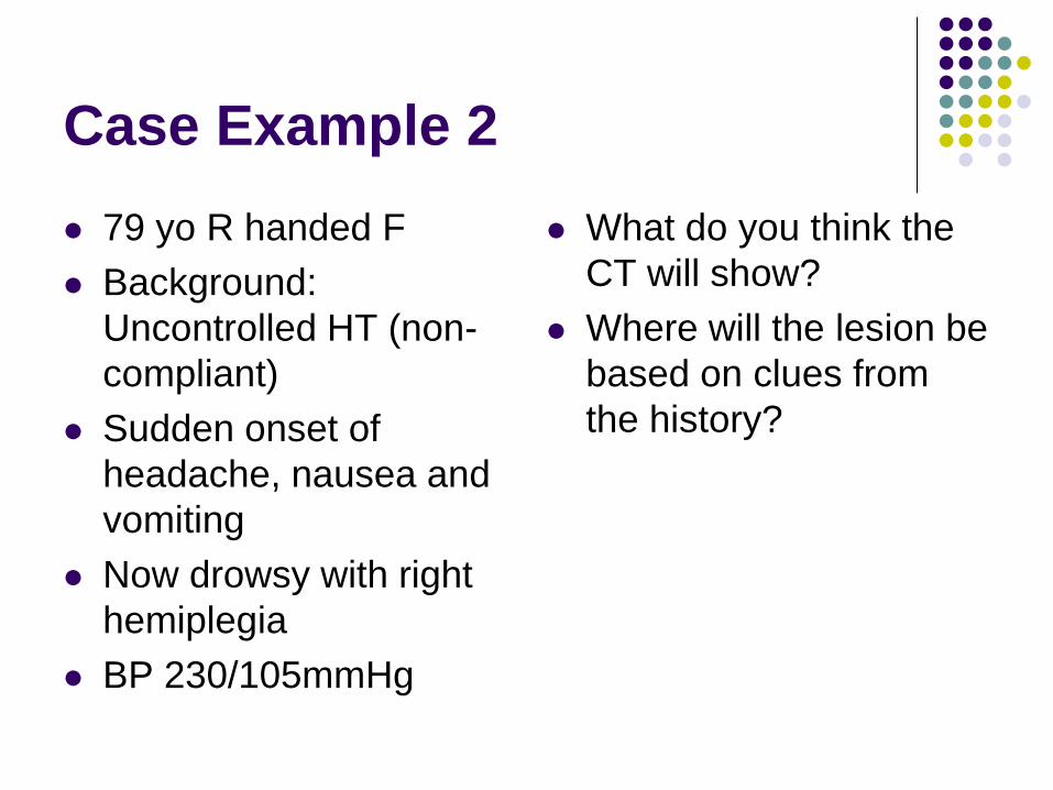
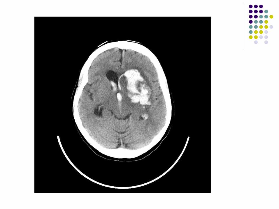
Case Example 2
79 yo R handed F
Background:
Uncontrolled HT (non-
compliant)
Sudden onset of
headache, nausea and
vomiting
Now drowsy with right
hemiplegia
BP 230/105mmHg
What do you think the
CT will show?
Where will the lesion be
based on clues from
the history?
Case Example 3
80 year old woman
1 hour history
Acute hemiplegia
Gaze deviation
Hemispatial neglect
A fib
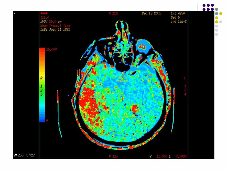

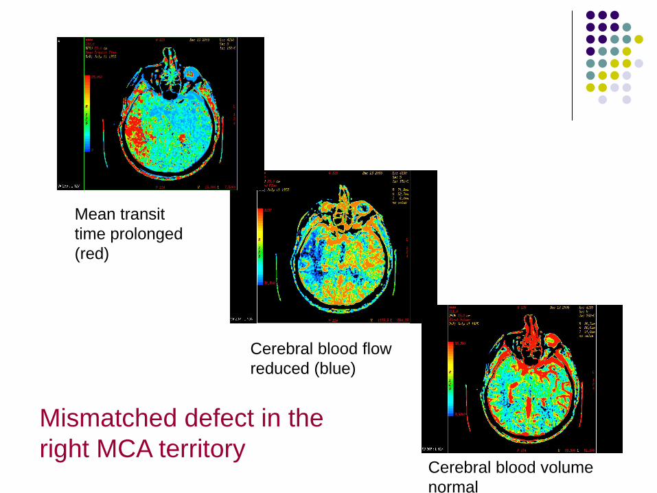
Mean transit
time prolonged
(red)
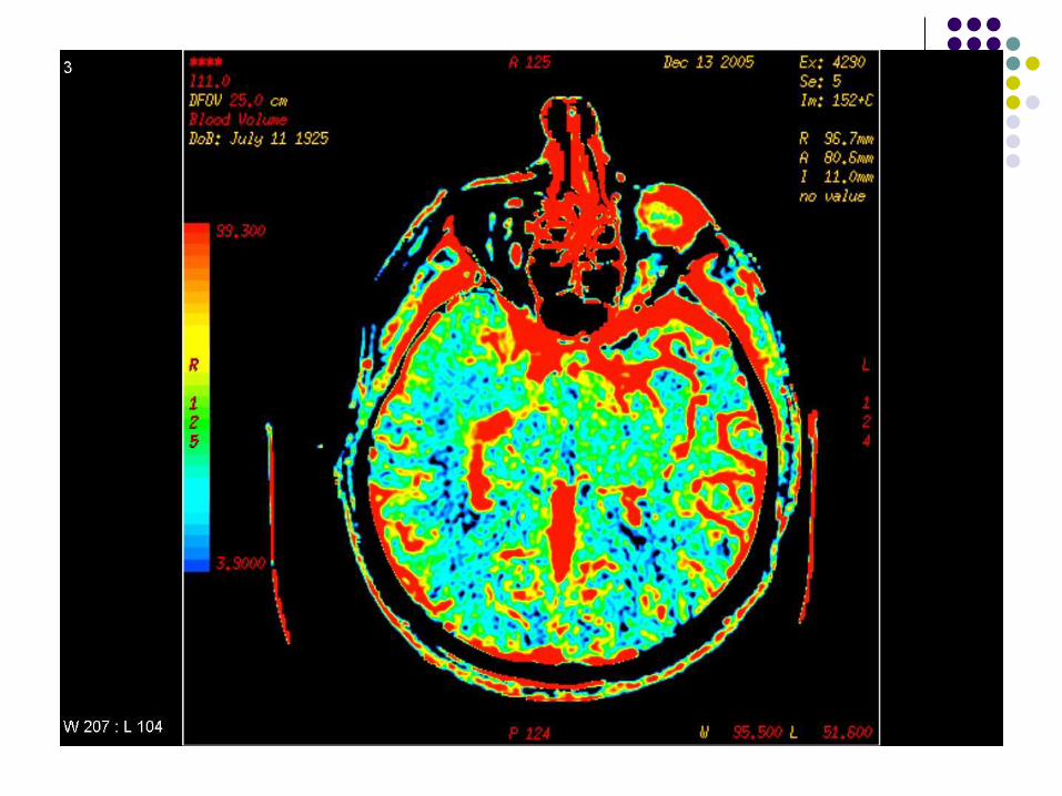
Cerebral blood flow
reduced (blue)
Cerebral blood volume
normal
Mismatched defect in the
right MCA territory
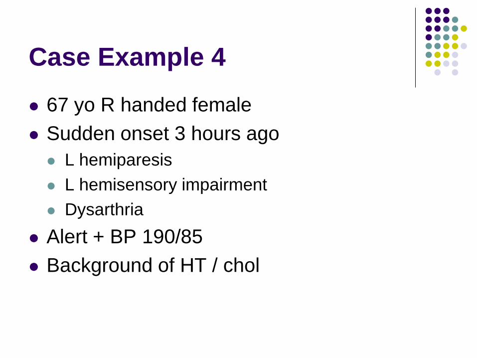
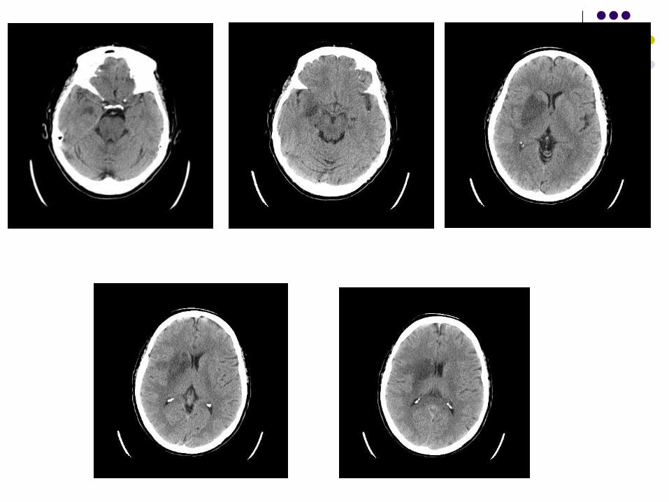
Case Example 4
67 yo R handed female
Sudden onset 3 hours ago
L hemiparesis
L hemisensory impairment
Dysarthria
Alert + BP 190/85
Background of HT / chol
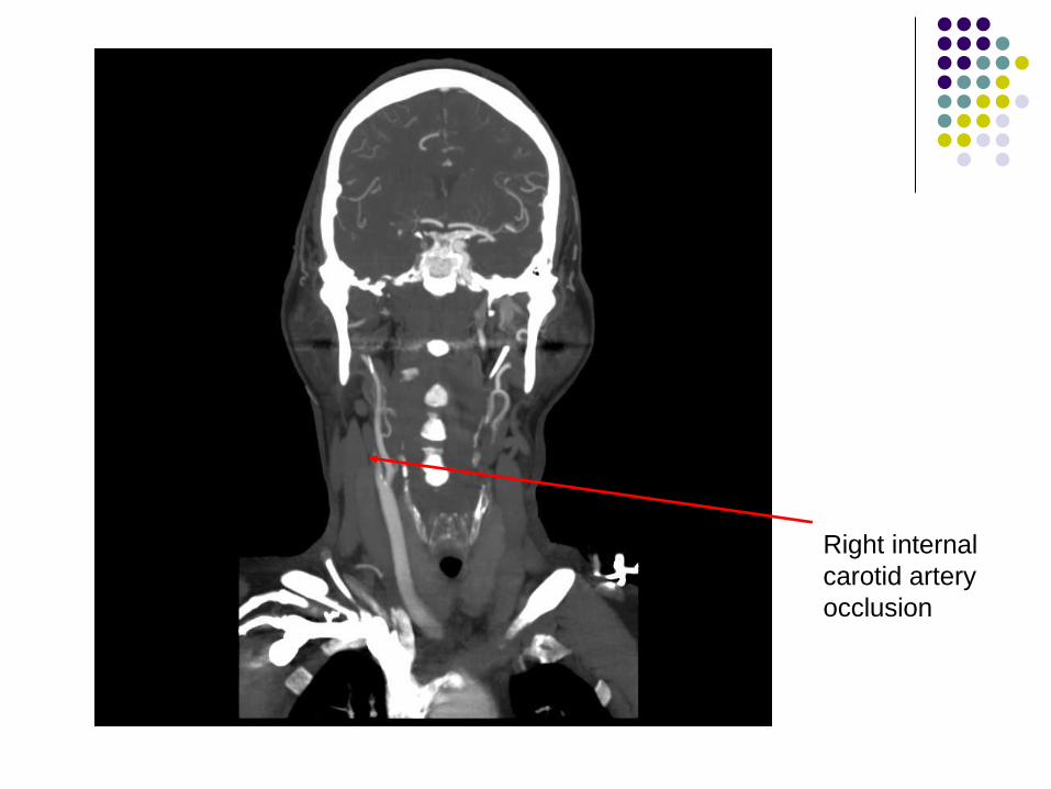
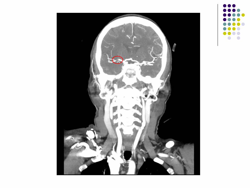
Right internal
carotid artery
occlusion
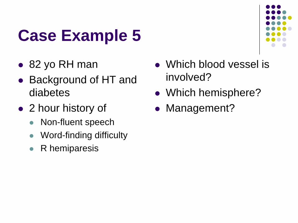
Case Example 5
82 yo RH man
Background of HT and
diabetes
2 hour history of
Non-fluent speech
Word-finding difficulty
R hemiparesis
Which blood vessel is
involved?
Which hemisphere?
Management?
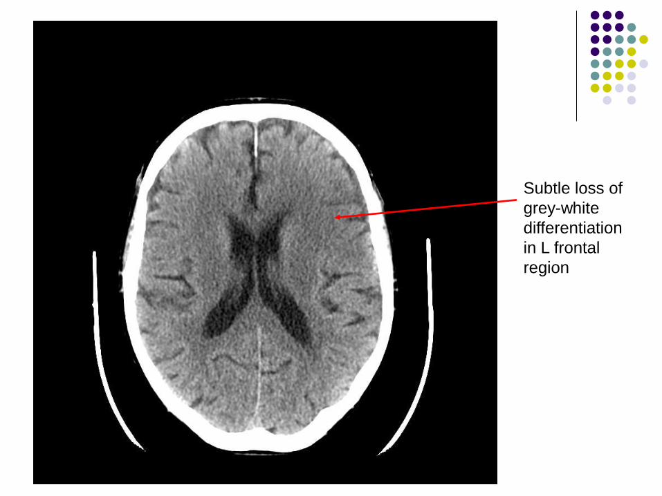
Subtle loss of
grey-white
differentiation
in L frontal
region
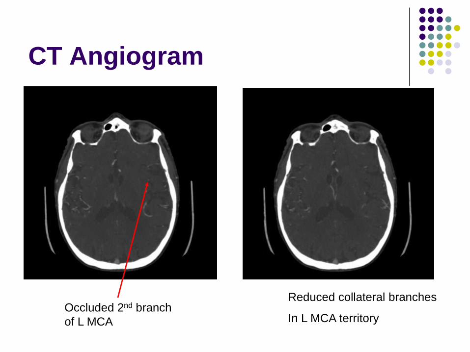
CT Angiogram
Occluded 2nd branch
of L MCA
Reduced collateral branches
In L MCA territory
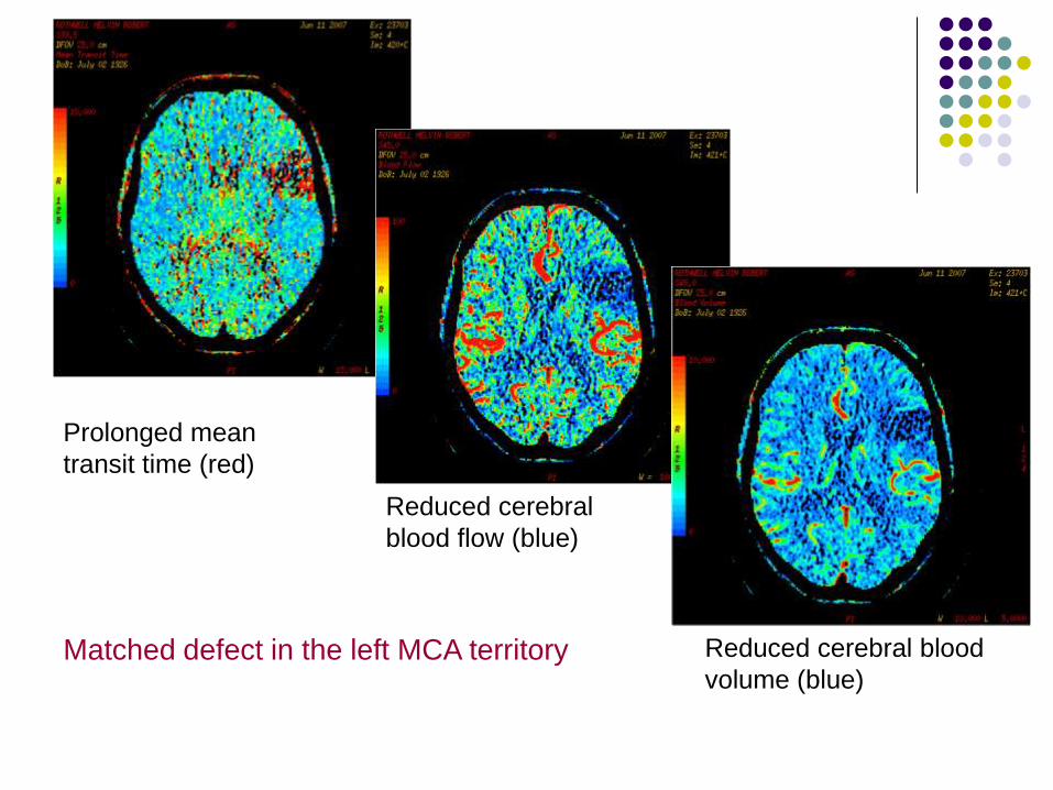
Matched defect in the left MCA territory
Prolonged mean
transit time (red)
Reduced cerebral
blood flow (blue)
Reduced cerebral blood
volume (blue)
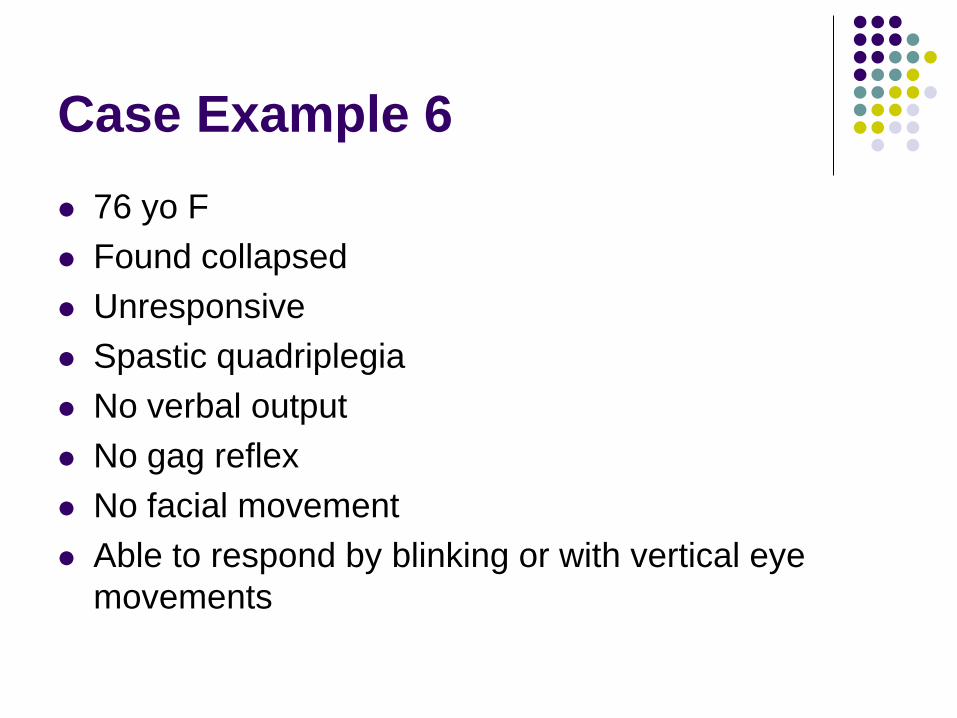
Case Example 6
76 yo F
Found collapsed
Unresponsive
Spastic quadriplegia
No verbal output
No gag reflex
No facial movement
Able to respond by blinking or with vertical eye
movements
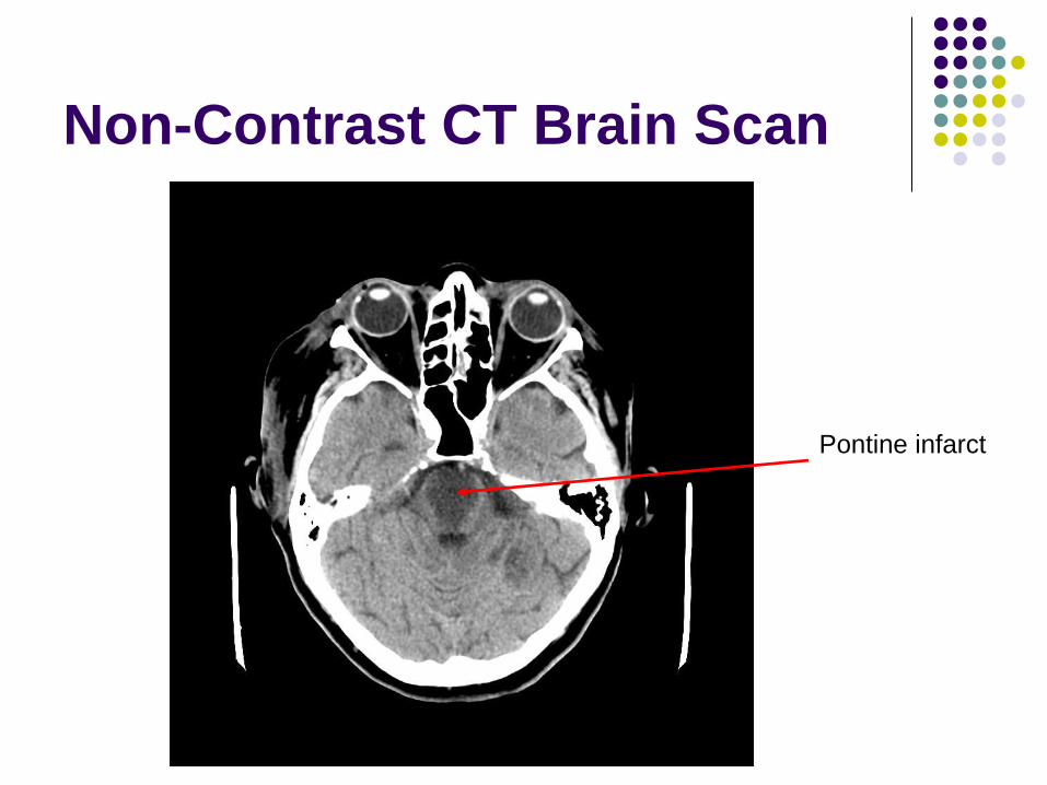
Non-Contrast CT Brain Scan
Pontine infarct
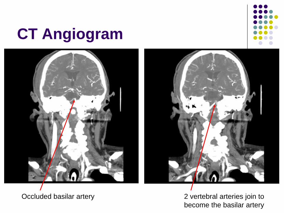
CT Angiogram
Occluded basilar artery 2 vertebral arteries join to
become the basilar artery