applied surface science - connecting repositories · zaca-morán et al. / applied surface science...
TRANSCRIPT
Mt
Pa
b
a
ARRAA
KMOLMHB
1
edBwmccsihca[
dt
dH
(((
h00
Applied Surface Science 392 (2017) 492–497
Contents lists available at ScienceDirect
Applied Surface Science
journa l h om epa ge: www.elsev ier .com/ locate /apsusc
icrocautery based on zinc metallic nanoparticles photodeposited onhe core of an optical fiber
. Zaca-Morána,∗, C.F. Pastelínb, C. Moránb, G.F. Pérez-Sáncheza, F. Cháveza
Departemento de Fisicoquímica de Materiales, Instituto de Ciencias, Universidad Autónoma de Puebla, 17 Nte 3417, Puebla 72050, MexicoDepartemento de Biología y Toxicologia de la Reproducción, Instituto de Ciencias, Universidad Autónoma de Puebla, 14 Sur 6301, Puebla 72570, Mexico
r t i c l e i n f o
rticle history:eceived 6 June 2015eceived in revised form 2 June 2016ccepted 11 September 2016vailable online 12 September 2016
a b s t r a c t
The experimental arrangement of a microcautery implemented by an optical fiber with zinc nanoparticles(ZnNPs) photodeposited on its core for the cauterization and coagulation in blood vessels hemostasisprocesses is presented. The interaction between a laser radiation source and the ZnNPS on the fibercore produces a controllable punctual heat source through the radiation intensity, which is capable ofreaching a temperature up to 200 ◦C covering an area of approximately ten micrometers. By using three-
eywords:icrocauteryptical fiberaseretallic nanoparticles
to-four-month-old rats of CIIZ-V strain, we made several microcauterization experimental tests to stopblood flow. The findings show that the microcautery obliterates the smooth muscle of the blood vesselsconcatenating mutually to tissue in an average time of three seconds, at the same time, the blood elementsresponsible for the coagulation are thermally activated and thus the bleeding is stopped.
© 2016 The Authors. Published by Elsevier B.V. This is an open access article under the CC BY-NC-ND
emostasislood vessels. Introduction
Cautery is widely used in medicine to seal minor injuries; forxample, in profuse bleeding in nasal mucous, eliminating a wart,estroy tissue or any other injury of less than 5 mm in length [1,2].efore the emergence of the electricity, the cauterizing of woundsas performed by using a hot piece of iron. In 1891, D’Arsonvalade the first study on the effects of high-frequency electrical
urrent in humans, and discovered that by applying an electri-al current above 100 kHz the cautery reduced the neuromuscularhock [3]. Following this principle, the first electrocautery wasmplemented as a transducer that transforms electrical current intoeat [4]. This electrical device is used as a surgical instrument forutting or sealing blood vesselsthrough a high-intensity currentllowing ablation, hemostasis, sealing and coagulation in surgeries5–9].
New systems applying the electrocautery principle have beeneveloped to both bipolar electrosurgery and radiofrequency abla-ion. In these systems, a high-frequency alternating current is
∗ Corresponding author at: Laboratorio de Fisicoquímica de Materiales, Institutoe Ciencias, Universidad Autónoma de Puebla, Privada 17 Nte 3417, San Miguelueyotlipan 72050, Puebla, Mexico.
E-mail addresses: zmoran [email protected]. Zaca-Morán), c [email protected]. Pastelín), [email protected] (C. Morán), f perez [email protected]. Pérez-Sánchez), [email protected] (F. Chávez).
ttp://dx.doi.org/10.1016/j.apsusc.2016.09.041169-4332/© 2016 The Authors. Published by Elsevier B.V. This is an open access article
/).
license (http://creativecommons.org/licenses/by-nc-nd/4.0/).
applied to living tissue to obtain different degrees of destruction[1,10,11], as well as to seal blood vessels in hysterectomy [2], andto accurately sectionalize nerves such as cochlear [5] and sciaticnerves [7] in animal models. This process can cause focal coagu-lation necrosis in tracheal stenosis after tracheotomy, which cancreate or disappear a stenotic injury in epithelial tissue [9], andremove tracheal hematoma [12]. The disadvantages of electrocau-terization lie on the effects that the electrical current have onliving beings such as faradic [13], electrolytic [14,15], or thermic[11,15,17].
The thermal probe heated by a coherent radiation source, a moreelaborated cauterization device, has been developed using an opti-cal fiber [18–21]. However, some studies have shown that by usingthis thermal probe (“hot tip”), it is possible to cause major injuriesto the diameter of the tip when the size of the injury is smaller thanthe tip. Because of this, non-invasive or minimal-invasive surgicaltechniques are of great importance for general health and exper-imental studies. Therefore, it is necessary to miniaturize surgicaldevices [22].
Nowadays, nanostructured materials have been widely appliedin the field of medicine. These materials are metallic, metal oxide,polymeric, or a combination of all of these. Metallic nanoparticleshave made it possible to develop medical therapies, new sensors to
detect illnesses and identify toxic products.In this research, the implementation of a microcautery based onapplied nanotechnology is described, in which metallic nanoparti-cles were photodeposited on the core of an optical fiber. This is
under the CC BY-NC-ND license (http://creativecommons.org/licenses/by-nc-nd/4.
P. Zaca-Morán et al. / Applied Surfac
Fa
atraoawni
2
2
nTtsct
cocfi1tws
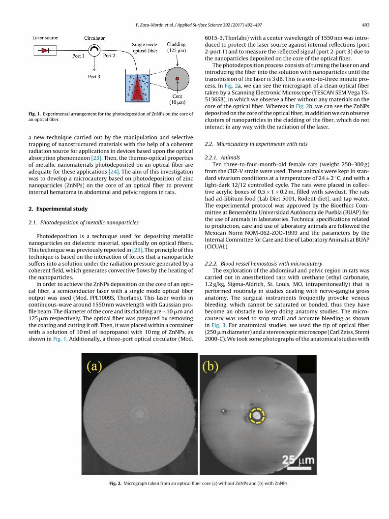
ig. 1. Experimental arrangement for the photodeposition of ZnNPs on the core ofn optical fiber.
new technique carried out by the manipulation and selectiverapping of nanostructured materials with the help of a coherentadiation source for applications in devices based upon the opticalbsorption phenomenon [23]. Then, the thermo-optical propertiesf metallic nanomaterials photodeposited on an optical fiber aredequate for these applications [24]. The aim of this investigationas to develop a microcautery based on photodeposition of zincanoparticles (ZnNPs) on the core of an optical fiber to prevent
nternal hematoma in abdominal and pelvic regions in rats.
. Experimental study
.1. Photodeposition of metallic nanoparticles
Photodeposition is a technique used for depositing metallicanoparticles on dielectric material, specifically on optical fibers.his technique was previously reported in [23]. The principle of thisechnique is based on the interaction of forces that a nanoparticleuffers into a solution under the radiation pressure generated by aoherent field, which generates convective flows by the heating ofhe nanoparticles.
In order to achieve the ZnNPs deposition on the core of an opti-al fiber, a semiconductor laser with a single mode optical fiberutput was used (Mod. FPL1009S, Thorlabs). This laser works inontinuous-wave around 1550 nm wavelength with Gaussian pro-le beam. The diameter of the core and its cladding are ∼10 �m and
25 �m respectively. The optical fiber was prepared by removinghe coating and cutting it off. Then, it was placed within a containerith a solution of 10 ml of isopropanol with 10 mg of ZnNPs, ashown in Fig. 1. Additionally, a three-port optical circulator (Mod.
Fig. 2. Micrograph taken from an optical fiber co
e Science 392 (2017) 492–497 493
6015-3, Thorlabs) with a center wavelength of 1550 nm was intro-duced to protect the laser source against internal reflections (port2-port 1) and to measure the reflected signal (port 2-port 3) due tothe nanoparticles deposited on the core of the optical fiber.
The photodeposition process consists of turning the laser on andintroducing the fiber into the solution with nanoparticles until thetransmission of the laser is 3 dB. This is a one-to-three minute pro-cess. In Fig. 2a, we can see the micrograph of a clean optical fibertaken by a Scanning Electronic Microscope (TESCAN SEM Vega TS-5136SB), in which we observe a fiber without any materials on thecore of the optical fiber. Whereas in Fig. 2b, we can see the ZnNPsdeposited on the core of the optical fiber, in addition we can observeclusters of nanoparticles in the cladding of the fiber, which do notinteract in any way with the radiation of the laser.
2.2. Microcautery in experiments with rats
2.2.1. AnimalsTen three-to-four-month-old female rats (weight 250–300 g)
from the CIIZ-V strain were used. These animals were kept in stan-dard vivarium conditions at a temperature of 24 ± 2 ◦C, and with alight-dark 12/12 controlled cycle. The rats were placed in collec-tive acrylic boxes of 0.5 × 1 × 0.2 m, filled with sawdust. The ratshad ad-libitum food (Lab Diet 5001, Rodent diet), and tap water.The experimental protocol was approved by the Bioethics Com-mittee at Benemérita Universidad Autónoma de Puebla (BUAP) forthe use of animals in laboratories. Technical specifications relatedto production, care and use of laboratory animals are followed theMexican Norm NOM-062-ZOO-1999 and the parameters by theInternal Committee for Care and Use of Laboratory Animals at BUAP(CICUAL).
2.2.2. Blood vessel hemostasis with microcauteryThe exploration of the abdominal and pelvic region in rats was
carried out in anesthetized rats with urethane (ethyl carbonate,1.2 g/kg, Sigma-Aldrich, St. Louis, MO, intraperitoneally) that isperformed routinely in studies dealing with nerve-ganglia grossanatomy. The surgical instruments frequently provoke venousbleeding, which cannot be saturated or bonded, thus they havebecome an obstacle to keep doing anatomy studies. The micro-
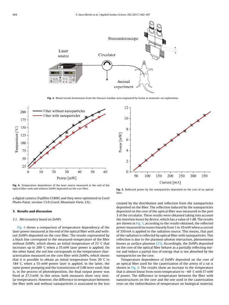
cautery was used to stop small and accurate bleeding as shownin Fig. 3. For anatomical studies, we used the tip of optical fiber(250 �m diameter) and a stereoscopic microscope (Carl Zeiss, Stemi2000-C). We took some photographs of the anatomical studies withre (a) without ZnNPs and (b) with ZnNPs.
494 P. Zaca-Morán et al. / Applied Surface Science 392 (2017) 492–497
Fig. 3. Blood vessels hemostasis from the thoracic-lumbar area originated by lesion in anatomic rat exploration.
Fo
aP
3
3
loawitat1sifilt
ig. 4. Temperature dependence of the laser source measured at the end of theptical fiber with and without ZnNPs deposited on the core fiber.
digital camera (Fujifilm S1800) and they were optimized in Corelhoto-Paint, version 13.0 (Corel, Mountain View, CA).
. Results and discussion
.1. Microcautery based on ZnNPs
Fig. 4 shows a comparison of temperature dependency of theaser power measured at the end of the optical fiber with and with-ut ZnNPs deposited on the core fiber. The results represented by
black line correspond to the measured-temperature of the fiberithout ZnNPs, which shows an initial temperature of 23 ◦C that
ncreases up to 200 ◦C when a 55 mW laser power is applied. Onhe other hand, the red line corresponds to the temperature char-cterization measured on the core fiber with ZnNPs, which showshat it is possible to obtain an initial temperature from 20 ◦C to84 ◦C, when a 55-mW-power laser is applied. In the latter, theame power pumping and the transmission of 3 dB were used; that
s, in the process of photodeposition, the final output power wasxed at 27.5 mW. In this sense, both measures show very simi-ar temperatures. However, the difference in temperature betweenhe fiber with and without nanoparticles is associated to the loss
Fig. 5. Reflected power by the nanoparticles deposited on the core of an opticalfiber.
created by the distribution and reflection from the nanoparticlesdeposited on the fiber. The reflection induced by the nanoparticlesdeposited on the core of the optical fiber was measured in the port3 of the circulator. These results were obtained taking into accountthe insertion losses by device, which has a value of 1 dB. The resultsare shown in Fig. 5, according to the results obtained, the reflectedpower measured increases linearly from 1 to 10 mW when a currentof 350 mA is applied to the radiation source. This means, that partof the radiation is reflected by optical fiber with nanoparticles. Thisreflection is due to the plasmon-photon interaction, phenomenonknows as surface plasmon [23]. Accordingly, the ZnNPs depositedon the core of the optical fiber behave as a partially reflecting mir-ror and induce a partial loss of energy that is not absorbed by thenanoparticles on the core.
Temperature dependence of ZnNPs deposited on the core ofan optical fiber used for the cauterization of the artery of a rat isshown in Fig. 6. The results show an increase in the temperaturethat is almost linear from room temperature to ∼60 ◦C with 57 mW
of power. The difference in temperature between the fiber withnanostructures on the core and the one used in the cauterizationrests on the redistribution of temperature on biological material,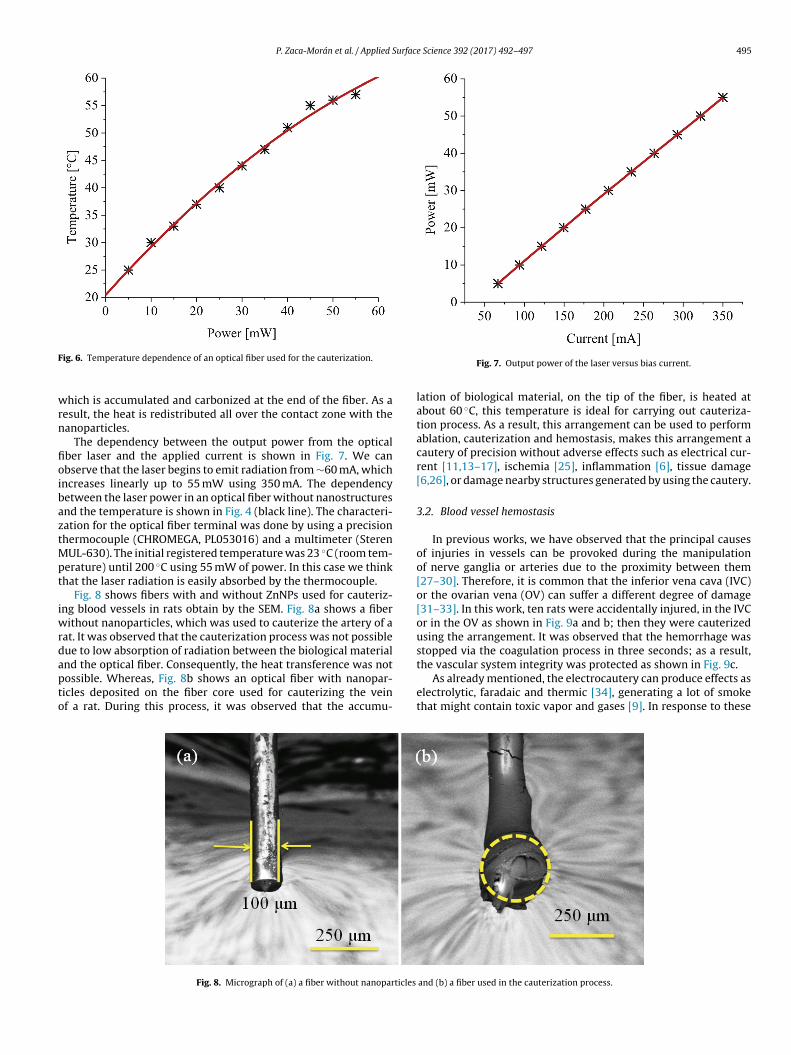
P. Zaca-Morán et al. / Applied Surface Science 392 (2017) 492–497 495
F
wrn
fioibaztMpt
iwrdapto
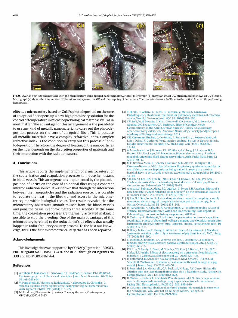
the vascular system integrity was protected as shown in Fig. 9c.
ig. 6. Temperature dependence of an optical fiber used for the cauterization.
hich is accumulated and carbonized at the end of the fiber. As aesult, the heat is redistributed all over the contact zone with theanoparticles.
The dependency between the output power from the opticalber laser and the applied current is shown in Fig. 7. We canbserve that the laser begins to emit radiation from ∼60 mA, whichncreases linearly up to 55 mW using 350 mA. The dependencyetween the laser power in an optical fiber without nanostructuresnd the temperature is shown in Fig. 4 (black line). The characteri-ation for the optical fiber terminal was done by using a precisionhermocouple (CHROMEGA, PL053016) and a multimeter (Steren
UL-630). The initial registered temperature was 23 ◦C (room tem-erature) until 200 ◦C using 55 mW of power. In this case we thinkhat the laser radiation is easily absorbed by the thermocouple.
Fig. 8 shows fibers with and without ZnNPs used for cauteriz-ng blood vessels in rats obtain by the SEM. Fig. 8a shows a fiber
ithout nanoparticles, which was used to cauterize the artery of aat. It was observed that the cauterization process was not possibleue to low absorption of radiation between the biological materialnd the optical fiber. Consequently, the heat transference was not
ossible. Whereas, Fig. 8b shows an optical fiber with nanopar-icles deposited on the fiber core used for cauterizing the veinf a rat. During this process, it was observed that the accumu-Fig. 8. Micrograph of (a) a fiber without nanoparticles
Fig. 7. Output power of the laser versus bias current.
lation of biological material, on the tip of the fiber, is heated atabout 60 ◦C, this temperature is ideal for carrying out cauteriza-tion process. As a result, this arrangement can be used to performablation, cauterization and hemostasis, makes this arrangement acautery of precision without adverse effects such as electrical cur-rent [11,13–17], ischemia [25], inflammation [6], tissue damage[6,26], or damage nearby structures generated by using the cautery.
3.2. Blood vessel hemostasis
In previous works, we have observed that the principal causesof injuries in vessels can be provoked during the manipulationof nerve ganglia or arteries due to the proximity between them[27–30]. Therefore, it is common that the inferior vena cava (IVC)or the ovarian vena (OV) can suffer a different degree of damage[31–33]. In this work, ten rats were accidentally injured, in the IVCor in the OV as shown in Fig. 9a and b; then they were cauterizedusing the arrangement. It was observed that the hemorrhage wasstopped via the coagulation process in three seconds; as a result,
As already mentioned, the electrocautery can produce effects aselectrolytic, faradaic and thermic [34], generating a lot of smokethat might contain toxic vapor and gases [9]. In response to these
and (b) a fiber used in the cauterization process.
496 P. Zaca-Morán et al. / Applied Surface Science 392 (2017) 492–497
F nologM ppingh
eocitpartot
4
tipibttmatpmhe
A
P3
R
[
[
[
[
[
[
[
[
[
[
ig. 9. Ovarian vein (OV) hemostasis with the microcautery using applied nanotechicrograph (c) shows the intervention of the microcautery over the OV and the sto
emostasis.
ffects, a microcautery based on ZnNPs photodeposited on the coref an optical fiber opens up a new high promissory solution for theontrol of temperature in microscopic biological matter as well as innert matter. The advantage for this arrangement is the possibilityo use any kind of metallic nanomaterial to carry out the photode-osition process on the core of an optical fiber. This is becausell metallic materials have a complex refractive index. Complexefractive index is the condition to carry out this process of pho-odeposition. Therefore, the degree of heating of the nanoparticlesn the fiber depends on the absorption properties of materials andheir interaction with the radiation source.
. Conclusions
This article reports the implementation of a microcautery forhe cauterization and coagulation processes to induce hemostasisn blood vessels. This arrangement is implemented by the photode-osition of ZnNPs on the core of an optical fiber using a coherent
nfrared radiation source. It was shown that through the interactionetween the nanoparticles and the radiation source, it is possibleo regulate the heat in the fiber tip, and access in the microme-er regime within biological tissues. The results revealed that the
icrocautery obliterates smooth muscle from the blood vesselsnd joins the tissue in approximately three seconds, at the sameime; the coagulation processes are thermally activated making itossible to stop the bleeding. One of the main advantages of thisicrocautery is related to the reduction of side effects that usually
appen in radio-frequency cautery process. To the best our knowl-dge, this is the first micrometric cautery that has been reported.
cknowledgements
This investigation was supported by CONACyT grant No 130 983,RODEP grant No. BUAP-PTC-476 and BUAP through VIEP grants No39 and No MORC-NAT-64.
eferences
[1] A. Taheri, P. Mansoori, L.F. Sandoval, S.R. Feldman, D. Pearce, P.M. Williford,Electrosurgery: part I. Basics and principles, J. Am. Acad. Dermatol. 70 (2014)591.e1–591.e14.
[2] V. Pergialiotis, D. Vlachos, A. Rodolakis, D. Haidopoulos, D. Christakis, G.Vlachos, Electrosurgical bipolar vessel sealing for vaginal hysterectomies,Arch. Gynecol. Obstet. 290 (2014) 215–222.
[3] M. Brodman, Electrocautery devices. The way the work, ContemporaryOB/GYN, (2007) 85–93.
[
y. Notes: Micrograph (a) shows an intact OV. Micrograph (b) shows an OV’s lesion. of hematoma. The zoom-in shows a ZnNPs onto the optical fiber while performing
[4] T. Hiraki, H. Gobara, T. Iguchi, H. Fujiwara, Y. Matsui, S. Kanazawa,Radiofrequency ablation as treatment for pulmonary metastasis of colorectalcancer, World J. Gastroenterol.: WJG 20 (2014) 988–996.
[5] C.E. Iseli, W.H. Merwin, C. Klatt-Cromwell, K.A. Hutson, M.G. Ewend, O.F.Adunka, D.C. Fitzpatrick, C.A. Buchman, Effect of Cochlear NerveElectrocautery on the Adult Cochlear Nucleus, Otology & Neurotology,American Otological Society, American Neurotology Society [and] EuropeanAcademy of Otology and Neurotology, 2014.
[6] C.R. Cervantes-Sánchez, C. Cu-Zetina, E. Serrano-Rico, J. Rojero-Vallejo, M.Lazos-Ochoa, R. Gutiérrez-Vega, Incisión cutánea: Bisturí vs electrocauterio.Estudio experimental en ratas, Rev. Med. Hosp. Gen. (Mex). 65 (2002)11–14.
[7] A. Moradzadeh, W.J. Brenner, E.L. Whitlock, A.Y. Tong, J.P. Luciano, D.A.Hunter, T.M. Myckatyn, S.E. Macninnon, Bipolar electrocautery. A rodentmodel of sunderland third-degree nerve injury, Arch. Facial Plast. Surg. 12(2010) 40–47.
[8] M.C. Navarro-Meza, R. Gonzáles-Baltazar, M.G. Aldrete-Rodríguez, D.E.Carmona-Navarrro, M.G. López-Cardona, Respiratory symtoms caused by theuse of electrocautery in physicans being trained in sugery in a mexicanhospital, Revista peruana de medicina experimental y salud publica 30 (2013)41–44.
[9] S.H. Oh, S.J. Lee, D.S. Kim, N.J. Na, E. Choi, S.J. Know, H.M. Cho, J.W. Son,Tracheal stenosis afther tracheostomy treated successful with papillotomeelectrocautery, Tuberculosis 75 (2014) 79–82.
10] A. Alpay, S. Bektas, A. Alpay, S.C. Ugurbas, C. Evren, S.H. Ugurbas, Effects of anew hemostatic agent Ankaferd Blood Stopper® on the intraocular tissues inrat model, Cutan. Ocul. Toxicol. 31 (2012) 128–131.
11] Q. Liu, X.B. Sun, Indirect electrical injuries from capacitive coupling: a rarelymentioned electrosurgical complication in monopolar laparoscopy, ActaObstet. Gynecol. Scand. 92 (2013) 238–241.
12] M. Panagiotou, A. Kalkanis, N. Karagiannidis, V. Polychronopoulos, A Case ofTracheal Hamartoma Resected with Loop Electrocautery, Case Reports inPulmonology, Hindawi publishing corporation, 20131–4.
13] D. Zadrozny, Z. Sledzinski, Small intestine perforation because of capacitivecoupling as a cause of abdominal wall gas gangrene and clostridial sepsis afterlaparoscopic cholecystectomy, Surg. Laparosc. Endosc. Percutaneous Tech. 10(2000) 412–414.
14] D. Berry, G. Garcea, C. Chong, E. Silman, G. Finch, A. Dennison, G.J. Maddern,Systematic reaction to electrolytic treatment of pig livers in vivo, ANZ J. Surg.74 (2004) 586–590.
15] C. Dobbins, C. Brennan, S.A. Wemyss-Holden, J. Cockburn, G.J. Maddern,Bimodal electric tissue ablation: positive electrode studies, ANZ J. Surg. 78(2008) 568–572.
16] K.K. Lim, S. Reddy, S. Desai, M. Smelley, S.S. Kim, J.F. Beshai, A.C. Lin, M.C.Burke, B.P. Knight, Effects of electrocautery on transvenous lead insulationmaterials, J. Cardiovasc. Electrophysiol. 20 (2009) 429–435.
17] R. Rothmund, D. Schaeller, A.A. Neugebauer, M.M. Scharpf, F.F. Fend, M.Schenk, D. Wallwiener, B. Kraemer, Evaluation of thermal damage in a pigmodel, J. Invest. Surg. 25 (2012) 43–50.
18] E. Rosenthal, J.K. Montarello, C.A. Bucknall, N. Fagg, P.V. Curry, His bundleablation with the laser thermal probe (hot tip): a feasibility study, Pacing Clin.Electrophysiol.: PACE 12 (1989) 812–822.
19] H. Weber, S. Enders, E. Keiditisch, Percutaneous Nd:YAG laser coagulation of
ventricular myocardium in dogs using a special electrode laser catheter,Pacing Clin. Electrophysiol.: PACE 12 (1989) 899–910.20] D.E. Haines, Thermal ablation of perfused porcine left ventricle in vitro withthe neodymium-YAG laser hot tip catheter system, Pacing Clin.Electrophysiol.: PACE 15 (1992) 979–985.
urfac
[
[
[
[
[
[
[
[
[
[
[
[32] I.E. Lawrence, H.W. Burden, The origin of the extrinsic adrenergic innervationto the rat ovary, Anat. Rec. 196 (1980) 51–59.
[33] A.P. Payer, Ultrastructural study of the nerve plexus accompanying the
P. Zaca-Morán et al. / Applied S
21] T. Seki, K. Oka, A. Naganawa, H. Yamashita, K. Kim, T. Chiba, Blood flowmeasurement system for fetoscopic laser photocoagulation of chorionic plateanastomosing vessels (FLPC), Minima. Invasive Ther. Allied Technol.: MITAT18 (2009) 350–355.
22] S. Thalhammer, G. Lahr, A. Clement-Sengewald, W.H. Heckl, R. Burgemeister,K. Schutze, Laser microtools in cell biology and molecular medicine, LaserPhys. 13 (2003) 681–691.
23] J.G. Ortega-Mendoza, F. Chavez, P. Zaca-Moran, C. Felipe, G.F. Perez-Sanchez,G. Beltran-Perez, O. Goiz, R. Ramos-Garcia, Selective photodeposition of zincnanoparticles on the core of a single-mode optical fiber, Opt. Express 21(2013) 6509–6518.
24] M.R. Huyeh, M.S. Havar, B. Palpant, Thermo-optical properties of embeddedsilver nanoparticles, J. Appl. Phys. 112 (2012) 1–4.
25] R.P. Ten Broek, J. Wilbers, H. Van Goor, Electrocautery causes more ischemicperitoneal tissue damage than ultrasonic dissection, Surg. Endosc. 25 (2011)
1827–1834.26] K.M. Keenan, G.T. Rodeheaver, J.G. Kenney, R.F. Edlich, Surgical cauteryrevisited, Am. J. Surg. 147 (1984) 818–821.
27] B. Baljet, J. Drukker, The extrinsic innervation of the abdominal organs in thefemale rat, Acta Anat. (Basel) 104 (1979) 243–267.
[
e Science 392 (2017) 492–497 497
28] B. Baljet, J. Drukker, The extrinsic innervation of thepelvic organs in thefemale rat, Acta Anat. (Basel) 107 (1980) 241–267.
29] H.R. Berthoud, T.L. Powley, Interaction between parasympathetic andsympathetic nerves in prevertebral ganglia: morphological evidence for vagalefferent innervation of ganglion cells in the rat, Microsc. Res. Tech. 35 (1996)80–86.
30] D.H. Hamer, R.M. Santer, Anatomy and blood supply of the coeliac-superiormesenteric ganglion complex of the rat, Anat. Embryol. (Berl.) 162 (1981)353–362.
31] T. Hanada, H. Hotta, Y. Aikawa, Number, size, conduction, and vasoconstrictorability of unmyelinated fibers of the ovarian nerve in adult and aged rats,Auton. Neurosci.: Basic Clin. 164 (2011) 6–12.
ovarian artery and vein in the rat, Anat. Rec. 190 (1978) 47–63.34] P.E. Arpi-Coellar, Electrobisturi, Universidad politecnica salesiana (2011) 1–3.