applied optoelectronics in medicine -...
TRANSCRIPT

1
© V. Blazek, MedIT, 2016All rights reservedLecture 7, Page 1
Scriptum AOM: Applied Optoelectronics in Medicine
7. Optoelectronic sensor concepts for vascular diagnostics – part I7. Optoelektronické koncepty pro vaskulární diagnostiku – část I
Applied Optoelectronics in Medicine
Aplikovaná optoelektronika v lékařství
Interdisciplinary course at the CTU Prague (P317APL-E, W, 4 credits)
© V. Blazek, MedIT, 2016All rights reservedLecture 7, Page 2
Scriptum AOM: Applied Optoelectronics in Medicine
Learning aims of the seventh AOM lecture
• Basics of quantitative Photoplethysmography (PPG) for transcutaneous detection ofdermal blood volume changes
• Alternative sensor concepts for blood volume studies in different compartments of human body
• Standardized PPG tests for non-invasive assessment of peripheral venous hemodynamics:a) Muscle pump test andb) Venous occlusion test

2
© V. Blazek, MedIT, 2016All rights reservedLecture 7, Page 3
Scriptum AOM: Applied Optoelectronics in Medicine
Comparison of non-invasive tests of the vascular system with phlebographyA scale of 1-5, in order of ascending quality, has been used
From: THE NEWS, Phlebology edition, No. 31,k Sept. 1992 * = invasive
© V. Blazek, MedIT, 2016All rights reservedLecture 7, Page 4
Scriptum AOM: Applied Optoelectronics in Medicine
Studies of peripheral hemodynamics using Photoplethysmography (PPG):transcutaneous monitoring of venous and/or arterial blood volume changes in dermal vascular plexus.
Milestones/Evolution steps:
1) 1938: first PPG system,created by Hertzman
2) 1979: LRR, first fullportable PPG system
?
3) 1989: first quantitative, PCcontrolled PPG system with calibration softwarefor each measurement
4) and today? - “intelligent” OES strategies likeSmart PPG
- camera based PPG Imagerfor spatial & time resolved skin perfusion studies

3
© V. Blazek, MedIT, 2016All rights reservedLecture 7, Page 5
Scriptum AOM: Applied Optoelectronics in Medicine
Discovery of the „optical windows“ through the skin in the 70ties l.c. at RWTHallowed the construction of advanced optoelectronic sensors (optrodes) for non-invasive measurements of venous and/or arterial blood volume changes
© V. Blazek, MedIT, 2016All rights reservedLecture 7, Page 6
Scriptum AOM: Applied Optoelectronics in Medicine
Fundamentals of Photoplethysmography (PPG) - one of the world wide well used methods for blood volume monitoring in dermal vascular network
Schematic visualisation of the correlation between the PPG signal and the blood volume changes in transilluminated vascular network under the sensor

4
© V. Blazek, MedIT, 2016All rights reservedLecture 7, Page 7
Scriptum AOM: Applied Optoelectronics in Medicine
ges
y
y
x
x
dq
ges
I
dzdydxzyxIzyxI
I
zIzS
max
max
max
max
00
00
,,,,
Fundamentals of Photoplethysmography (PPG) - one of the world wide well used methods for blood volume monitoring in dermal vascular network
© V. Blazek, MedIT, 2016All rights reservedLecture 7, Page 8
Scriptum AOM: Applied Optoelectronics in Medicine
Photon distribution in the skin and an estimation of the signal contents detected by the PPG sensor (T: tissue, V: venous signal, A: arterial signal). From 106 Photons injected to the skin in this model
scenario only 50 were detected (-43 dB)
Fundamentals of Photoplethysmography (PPG) - one of the world wide well used methods for blood volume monitoring in dermal vascular network

5
© V. Blazek, MedIT, 2016All rights reservedLecture 7, Page 9
Scriptum AOM: Applied Optoelectronics in Medicine
Time dependent visualisation of the typical PPG signal components
Fundamentals of Photoplethysmography (PPG) - one of the world wide well used methods for blood volume monitoring in dermal vascular network
© V. Blazek, MedIT, 2016All rights reservedLecture 7, Page 10
Scriptum AOM: Applied Optoelectronics in Medicine
PPG measuring system „for beginners“Optoelectronic components in the sensor
„Arterial sensor“: LD 242 as light source and BPW (photodiode) as light detector;both without focussing lenses.
„Venous sensor“: TIL31 as light source and TIL81 (phototransistor) as light detector; both with focussing lenses.
Possible sensor interfaces

6
© V. Blazek, MedIT, 2016All rights reservedLecture 7, Page 11
Scriptum AOM: Applied Optoelectronics in Medicine
PPG measuring system „for beginners“: schema of the electrical part
© V. Blazek, MedIT, 2016All rights reservedLecture 7, Page 12
Scriptum AOM: Applied Optoelectronics in Medicine
Block diagram of an C aided PPG system (so called quantitative Photoplethysmography). In a closed loopthe intensity of illumination will be adjusted to compensate individual optical transfer function of the skin.
Fundamentals of Photoplethysmography (PPG) - one of the world wide well used methods for blood volume monitoring in dermal vascular network

7
© V. Blazek, MedIT, 2016All rights reservedLecture 7, Page 13
Scriptum AOM: Applied Optoelectronics in Medicine
Functional monitoring of vascular hemodynamics - standardized clinical PPG tests
© V. Blazek, MedIT, 2016All rights reservedLecture 7, Page 14
Scriptum AOM: Applied Optoelectronics in Medicine
First and most used PPG test:MPT for functional assessment of the global blood transport properties of the leg vein system during standardized exercise and for evaluating the efficiency of the calf pump

8
© V. Blazek, MedIT, 2016All rights reservedLecture 7, Page 15
Scriptum AOM: Applied Optoelectronics in Medicine
Wie funktioniert der venöse Rückstrom des Blutes beim Menschen?
Venenklappen Muskel-Venen-Pumpe Thorax-Pumpe
© V. Blazek, MedIT, 2016All rights reservedLecture 7, Page 16
Scriptum AOM: Applied Optoelectronics in Medicine
Hl. Peregrinus, Votivbild in Mariazell, 1851Votivgabe zur Heilung von Krampfadern,Nationalmuseum Athen (um 400 v. u. Z.)

9
© V. Blazek, MedIT, 2016All rights reservedLecture 7, Page 17
Scriptum AOM: Applied Optoelectronics in Medicine
Why don't giraffes and other ani-mals suffer from varicose veins like the human beings do?
* Forschung 2-3/2001, DFG Bonn
Beispiel: BRACHIOSAURIUS *Fundstücke aus der späten Jura-zeit vor etwa 150 Mill. J. im Rah-men der deutschen Tendaguru-Expedition (1909-1913) inTansania, Ostafrika.
K.M.: 100 TB.V.: 3000 lH.G.: 230 kgS.V.: 15 lBPM: 17Psyst: 600 mmHg
© V. Blazek, MedIT, 2016All rights reservedLecture 7, Page 18
Scriptum AOM: Applied Optoelectronics in Medicine
Zur Häufigkeit der Venenleiden...

10
© V. Blazek, MedIT, 2016All rights reservedLecture 7, Page 19
Scriptum AOM: Applied Optoelectronics in Medicine
In Memoriam Robert May …
© V. Blazek, MedIT, 2016All rights reservedLecture 7, Page 20
Scriptum AOM: Applied Optoelectronics in Medicine
Klinisch eingeführte PPG-Geräte für funktionelle Venendiagnostik

11
© V. Blazek, MedIT, 2016All rights reservedLecture 7, Page 21
Scriptum AOM: Applied Optoelectronics in Medicine
Weitere Vorteile der quantitativen PhotoplethysmographenRechnergesteuerter Messablauf, einfache Bedienung, Datenspeicherung und -analyse
Blockschaltbild Hauptmenü des D-PPG-Gerätes
© V. Blazek, MedIT, 2016All rights reservedLecture 7, Page 22
Scriptum AOM: Applied Optoelectronics in Medicine
Selbsttest und automatische Signalkalibrierung durch Regelung des Sendestromsdes PPG-Sensors(succesive 8bit-Approximation).
Beispiel eines PPG-MPT-Protokolls:Messwiderholung an der gleichen Hautstelle,jedoch mit einem Graufilter zwischen Sensorund Haut

12
© V. Blazek, MedIT, 2016All rights reservedLecture 7, Page 23
Scriptum AOM: Applied Optoelectronics in Medicine
PPG muscle pump test
World wide standardized PPG degrees:
Normal : To > 25 secPPG stage I : To 24 to 20 secPPG stage II : To 19 to 10 secPPG stage III : To < 10 sec
© V. Blazek, MedIT, 2016All rights reservedLecture 7, Page 24
Scriptum AOM: Applied Optoelectronics in Medicine
MPT parameter venous refilling time T0 and venous drainage V0
in relation to clinical severity of CVI *

13
© V. Blazek, MedIT, 2016All rights reservedLecture 7, Page 25
Scriptum AOM: Applied Optoelectronics in Medicine
Quantitative Photoplethysmography: venous muscle pump test
Tourniquet test before invasive therapy (sclerotherapy or surgery)
X X
© V. Blazek, MedIT, 2016All rights reservedLecture 7, Page 26
Scriptum AOM: Applied Optoelectronics in Medicine
Quantitative Photoplethysmography: venous muscle pump testTourniquet test before invasive therapy (sclerotherapy or surgery)
Patient with improvable CVI. Measurements without (left) ant with tourniquet (right)

14
© V. Blazek, MedIT, 2016All rights reservedLecture 7, Page 27
Scriptum AOM: Applied Optoelectronics in Medicine
Example: Multi-wavelength venous recordings using “MedIT smart PPG”
© V. Blazek, MedIT, 2016All rights reservedLecture 7, Page 28
Scriptum AOM: Applied Optoelectronics in Medicine
PPG and invasive vein pressure measurement in comparison:Experimental setup and patient/sensor position during the study

15
© V. Blazek, MedIT, 2016All rights reservedLecture 7, Page 29
Scriptum AOM: Applied Optoelectronics in Medicine
PPG and invasive vein pressure measurement in comparison:Typical PPG (non-invasive) and VPM (invasive) records, measured simultaneously
© V. Blazek, MedIT, 2016All rights reservedLecture 7, Page 30
Scriptum AOM: Applied Optoelectronics in Medicine
PPG and invasive vein pressure measurement in comparison:Study results from 5 different research groups
n = 146R = 0.781

16
© V. Blazek, MedIT, 2016All rights reservedLecture 7, Page 31
Scriptum AOM: Applied Optoelectronics in Medicine
Second mostly used PPG test:Venous occlusion test (VOT) for quantitative assessment of venous outflow dynamics
© V. Blazek, MedIT, 2016All rights reservedLecture 7, Page 32
Scriptum AOM: Applied Optoelectronics in Medicine
Second mostly used PPG test: VOT for the quantitative assessment of venous outflow

17
© V. Blazek, MedIT, 2016All rights reservedLecture 7, Page 33
Scriptum AOM: Applied Optoelectronics in Medicine
Normaloutflow
DVT
© V. Blazek, MedIT, 2016All rights reservedLecture 7, Page 34
Scriptum AOM: Applied Optoelectronics in Medicine
Alternative sensor concepts for blood volume studies in different compartments of human body:1) Strain gauge Plethysmography *
* WHITNEY, R.J.: The measurement of changes in human limb volume by means of a mercury-in-rubber strain gauge. J. Physiol. 109, (1949), 5ff
Um die untersuchte Extremität wird ein dünner, mit leitendem Fluid gefüllter und hoch dehnbarerSchlauch gelegt, dessen Längenveränderung als Änderung des elektrischen Widerstandes desFluidmaterials registriert wird.
Umfangs – Querschnittsflächen - Relation
Näherung
Praxis
From: J. Physiol. 464 (1993), 407-422

18
© V. Blazek, MedIT, 2016All rights reservedLecture 7, Page 35
Scriptum AOM: Applied Optoelectronics in Medicine
Alternative sensor concepts for blood volume studies in different compartments of human body:1) Strain gauge Plethysmography *
© V. Blazek, MedIT, 2016All rights reservedLecture 7, Page 36
Scriptum AOM: Applied Optoelectronics in Medicine
Alternative sensor concepts for blood volume studies in different compartments of human body:1) Strain gauge Plethysmography *

19
© V. Blazek, MedIT, 2016All rights reservedLecture 7, Page 37
Scriptum AOM: Applied Optoelectronics in Medicine
Alternative sensor concepts for blood volume studies in different compartments of human body:2) Fiberoptic Plethysmography (RWTH Aachen)
CoatCoreLWL nnrf ,,,
Optical attenuation of FO banding sensor:
© V. Blazek, MedIT, 2016All rights reservedLecture 7, Page 38
Scriptum AOM: Applied Optoelectronics in Medicine
Alternative sensor concepts for blood volume studies in different compartments of human body:2) Fiberoptic Plethysmography (RWTH Aachen)

20
© V. Blazek, MedIT, 2016All rights reservedLecture 7, Page 39
Scriptum AOM: Applied Optoelectronics in Medicine
Alternative sensor concepts for blood volume studies in different compartments of human body:3) Venous Impedance Plethysmography (RWTH Aachen)
Methodology: Two “injection” electrodes are connected to the AC source (20-70 kHz; ca. 1mA), two “sensing “ electrodes detectchanges of tissue impedance as a function of total blood volume in the assessed extremity segment *
__________________________________* After WHEELER et al., 1971
© V. Blazek, MedIT, 2016All rights reservedLecture 7, Page 40
Scriptum AOM: Applied Optoelectronics in Medicine
Alternative sensor concepts for blood volume studies in different compartments of human body:4) Air-Plethysmography (NICOLAIDES, IC London)
5) Water-Plethysmography (PARTSCH, Univ. of Vienna)

21
© V. Blazek, MedIT, 2016All rights reservedLecture 7, Page 41
Scriptum AOM: Applied Optoelectronics in Medicine
Alternative sensor concepts for blood volume studies in different compartments of human body:6) Gravimetric Plethysmography (RWTH Aachen)
7) Microwave Plethysmography (RWTH Aachen)
© V. Blazek, MedIT, 2016All rights reservedLecture 7, Page 42
Scriptum AOM: Applied Optoelectronics in Medicine
Alternative sensor concepts for blood volume studies in different compartments of human body:8) Compression Interface Plethysmography (RWTH Aachen)
with a novel calibrated tandem sensor concept (Inflator with thin and flexible pneumatic sensor and piezo-electronicpressure converter), which avoids conceptional limitations of prevailing measuring systems.
Our prototype system data:
thin, flexible sensor14x8x4 cm size 245 gram incl. 9V accuanalogue & digital data outputmeasuring range 0-100 mmHghigh time resolutionhigh sensitivitylow cost conceptPC/laptop compatible
22
© V. Blazek, MedIT, 2016All rights reservedLecture 7, Page 43
Scriptum AOM: Applied Optoelectronics in Medicine
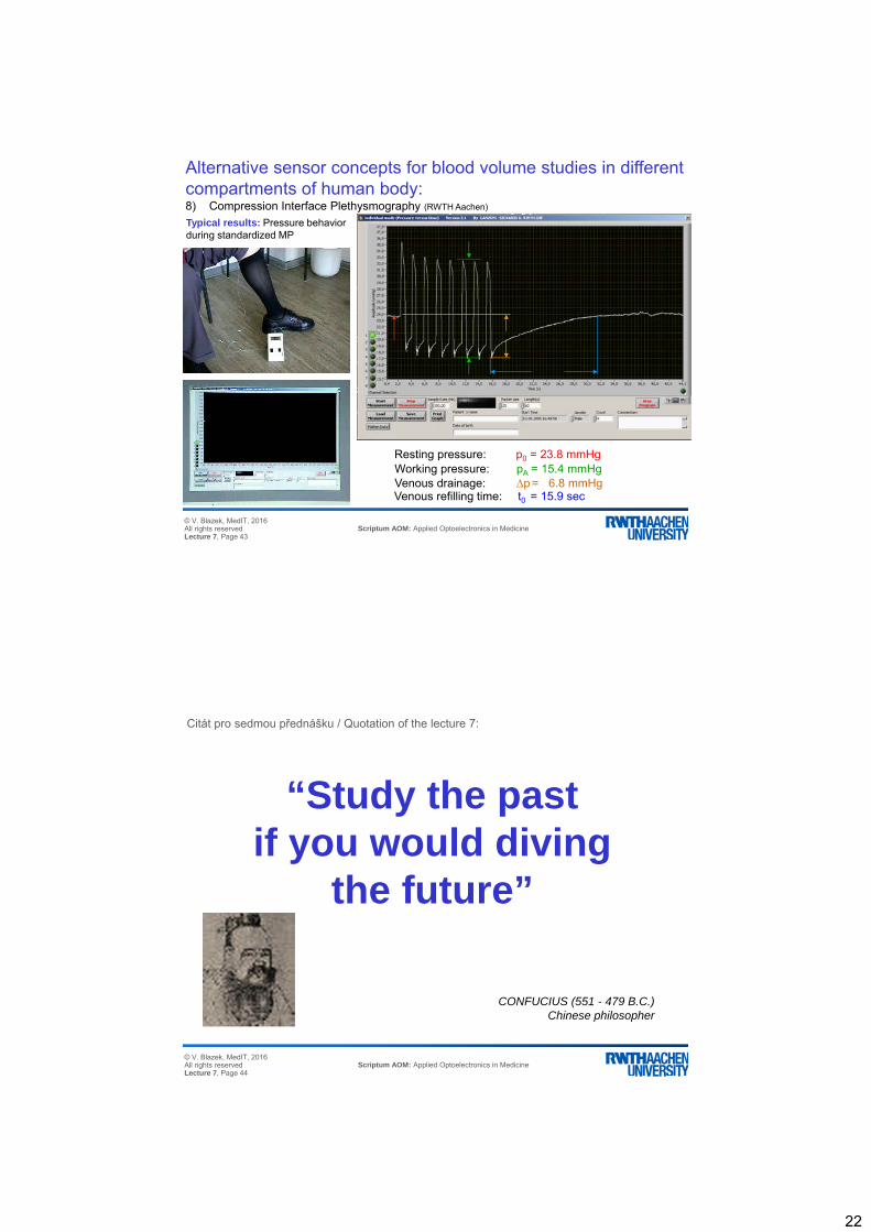
Typical results: Pressure behaviorduring standardized MP
Resting pressure: p0 = 23.8 mmHgWorking pressure: pA = 15.4 mmHgVenous drainage: p = 6.8 mmHgVenous refilling time: t0 = 15.9 sec
Alternative sensor concepts for blood volume studies in different compartments of human body:8) Compression Interface Plethysmography (RWTH Aachen)
© V. Blazek, MedIT, 2016All rights reservedLecture 7, Page 44
Scriptum AOM: Applied Optoelectronics in Medicine
“Study the pastif you would diving
the future”
CONFUCIUS (551 - 479 B.C.)Chinese philosopher
Citát pro sedmou přednášku / Quotation of the lecture 7: