applications of nano-materials in diverse dentistry regimes
TRANSCRIPT

RSC Advances
REVIEW
Ope
n A
cces
s A
rtic
le. P
ublis
hed
on 2
0 A
pril
2020
. Dow
nloa
ded
on 3
/23/
2022
9:0
3:52
PM
. T
his
artic
le is
lice
nsed
und
er a
Cre
ativ
e C
omm
ons
Attr
ibut
ion-
Non
Com
mer
cial
3.0
Unp
orte
d L
icen
ce.
View Article OnlineView Journal | View Issue
Applications of n
aInstitute of Research and Development, Du
NambDepartment of Chemistry, Kerman Branch,cNanobioelectrochemistry Research Center, B
Iran. E-mail: [email protected]; armitafor
Tel: +98 34331321750dStudent Research Committee, School of Pu
Sciences, Bam, IraneTehran Dental Branch, Islamic Azad UniverfCraniomaxilofacial Resarch Center, Teh
University, Tehran, IrangNeuroscience Research Center, Institute of N
Medical Sciences, Kerman, IranhDepartment of Pediatric Dentistry, School o
Sciences, Kerman, IraniRegional Centre of Advanced Technologies
Chemistry, Faculty of Science, Palacky Uni
Czech RepublicjCell Therapy and Regenerative Medicine Co
Medical Sciences, Kerman, Iran
Cite this: RSC Adv., 2020, 10, 15430
Received 24th January 2020Accepted 11th March 2020
DOI: 10.1039/d0ra00762e
rsc.li/rsc-advances
15430 | RSC Adv., 2020, 10, 15430–1
ano-materials in diverse dentistryregimes
Loke Kok Foong,a Mohammad Mehdi Foroughi, *b Armita Forutan Mirhosseini, *c
Mohadeseh Safaei,d Shohreh Jahani,cd Maryam Mostafavi,ef Nasser Ebrahimpoor,g
Maryam Sharifi,h Rajender S. Varma i and Mehrdad Khatami *cj
Research and development in the applied sciences at the atomic or molecular level is the order of the day
under the domain of nanotechnology or nano-science with enormous influence on nearly all areas of
human health and activities comprising diverse medical fields such as pharmacological studies, clinical
diagnoses, and supplementary immune system. The field of nano-dentistry has emerged due to the
assorted dental applications of nano-technology. This review provides a brief introduction to the general
nanotechnology field and a comprehensive overview of the synthesis features and dental uses of nano-
materials including current innovations and future expectations with general comments on the latest
advancements in the mechanisms and the most significant toxicological dimensions.
1. Introduction
Nowadays, nanotechnology has been integrated into differentareas of science as it provides various signicant ways to meetscientic and medical problems. Nanotechnology, which isa branch of technology, works in the dimensions of less than100 nm. It includes objects such as viruses of about 100 nm sizedown to glucose molecules of about 1 nm size. Therefore, itincludes the study of structures at the molecular and atomicscales.1–4 Assorted nano-materials may be categorized on thebasis of their morphology and the presence of nano-pores,which is exemplied by dendrimers, nano-tubes, quantumdots, liposomes, nano-rods, nano-wires, fullerenes, nano-
y Tan University, Da Nang, 550000, Viet
Islamic Azad University, Kerman, Iran
am University of Medical Sciences, Bam,
[email protected]; Fax: +98 3433210051;
blic Health, Bam University of Medical
sity, Tehran, Iran
ran Medical Sciences, Islamic Azad
europharmacology, Kerman University of
f Dentistry, Kerman University of Medical
and Materials, Department of Physical
versity, Slechtitelu 27, 783 71 Olomouc,
mprehensive Center, Kerman University of
5460
spheres, nano-belts, nano-rings, nano-shells, and nano-capsules (Fig. 1).
Most of the works that have been reported during the last 20–30 years have focused on nanoparticles; thus, it is clear thatthere is a great interest in nanotechnology and the features ofthe materials at these scales. For example, nanotechnology canmeasure the surface area of 1 g of a powder at different spher-ical sizes, and these data show that the surface area per gramrises exponentially below �100 nm. This leads to a change inthe phase of these materials such as the increase in the surfaceenergy per gram of the material. This massive increase in thesurface area can be applied for different purposes.5–10
Teeth within the oral cavity have various parts such as dentin,enamel, cementum, pulp, and periodontal ligament. Teeth cut andcrush food to make it easy to swallow and digest. Furthermore,teeth empower self-condence and improve the quality of life.Therefore, the loss of teeth due to a disease or decay can affect theeating pattern, speaking, or laughing. Thus, dentistry provides a lotof methods for protecting teeth.11–14 These efforts suffer from keydisadvantages, which require more efficient strategies and noveltechnologies in contemporary dentistry.15–18
Nanoparticles, unlike other biomaterials, present distinctbiological properties and can be used in novel applications inrestorative dentistry, prosthetic dentistry, endodontics,implantology, oral cancers, and periodontology. Nanoparticleshave immense potential because of their antimicrobial, anti-viral, and antifungal properties. The incorporation of nano-particles prevents biolm build up over the composite, whichavoids micro-leakage and secondary caries.19–21 These nano-particles enhance the mechanical properties of a restorativematerial and improve the overall bonding between dentin andbiomaterials, thus affecting the bond strength. Nanoparticle-
This journal is © The Royal Society of Chemistry 2020

Fig. 1 Representative structures of some nano-materials: (a) nanorings,228 (b) nanopellets,229 (c) nanorods,230 (d) nanosprings,228 (e) nanonails,231
(f) nanoflower.232
Review RSC Advances
Ope
n A
cces
s A
rtic
le. P
ublis
hed
on 2
0 A
pril
2020
. Dow
nloa
ded
on 3
/23/
2022
9:0
3:52
PM
. T
his
artic
le is
lice
nsed
und
er a
Cre
ativ
e C
omm
ons
Attr
ibut
ion-
Non
Com
mer
cial
3.0
Unp
orte
d L
icen
ce.
View Article Online
incorporated adhesive systems can be applied in orthodontictreatments to prevent white spot lesions. In vitro research hasshown that these nanoparticles prevent crack propagation andimprove the fracture toughness with dental ceramics, whichnegates the cracking of the porcelain restorations such ascrowns, bridges, and veneers.22–25 Although it is clear thatnanoparticles can be effective due to their incorporation withdental biomaterials, to use them for clinical applications, in vivoresults with long-term data are necessary. Besides the benetsof nanoparticles, the research on long-term in vivo results,methods of nanoparticle incorporation and characterization,and data on their long-term antibacterial action is needed forclinical applications.26–29
This paper provides an overview of the various kinds of nano-materials, their synthetic techniques, and characteristicsincluding the science, implications, and up-to-date uses ofnano-technology in dentistry. Novel designed materials intro-duced in the market as well as the summary of the contributionof dentists to the understanding of clinical relevance and effi-ciency of nano-materials is compared with those currentlydeployed in clinical practices.
2. Classification of nano-materials
The general classication of nano-materials comprisingorganic, inorganic, and carbon-based materials is presentedbelow (Scheme 1).
2.1. Organic nano-materials
Organic nano-materials or polymers usually encompass den-drimers, micelles, liposomes, ferritin, etc., which are biode-gradable and non-toxic. A number of such particles, including
This journal is © The Royal Society of Chemistry 2020
micelles and liposomes, have a hollow core, termed as nano-capsules that have the sensitivity towards thermal and electro-magnetic radiations, including heat and light.30 Such specicfeatures render them a perfect option for drug delivery. Due toits stability, capacity, and delivery systems, the absorbed drugsystem determines the respective type of uses and efficacy,regardless of the physical properties, including dimensions,compositions, and surface morphologies. Organic nano-materials have widespread usage in biomedicine for targeteddrug delivery.31–35
2.1.1. Polymeric nano-materials. This category is usuallyfor organic-based nano-materials, which basically have nano-sphere or nano-capsule shapes and can be easily functional-ized. Nanospheres are matrix particles with overall solid massand other molecules are absorbed at the external surface of thespherical surfaces. Nanocapsules are solid mass thoroughlyencapsulated into the particle.36,37
2.1.2. Lipid-based nano-materials. These nano-materials,ranging in diameter in between 10 and 1000 nm, include lipidmoieties with effective applications in numerous biomedicalelds. Similar to polymeric nano-materials, they possess a solidcore, which is made of a lipid and also a matrix containinglipophilic molecules while the emulsiers or the surfactantsstabilize the outer core. They nd varied applications in medi-cine carriers, delivery, and RNA release for treating cancer.38
2.2. Inorganic nano-materials
Metal and metal oxide-based nano-materials are usually clas-sied under this category.
2.2.1. Metal nano-materials. These are completely fabri-cated from metal precursors. In view of their common localizedsurface plasmon resonance (LSPR) properties, they enjoy
RSC Adv., 2020, 10, 15430–15460 | 15431

Scheme 1 General classification of nano-materials.
RSC Advances Review
Ope
n A
cces
s A
rtic
le. P
ublis
hed
on 2
0 A
pril
2020
. Dow
nloa
ded
on 3
/23/
2022
9:0
3:52
PM
. T
his
artic
le is
lice
nsed
und
er a
Cre
ativ
e C
omm
ons
Attr
ibut
ion-
Non
Com
mer
cial
3.0
Unp
orte
d L
icen
ce.
View Article Online
specic opto-electrical characteristics. Nano-materials of noblemetals such as Ag, Au, and Cu and alkali possess a wideadsorption peak in the observable region of the electro-
15432 | RSC Adv., 2020, 10, 15430–15460
magnetic solar spectrum; facets, sizes, and shape-monitoredmetal nano-materials are highly valued as cutting-edgeadvanced materials.39–41
This journal is © The Royal Society of Chemistry 2020

Review RSC Advances
Ope
n A
cces
s A
rtic
le. P
ublis
hed
on 2
0 A
pril
2020
. Dow
nloa
ded
on 3
/23/
2022
9:0
3:52
PM
. T
his
artic
le is
lice
nsed
und
er a
Cre
ativ
e C
omm
ons
Attr
ibut
ion-
Non
Com
mer
cial
3.0
Unp
orte
d L
icen
ce.
View Article Online
2.2.2. Metal oxide nano-materials. Mostly, metal oxidenano-materials are synthesized because of higher reactivity andeffectiveness. Some examples are cerium oxide (CeO2), zincoxide (ZnO), aluminium oxide (Al2O3), titanium oxide (TiO2),magnetite (Fe3O4), iron oxide (Fe2O3), and silicon dioxide (SiO2),which are frequently synthesized oxides. Such nano-materialsexhibit exceptional features in comparison to their metalanalogues.42,43
2.2.3. Ceramic nano-materials. These are inorganic non-metallic solids, which are synthesized through heating andconsecutive cooling and exist in polycrystalline, dense, amor-phous, porous, or hollow forms with applications in catalysis,photo-degradation of dyes, photo-catalysis, and imagingapplications.44,45
2.2.4. Semiconductor nano-materials. Semi-conductormaterials have features between metals and non-metals, andbecause of their broad bandgaps, their features are signicantlyaltered as the bandgaps are tuned. Thus, they are highly prominentmaterials in photo-catalysis, photo optics, and electronic devices.46
2.3. Carbon based nano-materials
Carbon-based nano-materials can be grouped into fullerenes,graphene, carbon nanotubes, carbon nanobers, carbon black,and occasionally, actuated carbon with nanometer size.47
2.3.1. Fullerenes. Fullerene (C60) is a spherical carbonmolecule made up of carbon atoms, which are bonded to eachother via sp2 hybridization with nearly 28 to 1500 carbon atoms,comprising spherical structures with a diameter of 8.2 nm foreach layer and 4 to 36 nm for the multilayers.48
2.3.2. Graphene. Graphene is one of the allotropes ofcarbon. It has a hexagonal network with a honeycomb latticeconsisting of carbon atoms in a 2D planar surface. In general,the thickness of a graphene sheet is �1 nm.49,50
2.3.3. Carbon nanotubes. Carbon nanotubes are graphenenano-foils with a honeycomb lattice of carbon atoms, which aretwisted in an empty cylinder, forming nano-tubes that are assmall as 0.7 nm for a one-layered carbon nanotube and 100 nmfor the multilayered carbon nanotubes, which varies from a fewmicro-meters to many millimeters. The end of nanotubes maybe unlled or closed via a half fullerene molecule.51–53
2.3.4. Carbon nanober. Similar to graphene, nano-foils areemployed for producing carbon nano-ber as carbon nanotubesbut they are twisted into a cup- or cone-shape rather thana conventional cylindrical tube.54
2.3.5. Carbon black. It is an unshaped carbon object that isusually spherical in shape with a diameter in the range of 20 to70 nm. The particles have a great interaction that leads tobinding of the aggregates so that approximately 500 nm-sizedagglomerates are established.55
3. Synthesis of nano-materials
Two main techniques are deployed for the synthesis of nano-materials and are broadly classied into top-down andbottom-up methods (Scheme 2).
This journal is © The Royal Society of Chemistry 2020
3.1. Top-down synthesis
Destructive or top down technique refers to the decrease in bulkmaterials to the nano-meter scale particles with mechanicalmilling, sputtering, nano-lithography, laser ablation, andthermal decomposition as the synthetic techniques with wide-spread applications.
3.1.1. Mechanical milling. Amongst different top downtechniques, mechanical milling is one of the widely usedmethods for generating different nano-particles and is appliedto mill and post-anneal nanoparticles during syntheses, inwhich various components are milled. Plastic deformation isa parameter affecting the mechanical milling, which results inthe particle shape and fracturing, resulting in the decrease inparticle sizes, and cold-welding, leading to an increase in theparticle size.56–58
3.1.2. Nano-lithography. Studying the fabrication of nano-meter scale structures with at least one dimension in the sizeranging between 1 and 100 nm is termed as nano-lithographywith different varieties of the nano-imprint, namely, optical,multi-photon, scanning probe lithography, and electronbeam.59,60 In general, lithography refers to the procedure ofprinting the intended form or structure on light sensitivematerials, which eliminates a part of the materials for creatingthe intended shape and structure in a selective manner.61
3.1.3. Laser ablation. One of the popular methods forproducing nano-materials in different solvents is laser ablationsynthesis, which entails the irradiation of a metal immersed ina liquid solution via a laser beam that condenses a plasmaplume, thus generating the nano-materials.62
3.1.4. Sputtering. NPs deposition on the surface via ejec-tion of particles from it through collision with the ions is calledsputtering.20 It is generally the deposition of a thin layer of NPsaccompanied by annealing. The NPs' sizes and shapes arespecied by the layer thickness, annealing period, andtemperature and type of the substrate.63
3.1.5. Thermal decomposition. Thermal decompositionprocess refers to the chemical decomposition through heating,which is endothermic and breaks down the chemical bonds inthe compounds. Nano-materials are generated via decomposi-tion of the metal at certain temperatures that undergo a chem-ical reaction, thus generating secondary products.64,65
3.2. Bottom-up synthesis
Generating materials from atoms to clusters to nano-materialsis called bottom-up or constructive technique with widespreadapplications for producing nano-materials via chemical vapordeposition (CVD), sol–gel, spinning, pyrolysis, and bio-synthesis.
3.2.1. Sol–gel. One of the widespread bottom-up methodsis the sol–gel method because of its straightforwardness. Sol–gel is a wet-chemical procedure with a chemical solution, whichis a precursor for discrete particles. Metal oxide and chloride aretwo precursors that are usually deployed in the sol–gel proce-dure.66–69 Aerwards, the precursors are scattered in a hostliquid by stirring, shaking, or sonicating. The nal systemcomprises a solid and a liquid phase. A separated phase is
RSC Adv., 2020, 10, 15430–15460 | 15433

Scheme 2 Conventional synthetic techniques for nano-materials.
RSC Advances Review
Ope
n A
cces
s A
rtic
le. P
ublis
hed
on 2
0 A
pril
2020
. Dow
nloa
ded
on 3
/23/
2022
9:0
3:52
PM
. T
his
artic
le is
lice
nsed
und
er a
Cre
ativ
e C
omm
ons
Attr
ibut
ion-
Non
Com
mer
cial
3.0
Unp
orte
d L
icen
ce.
View Article Online
applied for recovering the nano-materials via different tech-niques, including sedimentation, lteration, and centrifuga-tion. Moisture is oen additionally eliminated via drying.70,71
3.2.2. Spinning. Spinning disc reactor (SDR) has been usedto synthesize nano-materials via spinning, which involvesa spinning disc within a reactor/chamber, in which physicalvariables including temperature may be monitored. In general,nitrogen or other inert gases are lled in the reactor to removeoxygen and to avoid chemical reaction. The disc spins at variousvelocities, in which the solution with water and precursor arepumped in. The spinning process results in the fusion ofmolecules or atoms, which is followed by precipitation, collec-tion, and drying. The properties of the nano-materials synthe-sized from SDR are determined by distinct operational variablessuch as disc surfaces, disc rotation velocity, liquid ow rate,liquid or precursor ratio, and feed location.72–76
3.2.3. Chemical vapor deposition (CVD). CVD refers to thedeposition of a thin layer of gaseous reactants over a substrate. Thesedimentation process is performed at the ambient temperature ina reaction chamber via the combination of gas molecules whereinchemical reaction takes place when a heated substrate
15434 | RSC Adv., 2020, 10, 15430–15460
communicates with the fused gas.8 The reaction results in theformation of a thin layer of product on the substrate surface, whichis recyclable and reusable. The temperature of the substrate is oneof the factors affecting CVD. The benets of CVD include higherpurity, uniformity, and hardness, with the shortcoming that itrequires specic instruments and gaseous by-products that wouldbe strongly poisonous.77,78
3.2.4. Pyrolysis. Pyrolysis, the burning of a precursor witha ame is a procedure with widespread application in industrieswith large-scale production of NPs. The vapor or liquid form ofthe precursor is fed into the furnace with a high pressurethrough a little orice for combustion.79 Aerwards, thecombustion or by-product gases are categorized for recoveringthe nano-materials. A number of furnaces employ laser andplasma rather than ame for producing high temperatures forsimple evaporation.80,81 Pyrolysis enjoys benets such assimplicity, efficiency, affordability, and continual procedurewith great yields.
3.2.5. Bio-synthesis. Bio-synthesis refers to a green and eco-friendly strategy for synthesizing nano-materials, which isrelatively less toxic and with the possible use of biodegradable
This journal is © The Royal Society of Chemistry 2020
Review RSC Advances
Ope
n A
cces
s A
rtic
le. P
ublis
hed
on 2
0 A
pril
2020
. Dow
nloa
ded
on 3
/23/
2022
9:0
3:52
PM
. T
his
artic
le is
lice
nsed
und
er a
Cre
ativ
e C
omm
ons
Attr
ibut
ion-
Non
Com
mer
cial
3.0
Unp
orte
d L
icen
ce.
View Article Online
materials.15 It deploys bacteria, plant extracts, fungi, andenzymes accompanied by precursors for generating nano-particles rather than traditional chemicals for bio-reductionand capping purposes. The bio-synthesized nano-materialshave certain improved biocompatibility features that areuseful for bio-medical applications.82
4. Nano-materials features
In general, nano-material features are classied into physicaland chemical features.
4.1. Physical features
Physical features involve optical characteristics, includingnano-material color, light penetration, adsorption and reec-tion abilities, UV adsorption, and reection capability ina solution or coated over a surface. Moreover, it involvesmechanical features, including elasticity, ductility, tensilestrength, and exibility, which contribute signicantly to theirapplication. Notably, several contemporary industries use otherfeatures such as hydrophilicity, hydrophobicity, suspension,diffusion, and settling properties. Electrical and magneticfeatures, including conductivity, semiconductivity, and resis-tivity, provide the grounds for using nano-materials incontemporary electronics, thermal conductivity, and renewableenergy applications.83,84
4.2. Chemical characteristics
Chemical characteristics pertain to reactivity of the nano-materials with the target, stability, and sensitivity to variablessuch as atmosphere, humidity, light, and heat that determinesthe applications of nano-materials. Anti-bacterial, antifungal,disinfection, and toxicity are perfect nano-material features forbiomedical and environmental uses. Corrosive, anti-corrosive,oxidation, decline, and ammability properties of nano-materials determine their applications.85,86
5. Characterizing nano-materials
Various characterization methods have been developed toanalyze different physico-chemical features of nano-materials,namely, scanning electron microscopy (SEM), X-ray diffraction(XRD), infrared spectroscopy (IR), transmission electronmicroscopy (TEM), X-ray photo-electron spectroscopy (XPS), andBrunauer–Emmett–Teller (BET) and particle size analyses.
5.1. Morphological properties
The morphological properties of nano-materials have beenconsistently and greatly considered because morphologyinvariably affects a majority of the nano-material's features.Various characterization methods have been proposed formorphological examinations; however, microscopic methods,including SEM, polarized optical microscopy (POM), and TEMare the most prominent techniques. SEM is based on the elec-tron scanning principle, which presents information about thematerials at the nano-scale levels.87
This journal is © The Royal Society of Chemistry 2020
5.2. Structural features
Structural features are crucial for studying the compositionsand nature of the binding materials. XRD, EDX, XPS, Raman,IR, BET, and zeta potential and size analysis are the prevalentmethods employed for studying the structural features of nano-materials.88
5.3. Particle size and surface area
It is possible to use various procedures for estimating the size ofthe nano-materials such as TEM, XRD, and SEM, although thezeta potential and size analysis by dynamic light scattering(DLS) may be applied for nding the sizes of extremely smallnano-materials.89
5.4. Optical features
The optical features are crucial in photo-catalytic applicationsand the knowledge of the mechanisms can be exploited forphoto-chemical procedures. Such features are according to thepopular Beer–Lambert's law and the fundamental principles oflight. Such methods provide knowledge of the luminescence,adsorption, reectance, and phosphorescence features of thenano-materials.90
6. Dental applications of nano-materials
Injured dental tissues may result in dental caries, periodontaldiseases, tooth sensitivity, unpleasant breath, and oral pre-cancerous and cancerous conditions. All of the above compli-cations may be treated via therapeutic interventions andapplication of bio-compatible synthetic materials. Nano-medicines applied as dental materials possess certainphysico-chemical and biological features, which make themsuperior for overcoming the side effects related to moreconventional dental therapies.91 Research has shown thatvarious kinds of nano-materials mimic the host tissuefeatures,92,93 though the knowledge of such features amongstdental communities is not available. Hence, the present reviewfocuses on the characteristics of various metal and polymer-based nano-materials employed in adhesive and restorativedentistry, acrylic resins, periodontology, tissue engineering,endodontics, and implant dentistry.94–96
6.1. Nano-materials for preventive dentistry
Teeth function in the dynamic environment of the oral cavity,wherein it is a big challenge to prevent tooth decay. Because ofthe accumulated knowledge-base on oral diseases, preventivedentistry is imperative and plays a signicant role. Nano-materials are employed in preventive dentistry, managing bio-lms at the surface of teeth through nano-apatites, anddemineralizing the initial stage of submicron-sized enamellesions.97,98
Schwass et al. designed a silver NP (Ag NP) formulation asa targeted application for disinfecting carious dentine. Sodiumborohydrate (NaBH4) decreased silver nitrate (AgNO3)
RSC Adv., 2020, 10, 15430–15460 | 15435

RSC Advances Review
Ope
n A
cces
s A
rtic
le. P
ublis
hed
on 2
0 A
pril
2020
. Dow
nloa
ded
on 3
/23/
2022
9:0
3:52
PM
. T
his
artic
le is
lice
nsed
und
er a
Cre
ativ
e C
omm
ons
Attr
ibut
ion-
Non
Com
mer
cial
3.0
Unp
orte
d L
icen
ce.
View Article Online
chemically in the presence of sodium dodecyl sulfate (SDS) toform micelle aggregate structures with mono-dispersed stabi-lized Ag NPs with size in the range of 6.7 to 9.2 nm. On triplicatetesting of Ag NPs against Streptococcus gordonii DL1, C219,G102, and ATCC10558 strains, Streptococcus mutans UA159,Streptococcus mitis I18, and Enterococcus faecalis JH22 forplanktonic bacteria, the minimum suppressive concentrationswere determined to be as low as 7.6 mg mL�1 with the lowestbactericidal silver concentration of 19.2 mg mL�1. Microplatereadings, which detect crystal violet light adsorption at 590 nm,exhibited considerable difference among the Ag NP treated bio-lms. The presence of sucrose had no effect on the sensitivity ofbacteria. During the prevention of in vitro bio-lm creation fornumerous Streptococcus spp. and Enterococcus faecalis, this AgNP formulation showed potential for clinical applications insuppressing bio-lms.99
Favretto et al. conducted a study to evaluate the capability ofuoride toothpastes (1100 ppm F), which contain sodium tri-metaphosphate (TMP) NPs, in enhancing the obliteration ofdentinal tubules with or without acid challenges. They intendedto conrm if the reduction in the size of sodium trimetaphos-phate NPs could additionally increase these impacts or not.Sodium trimetaphosphate NPs enriched uoride toothpastesenjoy the same capability for occluding dentinal tubules asa toothpaste with 1100 ppm F, in which an acidic situationcould not change the obliterating dentinal canals and theparticle sizes did not affect the outputs. When sodium trime-taphosphate NPs have been added, uoride toothpastes can
Fig. 2 Schematic diagram of the rechargeable nanoparticles of the amorpdemineralization around the dental sealants: In (A), the recharge cycle diasealants. Three recharge/re-release cycles were performed and each rincreasing the NACP filler level. In (B), the TEM image of NACP from the
15436 | RSC Adv., 2020, 10, 15430–15460
occlude the dentinal canals with a capacity for reducing dentinhypersensitivity.100
Manikandan et al. explored the formation of silver oxide NPs(Ag2O NPs) via Ficus benghalensis prop root extract (FBPRE) asa stabilizing and decreasing agent and assessed its anti-bacterial activities versus dental bacterial strains; higherextract concentrations and time frame have been observed witha considerable enhancement in the formation of NPs. TheFBPRE and Ag2O NPs combination has been found to displayvery good anti-bacterial activity against both dental bacteriaLactobacilli sp and Streptococcus mutans. Their outcome indi-cated that blending the synthesized FBPRE and Ag2O NPS wouldbe benecial as a germicidal factor in toothpastes aer severalstudies on animal models.101
Mackevica et al. examined the release of Ag NP fromcommercial toothbrushes for children and adults by analyzingthe total Ag released and quantifying the particulate Ag NPrelease. Experimental ndings revealed the possible release ofAg NPs from the toothbrushes in the market that might result inpotential consumer oral and environmental exposures. Testingthese 2 toothbrushes found in the market showed that adulttoothbrushes have relatively greater Ag release with regard tothe Ag and Ag NP releases. The overall procedure is as follows:release of the particles considerably decreased aer six minutesof testing for the rst time and the release of total Ag reacheda plateau aer testing for 16 hours. The median particle size(43–47 nm) was identical for each toothbrush tested. Total Agrelease for the two toothbrush brands was at ng L�1 levels,
hous calcium phosphate (NACP) sealant approach to deal with enamelgram illustrates the re-release from the exhausted and recharged NACPe-release was measured for 14 days. The ion re-release increased onspray-drying technique, having sizes of about 100–300 nm.103
This journal is © The Royal Society of Chemistry 2020
Review RSC Advances
Ope
n A
cces
s A
rtic
le. P
ublis
hed
on 2
0 A
pril
2020
. Dow
nloa
ded
on 3
/23/
2022
9:0
3:52
PM
. T
his
artic
le is
lice
nsed
und
er a
Cre
ativ
e C
omm
ons
Attr
ibut
ion-
Non
Com
mer
cial
3.0
Unp
orte
d L
icen
ce.
View Article Online
which affirms that there is minor environmental and humanexposure from the toothbrushes; however, the safe levels of AgNP exposure is still unanswered.102
Salem Ibrahim et al. designed novel anti-bacterial resin-based sealants including NPs of amorphous calcium phos-phate (NACP) for PO4 and Ca ion release and re-charge char-acteristics (Fig. 2). They aimed at incorporating various massfractions of NACP into the parental re-chargeable anti-bacterialsealant, determining the impacts on the mechanical functions,and evaluating the method of studying the effect of the changesin the NACP concentrations on phosphate (PO4) and calcium(Ca) ions' release and recharge ability over time. It appears thatthe addition of an enhanced percentage of NACP had satisfac-tory physical and mechanical functions, while generatingconsiderable initial ion release and a lengthy iterated rechargeability.103
Wassel and Khattab proposed that applying the naturally-occurring products experimentally may be an efficient strategyto prevent caries; Varnish is a mix of natural products withoptimum concentrations of uoride in chitosan NPs (CS-NPs)and potential for ion release, re-mineralization potential, andclinical efficacy. The procured dental varnishes with miswak,propolis, and CS-NPs with or without sodium uoride (NaF)have been evaluated in terms of the anti-bacterial effects againstStreptococcus mutans employed in the disk diffusion test. Theprotecting effects of a single pre-treatment of the main teethenamel species against in vitro bacteria-induced enamel de-mineralization were evaluated for three days. Each naturalproduct with the varnish largely suppressed the growingbacteria more than that by 5% NaF varnish; however, NaFloaded CS-NPs (CSF-NPs) had maximum anti-bacterial impact,even though there was no signicant difference between themand other varnishes with the exception of miswak ethanolicextract varnish. Increased suppressive impact was observed forthe varnish with freeze-dried aqueous extract of miswak incomparison to the varnish with ethanolic extract of miswak,which may be caused by the anti-microbial substance concen-trations via freeze-drying. Adding natural ingredients to NaF ina dental varnish presented additional effects, particularly incomparison with uoride with varnish; 5% NaF varnish had themost acceptable suppression of the de-mineralization impact.Fluoride with miswak varnish and CSF-NPs varnish consider-ably suppressed the de-mineralizationmore favorably than eachexperimental varnish, although the CSF-NPs varnish exhibiteda small uoride concentration, which may be caused by themore acceptable existence of uoride ions andmore small-sizedNPs. One of the most efficient approaches to prevent caries, inparticular, miswak and propolis in a case of limited nancialresources, is the incorporation of natural products with uorideinto the dental varnishes.104
Nguyen et al. conducted a study to develop uoride loadedNPs on the basis of bio-polymers chitosan, alginate, and pectinfor use in dental delivery; polymer-based nano-particulateformulations were developed for providing and improving aninstrument for the effective topical delivery of uoride. In thepresence of NaF and a suitable cross-linker, simply chitosancould create stable mono-dispersed NPs. Alginate failed to
This journal is © The Royal Society of Chemistry 2020
create NPs because the optimum ionic strength was greater atthe experimented salt concentrations, while pectin producedlarge undened nanostructures. It was shown that uorideloading and the entrapment efficacy of chitosan NPs is 33–113and 3.6–6.2% ppm, respectively, under the testing conditions. Itis possible to optimize the aforementioned values for preparingthe variables during the process of incorporating uoride.Apparently, the release of uoride increased in an acidic envi-ronment while simulating a cariogenic attack. Such featuresmay be largely benecial for dental formulations, which targetpatients with high risk of development.105
Fathima et al. dealt with the synthesis of ZrO2 NPs witha crystalline nature and sizes between 15 and 21 nm, as veriedvia SEM, XRD, and TEM analyses. The anti-microbial activitiesof ZrO2 NPs versus Gram-positive and Gram-negative bacteriademonstrated the possible suppressive actions of ZrO2NPsagainst Gram-negative ones, in particular, Pseudomonas aeru-ginosa at greater concentrations because of the respective cellsurfaces with negative charges. Therefore, researchers haveillustrated the feasibility of exploitation of ZrO2 NPs in avoidingtooth decay by analyzing the tooth decay pathways. ZrO2 NPshave been suggested for applications in dental care and relatedbio-medical uses for future in vitro and in vivo research.106
Wang et al. exploited carboxymethyl chitosan (CMC) conju-gated with alendronate (ALN) for stabilizing amorphouscalcium phosphate (ACP) in the formation of CMC/ACP NPs.Sodium hypochlorite (NaClO) served as a protease decomposingamelogenin in vivo for degrading the CMC-ALN matrix andgenerating HAP@ACP core–shell NPs. HAP@ACP NPs werealtered by 10 mM glycine, as they were modied from anamorphous phase into well-ordered rod-shape apatite crystalsfor achieving oriented and ordered bio-mimetic re-mineralization on acid-etched enamel surfaces. The orientedbond of the NPs on the basis of the non-classical crystallizationtheory contributes to the bio-mimetic re-mineralization proce-dure. Researchers showed that one of the efficient approachesto remineralize the enamel is to nd and develop analogues ofnatural proteins, including amelogenin engaged in bio-mineralization via natural macro-molecular polymers andimitation of the biomineralization procedure. The above tech-nique could be a potential procedure for managing the initialcaries in minimal invasive dentistry.107
Liu et al. demonstrated the ability of ferumoxytol to disruptthe intractable oral bio-lms and prevention of tooth decay(dental caries) through intrinsic peroxidase-like activities. Fer-umoxytol binds to the bio-lm ultra-structure and produces freeradicals from hydrogen peroxide (H2O2), which causes in situbacterial mortality through cell membrane disruptions anddegradation of the extra-cellular polymeric substance matrix.When combined with a small concentration of H2O2, fer-umoxytol suppressed the bio-lm stacked on natural teeth ina human extracted ex vivo biolm model and stopped acidinjury of the mineralized tissues. Developing dental caries invivo is suppressed by topical oral therapy with H2O2 and fer-umoxytol, and prevents the initiation of serious tooth decay(cavity) in a rodent model of the disease. Histological andmicrobiome analysis did not present any consequences on the
RSC Adv., 2020, 10, 15430–15460 | 15437

RSC Advances Review
Ope
n A
cces
s A
rtic
le. P
ublis
hed
on 2
0 A
pril
2020
. Dow
nloa
ded
on 3
/23/
2022
9:0
3:52
PM
. T
his
artic
le is
lice
nsed
und
er a
Cre
ativ
e C
omm
ons
Attr
ibut
ion-
Non
Com
mer
cial
3.0
Unp
orte
d L
icen
ce.
View Article Online
oral microbiota diversities and gingival and mucosal tissues.Researchers have found a novel bio-medical application forferumoxytol as a topical therapy for the common and prevalentbio-lm, which results in oral diseases.108
A bio-nanocomposite of Carboxymethyl Starch (CMS)-Chitosan (CS)-Montmorillonite (MMT) has been designed todeliver Curcumin (Jahanizadeh et al.). They used ionic gelationtechnique and examined its anti-biolm activities versus Strep-tococcus mutans. Various formulations have been designed byresponse surface technique for obtaining the optimumcomposition with maximum medicine loading and minimumparticle sizes; entrapment efficacy and particles size wereinuenced by MMT amounts, surfactant concentrations, andpoly-saccharide concentrations. The results from the bacterialculture on the dental model demonstrated the powerful biolmreduction impact of the nano-composite with curcumin.109
Al Dulaijan et al. used Menschutkin reaction to synthesizedimethyl-aminohexadecyl methacrylate (DMAHDM). A spray-drying method was applied to synthesize NPs of amorphouscalcium phosphate (NACP). Resin included ethoxylatedbisphenol A dimethacrylate (EBPADMA) and pyromellitic glyc-erol dimethacrylate (PMGDM) and re-chargeable NACP and re-chargeable NACP-DMAHDM were the two constructedcomposites; ion release, mechanical features, and rechargewere evaluated. The bio-lm model of dental plaque microcosmwas experimented by using saliva. There was a match betweenthe modulus and commercial control composite and exuralstrength of rechargeable NACP and NACP-DMAHDM compos-ites (p > 0.1). Bio-lm metabolic events and lactic acid weresuppressed by NACP-DMAHDM, which declined the colony-forming units of the biolm (CFU) by 3–4 log. NACP andNACP-DMAHDM exhibited identical P and Ca ions' re-chargeand re-release (p > 0.1). Hence, the addition of DMAHDM didnot lead to compromise in the ion re-chargeability; continuousrelease was induced by 1 re-charge for 56 days and it was kept ata similar level when the number of recharge cycles wereenhanced, thus indicating the lengthy ion releases and re-mineralization ability. Researchers designed the 1st CaP re-chargeable and anti-bacterial composite. The addition ofDMAHDM to the re-chargeable NACP composite had no adverseeffect on the release and recharge of Ca and P ions, and thecomposite experienced highly lower bio-lm growth, lactic acidgeneration, and CFU decline by 3–4 log.110
Yan et al. synthesized mesoporous silica NPs (pMSN) forencapsulating chlorhexidine (CHX) as a classic anti-microbialagent. They used CHX@pMSN for modifying traditionaldental glass ionomer cement (GIC) for the rst time. It wasrevealed that CHX@pMSN modied GIC at 1% (w/w) couldattain the sustained release of CHX and effective inhibition ofthe formation of Streptococcus mutans biolm with no impact onthe mechanical features of GIC. The ndings disclosed thataddition of 1% (w/w) CHX@pMSN into the GIC had signicantpotential as a novel approach versus secondary caries, whichprolonged the conventional GIC service life. In addition, theenduring effects of incorporating CHX@pMSN into GIC have tobe assessed in more complicated scenarios via articial agingmethods, for example, pH cycling, sodium hypochlorite
15438 | RSC Adv., 2020, 10, 15430–15460
treatment, and lengthy storage. However, it is necessary thatfurther research should deal with more favorable incorporationapproaches for endowing GIC with inuential anti-microbialcapability and higher mechanical functions.111
Maghsoudi et al. examined the anti-biolm activity of nano-sized curcumin-loaded particles synthesized by desolvationtechnique. Nano-particle systems have been explored in termsof the properties against Streptococcus mutans functions ondental models when curcumin was applied as a biological anti-bacterial factor to load into NPs. The ndings determined thesize of the generated NPs with chitosan, starch, and alginate tobe 61.1, 66.3, and 78.8 nm, respectively; the corresponding zetapotential were �14.7, +21.7, and �23.4 mV, respectively. Thehighest amount of curcumin loaded onto the NPs was for chi-tosan (51.03); however, it was 24.59 and 29.69 for starch andalginate, respectively. It was estimated that the lowestsuppressive concentration (MIC) was 0.114 mg mL�1 for chito-san NPs while alginate and starch NPs had a MIC of 0.204 mgmL�1. Analyzing the release showed burst release aer 96 hoursfor chitosan and 48 hours for alginate; the release amounts were92.8% and 51.4%, respectively, while the starch NPs exhibiteda release with higher stability. When the equilibrium pointreached the end of 122 hours, the release of 81.6% of curcuminwas observed. Moreover, the impacts of curcumin-loaded NPson Streptococcus mutans bio-lms were evaluated for the dentalmodels. These ndings indicate that curcumin-loaded chitosanNPs could be applied in dental decay ghting products.112
Covarrubias et al. designed a study to develop hybrid NPs(CuCh NP) containing copper NPs with a chitosan shell. Anti-microbial features of CuCh NP have been evaluated againstStreptococcus mutans, which is a major bacterium causing toothdecay and their activities could be compared to the oral anti-microbial agents, including cetylpyridinium chloride andchlorhexidine. In particular, CuCh NP exhibited greater capac-ities for preventing the growth of Streptococcus mutans on thehuman tooth surface, disrupting and killing the bacterial cellsin the formed dental biolm. It is possible that there is aninteraction between the chitosan and tooth hydroxyapatite andthe bacterial cell wall, which enhances copper adherence to thetooth surface and increased their antibiolm actions. The anti-microbial features of CuCh NP may be advantageous for thedevelopment of more efficient therapies to control dental pla-que biolms.113
Gitipour et al. dealt with developing a nano-silver disinfec-tant (ASAP-AGX-32, an anti-microbial cleaner for dental units,0.0032% Ag) and a bio-lm. The researchers assembled an in-house dental unit water lines (DUWL) model for simulatingthe disinfection scenario so that the grounds for accumulatingthe biolm were provided. Gitipour et al. found that absorbingAg NPs on the bio-lm surfaces could be helpful in illustratingthe toxicity mechanism of Ag NPs on the biolm and bacteria.Therefore, this study might be an initial step in gaining moreknowledge as to how Ag NP transformation is dependent on theexposed conditions during their lifetime. So far, a majority ofthe studies have considered the assessment of the effects ofpristine (lab synthesized) nano-materials on differentsystems.114
This journal is © The Royal Society of Chemistry 2020

Review RSC Advances
Ope
n A
cces
s A
rtic
le. P
ublis
hed
on 2
0 A
pril
2020
. Dow
nloa
ded
on 3
/23/
2022
9:0
3:52
PM
. T
his
artic
le is
lice
nsed
und
er a
Cre
ativ
e C
omm
ons
Attr
ibut
ion-
Non
Com
mer
cial
3.0
Unp
orte
d L
icen
ce.
View Article Online
Ionescu et al. assessed bio-lm formation and bacterialadhesion on resin-based composites (RBC) such as dicalciumphosphate dihydrate NPs (nDCPD) wherein they illustratedanti-adherent or anti-biolm activity of nDCPD-lled RBC.Functionalizing nDCPD declined the surface roughness ofRBCs, which contributed to the decrease in biolm formationand adherence on the material surfaces. Therefore, an optimalformulation of the bio-mimetic RBCs would be as crucial as thebio-mimetic active principle alone in the regulation of micro-biological behaviors, which probably prevents the developmentof secondary caries.115
6.2. Nano-materials for edentulism
Edentulism has serious side effects, including reduced intake ofnutritious food and unsatisfactory appearance and has anincreased pervasiveness in numerous countries. In spite of theestimates of tooth loss declines, the age group, in whichedentulism would still be greatly common, has been gettingbroader. Therefore, it is strongly necessary for denture therapyin public health, which would enhance with the population'sage.116–118
Totu et al. procured polymethylmethacrylate (PMMA)/titanium dioxide NPs (TiO2) nano-composites and employednano-sized TiO2 ller synthesized using a modied sol–geltechnique; TiO2 nanoller experienced a homogeneousdispersion into the PMMA solution, which was veried bymorphological and structural analyses. Experimental dataconrmed that the addition of TiO2 NPs changed the polymerstructure and its certain features; 0.4% TiO2 NPs content in thenano-composite largely modied the FTIR spectrum. Theincorporation of TiO2 NPs in the PMMA polymer matrixprovided anti-bacterial impacts, particularly in the Candidaspecies, as conrmed by 0.4% nano-composite application viastereolithographic method for complete fabrication of thedenture.119
Rodrigues Magalhaes et al. described the application of TiO2
nano-tubes for enhancing the biological and mechanicalfeatures of dental materials. Yttria-stabilized tetragonal zirconiapoly-crystals (Y-TZP) have a growing application in dentistry asa substructure for xed partial prostheses and crowns.Regardless of its optimum clinical outputs, Y-TZP has suscep-tibility to failures such as micro-structure-associated defectivespresented in the fabrication procedure, which could decline itsclinical and structural reliability. Researchers assessed the roleof the production procedure of the blanks and their originalcomposition modications via the addition of TiO2 nano-tubes(0%, 1%, 2%, and 5% in volume) while monitoring each fabri-cation step. The addition of TiO2 nano-tubes in variouscombinations affected the experimental Y-TZP features andresulted in less exural strength. Moreover, the nano-tubesresulted in larger grain dimensions, more pores, and a minorenhancement in the mono-clinic phase, which inuenced themicro-structure of Y-TZP. Furthermore, the addition of TiO2
nano-tubes was accompanied by greater Weibull modulusvalues and higher structural reliability.120
This journal is © The Royal Society of Chemistry 2020
Gad et al. determined the effects of addition of zirconiumoxide (nano-ZrO2) NPs on the tensile and translucent strengthof polymethyl methacrylate (PMMA) denture base material; thetensile strength mean of PMMA in the test groups of 2.5% NZ,5% NZ, and 7.5% NZ was considerably greater compared to thecontrols. The tensile strength experienced a signicant increaseaer the addition of nano-ZrO2 and the highest amount ofincrease was seen in the 7.5% NZ group. The values of trans-lucency in the experimental group were remarkably less thanthe values in the controls. In the powered group, 2.5% NZ groupshowed greater translucency values in comparison with 5% NZand 7.5% NZ groups. The enhancement in the tensile strengthof the denture base acrylic was directly proportional to thenano-ZrO2 concentration while PMMA translucency declinedwhen nano-ZrO2 concentration was increased.121
Sarraf et al. built a hybrid biofunctionalized coatingencompassing nano-tubular rows of titanium dioxide (TiO2
NTs) with decorated silver oxide NPs (Ag2O NPs) on their edgesin order to improve the biological behaviors and anti-bacterialactivities of Ti6Al4V implants (Fig. 3). Aer making a nano-tubular structure via anodization of the substrate, Ag2O NPswere accumulated on the NTs through physical vapor deposi-tion in 30 s. In vitro bioactivity analysis revealed the depositionof apatite on Ag2O NPs that is decorated on TiO2 NTs aersoaking in simulated body uid for a day. Aer 14 days, theapatite quantity increased signicantly with the enhancementin submersion time and led to the formation of a thick layer ofapatite with a Ca/P ratio of 1.58. This novel Ag2O NPs-decoratedTiO2 NTs had good bactericidal effects against Escherichia coliand resulted in 100% eradication within two hours. In addition,osteointegration examinations via human osteoblast cells wereaccompanied by large nger-like protrusions and lopodialactivities of the cells, showing their efficient activation by theNT architecture. Moreover, consecutive rapid growththroughout the culture duration was shown by alamar blueassay and confocal laser scanning microscopy observations ofthe stained human osteoblast cells. Thus, this newly developedTiO2 NT coating covered with Ag2O NPs can efficiently amelio-rate the in vitro bioactivities of the implant alloys and establisha suitable bactericidal impact with minor cytotoxic responses.122
Gunputh et al. addressed two strategies for coating TiO2 NTswith Ag NPs, which also employed a less dangerous reducingagent, d-gluconolactone. These techniques were suitable forcreating Ag NPs with a main particle size �100 nm; however, anobvious difference was observed in the Ag NP clustering basedon the synthetic procedure. The mixing technique resulted inmicron clusters of Ag NPs; however, consecutive additiontechnique resulted in much smaller nanoclusters, whichshowed anti-bacterial effect on Staphylococcus aureus. In addi-tion, the amount of silver released from the coated NPs in therst 24 h was useful for patient healing. The maximum risk ofinfections was during the few hours immediately aer implantoperation.123
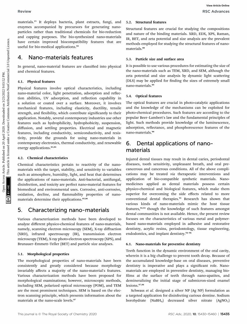
Yang et al. revealed feasible coating of dental implants underan extra-corporeal magnetic eld with lower concentration ofPLGA (Ag–Fe3O4) for improving the biological compatibilitywith no effects on the anti-bacterial efficacy (Fig. 4). A
RSC Adv., 2020, 10, 15430–15460 | 15439

Fig. 3 Schematic representation of the anodization setup and the development of TiO2 NTs as well as PVD decoration of Ag2O NPs on thenanotubular layer.122
RSC Advances Review
Ope
n A
cces
s A
rtic
le. P
ublis
hed
on 2
0 A
pril
2020
. Dow
nloa
ded
on 3
/23/
2022
9:0
3:52
PM
. T
his
artic
le is
lice
nsed
und
er a
Cre
ativ
e C
omm
ons
Attr
ibut
ion-
Non
Com
mer
cial
3.0
Unp
orte
d L
icen
ce.
View Article Online
permanent magnet was applied for building the magnetic eldas close to the PLGA (Ag–Fe3O4) as possible, which wasemployed in vivo to the implanted tooth containing a perma-nent magnet, thus providing Ag adhesion to the tooth surfaceswith no removal via ushing water. Bacterial infections,including the infection caused by Streptococcus mutans, trig-gered the host immune responses for producing reactive oxygenspecies (ROS), which led to the demolition of the tooth sup-porting tissues (Fig. 4, le). In the implanted tooth coated withPLGA (Ag–Fe3O4), bacterial adhesion was undermined. There-fore, ROS was not produced by the immune system and themicro-environment surrounding the implanted area triggeredosteoblast proliferation, which improved the transplant successrates.124
Jang et al. proposed bio-compatible Pd–Ag-HAp NPs, whichwere efficiently deposited onto the extended TiO2 obstacle layerin a 1.3 M (NH4)H2PO4 + 0.5 M NH4F electrolyte solution. Theydemonstrated that the protrusion patterns were slowly depos-ited over the TiO2 obstacle oxide lm and were rough. Abnormalpatterns due to Pd–Ag-HAp NPs might obviously be differenti-ated from the TiO2 nano-tube oxide layer formed by the anod-izing procedure. The element mapping dots usually hada homogeneous distribution throughout the surface of the lm.In particular, Pd and Ag had a uniform distribution throughoutthe surface areas of the protrusion patterns; however, P, Ca, Ti,
15440 | RSC Adv., 2020, 10, 15430–15460
and P were remarkably closer to the obstacle surface. Therepresentative protrusion patterns basically comprised Pd–Ag-HAp NPs linked to the TiO2 obstacle oxide lm. Pd, Ag, Ca, P,and Ti were found across the surface areas over the electro-deposited surfaces. Based on the bio-compatibility analyses ofthe surface, when it was soaked for 20, 23, and 26 days in theSBF solution, the entire surface was coated with HAp precipitatewith a turtle-shape crack because of the diffusing ions into thetriggered body uid solution. The Ca/P rate was 1.66, which wasnearly identical to the bulk Hap rate. Hence, the protrusionpattern surface contained Pd–Ag-HAp NPs on the TiO2 obstaclelayer, which affected the bio-compatibility.125
Rosenbaum examined the effects of copper extracted TiO2
surfaces (nCu-nT-TiO2) on the mortality of Escherichia coli andnosocomial Staphylococcus aureus. Anodic oxidation of puretitanium sheets in uorhydric solutions were used to make TiO2
nano-tube (nT-TiO2) arrays, which resulted in surface nano-structuration and the generation of certain reactive locations.Copper nano-cubes with a mean size of 20 nm were synthesizedand precipitated on the nT-TiO2 surfaces through pulsedelectro-deposition from a copper sulphate solution. Bacterialexamination implied higher biocide potential of the nCu-nT-TiO2 surfaces, leading to the total mortality of Staphylococcusaureus and Escherichia coli.126
This journal is © The Royal Society of Chemistry 2020
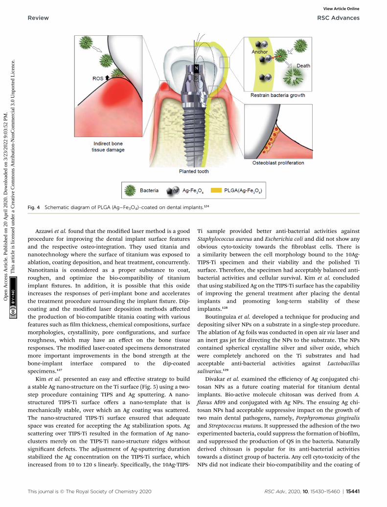
Fig. 4 Schematic diagram of PLGA (Ag–Fe3O4)-coated on dental implants.124
Review RSC Advances
Ope
n A
cces
s A
rtic
le. P
ublis
hed
on 2
0 A
pril
2020
. Dow
nloa
ded
on 3
/23/
2022
9:0
3:52
PM
. T
his
artic
le is
lice
nsed
und
er a
Cre
ativ
e C
omm
ons
Attr
ibut
ion-
Non
Com
mer
cial
3.0
Unp
orte
d L
icen
ce.
View Article Online
Azzawi et al. found that the modied laser method is a goodprocedure for improving the dental implant surface featuresand the respective osteo-integration. They used titania andnanotechnology where the surface of titanium was exposed toablation, coating deposition, and heat treatment, concurrently.Nanotitania is considered as a proper substance to coat,roughen, and optimize the bio-compatibility of titaniumimplant xtures. In addition, it is possible that this oxideincreases the responses of peri-implant bone and acceleratesthe treatment procedure surrounding the implant xture. Dip-coating and the modied laser deposition methods affectedthe production of bio-compatible titania coating with variousfeatures such as lm thickness, chemical compositions, surfacemorphologies, crystallinity, pore congurations, and surfaceroughness, which may have an effect on the bone tissueresponses. The modied laser-coated specimens demonstratedmore important improvements in the bond strength at thebone-implant interface compared to the dip-coatedspecimens.127
Kim et al. presented an easy and effective strategy to builda stable Ag nano-structure on the Ti surface (Fig. 5) using a two-step procedure containing TIPS and Ag sputtering. A nano-structured TIPS-Ti surface offers a nano-template that ismechanically stable, over which an Ag coating was scattered.The nano-structured TIPS-Ti surface ensured that adequatespace was created for accepting the Ag stabilization spots. Agscattering over TIPS-Ti resulted in the formation of Ag nano-clusters merely on the TIPS-Ti nano-structure ridges withoutsignicant defects. The adjustment of Ag-sputtering durationstabilized the Ag concentration on the TIPS-Ti surface, whichincreased from 10 to 120 s linearly. Specically, the 10Ag-TIPS-
This journal is © The Royal Society of Chemistry 2020
Ti sample provided better anti-bacterial activities againstStaphylococcus aureus and Escherichia coli and did not show anyobvious cyto-toxicity towards the broblast cells. There isa similarity between the cell morphology bound to the 10Ag-TIPS-Ti specimen and their viability and the polished Tisurface. Therefore, the specimen had acceptably balanced anti-bacterial activities and cellular survival. Kim et al. concludedthat using stabilized Ag on the TIPS-Ti surface has the capabilityof improving the general treatment aer placing the dentalimplants and promoting long-term stability of theseimplants.128
Boutinguiza et al. developed a technique for producing anddepositing silver NPs on a substrate in a single-step procedure.The ablation of Ag foils was conducted in open air via laser andan inert gas jet for directing the NPs to the substrate. The NPscontained spherical crystalline silver and silver oxide, whichwere completely anchored on the Ti substrates and hadacceptable anti-bacterial activities against Lactobacillussalivarius.129
Divakar et al. examined the efficiency of Ag conjugated chi-tosan NPs as a future coating material for titanium dentalimplants. Bio-active molecule chitosan was derived from A.avus Af09 and conjugated with Ag NPs. The ensuing Ag chi-tosan NPs had acceptable suppressive impact on the growth oftwo main dental pathogens, namely, Porphyromonas gingivalisand Streptococcus mutans. It suppressed the adhesion of the twoexperimented bacteria, could suppress the formation of biolm,and suppressed the production of QS in the bacteria. Naturallyderived chitosan is popular for its anti-bacterial activitiestowards a distinct group of bacteria. Any cell cyto-toxicity of theNPs did not indicate their bio-compatibility and the coating of
RSC Adv., 2020, 10, 15430–15460 | 15441

Fig. 5 Antibacterial and bioactive properties of stabilized silver on titanium with a nanostructured surface for dental implants.128
RSC Advances Review
Ope
n A
cces
s A
rtic
le. P
ublis
hed
on 2
0 A
pril
2020
. Dow
nloa
ded
on 3
/23/
2022
9:0
3:52
PM
. T
his
artic
le is
lice
nsed
und
er a
Cre
ativ
e C
omm
ons
Attr
ibut
ion-
Non
Com
mer
cial
3.0
Unp
orte
d L
icen
ce.
View Article Online
titanium dental implants with Ag-chitosan adds the advantageof being corrosion resistant to the dental implants, whichenhances the passivating impacts of the implants.130 Poly(lactic-
Fig. 6 Schematic illustration of the fabrication process of poly(lactic-co
15442 | RSC Adv., 2020, 10, 15430–15460
co-glycolic acid)/Ag/ZnO nano-rods coatings were introduced byXiang et al. over Ti metallic implant surface via a hydro-thermaltechnique and successive spin-coating of the mixture of
-glycolic acid)/Ag/ZnO nanorods composite coating.131
This journal is © The Royal Society of Chemistry 2020

Review RSC Advances
Ope
n A
cces
s A
rtic
le. P
ublis
hed
on 2
0 A
pril
2020
. Dow
nloa
ded
on 3
/23/
2022
9:0
3:52
PM
. T
his
artic
le is
lice
nsed
und
er a
Cre
ativ
e C
omm
ons
Attr
ibut
ion-
Non
Com
mer
cial
3.0
Unp
orte
d L
icen
ce.
View Article Online
poly(lactic-co-glycolic acid) and silver NPs (Fig. 6). Poly(lactic-co-glycolic acid)/Ag/ZnO nano-rods coating had very good anti-bacterial efficiency of >96% against Escherichia coli and Staph-ylococcus aureus while the initial content of Ag NPs was >3 wt%.in addition, the release of silver and zinc was prolonged for >100days because of the absorption of poly(lactic-co-glycolic acid).The rapid growth of mouse calvarial cells showed minimumcyto-toxicity of the poly(lactic-co-glycolic acid)/Ag/ZnO coatingwith an initial Ag NPs content of 1 wt% and 3 wt%, whereas itsuppressed the rapid growth of the cells when this value wasenhanced to 6 wt%. Finally, this poly(lactic-co-glycolic acid)/Ag/ZnO composite might present a lengthy anti-bacterial strategyand acceptable cyto-compatibility, which exhibited remarkablepotential for biomedical applications in orthopedic and dentalimplants with very good self-antibacterial activities and satis-factory bio-compatibility.131
Jadhav et al. evaluated the osteo-inductive potential of goldNPs (Au NPs) synthesized via phyto-chemicals from Salaciachinensis (Fig. 7). They conrmed that functionally bio-compatible and stable Au NPs can be successfully synthesizedvia an easy, affordable, and environment-friendly green chem-istry technique with applications in bone regeneration. The invitro examinations showed the considerable stability of the goldcolloidal dispersion in different blood elements. Theresearchers indicated that Au NPs are not toxic, as assessed bytheir cyto-compatibility and blood compatibility with peri-odontal broblasts and erythrocytes. The GNPs showed higherpercentage of cell viability (138� 27.4) of the MG-63 cell lines incomparison with the controls (96 � 3.7), indicating their osteo-inductive potential. They found that the bio-compatible and
Fig. 7 Phytosynthesis of gold nanoparticles and evaluation of its osteoin
This journal is © The Royal Society of Chemistry 2020
eco-friendly Au NPs may be applied as efficient bone-inductiveadjuvants during implant treatment to form an osteous inter-face and maintain the emerging peri-implant bone.132
6.3. Nano-materials for endodontics
The pervasiveness and seriousness of tooth root caries increasewith aging from 7% among the young to 56% in seniors with$75 years of age. This is an increasing public health problembecause of the fast enhancement in the elderly population astooth retention enhances in seniors.14,15 The vulnerability to theroot caries may be increased by gingival recession because ofaging, periodontal diseases, or traumatic tooth-brushinghabits.133,134 Moreover, small salivary ows in seniors andpatients suffering from dry mouth have an additional role inbiolm and plaque formation, and occurrence of root caries.Class V restorations may treat tooth caries. Nonetheless,cleaning and restoration with sub-gingival margins is difficultso that it would augment the developing periodontitis and lossof the tooth's attachment.135–137 Hence, it is necessary to developa bio-active Class V composite for eliminating secondary cariesand root caries.
Xiao et al. performed a study to develop a bio-active multi-functional composite (BMC) through the NPs of amorphouscalcium phosphate (NACP), dimethyl-aminohexadecyl methac-rylate (DMAHDM), 2-methacryloyloxyethyl phosphorylcholine(MPC), and silver NPs (NAg), and determined the impacts ofblended BMC + poly(amido amine) (PAMAM) on remineralizingthe demineralized root dentin in a cyclic articial saliva/lacticacid environment. The mechanical features of BMC were the
ductive potential for application in the implant dentistry.132
RSC Adv., 2020, 10, 15430–15460 | 15443
RSC Advances Review
Ope
n A
cces
s A
rtic
le. P
ublis
hed
on 2
0 A
pril
2020
. Dow
nloa
ded
on 3
/23/
2022
9:0
3:52
PM
. T
his
artic
le is
lice
nsed
und
er a
Cre
ativ
e C
omm
ons
Attr
ibut
ion-
Non
Com
mer
cial
3.0
Unp
orte
d L
icen
ce.
View Article Online
same as that of commercial control composites (p ¼ 0.913).BMC possessed very good release of P and Ca ions and acid-neutralization ability. BMC or PAMAM individually obtainedminor mineral re-generation in the demineralized root dentin.The blended BMC + PAMAM caused maximum root dentin re-mineralization and enhanced the hardness of the pre-demineralized root dentin that matched that of the healthyroot dentin (p ¼ 0.521).138
Rodrigues et al. assessed the anti-microbial actions of anirritant with silver NPs in an aqueous vehicle, such as sodiumhypochlorite, Ag NPs, and chlorhexidine against Enterococcusfaecalis bio-lm and infected dentinal tubules. The Ag NPsolution removed little bacteria; however, it could dissolve thebiolm better in comparison to chlorhexidine (P < 0.05). NaOClhad maximum anti-microbial activities and biolm dissolutioncapacities while the Ag NP solution had lower anti-microbialactions in the infected dentinal canals in comparison toNaOCl (P < 0.05). The Ag NP solution highly affected the elim-ination of planktonic bacteria in dentinal tubules compared tothe bio-lm aer 5 minutes; however, fewer viable bacteria werefound in the bio-lm in comparison to the intratubular dentine(P < 0.05) at 30 minutes. The Ag NP irritant was not as efficientagainst Enterococcus faecalis in comparison with the solutionswidely employed in treating root canal. NaOCl is suitable as anirritant as it caused the disruption of the bio-lm and elimi-nation of bacteria in bio-lms and dentinal canals.139
Bukhari et al. presented a therapeutic strategy forendodontic disinfections via nano-catalysis concept forincreasing bacterial destruction across the dentinal tubule. Ironoxide is a sustainable and bio-compatible material, which couldbe synthesized largely via simple and affordable chemicalsynthetic techniques in view of the current usage in food anddrug administration with the veried formulations for chronictreatment. The exibility in iron oxide chemistry provides thegrounds for producing the desired NP shape and size, whichcan additionally enhance the catalytic activities (probably withless H2O2 concentrations) with higher dentinal tubule pene-trations. It is necessary to validate the IO NPs/H2O2 systemefficiency by animal models and clinical research, which wouldresult in endodontic therapy with greater effectiveness andefficiency.140
6.4. Nano-materials for restoration
Composite resins play vital roles in dental restoration and haveseveral benets, including acceptable maneuverability, verygood esthetics, and satisfactory bio-compatibility.141–143 Theorganic elements of the composite resins have been nearlyconstant over time. The components are mostly methacrylate-type resins such urethane dimethacrylate (UDMA), bisphenolA-glycidylmethacrylate (Bis-GMA), and triethylene glycol dime-thacrylate (TEGDMA).144 However, several explorations havebeen conducted on inorganic llers to develop high-performance composite resins.145,146 To attain the acceptablelengthy clinical restoration, composite resins should showadequate mechanical features, small polymerization shrinkage,higher wear resistance, and anti-bacterial activities. Therefore,
15444 | RSC Adv., 2020, 10, 15430–15460
such necessities can be generally satised by using differentfunctionalized llers.147,148
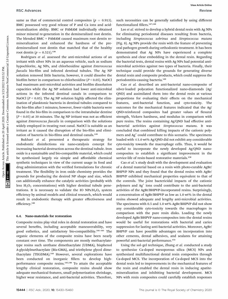
Lee et al. strived to develop a hybrid dental resin with Ag NPsfor eliminating periodontal diseases resulting from bacteria,including Streptococcus sobrinus and Streptococcus mutans(Fig. 8). Ag NPs provide the resin with the feature of preventingoral pathogen growth during orthodontic treatment. It has beendemonstrated that Ag NPs have experienced a completesynthesis and clear embedding in the dental resin. Regardingthe bacterial tests, dental resins with Ag NPs had potential anti-microbial activities against two types of bacteria. Finally, theirtechnique could provide the grounds for generating diversedental resin and composite products, which could suppress theperiodontitis-causing bacteria.149
Cao et al. described an anti-bacterial nano-composite ofsilver-loaded polycation functionalized nano-diamonds (Ag/QND) and assimilated them into the dental resin at variousproportions for evaluating their effects on the mechanicalfeatures, anti-bacterial function, and cyto-toxicity. Theoutcomes for the mechanical features indicated that the Ag/QND-reinforced composites had greater values of exuralstrength, Vickers hardness, and modulus in comparison withpure resins. The resins containing Ag/QND had effective anti-bacterial activities against Streptococcus mutans. It wasconcluded that combined killing impacts of the cationic poly-mers and Ag+ could contribute to this scenario. The specimensloaded with#1.0 wt% Ag/QND did not exhibit any considerablecyto-toxicity towards the macrophage cells. Thus, it would beuseful to incorporate the newly developed Ag/QND nano-composites to establish a signicant improvement in theservice-life of resin-based restorative materials.150
Cao et al.'s study dealt with the development and evaluationof a dental material based on a resin with photo-curable AgBr/BHPVP NPs and they found that the dental resins with AgBr/BHPVP exhibited mechanical properties equivalent to that ofthe controls. The joint bactericidal impacts of the cationicpolymers and Ag+ ions could contribute to the anti-bacterialactivities of the AgBr/BHPVP-incorporated resins. Surprisingly,a concentration of AgBr/BHPVP as low as 1.0 wt% in the dentalresins showed adequate and lengthy anti-microbial activities.The specimens with 0.5 and 1.0 wt% AgBr/BHPVP did not showany considerable cyto-toxicity towards the macrophages incomparison with the pure resin disks. Loading the newlydeveloped AgBr/BHPVP nano-composites into the dental resinswould be useful for restorations with bacterial and cariessuppression for lasting anti-bacterial activities. Moreover, AgBr/BHPVP can have possible advantages on incorporation intoother cements, dental adhesives, and sealants for attainingpowerful anti-bacterial performance.151
Using the sol–gel technique, Zhang et al. conducted a studyto synthesize Ca-doped mesoporous silica (MCS) NPs andsynthesized multifunctional dental resin composites throughCa-doped MCS. The incorporation of Ca-doped MCS into thedental resin led to improvements in the mechanical features ofthe resin and enabled the dental resin in inducing apatite-mineralization and inhibiting bacterial development. MCSNPs with resin composites could carefully establish responses
This journal is © The Royal Society of Chemistry 2020

Fig. 8 Schematic illustration of the preparation of Ag NPs and the hybrid dental resin.149
Review RSC Advances
Ope
n A
cces
s A
rtic
le. P
ublis
hed
on 2
0 A
pril
2020
. Dow
nloa
ded
on 3
/23/
2022
9:0
3:52
PM
. T
his
artic
le is
lice
nsed
und
er a
Cre
ativ
e C
omm
ons
Attr
ibut
ion-
Non
Com
mer
cial
3.0
Unp
orte
d L
icen
ce.
View Article Online
to wear and enhance mineralization-induced activities in thecase of wearing resin composites; MCS NPs or the resincomposites could be applied as multifunctional restorativematerials for potential dental applications.152
Cevik et al. evaluated the inuence of hydrophobic nano-particle silica and pre-polymer on the exural strength, surfaceroughness, surface hardness, and resilience of a denture baseacrylic resin. Statistical analyses found signicant differencebetween these groups. Each group possessed weak exuralstrength in comparison to the controls (p < 0.05). In terms of theresilience, silica 5% had the maximum value, while silica 1%possessed the minimum value. For Shore D hardness, silica 1%exhibited the minimum hardness, while addition of the poly-mer had no signicant effects on the acrylic resin's hardness (p< 0.05). Silica 1% showed maximum roughness in comparisonto the other groups (p < 0.05). The incorporation of silica andpre-polymer into the acrylic resin had contrary impacts on theacrylic resin's exural strength in comparison with the controls.For each concentration, pre-polymer incorporation led tohigher exural strength of the acrylic resins in comparison withsilica addition. Higher concentrations of the llers led to highermechanical features of the acrylic resin.153
Ghahremani et al. illustrated that the addition of TiO2 NPs tothe acrylic resin improved its mechanical features with a reverseimpact on its color; the mean tensile strength of the reinforcedgroup was considerably greater (difference of 11 MPa)compared to the controls (P ¼ 0.001). The mean effective
This journal is © The Royal Society of Chemistry 2020
strength of the potent group was about 7 MPa greater comparedto the controls and the differences were not statistically signif-icant (P ¼ 0.001). The color of the modied acrylic resinstrengthened with 1 wt% TiO2, which was accompanied byincrease in the tensile and impact strength in comparison withthe traditional acrylic resin. Hence, TiO2 NPs could be addedinto the acrylic resin powder for a modied color to enhance itstensile and impact strength, if they do not possess any reverseeffects on other features.154
Sodagar et al.'s study evaluated the mechanical and anti-microbial features of composite resins modied by the addi-tion of TiO2 NPs. Each concentration of TiO2 NPs showeda remarkable impact on the creation and extension of thesuppression region including the decline in the colony countsfor S. mutans and S. sanguinis. The composite with 10% TiO2
NPs had a considerable impact on the decrease in the colonycount for S. sanguinis and S. mutans (3 days). The controlsaccounted for the maximum mean shear bond strength,whereas the maximum amount was observed for the 10% NPscomposite. The incorporation of TiO2 NPs into the compositeresins gave anti-bacterial features to the adhesives, whereas themean shear bond of the composite with 1% and 5% NPs provedto be a reasonable range.155
Rodrigues et al. characterized the synthesized brushite NP(CaHPO4$2H2O) and demonstrated that functionalization canbe regulated based on the concentrations of triethylene glycoldimethacrylate (TEGDMA) employed in the synthesis. Although
RSC Adv., 2020, 10, 15430–15460 | 15445
RSC Advances Review
Ope
n A
cces
s A
rtic
le. P
ublis
hed
on 2
0 A
pril
2020
. Dow
nloa
ded
on 3
/23/
2022
9:0
3:52
PM
. T
his
artic
le is
lice
nsed
und
er a
Cre
ativ
e C
omm
ons
Attr
ibut
ion-
Non
Com
mer
cial
3.0
Unp
orte
d L
icen
ce.
View Article Online
the concentration of TEGDMA did not inuence the size of theNPs, no reduction in agglomeration was observed. Experi-mental composites with 10 vol% of the brushite agglomeratesand 50 vol% of the silanated glass particles had mechanicalfeatures identical to that of a commercial micro-hybrid restor-ative composite applied in regions exposed to serious occlusalloadings. Therefore, the composite comprising brushite NPsfunctionalized with higher TEGDMA contents exhibited betterperformance in the mechanical test in water aer 28 days,which had the same fracture strength as that of the commercialcontrols. The NPs functionalization with TEGDMA had noadverse impact on the ion releases.156
Meena et al. added various weight percents of nano-alumina(5–20 wt%) and marble dust powder (5–20 wt%) to the basemonomer system with bisphenol-A glycidyl methacrylate, cam-phorquinone, tri-ethylene glycol dimethacrylate, and dimethylaminoethyl methacrylate in order to fabricate a dentalcomposite. The experimental results showed that the additionof 5 wt% nano-alumina enhanced the hardness and compres-sive strength by about 88.46% and 23.25%, respectively, whilethe addition of 5 wt% marble powder augmented the hardnessand compressive strength by about 51.27% and 21.2%, respec-tively. Dynamic mechanical analyses demonstrated that theaddition of nano-alumina up to 20 wt% enhanced the storagemodulus by about 112.2%, while the addition of marble powderup to 20 wt% augmented the storage modulus by about 191.2%.The conclusion is that regardless of the economic aspects, themarble dust powder-lled dental composite had more accept-able thermo-mechanical and thermal features compared tothose of the nano-alumina-lled dental composite.157
Al-Ajely et al. described calcium uoride NPs (CaF2 NPs)applied in dental composites as the dental lling compositionof glass type. A coprecipitation technique through binary liquidwas employed to prepare the CaF2 NPs. X-ray diffraction wasused to predict the crystal structure characteristics andelemental compositions of the CaF2 NPs, showing crystallinepeaks of the material. EDX analysis was applied for obtainingthe elemental composition. Moreover, the SEM images showedthe particle size to be �58 � 21 nm.158
Campos et al. studied the experimental dental restorativecomposites and their development by the addition of mont-morillonite (MMT) NPs in a polymer matrix-based BisGMA/TEGDMA for assessing the feasibility of a distinct dimen-sional behavior during photo-polymerization. The researchersdemonstrated that the experimental composites lled withMMT NP have statistically identical polymerization shrinkagevalues (by thermal mechanical analyses) at the concentration of30% and less polymerization shrinkage values at the concen-tration of 50% than the composites that became full withbarium glass (BG-control groups).159
Al-Mosawi and Al-Badr assessed the efficiency of ZnO NPs,which were incorporated into the composite resin as anti-microbial agents against bacteria that cause dental caries inthe oral cavity. The anti-bacterial impacts of inorganic anti-bacterial agents was evaluated through agar disc diffusion testwith three different concentrations of the synthesized ZnO NPs(in vitro) on the caries-causing bacteria in this research. Finally,
15446 | RSC Adv., 2020, 10, 15430–15460
85% decline in the growth of various types of bacteria wasrevealed. The anti-bacterial agent (ZnO/NPs) had powerful anti-bacterial activities against a broad-spectrum of pathogeniccaries-causing bacteria in the oral cavity.160
Size-controlled mono-dispersed silica NPs (MSNPs) in thesize range from 20 to 330 nm were effectively synthesizedthrough internal circulation rotating packed bed (ICRPB) andblended with freeze-drying in a high-gravity environment (Yanget al.). In comparison with the conventional stirred tank reactor,ICRPB decreased the reaction duration by 1
3 times and MSNPswith a clearly lower dimensions from 75 nm to 35 nm wereobtained. Increasing the MSNPs contents from 40 wt% to70 wt% enhanced the composite's mechanical featuresconsiderably, so that it initially augmented and aerwardsdeclined due to enhancement of the particle size, and reachedthe highest value at 80 nm. Because of the very good dispersion,the highest MSNPs contents were 30% greater than thecommercial silica NPs with a similar size, which provided moreacceptable features for the composites.161
Bezerra Dias et al. made a composite resin that is commer-cial and contains TiO2 and Ag covered with TiO2 NPs forproviding anti-bacterial capacities without eliminating thephysical and mechanical features. The null hypothesis experi-mented in this research is that the addition of small amounts ofTiO2 and Ag decorated TiO2 NPs to a composite resin (acommercial one) had no effect on the compressive and dia-metric tensile strength, anti-bacterial activities, conversiondegree, and surface roughness.162
Ai et al. suggested a functional nano-brous ller andexperimented with it to prepare an anti-bacterial compositeresin. The aim was to produce hydroxyapatite (HA) nano-wiresthrough the hydrothermal method, dopamine (DA) modica-tion, and loading the silver NPs (Ag NPs). Such Ag NPs laden HAnano-wires were blended into the Bis-GMA/TEGDMA resin forpreparing the cured composites. It was assumed that thedispersion of the HA nano-wires and the Ag NPs in thecomposite resins was homogeneous in terms of the propertiesof polydopamine (PDA) surface modication. Then, themechanical features, release of silver ion, cyto-toxicity, and anti-bacterial activities were assessed. The researchers targeted theclinical applications and favorably postulated that the 1D AgNPs-laden HA nano-wires can be used as effective reinforce-ments for the composite resin with increased anti-bacterialactivities.163
Wang et al. dealt with the reinforcement and anti-bacterialimpacts of cellulose nanocrystal/zinc oxide (CNC/ZnO) nano-hybrids on dental resin composites (DRCs), which wasprocured via precipitation of Zn2+ on the CNC surface (Fig. 9). Incomparison to the DRCs with no CNC/ZnO nano-hybrids, DRCswith 2 wt% CNC/ZnO nano-hybrids had greater compressivestrength and exural modulus, and no signicant difference (P> 0.05) was observed in the exural strength and Vickers micro-hardness. The excessive application of the CNC/ZnO nano-hybrids degraded the mechanical features of the DRCs withthe exception of the exural modulus. The DRCs with CNC/ZnOnano-hybrids had very good anti-bacterial features and 78%decrease in the bacterial numbers were achieved with the
This journal is © The Royal Society of Chemistry 2020

Review RSC Advances
Ope
n A
cces
s A
rtic
le. P
ublis
hed
on 2
0 A
pril
2020
. Dow
nloa
ded
on 3
/23/
2022
9:0
3:52
PM
. T
his
artic
le is
lice
nsed
und
er a
Cre
ativ
e C
omm
ons
Attr
ibut
ion-
Non
Com
mer
cial
3.0
Unp
orte
d L
icen
ce.
View Article Online
addition of 2% CNC/ZnO nano-hybrids. The synthesized DRCswould be useful for addressing bulk fracture and secondarycaries.164
Wang et al. synthesized wrinkle-structured mesoporoussilica NPs for inclusion in dental composites wherein theBisGMA/TEGDMA based-resin composites were synthesized viasilanized silica NPs as the unimodal ller with various loadingperformances (25, 30, and 35 wt%). In comparison to regularsilica particles (Si507), the silica NPs possess center radialwrinkles that are suitable for forming micro-mechanical bonds,which enhanced the ller matrix interfacial interaction andmechanical features. Bimodal silica NPs llers with either silicaNPs-Si90 or silica NPs-Si190 were proposed for producing theresin composites, each of them providing 60 wt% ller loadingand eliminating the loading limitations of uni-modal silica NPs(35 wt%). It was found that bimodal silica NPs llers couldsignicantly augment the mechanical characteristics of theresulting composites. The silica NPs-Si190 lled compositehaving an optimal mass ratio of 10 : 90 (wt/wt) had the mostacceptable mechanical function, which indicated acceptablereinforcing efficacy of the silica NPs and the benets of the useof bimodal llers. Silica NPs are potential inorganic llers todesign and fabricate dental resin composites with reasonablemechanical features.165
Paiva et al. described the use of silver NPs absorbed intoa single phase, which was synthesized in a poly-electrolyte
Fig. 9 The preparation route of cellulose nanocrystal/zinc oxide (CNC/
This journal is © The Royal Society of Chemistry 2020
solution, which is the major compound of glass ionomercement. They carried this out via UV irradiation with nochanges in the viability of the net setting reaction. It was foundthat the molar ratio of Ag+/COO� and UV exposure duration aresignicant variables in the procedure. Group D-High Ag speci-mens generated AgNO3 0.5% and TA 10% bymass, for 90 min ofUV exposure, which showed considerable difference in inhib-iting bacterial development and Streptococcus mutans bio-lmviability in comparison with the negative controls (group A-with no Ag). Nano Ag-glass ionomer cement conrmed theanti-bacterial impacts of diffusion due to oxidative dissolutionof silver ions from the cement matrix, which inuenced cariesarrest and the prevention of developing oral bio-lms on theirsurfaces.166
Sundeep et al. conducted a study to synthesize clean, eco-friendly, and affordable silver NPs in bulk. The synthesizedsilver NPs were reinforced with glass ionomer cement formeeting the above two limitations concurrently. Reinforcing AgNPs in the glass ionomer cement augmented the hardness oftraditional glass ionomer cement and accordingly eliminatedthe limitations of secondary caries caused by bacterial colonysurrounding the glass ionomer cement xed restoration post-medication. A comparison was made between the increase inthe hardness of the Ag NP-reinforced glass ionomer cementsand traditional glass ionomer cement. Then, the micro-silverreinforced glass ionomer cement and the anti-microbial
ZnO) nanohybrids.164
RSC Adv., 2020, 10, 15430–15460 | 15447
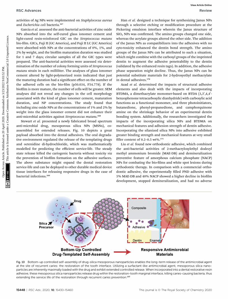
RSC Advances Review
Ope
n A
cces
s A
rtic
le. P
ublis
hed
on 2
0 A
pril
2020
. Dow
nloa
ded
on 3
/23/
2022
9:0
3:52
PM
. T
his
artic
le is
lice
nsed
und
er a
Cre
ativ
e C
omm
ons
Attr
ibut
ion-
Non
Com
mer
cial
3.0
Unp
orte
d L
icen
ce.
View Article Online
activities of Ag NPs were implemented on Staphylococus aureusand Escherichia coli bacteria.167
Garcia et al. assessed the anti-bacterial activities of zinc oxideNPs absorbed into the self-cured glass ionomer cement andlight-cured resin-reinforced GIC on the Streptococcus mutansbio-lm. GICs, Fuji II (GC America), and Fuji II LC (GC America)were absorbed with NPs at the concentrations of 0%, 1%, and2% by weight, and the biolm maturation duration was studiedfor 1 and 7 days; circular samples of all the GIC types wereprepared. The anti-bacterial activities were assessed via deter-mination of the number of colony forming units of Streptococcusmutans strain in each milliliter. The analyses of glass ionomercement altered by light-polymerized resin indicated that justthe maturing duration had a signicant effect on the number ofthe adhered cells on the bio-lm (p50.034, F54.778). If thebiolm is more mature, the number of cells will be greater. SEManalyses did not reveal any changes in the cell morphologyassociated with the kind of glass ionomer cement, maturationduration, and NP concentrations. The study found thatincluding zinc oxide NPs at the concentrations of 1% and 2% byweight into the glass ionomer cement did not enhance theiranti-microbial activities against Streptococcus mutans.168
Stewart et al. presented a newly fabricated broad spectrumanti-microbial drug, mesoporous silica NPs (MSNs), co-assembled for extended releases. Fig. 10 depicts a greatpayload absorbed into the dental adhesives. The oral degrada-tive environment regulated the release of the templating drugsand octenidine di-hydrochloride, which was mathematicallymodelled for predicting the efficient service-life. The steadystate release killed the cariogenic bacteria without toxicity viathe prevention of biolm formation on the adhesive surfaces.The above substance might expand the dental restorationservice-life and can be deployed to other durable medical devicetissue interfaces for releasing responsive drugs in the case ofbacterial infections.169
Fig. 10 Bottom-up controlled self-assembly of drug-silica mesoporousat the site of recurrent caries: the restoration of the tooth interface. Uparticles are inherently maximally loadedwith the drug and exhibit extendadhesive, thesemesoporous silica nanoparticles release drug within the reextending the service life of the restoration through recurrent caries pre
15448 | RSC Adv., 2020, 10, 15430–15460
Han et al. designed a technique for synthesizing Janus NPsthrough a selective etching or modication procedure at thePickering emulsion interface wherein the Janus structure ofSiO2 NPs was conrmed. The amino groups changed one side,whereas the acrylate groups altered the other side. The additionof the Janus NPs as compatibilizers into the adhesives with nocyto-toxicity enhanced the dentin bond strength. The aminogroups of the Janus NPs can be attributed to such a situation,whichmight combine with the carboxyl groups of the imprinteddentin to augment the adhesive penetrability to the dentin(validated by the enhanced resin tags). In addition, the adhesivephase separation might decline. Thus, the Janus NPs can bepotential substitute materials for 2-hydroxyethyl methacrylatein dental adhesives.170
Azad et al. determined the impacts of structural adhesiveelements and also dealt with the impacts of incorporatingBTDMA, a dimethacrylate monomer-based on BTDA (3,30,4,40-benzophenone tetracarboxylic dianhydride) with carboxylic acidfunctions as a functional monomer, and three photoinitiators,butanedione, phenyl-propanedione, and camphorquinoneamine on the shrinkage behavior of an experimental dentinbonding system. Additionally, the researchers investigated theimpacts of the incorporating silica NPs and BTDMA onmechanical features and adhesion strength of dentin adhesive.Incorporating the silanized silica NPs into adhesive exhibitedgreater binding strength and mechanical features at very smallller content of 0.2–0.5 wt%.171
Liu et al. found new orthodontic adhesive, which combinedthe anti-bacterial activities of 2-methacryloxylethyl dodecylmethyl ammonium bromide (MAE-DB) and demineralizationpreventive feature of amorphous calcium phosphate (NACP)NPs for combating the bio-lms and white spot lesions duringorthodontic therapy. In comparison with a commercial ortho-dontic adhesive, the experimentally lled PND adhesive with5% MAE-DB and 40% NACP showed a higher decline in biolmdevelopment, stopped demineralization, and had no adverse
nanoparticles enables the long-term release of the antimicrobial agenttilizing a surfactant-like antimicrobial agent, mesoporous silica nano-ed controlled release. When incorporated into a dental restorative resinstoration-toothmarginal interface, killing caries-causing bacteria, thusvention.169
This journal is © The Royal Society of Chemistry 2020

Review RSC Advances
Ope
n A
cces
s A
rtic
le. P
ublis
hed
on 2
0 A
pril
2020
. Dow
nloa
ded
on 3
/23/
2022
9:0
3:52
PM
. T
his
artic
le is
lice
nsed
und
er a
Cre
ativ
e C
omm
ons
Attr
ibut
ion-
Non
Com
mer
cial
3.0
Unp
orte
d L
icen
ce.
View Article Online
effect on the shear bond strength. If the anti-bacterial impactsof MAE-DB combined with the remineralization approach ofNACP, it can be followed by opportunities of applications innano-technology materials for reducing the burden of dentalcaries throughout the world.172
Yue et al. designed a self-healing adhesive containing anti-microbial and remineralizing abilities, and explored theunprecedented impacts of incorporation of micro-capsules,dimethyl-aminohexadecyl methacrylate (DMAHDM), and NPsof amorphous calcium phosphate (NACP). Poly(urea-formaldehyde) (PUF) shells with triethylene glycol dimethacry-late (TEGDMA) as a therapeutic liquid were used to synthesizeself-healing micro-capsules; the new adhesive included 10%DMAHDM, 7.5% micro-capsules, and 20% NACP. A single edgeV-notched beam technique was employed for measuring thefracture toughness KIC and the autonomous crack-healing effi-cacy with experiments on an oral plaque microcosm bio-lmmodel. This newly constructed self-healing, anti-microbial,and remineralizing dental adhesive corresponded to thedentin bond strength of a commercial control (p > 0.1); itattained substantial crack healing with very good KIC recovery of67%. It possessed powerful anti-microbial activities, decreasedthe bio-lm colony forming units by about 4 orders of magni-tude, and decreased the bio-lm acid generation to 1/100th ofthat of the bio-lms on the commercial control resin.173
Al-Qarni et al. conducted a study for developing a calcium(Ca) and phosphate (P) ion-rechargeable and protein repellentadhesive with NPs of amorphous calcium phosphate (NACP)and 2-methacryloyloxyethyl phosphorylcholine (MPC); theimpacts of MPC on the ion re-charge and re-release was exam-ined. It was found that the addition of NACP + MPC did notdemonstrate any negative effects on the dentin bindingstrength (p > 0.1). As the number of re-charge/re-release cyclesincreased, the Ca/P ion re-release achieved accordingly greaterlevels (p > 0.1), which indicated enduring re-mineralizationabilities; one re-charge made it possible for the adhesives tocontinue re-releases for 21 days. The incorporation of 3% MPCwas followed by a 10-fold decline in the protein absorption and1–2 log decline in the bio-lm CFU. The re-chargeable adhesivecontaining MPC + 30% NACP signicantly decreased proteinabsorption, bio-lm development, and lactic acid. The incor-poration of MPC did not endanger the very good Ca/P ionrelease, re-chargeability, and dentin bond strength. This adhe-sive has the potential for various tooth restoration applicationsfor inhibiting caries and increasing the restoration durability.174
Xie et al. designed an adhesive with the three advantages ofrecharging calcium phosphate ion, protein repellent, and anti-bacterial function. MPC and dimethyl-aminohexadecyl meth-acrylate (DMAHDM) were assimilated into a NACP re-chargeable adhesive for achieving protein repellent and anti-bacterial abilities for combating bio-lms and caries. The newbio-active adhesive exhibited powerful protein repellent abilityand considerably decreased the bacterial binding and viabilitiesby declining the bio-lm colony shaping units by nearly 4 log incomparison with the commercial control. The bio-active adhe-sive containing dental plaque microcosm bio-lm cultureretained a pH > 6, whereas the commercial control adhesive
This journal is © The Royal Society of Chemistry 2020
possessed a cariogenic pH ¼ 4. The adhesive with these threeadvantages has the potential for protecting tooth structures andinhibiting biolms and caries formation. The combinationprocedure for NACP, MPC, and DMAHDM with three benetswill hopefully nd application in a variety of dental restorativeand preventive substances for reducing plaque formation andinhibiting caries.175
Liang et al. examined poly(amido amine) (PAMAM), NPs ofamorphous calcium phosphate (NACP) adhesive, and PAMAM +NACP adhesive to remineralize dentin in lactic acid with noinitial P and Ca ions. Maximum dentin remineralization in thishighly challenging environment was used to obtain newPAMAM + NACP. It made the composite-dentin bond longer andsuppressed caries in patients with dry mouths, in which localpH is frequently acidic and there is a lack of saliva with P and Caions. PAMAM could not generate remineralization in lactic acid.Using NACP adhesive, P and Ca ions' release was observed,which increased the pH to 6.5, and attained minor reminerali-zation in such a challenging environment. PAMAM + NACP wassynergized and NACP presented necessary Ca and P ions,enhanced pH, and enabled PAMAM for fullling its nucleationtemplate functions. It was followed by maximum reminerali-zation and restoration of the hardness of the demineralizeddentin to the hardness of the healthy dentin. PAMAM + NACPhas potential for patients with lower saliva and radiation cariesand for a variety of dental restorations for prolonging thebonded interface, inhibiting secondary caries, remineraliza-tion, and protecting the tooth structures.176
Dutra-Correa et al. assessed the impacts of small Ag NPsabsorption concentration (50 ppm increment from 50 to 250ppm) into the primer of a 3-step, which is found in the marketas an etch-and-rinse adhesive system. NPs functionalized withappropriate organic stabilizers controlling the particle size andreducing agglomeration were substantially prepared andpoured into the primer, which generated hydrophilic andfunctional materials even at small Ag NP concentrations.
The lowest suppressive concentrations were observedbetween 25 and 50 ppm Ag NPs, the lowest being at 50 ppm ofAg NPs. The suppression of bacterial activities was greater withrespect to the controls in each Ag NP group in comparison withthe controls in the agar diffusion assay; biolm suppression wasgreater in 250 ppm Ag NPs as compared to the controls. Each AgNP group and Scotch bond Multi-Purpose adhesive systemshowed the same cyto-toxicity in all periods. The adhesivesbearing 200 and 250 ppm Ag NPs and Scotch bond Multi-Purpose adhesive system (controls) had maximum Micro-tensile Bond Test values, which are identical to the ones inScotch bond Multi-Purpose adhesive system control in both thecases (24 h and 6months) (p > 0.05). Ag NPs/primer relationshipmay defend the tooth adhesive interface, which increases thedental restoration durability.177
Polymethyl methacrylate (PMMA) thin lms absorbed withbio-fabricated silver NPs have been not employed for evaluatingthe in vitro anti-microbial and anti-biolm activities againstcariogenic bacterium Streptococcus mutans (Thomas et al.).Bacillus amylo-liquefaciens SJ14 culture (Mag NPs) and extractfrom Curcuma aromatica rhizome (Cag NPs) were used to
RSC Adv., 2020, 10, 15430–15460 | 15449
RSC Advances Review
Ope
n A
cces
s A
rtic
le. P
ublis
hed
on 2
0 A
pril
2020
. Dow
nloa
ded
on 3
/23/
2022
9:0
3:52
PM
. T
his
artic
le is
lice
nsed
und
er a
Cre
ativ
e C
omm
ons
Attr
ibut
ion-
Non
Com
mer
cial
3.0
Unp
orte
d L
icen
ce.
View Article Online
generate silver NPs. Moreover, the lowest suppressive concen-trations, lowest bactericidal concentrations, and anti-biolmactivities of Ag NPs against Streptococcus mutans were evalu-ated wherein Mag NPs displayed better anti-microbial activitiesin comparison to Cag NPs. Furthermore, the Mag NPs and CagNPs exhibited 99% and 94% suppression of biolm formationof Streptococcus mutans at concentrations of 3 mg mL�1 and 50mg mL�1, respectively. The Ag NPs were additionally absorbedinto the PMMA thin lms through solvent casting technique.PMMA/Mag NPs and PMMA/Cag NPs nano-composite thin lmswere exposed to anti-microbial and anti-biolm analyses. Inaddition, microbicidal activities were greater for the PMMA/Mag NPs thin lm, showing the potential of microbiallysynthesized Ag NPs as agents for inhibiting cariogenic bacteriafrom colonizing the dental restorative materials.178
A polymeric composition comprises modied-TiO2 nano-particles, with or without a solvent, and polymer precursors,wherein the modied-TiO2 nanoparticles comprise titaniumdioxide nanoparticles, which are modied with a short-chainunsaturated compound that comprises 2 to 10 carbon atoms.The polymeric composition can be used in dental compositionssuch as dental adhesives, dental composites, and dentalsealants.179
Balasankar et al. provided a treatment approach for deliv-ering chlorhexidine (CHX) loaded poly(lactic-co-glycolic acid)(PLGA) NPs via dentinal tubules of demineralized dentinsubstrates for probable applications in adhesive and restorativedentistry. The formulated CHX loaded PLGA NPs had inter-esting physico-chemical features, negligible cyto-toxicity,potential anti-bacterial efficiency, and slow degradationpatterns related to the capability of releasing CHX for up to 28days. CHX loaded PLGA NPs in aqueous solution can penetrateand inltrate via dentinal tubules up to 10 mm depth, whichhave a close association with the resin tags aer binding resininltration.180
Gutierrez et al. assessed the impact of adding copper NPs(Cu NPs) at various concentrations into a two-step etch-and-rinse (2 ER) adhesive on the anti-microbial activities (AMA),copper release (CR), degree of conversion (DC), water sorp-tion (WS), solubility (SO), immediate (IM), ultimate tensilestrength (UTS), 1 year resin dentin bond strength (mTBS),and nano-leakage (NL). The addition of Cu NPs presentedAMA towards the adhesives at each concentration. GreaterCR was seen in adhesives with greater concentrations of CuNPs. DC, UTS, SO, and WS was not affected. An increase wasseen for mTBS in the 0.1 and 0.5% copper groups. A consid-erable decline was found for NL in each group as comparedto the controls. Aer 1 year, any remarkable reductions inmTBS and any considerable increase in NL were not found forcopper with adhesives in comparison with the controls. Theaddition of Cu NPs at concentrations up to 1 wt% in the 2 ERadhesive can be an option for providing AMA and preservingthe bond to dentin with no reduction in the adhesive'smechanical features.181
The same group examined 2 year (2Y) resin dentin mTBS andNL. The addition of copper NPs up to 0.5 wt% exhibited anti-microbial features towards ER adhesives and stopped the
15450 | RSC Adv., 2020, 10, 15430–15460
degradation of the adhesive interface with no reduction in themechanical features of the formulations.182
Georgy et al. examined the dental adhesive compositioncontaining NPs of metals for gaining lengthy bactericidalfeatures by addition of Ag, V, Cu, Fe, Al, and Ta NPs in thedental adhesive, which resulted in its prolonged bacterio-static and bactericidal impacts versus dental plaque micro-ora. For tantalum NPs, the modications in theconcentrations resulting from the deviation in the workingmode of the electric discharge dispersion condensinginstrument had no effects on the anti-septic features of theresulting product of the dental adhesive. It was observed thattantalum NPs enhance the average value of shear strength inthe system lling the material adhesive dentin up to over40%. With regards to the bactericidal and high adhesionfeatures, tantalum NPs as an additive for dental adhesivehave advantages that need to be exploited.183
Rizk et al. demonstrated identical bioactive potentials ofpoly-hedraloligomeric silsesquioxanes (POSS) particles asthat of nanosized bio-active glass (BG-Bi) particles when theywere blended with the adhesive while maintaining suitablemechanical features of the system. The impacts of the multi-functional POSS NPs on the degrees of conversion, sol frac-tion, and water sorption indicated higher crosslink densityof the lled adhesive while maintaining low viscosities.Hence, multi-functional POSS is an attractive option fortesting in bond strength tests as it could enhance the bindingstrength due to the higher cross-linking capability and stopbinding strength degradation over time because of itsmineralization capacities. Though mono-functional POSSparticles resulted in a highly declined conversion and bio-active glass particles strongly enhanced the viscosity,apparently, their application in dental adhesives is limited tospecic uses and substances.184
Tooth whitening agents were necessarily nano-modied toincrease the whitening efficiency and decrease their harmfulside effects. For example, calcium peroxide nanoparticles couldpenetrate deeper into the tooth structure through micro andnano cracks, make longer surface contact, and increase theeffectiveness of the whitening agent.185
6.5. Nano-materials for periodontics
Periodontitis is a dental inammatory dysfunction, whichinuences the surrounding structures of the teeth (perio-dontium). This disease is described by inammation and decayof the surrounding teeth and supporting tissues, namely, peri-odontal ligament, alveolar bone, cementum, and gum, whichare commonly encountered due to anaerobic Gram-negativebacterial invasion of the teeth.186,187 New advancements innano-materials and nano-technology has offered hopefulchances and opportunities for the efficient management ofperiodontitis. A number of the most acceptable ndings andbenets can be attained from such newer strategies that includethe application of bio-adhesive polymers for achieving lengthydrug release, increase in intrapocket drug penetration, increaseof mechanical features via chemical cross-linkers, and possible
This journal is © The Royal Society of Chemistry 2020
Review RSC Advances
Ope
n A
cces
s A
rtic
le. P
ublis
hed
on 2
0 A
pril
2020
. Dow
nloa
ded
on 3
/23/
2022
9:0
3:52
PM
. T
his
artic
le is
lice
nsed
und
er a
Cre
ativ
e C
omm
ons
Attr
ibut
ion-
Non
Com
mer
cial
3.0
Unp
orte
d L
icen
ce.
View Article Online
loading of multiple drugs in a unit delivery system. The aboverewards could provide the ground for further research oppor-tunity in developing drug delivery systems, which will advancedental therapeutics.188–191
Khan et al. performed a study to synthesize bio-active electro-spun bers for bio-medical and dental application with higher bio-compatibility. The in situ precipitation of nanohydroxyapatite(nHA) was administered at different concentrations (0.5%, 1%,2%, 3%, and 5% wt/wt) of the functionalized multiwalled carbonnano-tubes (MWCNTs) through microwave (MW) irradiationmethod. HA/CNT and CNT were silanized with g-methacryloxypropyl-trimethoxysilane (MPTS), blended with poly-vinyl alcohol (10% wt/vol), and electrospun for fabricating thebers. The bio-compatibility of the two bers was accessed due totheir impacts on angiogenesis in chick chorioallantoic membrane(CAM) assay. The mechanical features indicated greater compres-sive strength of the 3% loaded HA/CNT (100.5 � 5.9 MPa) incomparison with the other features and failure behavior repre-sented the dispersion of CNT in the HA matrix. The HA/CNTelectrospun bers demonstrated higher blood vessel formationin comparison with the CNT bers. Such HA/CNT electrospunbers revealed advantageous objectives in terms of the bio-compatibility. Of course, due to the greater mechanical featuresof CNT reinforced composites, they may be employed for theregeneration of pulp-dentin and periodontal (Fig. 7).192
Khajuria et al. designed a chitosan-based risedronate or zinc-hydroxyapatite intra-pocket dental lm (CRZHDF) for use intreating alveolar bone loss in the rat model of periodontitis.Periodontitis was generated through Porphyromonas gingivalis-lipopolysaccharide injection surrounding the mandibular rstmolar and the rats were classied into ve groups (12 rats pergroup): periodontitis plus chitosan lm, periodontitis plusCRZHDF-B, healthy, untreated periodontitis, and periodontitisplus CRZHDF-A. Aer 4 weeks, blood samples and mandibleswere taken for bio-chemical, radiographic, and histologicalanalyses. Bone specic alkaline phosphatise activities andtartrate resistant acid phosphatase 5b were less in CRZHDF-Aand CRZHDF-B groups in comparison with the untreated peri-odontitis group (p < 0.0001); osteocalcin expression was greaterin the CRZHDF-A and CRZHDF-B groups in comparison withthe un-treated periodontitis group (p < 0.0001). Alveolar bonewas intact in the healthy group. A local injection of CRZHDF ledto remarkable improvement in the mesial and distal peri-odontal bone support (MPBS & DPBS) proportions (%), bonemineral density, and inverse alveolar bone resorption ascompared with the un-treated periodontitis group (p < 0.001).This study revealed that new CRZHDF therapy efficientlydeclined alveolar bone demolition and contributed to peri-odontal treatment in the rat model of experimentalperiodontitis.193
Mahapatra et al. synthesized cerium oxide nano-materials(Ce NMs) with various directional forms (aspect ratios) bya pH managed hydrothermal procedure and treated them withstem cells extracted from human dental pulp at differentdosages. Small Ce NMs (NPs & nano-rods) were internalizedquickly by the cells, whereas long Ce NMs (nano-wires) weregradually internalized, which resulted in various distributions
This journal is © The Royal Society of Chemistry 2020
of Ce NMs and inhibited the ROS levels intra-cellularly or extra-cellularly under H2O2-exposed condition. When the stem cellswere given a dose with Ce MNs, they were found to possess verygood cell survivability and damage from intra-cellular elementssuch as DNA fragmentation, lipid rupture, and protein degra-dation was considerably dropped. Therefore, ROS scavengingepisodes of Ce NMs require specic attention for aspect ratiodependent cellular internalization. Moreover, these ndingsindicated the potential application of Ce NMs for protectingstem cells from ROS environments that may eventually enhancestem cell potentials for tissue engineering and regenerativemedicine applications.194
Xia et al. developed iron oxide NPs, which incorporatedcalcium phosphate cement scaffolds (IONP-CPC) and ach-ieved considerable improvements in osteogenic differentia-tion of human dental pulp stem cells (hDPSCs) into IONP-CPC scaffolds. CPC features such as more satisfactorywetting, higher protein absorption, and higher cell bindingand spreading were ameliorated by addition of IONPs.Osteogenic differentiations of hDPSCs were considerablyincreased by incorporating IONP into CPC. A considerableimprovement was attained in ALP activities and expressionof the osteogenic gene. The bone matrix mineral, which wassynthesized by the cells, was enhanced two-to three-folds incomparison with the mineral with no IONPs. Higher cellfunction was ascribed to the IONP-CPC scaffold nano-topography and IONPs releases and intakes within thehDPSCs. Remarkable improvements were noted in osteo-genesis, indicating that the newly developed IONP-CPCscaffolds would be potentially useful for bone tissue engi-neering and re-generative medicine applications.195
Ji et al. specied a technique, which could efficiently delivertreatment agents to the pulp and was based on using NPs thatcould actively be directed by magnetic forces to the pulp andtravel across natural channels in dentin (middle layer of theteeth). The technique could reduce inammation of thedamaged pulp and enhance the penetration of dental adhesivesinto the dentin. The above delivery approach is affordable withlower pain and lower traumatic events compared to the currenttreatment choices for the treatment of the damaged dentalpulp. It is straightforward and its translation to clinical appli-cations can be accomplished easily (Fig. 11).196
Xia et al. made a calcium phosphate cement with gold NPs(GNP-CPC) and examined its osteogenic induction capability onhDPSCs. The incorporation of GNPs enhanced the hDPSCsbehavior on CPC such as more acceptable cell adhesion (abouttwo-fold enhancement in cell spreading) and rapid growth, andenhanced osteogenic differentiation (�2–3-fold increase in 14days). GNPs provided CPC with a nanostructure, whichimproved the surface features for cell adhesion and thefollowing behaviors. GNPs released from GNP-CPC were inter-nalized via hDPSCs, which was conrmed by transmissionelectron microscopy (TEM) and enhanced cell performance.Culture media with GNPs increased the cellular activity ofhDPSCs. This has\d consistency with GNP-CPC outputs andconrmed osteogenic induction. Finally, GNP-CPC remarkablyaugmented the osteogenic performance of hDPSCs, thus
RSC Adv., 2020, 10, 15430–15460 | 15451

Fig. 11 Fabrication and in vivo evaluation of the hydroxyapatite/carbon nanotube electrospun fibers for biomedical/dental applications.192
RSC Advances Review
Ope
n A
cces
s A
rtic
le. P
ublis
hed
on 2
0 A
pril
2020
. Dow
nloa
ded
on 3
/23/
2022
9:0
3:52
PM
. T
his
artic
le is
lice
nsed
und
er a
Cre
ativ
e C
omm
ons
Attr
ibut
ion-
Non
Com
mer
cial
3.0
Unp
orte
d L
icen
ce.
View Article Online
rendering GNPs as a good candidate for modifying CPC withnano-topography, which serve as bio-active additives forincreasing bone regeneration.197
Fig. 12 Technology for steering and delivering the drug-laden nanopartoccurring dentinal tubules that extend from the dentin to the pulp andtooth structure. This technology was tested on rat molar teeth and in fnanoparticles to the pulp or to enhance the bond strength of commerc
15452 | RSC Adv., 2020, 10, 15430–15460
Anton et al. examined the impact of nano-crystalline ceriumoxide doped with gadolinium (Ce1�xGdxOy) on the morpho-functional features and proliferative activities of mesenchymal
icles to the tooth pulp is described. This technology exploits naturallymagnetic forces to actively steer the iron nanoparticles deep into thereshly extracted human teeth, and can be used to deliver drug-ladenially available resin adhesives to the tooth dentin.196
This journal is © The Royal Society of Chemistry 2020

Review RSC Advances
Ope
n A
cces
s A
rtic
le. P
ublis
hed
on 2
0 A
pril
2020
. Dow
nloa
ded
on 3
/23/
2022
9:0
3:52
PM
. T
his
artic
le is
lice
nsed
und
er a
Cre
ativ
e C
omm
ons
Attr
ibut
ion-
Non
Com
mer
cial
3.0
Unp
orte
d L
icen
ce.
View Article Online
stem cells (MSCs) extracted from the dental pulp. They showedthat the introduction of Ce1�xGdxOy NPs into the culture ofdental MSCs activated the rapid growth of cells in a dosage-dependent manner. The increased concentration of Ce1�xGdx-OyNPs suppressed the cells' rapid growth but this condition didnot result in additional expansion of apoptosis and cellmortality. Anton et al. found that nano-crystalline cerium oxidemay be viewed as a foundation for developing largely effectiveand affordable complements to culture MSCs.198
Huang et al. conducted a study on fabricated mesoporouscalcium silicate (MesoCS) NPs with osteogenic, drug delivery,and anti-bacterial features for endodontic materials, which hasa very good capability for developing apatite mineralization.They found that 200 nm sized MesoCS NPs synthesized viaa simple template technique had a higher specic surface areaand pore volume with internal mesopores (average pore size ¼3.05 nm). MesoCS NPs might be employed as drug carriers formaintaining long-term release of gentamicin and broblastgrowth factor-2 (FGF-2). MesoCS-loaded FGF-2 could triggermore odontogenic proteins compared to CS due to FGF-2release. MesoCS NPs are potentially benecial endodonticmaterials as bio-compatible and osteogenic dental pulp tissueregenerative materials (Fig. 12).199
Table 1 lists the applications of nano-materials in dentistry.It shows the developments and individual procedures, whichhave been formerly discussed.
7. Toxicity of nano-materials
Although it is expected that nano-materials can have a greatimpact on a large range of industrial and economic sectors, thesustainability of green nano-solutions is not clear yet and itshould be carefully treated. In fact, the incorporation of nano-materials can be environmentally friendly, healthy, and safeas well as strongly compete with traditional technologies.200–208
Nano-materials covertly get introduced into the environmentvia soil, water, and air during different human activitiesincluding environmental treatments that intentionally depositengineered nano-materials into the soil or aquaticsystems.209–216 Magnetic nano-materials enjoy additional bene-ts, besides smaller sizes and higher reactivity and capacities;thus, they are potential lethal agents as they can induce toxicand harmful cellular impacts, which are not common in thebulkier micron-sized counterparts. Moreover, research hasshowed that nano-materials can enter organisms via ingestionor inhalation and can translocate into different organs andtissues, thus exhibiting hazardous impacts. Even thougha number of investigations have dealt with the toxic impacts ofnano-materials on plant cells and animal cells, toxicologicalexaminations of magnetic nano-materials on plants have beennot performed adequately. The use of Ag nano-materials inseveral consumer products has resulted in their release in theaquatic environment such that they have become the origin ofdissolved Ag with adverse impacts on aquatic organisms such asalgae, bacteria, daphnia, and sh.217–219 Respiratory systems arethe main target for the possible toxicity of nano-materials,which is caused by the addition of the inhaled particles to the
This journal is © The Royal Society of Chemistry 2020
portal entries. In addition, it accepts the whole cardiac outputs.Nano-materials have widespread application in biosystems;however, in spite of the fast development and initial acceptanceof nano-biotechnology, their potential impacts on the healthdue to the long-term exposure to different concentrations inhuman have still not been conrmed. Nonetheless, the study ofthe environmental impacts of nano-materials will be underextreme scrutiny in the future. Nano-material toxicity is itscapability of organizing around the protein molecules, whichare dependent on the sizes of the particle, curvature, shapes,surface characteristics, functionalized groups, charge, and freeenergy. As a result of such binding, a number of nanoparticleshave destructive biological outcomes such as protein unfolding,thiol cross-linking, brillation, and loss of enzymatic activities.One of the other paradigms is the release of poisonous ionswhen the thermo-dynamic features of the materials support thedissolving particles in a biological environment or a suspensionmedium.220–225 Nano-materials usually accumulate in seawaterand hard water and they are highly affected by organic materialsor other natural particles (colloids) in fresh water. The disper-sion mode can also modify the eco-toxicity; however, severalabiotic agents inuencing this are salinity, pH, and the exis-tence of organic matters, which should be carefully analyzedusing eco-toxicological examinations.226–228
8. Conclusion and futureperspectives
Nano-medicine should resolve challenges in its use, increaseunderstanding of pathophysiological basis of diseases, providegreater diagnostic chances and opportunities, and offer moreefficient treatments and preventive features. The purpose ofnanotechnology is to exploit the attractive physico-chemicalproperties of nano-materials in a number of innovative appli-cations that are energy saving as well as economically andenvironmentally useful, which are expected to impact severaleconomic sectors. These solutions may offer a chance todecrease the pressure of trading renewable energy, to improvethe reliability and safety of power delivery systems, as well as touse unconventional water sources or nano-enabled construc-tion products for providing better ecosystem and livelihoodconditions. However, the opportunities offered by nano-materials in economic solutions should be balanced bya number of practical challenges, critical environmental andsocial issues, as well as with human health and safety concerns.In particular, nano-materials may have signicant hazardousproperties that are still unknown, which are related to theirunique physico-chemical properties that can cause risks fora wide range of employees exposed through the overall lifecycleof the nano-materials. Therefore, scientic research, techno-logical, governmental, and workforce efforts should dene thehazardous effect of nano-materials to assess the risksadequately. This would provide helpful information inpreparing protective measures for comprehensive riskmanagement for both the general and occupationally exposedpopulations.
RSC Adv., 2020, 10, 15430–15460 | 15453
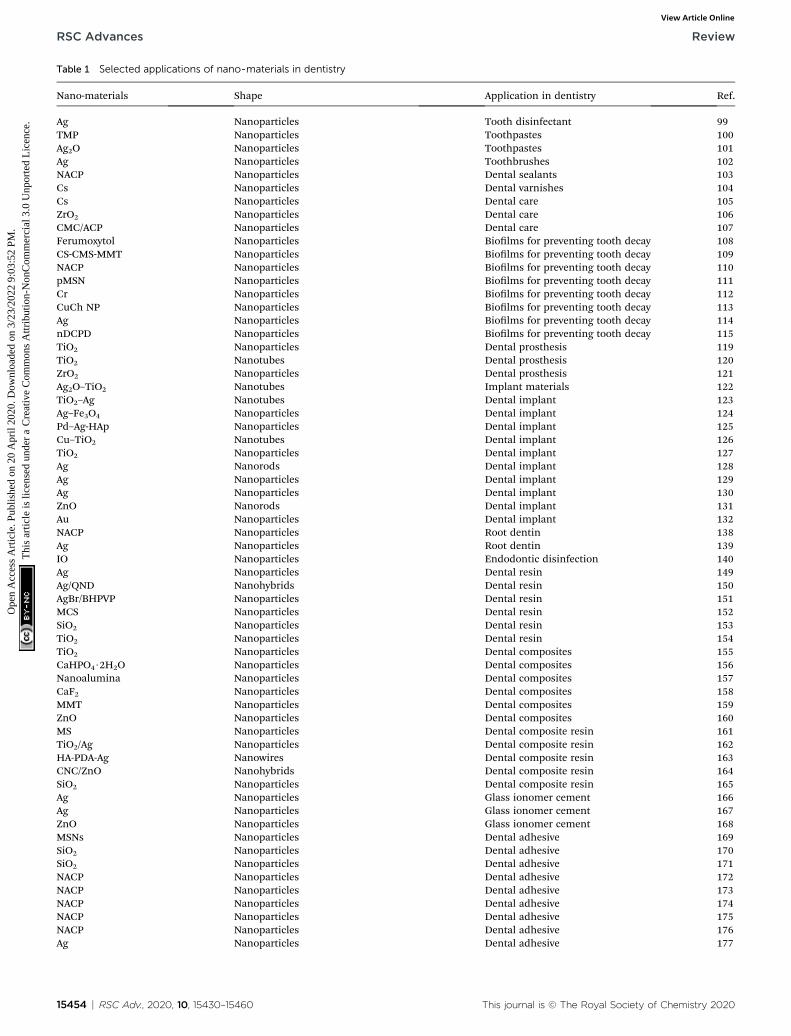
Table 1 Selected applications of nano-materials in dentistry
Nano-materials Shape Application in dentistry Ref.
Ag Nanoparticles Tooth disinfectant 99TMP Nanoparticles Toothpastes 100Ag2O Nanoparticles Toothpastes 101Ag Nanoparticles Toothbrushes 102NACP Nanoparticles Dental sealants 103Cs Nanoparticles Dental varnishes 104Cs Nanoparticles Dental care 105ZrO2 Nanoparticles Dental care 106CMC/ACP Nanoparticles Dental care 107Ferumoxytol Nanoparticles Biolms for preventing tooth decay 108CS-CMS-MMT Nanoparticles Biolms for preventing tooth decay 109NACP Nanoparticles Biolms for preventing tooth decay 110pMSN Nanoparticles Biolms for preventing tooth decay 111Cr Nanoparticles Biolms for preventing tooth decay 112CuCh NP Nanoparticles Biolms for preventing tooth decay 113Ag Nanoparticles Biolms for preventing tooth decay 114nDCPD Nanoparticles Biolms for preventing tooth decay 115TiO2 Nanoparticles Dental prosthesis 119TiO2 Nanotubes Dental prosthesis 120ZrO2 Nanoparticles Dental prosthesis 121Ag2O–TiO2 Nanotubes Implant materials 122TiO2–Ag Nanotubes Dental implant 123Ag–Fe3O4 Nanoparticles Dental implant 124Pd–Ag-HAp Nanoparticles Dental implant 125Cu–TiO2 Nanotubes Dental implant 126TiO2 Nanoparticles Dental implant 127Ag Nanorods Dental implant 128Ag Nanoparticles Dental implant 129Ag Nanoparticles Dental implant 130ZnO Nanorods Dental implant 131Au Nanoparticles Dental implant 132NACP Nanoparticles Root dentin 138Ag Nanoparticles Root dentin 139IO Nanoparticles Endodontic disinfection 140Ag Nanoparticles Dental resin 149Ag/QND Nanohybrids Dental resin 150AgBr/BHPVP Nanoparticles Dental resin 151MCS Nanoparticles Dental resin 152SiO2 Nanoparticles Dental resin 153TiO2 Nanoparticles Dental resin 154TiO2 Nanoparticles Dental composites 155CaHPO4$2H2O Nanoparticles Dental composites 156Nanoalumina Nanoparticles Dental composites 157CaF2 Nanoparticles Dental composites 158MMT Nanoparticles Dental composites 159ZnO Nanoparticles Dental composites 160MS Nanoparticles Dental composite resin 161TiO2/Ag Nanoparticles Dental composite resin 162HA-PDA-Ag Nanowires Dental composite resin 163CNC/ZnO Nanohybrids Dental composite resin 164SiO2 Nanoparticles Dental composite resin 165Ag Nanoparticles Glass ionomer cement 166Ag Nanoparticles Glass ionomer cement 167ZnO Nanoparticles Glass ionomer cement 168MSNs Nanoparticles Dental adhesive 169SiO2 Nanoparticles Dental adhesive 170SiO2 Nanoparticles Dental adhesive 171NACP Nanoparticles Dental adhesive 172NACP Nanoparticles Dental adhesive 173NACP Nanoparticles Dental adhesive 174NACP Nanoparticles Dental adhesive 175NACP Nanoparticles Dental adhesive 176Ag Nanoparticles Dental adhesive 177
15454 | RSC Adv., 2020, 10, 15430–15460 This journal is © The Royal Society of Chemistry 2020
RSC Advances Review
Ope
n A
cces
s A
rtic
le. P
ublis
hed
on 2
0 A
pril
2020
. Dow
nloa
ded
on 3
/23/
2022
9:0
3:52
PM
. T
his
artic
le is
lice
nsed
und
er a
Cre
ativ
e C
omm
ons
Attr
ibut
ion-
Non
Com
mer
cial
3.0
Unp
orte
d L
icen
ce.
View Article Online
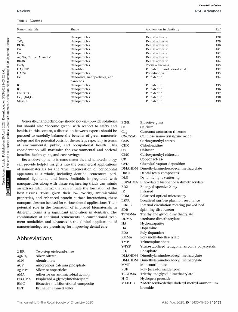
Table 1 (Contd. )
Nano-materials Shape Application in dentistry Ref.
Ag Nanoparticles Dental adhesive 178TiO2 Nanoparticles Dental adhesive 179PLGA Nanoparticles Dental adhesive 180Cu Nanoparticles Dental adhesive 181Cu Nanoparticles Dental adhesive 182Ag, Ta, Cu, Fe, Al and V Nanoparticles Dental adhesive 183BG-Bi Nanoparticles Dental adhesive 184CaO2 Nanoparticles Tooth whitening 185HA/CNT Nanober Pulp-dentin and periodontal 192HA/Zn Nanoparticles Periodontitis 193Ce Nanowires, nanoparticles, and
nanorodsPulp-dentin 194
IO Nanoparticles Pulp-dentin 195IO Nanoparticles Pulp-dentin 196GNP-CPC Nanoparticles Pulp-dentin 197Ce1�xGdxOy Nanoparticles Pulp-dentin 198MesoCS Nanoparticles Pulp-dentin 199
Review RSC Advances
Ope
n A
cces
s A
rtic
le. P
ublis
hed
on 2
0 A
pril
2020
. Dow
nloa
ded
on 3
/23/
2022
9:0
3:52
PM
. T
his
artic
le is
lice
nsed
und
er a
Cre
ativ
e C
omm
ons
Attr
ibut
ion-
Non
Com
mer
cial
3.0
Unp
orte
d L
icen
ce.
View Article Online
Generally, nanotechnology should not only provide solutionsbut should also “become green” with respect to safety andhealth. In this context, a discussion between experts should bepursued to carefully balance the benets of green nanotech-nology and the potential costs for the society, especially in termsof environmental, public, and occupational health. Thisconsideration will maximize the environmental and societalbenets, health gains, and cost savings.
Recent developments in nano-materials and nanotechnologycan provide helpful insights into the commercial applicationsof nano-materials for the ‘true’ regeneration of periodontalapparatus as a whole, including dentine, cementum, peri-odontal ligaments, and bone. Scaffolds impregnated withnanoparticles along with tissue engineering triads can mimican extracellular matrix that can initiate the formation of thehost tissues. Thus, given their low toxicity, antimicrobialproperties, and enhanced protein–surface interactions, thesenanoparticles can be used for various dental applications. Theirpotential role in the formation of improved biomaterials indifferent forms is a signicant innovation in dentistry. Thecombination of continual renements in conventional treat-ment modalities and advances in the clinical applications ofnanotechnology are promising for improving dental care.
Abbreviations
2 ER
This journal is
Two-step etch-and-rinse
AgNO3 Silver nitrate ALN Alendronate ACP Amorphous calcium phosphate Ag NPs Silver nanoparticles AMA Adhesive on antimicrobial activity Bis-GMA Bisphenol A-glycidylmethacrylate BMC Bioactive multifunctional composite BET Brunauer emmett teller© The Royal Society of Chemistry 2020
BG-Bi
Bioactive glass Ca Calcium Cag Curcuma aromatica rhizome CNC/ZnO Cellulose nanocrystal/zinc oxide CMS Carboxymethyl starch CHX Chlorhexidine CS Chitosan CMC Carboxymethyl chitosan CR Copper release CVD Chemical vapour deposition DMAHDM Dimethylaminohexadecyl methacrylate DRCs Dental resin composites DLS Dynamic light scattering EBPADMA Ethoxylated bisphenol A dimethacrylate EDX Energy dispersive X-ray IR Infrared POM Polarized optical microscopy LSPR Localized surface plasmon resonance ICRPB Internal circulation rotating packed bed SDR Spinning disc reactor TEGDMA Triethylene glycol dimethacrylate UDMA Urethane dimethacrylate HA Hydroxyapatite DA Dopamine PDA Poly dopamine PMMA Poly methylmethacrylate TMP Trimetaphosphate Y-TZP Yttria-stabilized tetragonal zirconia polycrystals PO4 Phosphate DMAHDM Dimethylaminohexadecyl methacrylate DMAHDM Dimethylaminohexadecyl methacrylate MMT Montmorillonite PUF Poly (urea-formaldehyde) TEGDMA Triethylene glycol dimethacrylate H2O2 Hydrogen peroxide MAE-DB 2-Methacryloxylethyl dodecyl methyl ammoniumbromide
RSC Adv., 2020, 10, 15430–15460 | 15455
RSC Advances Review
Ope
n A
cces
s A
rtic
le. P
ublis
hed
on 2
0 A
pril
2020
. Dow
nloa
ded
on 3
/23/
2022
9:0
3:52
PM
. T
his
artic
le is
lice
nsed
und
er a
Cre
ativ
e C
omm
ons
Attr
ibut
ion-
Non
Com
mer
cial
3.0
Unp
orte
d L
icen
ce.
View Article Online
MPC
15456 | RSC
2-Methacryloyloxyethyl phosphorylcholine
MMT Montmorillonite NaBH4 Sodium borohydrate NaF Sodium uoride PMMA Polymethyl methacrylate ROS Reactive oxygen species PLGA Poly-(lactic-co-glycolic acid) PMGDM Pyromellitic glycerol dimethacrylate CHX Chlorhexidine GIC Glass ionomer cement hDPSCs Human dental pulp stem cells UTS Ultimate tensile strength DC Degree of conversion WS Water sorption SO Solubility IM Immediate mTBS 1 year resin dentin bond strength FBPRE Ficus benghalensis prop root extract NaClO Sodium hypochlorite NL Nanoleakage POSS Polyhedraloligomeric silsesquioxanes RBC Resin-based composites TiO2 NPs Titanium dioxide nanoparticles NACP Nanoparticles of amorphous calcium phosphate CS-NPs Chitosan nanoparticles IO-NPs Iron oxide nanoparticles pMSN Pore mesoporous silica nanoparticles MSNPs Monodisperse silica nanoparticles CaF2 NPs Calcium uoride nanoparticle Cu NPs Copper nanoparticles nano-ZrO2 Zirconium oxide nanoparticles nDCPD Dicalcium phosphate dihydrate nanoparticle SEM Scanning electron microscope TEM Transmission electron microscopy XRD X-ray diffraction XPS X-ray photoelectron spectroscopyConflicts of interest
There are no conicts to declare.
Acknowledgements
This work was nancially supported by the Bilingual TeachingProgramme of Hainan University (hndsyk 201909); The Keyproject of the National Social Science Foundation of the year2018 (18AJY013); The 2017 National Social Science Foundationproject (17CJY072); The 2018 planning project of philosophyand social science of Zhejiang province (18NDJC086YB); The2018 Fujian Social Science Planning Project (FJ2018B067); ThePlanning Fund Project of Humanities and Social SciencesResearch of the Ministry of Education in 2019 (9YJA79010). Allprocedures conform to the principles outlined in the Declara-tion of Helsinki and Ethics committee of Islamic Azad Univer-sity of Kerman Branch has approved the experiments.
Adv., 2020, 10, 15430–15460
References
1 J. Wu, X. Wang, M. Khatami, M. S. Nejad, S. Salari andP. G. N. Zhu, IET Nanobiotechnol., 2016, 10, 237.
2 J. M. Granjeiro, R. Cruz, P. E. Leite, S. Gemini-Piperni,L. C. Boldrini and A. R. Ribeiro, Tin Oxide Materials,Elsevier, 2020, p. 133.
3 J. Han, L. Xiong, X. Jiang, X. Yuan, Y. Zhao and D. Yang,Prog. Polym. Sci., 2019, 91, 1.
4 S. Pramanik and D. Sundar Das, Two-DimensionalNanostructures for Biomedical Technology, Elsevier, 2020, p.281.
5 P. Liu, J. R. Williams and J. J. Cha, Nat. Rev. Mater., 2019, 4,479.
6 A. Zengin, P. Sutthavas and S. van Rijt, NanostructuredBiomaterials for Regenerative Medicine, WoodheadPublishing, 2020, p. 293.
7 M. Chen, Y. Sun, J. Liang, G. Zeng, Z. Li, L. Tang andB. Song, Environ. Int., 2019, 126, 690.
8 B. Noorani, F. Tabandeh, F. Yazdian, Z. S. Soheili,M. Shakibaie and S. Rahmani, Int. J. Polym. Mater. Polym.Biomater., 2018, 64, 754.
9 Z. Meng, R. M. Stolz, L. Mendecki and K. A. Mirica, Chem.Rev., 2019, 119, 478.
10 R. Fang, M. Liu and L. Jiang, Nano Today, 2019, 24, 48.11 G. J. Boelen, L. Boute, J. d'Hoop, M. EzEldeen, I. Lambrichts
and G. Opdenakker, Clin. Oral. Investig., 2019, 23, 2823.12 Y. Zhang and B. R. Lawn, J. Dent. Res., 2018, 97, 140.13 B. Duane, S. Harford, D. Ramasubbu, R. Stancliffe,
E. Pasdeki-Clewer, R. Lomax and I. Steinbach, Br. Dent. J.,2019, 226, 292.
14 F. Mirsiaghi, A. Leung, P. Fine, R. Blizard and C. Louca, Br.Dent. J., 2018, 225, 420.
15 N. Monteiro, G. Thrivikraman, A. Athirasala, A. Tahayeri,C. M. França, J. L. Ferracane and L. E. Bertassoni, Dent.Mater., 2018, 34, 389.
16 G. J. Boelen, L. Boute, J. d'Hoop, M. EzEldeen, I. Lambrichtsand G. Opdenakker, Clin. Oral. Investig., 2019, 23, 2823.
17 A. Schneider, J. Andrade, K. Tanja-Dijkstra and D. R. Moles,J. Anxiety. Disord., 2018, 58, 33.
18 I. Ferreira, C. L. Vidal, A. L. Botelho, P. S. Ferreira, M. L. daCosta Valente, M. A. Schiavon, O. L. Alves and A. Candidodos Reis, J. Prosthet. Dent., 2020, 123, 529.
19 H. Amini, J. D. Shenkin and D. L. Chi, Pediatr. Dent., 2019,159, 617.
20 N. Thomas, S. Blake, C. Morris and D. R. Moles, Int. J.Paediatr. Dent., 2018, 28, 226.
21 S. Sudenthiran, V. A. Ganesh, S. Harinee and M. Jacob, J.Dent. Educ., 2018, 3, 6.
22 G. Staunton and J. Hyland, J. Ir. Dent. Assoc., 2018, 64, 30.23 M. Salas, C. Lucena, L. J. Herrera, A. Yebra, A. Della Bona
and M. M. Perez, Dent. Mater., 2018, 34, 1168.24 I. Mirghani, F. Mushtaq, M. J. Allsop, L. M. Al-Saud,
N. Tickhill, C. Potter and M. Manogue, Eur. J. Dent. Educ.,2018, 22, 67.
This journal is © The Royal Society of Chemistry 2020
Review RSC Advances
Ope
n A
cces
s A
rtic
le. P
ublis
hed
on 2
0 A
pril
2020
. Dow
nloa
ded
on 3
/23/
2022
9:0
3:52
PM
. T
his
artic
le is
lice
nsed
und
er a
Cre
ativ
e C
omm
ons
Attr
ibut
ion-
Non
Com
mer
cial
3.0
Unp
orte
d L
icen
ce.
View Article Online
25 M. Eichenberger, N. Biner, M. Amato, A. Lussi and P. Perrin,Operat. Dent., 2018, 43, 501.
26 L. E. Silva Soares, S. Nahorny, V. de Faria Braga, F. RobertaMarciano, T. T. Bhattacharjee and A. Oliveira Lobo,Spectrochim. Acta, Part A, 2020, 228, 117818.
27 Y. Hassona, D. Malamos, M. Shaqman, Z. Baqain andC. Scully, Oral Dis., 2018, 24, 228.
28 J. Burns, N. McGoldrick and M. Muir, Evid. Based Dent.,2018, 19, 69.
29 C. T. Chang, G. R. Badger, B. Acharya, A. F. Gaw,M. S. Barratt and B. T. Chiquet, Pediatr. Dent., 2018, 40, 265.
30 M. E. Harb, S. Ebrahim, M. Soliman and M. Shabana, J.Electron. Mater., 2018, 47, 353.
31 S. Palazzolo, S. Bayda, M. Hadla, I. Caligiuri, G. Corona,G. Toffoli and F. Rizzolio, Curr. Med. Chem., 2018, 25, 4224.
32 Y. Y. Broza, R. Vishinkin, O. Barash, M. K. Nakhleh andH. Haick, Chem. Soc. Rev., 2018, 47, 4781.
33 Y. Yu, Y. Shi and B. Zhang, Acc. Chem. Res., 2018, 51, 1711.34 M. Oveisi, M. A. Asli and N. M. Mahmoodi, J. Hazard.
Mater., 2018, 347, 123.35 R. S. Andre, R. C. Sanfelice, A. Pavinatto, L. H. Mattoso and
D. S. Correa, Mater. Des., 2018, 156, 154.36 J. Zhu, J. Hou, Y. Zhang, M. Tian, T. He, J. Liu and V. Chen,
J. Membr. Sci., 2018, 550, 173.37 P. Makvandi, J. T. Gu, E. Nazarzadeh Zare, B. Ashtari,
A. Moeini, F. R. Tay and L. N. Niu, Acta Biomater., 2020,101, 69.
38 S. Palazzolo, S. Bayda, M. Hadla, I. Caligiuri, G. Corona,G. Toffoli and F. Rizzolio, Curr. Med. Chem., 2018, 25, 4224.
39 Y. Chen, Z. Fan, Z. Zhang, W. Niu, C. Li, N. Yang andH. Zhang, Chem. Rev., 2018, 118, 6409.
40 H. Cheng, N. Yang, Q. Lu, Z. Zhang and H. Zhang, Adv.Mater., 2018, 30, 1707189.
41 K. A. Roach, A. B. Stefaniak and J. R. Roberts, J.Immunotoxicol., 2019, 16, 87.
42 D. Zhang, C. Zhang, J. Liu, Q. Chen, X. Zhu and C. Liang,ACS Appl. Nano Mater., 2018, 2, 28.
43 L. Aline Pires, L. J. de Azevedo Silva, B. M. Ferrairo,R. Erbereli, J. F. Parreira Lovo, O. Ponce Gomes,J. Henrique Rubo, P. N. Lisboa-Filho, J. A. Griggs,C. A. Fortulan and A. F. Sanches Borges, Dent. Mater.,2020, 36, 38.
44 E. J. Foster, N. Zahed and C. Tallon, Small, 2018, 14,1870215.
45 D. H. Jung, A. Sharma and J. P. Jung, J. Alloys Compd., 2018,743, 300.
46 M. Singh, M. Goyal and K. Devlal, J. Taibah Univ. Sci., 2018,12, 470.
47 Q. Cai, K. Subramani, R. T. Mathew and X. Yang,Nanobiomaterials in Clinical Dentistry, Elsevier, Secondedition, 2019, p. 429.
48 A. J. Clancy, M. K. Bayazit, S. A. Hodge, N. T. Skipper,C. A. Howard and M. S. Shaffer, Chem. Rev., 2018, 118, 7363.
49 K. Ghosal and K. Sarkar, ACS Biomater. Sci. Eng., 2018, 4,2653.
50 M. Z. Islam Nizami, S. Takashiba and Y. Nishina, Appl.Mater. Today, 2020, 19, 100576.
This journal is © The Royal Society of Chemistry 2020
51 A. J. Clancy, M. K. Bayazit, S. A. Hodge, N. T. Skipper,C. A. Howard and M. S. Shaffer, Chem. Rev., 2018, 118, 7363.
52 R. Rao, C. L. Pint, A. E. Islam, R. S. Weatherup, S. Hofmann,E. R. Meshot and J. Carpena-Nunez, ACS Nano, 2018, 12,11756.
53 P. Laux, C. Riebeling, A. M. Booth, J. D. Brain, J. Brunner,C. Cerrillo and H. Jungnickel, Environ. Sci., 2018, 5, 48.
54 B. Yang, M. Zhang, Z. Lu, J. Luo, S. Song and Q. Zhang, ACSSustainable Chem. Eng., 2018, 6, 8954.
55 J. Bott and R. Franz, Appl. Sci., 2019, 9, 214.56 N. R. Panda, S. P. Pati and D. Das, Appl. Surf. Sci., 2019, 491,
313.57 P. Khade, T. Bagwaiya, S. Bhattacharaya, A. Singh, P. Jha
and V. Shelke, AIP Conf. Proc., 2018, 1942, 110052.58 S. A. Bello, J. O. Agunsoye, J. A. Adebisi and S. B. Hassan,
Eng. Appl. Sci. Res., 2018, 45, 262.59 A. Marcovici, G. Le Saux, V. Bhingardive, P. Rukenstein,
K. Flomin, K. Shreteh and M. Schvartzman, ACS Nano,2018, 12, 10016.
60 A. Bera, A. Bhattacharya, N. Tiwari, S. N. Jha andD. Bhattacharyya, Surf. Sci., 2018, 669, 145.
61 R. J. Varghese, S. Parani, S. Thomas, O. S. Oluwafemi andJ. Wu, Nanomater. Sol. Cell Appl., 2019, 2019, 75–95.
62 S. A. Davari, J. L. Gottfried, C. Liu, E. L. Ribeiro, G. Duscherand D. Mukherjee, Appl. Surf. Sci., 2019, 473, 156.
63 X. Wang, H. Li, M. Li, C. Li, H. Dai and B. Yang, DiamondRelat. Mater., 2018, 86, 179.
64 W. Shang, T. Cai, Y. Zhang, D. Liu and S. Liu, Tribol. Int.,2018, 118, 373.
65 F. Cazana, N. Latorre, P. Tarifa, J. Labarta, E. Romeo andA. Monzon, Catal. Today, 2018, 299, 67.
66 J. Lin, Y. He, X. Du, Q. Lin, H. Yang and H. Shen, Crystals,2018, 8, 384.
67 H. Mao and B. Li, Nano, 2018, 13, 1850027.68 M. Catauro, E. Tranquillo, G. Dal Poggetto, M. Pasquali,
A. Dell'Era and S. Vecchio Ciprioti, Materials, 2018, 11,2364.
69 F. Ansari, A. Sobhani and M. J. Salavati-Niasari, ColloidInterface Sci., 2018, 514, 723.
70 T. Budnyak, S. Aminzadeh, I. Pylypchuk, A. Riazanova,V. Tertykh, M. Lindstrom and O. Sevastyanova,Nanomater, 2018, 8, 950.
71 C. Aydin, J. Mater. Sci., 2018, 29, 20087.72 G. Molnar, S. Rat, L. Salmon, W. Nicolazzi and
A. Bousseksou, Adv. Mater., 2018, 30, 1703862.73 J. M. Salva, D. D. Gutierrez, L. A. Ching, P. M. Ucab,
H. Cascon and N. P. Tan, Nanotechnol, 2018, 29, 50LT01.74 B. Nafradi, M. Choucair and L. Forro, Nanomater, 2018, 1,
67.75 W. Tian, S. Liu, L. Deng, N. Mahmood and X. Jian,
Composites, 2018, 149, 92–98.76 N. A. Tien, C. H. Diem, N. T. T. Linh, V. O. Mittova and
I. Y. Mittova, Nanosystems, 2018, 9, 424.77 Y. Manawi, A. Samara, T. Al-Ansari and M. Atieh, Materials,
2018, 11, 822.78 R. Atchudan, T. N. J. I. Edison, S. Perumal, D. RanjithKumar
and Y. R. Lee, Int. J. Hydrogen Energy, 2019, 44, 2349.
RSC Adv., 2020, 10, 15430–15460 | 15457
RSC Advances Review
Ope
n A
cces
s A
rtic
le. P
ublis
hed
on 2
0 A
pril
2020
. Dow
nloa
ded
on 3
/23/
2022
9:0
3:52
PM
. T
his
artic
le is
lice
nsed
und
er a
Cre
ativ
e C
omm
ons
Attr
ibut
ion-
Non
Com
mer
cial
3.0
Unp
orte
d L
icen
ce.
View Article Online
79 W. Shang, T. Cai, Y. Zhang, D. Liu and S. Liu, Tribol. Int.,2018, 118, 373.
80 S. Nizamuddin, M. T. H. Siddiqui, N. M. Mubarak,H. A. Baloch, S. A. Mazari, M. M. Tunio and S. Riaz, Curr.Org. Chem., 2018, 22, 446.
81 S. Kalaiselvan, K. Balachandran, S. Karthikeyan andR. Venckatesh, Silicon, 2018, 10, 211.
82 Z. Guo, K. Cui, G. Zeng, J. Wang and X. Guo, Sci. Environ.,2018, 643, 1325.
83 Y. Chen, Z. Fan, Z. Zhang, W. Niu, C. Li, N. Yang andH. Zhang, Chem. Rev., 2018, 118, 6409.
84 J. Yao, H. Wang, M. Chen and M. Yang, Microchim. Acta,2019, 186, 395.
85 M. S. Zafar, A. A. Alnazzawi, M. Alrahabi, M. A. Fareed,S. Najeeb and Z. Khurshid, Adv. Dent. Biomater., 2019, 18,477.
86 X. Gao and G. V. Lowry, NanoImpact, 2018, 9, 14.87 J. Wang, R. Chen, L. Xiang and S. Komarneni, Ceram. Int.,
2018, 44, 7357.88 M. L. Cui, Y. S. Chen, Q. F. Xie, D. P. Yang and M. Y. Han,
Coord. Chem. Rev., 2019, 387, 450.89 Y. Li, Y. Xu, C. C. Fleischer, J. Huang, R. Lin, L. Yang and
H. Mao, J. Mater. Chem., 2018, 6, 9.90 M. G. Manera, A. Colombelli, A. Taurino, A. G. Martin and
R. Rella, Sci. Rep., 2018, 8, 12640.91 A. Besinis, T. De Peralta, C. J. Tredwin and R. D. Handy, ACS
Nano, 2015, 9, 2255.92 R. Pokrowiecki, K. Pałka and A. Mielczarek, Nanomedicine,
2018, 13, 639.93 Z. Khurshid, M. Zafar, S. Qasim, S. Shahab, M. Naseem and
A. AbuReqaiba, Materials, 2015, 8, 717.94 H. Fukuda, Dental implant, US Pat., US5174755A, 1992.95 X. Feng, A. Chen, Y. Zhang, J. Wang, L. Shao and L. Wei, Int.
J. Nanomed., 2015, 10, 3547.96 D. Elkassas and A. Arafa, Nanomed, 2017, 13, 1543.97 M. Hannig and C. Hannig, Nanobiomater. Clin. Dent., 2019,
201, 1.98 M. Goldberg, Dentistry and Oral Health Care, 2018, 1, 19.99 D. R. Schwass, K. M. Lyons, R. Love, G. R. Tompkins and
C. J. Meledandri, Adv. Dent. Res., 2018, 29, 117.100 C. O. Favretto, A. C. B. Delbem, J. C. S. Moraes,
E. R. Camargo, P. T. A. de Toledo and D. Pedrini, Clin.Oral Investig., 2018, 1, 3021–3029.
101 V. Manikandan, P. Velmurugan, J. H. Park, W. S. Chang,Y. J. Park, P. Jayanthi and B. T. Oh, Biotech, 2017, 7, 72.
102 A. Mackevica, M. E. Olsson and S. F. Hansen, J. Hazard.Mater., 2017, 322, 270.
103 M. Ibrahim, F. AlQarni, Y. Al-Dulaijan, M. Weir, T. Oates,H. Xu and M. Melo, Materials, 2018, 11, 1544.
104 M. O. Wassel and M. A. Khattab, J. Adv. Res., 2017, 8, 387.105 S. Nguyen, C. Escudero, N. Sediqi, G. Smistad and
M. Hiorth, Eur. J. Pharm. Sci., 2017, 104, 326.106 J. B. Fathima, A. Pugazhendhi and R. Venis,Microb. Pathog.,
2017, 110, 245.107 H. Wang, Z. Xiao, J. Yang, D. Lu, A. Kishen, Y. Li and
X. Yang, Sci. Rep., 2017, 7, 40701.
15458 | RSC Adv., 2020, 10, 15430–15460
108 Y. Liu, P. C. Naha, G. Hwang, D. Kim, Y. Huang, A. Simon-Soro and F. Alawi, Nat. Commun., 2018, 9, 2920.
109 S. Jahanizadeh, F. Yazdian, A. Marjani, M. Omidi andH. Rashedi, Int. J. Biol. Macromol., 2017, 105, 757.
110 Y. A. Al-Dulaijan, L. Cheng, M. D. Weir, M. A. S. Melo,H. Liu, T. W. Oates and H. H. Xu, J. Dent., 2018, 72, 44.
111 H. Yan, H. Yang, K. Li, J. Yu and C. Huang,Molecules, 2017,22, 1225.
112 A. Maghsoudi, F. Yazdian, S. Shahmoradi, L. Ghaderi,M. Hemati and G. Amoabediny, Mater. Sci. Eng., 2017, 75,1259.
113 C. Covarrubias, D. Trepiana and C. Corral, Dent. Mater. J.,2018, 37, 379.
114 A. Gitipour, S. R. Al-Abed, S. W. Thiel, K. G. Scheckel andT. Tolaymat, Chemosphere, 2017, 173, 245.
115 A. C. Ionescu, S. Hahnel, G. Cazzaniga, M. Ottobelli,R. R. Braga, M. C. Rodrigues and E. Brambilla, J. Mater.Sci., 2017, 28, 108.
116 M. Joana, M. Francisco, F. Ana, B. Maria Joao, M. PauloDurao and R. Jose Alexandre, Ann. Med., 2019, 51, 130.
117 R. B. Anchieta, I. A. E. Hoshino, A. C. F. Junior, E. O. deAlmeida, E. P. Rocha and W. G. Assunçao, J. Dent.Implants, 2019, 9, 41.
118 H. J. A. Judy, Dent. J., 2018, 6, 172.119 E. E. Totu, A. C. Nechifor, G. Nechifor, H. Y. Aboul-Enein
and C. M. Cristache, J. Dent., 2017, 59, 68.120 A. P. R. Magalhaes, C. A. Fortulan, P. N. Lisboa-Filho,
C. M. Ramos-Tonello, O. P. Gomes, P. F. Cesar andA. F. S. Borges, Ceram. Int., 2018, 44, 2959.
121 M. M. Gad, R. Abualsaud, A. Rahoma, A. M. Al-Thobity,K. S. Al-Abidi and S. Akhtar, Int. J. Nanomed., 2018, 13, 283.
122 M. Sarraf, A. Dabbagh, B. A. Razak, R. Mahmoodian,B. Nasiri-Tabrizi, H. R. Hosseini and N. L. Sukiman, Surf.Coat. Technol., 2018, 349, 1008.
123 U. F. Gunputh, H. Le, R. D. Handy and C. Tredwin, Mater.Sci. Eng., 2018, 91, 638.
124 Y. Yang, S. Ren, X. Zhang, Y. Yu, C. Liu, J. Yang and L. Miao,Int. J. Nanomed., 2018, 13, 3751.
125 J. M. Jang, S. D. Kim, T. E. Park and H. C. Choe, Appl. Surf.Sci., 2018, 432, 285.
126 J. Rosenbaum, D. L. Versace, S. Abbad-Andallousi, R. Pires,C. Azevedo, P. Cenedese and P. Dubot, Biomater. Sci., 2017,5, 455.
127 Z. G. Azzawi, T. I. Hamad, S. A. Kadhim and G. A. H. Naji, J.Mater. Sci., 2018, 29, 96.
128 S. Kim, C. Park, K. H. Cheon, H. D. Jung, J. Song, H. E. Kimand T. S. Jang, Appl. Surf. Sci., 2018, 451, 232.
129 M. Boutinguiza, M. Fernandez-Arias, J. del Val, J. Buxadera-Palomero, D. Rodrıguez, F. Lusquinos and J. Pou, Mater.Lett., 2018, 231, 126.
130 D. D. Divakar, N. T. Jastaniyah, H. G. Altamimi,Y. O. Alnakhli, A. A. Alkheraif and S. Haleem, Int. J. Biol.Macromol., 2018, 108, 790.
131 Y. Xiang, J. Li, X. Liu, Z. Cui, X. Yang, K. W. K. Yeung andS. Wu, Mater. Sci. Eng., 2017, 79, 629.
This journal is © The Royal Society of Chemistry 2020

Review RSC Advances
Ope
n A
cces
s A
rtic
le. P
ublis
hed
on 2
0 A
pril
2020
. Dow
nloa
ded
on 3
/23/
2022
9:0
3:52
PM
. T
his
artic
le is
lice
nsed
und
er a
Cre
ativ
e C
omm
ons
Attr
ibut
ion-
Non
Com
mer
cial
3.0
Unp
orte
d L
icen
ce.
View Article Online
132 K. Jadhav, H. R. Rajeshwari, S. Deshpande, S. Jagwani,D. Dhamecha, S. Jalalpure and D. Baheti, Mater. Sci. Eng.,2018, 93, 664.
133 J. J. Segura-Egea, K. Gould, B. H. Sen, P. Jonasson, E. Cotti,A. Mazzoni and P. M. H. Dummer, Int. Endod. J., 2018, 51,20.
134 N. H. F. Wilson, F. J. T. Burke, P. A. Brunton, S. Creanor,M. T. Hosey and F. Mannocci, Br. Dent. J., 2019, 226, 110.
135 K. Pineda, R. Bueno, C. Alvarado, F. Abella, M. Roig andF. Duran-Sindreu, Aust. Endod. J., 2018, 44, 40.
136 A. Torres, E. Shaheen, P. Lambrechts, C. Politis andR. Jacobs, Int. Endod. J., 2019, 52, 540.
137 S. G. Kim, M. Malek, A. Sigurdsson, L. M. Lin and B. Kahler,Int. Endod. J., 2018, 51, 1367.
138 S. Xiao, K. Liang, M. Weir, L. Cheng, H. Liu, X. Zhou andH. Xu, Materials, 2017, 10, 89.
139 C. T. Rodrigues, F. B. de Andrade, L. R. S. M. deVasconcelos, R. Z. Midena, T. C. Pereira, M. C. Kuga andN. Bernardineli, Int. Endod. J., 2018, 51, 901.
140 S. Bukhari, D. Kim, Y. Liu, B. Karabucak and H. Koo, J.Endod., 2018, 44, 806.
141 N. J. M. Opdam, K. Collares, R. Hickel, S. C. Bayne,B. A. Loomans, M. S. Cenci and N. H. F. Wilson, Dent.Mater., 2018, 34, 1.
142 W. F. Waggoner and T. Nelson, Pediatr. Dent., 2019, 304.143 F. Schwendicke and N. Opdam, Dent. Mater., 2018, 34, 29.144 R. Sarkis-Onofre, T. Pereira-Cenci, A. C. Tricco,
F. F. Demarco, D. Moher and M. S. Cenci, Journal ofEsthetic and Restorative Dentistry, 2019, 31, 222.
145 G. P. Ramiro, C. A. Coronel, A. F. Navarro, B. Hassan andF. Tamimi, Digit. Restor. Dent., 2019, 41, 7–39.
146 J. A. Rodrigues, L. Casagrande, F. B. Araujo, T. L. Lenzi andA. A. Mariath, Pediatr. Restor. Dent., 2019, 161.
147 T. E. Donovan, R. Marzola, K. R. Murphy, D. R. Cagna,F. Eichmiller, J. R. McKee and M. Troeltzsch, J. Prosthet.Dent., 2018, 120, 816.
148 A. Abdullah, F. Muhammed, B. Zheng and Y. Liu, J. Dent.Mater. Tech., 2018, 7, 1.
149 S. J. Lee, M. Heo, D. Lee, S. Han, J. H. Moon, H. N. Lim andI. K. Kwon, Appl. Surf. Sci., 2018, 432, 317.
150 W. Cao, X. Wang, Q. Li, Z. Ye and X. Xing,Mater. Lett., 2018,220, 104.
151 W. Cao, Y. Zhang, X. Wang, Y. Chen, Q. Li, X. Xing andZ. Ye, J. Mater. Sci., 2017, 28, 103.
152 Y. Zhang, C. Huang and J. Chang, J. Mater. Chem., 2018, 6,477.
153 P. Cevik and A. Z. Yildirim-Bicer, J. Prosthodontics, 2018, 27,763.
154 L. Ghahremani, S. Shirkavand, F. Akbari and N. Sabzikari, J.Clin. Experiment. Dent., 2017, 9, e661.
155 A. Sodagar, M. S. A. Akhoundi, A. Bahador, Y. F. Jalali,Z. Behzadi, F. Elhaminejad and A. H. Mirhashemi, DentalPress J. Orthodont., 2017, 22, 67.
156 M. C. Rodrigues, M. D. Chiari, Y. Alania, L. C. Natale,V. E. Arana-Chavez, M. M. Meier and R. R. Braga, Dent.Mater., 2018, 34, 746.
This journal is © The Royal Society of Chemistry 2020
157 A. Meena, H. S. Mali, A. Patnaik and S. R. Kumar, Polym.Chem., 2018, 39, E321.
158 M. S. Al-Ajely, K. M. Ziadan and R. M. Al-Bader, Int. J. Res.Granthaalayah, 2018, 6, 338.
159 L. M. Campos, L. C. Boaro, T. M. Santos, V. J. Santos,R. Grecco-Romano, M. J. Santos and D. F. Parra, J. Therm.Anal. Calorim., 2018, 131, 771.
160 R. M. Al-Mosawi and R. M. Al-Badr, IOSR J. Dent. Med. Sci.,2017, 16, 49.
161 D. L. Yang, J. Xiao, D. Wang, W. M. Lin, Y. Pu, X. F. Zengand J. X. Wang, Ind. Eng. Chem. Res., 2018, 57, 12809.
162 H. B. Dias, M. I. B. Bernardi, T. M. Bauab, A. C. Hernandesand A. N. de Souza Rastelli, Dent. Mater., 2019, 35, e36.
163 M. Ai, Z. Du, S. Zhu, H. Geng, X. Zhang, Q. Cai and X. Yang,Dent. Mater., 2017, 33, 12.
164 Y. Wang, H. Hua, W. Li, R. Wang, X. Jiang and M. Zhu, J.Dent., 2019, 80, 23.
165 R. Wang, E. Habib and X. X. Zhu, Dent. Mater., 2017, 33,1139.
166 L. Paiva, T. K. S. Fidalgo, L. P. da Costa, L. C. Maia, L. Balan,K. Anselme and R. M. S. M. Thire, J. Dent., 2018, 69, 102.
167 D. Sundeep, T. V. Kumar, P. S. Rao, R. V. S. S. N. Ravikumarand A. G. Krishna, Prog. Biomater., 2017, 6, 57.
168 P. P. N. S. Garcia, M. F. B. Cardia, R. S. Francisconi,L. N. Dovigo, D. M. P. Spolidorio, A. N. de Souza Rastelliand A. C. Botta, Microsc. Res. Tech., 2017, 80, 456.
169 C. A. Stewart, J. H. Hong, B. D. Hatton and Y. Finer, ActaBiomater., 2018, 76, 283.
170 B. Han, W. Xia, K. Liu, F. Tian, Y. Chen, X. Wang andZ. Yang, ACS Appl. Mater. Interfaces, 2018, 10, 8519.
171 E. Azad, M. Atai, M. Zandi, P. Shokrollahi and L. Solhi, Dent.Mater., 2018, 34, 1263.
172 Y. Liu, L. Zhang, L. N. Niu, T. Yu, H. H. Xu, M. D. Weir andJ. H. Chen, J. Dent., 2018, 72, 53.
173 S. Yue, J. Wu, Q. Zhang, K. Zhang, M. D. Weir, S. Imazatoand H. H. Xu, J. Dent., 2018, 75, 48.
174 F. D. Al-Qarni, F. Tay, M. D. Weir, M. A. Melo, J. Sun,T. W. Oates and H. H. Xu, J. Dent., 2018, 78, 91.
175 X. Xie, L. Wang, D. Xing, K. Zhang, M. D. Weir, H. Liu andH. H. Xu, Dent. Mater., 2017, 33, 553.
176 K. Liang, S. Xiao, M. D. Weir, C. Bao, H. Liu, L. Cheng andH. H. Xu, J. Biomed. Mater. Res., 2018, 106, 2414.
177 M. Dutra-Correa, A. A. Leite, S. P. de Cara, I. M. Diniz,M. M. Marques, I. B. Suffredini and I. S. Medeiros, J.Dent., 2018, 77, 66.
178 R. Thomas, S. Snigdha, K. B. Bhavitha, S. Babu, A. Ajith andE. K. Radhakrishnan, Biotech, 2018, 8, 404.
179 J. Sun and W. L. Wu, inventor, Google Patents, assignee.Toothpaste patent US20120172485A1, 2012.
180 B. M. Priyadarshini, K. Mitali, T. B. Lu, H. K. Handral,N. Dubey and A. S. Fawzy, Dent. Mater., 2017, 33, 830.
181 M. F. Gutierrez, P. Malaquias, T. P. Matos, A. Szesz, S. Souzaand J. Bermudez, Dent. Mater., 2018, 33, 309.
182 M. F. Gutierrez, P. Malaquias, V. Hass, T. P. Matos,L. Lourenço and A. Reis, J. Dent., 2019, 61, 12–20.
RSC Adv., 2020, 10, 15430–15460 | 15459
RSC Advances Review
Ope
n A
cces
s A
rtic
le. P
ublis
hed
on 2
0 A
pril
2020
. Dow
nloa
ded
on 3
/23/
2022
9:0
3:52
PM
. T
his
artic
le is
lice
nsed
und
er a
Cre
ativ
e C
omm
ons
Attr
ibut
ion-
Non
Com
mer
cial
3.0
Unp
orte
d L
icen
ce.
View Article Online
183 G. A. Frolov, Y. N. Karasenkov, A. A. Gusev, O. V. Zakharova,A. Y. Godymchuk, D. V. Kuznetsov and V. K. Leont'ev, NanoHybrids and Composites, 2017, 13, 39–46.
184 M. Rizk, L. Hohlfeld, L. T. Thanh, R. Biehl, N. Luhmann,D. Mohn and A. Wiegand, Dent. Mater., 2017, 33, 1056–1065.
185 V. Velkoborsky, inventor, Google Patents, assignee.Toothpaste patent US20100239618 A1, 2010.
186 S. Prakasam, P. Gajendrareddy, C. Louie, C. Lee andL. E. Bertassoni, Biomater. Immune Response, 2018, 211.
187 E. T. Knight and W. Murray Thomson, Periodontol, 2018,78, 195.
188 J. G. Deeb, C. K. Carrico, D. M. Laskin and T. E. Koertge, J.Dent. Educ., 2019, 83, 457.
189 G. Zucchelli, P. Sharma and I. Mounssif, Periodontol, 2018,77, 7–18.
190 Z. Danish, M. N. Shah, S. Rehmat, F. A. Hakam andH. A. Raza, Dent. J., 2019, 39, 60.
191 S. N. Papageorgiou, G. N. Antonoglou, C. Martin andT. Eliades, J. Orthod., 2019, 46, 101.
192 A. S. Khan, A. N. Hussain, L. Sidra, Z. Sarfraz, H. Khalid,M. Khan and I. U. Rehman, Mater. Sci. Eng., 2017, 80, 387.
193 D. K. Khajuria, S. F. Zahra and R. Razdan, J. Biomater. Sci.,2018, 29, 74.
194 C. Mahapatra, R. K. Singh, J. H. Lee, J. Jung, J. K. Hyun andH. W. Kim, Acta Biomater., 2017, 50, 142.
195 Y. Xia, H. Chen, F. Zhang, L. Wang, B. Chen, M. A. Reynoldsand H. H. Xu, Artif. Cells, Nanomed., Biotechnol., 2018, 46,423.
196 Y. Ji, S. K. Choi, A. S. Sultan, K. Chuncai, X. Lin,E. Dashtimoghadam and Z. Nie, Nanomed, 2018, 14, 919.
197 Y. Xia, H. Chen, F. Zhang, C. Bao, M. D. Weir,M. A. Reynolds and H. H. Xu, Nanomed, 2018, 14, 35.
198 A. L. Popov, O. G. Tatarnikova, N. R. Popova, I. I. Selezneva,A. Y. Akkizov, A. M. Ermakov and V. K. Ivanov, Nano Hybridsand Composites, 2017, 13, 26.
199 C. Y. Huang, T. H. Huang, C. T. Kao, Y. H. Wu, W. C. Chenand M. Y. Shie, J. Endod., 2017, 43, 69.
200 M. Ates, Z. Arslan, V. Demir, J. Daniels and I. O. Farah,Environ. Toxicol., 2015, 30, 119.
201 M. Coccia and L. Wang, Proc. Natl. Acad. Sci. U. S. A., 2016,113, 2057.
202 M. Giovanni, C. Y. Tay, M. I. Setyawati, J. Xie, C. N. Ong,R. Fan and D. T. Leong, Environ. Toxicol., 2015, 30, 1459.
203 M. Coccia, Scientometrics, 2018, 117, 1265.204 L. Li, T. Liu, C. Fu, L. Tan, X. Meng and H. Liu, Nanomed,
2015, 11, 1915.205 M. Coccia, Tech. Soc., 2020, 60, 1.206 I. Y. Kim, E. Joachim, H. Choi and K. Kim, Nanomed, 2015,
11, 407.207 N. Klinthoopthamrong, D. Chaikiawkeaw,
W. Phoolcharoen, K. Rattanapisit, P. Kaewpungsup,
15460 | RSC Adv., 2020, 10, 15430–15460
P. Pavasant and V. P. Hoven, Int. J. Biol. Macromol., 2020,149, 51.
208 M. Coccia, Int. J. Healthc. Tech. Manag., 2012, 13, 184.209 X. Jing, J. H. Park, T. M. Peters and P. S. Thorne, Toxicol,
2015, 29, 502.210 M. Coccia, U. Finardi and D. Margon, J. Technol. Trans.,
2012, 37, 777.211 N. Adam, A. Vakurov, D. Knapen and R. Blust, J. Hazard.
Mater., 2015, 283, 416.212 G. Libralato, G. Lofrano, A. Siciliano, E. Gambino,
G. Boccia, C. Federica, A. Francesco, E. Galdiero,R. Gesuele and M. Guida, Visible Light Active StructuredPhotocatalysts for the Removal of Emerging Contaminants,Elsevier, 2020, p. 195.
213 M. Coccia and J. Watts, J. Eng. Tech. Manag., 2020, 55,101552.
214 M. Kumari, S. P. Singh, S. Chinde, M. F. Rahman,M. Mahboob and P. Grover, Int. J. Toxicol., 2014, 33, 86.
215 M. Coccia, Technol. Forecast. Soc. Change, 2019, 141, 289.216 S. Aalapati, S. Ganapathy, S. Manapuram, G. Anumolu and
B. M. Prakya, Nanotoxicol, 2014, 8, 786.217 M. Coccia, Technol. Anal. Strateg. Manage., 2017, 29, 1048.218 B. Collin, E. Oostveen, O. V. Tsyusko and J. M. Unrine,
Environ. Sci. Technol., 2014, 48, 1280.219 E. O. Ogunsona, R. Muthuraj, E. Ojogbo, O. Valerio and
T. H. Mekonnen, Appl. Mater. Today, 2020, 18, 100473.220 M. Coccia, Technol. Anal. Strateg. Manage., 2014, 26, 733.221 N. S. Taylor, R. Merrield, T. D. Williams, J. K. Chipman,
J. R. Lead and M. R. Viant, Nanotoxicol, 2016, 10, 32.222 R. Abbasalipourkabir, H. Moradi, S. Zarei, S. Asadi,
A. Salehzadeh, A. Ghafourikhosroshahi and N. Ziamajidi,Food Chem. Toxicol., 2015, 84, 154.
223 N. R. Jacobsen, T. Stoeger, S. Van Den Brule, A. T. Saber,A. Beyerle, G. Vietti and A. Banerjee, Food Chem. Toxicol.,2015, 85, 84.
224 T. Y. Suman, S. R. Rajasree and R. Kirubagaran, Ecotoxicol.Environ., 2015, 113, 23.
225 J. Choi, H. Kim, P. Kim, E. Jo, H. M. Kim, M. Y. Lee andK. Park, J. Toxicol. Environ. Health, 2015, 78, 226.
226 Q. Fang, X. Shi, L. Zhang, Q. Wang, X. Wang, Y. Guo andB. Zhou, J. Hazard. Mater., 2015, 283, 897.
227 N. R. B. Younes, S. Amara, I. Mrad, I. Ben-Slama, M. Jeljeli,K. Omri and M. Sakly, Environ. Sci. Pollut. Res., 2015, 22,8728.
228 Z. L. Wang, MRS Bull., 2007, 32, 109.229 A. Ghasempour Ardakani and P. Raeipour, J. Opt. Soc. Am.
B, 2018, 35, 1708.230 D. B. Rihtnesberg, S. Almqvist, Q. Wang, A. Sugunan,
X. Yang, M. S. Toprak, Z. Besharat and M. Gothelid, inNanoelectronics Conference, INEC, 2011, vol. 2011, p. 1.
231 A. Umar, M. M. Rahman, S. H. Kim and Y. B. Hahn, J.Nanosci. Nanotechnol., 2008, 8, 3216.
232 S. Parasharam, Mater. Today, 2013, 16, 505–506.
This journal is © The Royal Society of Chemistry 2020