appendix c.9 toxicity profiles for copcs ameenuddin, s. and sunde, m.l. 1984. proceedings of the...
TRANSCRIPT

L2003-131 Mar 2004
Appendix C.9
Toxicity Profiles for COPCs

VOLATILE Acetone Acetone is an organic solvent with a wide variety of uses in industry, the laboratory and home. It is produced in large quantities and may be released to the environment as stack emissions, fugitive emissions and in waste water in its production and use as a chemical intermediate and solvent. Due to it’s volatile nature, most acetone used in solvents will ultimately be released into the air. In the atmosphere, acetone will be lost by photolysis and reaction with photochemically-produced hydroxyl radicals. Half-life estimates average 22 days and are shorter in the summer than winter. If released on soil, acetone will both volatilize and leach into the ground. If released to water, it will also be lost due to volatilization (half-life of 20 hours). Bioconcentration in aquatic organisms and adsorption to sediment should not be significant. (HSDB, 1995). Occupational exposure to acetone will be via dermal contact and inhalation of the vapor. It is highly volatile and rapidly absorbed by the respiratory system. Small quantities may be absorbed through the skin. The general population is exposed to acetone in the atmosphere from sources such as auto exhaust, solvents, tobacco smoke and fireplaces, as well as dermal contact with acetone-containing consumer products. Because of its solubility in water, acetone is readily absorbed into the bloodstream and transported rapidly throughout the body (HSDB, 1995). Excretion is rapid for 8 hours after a single oral does but was not complete in 24 hours. The ratio of excretion was approximately 40-70% in breath, 15-30% in urine and 10% through skin (Clayton and Clayton, 1981). Acute human exposure to high levels of acetone produces central nervous system depression and unconsciousness. Prolonged exposure may produce irritation of the respiratory tract, coughing, headache, drowsiness, loss of coordination and in severe cases, come (Clement Associates, 1985). Inhalation of 2000 ppm is fatal upon brief exposure (Arena, 1974). Prolonged or repeated dermal exposure causes skin irritation or contact dermatitis (HSDB, 1995). Other common symptoms associated with acetone exposure include bronchitis, gastritis, pharyngitis and conjunctivitis. Long-term exposure of rates to low levels of acetone produces increases in liver and kidney weights, as well as changes in red blood cell counts (HSDB, 1995). Acetone significantly reduced the percentage of hatchability in developing chick embryos and caused a high embryonic mortality during the first week of incubation (Ameenuddin and Sunde, 1984). However, no adverse effects were noted in cultured rat embryos exposed to 0.1% acetone. Acetone has not been tested in a carcinogenicity bioassay, but gave negative results in a skin painting study (Clement Associates, 1985). Acetone was not mutagenic in the Ames assay (Clement Associates, 1985) or in sister chromatid exchange assays (HSDB, 1995). No information is available on the carcinogenic effects of acetone in humane. Acetone is classified as a group D carcinogen.

C.8-2
Ameenuddin, S. and Sunde, M.L. 1984. Proceedings of the Society of ExperimentalBiological Medicine. 175(2): 176-178. Arena, J.M. 1974. Poisoning: Toxicology - Symptoms/Treatments. Third edition, Springfield, Illinois: Charles C. Thomas, ed. P. 686. Clement Associates, Inc. 1985. Chemical, physical and biolgical properties of compounds present at hazardous waste site. September, 1985. HSDB. 1995. Hazardous Substances Data bank. TOXNET computer database, National Library of Medicine. Benzene Benzene, also known as benzol, is a colorless liquid with a sweet odor. Benzene evaporates into air very quickly and dissolves slightly in water. Benzene is highly flammable. Benzene is made mostly from petroleum sources. Because of its wide use, benzene ranks in the top 20 in production volume for chemicals produced in the United States. Various industries use benzene to make other chemicals, such as styrene (for Styrofoam and other plastics), cumene (for various resins), and cyclohexane (for nylon and synthetic fibers). Benzene is also used for the manufacturing of some types of rubbers, lubricants, dyes, detergents, drugs, and pesticides. Natural sources of benzene, which include volcanoes and forest fires, also contribute to the presence of benzene in the environment. Benzene is also a part of crude oil and gasoline and cigarette smoke. Exposure to benzene can occur via inhalation, ingestion, especially of contaminated drinking water, and dermal contact (as in contact with liquid benzene found in gasoline.) (ATSDR, 1997) Benzene is readily absorbed by both test animals and humans from inhalation, oral, and dermal exposures (IRIS, 2003). Once in the bloodstream, benzene is distributed throughout the body, with the concentration in any one compartment dependent on the degree of perfusion of tissues by blood. Since benzene is lipid-soluble, it accumulates in fat, but the rate of accumulation is slow since fat is poorly perfused. The metabolites of benzene are responsible for its toxic effects. These include phenol (which is either formed via an unstable benzene oxide precursor or directly from benzene), catechol, hydroquinone and conjugated phenolic compounds. The primary site of benzene metabolism is the liver via the cytochrome P450 mixed function oxidase system. Some benzene metabolism may also occur in the bone marrow via the same enzyme system. Benzene is excreted either unchanged from the lungs or as metabolites in the urine (ATSDR, 1997). Benzene targets its effects on the hemopoietic, immune and nervous systems (ATSDR, 1997, USEPA, 2002). Exposure to benzene has produced irritation of the skin, eyes and upper respiratory tract. Acute exposure has produced central nervous system depression, headache, dizziness, nausea, convulsions, coma and death at extremely high concentrations (Sittig, 1981). Health effects in humans have been reported starting as low as 50 ppm via inhalation. Twenty-five ppm for 6 hrs had no obvious effects though benzene was detected in blood (Sandmeyer, 1981). Early autopsy reports found benzene-induced hemorrhages of the brain, pericardium, urinary tract, mucous membranes

C.8-3
and skin (Sittig, 1981). Chronic exposure to benzene produces blood changes involving an initial increase in levels of erythrocytes, leukocytes and thrombocytes, followed by aplastic anemia indicated by anemia, leukopenia and thrombocytopenia (Sittig, 1981). The following effects have been produced experimentally in laboratory animals, following exposure to benzene: decreased leukocyte and/or erythrocyte counts, reduction in cellular immunity and bone marrow depression (reduced number of granulopoietic stem cells). Animal studies do not indicate that benzene is teratogenic, but the following fetotoxic effects have been found: reduced fetal weight, altered fetal hematopoiesis, fetal skeletal variations and increased resorptions in pregnant exposed animals. In addition, benzene has produced histopathological changes in ovaries and testes of test animals (ATSDR, 1997). The RfD for benzene is based on route-to-route extrapolation of the results of benchmark dose (BMD) modeling of the absolute lymphocyte count (ALC) data from the occupational epidemiologic study by Rothman et al. (1996), in which workers were exposed to benzene by inhalation. The RfD is based upon a 95% lower bound benchmark concentration of 8.2 mg/m3 which was then converted to an equivalent oral dose of 1.2E+0 mg/kg-d which was divided by an uncertainty factor of 300 (3 for effect-level extrapolation, 10 for intraspecies differences (human variability), 3 for subchronic-to-chronic extrapolation, and 3 for database deficiencies) to arrive at a chronic RfD for benzene of 4E-03 mg/kg-day (IRIS, 2003). The RfC for benzene is based on the results of benchmark dose (BMD) modeling of the absolute lymphocyte count (ALC) data from the occupational epidemiologic study by Rothman et al. (1996), in which workers were exposed to benzene by inhalation. The RfD is based upon a 95% lower bound benchmark concentration of 8.2 mg/m3, which was divided by an uncertainty factor of 300 (3 for effect-level extrapolation, 10 for intraspecies differences (human variability), 3 for subchronic-to-chronic extrapolation, and 3 for database deficiencies) to arrive at a chronic RfC for benzene of 3E-02 mg/m3 (IRIS, 2003). Benzene and its metabolites have been shown to be mutagenic in a number of in vitro and in vivo studies. Genotoxic effects produced experimentally include structural and numerical chromosome aberrations in humans, animals and cell cultures, and sister chromatid exchanges and micronuclei in vivo animal studies. Benzene exposure has been found to produce an increase in the number of chromosome aberrations associated with myelotoxicity (Sittig, 1981). In addition, sperm head abnormalities, inhibition of DNA and RNA synthesis, DNA binding and interference with cell cycle progression have been shown in vitro studies (ATSDR, 1997). Epidemiologic studies and case studies provide clear evidence of a causal association between exposure to benzene and acute nonlymphocytic leukemia (ANLL) and also suggest evidence for chronic nonlymphocytic leukemia (CNLL) and chronic lymphocytic leukemia (CLL). Other neoplastic conditions that are associated with an increased risk in humans are hematologic neoplasms, blood disorders such as preleukemia and aplastic anemia, Hodgkin's lymphoma, and myelodysplastic syndrome (MDS). These human data are supported by animal studies. The experimental animal data add to the argument that exposure to benzene increases the risk of cancer in multiple species at multiple organ sites (hematopoietic, oral and nasal, liver,

C.8-4
forestomach, preputial gland, lung, ovary, and mammary gland) (IRIS, 2003). It is likely that these responses are due to interactions of the metabolites of benzene with DNA (Ross, 1996; Latriano et al., 1986). Recent evidence supports the viewpoint that there are likely multiple mechanistic pathways leading to cancer and, in particular, to leukemogenesis from exposure to benzene (Smith, 1996). Benzene is classified as a "known" human carcinogen (Category A) under the Risk Assessment Guidelines of 1986. Under the proposed revised Carcinogen Risk Assessment Guidelines (U.S. EPA, 1996), benzene is characterized as a known human carcinogen for all routes of exposure based upon convincing human evidence as well as supporting evidence from animal studies. (U.S. EPA, 1979, 1985, 1998; ATSDR, 1997). The oral slope factor for benzene is reported as a range, 1.5 x 10⁻² to 5.5 x 10⁻² per (mg/kg)/day. The quantitative oral unit risk estimate is an extrapolation from the known inhalation dose-response to the potential oral route of exposure. The inhalation risk estimate is reported as a range, from 2.2 × 10⁻⁶ to 7.8 × 10⁻⁶ per µg/m3. No relevant data exist in the published literature for oral absorption of benzene in humans. Inhalation absorption is assumed to be about 50% while that of oral is selected as 100% based upon a review of the relevant human and animal literature (U.S. EPA, 1999). Absorption of benzene via the dermal route of exposure is usually less than 1% of the applied dose and therefore it is not considered to contribute significantly the oral risk estimation. Agency for Toxic Substances and Disease Registry (ATSDR). 1997. Toxicological Profile For Benzene. September. Available online at: http://www.atsdr.cdc.gov/toxprofiles/tp3.html Integrated Risk Information System (IRIS). 2003. Benzene. U.S. Environmental Protection Agency. Available at: http://www.epa.gov/iris/ Latriano, L; Goldstein, BD; Witz, G. 1986. Formation Of Muconaldehyde, An Open-Ring Metabolite Of Benzene, In Mouse Liver Microsomes: An Additional Pathway For Toxic Metabolites. Proc Natl Acad Sci USA 83:8356-8360. Ross, D. 1996. Metabolic Basis Of Benzene Toxicity. Eur. J. Haematol. 57: 111-118. Rothman, N., G.L. Li, M. Dosemeci, W.E. Bechtold, G.E. Marti, Y.Z. Wang, M. Linet, L.Q. Xi, W. Lu, M.T. Smith, N. Titenko-Holland, L.P. Zhang, W. Blot, S.N. Yin, and R.B. Hayes. 1996. Hematotoxicity Among Chinese Workers Heavily Exposed To Benzene. Am. J. Ind. Med. 29: 236-246. Sandmeyer, E.E. 1981. Aromatic Hydrocarbons. In: Patty's Industrial Hygiene and Toxicology, Vol. 2, 3rd ed., Clayton G.D., Clayton F.E., eds. New York. Interscience Publishers. pp. 3253-3283. Sittig, M. 1981. Handbook of Toxic and Hazardous Chemicals. Noyes Publications. Smith, MT. 1996. The Mechanism Of Benzene-Induced Leukemia: A Hypothesis And Speculations On The Causes Of Leukemia. Environ Health Perspect 104 (Suppl 6):1219-1225.

C.8-5
U.S. Environmental Protection Agency. 1979. Final Report On Population Risk To Ambient Benzene Exposures. Prepared by the Carcinogen Assessment Group, Research Triangle Park, NC. EPA/450/5-80-004. U.S. EPA. 1985. Final Draft For Drinking Water Criteria Document On Benzene. Washington, DC: Office of Drinking Water, U.S. EPA. PB86-118122. U.S. EPA. 1998. Carcinogenic Effects Of Benzene: An Update. Prepared by the National Center for Environmental Health, Office of Research and Development. Washington, DC. EPA/600/P-97/001F. U.S. EPA. 1999. Extrapolation Of The Benzene Inhalation Unit Risk Estimate To The Oral Route Of Exposure. National Center for Environmental Health, Office of Research and Development. Washington, DC. NCEA-W-0517. USEPA. 2002. Toxicological Review of Benzene (Noncancer Effects). EPA/635/R-02/001F. Available online at: http://www.epa.gov/iris/toxreviews/0276-tr.pdf Bromomethane Bromomethane (also called methyl bromide) is a colorless gas without much smell. Some bromomethane is formed in the ocean, probably by algae or kelp. However, most is made by humans to kill various pests (rats, insects, fungus, etc.) that might be present in homes, foods, or soil. Some bromomethane is also used to make other chemicals (ATSDR, 1992). There is limited information regarding the absorption of Bromomethane in humans or animals. Medinsky et al. (1984) has shown that at least 97% of a single oral dose was absorbed from the gastrointestinal tract in rats. There is limited information regarding the distribution of chlorodibromomethane after exposure. Data seems to indicate that after inhalation and oral exposure, bromomethane is absorbed and distributed throughout the body (Bond et al. 1985; Jaskot et al. 1988; Medinsky et al. 1985). Bromomethane undergoes initial metabolism primarily by nucleophilic displacement of the bromide ion. When the attacking species is water, the products are methanol and bromide ion (Gargas and Andersen 1982; Honma et al. 1985). Bromomethane may also react with organic thiols (R-SH) to yield S-methyl derivatives (Iwasaki 1988b). No studies were located regarding excretion of bromomethane in humans after exposure. In animals exposed to bromomethane vapors, excretion occurs mainly by expiration of carbon dioxide or by urinary excretion of nonvolatile metabolites (Bond et al. 1985; Jaskot et al. 1988; Medinsky et al. 1985). Only small amounts are excreted in the feces. Very little parent bromomethane is exhaled (Jaskot et al. 1988; Medinsky et al. 1985). Bromomethane exists as a gas at ordinary temperatures, so the most likely route of human exposure is by inhalation. Only limited information is available on the effects of long-term inhalation exposure of humans to low levels of bromomethane. Headache, weakness, and increased prevalence of neurological signs such as muscle ache, fatigue, dizziness, and ataxia have been noted in workers exposed for extended periods in the workplace (Anger et al. 1986; Hine 1989; Kantarjian and

C.8-6
Shasheen 1963; Kishi et al. 1988). No cases of severe neurological effects from long-term exposure to low levels have been noted in humans. Acute inhalation exposure to bromomethane can result in marked lung irritation (edema, hemorrhagic lesions), and this may lead to moderate to severe impairment of respiratory function (Greenberg 1971; Prain and Smith 1952). Anuria and proteinuria are common signs of renal injury in acutely exposed humans (O'Neal 1987; Prain and Smith 1952; Viner 1945), but dose-response data are not available. Acute inhalation exposure to bromomethane can result in marked lung irritation (edema, hemorrhagic lesions) (Irish et al. 1940; Kato et al. 1986; Reuzel et al. 1987; Sato et al. 1985). Mild signs of liver injury (edema, focal hemorrhages, minimal necrosis) have been noted in some studies at levels of 150-600 ppm (Alexeeff et al. 1985; Eustis et al. 1988; Hurtt et al. 1987a; Kato et al. 1986), with no significant injury at levels of 66 ppm (Irish et al. 1940). Signs of renal injury have been reported in several animal studies, including swelling, edema, nephrosis, and tubular necrosis (Alexeeff et al. 1985; Eustis et al. 1988). Inhalation studies in animals confirm that the central nervous system is injured by inhalation exposure to bromomethane. Clinical effects that have been detected include tremors, ataxia, paralysis, and seizures (Alexeeff et al. 1985; Anger et al. 1981; Breslin et al. 1990; Drew 1984; Haber 1987; Hurtt et al. 1987a; Irish et al. 1940; Kato et al. 1986). Histological lesions in the brain (focal necrosis and hemorrhage) have also been detected(Alexeeff et al. 1985; Eustis et al. 1988; Hurtt et al. 1987a).
Several studies of animals exposed to bromomethane vapors up to 70 ppm did not detect developmental effects, even though these concentration levels resulted in maternal toxicity (Hardin et al. 1981; Sikov et al. 1980). However, exposure of rabbits to 80 ppm during gestation resulted in increased incidence of several developmental anomalies in the off-spring (Breslin et al. 1990). These data suggest bromomethane may cause developmental effects, but only at high doses where other effects would also be of concern. Inhalation exposure of male rats to high levels of bromomethane (120-400 ppm) has resulted in decreased sperm production along with testicular degeneration and atrophy (Drew 1984; Eustis et al. 1988; Kato et al. 1986). However, exposures up to 70 ppm do not appear to interfere with reproductive functions in male (McGregor 1981) or female rats (Hardin et al. 1981; McGregor 1981; Sikov et al. 1980). Several studies in animals have shown that repeated (90-day) administration of concentrated solutions of bromomethane (40-5,000 mg/L, dissolved in oil) by gavage to rats can result in irritation and hyperplasia of the epithelium in the forestomach (Boorman et al. 1986; Danse et al. 1984). The RfD for bromomethane is based on a 13-week subchronic oral study conducted in rats (Danse et al. 1984). The duration adjusted NOAEL of 1.4 mg/kg-day for gastric lesions was divided by an uncertainty factor of 1000 (10 to extrapolate from rats to humans; 10 to protect sensitive humans and 10 for extrapolating from subchronic to chronic exposure) to arrive at a chronic RfD for bromomethane of 1.4E-03 mg/kg-day (IRIS, 2003). The RfC for bromomethane is based on a chronic inhalation conducted in rats (Reuzel et al., 1987, 1991). Degenerative and proliferative lesions of the olfactory epithelium of the nasal cavity were selected as the critical effect for derivation of the RfC. The duration adjusted LOAEL was used to calculate a human equivalent concentration (HEC) of 0.48 mg/m3
which was divided by an

C.8-7
uncertainty factor of 100 (10 for intraspecies uncertainty, a factor of 3 for the use of a LOAEL for a mild effects and a factor of 3 for interspecies extrapolation because dosimetric adjustments have been applied. The factors of 3 represent operational application of a geometric half of the standard factor of 10, rounded to a single significant figure. As a result, multiplication of two factors of 3 results in a composite factor of 10) to arrive at a chronic RfC for bromomethane of 5E-03 mg/m3 (IRIS, 2003). Bromomethane has produced positive results in a number of mutagenicity test systems, both in vitro and in vivo. This effect does not appear to require metabolic activation, which is consistent with the fact the bromomethane is a direct-acting alkylating agent which can methylate DNA (Ikawa et al. 1986; Starratt and Bond 1988). This property suggests that bromomethane might be carcinogenic, but this has not been established. No epidemiological studies were located on cancer incidence in humans exposed specifically to bromomethane. Chronic inhalation studies performed in mice and rats revealed no evidence of carcinogenic effects at exposure levels of 33-90 ppm (Reuzel et al. 1987; Yang 1990). Rats given daily oral doses of 50 mg/kg/day for 90 days developed inflammation and keratosis of the forestomach, along with lesions that were originally interpreted as squamous carcinomas (Danse et al. 1984). However, reevaluation of the histological specimens by NTP scientists indicated that the forestomach lesions in this study were hyperplastic but not neoplastic (ATSDR, 1992). Bromomethane has been classified as a Group D carcinogen; not classifiable as to human carcinogenicity. This classification is based upon inadequate human and animal data: a single mortality study from which direct exposure associations could not be deduced and studies in several animal species with too few animals, too brief exposure or observation time for adequate power. Bromomethane has shown genotoxicity (IRIS, 2003). Agency for Toxic Substances and Disease Registry (ATSDR) 1992. Toxicological Profile For Bromomethane. U.S. Public Health Service. September. Available at: http://www.atsdr.cdc.gov/toxprofiles/tp27.html Alexeeff GV, Kilgore W, Munoz P, et al. 1985. Determination Of Acute Toxic Effects In Mice Following Exposure To Methyl Bromide. J Toxicol Environ Health 15:109-123. Anger WK, Setzer JV, Russo JM, et al. 1981. Neurobehavioral Effects Of Methyl Bromide Inhalation Exposures. Stand J Work Environ Health 7(Suppl 4):40-47. Bond JA, Dutcher JS, Medinsky MA, et al. 1985. Disposition Of [14C]Methyl Bromide In Rats After Inhalation. Toxicol Appl Pharmacol 78:259-267. Boorman GA, Hong HL, Jameson CW, et al. 1986. Regression Of Methyl Bromide Induced Forestomach Lesions In The Rat. Toxicol Appl Pharmacol 86:131-139. Breslin WJ, Zublotny CL, Bradley GJ, et al. 1990. Methyl Bromide Inhalation Teratology Study In

C.8-8
New Zealand White Rabbits. Midland, MI: Dow Chemical. Study K-00681-033. EPA MRID 415804-01. Danse, L.H.J.C., F.L. van Velsen and C.A. van der Heijden. 1984. Methylbromide: Carcinogenic Effects In The Rat Forestomach. Toxicol. Appl. Pharmacol. 72: 262-271. Drew RT. 1984. A 60-Day Inhalation Study Of Methyl Bromide Toxicity In Mice. (Unpublished study no. BNL 34506). Upton, NY: Brookhaven National Laboratory. Accession No. 40578401. EPA: 68D80056. Eustis SL, Haber SB, Drew RT, et al. 1988. Toxicology And Pathology Of Methyl Bromide In F344 Rats And B6C3F1 Mice Following Repeated Inhalation Exposure. Fundam Appl Toxicol 11:594-610. Gargas ML, Andersen ME. 1982. Metabolism Of Inhaled Brominated Hydrocarbons: Validation Of Gas Uptake Results By Determination Of A Stable Metabolite. Toxicol Appl Pharmacol 66:55-68. Greenberg JO. 1971. The Neurologoical Effects Of Methyl Bromide Poisoning. Ind Med 40:27-29. Haber SB. 1987. A Chronic Inhalation Study Of Methyl Bromide Toxicity In B6C3F1 Mice. Report to National Toxicology Program, Research Triangle Park, NC, by Brookhaven National Laboratory, Upton, NY. Hardin BD, Bond GP, Sikov MR, et al. 1981. Testing Of Selected Workplace Chemicals For Teratogenic Potential. Stand J Work Environ Health 7:66-75. Hine CH. 1969. Methylbromide Poisoning. J Occup Med ll: l-10. Honma T, Miyagawa M, Sato M, et al. 1985. Neurotoxicity And Metabolism Of Methyl Bromide In Rats. Toxicol Appl Pharmacol 81:183-191. Hurtt ME, Morgan KT, Working PK. 1987. Histopathology Of Acute Toxic Responses In Selected Tissues From Rats Exposed By Inhalation To Methyl Bromide. Fundam Appl Toxicol 9:352-365. Ikawa N, Araki A, Nozaki K, et al. 1986. Micronucleus Test Of Methyl Bromide By The Inhalation Method [Abstract]. Mutat Res 164:269. Integrated Risk Information System (IRIS). 2003. Bromomethane. U.S. Environmental Protection Agency. Available at: http://www.epa.gov/iris/ Irish DD, Adams EM, Spencer HC, et al. 1940. The Response Attending Exposure Of Laboratory Animals To Vapors Of Methyl Bromide. J Ind Hyg Toxicol 22:218-230. Iwasaki K. 1988. Determination Of S-Methycysteine In Mouse Hemoglobin Following Exposure To Methyl Bromide. Ind Health 26:187-190.
C.8-9
Jaskot RH, Grose EC, Most BM, et al. 1988. The Distribution And Toxicological Effects Of Inhaled Methyl Bromide In The Rat. J Am Co11 Toxicol 7:631-642. Kantarjian AD, Shasheen AS. 1963. Methyl Bromide Poisoning With Nervous System Manifestations Resembling Polyneuropathy. Neurology 13:1054-1058. Kato N, Morinobu S, Ishizu S. 1986. Subacute Inhalation Experiment For Methyl Bromide In Rats. Ind Health 24:87-103. Kishi R, Ishizu I, Ito I, et al. 1988. Health Research Of Methyl Bromide Manufacturing Workers. Part I. Symptoms Of Long-Term Exposure. Copenhagen, Denmark: World Health Organization, 122-134. McGregor DB. 1981. Tier II Mutagenic Screening Of 13 NIOSH Priority Compounds: Individual Compound Report Methyl Bromide. Report to National Institute for Occupational Safety and Health, Cincinnati, OH, by Inveresk Research International Limited, Musselburgh, Scotland. NTIS No. PB83-130211. Medinsky MA, Bond JA, Dutcher JS, et al. 1984. Disposition Of [14C]Methyl Bromide In Fischer-344 Rats After Oral Or Intraperitoneal Administration. Toxicology 32:187-196. O'Neal L. 1987. Acute Methyl Bromide Toxicity. JEN 13:96-98. *Prain JH, Smith GH. 1952. A Clinical-Pathological Report Of Eight Cases Of Methyl Bromide Poisoning. Br J Ind Med 9:44-49. Reuzel, P.G.J., C.F. Kuper, H.C. Dreef-van der Meulen and V.M.H. Hollanders. 1987. Chronic (29-Month) Inhalation Toxicity And Carcinogenicity Study Of Methyl Bromide In Rats. Report No. V86.469/221044. Netherlands Organization for Applied Scientific Research, Division for Nutrition and Food Research, TNO. EPA/OTS Document No. 86-8700001202. Reuzel, P.G.J., H.C. Dreef-van der Meulen, V.M.H. Hollanders, C.F. Kuper, V.J. Feron and C.A. van der Heijden. 1991. Chronic Inhalation Toxicity And Carcinogenicity Study Of Methyl Bromide In Wistar Rats. Fd. Chem. Toxic. 29(1): 31-39. Sato M, Miyagawa M, Honma T, et al. 1985. Subacute Effects Of Methyl Bromide Dosed By Inhalation Exposure To Rats. Ind Health 23:235-238. Sikov MR, Cannon WC, Carr DB. 1980. Teratologic Assessment Of Butylene Oxide, Styrene Oxide And Methyl Bromide. Report to National Institute for Occupational Safety and Health, Division of Biomedical and Behavioral Science, U.S. Department of Health, Education and Welfare, Cincinnati, OH, by Battelle, Pacific Northwest Laboratory, Richland, WA. NTIS No. PBSl-168510.

C.8-10
Starratt AN, Bond EJ. 1988. In Vitro Methylation Of DNA By The Fumigant Methyl Bromide. J Environ Sci Health [B] 23:513-524. Viner N. 1945. Methyl bromide poisoning: A New Industrial Hazard. Can Med Assoc J 53:43-45. Yang RSH. 1990. Personal Communication From Dr. Raymond Yang (NTP) To Dr. William Cibulas, Jr. (ATSDR) Regarding Findings And Publication Status Of Draft Technical Report On Inhalation Studies On Methyl Bromide. As cited in ATSDR, 1992. 2-Butanone (MEK) General Background Physical/Chemical Properties The chemical identify and physical/chemical properties of methyl ethyl ketone are summaried in Table 1. Characteristic/Property Data Reference CAS No. 78-93-3 Common Synonyms 2-Butanone, MEK Verschureren 1983 Molecular Formula C4H80 Chemical Structure ? 2 CH3-C-CH2-CH3 Physical State Liquid Budavari 1989 Molecular Weight 7.210 Budavari 1989 Melting Point -86iC Budavari 1989 Boiling Point 79.6iC Budavari 1989 Water Solubility 353 g/L @ 10iC; 190 g/L @ 90iC Verschueren 1983 Specific gravity 0.805 @ 20/4iC Verschueren 1983 Vapor Density (air = 1) 2.41 Verschueren 1983 KOC 4.5 -50 (estimated U.S. EPA 1989 Log KOW 0.29 (estimated) U.S. EPA 1989 Vapor Pressure 77.5 mm Hg @ 20iC Verschueren 1983 98.0 mm Hg @ 25iC U.S. EPA 1985 Henry’s Law Constant 4.16 to 6.11 X 10⁻⁵ atm-3/mol @25iC (estimated) U.S. EPA 1985 Odor Threshold 5 to 10 ppm (in air) U.S. Air Force 1989 There are three methyl ethyl ketone producers in the United States. In 1992, an estimated 494

C.8-11
million pounds of methyl ethyl ketone were produced in the US. During that same year, 55 million pounds were imported into the US and 112 million pounds were exported (Mannsville 1993). Methyl ethyl ketone is used in a number of industrial applications. The primary use of methyl ethyl ketone, accounting for approximately 63 percent of all use, is as a solvent in protective coatings. It is also used as a solvent in adhesives; printing inks; paint removers; in the production of magnetic tapes; and in dewaxing lubricating oil. Methyl ethyl ketone is used as a chemical intermediate in several reactions, including condensation; halogenation; ammonolysis; and oxidation. Small amounts of methyl ethyl ketone are also used as a sterilizer for surgical instruments, hypodermic needles, syringes and dental instruments; as an extraction solvent for hardwood pulping and vegetable oil; and as a solvent in pharmaceutical and cosmetic production (Mannsville 1993; HSDB 1994). Environmental Fate and Transport Of the total methyl ethyl ketone released to the environment almost all eventually enters the air. Methyl ethyl ketone is released into the environment from industrial and domestic uses. Methyl ethyl ketone has been found in cigarette smoke (500 ppm) and in gasoline engine exhaust (<0.1-1.0 ppm) (Verschueren 1983). Its use as a solvent in polyvinyl chloride pipe joint cement has introduced the chemical into drinking water (4.5 ppm, 6 months after installation) (HSDB 1994). It occurs naturally and has been found in a number of foods and beverages including swiss cheese (0.3 ppm), cream (0.154-0.177 ppm), barley, bread, honey, oranges, black tea, rum, non-alcoholic beverages (70 ppm), and ice cream (270 ppm) (HSDB 1994). Although the atmospheric concentration of methyl ethyl ketone increases during episodes of severe photochemical smog (up to 14 ppb in Los Angeles, CA during a photochemical pollution episode), ambient concentrations are usually below the detection limit (0.01 ppb) in most urban areas. Methyl ethyl ketone is not expected to be retarded by adsorption to soils rich in organic material [estimated Koc = 4.5-50 (U.S.EPA 1989)]; therefore, it is expected to be mobile in soil and, subject to leaching from landfills. The relatively high vapor pressure [98.0 mm Hg @ 25oC (U.S. EPA 1985)] and estimated Henry's Law constant (4.16-6.11 x 10⁻⁵ atm m3/mol @ 25oC) indicate that it can volatilize from moist and dry soil (HSDB 1994). It does not adsorb significantly to suspended solids, and will volatilize to the atmosphere from surface waters (HSDB 1994; U.S. EPA 1985). In wet or dry soil, methyl ethyl ketone will volatilize to air and may undergo photolysis on the soil surface (U.S. EPA 1985). It is also highly mobile and may be leached from the soil by water, and has been shown to be degradated by cultures of soil bacteria (U.S. EPA 1985). The most important fate process for methyl ethyl ketone in water is volatilization (estimated half-times of 3 and 12 days, for rivers and lakes, respectively). Complete aerobic biodegradation of methyl ethyl ketone has been reported in about 5-10 days following inoculation with sewage or polluted surface water. Longer times were required for degradation in marine water. Anaerobic degradation occurred after an acclimation period of about one week (HSDB 1994). Direct photolysis near the surface is also thought to be a possible mechanism, but was not measured (HSDB 1994). It is not expected to undergo chemical hydrolysis or to be bound to sediment or suspended organic matter (HSDB 1994). Methyl ethyl ketone is not expected to bioconcentrate in fish or aquatic organisms; its estimated fish bioconcentration factor is less than 1 (U.S. EPA 1985). Absorption Transport and Degradation

C.8-12
Studies in humans and animals have demonstrated that methyl ethyl ketone can be absorbed via the lungs, the skin, and the gastrointestinal system. Pulmonary absorption values range from 41.1% to 55.8% (WHO 1993). The relative uptake through the lungs by humans was about 53% through a 4 hour exposure at 200 ppm (HSDB 1994: WHO 1993). Oral studies in rats have demonstrated that the peak blood level of methyl ethyl ketone (0.95 mg/mL) was reached in 4 hours following oral administration of the chemical in water (1690 mg/kg) (U.S. EPA 1985). Methyl ethyl ketone can also be absorbed through intact human skin. Absorption is more rapid through moist skin than through dry skin, and the rate of percutaneous absorption has been estimated to range from 5 to 10 micrograms/cm2/min (WHO 1993). The relative solubility of methyl ethyl ketone in various human tissues and organs compared to blood (tissue/blood partition coefficient) was shown to be similar in the kidney, liver, brain, heart, and lung and in fat and muscle tissue. This indicates a potential for even and widespread distribution of methyl ethyl ketone in human tissues (U.S. EPA 1985). It has also been shown to cross the placenta and enter the human fetus (WHO 1993). Experiments with guinea pigs and rats have shown that methyl ethyl ketone is metabolized by oxidative hydroxylation, forming 3-hydroxy-2-butanone, which is further reduced to 2,3-butanediol. Methyl ethyl ketone is also reversibly reduced to 2-butanol (U.S. EPA 1985). The half-life of methyl ethyl ketone in the blood of guinea pigs was reported to be 270 minutes, following intraperitoneal injection of 450 mg/kg. The clearance time was 12 hours (U.S. EPA 1985). It has been estimated that humans eliminate 30 to 40% of methyl ethyl ketone intake in expired air (HSDB 1994). The unchanged chemical and its metabolite, 3-hydroxy-2-butanone were measured in the urine of workers occupationally exposed to 8 to 272 mg/m3. The metabolite was only identified in workers exposed to the higher levels, and was correlated with the level of exposure and the urinary concentration of methyl ethyl ketone (U.S. EPA 1985). The plasma half-time in humans has be estimated at 49-96 minutes with an apparent clearance rate of 0.6 L/minute (ATSDR 1992). Non-Carcinogenic Health Effects Acute Toxicity Humans - Volunteers complained of mild nose and throat irritation when exposed to 100 ppm. The irritation became objectionable when the concentration was raised to 300 ppm (ATSDR 1992). Workers occupationally exposure to an atmosphere containing 300 to 500 ppm (126 to 210 mg/kg/day) (see end note 2) complained of headaches, nausea, and respiratory tract irritation. Momentary exposure to 33,000 or 100,000 ppm caused intolerable irritation of the eyes, nose and throat (Krasavage et al. 1982). Workers exposed either by inhalation (300-600 ppm) or by skin contact to methyl ethyl ketone developed numbness in the extremities and dermatitis. High concentrations can result in central nervous system depression (see Section IV.G.1.). Eye contact with liquid methyl ethyl ketone causes painful irritation, and can result in comeal injury (Krasavage et al. 1982; HSDB 1994). Animals - Oral LD50 values of 2737 and 4050 mg/kg were reported for rats and mice, respectively. Inhalation LC50 values of 23,500 mg/m3/8 hours for rats and 40,000 mg/m3/2 hours for mice were reported (U.S. EPA 1985). Guinea pigs exposed to 10,000 ppm methyl ethyl ketone developed liver and kidney congestion, but no effects were seen at 3500 ppm (ATSDR 1992).

C.8-13
Subchronic/Chronic Effects Humans - Dermatitis, gastrointestinal upset, loss of appetite and weight, and neurological problems (see Section IV.G.I.) were reported by individuals occupationally exposed to methyl ethyl ketone, apparently in the absence of other solvents (WHO 1993). Neurological problems were also reported by workers chronically exposed to 300-600 ppm of the chemical (WHO 1993). All other available studies describing subchronic/chronic occupational exposure involved a mixture of organic solvents that contained methyl ethyl ketone. A variety of nervous system effects were consistently described after prolonged exposure to these mixtures (see Section IV.G.1.) (WHO 1993). Animals - Male and female Fischer 344 rats were exposed to 0, 1250, 2500, or 5000 ppm methyl ethyl ketone by inhalation 6 hours/day, 5 days/week, for 90 days. No treatment related differences in food consumption, in eye effects, in neurological functioning, or in histopathologies were reported (U.S. EPA 1985). Increased serum alanine amino transferase in females was the only effect at 2500 ppm; increased absolute and relative liver weight were seen in both sexes at 5000 ppm. Females also developed decreased relative brain weight, and increased blood levels of alkaline phosphatase, glucose, and potassium at 5000 ppm. The mean body weights of both sexes were decreased at 5000 ppm, but increased at the other dose levels compared to the controls. In another inhalation study Fischer 344 rats were exposed by inhalation to 5041 ppm (14,870 mg/m3), 2518 ppm, or 1254 ppm methyl ethyl ketone for 6 hours/ day, 5 days/week for 90 days. No significant effects on food consumption, eyes, or morphology were observed (WHO 1993). Increased absolute and relative liver weight, decreased body weight, increased relative kidney weight, decreased relative brain and spleen weights, and increased mean corpuscular hemoglobin were reported at the high dose. Increased body weights were reported at the low and intermediate dose. Male rats exposed to 10,000 ppm methyl ethyl ketone 8 hours/day, 7 days/week developed severe irritation in the upper respiratory tract within a few days and died during the 7th week of exposure of bronchopneumonia (ATSDR 1992). Carcinogenic Health Effects Humans - No excess cancer incidence was found in an epidemiological study of 446 male workers in two methyl ethyl ketone dewaxing plants. The deaths observed were below the expected, and overall deaths from cancer were also below the expected. An increase in tumors of the buccal cavity and pharynx was seen but the numbers were small (2 observed, 0.13 expected) and lung cancers were significantly decreased (I observed, 6.02 expected) (ATSDR 1992; WHO 1993). The overall cancer-related mortality was less than expected in another epidemiological study of 1008 male oil refinery workers exposed to 1-4 ppm methyl ethyl ketone. There were no increases in buccal and pharyngeal cancers seen in this study (ATSDR 1992). Animals - Methyl ethyl ketone was applied to the skin of mice twice weekly for one year (50 mg/dose). No tumors were reported (U.S. EPA 1989). No information on the carcinogenicity of

C.8-14
methyl ethyl ketone by other routes of administration were found in the secondary sources searched. Mutagenicitv Results from four out of the five short term mutagenicity assays, requested by and submitted to EPA under the Toxic Substances Control Act (TSCA), indicate methyl ethyl ketone is not mutagenic. Methyl ethyl ketone is negative in the Ames test (in Salmonella typhimurium strains TA98, TA 100, TA 1535, and TA 1537 with or without S-9 metabolic activation); the mouse lymphoma test; the cell transformation assay; and the mouse micronucleus test. Results from the Section 4 unscheduled DNA test was concluded to be positive (Cimino 1985). Most of the information from other secondary sources also indicate that methyl ethyl ketone is not genotoxic. Methyl ethyl ketone was negative in E. coli tester strains WP2 and WP2uvrA (WHO 1993). It was negative in the mitotic gene conversion assay in S. cerevisiae tester strain JD 1 (WHO 1993). It was negative in the micronucleus test in both CD I mice and Chinese hamsters (WHO 1993). It was a strong inducer of aneuploidy in S. cerevisiae strain D6 LM (U.S. EPA 1989). Developmental/Reproductive Toxicity Animal studies indicate that exposure to high methyl ethyl ketone concentrations in air breathed during pregnancy can cause fetal toxicity and possibly adverse developmental effects. EPA has derived an oral reference dose (RfD) (see end note 3) of 0.6 mg/kg/day, based on developmental effects of a metabolite (2-butanol) of MEK. EPA has derived an inhalation reference concentration (RfC) (see end note 4) of 1 mg/m') for methyl ethyl ketone, based on its developmental effects. Humans - No information on the developmental/reproductive toxicity of methyl ethyl ketone was found in the secondary sources searched. Animals - Since there are no appropriate oral studies on methyl ethyl ketone, the U.S. EPA (1994) calculated a chronic oral RfD for methyl ethyl ketone of 0.6 mg/kg/day, based on decreased birth weights seen in a multigeneration study with its metabolic intermediate, 2-butanol. Male and female Wistar rats (30/sex/group) were given 2-butanol in drinking water at 0, 0.3, 1.0, or 3.0% nine weeks before mating through gestation and lactation. The high dose was reduced to 2.0°/a for the second generation. Decreased fetal weight and decreased pup survivability were reported for the first generation at 3.0% 2-butanol. Decreased fetal weight was seen in the second generation at 2.0%. Increases in the incidences of missing stemebrae, wavy ribs, and incomplete vertebrae ossification at 2%, compared to the 1.0 and 0.3% groups, were seen. However, because of high incidences of these developmental effects in the control group, they could not be determined to be treatment related. The 1.0% dose, equivalent to 1771 mg/kg/day, was identified as a no-adverse-effect-level (NOAEL), and the 2.0% dose was identified as a lowest-adverse-effect-level (LOAEL)(U.S. EPA 1994). The U.S. EPA (1994) calculated a chronic inhalation reference concentration (RfC) of 1.0 mg/m3 for methyl ethyl ketone, based on decreased fetal birth weight seen in a mouse inhalation study. Pregnant mice were exposed by inhalation to atmospheric concentrations of 0, 398, 1010, or 3020
C.8-15
ppm methyl ethyl ketone 7 hours/day during days 6-15 of gestation. Decreased fetal body weight was seen at 3020 ppm. There was an increased incidence of fetuses and litters with malformations in the treated groups, but the increases were not statistically significant. A NOAEL of 1010 ppm and a LOAEL of 3020 ppm were identified (U.S. EPA 1994). Groups of 25 pregnant Sprague-Dawley rats were exposed to air concentrations of 400, 1000, or 3000 ppm methyl ethyl ketone 7 hours/day during days 6-15 of gestation. The control group contained 35 rats. Maternal body weight gain was decreased and water consumption was increased in dams receiving 3000 ppm. There were no treatment related effects on the pregnancy rate, the number of implantations per litter, or the percent live fetuses. A total of five fetuses with at least one external or soft tissue malformation were reported, but were distributed in all groups including the control. Increased incidences of delayed ossification and extra ribs at the 3000 ppm dose were attributed to fetal toxicity (U.S. EPA 1989). References ACGIH. 1991. Documentation of the Threshold Limit Values and Biological Exposure Indices, Sixth edition. American Conference of Governmental Industrial Hygienists, Inc., Cincinnati, OH. AQUIRE. 1994. Aquatic Information Retrieval online data base. Chemical Information Systems, Inc., a subsidiary of Fein-Marquart Assoc. Retrieved Aug, 1994. ATSDR. 1992. Agency for Toxic Substances and Disease Registry. Toxicological Profile for 2-Butanone. U.S. Department of Health and Human Services, Public Health Service. Atlanta, GA. Budavari S, O'Neil MJ, Smith A, Heckehnan PE (Ed's.). 1989. The Merck Index, 1 lth ed. Merck & Co., Inc., Rahway, N.J., p. 820. Cimino. 1985. Review of Mutagenicity Test Data on MIBK and MEK. Memorandum from M. Cimino (Toxic Effects Branch) to J. Kariya (Chemical Review and Evaluation Branch), Office of Toxic Substances, Washington, D.C. January 23, 1985. Federal Register. 1992. Part 51 - Requirements for Preparation, Adoption, and Submittal of Implementation Plans. Fed. Reg. 57:3945. HSDB. 1994. Hazardous Substances Data Bank. MEDLARS Online Information Retrieval system, National Library of Medicine. Retrieved 8/3/94. Krasavage, W.J., J.L. O'Donoghue and G.D. Divincenzo. 1982. Ketones.in: Clayton GD, Clayton FE. 1981-1982. Patty's Industrial Hygiene and Toxicology, 3rd ed., Vol. 2C. John Wiley & Sons, New York, pp. 4747-4751. Mannsville, 1993. Chemical Products Synopsis, Methyl Ethyl Ketone. Mannsville Chemical Products Corporation, 1993.
C.8-16
NIOSH (National Institute for Occupational Safety and Health). 1992. NIOSH Recommendations for Occupational Safety and Health, Compendium of Policy Documents and Statements. Cincinnati OH. U.S. Department of Commerce, National Technical Information Service, PB92-162536, Table 1. OSHA. 1993. Occupational Safety and Health Administration. Air Contaminants Rule, Table Z-1, Limits for Air Contaminants. 29 CFR part 1910, Part V, p. 35345. TRI92. 1994. 1992 Toxics Release Inventory, Public Data Release. U.S. EPA, Office of Pollution Prevention and Toxics (7408). U.S. Air Force. 1989. Methyl Ethyl Ketone. in: The Installation Restoration Toxicology Guide, Vols. 1-5. WrightPatterson Air Force Base, OH. pp: 41-I - 41-31. U.S. EPA. 1985. Health and Environmental Effects Profile for Methyl Ethyl Ketone. U.S. Environmental Protection Agency, Office of Solid Waste and Emergency Response, Environmental Criteria and Assessment Offce, Offce of Health and Environmental Assessment, Cincinnati, OH. ECAO-CIN-P143. U.S. EPA. 1988. U.S. Environmental Protection Agency. Methodology for Evaluating Potential Carcinogenicity in Support of Reportable Quantity Adjustments Pursuant to CERCLA Section 102. Carcinogen Assessment Group, Offce of Health and Environmental Assessment, U.S. EPA, Washington, D.C., pp. 21, 22. OHEA-C-073. U.S. EPA. 1989. Updated Health Effects Assessment for Methyl Ethyl Ketone. U.S. Environmental Protection Agency, Offce of Solid Waste and Emergency Response, Environmental Criteria and Assessment Office, Offce of Health and Environmental Assessment, Cincinnati, OH. ECAO-CIN-H003a. U.S. EPA. 1994. Integrated Risk Information System (IRIS) Online. Coversheet for Methyl ethyl ketone. Office of Health and Environmental Assessment, U.S. EPA, Cincinnati, OH. Retrieved 8/94. Verschueren, K. 1983. Methyl ethyl ketone. in: Handbook of Environmental Data on Organic Chemicals, Second Edition. Van Nostrand Reinhold Co., New York, pp: 850-852. WHO (World Health Organization). 1993. Environmental Health Criteria 143, Methyl Ethyl Ketone. First draft prepared by R.B. Williams, U.S. EPA. Sponsored by the United Nations Environment Programme, the International Labour Organization, and the World Health Organization.WHO, Geneva. Carbon Disulfide Carbon disulfide is a colorless liquid with a pleasant, sweet odor. If impure, it takes on a yellowish

C.8-17
color and an unpleasant odor. It evaporates at room temperature. The vapor is twice as heavy as air. It is flammable and explosive. Commercial carbon disulfide is made by combining carbon and sulfur at high temperatures. It is used to make rayon, cellophane, and carbon tetrachloride. In smaller amounts, it is used to dissolve rubber and in the manufacture of some pesticides. Carbon disulfide evaporates rapidly when released to the environment (half-life of 1 to 10 weeks). Most carbon disulfide is air and surface water is from manufacturing and processing activities. Once released to the air, it will break down into simpler components after approximately 12 days. If released to soil, it moves through the soil relatively quickly (i.e., it does not bind to soil) into groundwater. Once dissolved in water, it is relatively stable. Humans may be exposed to carbon disulfide in the air that is breather, and in the food and water that is ingested. Exposure by skin contact may also occur. By all three routes of exposure, carbon disulfide rapidly enters the bloodstream and widely distributes throughout the body. A small amount of carbon disulfide will exit the body in an unchanged form via the lungs. Most is biotransformed with breakdown products eliminated from the body via urine. At high levels of exposure, carbon disulfide may cause life-threatening effects on the nervous system and heart. Lesser exposures result in headaches, tiredness, and difficulty sleeping. Long-term exposures may also result in liver damage. Pregnant animals, after exposure, had miscarriages or offspring with birth defects. Skin contact can result in chemical burns and blistering. No information is available on the potential of carbon disulfide to produce cancer in humans or laboratory animals. Agency for Toxic Substances and Disease Registry (ATSDR). 2002. Toxicological Profile for Carbon Disulfide. U.S. Department of Health and Human Services. Public Health Service. Chlorobenzene Chlorobenzene occurs as a colorless flammable liquid, with low solubility in water. The primary uses of chlorobenzene are as a solvent for pesticide formulations, diisocyanate manufacture, and degreasing automobile parts and for the production of nitrochlorobenzene. In the past, chlorobenzene was used as an intermediate in phenol and DDT production (ATSDR, 1997). Chlorobenzene is readily absorbed via inhalation and oral exposure. After inhalation exposure Chlorobenzene appears to preferentially distribute to adipose tissue due to its lipophilic nature. Chlorobenzene is primarily excreted as a mercapturic acid conjugate in the urine (ATSDR, 1990). Human exposure to chlorobenzene appears to be primarily occupational. In urban areas, chlorobenzene may be released to the ambient air during its manufacture and use. Chlorobenzene or its breakdown products can be detected in urine, exhaled breath, blood, and body fat to determine whether or not exposure has occurred. A child who ingested chlorobenzene became unconscious and cyanotic and had muscle spasms but recovered completely (ATSDR, 1997). Acute (short-term) inhalation exposure of cats to chlorobenzene produced narcosis, restlessness, tremors, and muscle spasms (HSDB, 2003). Acute animal tests, such as the LC50 and LD50 tests in rats, mice, rabbits,

C.8-18
and guinea pigs, have demonstrated chlorobenzene to have low acute toxicity by inhalation and moderate acute toxicity from oral exposure (RTECS, 1993). Chronic (long-term) exposure of humans to chlorobenzene affects the CNS. Signs of neurotoxicity include numbness, cyanosis, hyperesthesia (increased sensation), and muscle spasms. Headaches and irritation of the mucosa of the upper respiratory tract and eyes have also been reported in humans chronically exposed via inhalation (USEPA, 1989). No studies were located demonstrating that chlorobenzene causes hepatic toxicity in humans by any route of exposure (ATSDR, 1990). The CNS, liver, and kidneys have been affected in animals chronically exposed to chlorobenzene by inhalation (ATSDR, 1997). Chronic ingestion of chlorobenzene has resulted in damage to the kidneys and liver in animals (USEPA, 1989). No studies were found regarding the developmental and reproductive toxicity of chlorobenzene in humans. In inhalation and oral exposure studies, the animals did not demonstrate significant developmental toxicity when compared with untreated controls. Negative responses in two animal species suggest that developmental toxicity may not be an area of concern for chlorobenzene. In a two-generation inhalation study, chlorobenzene did not adversely affect various reproductive parameters in rats (Nair et al. 1987). The RfD for chlorobenzene is based on a 13-week subchronic oral study conducted in dogs (Monsanto, 1967; Knapp, et al., 1971). The duration adjusted NOAEL of 19 mg/kg-day for histopathologic changes in the liver was divided by an uncertainty factor of 1000 (10 to extrapolate from rats to humans; 10 to protect sensitive humans and 10 for extrapolating from subchronic to chronic exposure) to arrive at a chronic RfD for Chlorobenzene of 2E-02 mg/kg-day (IRIS, 2003). The NCEA Superfund Technical Support Center has derived a provisional RfC for Chlorobenzene. The provisional RfC is based on a two-generation reproductive study in rats (Nair et al, 1987). Liver enlargement with associated centrilobular hepatocellular hypertrophy was selected as the critical effect for derivation of the RfC. The duration adjusted NOAEL was used to calculate a human equivalent concentration (HEC) of 58 mg/m3
was divided by an uncertainty factor of 1000 (3 extrapolate from rats to humans using dosimetric adjustments; 10 to protect sensitive humans; 10 to extrapolate from a subchronic to a chronic study; and 3 for database deficiencies) to arrive at a chronic RfC for chlorobenzene of 6E-2 mg/m3 (NCEA, 1998). No studies were located regarding the genotoxic effects of chlorobenzene in humans. No in vivo animal assays were found, except the micronuclear test in mice, which was moderately positive (Mohtashamipur et al. 1987). Furthermore, in vitro tests employing bacterial and yeast assay systems with and without metabolic activation were negative (Haworth et al. 1983; NTP 1985; Prasad 1970). No studies were found regarding the carcinogenicity of chlorobenzene in humans. In a chronic bioassay in animals, chlorobenzene (up to 120 mg/kg/day) did not produce increased tumor incidences in mice of both sexes or in female rats (NTP 1985). It was noted, however, that male rats showed a statistically significant increase in neoplastic nodules at the highest dose level tested. While there is strong evidence for neoplastic nodules, existing data are inadequate to characterize

C.8-19
the potential for chlorobenzene to cause cancer in humans and animals (ATSDR, 1990). The USEPA has classified Chlorobenzene as a Group D carcinogen; not classifiable as to human carcinogenicity. The basis for this classification is a lack of human data, inadequate animal data and predominantly negative genetic toxicity data in bacterial, yeast, and mouse lymphoma cells (IRIS, 2003). Agency for Toxic Substances and Disease Registry (ATSDR). 1990. Toxicological Profile for Chlorobenzene. U.S. Public Health Service, U.S. Department of Health and Human Services, Atlanta, GA. Available at: http://www.atsdr.cdc.gov/toxprofiles/tp131.html Agency for Toxic Substances and Disease Registry (ATSDR). 1997. Toxicological Profile for Chlorobenzene. U.S. Public Health Service, U.S. Department of Health and Human Services, Atlanta, GA. 1997. Haworth S, Lawlor T, Mortelmans K, et al. 1983. Salmonella mutagenicity test results for 250 chemicals. Environ Mutagen [Suppl 1]:3-142. Hazardous Substances Data Bank (HSDB, online database) 2003. U.S. Department of Health and Human Services. National Toxicology Information Program, National Library of Medicine, Bethesda, MD. Available at: http://toxnet.nlm.nih.gov/cgi-bin/sis/htmlgen?HSDB Integrated Risk Information System (IRIS). 2003. Chlorobenzene. U.S. Environmental Protection Agency. Available at: http://www.epa.gov/iris/ Knapp, W.K., W.M. Busey and W. Kundzins. 1971. Subacute oral toxicity of monochlorobenzene in dogs and rats. Toxicol. Appl. Pharmacol. 19: 393 (Abstract). Monsanto Company. 1967. 13-week oral administration - dogs. Monchlorobenzene. Final report. Prepared by Hazelton Laboratories, Project No. 241-105, February 24. Nair RS, Barter JA, Schroeder RE, et al. 1987. A two generation reproduction study with monochlorobenzene vapor in rats. Fundam Appl Toxicol 9:678-686. National Center for Environmental Assessment (NCEA). 1998. Risk Assessment Issue Paper for: Derivation of Provisional Chronic RfC for Chlorobenzene (CAS No. 108-90-7). Superfund Technical Support Center. U.S. Environmental Protection Agency. 98-020/09-18-98. NTP. 1985. Toxicology and carcinogenesis studies of chlorobenzene (CAS No. 108-90-70) in F344/N rats and B6C3F mice (gavage studies). Technical report series No. 261. Research Triangle Park, NC: U.S. Department of Health and Human Services, Public Health Service, National Institutes of Health, National Toxicology Program. NIH Publication No. 86-2517. Prasad I. 1970. Mutagenic effects of the herbicide 3',4'-dichloropropionanilide and its degradation

C.8-20
products. Can J Microbial 16:369-372.
Registry of Toxic Effects of Chemical Substances (RTECS, online database). 1993. U.S. Department of Health and Human Services. National Toxicology Information Program, National Library of Medicine, Bethesda, MD. U.S. Environmental Protection Agency (USEPA). 1989. Updated Health Effects Assessment for Chlorobenzene. EPA/600/8-89/099. Environmental Criteria and Assessment Office, Office of Health and Environmental Assessment, Office of Research and Development, Cincinnati, OH. Chlorodibromomethane Chlorodibromomethane is colorless, heavy, nonburnable liquid with the sweetish odor. It was used in the past to make other chemicals, such as fire extinguisher fluids, spray can propellants, refrigerator fluid, and pesticides. Currently, it is produced only in small compounds for use in laboratories. Another source of chlorodibromomethane is drinking water. When chlorine is added to tricky water to kill any disease-causing organisms, which might be present, the chlorine reacts with natural substances found in the water, reducing low levels of chlorodibromomethane as an undesired byproduct. Small amounts are also produced by plants in the ocean (ATSDR, 1990). There is limited information regarding chlorodibromomethane absorption in humans or animals following exposure. Based on the physical-chemical properties of this compound, and by an analogy with other related halomethanes, such as chloroform, it is expected that chlorodibromomethane would be across the lung and skin. Mink et al. (1986) found that 60% to 90% of single oral doses of this compound given in corn oil to rats or mice were recovered in expired air, urine, or in internal organs. This indicates that gastrointestinal absorption was at least 60% to 90% complete. This is consistent with the ready gastrointestinal absorption observed for other halomethanes, such as chloroform or carbon tetrachloride (ATSDR, 1990). There is limited information regarding the distribution of chlorodibromomethane after exposure. Data seems to indicate that after oral exposure, chlorodibromomethane is absorbed from the stomach and distributed throughout the body (Roth, 1904, as cited in von Oettingen 1955, Mink et al., 1986). The first step in the metabolism of chlorodibromomethane is oxidation by the cytochrome P-450 mixed function oxidase system of liver to a dihalocarbonyl molecule. This dihalocarbonyl molecule (an analogue of phosgene) is highly reactive, and may undergo a number of reactions, including: (a) direct reaction with cellular nucleophiles to yield covalent adducts; (b) reaction with two moles of glutathione (GSH) to yield carbon monoxide and oxidized glutathione (GSSG): and (c) hydrolysis to yield CO2 (ATSDR, 1990). Excretion primarily occurs via excretion of the parent compound or of CO2. The chief systemic effects recognized following exposure to chlorodibromomethane is injury to the liver and the kidneys. These effects have been investigated mostly in animals exposed by the oral route, but there is limited data indicating that similar effects occur following inhalation exposure as well. Typical effects in liver include increased liver weight, vacuolization, and fat accumulation (Condie et al., 1983, NTP, 1985, Tobe et al., 1982, Munson et al., 1982). Effects in kidney are

C.8-21
usually characterized by tubular degeneration and mineralization, leading to nephrosis and decreased renal function (Condie et al. 1983: NTP 1985). Oral dose levels leading to renal and hepatic effects in animals vary somewhat between species and sexes. Other systemic effects of chlorodibromomethane appear to be minor or absent. Exposure to chlorodibromomethane, like other volatile halogenated hydrocarbons, can lead to marked central nervous system depression. The developmental effects of oral exposure to chlorodibromomethane have not been extensively investigated, but limited data suggest this chemical has relatively low toxicity on the developing fetus (Ruddick et al., 1983). Chronic exposure of rats and mice to chlorodibromomethane (80 to 100 mg/kg/day) resulted in no detectable histological effects in reproductive tissues of males (testes, prostate, and seminal vesicles) or females (ovaries, uterus, and mammary gland) (NTP, 1985). The RfD for chlorodibromomethane is based on a 13-week subchronic oral study conducted in rats (NTP, 1985). The duration adjusted NOAEL of 21.4 mg/kg-day for hepatic lesions was divided by an uncertainty factor of 1000 (10 to extrapolate from rats to humans; 10 to protect sensitive humans and 10 for extrapolating from subchronic to chronic exposure) to arrive at a chronic RfD for chlorodibromomethane of 2E-02 mg/kg-day (IRIS, 2003). Neither IRIS (2003) nor HEAST (1997) carry a verified RfC for chlorodibromomethane. The genotoxicity of chlorodibromomethane has been investigated in a number of studies, both in vitro and in vivo. The results of the studies are generally mixed and are occasionally inconsistent, perhaps because of variations in the efficiency of extrinsic or intrinsic metabolic activation of the parent compounds under test conditions. Still, a number of studies found evidence for both mutagenic and cytogenic effects by chlorodibromomethane (ATSDR, 1990). No studies are located regarding carcinogenic effects in humans following oral exposure to chlorodibromomethane. Chronic oral studies in animals indicate that chlorodibromomethane has carcinogenic effects. Chronic exposure to chlorodibromomethane resulted in an increased incidence of liver tumors (adenomas or carcinomas) in mice (but not in rats) (NTP, 1985). The EPA has classified chlorodibromomethane as a Group C compound, possible human carcinogen, based on adequate human data and limited evidence of carcinogenicity in animals; namely, positive carcinogenic evidence in B6C3F1 mice (males and females), together with positive mutagenicity data, and structural similarity to other trihalomethanes, which are known animal carcinogens (IRIS, 2003). The oral slope factor for this compound is 8.4E-02 (mg/kg-d)-1 and is based upon hepatocellular adenoma or carcinomas in female mice in a two-year carcinogenicity bioassay (NTP, 1985). There is no inhalation unit risk value available for this compound. Agency for Toxic Substances and Disease Registry (ATSDR). 1990. Toxicological Profile for Chlorodibromomethane. U.S. Public Health Service. December. Available at: http://www.atsdr.cdc.gov/toxprofiles/tp130.html Condie LW, Smallwood CL, Laurie RD. 1983. Comparative Renal And Hepatotoxicity Of

C.8-22
Halomethanes: Bromodichloromethane, Bromoform, Chloroform, Dibromochloromethane And Methylene Chloride. Drug Chem Toxicol 6:563-578. Health Effects Assessment Summary Tables (HEAST). 1997. U.S. EPA. FY 1997 Update. EPA-540-R-97-036. Integrated Risk Information System (IRIS). 2003. Dibromochloromethane. U.S. Environmental Protection Agency. Available at: http://www.epa.gov/iris/ Mink FL, Brown JT, Rickabaugh J. 1986. Absorption, Distribution, And Excretion Of 14C-Trihalomethanes In Mice And Rats. Bull Environ Contam Toxicol 37:752-758. Munson AE, Sain LE, Sanders VM, et al. 1982. Toxicology Of Organic Drinking Water Contaminants: Trichloromethane, Bromodichloromethane, Dibromochloromethane, And Tribromomethane. Environ Health Perspect 46:117-126. NTP. 1985. Toxicology And Carcinogenesis Studies Of Chlorodibromomethane (CAS No. 124-48-1) In F344/N Rats And B6C3F, Mice (Gavage Studies). Technical report series no. 282. Research Triangle Park, NC: U.S. Department of Health and Human Services, Public Health Service, National Institutes of Health, National Toxicology Program. NIH Publication No. 88-2538. Roth 0. 1904. Zeitschr f Medizinalbeamte 17:233. (Cited in von Ottingen 1955). Ruddick JA, Villeneuve DC, Chu I, et al. 1983. A Teratological Assessment Of Four Trihalomethanes In The Rat. Environ Sci Health B18:333-349. Tobe M, Suzuki Y, Aida K, et al. 1982. Studies On The Chronic Oral Toxicity Of Tribromomethane, Dibromochloromethane, And Bromodichloromethane. National Institute of Hygienic Sciences, Division of Toxicology, Biological Safety Research Center, 8-43. Von Oettingen WF. 1955. The Halogenated Aliphatic, Olefinic, Cyclic, Aromatic, And Aliphatic-Aromatic Hydrocarbons Including The Halogenated Insecticides, Their Toxicity And Potential Dangers. Washington, DC: U.S. Department of Health, Education and Welfare, Public Health Service, 65-67. Chloroethane Chloroethane, also called ethyl chloride, is a colorless gas at room temperature and pressure. In pressurized containers, chloroethane exists as a liquid. However, the liquid evaporates quickly when exposed to air. It catches fire easily and is very dangerous when exposed to heat or flame. Chloroethane does not occur naturally in the environment. It is present in the environment as a result of human activity. In the past, the largest single use for chloroethane was for the production of tetraethyl lead, which is a gasoline additive. However, production of chloroethane has decreased dramatically as a result of stricter government regulations controlling lead in gasoline. Other
C.8-23
applications include use in the production of ethyl cellulose, dyes, medicinal drugs, and other commercial chemicals, and use as a solvent and refrigerant. It is used to numb skin prior to medical procedures such as ear piercing and skin biopsies, and it is used in the treatment of sports injuries (ATSDR, 1998). Chloroethane is readily absorbed through the lungs in humans and animals (Konietzko 1984; Lehman and Flury 1943; Torkelson and Rowe 1981). No studies were located regarding absorption in humans or animals following oral exposure to chloroethane. A dermal flux rate of 0.99 mg/cm2/hour was estimated based on the physical properties of chloroethane (Fiserova-Bergerova et al. 1990). Based on physical properties, the study authors considered chloroethane to have no significant dermal absorption potential. No quantitative studies were located regarding absorption in humans or animals following dermal exposure to chloroethane (ATSDR, 1998). Chloroethane has a higher affinity for fat than for blood, liver, or muscle (Gargas et al. 1989). It is unknown if chloroethane can reach and cross the placenta or its precursors. However, based on physical-chemical characteristics of the compound, it is likely that it can. In addition, it is known that some chloroethane metabolites such as ethanol (Guzelian et al. 1992) can cross the placental barrier. One study to date (Pellizari et al. 1982) determined that chloroethane does enter the milk of a lactating woman and can be detected. However, this study did not quantify the chloroethane in milk, few women were tested, and the route of exposure to chloroethane was not determined. No studies were located regarding metabolism of chloroethane by humans. A review indicates that a small amount of chloroethane was metabolized to ethanol via dechlorination in animals following administration of high anesthetic doses (Konietzko 1984). The species was not identified. The two major proposed pathways in rats and mice are the production of acetaldehyde by cytochrome 450, and conjugation of chloroethane with glutathione to form S-ethyl-glutathione (Fedtke et al. 1994b). Since chloroethane primarily exists as a gas, inhalation is the primary route of exposure. Although chloroethane has been used as an anesthetic agent in humans, there is little data pertaining to the effects of chloroethane at low concentrations. Animal studies regarding the effects of chloroethane are predominantly focused on the inhalation route of exposure. At high concentrations for short periods of time, chloroethane clearly results in neurological effects producing unsteadiness followed by unconsciousness. A number of toxicity studies have not clearly identified a target organ of toxicity for chloroethane (Landry et al. 1982, 1987,1989; NTP 1989). One target of chloroethane toxicity in animals exposed to high concentrations is the uterus. Chloroethane has been shown to decrease uterine weight by 35% in mice exposed to 15000 ppm chloroethane, 6 hours/day for 5 days (Fedtke et al. 1994a). Significant decreases in uterine glutathione levels were also observed in both rats and r-nice (Fedtke et al. 1994b). Chloroethane has also been shown to produce uterine cancer in mice but not rats exposed to 15,000 ppm chloroethane for approximately 2 years (NTP 1989). In addition to uterine effects, limited studies of reproduction and development in mice have shown effects. A small increase in the average duration of the estrous cycle was observed in mice exposed to 15,000 ppm chloroethane 6 hours/day for 21 days (Bucher et al. 1995). Evidence of fetotoxicity, a statistically significant increase in small centers of unossified bones of the skull, was observed in the offspring of mice exposed to 4,946 ppm chloroethane during gestation days 6-15 (Scortichini et al. 1986). Further studies are needed to confirm the reproductive and developmental toxicity of
C.8-24
chloroethane and to determine that the effects are observed in another species in addition to mice. Neither IRIS nor HEAST carry a verified toxicity values such as an RfD for chloroethane. The NCEA has developed a provisional RfD for chloroethane of 4E-01 mg/kg/day as cited in the Region IX PRG Tables (USEPA, 2002). The RfC for chloroethane is based on delayed fetal ossification in a mouse developmental inhalation study (Scortichini et al., 1986). The duration adjusted NOAEL was used to calculate a human equivalent concentration (HEC) of 4000 mg/m3 which was divided by an uncertainty factor of 100 (a factor of 10 is used to account for sensitive populations. An uncertainty factor of 3 (rather than 10) is used for interspecies extrapolation due to dosimetric adjustment of the inhaled concentration. As no multigeneration reproductive study and no definitive developmental toxicity studies were available, a full factor of 10 is proposed for data base deficiencies) to arrive at a chronic RfC for chloroethane of 1E+1 mg/m3 (USEPA, 2003). Chloroethane is an alkylating agent and is mutagenic to Salmonella (NTP 1989). Chloroethane has not been shown to cause genotoxic effects in in vivo assays in mice (Ebert et al. 1994). In a single high concentration (15,000 ppm) study, chloroethane clearly caused uterine cancer in female mice, with equivocal evidence of carcinogenicity in rats (increased skin tumors in male rats and astrocytomas in the brains of female rats) (NTP 1989). Because only one concentration was tested, it cannot be determined whether or not the carcinogenic effect of chloroethane is a high-concentration phenomenon. A carcinogenicity assessment for lifetime exposure is not available at this time (IRIS, 2003; HEAST, 1997). The NCEA has developed a provisional oral and inhalation slope factor for chloroethane of 2.9E-03 (mg/kg/day)-1 as cited in the Region IX PRG Tables (USEPA, 2002). Agency for Toxic Substances and Disease Registry (ATSDR). 1998. Toxicological Profile for Chloroethane. U.S. Public Health Service. December. Available at: http://www.atsdr.cdc.gov/toxprofiles/tp105.html Bucher JR Morgan DL, Adkins B Jr, et al. 1995. Early Changes In Sex Hormones Are Not Evident In Mice Exposed To The Uterine Carcinogens Chloroethane Or Bromoethane. Toxicol Appl Pharmacol 130: 169-l 73. Ebert R, Fedtke N, Certa H, et al. 1994. Genotoxicity Studies With Chloroethane. Mutat Res 322:33-44 Fedtke N, Certa H, Ebert R, et al. 1994a. Species Differences In The Biotransformation Of Ethyl Chloride: I. Cytochrome P450-Dependent Metabolism. Arch Toxicol 68: 158-l 66. Fedtke N, Certa H, Ebert R, et al. 1994b. Species Differences In The Biotransformation Of Ethyl Chloride: II. GSH-Dependent Metabolism. Arch Toxicol 68:217-223.

C.8-25
Fiserova-Bergerova V, Pierce JT, Droz PO. 1990. Dermal Absorption Potential Of Industrial Chemicals: Criteria For Skin Notation. Am J Ind Med 17:6 17-635. Gargas ML, Burgess RJ, Voisard DE, et al. 1989. Partition Coefficients Of Low-Molecular Weight Volatile Chemicals In Various Liquids And Tissues. Toxicol Appl Pharmacol 98:87-99. Guzelian PS, Henry CJ, Olin SS. 1992. Similarities And Differences Between Children And Adults: Implications For Risk Assessment. Washington, DC: International Life Sciences Institute Press. Health Effects Assessment Summary Tables (HEAST). 1997. U.S. EPA. FY 1997 Update. EPA-540-R-97-036. Integrated Risk Information System (IRIS). 2003. Ethyl Chloride. U.S. Environmental Protection Agency. Available at: http://www.epa.gov/iris/ Konietzko H. 1984. Chlorinated Ethanes: Sources, Distribution, Environmental Impact And Health Effects. Hazard Assessment of Chemicals 3:401-448. Landry TD, Ayres JA, Johnson KA, et al. 1982. Ethyl chloride: A Two-Week Inhalation Toxicity Study And Effects On Liver Non-Protein Sulfhydryl Concentrations. Fundam Appl Toxicol 2:230-234. Landry TD, Johnson KA, Momany-Pfruender JJ, et al. 1987. Ethyl Chloride: 11 -Day Continuous Exposure Inhalation Toxicity Study In B6C3Fl Mice. The Dow Chemical Company, Midland, MI. NTIS no.OTS0517040. Doc # 86-870002250. Landry TD, Johnson KA, Phillips JE, et al. 1989. Ethyl Chloride (EtCl): 1 L-Day Continuous Exposure Inhalation Toxicity Study In B6C3Fl Mice. Fundam Appl Toxicol 135 16-522. Lehmann KB, Flury F. 1943. Toxicology And Hygiene Of Industrial Solvents. Baltimore, MD: William and Wilkins Co. 154- 157. NTP. 1989. Toxicology And Carcinogenesis Studies Of Chloroethane (Ethyl Chloride) (CAS No. 75-00-3) In F344/N Rats And B6C3Fl Mice (Inhalation Studies). National Toxicology Program. Research Triangle Park, NC. NTP Technical Report Series No. 346. NTIS No. PB90-225053. Pellizzari ED, Hartwell TD, Harris BSH, et al. 1982. Purgeable Organic Compounds In Mother’s Milk. Bull Environ Contam Toxicol 28:322-328. Scortichini BH, Johnson KA, Momany-Pfruenderd JJ, et al. 1986. Ethyl Chloride: Inhalation Teratology Study In CF-1 Mice. Dow Chemical Company, Mammalian and Environmental Toxicology Research Laboratory, Health and Environmental Sciences, Midland, MI. NTIS no. OTS0001135.

C.8-26
Torkelson TR, Rowe VK. 198 1. Ethyl Chloride. In: Clayton GD and Clayton FE, eds. Patty’s Industrial Hygiene And Toxicology. Vol2B. 3rd ed. New York: John Wiley and Sons, Inc., 3480-3483. USEPA, 2002. Region IX PRG Tables, October 01. Chloroform Chloroform is a colorless, volatile liquid that is widely used as a general solvent and as an intermediate in the production of refrigerants, plastics, and pharmaceuticals (Torkelson and Rowe, 1976; IARC, 1976). Chloroform is rapidly absorbed from the lungs and the gastrointestinal tract, and to some extent through the skin. It is extensively metabolized in the body, with carbon dioxide as the major end product. The primary sites of metabolism are the liver and kidneys. Excretion of chloroform occurs primarily via the lungs, either as unchanged chloroform or as carbon dioxide (ATSDR, 1997). Target organs for chloroform toxicity are the liver, kidneys, and central nervous system. Liver effects (hepatomegaly, fatty liver, and hepatitis) were observed in individuals occupationally exposed to chloroform (Bomski et al., 1967). Several subchronic and chronic studies by the oral or inhalation routes of exposure documented hepatotoxic effects in rats, mice, and dogs (Palmer et al., 1979; Munson et al., 1979; Heywood et al., 1979). Renal effects were reported in rats and mice following oral and inhalation exposures (Roe et al., 1979; Reuber, 1976; Torkelson et al., 1976), but evidence for chloroform-induced renal toxicity in humans is sparse. Chloroform is a central nervous system depressant, inducing narcosis and anesthesia at high concentrations. Lower concentrations may cause irritability, lassitude, depression, gastrointestinal symptoms, and frequent and burning urination (ATSDR, 1997). Developmental toxicity studies with rodents indicate that inhaled and orally administered chloroform is toxic to dams and fetuses. Possible teratogenic effects were reported in rats and mice exposed to chloroform by inhalation (Schwetz et al.; 1974; Murray et al., 1979). Chloroform may cause sperm abnormalities in mice and gonadal atrophy in rats (Palmer et al, 1979; Reuber, 1979; Land et al., 1981). A Reference Dose (RfD) of 0.01 mg/kg/day for subchronic and chronic oral exposure was calculated from a lowest-observed-adverse-effect level (LOAEL) of 15 mg/kg/day based on fatty cyst formation in the liver of dogs exposed to chloroform for 7.5 years (Heywood et al., 1979). Epidemiological studies indicate a possible relationship between exposure to chloroform present in chlorinated drinking water and cancer of the bladder, large intestine, and rectum. Chloroform is one of several contaminants present in drinking water, but it has not been identified as the sole or primary cause of the excess cancer rate (ATSDR, 1997; U.S. EPA, 1985). In animal carcinogenicity studies, positive results included increased incidences of renal epithelial tumors in male rats, hepatocellular carcinomas in male and female mice, and kidney tumors in male mice (Jorgensen et
C.8-27
al., 1985; Roe et al., 1979; NCI, 1976). Based on U.S. EPA guidelines, chloroform was assigned to weight-of-evidence Group B2, probable human carcinogen, on the basis of an increased incidence of several tumor types in rats and in three strains of mice. The carcinogen slope factor (q1
*) for chloroform is 6.1E-3 (mg/kg/day)-1 for oral exposure and 8.1E-2 (µg/m3)-1 for inhalation exposure. An inhalation unit risk of 2.3E-5 (µg/m3)-1 is based on hepatocellular carcinomas in mice in an oral gavage study. ATSDR (Agency for Toxic Substances and Disease Registry). 1997. Toxicological Profile for Chloroform. Prepared by Syracuse Research Corporation, under Contract 68-C8-0004. U.S. Public Health Service. ATSDR/TP-88/09. Bomski, H., A. Sobolweska and A. Strakowski. 1967. Toxic damage to the liver by chloroform in chemical industry workers. Arch. Gewerbepathol. Gewerbehyg. 24: 127-134. (In German; cited in ATSDR, 1989; Torkelson and Rowe, 1981; IARC, 1979) Heywood, R., R.J. Sortwell, P.R.B. Noel, et al. 1979. Safety evaluation of toothpaste containing chloroform. III. Long-term study in beagle dogs. J. Environ. Pathol. Toxicol. 2: 835-851. IARC (International Agency for Research on Cancer). 1979. Chloroform. In: Some Halogenated Hydrocarbons. IARC Monographs on the Evaluation of the Carcinogenic Risk of Chemicals to Humans, Vol. 20. World Health Organization, Lyon, France, pp. 401-427. Jorgenson, T.A., E.F. Meierhenry, C.J. Rushbrook, et al. 1985. Carcinogenicity of chloroform in drinking water to male Osborne-Mendel rats and female B6C3F1 mice. Fund. Appl. Toxicol. 5: 760-769. Land, P.D., E.L. Owen and H.W. Linde. 1981. Morphological changes in mouse spermatozoa after exposure to inhalational anesthetics during early spermatogenesis. Anesthesiology 54: 53-56. Munson, A.E., L.E. Sain, V.M. Sanders, et al. 1982. Toxicology of organic drinking water contaminants: Trichloromethane, bromodichloromethane, dibromochloromethane, and tribromomethane. Environ. Health Perspect. 46: 117-126. Murray, A.E., B.A. Schwetz, J.G. McBride and R.E. Staples. 1979. Toxicity of inhaled chloroform in pregnant mice and their offspring. Toxicol. Appl. Pharmacol. 50: 515-522. NCI (National Cancer Institute). 1976. Report on Carcinogenesis Bioassay of Chloroform. National Cancer Institute, Washington, DC. NTIS PB 264018. Palmer, A.K., A.E. Street, F.J.C. Roe, et al. 1979. Safety evaluation of toothpaste containing chloroform. II. Long term studies in rats. J. Environ. Path. Toxicol. 2: 821-833. Reuber, M.D. 1979. Carcinogenicity of chloroform. Environ. Health Perspect. 31: 171-182. (Cited

C.8-28
in U.S. EPA, 1985) Roe, F.J.C., A.A.K. Palmer, A.N. Worden, et al. 1979. Safety evaluation of toothpaste containing chloroform. I. Long-term studies in mice. J. Environ. Toxicol. 2: 799-819. Schwetz, B.A., B.K.L. Leong and P.J. Gehring. 1974. Embryo- and fetotoxicity of inhaled chloroform in rats. Toxicol. Appl. Pharmacol. 28: 442-451. Torkelson, T.R. and V.K. Rowe. 1981. Halogenated aliphatic hydrocarbons containing chlorine, bromine and iodine. In: G.D. Clayton and E. Clayton, Eds. Patty's Industrial Hygiene and Toxicology, Vol. 2B. John Wiley & Sons, New York, pp. 3462-3469. Torkelson, T.R., F. Oyen and V.K. Rowe. 1976. The toxicity of chloroform as determined by single and repeated exposure of laboratory animals. Am. Ind. Hyg. Assoc. 37: 697-704. U.S. EPA. 1985. Health Assessment Document for Chloroform. Final Report. Office of Health and Environmental Assessment, Washington, DC. EPA/600/8-84/004F, NTIS PB86-105004/XAB. Chloromethane Chloromethane, also known as methyl chloride, is a clear, colorless gas. It has a faint, sweet odor that is noticeable only at toxic levels. It is heavier than air and extremely flammable. Chloromethane is produced in industry, but it also occurs naturally with up to 99% of it released to the environment emanating from natural sources. Natural sources include chemical reactions occurring in the oceans or the burning of grass, wood, charcoal and coal. In the past, chloromethane was widely used as a refrigerant, foam-blowing agent and as a pesticide or fumigant. Today, nearly all commercially produced chloromethane is used to make silicones, agricultural chemicals, methyl cellulose, quaternary amines and butyl rubber. It is also found as a pollutant in municipal waste streams and industrial waste streams as a result of formation or incomplete removal. Most releases of chloromethane will be to the air. It breaks down very slowly (months to years) in the air. Chloromethane can dissolve in water, and small amounts may be found in surface water. Because chloromethane is a gas at room temperature, it evaporates quickly from water. Small amounts may move below the surface to groundwater where it breaks down slowly (months to years) unless acted upon by certain bacteria. Chloromethane does not concentrate in sediments, or in animals and fish in the food chain. Chloromethane may enter the body by inhalation or ingestion. It rapidly enters the blood stream by both pathways, and distributes throughout the body. Most chloromethane is chemically changed in the body. It’s breakdown products are eliminated by exhalation. A small amount of the breakdown products are eliminated in urine within a few days of exposure. Because chloromethane is a gas at room temperature, dermal contact is unlikely.

C.8-29
Brief exposures to high levels of chloromethane can cause convulsions, coma, and death. Low exposure levels result in blurred or double vision, dizziness, fatigue, personality changes, confusion, tremors, uncoordinated movements, nausea and vomiting. Symptoms can last for several months or years. Chloromethane can also cause liver and kidney damage, and can affect heart rate and blood pressure. Young animals exposed to chloromethane exhibited slow growth and brain damage. Male animals may have reduced fertility or sterility. Pregnant females lost their developing young upon exposure. It is not known whether chloromethane can cause sterility, miscarriages, birth defects or cancer in humans. Male mice, after inhalation exposure to chloromethane for 2 years, developed kidney tumors. This effect was not noted in female mice or male/female rats. The EPA has classified chloromethane into Group C, possible carcinogen. Agency for Toxic Substances and Disease Registry (ATSDR). 2002. Toxicological Profile for Chloromethane. U.S. Department of Health and Human Services. Public Health Service. 1,2-Dibromo-3-chloropropane 1,2-Dibromo-3-chloropropane is a colorless liquid with a sharp smell. It evaporates about as fast as water does, which is not very quickly. 1,2-Dibromo-3-chloropropane will dissolve in water to a very limited extent. It is a man-made chemical not found naturally in the environment. Some industries use 1,2-dibromo-3-chloropropane to make a chemical that is used to make materials resistant to burning. Large amounts of 1,2-dibromo-3-chloropropane were used in the past on certain farms to kill pests that were harmful to the crops. Farmers in Hawaii stopped using this chemical in 1985; use in other states stopped in 1979 (ATSDR, 1992). No studies were located regarding the absorption and distribution of 1,2-dibromo-3-chloropropane by inhalation or dermal exposure in humans or animals. No studies were located regarding the absorption and distribution of 1,2-dibromo-3-chloropropane by humans after oral exposure. Animal studies show that 1,2-dibromo-3-chloropropane extensively absorbed from the gastrointestinal tract (Gingell, et al., 1987a, Kato, et al., 1979). In rats administered 14C-1,2-dibromo-3-chloropropane in corn oil by oral exposure, unchanged 1,2-dibromo-3-chloropropane accumulated only in the adipose tissues, while the unextractable metabolites were found in kidneys and livers (Kato et al. 1979). The unextractable metabolites were detected in most tissues, possibly as reactive metabolites bound to tissue macromolecules. The highest level of radioactivity was found in livers and kidneys (Kato et al. 1980) 6 and 20 hours postexposure. The metabolism of 1,2-dibromo-3-chloropropane was studied in rats. 1,2-dibromo-3-chloropropane is converted to epoxy derivatives, which are further hydrolyzed and debrominated. Bromide accumulates in the kidneys. Beside other metabolites, epichlorohydrin and epibromohydrin were found, which can be further metabolized to oxalic acid. Mercapturic acids were detected in urine and this indicates that metabolic intermediates reacted with nonprotein sulfhydryl (NPS) groups (Jones et al. 1979). Epidemiological studies have indicated that the testes are the main target organ of 1,2-dibromo-3-

C.8-30
chloropropane toxicity following occupational exposures. Decreased spermatogenesis, atrophy of the seminiferous epithelium with azoospermia, and possible sex ratio differences in offspring were observed in exposed workers (Biava, et al., 1978, Potashnik, et al., 1978). Studies indicate that the testicular damage can be permanent (Egnatz, et al., 1980, Lantz, et al., 1981). Other effects reported by exposed workers include headache, nausea, lightheadedness, and weakness. No reproductive or carcinogenic effects were detected in a population exposed to concentrations of 1,2- dibromo-3-chloropropane ranging from 0.004 ppb to 5.75 ppb in drinking water (Wong et al. 1988, 1989). In animals, effects after inhalation and oral exposures include mortality, fetotoxicity, hepatic (Torkelson, et al., 1961, Rao, et al., 1982, 1983, NTP, 1982) and renal lesions (Torkelson, et al., 1961, Saegusa, et al., 1982, NTP, 1982), gonadal atrophy (Torkelson, et al., 1961, Saegusa, et al., 1982, NTP, 1982), and cancer (NTP, 1982). Respiratory lesions and carcinomas of the respiratory tract were observed after inhalation exposure (Torkelson, et al, 1961, NTP, 1982), while gastrointestinal lesions and stomach carcinomas (Ghanayem, et al., 1986, NCI, 1978) were seen after oral exposure. In addition, anemia (Torkelson et al., 1961), central nervous system depression, and brain lesions were observed in animals after inhalation exposures (Torkelson, et al., 1961, Rao, et al, 1983, NTP, 1982). Neither IRIS nor HEAST carry a verified toxicity values such as an RfD for 1,2-dibromo-3-chloropropane. The RfC for 1,2-dibromo-3-chloropropane is based on testicular effects in a subchronic reproductive study in rabbits (Rao, et al., 1982). The duration adjusted NOAEL was used to calculate a human equivalent concentration (HEC) of 0.17 mg/m3 which was divided by an uncertainty factor of 1000 (an uncertainty factor of 10 is used for the protection of sensitive human subpopulations. A factor of 3 is used for interspecies extrapolation, as the concentration was dosimetrically adjusted to humans. A full factor of 10 is applied for the use of a subchronic study to reflect the marginal NOAEL in the principal study, as the minor testicular effects seen at the NOAEL were consistent with the effects seen at the higher LOAEL in this and other investigations. A study of chronic duration could result in these minor effects progressing into more delineated adverse effects. A factor of 3 is used for data base deficiency because of the lack of a multigenerational reproductive study, and inhalation development toxicity studies) to arrive at a chronic RfC for 1,2-dibromo-3-chloropropane of 2E-04 mg/m3 (IRIS, 2003). Positive results were found in the reverse mutation assay in Salmonella typhimurium TA1535, TAlOO, and TA98 with metabolic activation but not without activation (Ratpan and Plauman 1988; Stolzenberg and Hine 1979). Purified 1,2-dibromo-3-chloropropane was considered a potent indirect mutagen. The mutagenic potential of 1,2-dibromo-3-chloropropane was demonstrated in humans by the evidence of a change in sex ratio among the offspring of exposed workers (Potashnik et al. 1984). Increased dominant lethality was reported in rats after inhalation (Rao 1983) and oral exposures to 1,2-dibromo-3-chloropropane (Teramoto et al. 1980). In contrast, no dominant lethal effect was observed in mice treated either orally for 5 days (Teramoto et al. 1980) or intraperitoneally or subcutaneously with a single injection of 1,2-dibromo-3-chloropropane

C.8-31
(Generoso et al. 1985). Positive results were obtained in mice in the spot test (Sasaki et al. 1986) but not in the specific locus gene mutation test (Russell et al. 1986). Information regarding carcinogenicity of 1,2-dibromo-3-chloropropane in humans is sparse. Only two epidemiological studies regarding cancer risk were located. One did not report any increased incidence of cancer among exposed workers (Hearn et al. 1984). The other study found no correlation between the risk of gastric cancer in a population residing in an area where drinking water chloropropane (Wong et al. 1989). There is conclusive evidence of the carcinogenicity of 1,2-dibromo-3-chloropropane in experimental animals. Rats that were exposed to 1,2-dibromo-3-chloropropane by inhalation for 84-103 weeks developed multiple site neoplasms (NTP 1982). Adenomas and carcinomas of the respiratory tract and tongue in both sexes, fibroadenomas of the mammary gland and adenomas of the adrenal cortex in females, and trichoadenomas of the skin and mesotheliomas of the tunica vaginalis in males were observed in the exposed animals. In contrast, the development of neoplasms was restricted only to the respiratory tract in mice exposed to the same concentrations for 76-103 weeks (NTP 1982). When administered chronically by gavage, 1,2-dibromo-3-chloropropane induced squamous cell carcinomas of the forestomach in rats and mice of both sexes and carcinomas of the mammary gland in female rats (NCI 1978). A carcinogenicity assessment for lifetime exposure is not available at this time on IRIS. An oral slope factor is available in HEAST (HEAST, 1997) and has been derived based upon a 104 week feeding study in rats (Hazleton Laboratories, 1977). HEAST has classified 1,2-dibromo-3-chloropropane as a B2 carcinogen and has developed an oral slope factor of 1.4E+0 (mg/kg-d)-1. HEAST has developed an inhalation slope factor of 2.4E-03 (mg/kg-d)-1 based upon an inhalation carcinogenesis bioassay conducted in rats and mice (NTP, 1982). Agency for Toxic Substances and Disease Registry (ATSDR). 1992. Toxicological Profile for Dibromochloropropane. U.S. Public Health Service. September. Available at: http://www.atsdr.cdc.gov/toxprofiles/tp36.html Biava CG, Smuckler EA, Whorton D. 1978. The Testicular Morphology Of Individuals Exposed To Dibromochloropropane. Exp Mol Pathol 29:448-458. Egnatz D, Ott M G, Townsend J, et al. 1980. DBCP And Testicular Effects In Chemical Workers: An Epidemiological Survey In Midland, Michigan. J Occup Med 22:727-732. Generoso W, Cain K, Hughes L. 1985. Tests For Dominant-Lethal Effects Of 1,2- Dibromo-3-Chloropropane (DBCP) In Male And Female Mice. Mutat Res 156:103-168. Ghanayem BI, Maronpot RR, Matthews HB. 1986. Association Of Chemically Induced Forestomach Cell Proliferation And Carcinogenesis. Cancer Lett 321271-278. Gingell R, Beatty PW, Mitschke HR, et al. 1987a. Toxicokinetics Of 1,2-Dibromo 3-Chloropropane (DBCP) In The Rat. Toxicol Appl Pharmacol 91:386-394.

C.8-32
Hazleton 1977. 104-Week Dietary Study in Rats: 1,2-Dibromo-3-Chloropropane (DBCP) Final Report. Unpublished data submitted to Dow Chemical Company. Midland, Michigan. Project No. 174-122. Hearn S, Ott MG, Kolesar RC, et al. 1984. Mortality Experience Of Employees With Occupational Exposure To DBCP. Arch Environ Health 39:49-55. Health Effects Assessment Summary Tables (HEAST). 1997. U.S. EPA. FY 1997 Update. EPA-540-R-97-036. Integrated Risk Information System (IRIS). 2003. 1,2-Dibromo-3-Chloropropane. U.S. Environmental Protection Agency. Available at: http://www.epa.gov/iris/ Jones A, Fakhouri G, Gadiel P. 1979. The Metabolism Of The Soil Fumigant 1,2- Dibromo-3-Chloropropane In The Rat. Experientia 35:1432-1434. Kato Y, Sato K, Maki S, et al. 1979. Metabolic Fate Of 1,2-Dibromo-3-Chloropropane (DBCP) In Rats. J Pestic Sci 4:195-203. Kato Y, Sato K, Harada T, et al. 1980. Correlation Between Macromolecular Binding Of DBCP-Metabolite And Pathogenicity Of Necrosis. J Pestic Sci 5:81-88. Lantz GD, Cunningham GR, Huckins C, et al. 1981. Recovery From Severe Oligospermia After Exposure To Dibromochloropropane. Fertil Steril 35:46-53. NCI. 1978. Bioassay Of Dibromochloropropane For Possible Carcinogenicity. Prepared by Carcinogenesis Program, Division of Cancer Cause & Prevention, National Cancer Institute, Bethesda, MD. NTIS PB-277 472. NIH 78-828. NTP. 1982. NTP Technical Report On The Carcinogenesis Bioassay Of 1,2-Dibromo- 3-Chloropropane (CAS No. 96-12-8) In F344 Rats And B6C3Fl Mice (Inhalation Study). National Toxicology Program, Research Triangle Park, NC and Bethesda, MD. NTP-81-21. Potashnik G, Ben-Aderet N, Israeli R, et al: 1978. Suppressive Effect Of 1,2-Dibromo-3-Chloropropane On Human Spermatogenesis. Fertil Steril 30:444-447. Potashnik G, Goldsmith J, Insler V. 1984. Dibromochloropropane-Induced Reduction Of The Sex-Ratio In Man. Andrologia 16:213-218. Rao, K.S., J.D. Burek, F. Murray, et al. 1982. Toxicologic and reproductive effects of inhaled 1,2-dibromo-3-chloropropane in male rabbits. Fund. Appl. Toxicol. 2(5): 241-251. Rao K, Burek J, Murray F, et al. 1983. Toxicologic And Reproductive Effects Of Inhaled 1,2-Dibromo-3-Chloropropane In Rats. Fundam Appl Toxicol 3:104-110.

C.8-33
Ratpan F, Plaumann H. 1988. Mutagenicity Of Halogenated Propanes And Their Methylated Derivatives. Environ Mol Mutagen 12:253-259. Russell LB, Hunsicker PR, Cacheiro NLA. 1986. Mouse Specific-Locus Test For The Induction Of Heritable Gene Mutations By Dibromochloropropane (DBCP). Mutat Res 170:161-166. Saegusa J, Hasegawa H, Kawai K. 1982. Toxicity Of 1,2-Dibromo-3-Chloropropane (DBCP): I. Histopathological Examination Of Male Rats Exposed To DBCP Vapour. Ind Health 20:315-323. Sasaki YF, Imanishi H, Watanabe M, et al. 1986. Mutagenicity Of 1,2-Dibromo- 3-Chloropropane (DBCP) In The Mouse Spot Test. Mutat Res 174:145-147. Stolzenberg SJ, Hine CH. 1979. Mutagenicity Of Halogenated And Oxygenated Three- Carbon Compounds. J Toxicol Environ Health 5:1149-1158. Teramoto S, Saito R, Aoyama H, et al. 1980. Dominant Lethal Mutation Induced In Male Rats By 1,2-Dibromo-3-Chloropropane (DBCP). Mutat Res 77:71-78. Torkelson TR, Sadek SE, Rowe VK. 1961. Toxicologic Investigations Of 1,2- Dibromo-3-Chloropropane. Toxicol Appl Pharmacol 3:545-559. Wong 0, Whorton MD, Gordon N, et al. 1988. An Epidemiologic Investigation Of The Relationship Between DBCP Contamination In Drinking Water And Birth Rates In Fresno County, California. Am J Public Health 78:43-46. Wong 0, Morgan RW, Whorton MD, et al. 1989. Ecological Analyses And Case Control Studies Of Gastric Cancer And Leukemia In Relation To DBCP In Drinking Water In Fresno County, California. Br J Ind Med 46:521-528. Dichlorobenzenes 1,4-Dichlorobenzene, also referred to as para-DCB, p-DCB, paracide, Paramoth®, Parazene®, PDB, and Santochlor®, has a benzene ring with two chlorine atoms attached at the 1 and 4 carbon atoms; it does not occur naturally (ATSDR, 1997). Two additional isomers, 1,3-dichlorobenzene and 1,2-dichlorobenzene, also exist. 1,4-Dichlorobenzene is used to make mothballs, deodorant blocks used in restrooms, and in animal holding facilities to control odors (ATSDR, 1997). It also has applications in fumigants, insecticides, lacquers, paints, and seed disinfection products (Leber and Benya, 1994). Of the 1300 sites on the United States Environmental Protection Agency’s National Priorities List, dichlorobenzenes have been identified on at least 244 sites. Drinking water samples from U.S. surface water sources, environmental hazardous waste sites, and food have been reported to contain dichlorobenzenes (ATSDR, 1997).

C.8-34
Detectable concentrations of dichlorobenzenes were found in adipose tissue and blood samples taken from Tokyo residents (Morita and Ohi, 1975; Morita et al., 1975). A national survey of various volatile organic chemicals demonstrated dichlorobenzenes in the three adipose tissues sampled. In addition, studies have shown that babies can receive dichlorobenzenes from mother’s milk (ATSDR, 1997). Dichlorobenzenes are absorbed by experimental animals via inhalation, gavage, or subcutaneous injection (Hawkins et al., 1980). Data from oral administration of 1,4-dichlorobenzene to rabbits indicated oxidation to 2,5-dichlorophenol, which was found in the urine as a conjugate of glucuronic and sulfuric acids (Azouz et al., 1955). Other metabolites identified in the blood and urine of rats were 2,5-dichlorophenyl methyl sulfoxide and 2,5-dichlorophenyl methyl sulfone. Severe hypochromic, microcytic anemia with excessive polychromasia, marginal nuclear hypersegmentation of the neutrophils, and a small number of red blood cells with Heinz bodies developed in a pregnant woman (21 years old) who consumed 1–2 blocks of 1,4-dichlorobenzene toilet air freshener per week throughout her pregnancy (Campbell and Davidson, 1970). A 19-year-old female who consumed 4–5 moth pellets containing 1,4-dichlorobenzene on a daily basis for 2.5 years developed symmetrical, well-demarcated areas of increased pigmentation over various parts of her body, which disappeared over a 4-month period after discontinuing the ingestion (Frank and Cohen, 1961). In rats, 13-week gavage studies resulted in decreased hematocrit levels, red blood cell counts, and hemoglobin concentrations at 300 mg/kg/day (NTP, 1987). Oral administration of 1200 and 1500 mg/kg/day resulted in degeneration and necrosis of rat hepatocytes. Increased incidences of hepatocellular degeneration and individual cell necrosis were observed in male and female mice gavaged with 600–1800 mg/kg/day. Rats exposed via inhalation to 96–341 ppm of 1,4-dichlorobenzene intermittently for 5–7 months had cloudy swelling and degeneration of hepatic parenchymal cells in the central zone of the liver. Increased liver weights in the male and/or female rats occurred above 96 ppm (Hollingsworth et al., 1956). During a 2-generation study, adult rats exposed to 538 ppm exhibited tremors, ataxia, and hyperactivity; decreased grooming behavior; and an unkempt appearance (Tyl and Neeper-Bradley, 1989). Both generations of offspring in the 538 ppm group had lower body weights at lactation day 4, and average litter size and survival were decreased. Selected animals from the first filial generation still had reduced body weights at 5 weeks post-exposure. No epidemiologic studies or case reports addressing the carcinogenicity of 1,4-dichlorobenzene in humans were available. In a 2-year study, female rats and male and female mice were gavaged with 300 and 600 mg/kg/day and male rats were gavaged with 150 and 300 mg/kg/day (NTP, 1987). Nephropathy, epithelial hyperplasia of the renal pelvis, mineralization of the collecting tubules in the renal medulla, and focal hyperplasia of the renal tubular epithelium were noted in male rats receiving 150 and 300 mg/kg/day. Female rats gavaged with 300 and 600 mg/kg/day had an increased incidence of nephropathy and minimal hyperplasia of the renal pelvis or tubules. The following tumors were described as being present in the animals: renal tubular adenocarcinomas in male rats (controls, 2%; low dose, 6%; high dose, 14%), a marginal increase in mononuclear cell

C.8-35
leukemia in male rats (control, 10%; low dose, 14%; high dose, 22%), hepatocellular carcinomas in male mice (controls, 28%; low dose, 22.5%; high dose, 64%) and in female mice (controls, 10%; low dose, 10.4%; high dose, 38%), and hepatocellular adenomas in male mice (controls, 10%; low dose, 26.2%; high dose, 32%) and in female mice (controls, 20%; low dose, 12.5%; high dose, 42%). In this NTP study, the tumor incidence in female controls was higher than the historical control. In both male and female mice, hepatocellular degeneration with resultant initiation of tissue repair was present. These findings resulted in a speculation by NTP (1987) that 1,4-dichlorobenzene was acting as a tumor promotor for liver tumors in male and female mice. Reference concentrations (RfC) of 2.5 mg/m3 (0.42 ppm) for subchronic inhalation exposure (EPA 1995b) and 0.8 mg/m3 (0.13 ppm) for chronic inhalation exposure for 1,4-dichlorobenzene were derived based on increased liver weights in the P1 males exposed via inhalation to 1,4-dichlorobenzene from the study of Tyl and Neeper-Bradley (1989). The No Observed Adverse Effects Level (NOAEL) was 301 mg/m3 (50 ppm). The Lowest Observed Adverse Effects Level (LOAEL) was 902 mg/m3 (150 ppm). 1,4-Dichlorobenzene has been classified as C, possible carcinogen to humans. For oral exposure, the slope factor was 0.024 (mg/kg/day)-1, and the unit risk was 6.8E-7 (µg/L)-1. ATSDR (Agency for Toxic Substances and Disease Registry). 1997. Toxicological Profile for 1,4-Dichlorobenzene., ATSDR/TP-92/10, prepared by Life Sciences, Inc., under Subcontract to Clement Associates, Inc., Contract No. 205-88-0608. Azouz, W. M., D. V. Parke, and R. T. Williams. 1955. “Studies in detoxication. The metabolism of halogenobenzenes. Ortho- and para-dichlorobenzenes,” Biochem. J. 59: 410–415. Campbell, D. M. and R. J. Davidson. 1970. “Toxic haemolytic anemia in pregnancy due to a pica for paradichlorobenzene,” J. Obstet. Gynaec. Br. Commonw. 77: 657–659. Frank, S. B. and H. J. Cohen. 1961. “Fixed drug eruption due to paradichlorobenzene,” NY State J. Med. 61:4079. Hawkins, D. R., L. F. Chasse, R. N. Woodhouse and D. G. Cresswell. 1980. “The distribution, excretion, and biotransformation of p-dichloro[14C]benzene in rats after repeated inhalation, oral, and subcutaneous doses,” Xenobiotica 10: 81–95. Hollingsworth, R. L., V. K. Rowe, F. Oyen, et al. (1956). “Toxicity of paradichlorobenzene: Determinations on experimental animals and human subjects,” AMA Arch. Ind. Health 14: 138–147. Leber, A. P. and T. J. Benya. 1994. “Halogenated Benzene,” in Patty’s Industrial Hygiene and Toxicology, Fourth Edition, Volume II, Part B, edited by G. D. Clayton and F. E. Clayton. Morita, M. and G. Ohi. 1975. “Para-dichlorobenzene in human tissue and atmosphere in Tokyo metropolitan area,” Environ. Pollut. 8: 269–274.

C.8-36
Morita, M. S., Minur, G. Ohi, el at. 1975. “A systematic determination of chlorinated benzenes in human adipose issues,” Environ. Pollut. 9: 175–179. NTP (National Toxicology Program). 1987. Toxicology and Carcinogenesis Studies of 1,4-Dichlorobenzene (CAS No. 106-46-7) in F344/N Rats and B6C3F1 Mice (gavage studies). NTP TR 319, NIH Publication No. 87-2575, Research Triangle Park, North Carolina. Tyl, R. W. and T. L. Neeper-Bradley. 1989. “Paradichlorobenzene: Two Generation Reproductive Study of Inhaled Paradichlorobenzene in Sprague-Dawley (CD) Rats,” Laboratory Project 86-81-90605. Washington, D.C., Chemical Manufacturers Association, Chlorobenzene Producers Association.
Dichlorodifluoromethane Dichlorofluoromethane (also known as Freon 12 or CFC-12) is a colorless, nonflammable gas with a characteristic ether-like odor. It is pressurized and used in liquid form as a refrigerant. It is also used as an aerosol propellant and in fire extinguishers (Sittig, 1991). Exposure to dichlorodifluoromethane may occur through inhalation and eye/skin contact. At high inhalation doses in animals, dichlorodifluoromethane produces narcosis and asphyxiation. Surviving animals may exhibit lung tissue alterations. Lower levels of exposure result in tremors, liver injury, cardiac arrhythmias and airway constriction. Direct eye contact with the liquid form may result in freezing of the eye surface (ACGIH, 1991). In humans, inhalation of high doses results in narcosis, unconsciousness, cardiac arrhythmias, cardiac arrest and asphyxiation. Lower levels of exposure may result in generalized sensroy losses, ringing in the ears, apprehension and slurred speech. Contact with the liquid can cause pain, redness and frost bite because of the rapid evaporation of the chemical (Hathaway et al., 1991). No signs and symptoms have been associated with chronic, low-level exposures (Sittig, 1991). No information is available related to the carcinogenic potential of dichlorodifluoromethane. ACGIH. 1991. Documentation of the threshold limit values and biological exposure indices. 6th edition. Cincinnati, OH: American Conference of Governmental Industrial Hygienists. Hathaway, G.J., Proctor, N.H., Hughes, J.P. and Fischman, M.L. 1991. Proctor and Hughes’ chemical hazards of the workplace. 3rd edition. New York, N.Y.: Van Nostrand Reinhold. Sittig, M. 1991. Handbook of toxic and hazardous chemicals. 3rd edition. Park Ridge, N.J.: Noyes Publications. 1,1-Dichloroethene 1,1-dichloroethene, also known as vinylidene chloride, is a chemical used to make certain plastics (such as packaging materials, flexible films like SARAN wrap) and flame retardant coatings

C.8-37
for fiber and carpet backing. It is a colorless liquid that evaporates quickly at room temperature. It has a mild sweet smell and burns quickly. 1,1-dichloroethene is a man-made chemical and is not found naturally in the environment. Although 1,1-dichloroethene is manufactured in large quantities, most of it is used to make other substances or products such as polyvinylidene chloride (ATSDR, 1994). There is no data regarding the absorption, distribution or metabolism of 1,1-dichloroethene in humans. Studies in laboratory animals have demonstrated that 1,1-dichloroethene was rapidly absorbed following inhalation exposure (Dallas et al. 1983; McKenna et al. 1978a). Studies in animals clearly indicated that doses of 1,1-dichloroethene ranging from 10 to 100 mg/kg were rapidly and almost completely absorbed from the gastrointestinal tract of rats and mice following oral administration in corn oil (Jones and Hathway 1978a; Putcha et al. 1986). Although there is no information regarding the dermal absorption of 1,1-dichloroethene in animals, the physical/chemical properties of 1,1-dichloroethene indicate that dermal absorption of 1,1-dichloroethene is probable. 1,1-dichloroethene is a small organic molecule with properties similar to the lipid-soluble anesthetics. Thus, liquid 1,1-dichloroethene is expected to readily penetrate the skin, which is a lipid-rich tissue. However, with a vapor pressure of greater than 500 torr at room temperature, the rate of evaporation would be rapid leaving only a short time for skin penetration (ATSDR, 1994). Preferential accumulation of 1,1-dichloroethene was reported in the kidney and liver of rats exposed orally and via inhalation (Jaeger 1977, Jones and Hathway 1978b). The pathways of excretion are dose dependent regardless of oral or inhalation exposure. Administration of low doses results in the majority of the compound excreted as water soluble metabolites. As the dose increases and the metabolizing enzymes become saturated, more of the compound is excreted via exhalation (Jones and Hathway 1978b; McKenna et al. 1978a, 1978b; Reichert et al. 1979, Dallas et al. 1983). Limited information is available on the human health effects following exposure to 1,1-dichloroethene. This information comes primarily from case reports and/or insufficiently detailed mortality studies wherein the concentration and duration of exposure to 1,1-dichloroethene were not quantified. Concurrent exposure to other toxic substances cannot be ruled out in most of these cases. Nevertheless, the information available indicates that relatively high concentrations of inhaled 1,1-dichloroethene can induce adverse neurological effects after acute-duration exposure (EPA, 1979b), and that 1,1-dichloroethene is associated with liver (Ott et al., 1976, EPA, 1976) toxicity in humans after repeated, low-level exposure. Considerable information exists regarding the effects of 1,1-dichloroethene in animals after inhalation and oral exposure. The liver (Anderson and Jenkins, 1977, McKenna et al., 1978a, Anderson et al, 1978) and kidney (Jackson and Conolly 1985; Lee et al. 1977; McKenna et al. 1978a), and possibly the lungs (Gage 1970; Quast et al. 1986 Forkert et al. 1985), can be considered target organs for 1,1-dichloroethene by both routes of exposure. In addition, cardiovascular (Siletchnik and Carlson, 1974), neurological and developmental (Dawson et al. 1990, Short et al. 1977a, Murray et al. (1979) effects were reported after inhalation of 1,1-dichloroethene, and gastrointestinal effects occurred after oral exposure (Chieco et al.1981).
C.8-38
The RfD for 1,1-dichloroethene is based on liver toxicity (fatty changes) in a 2-yr chronic toxicity and carcinogenicity drinking water study conducted in rats (Quast, et al., 1983). Although this minimal effect might not be considered adverse—as there is no evidence of a functional change in the liver in rats exposed and glutathione levels are not reduced in this bioassay—the bench mark dose low (BMDL10) was used to derive the RfD, because limiting exposure to the BMDL10 will protect the liver from more serious damage (fatty liver or necrosis) that could compromise liver function. The duration adjusted BMDL10of 4.6 mg/kg-day was divided by an uncertainty factor of 100 (10 each were used for interspecies extrapolation and intraspecies variability because there were no applicable data to justify departure from the default values) to arrive at a chronic RfD for 1,1-dichloroethene of 5E-02 mg/kg-day (IRIS, 2003). The RfC for 1,1-dichloroethene is based on liver toxicity (fatty changes) in a chronic inhalation study in rats (Quast et al. 1986). Although this minimal effect might not be considered adverse—as there is no evidence of a functional change in the liver in rats exposed at this level and glutathione levels are not reduced—it is used to derive the RfC, because limiting exposure to this level will protect the liver from more serious damage (fatty liver or necrosis) that could compromise liver function. The duration adjusted NOAEL was used to calculate a human bench mark concentration low (BMCL) of 6.9 mg/m3 which was divided by an uncertainty factor of 30 (a UF of 3 is used for interspecies extrapolation because a dosimetric adjustment was used. There is some suggestion that the effects in the kidney of male mice might occur at an exposure lower than the level that produced effects in the liver of rats. Thus, there is some uncertainty as to whether the most sensitive species has been used to derive the RfC. A UF of 10 is used for intraspecies variability because there were no applicable data to depart from the default value.) to arrive at a chronic RfC for 1,1-dichloroethene of 2E-01 mg/m3 (USEPA, 2003). 1,1-dichloroethene was genotoxic in several in vitro test systems. A metabolic activation system is usually required for activity. Gene mutations were observed in bacteria, yeast, and plant cells (Bartsch et al. 1975, Bronzetti et al. 1981, Jones and Hathway 1978c, Van’t Hof and Schairer 1982) and it induced gene conversion in yeast (Bronzetti et al. 1981; Koch et al. 1988). 1,1-dichloroethene was mutagenic in Salmonella after metabolic activation with an exogenous activation system derived from human liver cells (Jones and Hathway 1978c). In a mouse host-mediated assay system, 1,1-dichloroethene was mutagenic and induced gene conversion in yeast (Bronzetti et al.1981). The carcinogenicity of 1,1-dichloroethene following inhalation, oral, dermal, and subcutaneous exposure has been evaluated in mice (Hong et al. 1981; Lee et al. 1978; Maltoni et al. 1985; Van Duuren et al. 1979) rats (Hong et al. 1981; Maltoni et al. 1985; Quast et al. 1983, 1986) and Chinese hamsters (Maltoni et al. 1985). Of the carcinogenicity bioassays conducted to date, only the results of a single inhalation study in mice by Maltoni et al. (1985) provide evidence of a positive carcinogenic effect from 1,1-dichloroethene exposure. In this study, increases in renal adenocarcinomas were noted in male Swiss mice exposed to 25 ppm 1,1-dichloroethene via inhalation. Mammary gland carcinomas and lung tumors, most of which were benign pulmonary adenomas, were also observed in this study. The USEPA has classified 1,1-dichloroethene as a Group C carcinogen, possible human carcinogen.

C.8-39
EPA has concluded that 1,1-DCE exhibits suggestive evidence of carcinogenicity but not sufficient evidence to assess human carcinogenic potential following inhalation exposure in studies in rodents and therefore has not developed an oral slope factor or an inhalation unit risk for this compound (IRIS, 2003). USEPA’s HEAST document does provide an inhalation slope factor of 1.2 mg/kg-d-1 based upon adenocarcinomas in the kidneys of mice after a 12-month inhalation exposure (HEAST, 1997, Maltoni et al. 1985). Agency for Toxic Substances and Disease Registry (ATSDR) 1994. Toxicological Profile For 1,1-Dichloroethene. U.S. Public Health Service. May. Available at: http://www.atsdr.cdc.gov/toxprofiles/tp39.html Andersen ME, Jenkins J. 1977. Oral Toxicity Of 1,1-Dichloroethylene In The Rat: Effects Of Sex, Age, And Fasting. Environ Health Perspect 21:157-163 Andersen ME, Jones RA, Jenkins LJ. 1978. The Acute Toxicity Of Single, Oral Doses Of 1,1-Dichloroethylene In The Fasted Male Rat: Effect Of Induction And Inhibition Of Microsomal Enzyme Activities On Mortality. Toxicol Appl Pharmacol 46:227-234. Bartsch H, Malaveille C, Montesano R, et al. 1975. Tissue-Mediated Mutagenicity Of Vinylidene Chloride And 2-Chlorobutadiene In Salmonella Typhimurium. Nature 255:641-643. Bronzetti G, Bauer C, Corsi C, et al. 1981. Genetic Activity Of Vinylidene Chloride In Yeast. Mutat Res 89:179-185. Chieco P, Moslen MT, Reynolds ES. 1982. Histochemical Evidence That Plasma And Mitochondrial Membranes Are Primary Foci Of Hepatocellular Injury Caused By 1,1-Dichloroethylene. Lab Invest 46(4):413-421. Dallas CE, Weir FW, Feldman S, et al. 1983. The Uptake And Disposition Of 1,1-Dichloroethylene In Rats During Inhalation Exposure. Toxicol Appl Pharmacol 68:140-151. Dawson BV, Johnson PD, Goldberg SJ, et al. 1990. Cardiac Teratogenesis Of Trichloroethylene And Dichloroethylene In A Mammalian Model. J Am Coll Cardiol 16:1304-1309. EPA. 1976. Summary Characteristics Of Selected Chemicals Of Near-Term Interest. Washington, DC: U.S. Environmental Protection Agency. EPA-56014-76-004. EPA. 1979. Status Assessment Of Toxic Chemicals: Vinylidene Chloride. Cincinnati, OH: US Environmental Protection Agency. EPA 600/2-79-2100 (PB80-146442). Forkert PG, Forkert L, Farooqui M, et al. 1985. Lung Injury And Repair: DNA Synthesis Following 1,1-Dichloroethylene. Toxicology 36:199-214. Gage JC. 1970. The Subacute Inhalation Toxicity Of 109 Industrial Chemicals. Br J Ind Med 27:1-

C.8-40
18 Health Effects Assessment Summary Tables (HEAST). 1997. U.S. EPA. FY 1997 Update. EPA-540-R-97-036. Hong CB, Winston JM, Thornburg LP, et al. 1981. Follow-Up Study On The Carcinogenicity Of Vinyl Chloride And Vinylidene Chloride In Rats And Mice: Tumor Incidence And Mortality Subsequent To Exposure. J Toxicol Environ Health 7:909-924. Integrated Risk Information System (IRIS). 2003. 1,1-Dichloroethene. U.S. Environmental Protection Agency. Available at: http://www.epa.gov/iris/ Jackson NM, Conolly RB. 1985. Acute Nephrotoxicity Of 1,1-Dichloroethylene In The Rat After Inhalation Exposure. Toxicol Lett 29:19’1-199. Jaeger RJ. 1977. Effect Of 1,1-Dichloroethylene Exposure On Hepatic Mitochondria. Res Commun Chem Pathol Pharmacol 1X(1):83-94. Jones BK, Hathway DE. 1978a. Differences In Metabolism Of Vinylidene Chloride Between Mice And Rats. Br J Cancer 37:411-417. Jones BK, Hathway DE. 1978b. The Biological Fate Of Vinylidene Chloride In Rats. Chem Biol Interact 20:27-41. Jones BK, Hathway DE. 1978c. Tissue-mediated mutagenicity of vinylidene chloride in Salmonella typhimurium TA1535. Cancer Lett 5:1-6. Koch R, Schlegelmilch R, Wolf HU. 1988. Genetic Effects Of Chlorinated Ethylenes In The Yeast Sacchyromyces Cerevisiae. Mut Res 206:209-216. Lee CC, Bhandari JC, Winston JM, et al. 1977. Inhalation Toxicity Of Vinyl Chloride And Vinylidene Chloride. Environ Health Perspect 21:25-32. Lee CC, Bhandari JC, Winston JM, et al. 1978. Carcinogenicity Of Vinyl Chloride And Vinylidene Chloride. J Toxicol Environ Health 4:15-30. Maltoni, C, Lefemine, G, Chieco, P, et al. 1985. Experimental Research on Vinylidene Chloride Carcinogenesis. In: Archives of Research On Industrial Carcinogenesis. Vol 3. C. Maltoni and M. Mehlman Ed. Princeton Scientific Publishers, Princeton, NJ. McKenna MJ, Zempel JA, Madrid EO, et al. 1978a. The Pharmacokinetics Of [14C] Vinylidene Chloride In Rats Following Inhalation Exposure. Toxicol Appl Pharmacol 45:599-610. McKenna MJ, Zempel JA, Madrid EO, et al. l978b. Metabolism And Pharmacokinetic Profile Of

C.8-41
Vinylidene Chloride In Rats Following Oral Administration. Toxicol Appl Pharmacol 45:82 l-835. Murray FJ, Nitschke KD, Rampy LW, et al. 1979. Embryotoxicity And Fetotoxicity Of Inhaled Or Ingested Vinylidene Chloride In Rats And Rabbits. Toxicol Appl Pharmacol 49:189-202. Ott MG, Fishbeck WA, Townsend JC, et al. 1976. A Health Study Of Employees Exposed To Vinylidene Chloride. J Occup Med 18:735-743. Putcha L, Bruchner JV, D’Soyza R, et al. 1986. Toxicokinetics And Bioavailability Of Oral And Intravenous 1,1 -Dichloroethylene. Fund Appl Toxicol 6:240-250. Quast, JF; Humiston, CG; Wade, CE; et al. 1983. A Chronic Toxicity And Oncogenicity Study In Rats And Subchronic Toxicity Study In Dogs On Ingested Vinylidene Chloride. Fundam Appl Toxicol 3:55-62. Quast, JF; McKenna, MJ; Rampy, LW; et al. 1986. Chronic Toxicity And Oncogenicity Study On Inhaled Vinylidene Chloride In Rats. Fundam Appl Toxicol 6:105-144. Short RD, Minor JL, Winston JM, et al. 1977. Toxicity Studies Of Selected Chemicals: Task II. The Development Of Vinylidene Chloride Inhaled By Rats And Mice During Gestation. Washington, DC: US Environmental Protection Agency. Publication No. EPA-560/6-77-022. Siletchnik LM, Carlson GP. 1974. Cardiac Sensitizing Effects Of 1,1-Dichloroethylene: Enhancement By Phenobarbital Pretreatment. Arch Int Pharmacodyn 210359-364. Van’t Hof J, Schairer LA. 1982. Tradescantia Assay System For Gaseous Mutagens: A Report Of The US Environmental Protection Agency Gene-Tox Program. Mutat Res 99:303-315.
1,1-Dichloroethene 1,1-Dichloroethene, also known as vinylidene chloride, is a chemical used to make plastics and flame retardant coatings. It is a colorless liquid that evaporates quickly at room temperature. It has a mild sweet smell and burns quickly. 1-1-Dichloroethene enters the environment when it is released to the air during its production, or released to surface water or soil as a result of waste disposal. Most 1,1-dichloroethene evaporates quickly and mainly enters the environment through the air, although some enters into rivers and lakes. In air, it breaks down quickly (half-life of 4 days). It tends to break down slowly in water. It is not readily transferred to fish or birds, and only very small amounts enter the food chain. In soil, 1,1-dichloroethene either evaporates or percolates through soil and enters groundwater. Bacteria in soil and groundwater slowly transform it into other lass harmful substances. 1,1-Dichloroethene easily enters the body through the lungs, if present in air, or through the stomach

C.8-42
and intestines, if contaminated materials are ingested. It can also penetrate through human skin. Following exposure, 1,1-dichloroethene partially leaves the body through the lungs. The remaining amount is broken down into other substances that then leave the body in urine within 1 to 2 days. Some of the breakdown products are more harmful than the parent compound. Inhalation of high levels of 1,1-dichloroethene result in loss of breath and fainting. Prolonged inhalation exposures in humans and animals may result in adverse neurological effects and liver and kidney disease. Exposure of pregnant rats to 1,1-dichloroethene in air resulted in birth defects in the offspring. Oral exposures did not result in birth defects. Contact of 1,1-dichloroethene with skin and eyes can cause irritation. The carcinogenic effects of 1,1-dichloroethene in humans is unknown. One study found that mice breathing 1,1-dichloroethene for one year developed kidney cancer. EPA has classified 1,1-dichloroethene as a group C carcinogen. Agency for Toxic Substances and Dusease registry (ATSDR). 2002. Toxicological profile for 1,1-dichloroethene. U.S. Department of Health and Human Services. 1,2-Dichloroethene 1,2-Dichloroethene is a highly flammable, colorless liquid with a sharp, harsh odor. There are two isomers of 1,2-dichloroethene: cis- and trans-. Both forms may be present in a mixture, or the individual isomers may be used in their pure form. 1,2-Dichloroethene is used to produce solvents and in chemical mixtures. It enters the environment through industrial activity. It may be released from chemical factories during its manufacture, from landfills and hazardous waste sites, from chemical spills, from burning objects made of vinyl and from the breakdown of other chlorinated solvents. 1,2-Dichloroethene evaporates rapidly when exposed to air. It’s half-life in air is 5 to 12 days. If present in soil, it may seep deeper into the soil and dissolve into groundwater. It’s half-life in groundwater is 13 to 48 weeks. Small amounts of 1,2-dichloroethene may break down into vinyl chloride, a more hazardous chemical. 1,2-Dichloroethene enters the body through the lungs when contaminated air is breathed, through the stomach and intestines when contaminated food and water are ingested, or through the skin upon dermal contact with the chemical. Once absorbed, it rapidly distributes into blood and other tissues and is broken down into other compounds by the liver. Inhalation of high levels of 1,2-dichloroethene can result in nausea, drowsiness, and fatigue. Higher levels can result in death. The lungs, liver and heart may be damaged after prolonged inhalation exposures. Oral doses have produced effects on the liver and blood (e.g., anemia). Animal studies suggest that exposure in utero may result in developmental delay. No studies have been done to determine the carcinogenic potential of 1,2-dichloroethene in humans or animals.

C.8-43
Agency for Toxic Substances and Dusease registry (ATSDR). 2002. Toxicological profile for 1,2-dichloroethene. U.S. Department of Health and Human Services. Ethylbenzene Ethylbenzene is a colorless liquid that smells like gasoline. It is volatile and flammable. Ethylbenzene occurs naturally in coal tar and petroleum. It is also found in many products, including paints, inks, and insecticides. Gasoline contains about 2% (by weight) ethylbenzene. Ethylbenzene is used primarily in the production of styrene. It is also used as a solvent, a component of asphalt and naphtha, and in fuels. In the chemical industry, it is used in the manufacture of acetophenone, cellulose acetate, diethylbenzene, ethyl anthraquinone, ethylbenzene sulfonic acids, propylene oxide, and α-methylbenzyl alcohol. Consumer products containing ethylbenzene include pesticides, carpet glues, varnishes and paints, and tobacco products. In 1994, approximately 12 billion pounds of ethylbenzene were produced in the United States. Ethylbenzene has a wide environmental distribution due to its widespread use (ATSDR, 1999). Ethylbenzene has been shown to be readily absorbed via inhalation, ingestion, and dermal exposure in humans as well as in laboratory animals. Following exposure, ethylbenzene is distributed throughout the body, with the highest levels detected in the kidney, lung, adipose tissue, digestive tract, and liver (Chin et al., 1980). There appears to be quantitative differences in metabolism of the chemical in humans and laboratory animals. However, in all species, ethylbenzene undergoes a variety of microsomally mediated side-chain hydroxylations to yield the major metabolites, mandelic acid and phenylglyoxylic acid (Engstrom et al., 1984). The oxidation products are conjugated followed by urinary excretion, which appears to be complete within 2 days of exposure (ATSDR, 1999). Humans exposed to low levels of ethylbenzene in air for short periods of time experience eye and throat irritation. Exposure to higher levels may cause more severe effects such as respiratory effects (irritation and chest constriction), central nervous system depression, decreased movement and dizziness, and more severe mucous membrane irritation. No studies have reported death in humans following exposure to ethylbenzene. No information was located to indicate that ethylbenzene produces toxicity in other organ systems upon short-term or prolonged exposure (ATSDR, 1999). Animal studies indicate that the primary symptoms resulting from acute exposure to ethylbenzene are manifested as neurological and respiratory depression. Other studies suggest that the liver, kidney and hematopoietic system may also be targets of ethylbenzene toxicity (ATSDR, 1999). Studies indicate that ethylbenzene exposure of pregnant rats can produce fetotoxic effects at doses that also induce maternal toxicity (Andrew et al., 1981). Additionally, oral administration resulted in blockage of the estrus cycle in female rats (Ungvary, 1986). A NTP-sponsored 2-year bioassay revealed that male reproductive tissues may be a target for ethylbenzene toxicity (NTP, 1992). The RfD for ethylbenzene is based on a 182 day oral bioassay conducted in rats (Wolf, et al., 1956). The duration adjusted NOAEL of 97.1 mg/kg-day for liver and kidney toxicity was divided by an uncertainty factor of 1000 (10 to account for interspecies differences; 10 for extrapolating from

C.8-44
subchronic to chronic exposure and 10 for protection of sensitive individuals) to arrive at a chronic RfD for ethylbenzene of 1E-01 mg/kg-day (IRIS, 2003). The RfC for ethylbenzene is based upon a developmental study conducted in rats and rabbits (Andrew, et al., 1981). Developmental effects were selected as the critical effect for derivation of the RfC. The duration adjusted NOAEL was used to calculate a human equivalent concentration (HEC) of 434 mg/m3
which was divided by an uncertainty factor of 300 (10 to protect sensitive humans; 3 to adjust for interspecies conversion; and 10 to adjust for the absence of multigenerational reproductive and chronic studies) to arrive at a chronic RfC for ethylbenzene of 1E+00 mg/m3 (IRIS, 2003). Results of in vitro genotoxicity test generally indicate that ethylbenzene is not mutagenic in the presence or absence of metabolic activation (ATSDR, 1999). In one in vivo study, there was no dose-dependent increase in the frequency of micronucleated polychromic erythrocytes (Mohtashamipur et al., 1985). Ethylbenzene did cause a mutagenic effect in mouse lymphoma cells and has been shown to induce a marginal yet significant increase in SCE in human lymphocytes. Therefore, ethylbenzene may cause an increased potential for genotoxicity in humans (ATSDR, 1999). No association between increased cancer incidence in humans and exposure to ethylbenzene has been reported. In animal studies, the only chronic bioassay produced inconclusive results of the tumorigenicity of oral ethylbenzene (Maltoni et al., 1985). The USEPA has classified ethylbenzene as a Group D carcinogen; not classifiable as to human carcinogenicity. The basis for this classification is due to lack of animal bioassays and human studies (IRIS, 2003). Agency for Toxic Substances and Disease Registry (ATSDR) 1999. Toxicological Profile For Ethylbenzene. U.S. Public Health Service. Available at: http://www.atsdr.cdc.gov/toxprofiles/tp110.html Andrew, F.D., R.L. Buschbom, W.C. Cannon, R.A. Miller, L.F. Montgomery, D.W. Phelps, et al. 1981. Teratologic assessment of ethylbenzene and 2- ethoxyethanol. Battelle Pacific Northwest Laboratory, Richland, WA. PB 83- 208074, 108. Chin, B.J., McKelvey, J.A. and Tyler, T.R. 1980. Absorption, Distribution And Excretion Of Ethylbenzene, Ethylcyclohexane And Methylethylbenzene Isomers In Rats. Bull. Env. Contam. Toxicol. 24:477-483. Engstrom, K., Riihimaki, V. and Laine, A. 1984. Urinary Disposition Of Ethylbenzene And M-Xylene In Man Following Separate And Combined Exposure. Xenobiotica 15:281-286. Integrated Risk Information System (IRIS). 2003. Ethylbenzene. U.S. Environmental Protection Agency. Available at: http://www.epa.gov/iris/ Maltoni, C., Conti, B. and Cotti, G. 1985. Experimental Studies On Benzene Carcinogenicity At The

C.8-45
Bologna Institute Of Oncology: Current Results And Ongoing Research. Am. J. Ind. Med. 7:415-446. Mohtashamipur, E., Norpoth, K. and Woelke, U. 1985. Effects Of Ethylbenzene, Toluene, And Xylene On The Induction Of Micronuclei In Bone Marrow Polychromatic Erythrocytes Of Mice. Arch. Toxicol. 58:106-109. NTP. 1992. Draft, Subchronic And Chronic Toxicity Study Of Ethylbenzene: 90-Day Subchronic Study Report On Inhalation Exposure Of F344/N Rats B6C3Fl Mice. Prepared for National Toxicology Program of the National Institute of Health by IIT Research Institute, Chicago, Illinois. October 27, 1988. Ungvary, G. 1986. Solvent Effects On Reproduction. Exp. Toxicol. 220:169-177. Wolf, M.A., V.K. Rowe, D.D. McCollister, R.L. Hollingsworth and F. Oyen. 1956. Toxicological studies of certain alkylated benzenes and benzene. Arch. Ind. Health. 14: 387-398. Ethylene Dibromide Ethylene dibromide (1,2-Dibromoethane) is a pesticide and gasoline additive. It is mostly man-made, but it may occur naturally in the ocean in very small amounts. In the 1970s and early 1980s, it was used in soil to kill insects and worms that get on fruits, vegetables, and grain crops. It was also used in soil to protect grass, such as on golf courses. Another use was to kill fruit flies on citrus fruits, mangoes, and papayas after they were picked. EPA stopped most of these uses in 1984. Ethylene dibromide was added to leaded gasoline to produce better fuel efficiency. Because use of leaded gasoline has fallen, less ethylene dibromide is made for this use. The chemical is a colorless liquid with a mild, sweet odor. It evaporates easily and can dissolve in water (ATSDR, 1992). There are no studies evaluating the absorption, distribution, metabolism or excretion of ethylene dibromide in humans. From the available data it appears that ethylene dibromide is absorbed via the inhalation, oral and dermal routes of exposure (Rowe et al. 1952; Stott and McKenna 1984, Botti et al. 1982; Nachtomi 1981; Plotnick et al. 1979; Van Bladeren et al. 1980, Jakobson et al. 1982). Indirect evidence in humans (intentional ingestion) indicates the distribution of ethylene dibromide to the kidneys and liver (Olmstead 1960; Saraswat et al. 1986). The tissue distribution of ethylene dibromide has been studied in rats following exposure by the oral route. Although retention was limited, the kidneys, liver, and spleen appear to retain the highest amounts of the administered dose (Plotnick et al. 1979). Ethylene dibromide is metabolized to active forms capable of inducing toxic effects by either of two systems- -the microsomal monooxygenase system (cytochrome P-450 oxidation) (Tamura et al. 1986; Van Duuren et al. 1985) and the cytosolic activation system (glutathione conjugation) (Peterson et al. 1988). Oral administration of 1,2-dibromoethane to rats primarily results in mercapturic acid derivatives excreted in the urine (approximately 74% of the administered dose) (Plotnick et al. 1979). Unmetabolized 1,2-dibromoethane may be excreted via the lungs; fecal excretion of metabolites accounts for approximately 3% of the administered dose (Plotnick et al. 1979).

C.8-46
Humans are susceptible to the acute toxic effects of 1,2-dibromoethane from various routes of exposure. Except for adverse reproductive effects in men after occupational exposure, chronic effects of 1,2-dibromoethane exposure have not been documented in humans. Based on data derived from animal studies, mechanisms of action of 1,2-dibromoethane at a cellular level, toxicokinetics, and genotoxicity tests, there is a potential for certain adverse health effects in humans exposed chronically to low environmental levels of 1,2-dibromoethane that could exist near hazardous waste sites or areas of former agricultural use. Clinical signs in humans related to acute toxic exposure to 1,2-dibromoethane are depression and collapse, indicative of neurologic effects, and erythema and necrosis of tissue at the point of contact (oral and pharyngeal ulcers for ingestion, skin blisters and sloughing for dermal exposure). Target organs of 1,2-dibromoethane are of two types. The first is the point of contact with the chemical, i.e., skin for dermal exposure, oropharynx for ingestion and upper respiratory tract for inhalation exposure. Although there is little information on toxicity of 1,2-dibromoethane in humans after inhalation, the testis was a target organ in exposed workers; the liver and kidney have been identified as target organs after dermal and oral exposure in humans (ATSDR, 1992). The liver, kidney, and testis are target organs in experimental animals irrespective of the exposure route. The respiratory tract is a target organ after inhalation exposure due to point of contact exposure. Subchronic inhalation of 1,2-dibromoethane (Nitschke et al. 1981) resulted in hyperplasia of nasal turbinate epithelium at the mid and high doses; rats at the highest dose also exhibited nonkeratinizing squamous metaplasia of respiratory epithelium of the nasal turbinates. The liver is a target organ for toxic effects of 1,2-dibromoethane following exposure by a variety of routes (Botti et al. 1986; Brandt et al. 1987; Broda 1976; NTP 1982). 1,2-Dibromoethane, as well as inducing necrosis, can also act as a hepatocellular mitogen in rats (Ledda-Columbano et al. 1987). Following chronic inhalation exposure to 1,2-dibromoethane, rats developed toxic nephropathy (NTP 1982). 1,2-Dibromoethane can be activated in the kidney of rodents by a glutathione-dependent pathway to toxic metabolites, as well as having such metabolites reach the kidney via the enterohepatic circulation (Rush et al. 1984; Working et al. 1986). In rats and mice exposed to 1,2-dibromoethane by inhalation, most developmental effects have been observed at doses that produced maternal toxicity (Short et al. 1978, 1979). The available data from animal studies indicate that the male reproductive system in rats is affected by exposure to 1,2-dibromoethane at high doses (atrophy of the testis, epididymis, prostate, and seminal vesicles)(Short et al. 1979, NTP, 1982). Neither IRIS (2003) nor HEAST (1997) has a verifiable chronic oral RfD for ethylene dibromide. IRIS does not have a chronic inhalation RfC for ethylene dibromide. US EPA’s HEAST (HEAST, 1997) presents a chronic RfC for ethylene dibromide based upon sperm effects in humans (Ratcliffe, et al., 1987, Schrader et al., 1988). The LOAEL of 88 ppb and the uncertainty factor of 1000 were used to arrive at a chronic RfC for ethylene dibromide of 2E-04 mg/m3 (HEAST, 1997). 1,2-Dibromoethane has been tested extensively to assess its genotoxic potential in prokaryotic, eukaryotic, and mammalian systems. The results of these studies indicate that 1,2-dibromoethane is
C.8-47
a potent mutagen, producing a broad spectrum of mutations in various test systems. In bacterial systems, 1,2-dibromoethane is a direct-acting mutagen and primarily causes mutations of the base-pair substitution type (Barber et al. 1981; McCann et al. 1975; Moriya et al. 1983; Principe et al. 1981; Rosenkranz 1977). 1,2-dibromoethane has been shown to bind covalently to DNA both in vitro (Banerjee and Van Duuren 1979, 1986; DiRenzo et al. 1982) and in vivo (Hill et al. 1978; Inskeep et al. 1986; Koga et al. 1986; Prodi et al. 1986), forming a stable adduct. There are no reports of cancer in humans associated with occupational exposure to 1,2-dibromoethane, although the negative epidemiologic studies have some limitations. 1,2-dibromoethane has been positive in short-term tests in animals used to predict carcinogenic potential of a chemical (Milks et al. 1982; Moslen 1984). In addition, there is dramatic tissue-specific binding of metabolites in experimental animals. 1,2-dibromoethane is a potent carcinogen in rats and mice, causing malignant and benign neoplasms of epithelial and mesenchymal origin in multiple organ systems when administered by inhalation, oral, or dermal routes. Cancer was also induced at initial point of contact with 1,2-dibromoethane--nasal cavity for inhalation exposure, forestomach for oral (gavage and drinking water) exposure, and skin for dermal exposure (NTP, 1982, NCI, 1978, Van Duuren et al. 1979). The EPA has classified ethylene dibromide as a Group B2 carcinogen; probable human carcinogen based upon increased incidences of a variety of tumors in rats and mice in both sexes by three routes of administration at both the site of application and at distant sites. Ethylene dibromide is mutagenic in various in vitro and in vivo assays. Ethylene dibromide is structurally similar to dibromochloropropane, a probable human carcinogen and to ethylene dichloride, a probable human carcinogen (IRIS, 2003). The oral slope factor for ethylene dibromide is 8.5E+1 (mg/kg-d)-1 based upon squamous cell carcinoma of the forestomach in rats. The inhalation unit risk factor for ethylene dibromide is 2.2E-04 (ug/m3)-1 and is based upon nasal cavity tumors (including adenoma, adenocarcinoma, papillary adenoma, squamous cell carcinoma, and or/papilloma) (IRIS, 2003). Agency for Toxic Substances and Disease Registry (ATSDR) 1992. Toxicological Profile For 1,2-Dibromoethane. U.S. Public Health Service. July. Available at: http://www.atsdr.cdc.gov/toxprofiles/tp37.html Banerjee S, Van Durren BL. 1979. Binding Of Carcinogenic Halogenated Hydrocarbons To Cell Macromolecules. J Nat1 Cancer Inst 63:707-711. Barber ED, Donish WH, Mueller KR. 1981. A Procedure For The Quantitative Mwasurement Of The Mutagenicity Of Volatile Liquids In The Ames Salmonella/Microsome Assay. Mutat Res. 90:31-48. Botti B, Moslen MT, Trieff NM, et al. 1982. Transient Decrease Of Liver Cytosolic Glutathione S-Transferase Activity In Rats Given 1,2-Dibromoethane Or CCl4. Chem Biol Interact 42:259-270. Botti B, Bini A, Calligaro A, et al. 1986. Decrease Of Hepatic Mitochondrial Glutathione And Mitochondrial Injury Induced By 1,2-Dibromoethane In The Rat In Vivo: Effect Of Diethylmaleate
C.8-48
Pretreatment. Toxicol Appl Pharmacol 83:494-505. Brandt I, Brittebo EB, Kowalski B, et al. 1987. Tissue Binding Of 1,2-Dibromoethane In The Cynomolgus Monkey (Macaca fascicularis). Carcinogenesis 8:1359-1361. Broda C, Nachtomi E, Alumot (Olomucki) E. 1976. Differences In Liver Morphology Between Rats And Chicks Treated With Ethylene Dibromide. Gen Pharmac 7:345-348. DiRenzo AB, Gandolfi AJ, Sipes IG. 1982. Microsomal Bioactivation And Covalent Binding Of Aliphatic Halides To DNA. Toxicol Lett 11:243-252. Health Effects Assessment Summary Tables (HEAST). 1997. U.S. EPA. FY 1997 Update. EPA-540-R-97-036. Hill DL, Tzu-Wen S, Johnson TP, et al. 1978. Macromolecular Binding And Metabolism Of The Carcinogen 1,2-Dibromoethane. Cancer Res 38:2438-2442. Inskeep PG, Koga N, Cmarid JL, et al. 1986. Covalent Binding Of 1,2-Dihaloalkanes To DNA And Stability Of The Major DNA Adduct, S-[2-(N7-Guanyl)Ethyl]Glutathione. Cancer Res 46:2839-2844. Integrated Risk Information System (IRIS). 2003. 1,2-Dibromoethane. U.S. Environmental Protection Agency. Available at: http://www.epa.gov/iris/ Jakobson I, Wahlberg JE, Holmberg B, et al. 1982. Uptake Via The Blood And Elimination Of 10 Organic Solvents Following Epicutaneous Exposure Of Anesthetized Guinea Pigs. Toxicol Appl Pharmacol 63:181-187. Koga N, Inskeep PB, Harris TM, et al. 1986. S-[2-(N7-Guanyl)-Ethyl]Glutathione, the Major DNA Adduct Formed From 1,2-Dibromoethane. Biochemistry 25:2192-2198. Ledda-Columbano GM, Columbano A, Coni P, et al. 1987. Liver Cell Proliferation Induced By The Mitogen Ethylene Dibromide, Unlike Compensatory Cell Proliferation, Does Not Achieve Initiation Of Rat Liver Carcinogenesis By Diethylnitrosoamine. Cancer Lett 36:247-252. McCann J, Choi E, Yamasaki E, et al. 1975. Detection of carcinogens as mutagens in the Salmonella/microsome test: Assay of 300 chemicals. Proc Nat1 Acad Sci 72:5135-5139. Milks MM, Wilt SR, Ali I, et al. 1982. Dibromoethane Effects On The Induction Of Ã-Glutamyl-Transpeptidase Positive Foci In Rat Liver. Arch Toxicol 51:27-35. Moslen MT. 1984. Increased Incidence Of Hepatic Foci And Nodules In Rats Given One Or Two Doses Of 1,2-Dibromoethane. Toxicol Pathol 12:307-314.

C.8-49
Moriya M, Ohta T, Watanabe K, et al. 1983. Further Mutagenicity Studies On Pesticides In Bacterial Reversion Assay Systems. Mutat Res 116:185-216. Nachtomi E. 1981. Role Of Diethyldithiocarbamate In Ethylene Dibromide Metabolism And Covalent Binding. Toxicol Appl Pharmacol 57:247-253. NCI . 1978. Bioassay Of 1,2-Dibromoethane For Possible Carcinogenicity. Bethesda, MD: National Cancer Institute. NTIS no. PB 288428 Nitschke KD, Kociba RJ, Keyes DG, et al. 1981. A Thirteen Week Repeated Inhalation Study Of Ethylene Dibromide In Rats. Fundam Appl Toxicol 1:437-442. NTP. 1982. Carcinogenesis Bioassay Of 1,2-Dibromoethane (CAS No. 106-93-4) In F344 Rats And B6C3F1 Mice (Inhalation Study). National Toxicology Program Technical Report Series. Research Triangle Park, NC: US National Toxicology Program. NTIS no. PB82-181710. Olmstead EV. 1960. Pathological Changes In Ethylene Dibromide Poisoning. AMA Archives of Industrial Health 21:45-49, 525-529. Peterson LA, Harris TM, Guengerich FP. 1988. Evidence For An Episulfonium Ion Intermediate In The Formation Of S-(2-N7-Guanyl)Ethyl)Glutathione In DNA. J Am Chem Sot 110:3284-3291. Plotnick HB, Weigel WW, Richards DE, et al. 1979. The Effect Of Dietary Disulfiram Upon The Tissue Distribution And Excretion Of 14C-1,2-Dibromoethane in the Rat. Res Commun Chem Pathol Pharmacol 26:535-543. Principe P, Dogliotti E, Bignami M, et al. 1981. Mutagenicity Of Chemicals Of Industrial And Agricultural Relevance In Salmonella, Streptomyces And Aspergillus. J Sci Food Agric 32:826-832. Prodi G, Arfellini F, Colacci A, et al. 1986. Interaction Of Halocompounds With Nucleic Acids. Toxicol Pathol 14:444. Ratcliffe JM, Schrader SM, Steenland K, et al. 1987. Semen Quality In Papaya Workers With Long Term Exposure To Ethylene Dibromide. Br J Ind Med 44:317-326. Rosenkranz HS. 1977. Mutagenicity Of Halogenated Alkanes And Their Derivatives. Environ Health Perspect 21:79-84. Rowe VK, Spencer HC, McCollister DD, et al. 1952. Toxicity Of Ethylene Dibromide Determined On Experimental Animals. Industrial Hygiene and Occupational Medicine 6:158-173. Rush GF, Smith JH, Newton JF, et al. 1984. Chemically-Induced Nephrotoxicity: Role Of Metabolic Activation. CRC Critical Reviews in Toxicology 13:99-160.

C.8-50
Saraswat PK, Kandara M, Dhurva AK, et al. 1986. Poisoning By Ethylene Dibromide-Six Cases: A Clinicopathological And Toxicological Study. Indian J Med Sci 40:121-123. Schrader SM, Turner TW, Ratcliffe JM. 1988. The Effects of Ethylene Bromide on Semen Quality: A Comparison of Short-Term and Chronic Exposure. Reprod. Toxicology. 2: 191-198. Short RD, Minor JL, Winston HM, et al. 1978. Inhalation Of Ethylene Dibromide During Gestation By Rats And Mice. Toxicol Applied Pharmacol 46:173-182. Short RD, Winston JM, Hong CB, et al. 1979. Effects Of Ethylene Dibromide On Reproduction In Male And Female Rats. Toxicol Appl Pharmacol 49:97-105. Stott WT, McKenna MJ. 1984. The Comparative Absorption And Excretion Of Chemical Vapors By The Upper, Lower, And Intact Respiratory Tract Of Rats. Fundam Appl Toxicol 4:594-602. Tamura S, Sugiyama T, Minami Y, et al. 1986. Analysis Of Debromination Of 1,2-Dibromoethane By Cytochrome P-450-Linked Hydroxylation Systems As Observed By Bromide Electrode. J Biochem 99:163-171. Van Bladeren PJ, Breimer DD, Rotteveel-Smijs GMT, et al. 1980. The Role Of Glutathione Conjugation In The Mutagenicity Of 1,2-Dibromoethane. Biochem Pharmacol 29:2975-2982. Van Duuren BL, Goldschmidt BM, Loewengart G, et al. 1979. Carcinogenicity Of Halogenated Olefinic And Aliphatic Hydrocarbons In Mice. J Nat1 Cancer Inst 63:1433-1439. Van Duuren BL, Seidman I, Melchionne S, et al. 1985. Carcinogenicity Bioassays Of Bromoacetaldehyde And Bromoethanol--Potential Metabolites Of Dibromoethane. Teratogenesis Carcinog Mutagen 5:393-403. Van Duuren BL, Melchionne S, Seidman I, et al. 1986. Chronic Bioassays Of Chlorinated Humic Acids In B6C3Fl Mice. Environ Health Perspect 69:109-117. Working PK, Smith-Oliver T, White RD, et al. 1986. Induction Of DNA Repair In Rat Spermatocytes And Hepatocytes By 1,2-Dibromoethane: The Role Of Glutathione Conjugation. Carcinogenesis 7:467-472. Isopropylbenzene Isopropylbenzene, also known as cumene, is a potent narcotic and a primary skin and eye irritant. It is an aromatic organic solvent (much like toluene or xylene) and is found most commonly as a component of hydrocarbon fuels (e.g., gasoline). Isopropylbenzene is absorbed efficiently from the respiratory tract . It is absorbed through intact

C.8-51
skin more rapidly than other aromatic solvents. After absorption, isopropylbenzene is widely distributed with most of the parent compound undergoing biotransformed. Breakdown products are excreted via urine within 40 hours of exposure. No human information on the toxicity of this compound was located. However, neurotoxicological effects from long-term exposure to isopropylbenzene would be expected. Upon short-term, high level exposure, animals exhibited damage to the spleen and fatty changes to the liver, but no renal or pulmonary effects (Sandmeyer, 1981). Long-term gavage exposure results in a slight increase in liver weight in rats (Wolf at al., 1956). Short-term exposures result in transient symptoms typical of central nervous system depression. No indications of developmental toxicity were noted in inhalation studies in which pregnant rats and rabbits were exposed to isopropylbenzene vapors. Sandmeyer, E.E. 1981. Aromatic Hydrocarbons. In: Patty’s Industrial Hygiene and Toxicology, Vol. 2, 3rd ed. Clayton and Clayton, eds. New York, NY. Interscience Publishers. Pp. 3253-3283. Wolf, et al. 1956. Toxicological studies of certain alkylated benzenes and benzenes. Arch. Ind. Health. 14: 387-398. Methylcyclohexane Methylcyclohexane is a component of fuels and has similar toxicity to the straight chain hydrocarbons of similar size (e.g., hexane). It is a liquid at room temperature, but will volatilize to air with relative ease. Methylcyclohexane is a six-membered aliphatic ring compound with a single methyl-substitution. In humans, methylcyclohexane can produce central nervous system depression upon low level inhalation exposures, and dermal irritation following skin contact. Limited quantitative toxicity information in laboratory animals is available for this compound. A one-year inhalation study in rats demonstrated mineralization and papillary hyperplasia in affected kidneys. A NOAEL was identified as 287 mg/m3. This study serves as the basis for the inhalation RfC (HEAST, 1997). Health Effects Assessment Summary Tables (HEAST). 1997. Environmental Protection Agency. Solid Waste and Emergency Response. EPA-540-R-97-036. July 1997. Methyl tert-butyl ether Methyl tert-butyl ether (MTBE) occurs as a colorless liquid and is used as an octane booster in unleaded gasoline. In city areas where there are concerns over pollutants like carbon monoxide, EPA may require the use of MTBE or ethanol as an oxygenating agent to make the fuel burn more cleanly during the winter months. Fuels containing these additives are called reformulated gasolines (ATSDR, 1996). MTBE is also used in the manufacture of isobutene (HSDB, 2003; Merck, 1989). The general population may be exposed to MTBE via the inhalation of air contaminated from its use as an octane booster or a pollution reducer in unleaded gasoline, ingestion of food or water

C.8-52
contaminated with MTBE or by dermal contact. Workers may be occupationally exposed via inhalation or dermal contact (ATSDR, 1996). Acute (short-term) exposure of humans to MTBE has occurred via injection into the gallbladder during its use as a medical treatment to dissolve cholesterol gallstones. Nausea, vomiting, and sleepiness have been observed; in one case renal failure was reported (HSDB, 2003). Acute inhalation exposure has resulted in ataxia and abnormal gait in rats. Acute animal tests, such as the LC50 and LD50 test in rats, have demonstrated MTBE to have low acute toxicity via inhalation and moderate acute toxicity via ingestion (RTECS, 1993). No information is available on the chronic (long-term) health effects of MTBE in humans. Increased liver and kidney weights, decreased brain weight, swollen periocular tissue, and ataxia have been reported in rats following chronic inhalation exposure. Increased severity of spontaneous renal lesions and increased prostration (lying flat or exhaustion) were reported in females only. Increased liver, kidney, spleen, and adrenal weights; ataxia; and decreased brain weight, body weight, and body weight gain have been observed in mice chronically exposed to MTBE by inhalation. Increased prostration was reported in females (HSDB, 2003). No information is available on the reproductive or developmental toxicity of MTBE in humans. In rats exposed via inhalation, reduced body weight and body weight gain in pups and decreased pup viability have been reported. A decreased number of viable implantations, increased maternal toxicity, dead fetuses, late resorptions, and skeletal variations have been reported in mice exposed via inhalation (HSDB, 2003). A RfD for MTBE is not available on IRIS at this time (IRIS, 2003). The NCEA has developed a provisional RfD for MTBE of 8.6E-01 mg/kg/day as cited in the Region IX PRG Tables (USEPA, 2002). The RfC for MTBE is based on increased liver and kidney weights, increased prostration in females, and swollen periocular tissues in male and female rats (Chun, et al., 1992). The duration adjusted NOAEL was used to calculate a human equivalent concentration (HEC) of 259 mg/m3
was divided by an uncertainty factor of 100 (an uncertainty factor of 10 is applied to account for extrapolation to sensitive human subpopulations, an additional factor of 3 is used to account for interspecies extrapolation. A full 10-fold adjustment for interspecies extrapolation is not deemed necessary due to the use of dosimetric adjustments. An uncertainty factor of 3 is applied for data base deficiencies because of the lack of certain information from the chronic exposure bioassay (e.g., urinalysis results, serum chemistry, and limited reporting of motor activity/clinical signs during exposure)) to arrive at a chronic RfC for MTBE of 3 mg/m3 (IRIS, 2003). Information on the genotoxicity of MTBE indicates that it has little if any genotoxic activity (ATSDR, 1996). Available carcinogenicity studies indicate the MTBE can produce tumors in multiple species of animals, multiple strains of rats, and at multiple sites, the relevance of the carcinogenic effects of MTBE to humans is of concern. However, the NTP, EPA, and International Agency for Research on Cancer (IARC) have not yet classified MTBE as to its carcinogenicity potential for humans (ATSDR, 1996, IRIS, 2003). The NCEA has developed an oral slope factor of 3.3E-03 (mg/kg/day)-
1 and an inhalation slope factor of 3.5E-04 (mg/kg/day)-1 (USEPA, 2002)

C.8-53
Agency for Toxic Substances and Disease Registry (ATSDR). 1996. Toxicological Profile for Methyl tert-Butyl Ether. U.S. Public Health Service. Available at: http://www.atsdr.cdc.gov/toxprofiles/tp91.html Chun, J.S., H.D. Burleigh-Flayer, and W.J. Kintigh. 1992. Methyl tertiary butyl ether: vapor inhalation oncogenicity study in Fischer 344 rats (unpublished material). Prepared for the MTBE Committee by Bushy Run Research Center, Union Carbide Chemicals and Plastics Company Inc. Docket No. OPTS- 42098. Hazardous Substances Data Bank (HSDB). 2003. U.S. Department of Health and Human Services. National Toxicology Information Program, National Library of Medicine, Bethesda, MD. Available at: http://toxnet.nlm.nih.gov/cgi-bin/sis/htmlgen?HSDB Integrated Risk Information System (IRIS). 2003. Methyl tert-Butyl Ether. U.S. Environmental Protection Agency. Available at: http://www.epa.gov/iris/ The Merck Index. 1989. An Encyclopedia of Chemicals, Drugs, and Biologicals. 11th ed. Ed. S. Budavari. Merck and Co. Inc., Rahway, NJ. Registry of Toxic Effects of Chemical Substances (RTECS). 1993. U.S. Department of Health and Human Services. National Toxicology Information Program, National Library of Medicine, Bethesda, MD. USEPA, 2002. Region IX PRG Tables, October 01. Methylene Chloride Methylene chloride, also known as dichloromethane, is a colorless volatile liquid with a penetrating ether-like odor. In industry, methylene chloride is widely used as a solvent in paint removers, degreasing agents, and aerosol propellants; as a polyurethane foam-blowing agent; and as a process solvent in the pharmaceutical industry. The compound is also used as an extraction solvent for spice oleoresins, hops, and caffeine (ATSDR, 2000; IARC, 1986). Methylene chloride is readily absorbed from the lungs, the gastrointestinal tract, and to some extent through the skin. Metabolism of methylene chloride produces CO2 and CO, which readily binds with blood hemoglobin to form carboxyhemoglobin (CO-Hb). The primary adverse health effects associated with methylene chloride exposure are central nervous system (CNS) depression and mild liver effects. Neurological symptoms described in individuals occupationally exposed to methylene chloride included headaches, dizziness, nausea, memory loss, paresthesia, tingling hands and feet, and loss of consciousness (Welch, 1987). Major effects following acute inhalation exposure include fatigue, irritability, analgesia, narcosis, and death (ATSDR, 2000). CNS effects have also been demonstrated in animals following acute exposure to methylene chloride (Weinstein et al., 1972; Berger and Fodor, 1968).

C.8-54
Impaired liver function has been associated with occupational exposure to methylene chloride (Welch, 1987). Liver effects have also been documented in a number of inhalation studies with laboratory animals. Subchronic exposure of rats, mice, dogs, and monkeys caused mild hepatic effects such as cytoplasmic vacuolization and fatty changes (USEPA, 1983; Haun et al., 1972; Weinstein and Diamond, 1972; Heppel, 1944). Hepatocellular foci, fatty changes, and necrosis were reported following chronic inhalation exposure of rats and mice (Nitschke et al., 1988a; NTP, 1986). Chronic oral exposure to methylene chloride via drinking water resulted in histopathological alterations of the liver in rats and mice (NCA, 1982, 1983). In addition, inhalation exposure of rats caused nonspecific degenerative and regenerative changes in the kidneys (USEPA, 1983; Haun et al., 1972). The RfD for methylene chloride is based on a 2-year study conducted in rats (NCA, 1982). This value is based on a NOAEL of 5.85 mg/kg/day divided by an uncertainty factor of 100 (the 100-fold factor accounts for both the expected intra- and interspecies variability to the toxicity of this chemical in lieu of specific data) to arrive at a chronic RfD for methylene chloride of 6E-2 mg/kg/d) (IRIS, 2003). A RfC for methylene chloride is not available on IRIS (2003) at this time. A RfC for methylene chloride is available in HEAST (1997) and is based on a 2 year inhalation study in rats (Nitschke, et al., 1988a). The RfC is based upon a NOAEL of 694.8 mg/m3 divided by an uncertainty factor of 100 (the 100-fold factor accounts for both the expected intra- and interspecies variability to the toxicity of this chemical in lieu of specific data) to arrive at a chronic RfC for methylene chloride of 3 mg/m3 (HEAST, 1997). Studies of workers exposed to methylene chloride have not recorded a significant increase in cancer cases above the number of cases expected for nonexposed workers (Hearne et al., 1987; Ott et al., 1983a; Friedlander et al., 1978). However, long-term inhalation studies with rats and mice demonstrated that methylene chloride causes cancer in laboratory animals. Mice exposed via inhalation to high concentrations of methylene chloride (2000 or 4000 ppm) exhibited a significant increase of malignant liver and lung tumors compared with nonexposed controls (NTP, 1986). Rats of both sexes exposed to concentrations of methylene chloride ranging from 500 to 4000 ppm showed increases of benign mammary tumors (Nitschke et al., 1988a; NTP, 1986; Burek et al., 1984). An inhalation study with rats and hamsters revealed sarcomas of the salivary gland in male rats, but not in female rats or hamsters (Burek et al., 1984). Liver tumors observed in rats and mice that ingested methylene chloride in drinking water for 2 years provided suggestive evidence of carcinogenicity (NCA, 1982, 1983). EPA has classified methylene chloride as a B2; probable human carcinogen. This classification is based on inadequate human data and sufficient evidence of carcinogenicity in animals; increased incidence of hepatocellular neoplasms and alveolar/bronchiolar neoplasms in male and female mice, and increased incidence of benign mammary tumors in both sexes of rats, salivary gland sarcomas in male rats and leukemia in female rats. This classification is supported by some positive genotoxicity data, although results in mammalian systems are generally negative. A slope factor and unit risk of

C.8-55
7.5E-3 (mg/kg/day)-1 and 2.1E-7 (ug/L)-1, respectively, was derived for oral exposure to methylene chloride. The inhalation unit risk is 4.7E-7 (ug/m3)-1(IRIS, 2003). ATSDR (Agency for Toxic Substances and Disease Registry). 1997. Toxicological Profile for Methylene Chloride. Prepared by Life Systems, under Contract No. 68-02-4228. ATSDR/TP-88-18. Available at: http://www.atsdr.cdc.gov/toxprofiles/tp14.html Berger, M. and G.G. Fodor. 1968. CNA Disorders Under The Influence Of Air Mixtures Containing Dichloromethane. Zentralbl. Bakteriol. 215: 417. Burek, J.D., K.D. Nitschke, T.J. Bell, et al. 1984. Methylene Chloride: A Two-Year Inhalation Toxicity And Carcinogenicity Study In Rats And Hamsters. Fund. Appl. Toxicol. 4: 30-47. Friedlander, B.R., F.T. Hearne and S. Hall. 1978. Epidemiologic Investigation Of Employees Chronically Exposed To Methylene Chloride. J. Occup. Med. 20: 657-666. Haun, C.C., E.H. Vernot, K.I. Darmer, et al. 1972. Continuous Animal Exposure To Low Levels Of Dichloromethane. Proc. 3rd. Ann,. Conf. Env. Toxicol. Aerospace Med. Res. Lab., Wright-Patterson Air Force Base, Ohio. AMRL-TR-130., Paper No. 12, pp. 199-208. Hearne, F.T., F. Grose, J.W. Pifer, et al. 1987. Methylene Chloride Mortality Study: Dose-Response Characterization And Animal Model Comparison. J. Occup. Med. 29: 217-228. Health Effects Assessment Summary Tables (HEAST). 1997. U.S. EPA. FY 1997 Update. EPA-540-R-97-036. Heppel, L., P. Neal, T. Perrin, et al. 1944. Toxicology Of Dichloromethane (Methylene Chloride). I. Studies On Effects Of Daily Inhalation. J. Ind. Hyg. Toxicol. 26: 8-16. IARC (International Agency for Research on Cancer). 1986. Dichloromethane. In: IARC Monographs on the Evaluation of the Carcinogenic Risk of Chemicals to Humans. Some Halogenated Hydrocarbons and Pesticide Exposures. Vol. 41. World Health Organization, Lyon, France, pp. 43-85. Integrated Risk Information System (IRIS). 2003. Dichloromethane. U.S. Environmental Protection Agency. Available at: http://www.epa.gov/iris/ NCA (National Coffee Association). 1982. 24-Month Chronic Toxicity and Oncogenicity Study of Methylene Chloride in Rats. Final Report. Prepared by Hazleton Laboratories America, Inc., Vienna, VA. NCA (National Coffee Association). 1983. 24-Month Chronic Toxicity and Oncogenicity Study of Methylene Chloride in Mice. Final Report. Prepared by Hazleton Laboratories America, Inc., Vienna, VA.

C.8-56
Nitschke, K.D., J.D. Bured, T.J. Bell, et al. 1988a. Methylene Chloride: A 2-Year Inhalation Toxicity And Oncogenicity Study In Rats. Fund. Appl. Toxicol. 11: 48-59. Nitschke, K.D., D.L. Eisenbrandt, L.G. Lomax, et al. 1988b. Methylene Chloride: Two-Generation Reproductive Study In Rats. Fund. Appl. Toxicol. 11: 60-67. NTP (National Toxicology Program). 1986. Toxicology and Carcinogenesis Studies of Dichloromethane (Methylene Chloride) (CAS No. 75-09-2) in F344/N Rats and B6C3F1 Mice (Inhalation Studies). U.S. Department of Health and Human Services, Public Health Service, Research Triangle Park, N.C., NTP TR 306. Ott, M.G., L.K. Skory, B.B. Holder, et al. 1983a. Health Surveillance Of Employees Occupationally Exposed To Methylene Chloride. I. Mortality. Scand. J. Work Environ. Health 9 (Suppl. 1): 8-16. Ott, M.G., L.K. Skory, B.B. Holder, et al. 1983b. Health Surveillance Of Employees Occupationally Exposed To Methylene Chloride. Twenty-Four Hour Electrocardiographic Monitoring. Scand. J. Work Environ. Health 9 (Suppl. 1): 26-30. U.S. EPA (U.S. Environmental Protection Agency). 1983. Reportable Quantity for Dichloromethane. Prepared by the Office of Health and Environmental Assessment, Environmental Criteria and Assessment Office, Cincinnati, OH, for the Office of Emergency and Remedial Response, U.S. Environmental Protection Agency, Washington, DC. Weinstein, R.S. and S.S. Diamond. 1972. Hepatotoxicity of dichloromethane (methylene chloride) with continuous inhalation exposure at a low dose level. Proc. 3rd. Ann,. Conf. Env. Toxicol. Aerospace Med. Res. Lab., Wright-Patterson Air Force Base, Ohio. AMRL-TR-72-130., pp. 209-220. Weinstein, R.S., D.D. Boyd and K.C. Back. 1972. Effects of continuous inhalation of dichloromethane in the mouse--morphological and functional observations. Toxicol. Appl. Pharmacol. 23: 660. Welch, L. 1987. Reports of clinical disease secondary to methylene chloride exposure -- a collection of 141 cases. Unpublished study. Submitted to OPTS/EPA 3/31. Tetrachloroethene Tetrachloroethene (PCE) is readily absorbed following inhalation and oral exposure (ATSDR 1992). Tetrachloroethene vapors and liquid also can be absorbed through the skin (USEPA 1985a,b). Humans acutely exposed to tetrachloroethene at levels as low as 216 mg/m3 experienced respiratory irritation, dizziness, and sleepiness (Rowe et al. 1952). The principal toxic effects of tetrachloro-ethene in humans and animals following acute and longer-term exposures in-clude CNS depression and fatty infiltration of the liver and kidney with concomitant changes in serum enzyme
C.8-57
activity levels indicative of tissue damage (U.S. EPA 1985a,b; Buben and O'Flaherty 1985). Mice subchronically exposed to tetrachloroethene did not show any adverse liver effects at 20 mg/kg/day (Buben and O'Flaherty 1985). Humans exposed to doses of between 136 and 1,018 mg/m3 for 5 weeks develop CNS effects, such as lassitude and signs of inebriation (Stewart et al. 1974). The offspring of female rats and mice exposed to high concentrations of tetrachloroethene (300 mg/m3) for 7 hours daily on days 6 to 15 of gestation developed toxic effects, including a decrease in fetal body weight in mice and a small but significant increase in fetal resorption in rats (Schwetz, Leong, and Gehring 1975). Mice also exhibited develop-men-tal effects, including subcutaneous edema and delayed ossification of skull bones and sternebrae (Schwetz, Leong, and Gehring 1975). In an NCI (1977) bioassay, increased incidence of hepatocellular carcinoma were observed in both sexes of B6C3F1 mice administered tetrachloroethylene (386–1,072 mg/kg/day) in corn oil by gavage for 78 weeks. Increased incidence of mononuclear cell leukemia and renal adenomas and carcinomas (combined) have also been observed in long-term bioassays in which rats were exposed to tetrachloroethene by inhalation at air concentrations of 200–400 mg/m3 (NTP 1986). Tetrachloroethene is currently under review by the Carcinogen Risk Assessment Verification Endeavor (CRAVE) and estimates of cancer potency were recently withdrawn by USEPA (1995). However, the USEPA National Center for Environmental Assessment (USEPA 1996a) currently classifies tetrachloroethene as a Group B2/C carcinogen (Probable/Possible Human Carcinogen). USEPA (1996a) has reported an oral slope factor of 5.2�10-2 (mg/kg/day)-1 based on liver tumors observed in the NCI (1977) gavage bioassay for mice. The cancer slope factor is currently under review by USEPA. USEPA (1996b) has also derived an oral RfD of 1�10-2 mg/kg/day for tetrachloroethene based on a 6-week gavage study by Buben and O'Flaherty (1985). In this study, liver weight/body weight ratios were significantly increased in mice and rats treated with 71 mg/kg/day tetrachloroethene but not in animals treated with 14 mg/kg/day. Using a NOAEL of 14 mg/kg/day and applying an uncertainty factor of 1,000 the RfD was derived. Agency for Toxic Substances and Disease Registry (ATSDR). 1992. Toxicological profile for tetrachloroethene. Draft. U.S. Department of Health and Human Services. October. American Conference of Governmental Industrial Hygienists (ACGIH). 1991. Threshold limit values and biological exposure indices for 1990-1991. Cincinnati: ACGIH. Buben, J.A., and O'Flaherty, E.J. 1985. Delineation of the role of metabolism in the hepatotoxicity of trichloroethylene and perchloroethylene: A dose-effect study. Toxicol. Appl. Pharmacol. 78:105-122. International Agency for Research on Cancer (IARC). 1979. IARC Monographs on the evaluation of the carcinogenic risks of chemicals to humans. Vol. 20: Some Halogenated Hydrocarbons. Lyon, France: World Health Organization. National Cancer Institute (NCI). 1977. Bioassay of tetrachloroethylene for possible carcinogenicity. CAS No. 127-18-4. NCI Carcinogenesis Technical Report Series No. 13. DHEW (NIH) Publication No. 77-813

C.8-58
National Toxicology Program (NTP). 1986. Toxicology and carcinogenesis studies of tetrachloroethylene (perchloroethylene) (CAS no. 127-18-4) in F344/N rats and B6C3F1 mice (inhalation studies). NTP Technical Report Series No. 311, Research Triangle Park, N.C. DHEW (NIH) Publication No. 86-2567. Rowe, V.K.; McCollister, D.D.; Spencer, H.C.; et al. 1952. Vapor toxicity of tetrachloroethylene for laboratory animals and human subjects. Arch. Ind. Hyg. Occup. Med. 5:566-579. Schwetz, B.A.; Leong, B.K.J.; and Gehring, P.J. 1975. The effect of maternally inhaled trichloroethylene, perchloroethylene, methyl chloroform, and methylene chloride on embryonal and fetal development in mice and rats. Toxicol. Appl. Pharmacol. 55:207-219. Stewart, R.D.; Hake, C.L.; Forster, H.V.; Lebrun, A.J.; Peterson, J.F.; and Wu, A. 1974. Tetrachloroethylene: development of a biologic standard for the industrial worker by breath analysis. Milwaukee, WI: Medical College of Wisconsin. NIOSH-MCOW-ENUM-PCE-74-6 U.S. Environmental Protection Agency (USEPA). 1985a. Health assessment document for tetrachloroethylene (perchloroethylene). Washington, D.C.: Office of Health and Environmental Assessment. July 1985. EPA 600/8-82-005F. U.S. Environmental Protection Agency (USEPA). 1985b. Drinking water criteria Document for tetrachloroethylene. Washington, D.C.: Office of Drinking Water, Criteria and Standards Division. June 1985. U.S. Environmental Protection Agency (USEPA). 1995. Health effects assessment summary tables. Cincinnati, OH: Office of Health and Environmental Assessment, Environmental Assessment and Criteria Office, Washington, D.C.: Office of Solid Waste and Emergency Response, Office of Remedial Response. FY-1995. U.S. Environmental Protection Agency (USEPA). 1996a. Provisional slope factors for tetrachloroethene. National Center for Environmental Assessment (NCEA). March. U.S. Environmental Protection Agency (USEPA). 1996b. Integrated Risk Information System (IRIS). Cincinnati: Environmental Criteria and Assessment Office. Toluene Toluene is a clear, colorless organic liquid with a sweet smell and a high degree of lipid solubility. It is used as an industrial solvent/degreaser, as an intermediate in the manufacture of chemicals and pharmaceuticals, and is present as a component of gasoline and other fuels, paints, lacquers, adhesives, rubber and printing ink. Toluene is a volatile molecule with relatively low water solubility. It is flammable and may pose a fire hazard if handled improperly (ATSDR, 2000).

C.8-59
Toluene is readily absorbed by all routes of exposure. Once absorbed, it is rapidly distributed to all organ systems, including fetal tissue, with highest concentrations occurring in organs with high lipid content such as adipose tissue, brain and bone marrow. Toluene undergoes primarily oxidative metabolism to benzyl alcohol mediated by the mixed function oxidase enzyme system. Benzyl alcohol is further oxidized by alcohol and aldehyde dehydrogenase to produce benzoic acid, which is primarily conjugated with glycine or glucuronic acid and excreted in urine as hippuric acids or benzoyl glucuronide. Toluene may also be excreted unchanged in exhaled air. Metabolism and excretion occurs rapidly, with the major portion occurring within 12 hours of exposure (ATSDR, 2000). In humans, the most profound effects of toluene are on the central nervous system. Acute exposure results in reversible depression of the central nervous system, neurological dysfunction, impaired performance and narcosis. Chronic exposure has been reported to result in permanent central nervous system effects such as ataxia, tremors and impaired speech, hearing and vision (ATSDR, 2000). Toluene vapors cause irritation of the upper respiratory tract, mucous membranes and eyes, and may produce cardiac arrhythmias upon chronic exposure (Anderson et al., 1982). Reports of effects on the hematological system, liver, kidney, immune system, reproductive organs and the developing fetus are confounded by exposure to multiple solvents (ATSDR, 1989). Case reports of birth defects in children of mothers who abused toluene during pregnancy suggest that exposure to high levels of toluene may be toxic to the developing fetus. However, results from animal studies indicate that toluene is not a teratogenic agent, but can retard fetal growth and skeletal development and adversely influence behavior of offspring at exposure levels that overwhelm maternal mechanisms protecting the developing fetus from exposure (ATSDR, 2000). Toluene has been demonstrated to produce similar effects in humans and animals. The major target organ following acute or chronic exposure is the central nervous system. Signs of central nervous system damage include impaired motor abilities, narcosis, tremors, alterations in EEG activity, changes in the levels of brain neurotransmitters and morphological effects (ATSDR, 1989). High-level inhalation exposure resulted in respiratory irritation and inflammation and pulmonary lesions (NTP, 1989). Toluene does not appear to be directly toxic to the cardiovascular system (Bruckner and Peterson, 1981). Decreased leukocyte counts were observed in dogs following exposure to toluene (Hobara et al., 1984). In addition, exposed mice exhibited increased susceptibility to respiratory infection (Aranyi et al., 1985). Hepatic effects appear to be relatively mild with reported increases in liver weight and minor ultrastructural changes (Ungvary et al., 1982). Renal toxicity has not been observed (NTP, 1989; Bruckner and Peterson, 1981). Studies with animals provide evidence that toluene may be a developmental toxicant. Exposure in utero resulted in skeletal anomalies, retarded skeletal growth and low fetal weights (Ungvary, 1985). No reproductive effects have been reported (API, 1985; NTP, 1989). The RfD for toluene is based on a 13-week subchronic gavage study conducted in rats (NTP, 1989). The duration adjusted NOAEL of 223 mg/kg-day for changes in liver and kidney weights was divided by an uncertainty factor of 1000 (10 to account for intra and interspecies differences; 10 for extrapolating from subchronic to chronic exposure and 10 for limited reproductive and developmental exposure data) to arrive at a chronic RfD for toluene of 2E-01 mg/kg-day (IRIS,

C.8-60
2003). The RfC for toluene is based upon occupational study of exposed females in a glue factory (Foo, et al., 1990). Neurological effects were selected as the critical effect for derivation of the RfC. The duration adjusted NOAEL was used to calculate a human equivalent concentration (HEC) of 119 mg/m3
for neurobehavioral effects was divided by an uncertainty factor of 300 (10 to protect sensitive humans; 10 to extrapolate from a LOAEL to a NOAEL; and 3 for database deficiencies, including the lack of data and well-characterized laboratory animal exposures evaluating neurotoxicity and respiratory irritation) to arrive at a chronic RfC for toluene of 4E-1 mg/m3 (IRIS, 2003). Available in vitro studies suggest that toluene is nongenotoxic (ATSDR, 1989). In vivo studies in animals provide additional supportive evidence (API, 1981). A small number of human studies have reported an increased incidence in chromosomal abnormalities; however, these studies are confounded by possible co-exposure to other chemicals (Schmid et al., 1985; Bauchinger et al., 1982). Other human studies have found no correlation between exposure to toluene and increased frequencies of chromosomal abnormalities (Haglund et al., 1980; Maki-Paakkanen et al., 1980). Human and animal studies generally do not support a concern for the carcinogenicity of toluene. The only available human epidemiological studies were negative but inconclusive due to limitations in design. The validated animal inhalation bioassays were negative (CIIT 1980; NTP 1990); however, one available oral study showed a nondose-related increase in a variety of tumors (Maltoni et al. 1997). Thus, the data do not support a firm conclusion regarding the carcinogenicity of toluene. The USEPA has classified toluene as a Group D carcinogen; not classifiable as to human carcinogenicity. The basis for this classification is a lack of human data, inadequate animal data and predominantly negative results in the majority of genotoxic assays (IRIS, 2003). Agency for Toxic Substances and Disease Registry (ATSDR). 1989. Toxicological Profile for Toluene. U.S. Public Health Service. Agency for Toxic Substances and Disease Registry (ATSDR). 2000. Toxicological Profile for Toluene. U.S. Public Health Service. Available at: http://www.atsdr.cdc.gov/toxprofiles/tp56.html Anderson, H.R., Dick, B. and Macnair, R.S. 1982. An Investigation Of 140 Deaths Associated With Volatile Substance Abuse In The United Kingdom. Hum. Toxicol. 1:207-221. API. 1981. American Petroleum Institute. Mutagenicity Evaluation Of Toluene In The Mouse Dominant Lethal Assay. Litton Bionetics, Inc. Proj. No. 21141-05. API. 1985. American Petroleum Institute. Two-Generation Reproduction/Fertility Study On A Petroleum-Derived Hydrocarbon: Toluene. Volume 1. Aranyi, C., O'Shea, W.J. and Sherwood, R.L. 1985. Effects Of Toluene Inhalation On Pulmonary Host Defenses Of Mice. Toxicol. Lett. 25:103-110.

C.8-61
Bauchinger, M., Schmid, E. and Dresp, J. 1982. Chromosome Change In Lymphocytes After Occupational Exposure To Toluene. Mutat. Res. 102:439-445. Bruckner, J.V. and Peterson, R.G. 1981. Evaluation Of Toluene And Acetone Inhalant Abuse. II. Model Development And Toxicology. Toxicol. Appl. Pharmacol. 61:302-312. CIIT. 1980. Chemical Industry Institute of Toxicology. A 24 Month Inhalation Toxicology Study In Fischer-344 Rat Exposed To Atmospheric Toluene. Executive Summary and Data Tables. Conducted by Industrial Bio-Test Laboratories, Inc., Decatur, IL, and Experimental Pathology Laboratories, Inc., Raleigh, NC for CIIT, Research Triangle Park, NC. October 15, 1980. Foo, S., J. Jeyaratnam and D. Koh. 1990. Chronic Neurobehavioral Effects Of Toluene. Br. J. Ind. Med. 47(7): 480-484. Haglund, U., Lundberg, I. and Zech, L. 1980. Chromosome Aberrations And Sister Chromatid Exchanges In Swedish Paint Industry Workers. Scan. J. Environ. Health 6:291-298. Hobara, T., Kobayashi, H. and Higashihara, E. 1984. Acute Effects Of 1,1,1-Trichloroethane, Trichloroethylene And Toluene On The Hematological Parameters In Dogs. Arch. Environ. Contam. Toxicol. 13:589-593. Integrated Risk Information System (IRIS). 2003. Toluene. U.S. Environmental Protection Agency. Available at: http://www.epa.gov/iris/ Maltoni C, Ciliberti A, Pinto C et al. 1997. Results Of Long-Term Experimental Carcinogenicity Studies Of The Effects Of Gasoline, Correlated Fuels, And Major Gasoline Aromatics On Rats. Ann N Y Acad Sci 837:15-52. Maki-Paakkanen, J., Husgafvel-Pursiainen, K. and Kalliomaki, P.L. 1980. Toluene-Exposed Workers And Chromosome Aberrations. J. Toxicol. Environ. Health 6:775-781. NTP. 1989. NTP Technical Report On The Toxicology And Carcinogenesis Studies Of Toluene (CAS No. 108-88-3) In F344/N Rats And B6C3F1 Mice. NIH Publication No. 89-2826. NTP. 1990. National Toxicology Program Technical Report Series Toxicology And Carcinogenesis Studies Of Toluene (CAS No. 108-88-3) In F344/N Rats And 86C3F Mice (Inhalation Studies). Research Triangle Park, NC: U.S. Environmental Protection Agency, Department of Health and Human Services. No. 371. PB90-256371. Schmid, E., Bauchinger, M. and Hauf, R. 1985. Chromosome Changes With Time In Lymphocytes After Occupational Exposure To Toluene. Mutat. Res. 142:37-39. Ungvary, G. 1985. The Possible Contribution Of Industrial Chemicals (Organic Solvents) To The

C.8-62
Incidence Of Congenital Defects Caused By Teratogenic Drugs And Consumer Goods: An Experimental Study. In: Marois, M., ed. Prevention Of Physical And Mental Congenital Defects. Part B: Epidemiology, Early Detection And Therapy, And Environmental Factors. New York: Alan R. Liss, Inc. pp. 295-300. 1,2,4-Trichlorobenzene 1,2,4-Trichlorobenzene usually present in a mixture of three trichlorobenzene isomers and has been used as a solvent in the manufacturing of chemicals, dyes & intermediates, dielectric fluid, synthetic transformer oils, lubricants, heat-transfer medium, and insecticides (Lewis, 1993). It is also used in degreasing agents, septic tank and drain cleaners, wood preservatives, and abrasive formulations (McNamara, et al, 1981). All three isomers of trichlorobenzene are absorbed from the GI tract, intact skin, and lung (USEPA, 1980). After oral administration of radioactively labeled 1,2,4-trichlorobenzene, the bladder, kidney, fat, skin, liver, and adrenals, showed high 1,2,4-trichlorobenzene activity up to 24 hr post treatment (Chu, et al., 1987). Lingg, et al., (1980) demonstrated that there is a sharp distinction between the types of conjugates formed by rats and rheusus monkeys. Glucuronide conjugates and unconjugated trichlorophenols comprised the majority of the urinary metabolites after oral and i.v. administration of 1,2,4-trichlorobenzene to rheusus monkeys. Cysteine conjugates and isomers of trichlorothiophenol comprised the majority of the urinary metabolites after oral and i.v. administration of 1,2,4-trichlorobenzene to rats. There is limited information regarding the human toxicity of 1,2,4-trichlorobenzene. Industrial experience has shown this compound to be a skin, eye and throat irritant. There is the potential for trichlorobenzene induced hepatic toxicity in situations where exposures to high concentrations are encountered (ACGIH, 1992). Male rats, rabbits and beagle dogs were exposed to 0, 30, or 100 ppm of 1,2,4-trichlorobenzene for 7 hr/day, 5 days/week for 30 exposures in 44 days. In all 3 species there were no significant effects on body weight gain, hematologic and serum biochemical tests or gross and histopathologic appearance of tissues. At 100 ppm 1,2,4-trichlorobenzene, both rats and dogs had increased liver weights, and the rats also had increased relative kidney weight. Urinary excretion of porphyrins increased in rats exposed to 30 or 100 ppm 1,2,4-trichlorobenzene, most likely as a result of hepatic microsome induction (Kociba, et al. 1981). Possible maternal and hepatic reproductive effects of 1,2,4-trichlorobenzene were assessed in rats given 0, 36, 120, 360, and 1200 mg/kg/day on days 9-13 of gestation. Animals were sacrificed on day 14 of pregnancy. Although pretreatment with 360 mg/kg/day did not increase resorptions, embryolethality, or teratogenicity, embryonic development was significantly retarded by all 4 growth criteria used (head length, crown-rump length, somite number, and protein content) (Kitchin and Ebron, 1983). In a multigeneration reproductive study, litters (17-23 litters/dose group) of the F0 generation were randomly reduced to 4 males and 4 females at birth. Male and female progeny were dosed with 0, 25, 100 or 400 ppm of 1,2,4-trichlorobenzene in the drinking water. The study

C.8-63
ended when the F2 generation was 32 days old. Fertility (as indexed by conception rate of dams) of the F0 and F1 generation rats was not affected by treatment. A LOAEL was derived from a significant increase (11% in males, 13% in females) in adrenal gland weights observed in the 400-ppm groups of males and females of the F0 and F1 generations (Robinson, et al., 1980). The RfD for 1,2,4-trichlorobenzene is based on a multigeneration reproductive study conducted in rats (Robinson, et al., 1980). The critical effect on which the RfD is based is increased adrenal weights and vacuolization of zona fasciculate in the cortex. The NOAEL of 14.8 mg/kg-day was divided by an uncertainty factor of 1000 (10 was used to account for extrapolation from laboratory studies to humans. An additional factor of 10 was used to allow for sensitive subpopulations among humans. An additional factor of 10 was used to account for a lack of chronic studies) to arrive at a chronic RfD for 1,2,4-trichlorobenzene of 1E-02 mg/kg-day (IRIS, 2003). An RfC for 1,2,4-trichlorobenzene is not available on IRIS (2003) at this time. However, USEPA’s HEAST document provides a chronic RfC of 2E-01 mg/m3. The RfC is based upon a inhalation study conducted in rats, rabbits, dogs and monkeys exposed to 1,2,4-trichlorobenzene for 6 and 26 weeks (Kociba, et al., 1981; Coate, et al., 1977; Cote, et al., 1988). The NOEL of 104 ppm is based upon non-adverse liver weight changes. An uncertainty factor of 1000 was applied to this NOEL to obtain the RfC (HEAST, 1997). 1,2,4-Trichlorobenzene was evaluated for mutagenicity and hepatic enzyme induction potential in the Salmonella microsomal assay. The compound did not revert strains TA1535, TA1537, TA98, or TA100 when tested with or without metabolic activation at seven or eight concentrations over a three-log dose range (Schoeny, et al., 1979; Lawlor, et al., 1979). American Conference of Governmental Industrial Hygienists, Inc. (ACGIH). 1992. Documentation of the Threshold Limit Values and Biological Exposure Indices. 6th ed. Volumes I, II, III. Cincinnati, OH: ACGIH, p. 1605. Chu I, Murdoch, DJ., Villeneuve, DC, Viau, A. 1987. Tissue Distribution and Elimination of Trichlorobenzenes in the Rat. J Environ Sc Health Part B: Pesticides, Foods, Contaminants, and Agricultural Wastes, 22 (4): 439-53. Coate, W.B., T.R. Lewis, W.M. Busey and W.H. Schoenfisch. 1977. Chronic inhalation exposure of rats, rabbits, and monkeys to 1,2,4-trichlorobenzene. Arch. Environ. Health. 32(6): 249-255. Cote, M., Chu, I., Velleneuve, DC, Secours, VE, Valli, VE. 1988. Trichlorobenzenes: Results of a Thirteen Week Feeding Study in the Rat. Drug and Chemical Toxicology. 11(1):11-28. Health Effects Assessment Summary Tables (HEAST). 1997. U.S. EPA. FY 1997 Update. EPA-540-R-97-036. Integrated Risk Information System (IRIS). 2003. 1,2,4-Trichlorobenzene. U.S. Environmental Protection Agency. Available at: http://www.epa.gov/iris/

C.8-64
Kitchin, K.T. and M.T. Ebron. 1983. Maternal Hepatic And Embryonic Effects Of 1,2,4-Trichlorobenzene In The Rat. Environ. Res. 31: 362-373. Kociba, R.J., B.K.J. Leong and R.E. Hefner, Jr. 1981. Subchronic Toxicity Study Of 1,2,4-Trichlorobenzene In The Rat, Rabbit And Beagle Dog. Drug Chem. Toxicol. 4(3): 229-249. Lawlor, T., S.R. Haworth and P. Voytek. 1979. Evaluation Of The Genetic Activity Of Nine Chlorinated Phenols, Seven Chlorinated Benzenes, And Three Chlorinated Hexanes. Environ. Mutagen. 1: 143. (Abstract) Lewis, R.J., Sr (Ed.). 1993. Hawley's Condensed Chemical Dictionary. 12th ed. New York, NY: Van Nostrand Rheinhold Co., p.1169. As cited in HSDB, 2003. Lingg, RD, Kaylor, WH, Pyle, SM, Kopfler, FC, Smith, CC, Wolfe, GF, Cragg, S. 1982. Comparative Metabolism Of 1,2,4-Trichlorobenzene In The Rat And Rhesus Monkey. Drug Metab Dispos 10 (2): 134-41. As cited in HSDB, 2003. McNamara PW et al. 1981. Exposure and Risk Assessment for 1,2,4-Trichlorobenzene. USEPA-440/4-85-017. As cited in HSDB, 2003. Robinson, K.S., R.J. Kavlock, N. Chernoff and L.E. Gray. 1981. Multigeneration Study Of 1,2,4-Trichlorobenzene In Rats. J. Toxicol. Environ. Health. 8: 489-500. Schoeny, R.S., C.C. Smith and J.C. Loper. 1979. Non-Mutagenicity For Salmonella Of The Chlorinated Hydrocarbons Arochlor 1254, 1,2,4- Trichlorobenzene, Mirex And Kepone. Mutat. Res. 68(2): 125-132. USEPA, 1980. Ambient Water Quality Criteria Doc: Chlorinated Benzenes p. C-35, EPA 440/5-80-028. Trichloroethene Absorption of trichloroethene (TCE) from the gastrointestinal tract is virtually complete. Absorption following inhalation exposure is proportional to concentration and duration of exposure (USEPA, 1985). TCE is a CNS depressant following acute and chronic exposures. In humans, single oral doses of 15–25 mL (21–35 grams) have resulted in vomiting and abdominal pain, followed by transient unconsciousness (Stephens, 1945). High-level exposure can result in death due to respiratory and cardiac failure (ATSDR, 1995). Hepatotoxicity has been reported in human and animal studies following acute exposure to TCE (ATSDR, 1995). Nephrotoxicity has been observed in animals following acute exposure to TCE vapors (ACGIH, 1986; Torkelson and Rowe, 1981). Subacute inhalation exposures in mice have resulted in transient increased liver weights (Kjellstrand et al., 1983a,b). Industrial use of TCE is often associated with adverse dermatological effects including reddening and skin burns on contact with the liquid form, and dermatitis resulting

C.8-65
from vapors. These effects are usually the result of contact with concentrated solvent. However, no effects have been reported following exposure to TCE in dilute, aqueous solutions (USEPA, 1985). TCE has caused significant increases in the incidence of hepatocellular carcinomas in mice (NCI, 1976), renal tubular-cell neoplasms in rats exposed by gavage (NTP, 1983), and pulmonary adenocarcinomas in mice following inhalation exposure (Fukuda et al., 1983; Maltoni et al., 1986). TCE was mutagenic in Salmonella typhimurium and in E. coli (strain K-12), utilizing liver microsomes for activation (Greim et al., 1977). USEPA is currently reviewing the carcinogenicity of TCE. The National Center for Environmental Assessment (NCEA) currently classifies TCE as a Group B2/C (Probable/Possible Human Carcinogen) based on inadequate evidence in humans and sufficient evidence of carcinogenicity from animal studies. NCEA (USEPA, 1998) reports a provisional oral cancer slope factor of 1.1�10-2 (mg/kg-day)-1 based on two gavage studies conducted in mice in which an increased incidence of liver tumors were observed (Maltoni et al., 1986; Fukuda et al., 1983). The cancer estimate is currently under review by USEPA. Agency for Toxic Substances and Disease Registry (ATSDR). 1995. Toxicological profile for
trichloroethylene. August 1995. American Conference of Governmental Industrial Hygienists (ACGIH). 1986. Documentation of
the Threshold Limit Values and Biological Exposure Indices, 5th ed. Cincinnati, OH. Fukuda, K., K. Takemoto, and H. Tsuruta. 1983. Inhalation carcinogenicity of trichloroethylene in
mice and rats. Ind. Health 21:243-254. Greim, H., D. Bimboes, G. Egert, W. Giggelmann and M. Kramer. 1977. Mutagenicity and
chromosomal aberrations as an analytical tool for in vitro detection of mammalian enzyme-mediated formation of reactive metabolites. Arch. Toxicol. 39:159.
Kjellstrand, P., B. Holmquist, N. Mandahl and M. Bjerkemo. 1983a. Effects of continuous
trichloroethylene inhalation on different strains of mice. Acta Pharmacol. Toxicol. 53:369-374.
Kjellstrand, P., B. Holmquist, P. Alm, M. Kanje, S. Romare, I. Jonsson, L. Mansson and M.
Bjerkemo. 1983b. Trichloroethylene: Further studies of the effects on body and organ weights and plasma butylcholinesterase activity in mice. Acta Pharmacol. Toxicol. 53:375-384.
Maltoni, C., G. Lefemine and G. Cotti. 1986. Experimental research on trichloroethylene.
Carcinogenesis Arch. Res. Industrial Carcinogenesis Series, eds. C. Maltoni, M.A. Mehlman, Vol. V. Princeton, N.J.: Princeton Scientific Publishing Co., Inc.
National Cancer Institute (NCI). 1976. Carcinogenesis bioassay of trichloroethylene. CAS
No. 79-01-6. Carcinogenesis Technical Report Series No. 2. PB-264 122.

C.8-66
National Toxicology Program (NTP). 1983. Carcinogenesis studies of trichloroethylene (without
epichlorohydrin), CAS No. 79-01-6, in F344/N rats and B6C3F1 mice (gavage studies). Draft. NTP 81-84, NTP TR 243. August 1983.
Stephens, C. 1945. Poisoning by accidental drinking of trichloroethylene. Br. Med. J. 2:218. Torkelson, T.R. and V.K. Rowe. 1981. Halogenated aliphatic hydrocarbons. In: Patty's industrial
hygiene and toxicology, eds. G. Clayton and F. Clayton, 3rd ed. Vol. 2B. John Wiley and Sons. New York, New York. pps. 3553-3559.
U.S. Environmental Protection Agency (USEPA). 1985. Health assessment document for
trichloroethylene. Environmental Criteria and Assessment Office. EPA/600/8-82/006F. U.S. Environmental Protection Agency (USEPA). 1998. Provisional oral slope factor documentation. National Center for Environmental Assessment (NCEA). March 1998. 1,1,1-Trichloroethane 1,1,1-Trichloroethane (also known as 1,1,1-TCA) is a colorless man-made chemical. It can be found in a liquid state, vapor, or dissolved in water or other chemicals. When found as a liquid, it evaporates rapidly and becomes a vapor in the air. 1,1,1-TCA has a sweet, sharp odor (ATSDR, 1990). 1,1,1-Trichloroethane is often used as a solvent to dissolve other substances such as glue or paint. Industrially, it is used to remove oil or grease from manufactured metal parts. Residentially, it is used for spot removal cleaners, aerosol sprays and glues. 1,1,1-Trichloroethane can be found in hazardous waste sites in the soil, water and in the air (ATSDR, 1990). It can be found in rivers, lakes, soil, drinking water, and drinking water from underground wells. 1,1,1-Trichloroethane is rapidly and completely absorbed by ingestion and inhalation (ATSDR, 1995). It distributes throughout the body and crosses the blood-brain barrier (ATSDR, 1995). 1,1,1-TCA is absorbed through human skin. The amount of 1,1,1-trichloroethane absorbed depended on the surface area of exposed skin and the method of exposure (i.e., immersion or topical application). 111-TCA appears to preferentially distribute to fatty tissues (ATSDR, 1995). Regardless of how 1,1,1-trichloroethane enters the body, most will quickly leave as exhalation occurs (ATSDR, 1990). What does not exit from expiration (metabolites) will be excreted through the urine and breath in a few days. The toxic effects of 1,1,1-TCA are generally seen at concentrations well above those likely in an ambient environment. The most notable toxic effects of 1,1,1-TCA in humans are central nervous system depression, including anesthesia at very high concentrations, and impairment of coordination, equilibrium, and judgment at lower concentrations. Exposure to high concentrations may also result in cardiovascular effects, including premature ventricular contractions, decreased blood pressure and sensitization of the heart to epinephrine-induced arrhythmias, leading possibly to cardiac arrest (U.S. EPA, 1985; ATSDR, 1990). Acute exposure to minimal concentrations of 1,1,1-

C.8-67
trichloroethane did not produce respiratory or lung volume effects (Dornette, 1960; Torkelson et al., 1958). Similar effects as noted above are observed in animals exposed to 1,1,1-TCA. In addition, animal experiments investigating the influence of 1,1,1-TCA on liver and kidney function yield conflicting results highly dependent on species, doses, and treatment schedules. Fatty changes in rodent livers following exposure by inhalation have been reported (U.S. EPA, 1985). Neither developmental effects nor adverse effects of 1,1,1-trichloroethane on reproduction in humans have been reported. Histological evaluation of reproductive organs and tissues from rats and mice of either sex revealed no lesions attributable to 1,1,1-trichloroethane exposure (Adams et al. 1950; Calhoun et al. 1981; Eben and Kimmerle 1974; Quast et al. 1988; Torkelson et al. 1958; Truffert et al. 1977). However, testicular degeneration was observed in guinea pigs (Adams et al. 1950). Minor embryotoxic effects were observed in rats and rabbits after inhalation exposure to high concentrations of 1,1,1-trichloroethane (BRRC 1987a, 1987b; York et al. 1982). Effects included decreased fetal weights, increased minor soft tissue and skeletal anomalies, and delayed ossification. The developmental defects reported in two of these studies (BRRC 1987a, 1987b) may have been associated with significant maternal toxicity. Neither IRIS nor HEAST carry verified toxicity values such as an RfD or RfC for 1,1,1-TCA. Due to the lack of adequate oral toxicity data pertaining to sensitive effects and species of 1,1,1-TCA the NCEA Superfund Technical Support Center derived the provisional RfD using inhalation data. The provisional RfD is based on a 12-month inhalation study in rats (Quast et al. 1978). Hepatotoxicity was selected as the critical effect for derivation of the RfD. The RfD was derived using physiologically based pharmacokinetic modeling to arrive at a provisional RfD of 2.8 E-1 mg/kg-d (NCEA, 1999a). The NCEA Superfund Technical Support Center has derived a provisional RfC for 1,1,1-TCA. The provisional RfC is based on a 3-month inhalation study in gerbils (Rosengren et al., 1985). Increased glial fibrillary acidic (GFA) protein indicating brain astrogliosis in gerbils was selected as the critical effect for derivation of the RfC. The RfC was derived using physiologically based pharmacokinetic modeling to arrive at a provisional RfC of 2.2 mg/m3 (NCEA, 1999b). The genotoxic effects of 1,1,1-trichloroethane have been studied, extensively. Although most tests of mutagenicity in the Ames Salmonella assay produced negative results, those conducted in a desiccator, to minimize evaporation and maximize exposure were mostly positive (Gocke et al. 1981; Nestmann et al. 1980, 1984; Simmon et al. 1977). These results indicate that 1,1,1-trichloroethane may be mutagenic in Salmonella. The results were negative in other tests of genotoxicity in bacteria and fungi. Although 1,1,1-trichloroethane was mutagenic in a few assays with Salmonella, induced chromosomal aberrations in a Chinese hamster ovary cell assay, and was positive in most mammalian cell transformation assays, the existing genotoxicity data are largely negative. In addition, positive results may have been produced by stabilizers and not 1,1,1-trichloroethane itself. Therefore, a firm conclusion regarding the genotoxic potential of 1,1,1-

C.8-68
trichloroethane in humans is not possible (ATSDR, 1995). A relationship between exposure to 1,1,1-trichloroethane and cancer in humans has not been established. Animal studies fail to provide any definitive link between exposure and carcinogenicity (ATSDR, 1995). The USEPA has classified 1,1,1-TCA as a Group D carcinogen; not classifiable as to human carcinogenicity. The basis for this classification is that there are no reported human data and animal studies (one lifetime gavage, one intermediate-term inhalation) have not demonstrated carcinogenicity. Technical grade 1,1,1-trichloroethane has been shown to be weakly mutagenic, although the contaminant, 1,4-dioxane, a known animal carcinogen, may be responsible for this response (IRIS, 2003). Adams EM, Spencer HC, Rowe VK, et al. 1950. Vapor Toxicity Of 1,1,1-Trichloroethane (Methylchloroform) Determined By Experiments On Laboratory Animals. Archives of Industrial Hygiene and Occupation Medicine 1:225-236. Agency for Toxic Substances and Disease Registry (ATSDR). 1990. Toxicology Profile For 1,1,1-Trichloroethane. U.S. Public Health Service. Agency for Toxic Substances and Disease Registry (ATSDR). 1995. Toxicology Profile For 1,1,1-Trichloroethane. U.S. Public Health Service. August. Available at: http://www.atsdr.cdc.gov/toxprofiles/tp70.html BRRC. 1987a. Developmental Toxicity Study Of Inhaled 1,1,1-Trichloroethane In CD (Sprague-Dawley) Rats. Project report 50-517. Bushy Run Research Center, Export, PA. BRRC. 1987b. Developmental Toxicity Study Of Inhaled 1,1,1-Trichloroethane In New Zealand White Rabbits. Project report 50-517. Bushy Run Research Center, Export, PA. Calhoun, LL, Quast FJ, Schumann AM, et al. 1981. Chloroethene VG: Preliminary Studies To Establish Exposure Concentrations For A Chronic Inhalation Study With Rats And Mice. Toxicology Research Laboratory, Health and Environmental Sciences, The Dow Chemical Company, Midland, MI. Dornette, W.H.L. and Jones, J.P. 1960. Clinical experiences with 1,1,1-trichloroethane: a preliminary report of 50 anesthetic administrations. Anesthesia and Analgesia. 39:249-252. Eben A, Kimmerle G. 1974. Metabolism, Excretion And Toxicology Of Methylchloroform In Acute And Subacute Exposed Rats. Arch Toxicol 31:233-242. Gocke E, King MT, Eckhardt K, et al. 1981. Mutagenicity Of Cosmetics Ingredients Licensed By The European Communities. Mutat Res 90:9 l- 109.

C.8-69
Integrated Risk Information System (IRIS). 2003. 1,1,1-Trichloroethane. U.S. Environmental Protection Agency. Available at: http://www.epa.gov/iris/ National Center for Environmental Assessment (NCEA). 1999a. Risk Assessment Issue Paper for: Derivation of Provisional Oral Chronic RfD and Subchronic RfDs for 1,1,1-Trichloroethane (CAS No. 71-55-6). Superfund Technical Support Center. U.S. Environmental Protection Agency. 98-025/8-4-99. National Center for Environmental Assessment (NCEA). 1999b. Risk Assessment Issue Paper for: Derivation of Provisional Chronic RfC and Subchronic RfCs for 1,1,1-Trichloroethane (CAS No. 71-55-6). Superfund Technical Support Center. U.S. Environmental Protection Agency. 98-025/8-4-99. Nestmann ER, Lee EGH, Matula TI, et al. 1980. Mutagenicity Of Constituents Identified In Pulp And Paper Mill Effluents Using The Salmonella/Mammalian-Microsome Assay. Mutat Res 79:203-212. Nestmann ER, Otson R, Kowbel DJ, et al. 1984. Mutagenicity In A Modified Salmonella Assay Of Fabric-Protecting Products Containing 1,1,1 -Trichloroethane. Environ Mutagen 6:7 l-80. Quast JF, Calhoun LL, Frauson LE. 1988. 1,1,1-Trichloroethane Formulation: A Chronic Inhalation Toxicity And Oncogenicity Study In Fischer 344 Rats And B6C3Fl Mice. Fundam Appl Toxicol 11:61 l-625. Quast, J.F., Rampy, L.W., Balmer, M.F., Leong, B.K.J., and Gehring, P.J. 1978. Toxicologic and Carcinogenic Evaluation of a 1,1,1-Trichloroethane Formulation by Chronic Inhalation in Rats. Unpublished Study. Toxicology Research Laboratory, Health and Environmental Sciences, The Dow Chemical Company, Midland, MI (as cited in NCEA, 1999a). Rosengren, L.E., Aurell, A., Kjellstrand, P. and Haglid, K.G. 1985. Astrogliosis in the Cerebral Cortex of Gerbils After Long-Term Exposure to 1,1,1-Trichloroethane. Scand. J. Work. Environ. Health. 11:447-456. Simmon VF, Kauhanen K, Tardiff RG. 1977. Mutagenic Activity Of Chemicals Identified In Drinking Water. Dev Toxicol Environ Sci 2:249-258. Torkelson, T.R., Oyen, F., McCollister, D.D., and Rowe, V.K. (1958) Toxicity Of 1,1,1-Trichloroethane As Determined On Laboratory Animals And Human Subjects. Am Ind. Hyg. Assoc. J. 19:353-362. Truffert L, Girard-Wallon C, Emmerich E, et al. 1977. Early Experimental Demonstration Of The Hepatotoxicity Of Some Chlorinated Solvents By The Study Of The Synthesis Of Hepatic DNA. Arch Ma1 Prof 38:261-263.

C.8-70
U.S. Environmental Protection Agency (EPA) (1985) Drinking water criteria for 1,1,1-trichloroethane. Office of Drinking Water, Criteria and Standards Division, Washington D.C. York RG, Sowry BM, Hastings L, et al. 1982. Evaluation Of Teratogenicity And Neurotoxicity With Maternal Inhalation Exposure To Methyl Chloroform. J Toxicol Environ Health 9:251-266. Trimethylbenzene Trimethylbenzene is a clear, colorless liquid with a pleasant aromatic odor. All three isomers of trimethylbenzene are present in petroleum and coal tar. The isomers of trimethylbenzene are used as raw materials in chemical syntheses and as ultraviolet stabilizers, e.g., in plastics. They are present in many high-boiling hydrocarbon solvents, especially those enriched in aromatics (ACGIH, 1992). 1,2,4-Trimethylbenzene and 1,3,5-Trimethylbenzene, also known as pseudocumene and mesitylene, respectively, have been identified to be pharmacologically and toxicologically similar (Gerarde, 1960). Trimethylbenzenes have been shown to accumulate in adipose tissue in both humans and rats following both inhalation and oral exposure (Jarnberg, et al., 1996, Jarnberg, et al., 1997, Zahlsen, 1990, Hou, et al., 1989). In humans exposed to trimethylbenzenes via inhalation, approximately 20 to 37% of the dose was cleared by exhalation. Urinary excretion of the compounds amounted to less than 0.002% of the dose. The authors concluded that the trimethylbenzene isomers are rapidly absorbed following inhalation exposure and metabolized at a moderately rapid rate. The low urinary excretion rate indicates that extensive accumulation of the compounds in adipose tissue occurs (Jarnberg, et al., 1996, Jarnberg, et al., 1997). Rats given single oral doses of 1,2,4-trimethylbenzene exhibited rapid distribution throughout the body, with highest levels in adipose tissue. No evidence was obtained of any of the other organs or tissues having preferential uptake. The tissue levels declined rapidly within 24 hours after dosing, with more than 99 percent of the administered radioactivity being recovered in the urine in this period of time. More than 81 percent of the administered dose was accounted for in the urine by a complex mixture of isomeric trimethylphenols, dimethylbenzyl alcohols, dimethylbenzoic acids, and dimethylhippuric acids. The major metabolites identified were 3,4-dimethylhippuric-acid, 2,4-dimethylbenzylalcohol, and 2,5-dimethylbenzyl-alcohol (Hou, et al., 1989). Studies indicated that these compounds enter the body primarily through inhalation but can also be absorbed through the skin. Pneumonitis, edema, tissue necrosis and hemorrhage of the lungs have been brought about by the aspiration of liquid trimethylbenzenes. The use of an organic solvent containing 50 percent pseudocumene, 30 percent mesitylene, and traces of hemimellitene caused hypochromic anemia, hemopoietic disturbances, chronic asthmatic bronchitis, and central nervous system depression in humans (Anon. 1981). Subchronic inhalation exposure of 1,2,4-trichlorobenzene to rats resulted in some disturbances in hematological parameters characterized by decrease in red and increase in white blood cells at the high concentration of 1230 mg/m3. Pulmonary lesions observed in male and female rats were

C.8-71
statistically significant at mid and high concentrations of 1,2,4-trichlorobenzene (Korsak et al., 2000). Exposure of rats to each of the trimethylbenzene isomers in a subchronic inhalation study resulted in concentration-dependent neurological disturbances such as a decrease in rotarod performance and decrease in pain sensitivity in rats. Two weeks after cessation of inhalation exposure to pseudocumene or hemimellitene no recovery in rotarod performance behavior was observed (Korsak and Rydzynski, 1996). No information was found on chronic, developmental or reproductive toxicity. Neither IRIS nor HEAST carry verified toxicity values such as an RfD or RfC for the trimethylbenzenes. The NCEA has developed a provisional oral and inhalation RfDs for 1,2,4-trimethylbenzene and 1,3,5-trimethylbenzene. The oral RfD for both isomers is 5E-02 mg/kg-d while the inhalation RfD for both isomers is 1.7E-03 mg/kg-d as cited in the Region IX PRG Tables (USEPA, 2002). The three trimethyl isomers of benzene (hemimellitene, 1,2,3-TMB; pseudocumene, 1,2,4-TMB and mesitylene, 1,3,5-TMB) were investigated for different genotoxicity endpoints: in vitro, in the Ames test with Salmonella typhimurium TA97a, TA98, TA100 and TA102 strains in the presence and absence of rat liver S9 metabolic activation; in vivo, in the micronucleus and sister chromatid exchange (SCE) tests with bone marrow cells of Imp:Balb/c mice. Only the isomer of benzene with the methyl-group at positions 1, 2, and 3 was found to have mutagenic effect on S. typhimurium cells. Increase in bacterial reversions was observed in four conventional strains used in this study, but most clearly in TA97a. The mutagenic responses of 1,2,3-TMB with the Salmonella tester strains were observed in the experiments performed in the absence of enzymatic activation. None of the compounds had an influence on the frequency of micronucleated polychromatic erythrocytes in bone marrow cells of mice (Janik-Spiechowicz, et al., 1998). Neither IRIS nor HEAST carry verified toxicity values such as oral slope factors and inhalation unit risk values. American Conference of Governmental Industrial Hygienists, Inc. (ACGIH). 1992. Documentation of the Threshold Limit Values and Biological Exposure Indices. 6th ed. Volumes I, II, III. Cincinnati, OH: ACGIH, p. 1648. Anonymous. 1981. Information Profiles on Potential Occupational Hazards: Trimethylbenzenes. Center for Chemical Hazard Assessment, Syracuse Research Corporation, Syracuse, New York, Report No. SRC TR 81-526, Contract No. 210-79-0030. Gerarde, H.W. 1960. Toxicology and Biochemistry of Aromatic Hydrocarbons. Pp. 181-199. Elsevier Publishing Co., New York. Health Effects Assessment Summary Tables (HEAST). 1997. U.S. EPA. FY 1997 Update. EPA-540-R-97-036. Huo, J-Z, Aldous, S, Campbell, K, Davies, N. 1989. Distribution and Metabolism of 1,2,4-

C.8-72
Trimethylbenzene (Pseudocumene) in the Rat. Xenobiotica, Vol. 19, No. 2, pages 161-170. Integrated Risk Information System (IRIS). 2003. U.S. Environmental Protection Agency. Available at: http://www.epa.gov/iris/ Janik-Spiechowicz E Wyszynska K Dziubaltowska E 1998. Genotoxicity Evaluation Of Trimethylbenzenes. Mutation Research; 412 (3). 299-305 Jarnberg, J, Johanson, G, Lof, A. 1996. Toxicokinetics of Inhaled Trimethylbenzenes in Man. Toxicology and Applied Pharmacology, Vol. 140, No. 2, pages 281-288. Jarnberg, J, Johanson, G, Lof, A, Stahlbom, B. 1997. Inhalation Toxicokinetics of 1,2,4-Trimethylbenzene in Volunteers: Comparison between Exposure to White Spirit and 1,2,4-Trimethylbenzene Alone. Science of the Total Environment, Vol. 199, Nos. 1/2, pages 65-71. Korsak Z, Rydzynski K. 1996. Neurotoxic Effects Of Acute And Subchronic Inhalation Exposure To Trimethylbenzene Isomers (Pseudocumene, Mesitylene, Hemimellitene) In Rats. Int J Occup Med Environ Health. 9(4):341-9 Korsak Z, Stetkiewicz J, Majcherek W, Stetkiewicz I, Jajte J, Rydzynski K. 2000. Sub-Chronic Inhalation Toxicity Of 1,2,4-Trimethylbenzene (Pseudocumene) In Rats. Int J Occup Med Environ Health.13(2):155-64. USEPA, 2002. Region IX PRG Tables, October 01. Zahlsen, K., Nilsen, AM, Eide, I, Nilsen, OG. 1990. Accumulation and Distribution of Aliphatic (n-Nonane), Aromatic (1,2,4-Trimethylbenzene) and Naphthenic (1,2,4-Trimethylcyclohexane) Hydrocarbons in the Rat after Repeated Inhalation. Pharmacology and Toxicology, Vol. 67, No. 5, pages 436-440 1,1,2-Trichloroethane 1,1,2-Trichloroethane is a colorless, sweet-smelling liquid that does not burn easily and boils at a higher temperature than water (ATSDR, 1989). 1,1,2-Trichloroethane is an intermediate in the production of vinylidene chloride, and is used as a solvent and component of adhesives (IARC, 1972). Studies in humans indicate that 1,1,2-trichloroethane is absorbed rapidly after inhalation exposure (Morgan et al, 1970, 1972). No studies were located regarding absorption in humans following oral or dermal exposure to 1,1,2-trichloroethane (ATSDR, 1989). Studies in animals indicate that 1,1,2-trichloroethane is absorbed following inhalation, oral and dermal exposure (Filser et al. 1982; Mitoma et al. 1985; Jakobson et al. 1977; Tsuruta 1975). Little information is available on the distribution of 1,1,2-trichloroethane in humans or animals. Data in animals suggest distribution to fat and to the liver in animal models (Takahara 1986, Mitoma et al. 1985). The primary metabolites

C.8-73
identified in rats and mice given 1,1,2-trichloroethane by gavage were chloroacetic acid, S-carboxymethylcysteine, and thiodiacetic acid (Mitoma et al. 1985). S-carboxymethycysteine and thiodiacetic acid are formed from 1,1,2-trichloroethane following conjugation with glutathione (Yllner 1971). Chloroacetic acid is formed by hepatic cytochrome P-450 (Ivanetich and Van Den Honert 1981).Cytochrome P-450 can also produce free radicals from 1,1,2-trichloroethane (Mazzullo et al. 1986). Little is known about the excretion of 1,1,2-trichloroethane in humans. The excretion routes were shown to be similar in rats and mice, regardless of whether the chemical was given orally (Mitoma et al. 1985) or intraperitoneally (Yllner et al. 1971). The majority of 1,1,2-trichloroethane was found as metabolites in the urine, followed by exhalation as the second route of excretion (Yllner et al. 1971). Other than studies on dermal irritation, no studies were located regarding health effects in humans following inhalation, oral, or dermal exposure to 1,1,2-trichloroethane; therefore, all implications for public health are derived from animal studies (ATSDR, 1989). 1,2-Trichloroethane had adverse effects the liver (Gehring 1968, Tyson et al. 1983, White et al. 1985), kidneys (Plaa et al. 1958, Wright and Schaffer 1932), immune system (Sanders et al. 1985), and central nervous system (Gehring 1968, De Ceaurriz et al.1981) in animal models. The RfD for 1,1,2-trichloroethane is based on effects on erythrocytes and depressed humoral immune status in a subchronic toxicity and drinking water study conducted in rats (Sanders, et al., 1985; White, et al., 1985). The NOAEL of 3.9 mg/kg-day was divided by an uncertainty factor of 1000 (which includes the standard uncertainty factors for interspecies and intrahuman variability, and a factor of 10 for extrapolation to lifetime exposure from an intermediate exposure duration) to arrive at a chronic RfD for 1,1,2-trichloroethane of 4E-03 mg/kg-day (IRIS, 2003). Neither IRIS (IRIS, 2003) nor HEAST (HEAST, 1997) include a verifiable chronic inhalation RfC for 1,1,2-trichloroethane. It is evident that 1,1,2-trichloroethane does have some genetic effects both in vitro and in vivo. In vitro mutagenicity assays were negative in Salmonella typhimurium (Simmon et al. 1977) and positive in Saccharomyces cerevisiae (Bronzetti et al. 1987). A cell transformation assay performed in the absence of activation on mouse BALB/c-3T3 cells was negative (Tu et al. 1985). A test of DNA repair in cultured rat hepatocytes was positive, but one in mouse hepatocytes was not (Williams 1983). There is no evidence for carcinogenicity of 1,1,2-trichloroethane in humans. Among animals, 1,1,2-trichloroethane was carcinogenic in B6C3Fl mice, but not Osbourne-Mendel rats. In a gavage study by NCI (1978), this compound produced significant increases in the incidence of hepatocellular carcinomas and adrenal pheochromocytomas in mice. The USEPA has classified 1,1,2-trichloroethane as a Group C carcinogen; possible human carcinogen. This classification is based upon hepatocellular carcinomas and pheochromocytomas in one strain of mice. Carcinogenicity was not shown in rats. 1,1,2-Trichloroethane is structurally

C.8-74
related to 1,2-dichloroethane, a probable human carcinogen (IRIS, 2003). The oral slope factor of 5.7E-02 mg/kg-d-1 is based upon hepatocellular carcinomas in mice following oral exposure (NCI, 1978). The inhalation unit risk value of 1.6E-05 (ug/m3)-1 is based upon route-to route extrapolation from the oral slope factor. Agency for Toxic Substances and Disease Registry (ATSDR) 1989. Toxicological Profile For 1,1,2-Trichloroethane. U.S. Public Health Service. December. Available at: http://www.atsdr.cdc.gov/toxprofiles/tp148.html Bronzetti G, Galli A, Vellosi R, et al. 1987. [Genetic Activity Of Chlorinated Ethanes.] Eur J Cancer Clin Oncol 23:1737-1738. (Italian) As cited in ATSDR, 1989. De Ceaurriz J, Bonnet P, Certin C, et al. 1981. [Chemicals As Central Nervous System Depressants. Benefits Of An Animal Model.] Cah Notes Dot 104:351-355. (French). As cited in ATSDR, 1989. Filser JG, Jurg P, Bolt HM. 1982. Decreased Acetone Exhalation Induced By Metabolites Of Halogenated Cl And C2 Compounds. Arch Toxico1 49:107-116 Gehring PJ. 1968. Hepatotoxic Potency Of Various Chlorinated Hydrocarbon Vapours Relative To Their Narcotic And Lethal Potencies In Mice. Toxico1 Appl Pharmacol 13:287-298. Health Effects Assessment Summary Tables (HEAST). 1997. U.S. EPA. FY 1997 Update. EPA-540-R-97-036. IARC. 1972. Monographs on the Evaluation of the Carcinogenic Risk of Chemicals to Man. Geneva: World Health Organization, International Agency for Research on Cancer V52, p. 337. Integrated Risk Information System (IRIS). 2003. 1,1,2-Trichloroethane. U.S. Environmental Protection Agency. Available at: http://www.epa.gov/iris/ Ivanetich KM, Van Den Honert LH. 1981; Chloroethanes: Their Metabolism By Hepatic Cytochrome P-450 In Vitro. Carcinogenesis 2:697-702. Jakobson I, Holmberg B Wahlberg JE. 1977. Variations In The Blood Concentration Of 1,1,2-Trichloroethane By Percutaneous Absorption And Other Routes Of Administration In The Guinea Pig. Acta Pharmacol Toxico1 41:497- 506. Mazzullo M, Colacci A, Grilli S, et al. 1986. 1,1,2-Trichloroethane: Evidence Of Genotoxicity From Short-Term Tests. Jpn J Cant Res 77:532-539. Mitoma C, Tyson CA, Riccio ES. 1985. Investigation Of The Species Sensitivity And Mechanism Of Carcinogenicity Of Halogenated Hydro- Carbons Final Report EPA Contract 68-01-5079. EPA/OTS; Document #40-842-8424225. Morgan A, Black A, Belcher DR. 1970. The Excretion In Breath Of Some Aliphatic Halogenated
C.8-75
Hydrocarbons Following Administration By Inhalation. Ann Occup Hyg 13:219-233. Morgan A, Black A, Belcher DR. 1972. Studies On The Absorption Of Halogenated Hydrocarbons And Their Excretion In Breath Using 38C1 Tracer Techniques. Ann Occup Hyg 15:273-282. NCI. 1978. Bioassay Of 1,1,2-Trichloroethane For Possible Carcinogenicity. Report. ISS DHEW/PUB/NIH-78-1324. NCI-CG-TR-74. PB-283337. Plaa GL, Evans EA, Hine CH. 1958. Relative Hepatotoxicity Of Seven Halogenated Hydrocarbons. J Pharmacol Exper Therap 123:224-229. Sanders, V.M., K.L. White, Jr., G.M. Shopp, Jr. and A.E. Munson. 1985. Humoral And Cell-Mediated Immune Status Of Mice Exposed To 1,1,2- Trichloroethane. Drug Chem. Toxicol. 8(5): 357-372. Simmon VF, Kauhanen K, Tardiff RG. 1977. Mutagenic Activity Of Chemicals Identified In Drinking Water. Dev Toxico1 Environ Sci 2:249-258. Takahara K. 1986. [Experimental Study On Toxicity Of Trichloroethane: Part 1. Organ Distribution Of L,L,L- And 1,1,2-Trichloroethanes In Exposed Mice.] Okayama Igakkai Zasshi 98:1079-1090. (Japanese) As cited in ATSDR, 1989. Tsuruta H. 1975. Percutaneous Absorption Of Organic Solvents. 1) Comparative Study Of The In Vivo Percutaneous Absorption Of Chlorinated Solvents In Mice. Ind Health 13:227-236. Tu AS, Murray TA, Hatch KM, et al. 1985. In Vitro Transformation Of BALB/C-3T3 Cells By Chlorinated Ethanes And Ethylenes. Cancer Lett 28:85-92. Tyson CA, Hawk-Prather K, Story DL, et al. 1983. Correlations Of In Vitro And In Vivo Hepatotoxicity For Five Haloalkanes. Toxico1 Appl Pharmacol 70:289-302. White, K.L., Jr., V.M. Sanders, V.W. Barnes, G.M. Shopp, Jr. and A.E. Munson. 1985. Toxicology Of 1,1,2-Trichloroethane In The Mouse. Drug Chem. Toxicol. 8(5): 333-355. Williams, G. 1983. DNA Repair Tests Of 11 Chlorinated Hydrocarbon Analogs. Final Report EPA contract. EPA/OTS, Document No. 40+8324292. NTIS/OTS0509403. Wright WH, Schaffer JM. 1932. Critical Anthelmintic Tests Of Chlorinated Alkyl Hydrocarbons And A Correlation Between The Antihelmitic Efficacy, Chemical Structure And Physical Properties. Am J Hyg 16:325-426. Yllner S. 1971. Metabolism Of 1,1,2-Trichloroethane-1,2-(14)C In The Mouse. Acta Pharmacol Toxicol 30:248-256.
C.8-76
Vinyl Chloride Vinyl chloride, a colorless gas, is a halogenated aliphatic hydrocarbon with the empirical formula of C2H3Cl. It is used primarily as an intermediate in the manufacture of polyvinyl chloride (PVC); limited quantities are used as a refrigerant and as an intermediate in the production of chlorinated compounds (ATSDR, 1989). Vinyl chloride is rapidly absorbed from the gastrointestinal tract and lungs. Metabolism of vinyl chloride occurs primarily in the liver via oxidation by hepatic microsomal enzymes to polar compounds which can be conjugated with glutathione and/or cysteine. These covalently bound metabolites are then excreted in the urine (U.S. EPA, 1980, 1985). In humans and animals, vinyl chloride is a CNS depressant, inducing narcosis and anesthesia at high concentrations (Torkelson and Rowe, 1981; Patty et al., 1930). Nonneoplastic toxic effects observed in workers exposed by inhalation to vinyl chloride include hepatotoxicity, acroosteolysis and scleroderma, and Raynaud's syndrome, a vascular disorder of the extremities. Also reported were abnormalities of CNS function, high blood pressure, and occasional pulmonary effects (ATSDR, 1989; U.S. EPA, 1985; Lloyd et al., 1984; Langauer-Lewowicka et al., 1983; Waxweiler et al., 1977). The evidence for potential developmental effects in humans (increased fetal loss and birth defects) is equivocal (ATSDR, 1989; Waxweiler et al., 1977; Infante et al., 1976). Occupational exposure to vinyl chloride has been associated with reduced sexual function in both sexes and gynecological effects in women (Makarov, 1984; Makarov et al., 1984). For the oral route of exposure, the primary target organ of vinyl chloride toxicity in animals is the liver. Chronic oral administration of 1.7-14.1 mg/kg/day of vinyl chloride induced dose-related increases in nonneoplastic lesions of the liver of rats (Feron et al., 1981). In addition to the CNS, target organs for inhalation exposure include the liver, kidneys, lungs, spleen, and testes. Subchronic inhalation studies with rodents documented hepatic effects at concentrations as low as 50 ppm (Sokal et al., 1980) and degenerative changes of the liver and kidneys at >= 500 ppm (Torkelson et al., 1961). Exposure to higher concentrations caused proliferative changes in the lungs of mice (Suzuki, 1980), extensive liver and kidney damage in rats and guinea pigs, cerebral and cerebellar nephrosis in rats, and degeneration of the spleen in guinea pigs (Prodan et al., 1975; Viola, 1971). Subchronic exposure of rats to 100 ppm vinyl chloride produced significantly decreased testes weights and testicular regeneration (Bi et al., 1985). Evidence of developmental toxicity was seen in rats exposed to vinyl chloride during the first trimester of gestation (Ungvary et al., 1978). The carcinogenicity of vinyl chloride in humans has been demonstrated in a number of epidemiological studies and case reports, many of which associated occupational exposure to vinyl chloride to the development of angiosarcomas of the liver. In addition to liver cancer, exposure to vinyl chloride also has been linked to an increased risk of lung, brain, hematopoietic, and digestive tract cancers (U.S. EPA, 1985; Heldaas et al., 1984; IARC, 1979; Byren et al., 1976; Waxweiler et al., 1976; Monson et al., 1974). Vinyl chloride has been shown to be carcinogenic in numerous animal studies. Inhalation exposure to vinyl chloride induced an increased incidence of liver angiosarcomas; kidney nephroblastomas; and lung, brain, and forestomach tumors in rodents

C.8-77
(Maltoni et al., 1980, 1981; Feron et al., 1981; Hong et al., 1981; Suzuki, 1978; Lee et al., 1977, 1978). Oral administration of vinyl chloride induced liver, lung, and kidney tumors in rodents (Feron et al., 1981; Maltoni, 1977). Angiosarcomas observed in offspring of rats exposed by inhalation during gestation indicates that vinyl chloride has the potential to initiate cancer in utero (Radike et al., 1988). EPA has classified vinyl chloride as a Group A chemical, human carcinogen (U.S. EPA, 1985). Xylenes Xylenes are colorless liquid organic molecules with a sweet odor and a high degree of lipid solubility. There are three isomers of xylene: meta- ortho- and para-xylene (m-, o- and p-xylene, respectively). The term "total xylenes" is used to designate a mixture of the three possible isomers, in any proportions. They are commonly used as industrial solvents, as components of paints, varnishes, cleaners, degreasers and gasoline and as chemical intermediates in the manufacture of other chemicals, plastics and synthetic fibers. Xylenes are volatile molecules and therefore, evaporate quickly. They are also flammable and may pose a fire hazard if handled improperly (ATSDR, 1995). Xylenes are readily absorbed by all routes of exposure. Xylenes are very soluble in blood and therefore are absorbed easily into the systemic circulation during exposure (Astrand, 1982). Following absorption, distribution occurs rapidly to all organs, including fetal tissue, with greatest distribution occurring to organs having a high lipid content, such as adipose tissue, bone marrow and brain (Astrand, 1982; Engstron and Bjurstrom, 1978; Riihimaki et al., 1979). In humans, xylenes are primarily metabolized by the mixed function oxidase enzyme system in the liver to methylbenzyl alcohols that are further oxidized by alcohol and aldehyde dehydrogenase to yield methyl benzoic acids. The acids are readily conjugated and excreted in urine (Fishbein, 1985). In addition, a small percentage (3-6%) is exhaled unchanged due to the volatile nature of these compounds. Human data suggests that the three xylene isomers all produce qualitatively similar effects, although the individual isomers are not necessarily equal in potency with regard to a given effect (ATSDR, 1995). Exposure, by any route, results in primarily central nervous system effects that may include headaches, nausea, mental confusion, narcosis, impaired learning and memory, dizziness, tremors, unconsciousness and coma, depending on dose and length of exposure. High doses may result in death. The respiratory system may also be a target of xylene toxicity in humans, producing respiratory tract irritation, pulmonary edema and inflammation after inhalation. Ocular irritation may result following exposure to xylene vapors. Skin irritation, dryness and scaling may result following dermal exposure. Limited data are available concerning effects of exposure on the hepatic, renal, cardiovascular, musculoskeletal or hematological system. Insufficient information is available regarding the developmental and reproductive toxicity of xylenes in humans (ATSDR, 1995) Exposure to xylenes produces similar effects in humans and laboratory animals. The central nervous system is the primary target for both short-term and long-term exposures. Respiratory effects are observed following inhalation exposure. Data from animal studies provide limited evidence that

C.8-78
xylene may produce cardiovascular effects (arrhythmias, atrial fibrillation and alterations in blood vessels and blood flow) (Morvai et al., 1976, 1987), hepatic effects (enzyme induction, increased liver weight, ultrastructural alterations) (Condie et al., 1988; Elovaara et al., 1980; Elovaara, 1982) and renal effects (enzyme induction, renal atrophy, tubular alterations) (Condie et al., 1988; Elovaara, 1982; Toftgard and Nilsen, 1982). These results suggest that humans might be at increased risk of developing such effects following exposure. Although the human data regarding the developmental effects of xylene suggest a possible relationship between solvent (unspecified) exposure and developmental toxicity (Holmberg and Nurminen 1980; Kucera 1968; Taskinen et al. 1989; Windham et al. 1991), these data are limited for assessing the relationship between inhalation of xylene and developmental effects because the available studies involved concurrent exposure to other solvents in addition to xylene in the workplace (Holmberg and Nurminen 1980; Kucera 1968; Taskinen et al. 1989; Windham et al. 1991) and because of the small number of subjects ranging from 9 to 61 (Taskinen et al. 1989; Windham et al. 1991). Findings in animal studies suggest that xylenes may produce developmental defects including increased fetal death, decreased fetal weight, delayed skeletal development and gross anomalies (Marks et al., 1982; Ungvary et al., 1980). No animal data exists suggesting effects on reproductive organs, the musculoskeletal system or hematological system. The RfD for xylenes is based on a 2-year study conducted in rats (NTP, 1986). The duration-adjusted NOAEL of 179 mg/kg-day for decreased body weight gain 5 –8 % of controls) in male rats was divided by an uncertainty factor of 1,000 (10 to extrapolate from rats to humans; 10 to protect sensitive humans and 10 for database deficiencies, including the lack of adequate studies of the oral neurotoxicity of xylenes as well as multigenerational reproductive toxicity and developmental neurotoxicity studies) to arrive at a chronic RfD for xylenes of 2E-1 mg/kg-day (IRIS, 2003). Because available human data are insufficient for derivation of an RfC and chronic animal inhalation data are lacking, the subchronic study by Korsak et al. (1994) was selected as the principal study. Neurological effects (impaired motor coordination) were selected as the critical effect for derivation of the RfC. The duration adjusted NOAEL was used to calculate a human equivalent concentration (HEC) of 39 mg/m3 for neurobehavioral effects was divided by an uncertainty factor of 300 (3 to extrapolate from rats to humans; 10 to protect sensitive humans; 3 to extrapolate from a subchronic to a chronic study; and 3 for database deficiencies, including the lack of a two-generation reproductive toxicity study and chronic inhalation data) to arrive at a chronic RfC for xylenes of 1E-1 mg/m3 (IRIS, 2003). Xylenes have been tested for genotoxicity in a variety of in vitro and in vivo assays. Results of the various assays indicate that xylenes are nongenotoxic following in vitro and in vivo exposure (ATSDR, 1995). No evidence of carcinogenicity exists in humans or laboratory animals (ATSDR, 1995). Under the Draft Revised Guidelines for Carcinogen Risk Assessment (U.S. EPA, 1999), data are inadequate for an assessment of the carcinogenic potential of xylenes. Adequate human data on the

C.8-79
carcinogenicity of xylenes are not available, and the available animal data are inconclusive as to the ability of xylenes to cause a carcinogenic response. Evaluations of the genotoxic effects of xylenes have consistently given negative results (USEPA, 2003). Agency for Toxic Substances and Disease Registry (ATSDR). 1995. Toxicological Profile for Xylenes. U.S. Public Health Service. Available at: http://www.atsdr.cdc.gov/toxprofiles/tp71.html Astrand, I. 1982. Workload And Uptake Of Solvents In Tissues Of Man. In: Mehlman, M.A., ed. Advances In Modern Environmental Toxicology. Vol. 2, Princeton Junction, N.J.: Senate Press, pp. 141-152. Condie, L.W., Hill, J.R. and Borzelleca, J.F. 1988. Oral Toxicology Studies With Xylene Isomers And Mixed Xylenes. Drug Chem. Toxicol. 11:329-354. Elovaara, E. 1982. Dose-Related Effects Of M-Xylene Inhalation On The Xenobiotic Metabolism Of The Rat. Xenobiotica 12:345-352. Elovaara, E., Collan, Y. and Pfaffli, P. 1980. The Combined Toxicity Of Technical Grade Xylene And Ethanol In The Rat. Xenobiotica 10:435-445. Engstrom, J. and Bjurstrom, R. 1978. Exposure To Xylene And Ethylbenzene. II. Concentration In Subcutaneous Adipose Tissue. Scand. J. Work Environ. Health 4:195-203. Fishbein, L. 1985. An Overview Of The Environmental And Toxicological Aspects Of Aromatic Hydrocarbons: III. Xylene. Sci. Total Environ. 43:165-183. Holmberg PC, Nurminen M. 1980. Congenital Defects Of The Central Nervous System And Occupational Factors During Pregnancy, Case-Referent Study. Am J Ind Med 1:167-176. Integrated Risk Information System (IRIS). 2003. Xylenes. U.S. Environmental Protection Agency. Available at: http://www.epa.gov/iris/ Korsak, Z; Wisniewska-Knypl, J; Swiercz, R. 1994. Toxic Effects Of Subchronic Combined Exposure To N-Butyl Alcohol And M-Xylene In Rats. Int J Occup Med Environ Health. 7:155–166. Kucera J. 1968. Exposure To Fat Solvents: A Possible Cause For Sacral Agenesis In Man. J Pediatr 72:857-859. Marks, T.A., Ledoux, T.A. and Moore, J.A. 1982. Teratogenicity Of A Commercial Xylene Mixture In The Mouse. J. Toxicol. Environ. Health 9:97-105. Morvai, V., Hudak, A. and Ungvary, G. 1976. ECG Changes In Benzene, Toluene And Xylene Poisoned Rats. Acta. Med. Acad. Sci. Hung. 33:275-286.
C.8-80
Morvai, V., Ungvary, G. and Herrmann, H.J. 1987. Effects Of Quantitative Under Nourishment, Ethanol And Xylene On Coronary Microvessels Of Rats. Acta Morphol. Hung. 35:199-206. NTP (National Toxicology Program). 1986. Technical Report on the Toxicology and Carcinogenesis of Xylenes (mixed) (60% m-xylene, 13.6% p-xylene, 17.0% ethylbenzene, and 9.1% o-xylene) in F344/N Rats and B6C3F1 Mice (Gavage Studies). Research Triangle Park, NC. NTP TR 327, NIH Publ. No. 86-2583. Riihimaki, V., Pfaffli, P. and Savolainen, K. 1979. Kinetics Of M-Xylene In Man: General Features Of Absorption, Distribution, Biotransformation And Excretion In Repetitive Inhalation Exposure. Scand. J. Work Environ. Health 5:217-231. Taskinen H, Anttila A, Lindbohm ML, et al. 1989. Spontaneous Abortions And Congenital Malformations Among The Wives Of Men Occupationally Exposed To Organic Solvents. Stand J Work Environ Health 15:345-352. Toftgard, R. and Nilsen, O.G. 1982. Effects Of Xylene And Xylene Isomers On Cytochrome P-450 And In Vitro Enzymatic Activities In Rat Liver, Kidney And Lung. Toxicology 23:197-212. Ungvary, G., Tatrai, E., Hudak, A. 1980. Studies On The Embryo Toxic Effects Of Ortho-, Meta-, And Para-Xylene. Toxicology 18:61-74. U.S. EPA. 1999. Guidelines For Carcinogen Risk Assessment. Review Draft. NCEA-F-0644, July. Risk Assessment Forum, Washington, DC. Available at: http://www.epa.gov/ncea/raf/cancer.htm. U.S. EPA. 2003. Toxicology Review of Xylenes. In Support of Information on the Integrated Risk Information System (IRIS). EPA 635/R-03/001. Available at: http://www.epa.gov/iris/toxreviews/0270-tr.pdf Windham GC, Shusterman D, Swan SH, et al. 1991. Exposure To Organic Solvents And Adverse Pregnancy Outcome. Am J Ind Med 20:241-259.

C.8-1
SEMI-VOLATILE Acetophenone Acetophenone occurs as a colorless liquid that is slightly soluble in water (USEPA, 1987; HSDB, 2003; Merck, 1989). Acetophenone is used in perfumery as a fragrance ingredient in soaps, detergents, creams, lotions, and perfumes; as a flavoring agent in foods, nonalcoholic beverages, and tobacco; as a specialty solvent for plastics and resins; as a catalyst for the polymerization of olefins; and in organic syntheses as a photosensitizer (USEPA, 1987; HSDB, 2003; Merck, 1989). Occupational exposure to acetophenone may occur during its manufacture and use (Sittig, 1985). Acetophenone has been detected in ambient air and drinking water; exposure of the general public may occur through the inhalation of contaminated air or the consumption of contaminated water (USEPA, 1987). Hippuric acid may be monitored in the urine to determine whether or not exposure to acetophenone has occurred (Sittig, 1985). Acute (short-term) exposure of humans to acetophenone vapor may produce skin irritation and transient corneal injury. One study noted a decrease in light sensitivity in exposed humans (USEPA, 1987; HSDB, 2003). Acute oral exposure has been observed to cause hypnotic or sedative effects, hematological effects, and a weakened pulse in humans (Sittig, 1985; HSDB, 2003). Congestion of the lungs, kidneys, and liver were reported in rats acutely exposed to high levels of acetophenone via inhalation (HSDB, 2003). Tests involving acute exposure of animals, such as the LD50 test in rats, mice, and rabbits, have demonstrated acetophenone to have moderate acute toxicity from oral or dermal exposure (RTECS, 1993). No information is available on the chronic (long-term) effects of acetophenone in humans. Degeneration of olfactory bulb cells was reported in rats chronically exposed via inhalation. In another study, chronic inhalation exposure of rats produced hematological effects and, at high doses, congestion of cardiac vessels and pronounced dystrophy of the liver (USEPA, 1987; HSDB, 1993). In two studies, no effects were observed in rats chronically exposed to acetophenone in their diet (USEPA, 1987; HSDB, 2003). No information is available on the reproductive or developmental effects of acetophenone in humans. In one study of pregnant rats exposed dermally, no effects on reproduction or development were noted (USEPA, 1987; HSDB, 2003). The RfD for acetophenone based on a subchronic study in rats (Hagen et al., 1967). The adjusted NOAEL of 423 mg/kg-day for general toxicity was divided by an uncertainty factor of 3,000 (10 to extrapolate from rats to humans; 10 to protect sensitive humans, 10 to extrapolate from a subchronic study to a chronic study and 3 for database deficiencies, including the lack of reproductive toxicity developmental studies) to arrive at a chronic RfD for acetophenone of 1E-1 mg/kg-day (IRIS, 2003). There is no RfC for acetophenone at this time (IRIS, 2003).

C.8-2
No information is available on the carcinogenic effects of acetophenone in humans or animals. EPA has classified acetophenone as a Group D, not classifiable as to human carcinogenicity (IRIS, 2003). Hagan, E.C., W.H. Hansen, O.G. Fitzhugh, et al. 1967. Food Flavorings And Compounds Of Related Structure. II. Subacute And Chronic Toxicity. Food Cosmet. Toxicol. 5: 141-157. Hazardous Substances Data Bank (HSDB). 2003. U.S. Department of Health and Human Services. National Toxicology Information Program, National Library of Medicine, Bethesda, MD. Available at: http://toxnet.nlm.nih.gov/cgi-bin/sis/search/f?./temp/~BAAMxaWk1:1 Integrated Risk Information System (IRIS). 2003. Acetophenone. U.S. Environmental Protection Agency. Available at: http://www.epa.gov/iris/subst/0321.htm The Merck Index. 1989. An Encyclopedia of Chemicals, Drugs, and Biologicals. 11th ed. Ed. S. Budavari. Merck and Co. Inc., Rahway, NJ. Registry of Toxic Effects of Chemical Substances (RTECS). 1993. U.S. Department of Health and Human Services. National Toxicology Information Program, National Library of Medicine, Bethesda, MD. Sittig, M. 1985. Handbook of Toxic and Hazardous Chemicals and Carcinogens. 2nd ed. Noyes Publications, Park Ridge, NJ. U.S. EPA. 1987. Health and Environmental Effects Document for Acetophenone. ECAO-CIN-G001. Environmental Criteria and Assessment Office, Office of Health and Environmental Assessment, Office of Research and Development, Cincinnati, OH. 1,1-Biphenyl Chronic exposure to 1,1-biphenyl is characterized by central nervous system effects, fatigue, headache, tremors, insomnia, sensory impairment accompanied by clinical findings of cardiac and hepatic impairment, irregularities of the peripheral and central nervous system and possible brain lesions (Sandmeyer, 1981). A feeding study in rats resulted in kidney damage, reduced hemoglobin levels, decreased food intake and decreased longevity (Ambrose et al, 1960). Limited teraotgenicity data indicate that 1,1-biphenyl is not teratogenic. Some evidence of fetotoxicity has been noted following high-dose exposure to pregnant laboratory animals. Based on a lack of human carcinogenicity data and inadequate studies in laboratory animals, 1,2-biphenyl is classified as a Group D carcinogen (i.e., not classifiable) by EPA. This compound was not mutagenic in reverse mutation testing and in a DNA repair test. 1,1-Biphenyl did induce forward mutations in mouse lymphoma cells and sister chromatid exchanges in Chinese hamster cells, although a dose-response relationship was not observed (EPA, 2003).

C.8-3
Ambrose, et al., 1960. A toxicological study of biphenyl, a citrus fungistat. Food Research 25: 328-336. Sandmeyer, E.E. 1981. Aromatic Hydrocarbons. In: Patty’s Industrial Hygiene and Toxicology, Vol. 2, 3rd ed. Clayton and Clayton, eds. New York, NY. Interscience Publishers. Pp. 3253-3283. Environmental Protection Agency (EPA). 2003. Integrated Risk Information System. August, 2003. 4-Methylphenol 4-Methylphenol (p-cresol) belongs to the cresol family. Three types of closely related cresols exist: ortho-cresol (o-cresol), meta-cresol (m-cresol), and para-cresol (p-cresol). Pure cresols are colorless chemicals, but they may be found in brown mixtures such as creosote and cresylic acids (e.g., wood preservatives). Because these three types of cresols are manufactured separately and as mixtures, they can be found both separately and together. Cresols can be either solid or liquid, depending on how pure they are; pure cresols are solid, while mixtures tend to be liquid. Cresols are natural products that are present in many foods and in animal and human urine. They are also present in wood and tobacco smoke, crude oil, and coal tar. In addition, cresols also are man-made and used as disinfectants and deodorizers, to dissolve substances, and as starting chemicals for making other chemicals (ATSDR, 1992). No studies were located regarding the rate and extent of absorption in humans following inhalation and oral exposure to cresols. The occurrence of coma, death, and systemic effects in two humans dermally exposed to cresols (Cason 1959; Green 1975) indicates that these compounds can be absorbed through the skin. No studies were located that sought to quantify the rate or extent of absorption in intact humans. An in vitro study of the permeability of human skin to cresols found that these substances had permeability coefficients greater than that for phenol, which is known to be readily absorbed across the skin in humans (Roberts et al. 1977). There is limited information regarding the rate and extent of absorption in animal models following inhalation, oral and dermal exposure. It is known that cresol can be absorbed after inhalation due to mortality and other effects seen in animal studies (Campbell 1941; Kurlyandsky et al. 1975). Rabbits orally exposed to cresols excreted 65% to 84% of the administered dose in the urine (Bray et al., 1950). No studies were located regarding the distribution of cresols after oral, inhalation and dermal exposure in humans or animals. No studies were located regarding metabolism in humans following exposure to cresols. A few studies reported on the metabolism of cresols in animals. Cresols in the urine are found primarily as sulfate and glucuronide conjugates (Bray et al., 1950). Effects reported in humans include mucosal irritation following inhalation; mouth and throat burns, abdominal pain, vomiting, tachycardia and ventricular fibrillation, hemolytic anemia, and impaired kidney function following ingestion of highly concentrated solutions; and hemolytic anemia, anuria, elevated blood urea levels, and severe skin corrosion following spilling of highly concentrated solutions on the skin (ATSDR, 1992).

C.8-4
Data from animal studies generally support the portal of entry effects reported in humans, such as mucosal irritation following inhalation, gastrointestinal irritation following oral administration, and severe skin damage following dermal application. Other effects, such as hemolytic anemia, have not been reported in animals; however, the doses given in the animal studies that examined toxic effects to the blood are probably well below those to which the humans were exposed. Other acute effects reliably reported to occur in animals include labored breathing, ocular discharge, and reduced body weight gain. In rats, oral exposure to 4-methylphenol produced reductions in body weight gain and occasional organ weight changes as well as some more notable changes, such as an increased incidence of epithelial metaplasia in the trachea, mild reductions in hemoglobin, hematocrit and red blood cell counts, increased serum transaminase levels (indicative of liver damage and associated with liver inflammation in this study), and mild nephropathy (ATSDR, 1992). No developmental effects have been reported in humans exposed to cresols. Slightly elevated incidences of minor variations in rats and rabbits exposed 4-methylphenol at maternally toxic doses indicate that this chemical is a weak developmental toxicant capable of producing mild fetotoxic effects in these species (BRRC 1988, 1989). No reproductive effects have been reported in humans exposed to cresols. No adverse reproductive effects were seen even at parenterally toxic doses in two-generation studies in rats (BRRC 1988, BRRC 1989). The Oral RfD for 4-methylphenol was withdrawn from IRIS on 08/01/1991 as a result of further review. A new RfD summary is in preparation by the RfD/RfC Work Group (IRIS, 2003). EPA’s HEAST (HEAST, 1997a) provides a chronic RfD of 5E-03 mg/kg-d, which is based upon a developmental study in rabbits (CMA, 1988). The uncertainty factor applied to the NOAEL is 1000. Neither IRIS nor HEAST carry an RfC for 4-methylphenol. The health effects data for 4-methylphenol were reviewed by the U.S. EPA RfD/RfC Work Group and determined to be inadequate for the derivation of an inhalation RfC. The verification status for this chemical is currently not verifiable (IRIS, 2003). The genotoxic effects of cresols have been well studied. 4-metholphenol produced chromosomal aberrations in Chinese ovary cells (Hazleton Labs 1988a), but did not produce sister chromatid exchange in in vitro or in vivo assays by Cheng and Kligerman (1984). 4-methylphenol also produced cell transformation in mouse BALB/C-3T3 cells (Hazleton Labs, 1988b) and a minor increase in DNA synthesis in human peripheral lymphocytes in vitro (Daugherty and Franks 1986). Studies found no relationship between endogenous 4-methylphenol levels in the urine and the occurrence of large bowel cancer (Bone et al. 1976) or bladder cancer (Renwick et al. 1988) in humans. There are no data available regarding the carcinogenicity of exogenous cresols in humans. No cancer bioassays have been conducted in animals, but the results of a promotion study in mice suggested that cresols could be cancer promoters (Boutwell and Bosch 1959). The USEPA has classified 4-methylphenol as a Group C carcinogen; possible human carcinogen. The basis for this classification is due to an increased incidence of skin papillomas in mice in an

C.8-5
initiation-promotion study. The three cresol isomers produced positive results in genetic toxicity studies both alone and in combination. A quantitative estimate of carcinogenic risk from oral or inhalation exposure to 4-methylphenol has not been conducted (IRIS, 2003). ATSDR (Agency for Toxic Substances and Disease Registry). 1992. Toxicological Profile for Cresols. July. Available at: http://www.atsdr.cdc.gov/toxprofiles/tp34.html Bone E, Tamm A, Hill M. 1976. The Production Of Urinary Phenols By Gut Bacteria And Their Possible Role In The Causation Of Large Bowel Cancer. Am J Clin Nutr 29:1448-1454. Boutwell, R.K. and D.K. Bosch. 1959. The Tumor-Promoting Action Of Phenol And Related Compounds For Mouse Skin. Cancer Res. 19: 413-424. Bray HG, Thrope WV, White K. 1950. Metabolism Of Derivatives Of Toluene. Biochem J 46,275-278. BRRC. 1988. Bushy Run Research Center. Developmental Toxicity Evaluation of o-, m-, or p-Cresol Administered By Gavage To Sprague Dawley (CD) Rats. Unpublished data submitted to EPA/OTS. Fiche no. OTS0517695. As cited in ATSDR, 1992. BRRC. 1989. Bushy Run Research Center. Two-Generation Reproduction Study of p-Cresol (CAS No. 106-44-5) Administered By Gavage To Sprague-Dawley (CD®) Rats. Project report 52-512. Unpublished data submitted to The Chemical Manufacturers Association Cresols Panel, Washington, DC. As cited in ATSDR, 1992. Campbell I. 1941. Petroleum cresylic acids. A Study Of Their Toxicity And The Toxicity Of Cresylic Disinfectants. Soap Sanit Chem 17(4):103. Cason JS. 1959. Report On Three Extensive Industrial Chemical Burns. Br Med J 1:827-829. Cheng M, Kligerman AD. 1984. Evaluation Of The Genotoxicity Of Cresols Using Sister-Chromatid Exchange (SCE). Mutat Res 137(1):51-55. CMA (Chemical Manufacturer’s Association). 1988. Developmental Toxicity Evaluation of o-, m-, or p-Cresol Administered By Gavage to New Zealand White Rabbits. Final Project Report. 51-508. Fiche No. OTS0517695. As cited in HEAST, 1997). Daugherty JP, Franks H. 1986. Effect Of Monocyclic Derivatives On DNA Repair In Human Lymphocytes. Res Commun Chem Pathol Pharmacol 54(1):133-136. Hazleton Labs. 1988a. Mutagenicity Tests On o-, m-, and p-Cresol In An In Vitro Cytogenetic Assay Measuring Chromosomal Aberration Frequencies In CHO Cells. Unpublished data submitted to EPA/OTS. Fiche no. OTS0517691. As cited in ATSDR, 1992.

C.8-6
Hazleton Labs. 1988b. Mutagenicity Tests On Meta-Cresol And Para-Cresol In The In Vitro Transformation Of BALB/C-3T3 Cells Assay. Unpublished data submitted to EPA/OTS. Fiche no. OTS0517694. As cited in ATSDR, 1992. Health Effects Assessment Summary Tables (HEAST). 1997b. p-Cresol. Table 1. U.S. EPA. FY 1997 Update. EPA-540-R-97-036. Integrated Risk Information System (IRIS). 2003. 4-Methylphenol. U.S. Environmental Protection Agency. Available at: http://www.epa.gov/iris/ Kurlyandsky BA, Partsef DP, Chernomorsky AR. 1975. [A Procedure For Determining The Mean Daily Maximum Permissible Concentration Of Tricresol In Atmospheric Air.] Gig Sanit 5:85-87. (Russian) As cited in ATSDR, 1992. Renwick AG, Thakrar A, Lawrie CA, et al. 1988. Microbial Amino Acid Metabolites And Bladder Cancer: No Evidence Of Promoting Activity In Man. Hum Toxicol 7(3):267-272. Roberts MS, Anderson RA, Swabrick J. 1977. Permeability Of Human Epidermis to The Phenolic Compounds. J Pharm Pharmacol. 29:677-683.

C.8-1
SEMI-VOLATILE – POLYCYCLIC AROMATIC HYDROCARBONS Polycyclic Aromatic Hydrocarbons (Carcinogenic) Polycyclic aromatic hydrocarbons (PAHs) occur in the environment as complex mixtures containing numerous PAHs of varying carcinogenic potencies. Only a few components of these mixtures have been adequately characterized, and only limited information is available on the relative potencies of different compounds. PAH absorption following oral and inhalation exposure is inferred from the demonstrated toxicity of PAHs following ingestion and inhalation, respectively (USEPA, 1984a). PAHs are also absorbed following dermal exposure (Kao et al., 1985). It has been suggested that simultaneous exposure to carcinogenic PAHs (cPAHs) such as benzo(a)pyrene and particulate matter can increase the effective dose of the compound (ATSDR, 1995). Acute effects from direct contact with PAHs and related materials are limited primarily to phototoxicity; the primary effect is dermatitis (NIOSH, 1977). PAHs have also been shown to cause cytotoxicity in rapidly proliferating cells throughout the body; the hematopoietic system, lymphoid system, and testes are frequent targets (Santodonato et al., 1981). Destruction of the sebaceous glands, hyperkeratosis, hyperplasia, and ulceration have been observed in mouse skin following dermal application of the cPAHs (Santodonato et al., 1981). Benzo(a)pyrene has also been shown to have an immunosuppressive effect in animals (ATSDR, 1995). Nonneoplastic lesions have been observed in animals exposed to the more potent cPAHs, but only after exposure to levels well above those required to elicit a carcinogenic response. Benzo(a)pyrene has been demonstrated to induce adverse developmental and reproductive effects in experimental animals following oral exposure (ATSDR, 1995). These effects were manifested as reduced pup weights during postnatal development, sterility, reduced fertility, and an increased incidence of stillborns and resorptions (ATSDR, 1995). cPAHs are believed to induce tumors both at the site of application and systemically. Studies in laboratory animals have demonstrated that the cPAHs benzo(a)anthracene, benzo(b)fluoranthene, benzo(k)fluoranthene, benzo(a)pyrene, chrysene, dibenz(a,h)anthracene and indeno(1,2,3-cd)pyrene have the ability to induce skin tumors following dermal exposure (ATSDR, 1995). Neal and Rigdon (1967) reported that oral administration of 250 ppm benzo(a)pyrene for approximately 110 days led to forestomach tumors in mice. Thyssen et al. (1981) observed respiratory tract tumors in hamsters exposed to up to 9.5 mg/m3 benzo(a)pyrene for up to 96 weeks. Benzo(a)pyrene, benzo(a)anthracene, benzo(b)fluoranthene, benzo(k)fluoranthene, chrysene, dibenz(a,h)anthracene, and indeno(1,2,3-cd)pyrene are classified by USEPA in Group B2—Probable Human Carcinogen (USEPA, 1998). USEPA has developed an oral slope factor of 7.3 (mg/kg-day)-1 for benzo(a)pyrene based on the geometric mean of four slope factors calculated from three studies (Neal and Rigdon, 1967; Brune et al., 1981; Rabstein et al., 1973). Oral cancer slope factor for the other six cPAHs are derived by applying relative potency factors developed by USEPA (1993) to the oral slope factor for benzo(a)pyrene (benzo(a)anthracene, 0.73 (mg/kg-day)-1; benzo(b)fluoranthene, 0.73 (mg/kg-day)-1; benzo(k)fluoranthene, 0.073 (mg/kg-day)-1; chrysene, 0.0073 (mg/kg-day)-1; dibenz(a,h)anthracene, 7.3 (mg/kg-day)-1; indeno(1,2,3-cd)pyrene, 0.73 (mg/kg-day)-1).

C.8-2
Agency for Toxic Substances and Disease Registry (ATSDR). 1995. Toxicological profile for polycyclic aromatic hydrocarbons (PAHs). August 1995. Brune, H., R.P. Deutsch-Wenzel, M. Habs, S. Ivankovic and D. Schmhl. 1981. Investigation of the tumorigenic response to benzo(a)pyrene in aqueous caffeine solution applied orally to Sprague-Dawley rats. J. Cancer Res. Clin. Oncol. 102:153-57. Kao, J.K., F.K. Patterson and J. Hall. 1985. Skin penetration and metabolism of topically applied chemicals in six mammalian species including man: An in vitro study with benzo[a]pyrene and testosterone. Toxicol. Appl. Pharmacol. 81:502-516. National Institute for Occupational Safety and Health (NIOSH). 1977. Criteria for a Recommended Standard—Occupational Exposure to Coal Tar Products. DHEW (NIOSH) 78-107. Neal, J. and R.H. Rigdon. 1967. Gastric tumors in mice fed benzo(a)pyrene: A quantitative study. Tex. Rep. Biol. Med. 25:553-557. Rabstein, L.S., R.L. Peters and G.H. Spahn. 1973. Spontaneous tumors an pathologic lesions in SWR/J mice. J. Natl. Cancer Inst. 50:751-758. Santodonato, J., P. Howard and D. Basu. 1981. Health and ecological assessment of polynuclear aromatic hydrocarbons. J. Environ. Pathol. Toxicol. 5:1-364. Thyssen, J., J. Althoff, G. Kimmerle and U. Mohr. 1981. Inhalation studies with benzo(a)pyrene in Syrian golden hamsters. J. Natl. Cancer Inst. 66:575-577. U.S. Environmental Protection Agency (USEPA). 1984. Health effects assessment for polycyclic aromatic hydrocarbons (PAHs). Environmental Criteria and Assessment Office. EPA 540/1-86-013. September 1984. U.S. Environmental Protection Agency (USEPA). 1993. Provisional guidance for quantitative risk assessment of polycyclic aromatic hydrocarbons. Office of Research and Development. EPA/600/R-93/089. July 1993. U.S. Environmental Protection Agency (USEPA). 1998. Integrated Risk Information System (IRIS). Environmental Criteria and Assessment Office. July 1998. Acenaphthylene Acenaphthylene is a member of the polycyclic aromatic hydrocarbons (PAH). PAHs are a class of non-polar compounds that contain two or more aromatic rings. They are ubiquitous in nature and are both naturally occurring and man-made. The data on acenaphthylene are limited.

C.8-3
No data were found regarding the pharmacokinetics, human toxicity or mammalian toxicity of acenaphthylene. Data from a single mutagenicity assay using acenaphthylene were positive (U.S. EPA, 1982). U.S. Environmental Protection Agency (U.S. EPA) (1982) An exposure and risk assessment for polynuclear aromatic hydrocarbons (acenaphthylene). U.S. EPA Contract 68-01-6017. Office of Water Regulations and Standards. Washington, D.C. Dibenzofuran The general population is primarily exposed to dibenzofuran through inhalation of air which has been contaminated by a variety of combustion sources; dibenzofuran has been identified in tobacco smoke. Exposure may also occur through consumption of contaminated food and drinking water. Occupational exposure can occur through inhalation and dermal contact, particularly at sites engaged in combustion/carbonization processes such as coal tar and coal gasification operations. (SRC) Dibenzofuran has; a boiling point of 287 DEG C AT 760 MM HG, a melting point of 86-87 DEG C, a molecular weight of 168.19O, a log Kow of 4.12 (measured), measured water solubility of 10 ppm at 25 deg C., and a vapor pressure of 0.0044 mm Hg at 25 deg C. No data were found regarding the pharmacokinetics of dibenzofuran. The workplace air in a carbon-paste plant in Norway was found to contain a dibenzofuran particulate conc. of 0.7-1.8 ug/cu m(Bjoerseth et al., 1977). Monitoring of the workplace air in an aluminum reduction plant in Norway found gas-phase dibenzofuran levels of 0.44-61.0 ug/cu m and particulate phase levels of 0.02-0.14 ug/cu m(Bjoerseth et al., 1978). Cumulative oral doses of 100 ug/kg are estimated to be the minimum toxic dose. Dermal exposure to soil concentrations of greater than 100 ppm are likely to produce chloracne. Bjoerseth A, Lunde G; J Am Ind Hyg Assoc 38: 224-8 (1977) Bjoerseth A et al; Scand J Work Environ Health 4: 212-23 (1978) Acenaphthene Acenaphthene is a member of the polycyclic aromatic hydrocarbons (PAH). PAHs are a class of non-polar compounds that contain two or more aromatic rings. They are ubiquitous in nature and are both naturally occurring and man-made. The database for acenaphthene is very limited. No data were found regarding the pharmacokinetics of acenaphthene. No data were found regarding the human toxicology of acenaphthene.

C.8-4
Adverse effects on the lungs, glands, and blood were observed in rats following aerosol administration of 12 mg/m3 acenaphthene for 5 months (U.S. EPA, 1981). Mutagenicity tests for acenaphthene were negative (U.S. EPA, 1981). Carcinogenicity tests were negative (IARC, 1983). International Agency for Research on Cancer (IARC) (1983) Monograph on the evaluation of carcinogenic risk of chemicals to man: polynuclear aromatic hydrocarbons. 32:33-43. U.S. Environmental Protection Agency (U.S. EPA) (1981) An exposure and risk assessment for acenaphthylene. U.S. EPA Contract No. 68-01-6017. Office of Water Regulations and Standards, Washington, D.C. Anthracene Anthracene is a polycyclic aromatic hydrocarbon (PAH). PAHs are a class of compounds which are non-polar and contain two or more aromatic rings. They are ubiquitous in nature and are both naturally occurring and man-made. As a PAH, anthracene is found in tobacco smoke, certain foods, and the emissions from industrial or natural burning. Little data were found regarding the pharmacokinetics of anthracene. The intestinal absorption of anthracene is less dependent on the presence of bile in the stomach than is the absorption of larger PAHs such as benzo(a)pyrene (Rahman et al, 1986). Anthracene is a skin irritant and allergen (Sax, 1984). Humans exposed to anthracene in an occupational setting may demonstrate skin disorders (Clement, 1985). Anthracene has been associated with gastrointestinal tract toxicity in humans (Badiali et al, 1985). However, the usefulness of this study is limited due to confounding factors. Hematopoietic toxicity has also been observed in cancer patients who have been treated with anthracene-containing chemotherapeutics (Falkson et al, 1985). No control groups and concomitant exposure to other ingredients in the therapeutic agents prevents any definitive conclusions. A subchronic study where anthracene was administered to mice by gavage for at least 90 days found no treatment-related effects at doses up to 1000 mg/kg-day (US EPA, 1989). The data on the carcinogenicity of anthracene are considered inadequate by EPA. Tests for DNA damage, mutation, chromosome effects and cell transformation in a variety of eukaryotic cell preparations have shown negative results. The majority of tests using anthracene in prokaryotes are negative, but positive results are reported in one or two tests (ATSDR, 2002). Agency for Toxic Substances and Disease Registry (ATSDR) (2002) Toxicological summary for polycyclic aromatic hydrocarbons. U.S. Public Health Service.

C.8-5
Badiali, D. et al. (1985) Melanosis of the rectum in patients with constant constipation. Dis Colon Rectum 28:241-245. Clement (1985) Chemical, physical and biological properties of compounds present at hazardous waste sites. Falkson, G. Klein, B., Falkson, H. (1985) Hematological toxicity: experience with antracyclines adn anthracenes. Exp. Hematol 13:64-71. International Agency for Research on Cancer (IARC) (1983) Monograph on the evaluation of carcinogenic risk of chemicals to man, anthracene. 32:433-440. Sax, N.I. (1984) Dangerous Properties of Industrial Materials. 6th edition. Van Nostrand Reinhold Company, N.Y. Fluorene Fluorene is a member of the polyaromatic hydrocarbons (PAH). PAHs constitute a class of non-polar compounds that contain two or more aromatic rings. They are ubiquitous in nature and are both naturally occurring and man-made. The data on fluorene are very limited. Low levels of (5 to 67 ug/kg) have been detected in smoked meats (U.S. EPA, 1982). No data were found regarding the pharmacokinetics of fluorene. The database for the toxicological effects of fluoranthene on humans, separate from other PAHs, is limited. Toxic effects attributable to mixtures of PAHs include a variety of skin lesions and non-cancer lung diseases such as bronchitis (IARC, 1973). Limited information is available on the threshold effects of fluorene. An EPA study (EPA, 1989) indicated that CD-1 mice exposed by gavage to up to 500 mg/kg-day of fluorene showed hypoactivity as well as a decrease in red blood cell count and packed cell volume and hemoglobin. Increases in absolute and relative liver, spleen and kidney weights was also observed. Gershbein (1975) reported that partially hepatectomized rats fed a diet of 180 mg/kg-day of fluorene for 10 days showed a statistically significant increase in liver regeneration, which is indicative of the ability to induce a proliferative response. Fluorene is not reported to be a complete skin carcinogen (ATSDR, 2002). It was inactive as a tumor initiator when an estimated total dose of 1.0 mg was applied prior to the application of tetradecanoyl phorbol acetate (LaVoie et al, 1980). There is no evidence that fluorene is genotoxic, but genotoxicity has been studied only in a few in vitro assays (ATSDR, 2002). Agency for Toxic Substances and Disease Registry (ATSDR) (2002) Toxicological profile for polycyclic aromatic hydrocarbons. U. S. Public Health Service.

C.8-6
Gershbein, L.L. (1975) Liver regeneration as influenced by the structure of aromatic and heterocyclic compounds. Res Commun Chem Pathol Pharmacol 11:445-466. International Agency for Research on Cancer (IARC) (1983) Monograph on the evaluation of carcinogenic risk of chemicals to man, fluorene. 32: 419-430. LaVoie, E.J. (1980) Identification of mutagenic hydrodiols as metabolites of benzo(j)fluoranthene and bnezo(k)fluoranthene. Cancer Research 40:4528-4532. U.S. Environmental Protection Agency (U.S. EPA) (1982) An exposure and risk assessment of polycyclic aromatic hydrocarbons (fluorene). U.S. EPA Contract 68-01-6017. Office of Water Regulations and Standards, Washington, D.C. U.S. Environmental Protection Agency (EPA) (1989) Mouse oral subchronic toxicity study. Prepared by Toxicity Research Laboratories, Ltd. Muskegon, MI for the Office of Solid Waste, Washington, D.C.

C.8-1
PESTICIDES AND POLYCHLORINATED BIPHENYLS Alpha-Chlordane
Chlordane is a manmade pesticide used in the United States from 1948 to 1988. It was used to treat field crops and as a soil treatment to kill termites. Chlordane is not water soluble, and in soil, adsorbs strongly to the upper layers of soil especially heavy clayey soils and organic soils. Breakdown is slow; most is lost by evaporation in the first two to three days after application. However, chlordane can persist up to 20 years (ATSDR, 1994). The effects observed in humans and animals exposed to chlordane do not appear to be route dependent. Absorption occurs readily by any route of exposure. Gastrointestinal symptoms are an early and consistent observation in acute human oral and inhalation exposure (Curley and Garrettson, 1969; Dadey and Kramer, 1953; USEPA, 1980; Olanoff et al., 1983). Chlordane causes neurological effects in humans following acute or prolonged oral, inhalation, or dermal exposures. Neurological effects, such as headache, dizziness, irritability, muscle tremors, confusion, convulsions, and coma are the first signs reported. Central nervous system effects have been reported in children following oral exposure (Aldrich and Holmes, 1969). Jaundice has been reported by persons lining in homes treated with chlordane (USEPA, 1980). Subtle serum enzyme level changes were observed in pesticide application workers in Japan (Ogata and Izushi, 1991). Acute oral and parenteral studies of animals exposed to low levels of chlordane are reported to show enzyme induction, minor histochemical and histomorphological changes, and liver hypertrophy within hours of exposure (Casterline and Williams, 1971; Cram et al., 1956; Den Tonkelaar and Van Esch, 1974; Hart et al., 1963; Johnson et al., 1986; Truhaut et al., 1974, 1975). Chlordane is classified as Group B2 - Probable Human Carcinogen based on inadequate evidence of carcinogenicity from human studies and sufficient evidence of carcinogenicity from animal studies (USEPA, 1998). USEPA developed an oral cancer slope factor of 3.5x10⁻¹(mg/kg-day)-1 based on the geometric mean of five data sets of hepatocellular carcinomas in mice. USEPA developed a chronic oral RfD of 5x10⁻⁴ mg/kg-day based on several 24-month studies in which hepatic necrosis was seen in mice fed 5 ppm (0.75 mg/kg-day) and not in those fed 1 ppm (0.15 mg/kg-day) (Khasawinah and Grutsch, 1989). An uncertainty factor of 300 was used to derive the RfD. Agency for Toxic Substances and Disease Registry (ATSDR). 1994. Toxicological profile for chlordane. May 1994. Aldrich, F.D. and J.H. Holmes. 1969. Acute chlordane intoxication in a child: Case report with toxicological data. Environ. Health 19:129-132. Casterline, J.L. and C.H. Williams. 1971. The effects of 28-day pesticide feeding on serum and tissue enzyme activities of rats fed diets of varying casein content. Toxicol. Appl. Pharmacol. 18:607-618.

C.8-2
Cram, R.L., M.R. Juchau and J.R. Fouts. 1956. Stimulation by chlordane of hepatic drug metabolism in the squirrel monkey. J. Lab. Clin. Med. 66:906-911. Curley, A. and L.K. Garrettson. 1969. Acute chlordane poisoning. Clinical and chemical studies. Arch. Environ. Health 18:211-215. Dadey, J.L. and A.G. Kammer. 1953. Chlordane intoxication. J. Am. Med. Assoc. 153:723. Den Tonkelaar, E.M. and G.J. Van Esch. 1974. No-effect levels of organochlorine pesticides based on induction of microsomal liver enzymes in short-term toxicity experiments. Toxicology 2:371-380. Hart, L.G., R.W. Shultice and J.R. Fouts. 1963. Stimulatory effects of chlordane on hepatic microsomal drug metabolism in the rat. Toxicol. Appl. Pharmacol. 5:371- 386. Johnson, K.W., M.P. Holsapple and A.E. Munson. 1986. An immunotoxicological evaluation of gamma-chlordane. Fund. Appl. Toxicol. 6:317-326. Khasawinah, A.M. and J.F. Grutsch. 1989. Chlordane: 24-month tumorigenicity and chronic toxicity test in mice. Regul. Toxicol. Pharmacol. 10:244-254. Ogata, M. and F. Izushi. 1991. Effects of chlordane on parameters of liver and muscle toxicity in man and experimental animals. Toxicol. Lett. 56:327-337. Olanoff, L.S., W.J. Bristow and J. Colcolough. 1983. Acute chlordane intoxication. J. Toxicol. Clin. Med. 20:291-306. Truhaut, R., J.C. Gak and C. Graillot. 1974. Organochlorine insecticides, Research work on their toxic action (its modalities and mechanisms): I. Comparative study of the acute toxicity on the hamster and the rat. J. Eur. Toxicol. 7:159-166. Truhaut, R., C. Graillot and J.C. Gak. 1975. The problem of selecting animal species for assessing the toxicity of organochlorine pesticide residues for extrapolation to man: Comparative study of the sensitivity of the rat, mouse and hamster. Comm. Eur. Communities 5196:477-498. U.S. Environmental Protection Agency (USEPA). 1998. Integrated Risk Information System (IRIS). Environmental Criteria and Assessment Office. July 1998. U.S. Environmental Protection Agency (USEPA). 1980. Summary of Reported Pesticide Incidents Involving Chlordane. Pesticide Incident Monitoring System Report No. 360. Office of Pesticide Programs. Gamma-Chlordane Chlordane is a manmade pesticide used in the United States from 1948 to 1988. It was used to treat

C.8-3
field crops and as a soil treatment to kill termites. Chlordane is not water soluble, and in soil, adsorbs strongly to the upper layers of soil especially heavy clayey soils and organic soils. Breakdown is slow; most is lost by evaporation in the first two to three days after application. However, chlordane can persist up to 20 years (ATSDR, 1994). The effects observed in humans and animals exposed to chlordane do not appear to be route dependent. Absorption occurs readily by any route of exposure. Gastrointestinal symptoms are an early and consistent observation in acute human oral and inhalation exposure (Curley and Garrettson, 1969; Dadey and Kramer, 1953; USEPA, 1980; Olanoff et al., 1983). Chlordane causes neurological effects in humans following acute or prolonged oral, inhalation, or dermal exposures. Neurological effects, such as headache, dizziness, irritability, muscle tremors, confusion, convulsions, and coma are the first signs reported. Central nervous system effects have been reported in children following oral exposure (Aldrich and Holmes, 1969). Jaundice has been reported by persons lining in homes treated with chlordane (USEPA, 1980). Subtle serum enzyme level changes were observed in pesticide application workers in Japan (Ogata and Izushi, 1991). Acute oral and parenteral studies of animals exposed to low levels of chlordane are reported to show enzyme induction, minor histochemical and histomorphological changes, and liver hypertrophy within hours of exposure (Casterline and Williams, 1971; Cram et al., 1956; Den Tonkelaar and Van Esch, 1974; Hart et al., 1963; Johnson et al., 1986; Truhaut et al., 1974, 1975). Chlordane is classified as Group B2 - Probable Human Carcinogen based on inadequate evidence of carcinogenicity from human studies and sufficient evidence of carcinogenicity from animal studies (USEPA, 1998). USEPA developed an oral cancer slope factor of 3.5x10⁻¹(mg/kg-day)-1 based on the geometric mean of five data sets of hepatocellular carcinomas in mice. USEPA developed a chronic oral RfD of 5x10⁻⁴ mg/kg-day based on several 24-month studies in which hepatic necrosis was seen in mice fed 5 ppm (0.75 mg/kg-day) and not in those fed 1 ppm (0.15 mg/kg-day) (Khasawinah and Grutsch, 1989). An uncertainty factor of 300 was used to derive the RfD. Agency for Toxic Substances and Disease Registry (ATSDR). 1994. Toxicological profile for chlordane. May 1994. Aldrich, F.D. and J.H. Holmes. 1969. Acute chlordane intoxication in a child: Case report with toxicological data. Environ. Health 19:129-132. Casterline, J.L. and C.H. Williams. 1971. The effects of 28-day pesticide feeding on serum and tissue enzyme activities of rats fed diets of varying casein content. Toxicol. Appl. Pharmacol. 18:607-618. Cram, R.L., M.R. Juchau and J.R. Fouts. 1956. Stimulation by chlordane of hepatic drug metabolism in the squirrel monkey. J. Lab. Clin. Med. 66:906-911. Curley, A. and L.K. Garrettson. 1969. Acute chlordane poisoning. Clinical and chemical studies. Arch. Environ. Health 18:211-215.

C.8-4
Dadey, J.L. and A.G. Kammer. 1953. Chlordane intoxication. J. Am. Med. Assoc. 153:723. Den Tonkelaar, E.M. and G.J. Van Esch. 1974. No-effect levels of organochlorine pesticides based on induction of microsomal liver enzymes in short-term toxicity experiments. Toxicology 2:371-380. Hart, L.G., R.W. Shultice and J.R. Fouts. 1963. Stimulatory effects of chlordane on hepatic microsomal drug metabolism in the rat. Toxicol. Appl. Pharmacol. 5:371- 386. Johnson, K.W., M.P. Holsapple and A.E. Munson. 1986. An immunotoxicological evaluation of gamma-chlordane. Fund. Appl. Toxicol. 6:317-326. Khasawinah, A.M. and J.F. Grutsch. 1989. Chlordane: 24-month tumorigenicity and chronic toxicity test in mice. Regul. Toxicol. Pharmacol. 10:244-254. Ogata, M. and F. Izushi. 1991. Effects of chlordane on parameters of liver and muscle toxicity in man and experimental animals. Toxicol. Lett. 56:327-337. Olanoff, L.S., W.J. Bristow and J. Colcolough. 1983. Acute chlordane intoxication. J. Toxicol. Clin. Med. 20:291-306. Truhaut, R., J.C. Gak and C. Graillot. 1974. Organochlorine insecticides, Research work on their toxic action (its modalities and mechanisms): I. Comparative study of the acute toxicity on the hamster and the rat. J. Eur. Toxicol. 7:159-166. Truhaut, R., C. Graillot and J.C. Gak. 1975. The problem of selecting animal species for assessing the toxicity of organochlorine pesticide residues for extrapolation to man: Comparative study of the sensitivity of the rat, mouse and hamster. Comm. Eur. Communities 5196:477-498. U.S. Environmental Protection Agency (USEPA). 1998. Integrated Risk Information System (IRIS). Environmental Criteria and Assessment Office. July 1998. U.S. Environmental Protection Agency (USEPA). 1980. Summary of Reported Pesticide Incidents Involving Chlordane. Pesticide Incident Monitoring System Report No. 360. Office of Pesticide Programs. Dieldrin Dieldrin is a chlorinated cyclodiene insecticide that is structurally related to aldrin. Both aldrin and dieldrin are well absorbed through the lungs, skin, and gastrointestinal tract (Shell, 1984; Heath and Vanderkar, 1964; Hunter and Robinson, 1967, 1969; Sundaram et al., 1978a,b; Iatropoulous et al., 1975). Aldrin is metabolically converted to dieldrin in fatty tissues (ACGIH, 1986) and both are considered to have similar chemical and toxic effects (USEPA, 1988). Several human and animal studies have shown that adipose tissue is the primary storage depot for dieldrin, followed by the liver, brain, and whole blood (ATSDR, 1993). Acute symptoms of dieldrin intoxication in humans

C.8-5
and animals following ingestion or inhalation indicate CNS stimulation manifested primarily as irritability, salivation, tremors, and convulsions. Experimental studies indicate that dogs exposed for longer periods of time to levels as low as 1 mg/kg developed hepatic and renal toxicity (Fitzhugh et al., 1964; Treon and Cleveland, 1955; Walker et al., 1969). Rats fed dieldrin for 2 years developed hepatic lesions and nephritis at doses of 0.5 and 50 ppm, respectively (Fitzhugh et al., 1964). Dieldrin produced fetotoxic and/or teratogenic effects in hamsters fed a single oral dose of 50 mg/kg (approximately 84 ppm) and in mice fed a single oral dose of 25 mg/kg (approximately 6 ppm) (Ottolenghi et al., 1974). Dieldrin produced marked effects on fertility, gestation, viability, and lactation in mice given 25 mg/kg-day in a six-generation study (Deichmann, 1972). Dieldrin produces chromosomal aberrations in mouse, rat, and human cells and unscheduled DNA synthesis in rats and humans (Probst et al., 1981). Chronic oral exposure to dieldrin has produced an increase in hepatocellular tumors in mice (Davis, 1965; Epstein, 1975; NCI, 1978). In contrast, chronic feeding studies with dieldrin in rats indicate that exposure was associated with nonneoplastic changes in the liver (NCI, 1978; Fitzhugh et al., 1964). Ingestion of dieldrin by laboratory animals results in a decreased immune response (Loose 1982; Loose et al., 1981). USEPA (1998) classified dieldrin as group B2 - Probable Human Carcinogen and developed an oral cancer slope factor of 1.6�10+1 (mg/kg-day)-1 based on the increased incidence of liver carcinoma observed in male and female C3H mice (Davis, 1965; Epstein, 1975) and in male B6C3F1 mice (NCI, 1978). USEPA (1998) derived a chronic oral RfD for dieldrin of 5�10-5 mg/kg-day based on a study in which rats were fed dieldrin for 2 years and displayed liver lesions at dose levels of 0.005 mg/kg-day (1 ppm) and greater (Walker et al., 1969). An uncertainty factor of 100 was used to calculate the chronic RfD. Agency for Toxic Substances and Disease Registry (ATSDR). 1993. Toxicological profile for aldrin/dieldrin. April 1993. American Conference of Governmental Industrial Hygienists (ACGIH). 1986. Documentation of the threshold limit values and biological exposure indices. 5th ed. Cincinnati: ACGIH. pp. 17, 196. Davis, L. 1965. Pathology report on mice fed dieldrin, aldrin, heptachlor, or heptachlor epoxide for two years. Internal FDA memorandum to Dr. A.J. Lehrman, July 19, 1965. Deichmann, W. 1972. Toxicology of DDT and related chlorinated hydrocarbon pesticides. J. Occup. Med. 14:285. Epstein, S. 1975. The carcinogenicity of dieldrin. Part 1. Sci. Total Environ. 4:1-52. Fitzhugh, O., A. Nelson and M. Quaife. 1964. Chronic oral toxicity of aldrin and dieldrin in rats and dogs. Food Cosmet. Toxicol. 2:551-562. Heath, D. and M. Vandekar. 1964. Toxicity and metabolism of dieldrin in rats. Br. J. Ind. Med. 21:269-279.

C.8-6
Hunter, C. and J. Robinson. 1967. Pharmacodynamics of dieldrin (HEOD). I. Ingestion by human subjects for 18 months. Arch. Environ. Health 15:614-626. Hunter, C. and J. Robinson. 1969. Pharmacodynamics of dieldrin (HEOD) ingestion by human subjects for 18 to 24 months, and postexposure for 8 months. Arch. Environ. Health 18:12-21. Loose, L.D. 1982. Macrophage induction of T-suppressor cells in pesticide-exposed and protozoan-infected mice. Environ. Health Perspect. 43:89-97. Loose, L.D., J.B. Silkworth and T. Charbonneau. 1981. Environmental chemical induced macrophage dysfunction. Environ. Health Perspect. 39:79-92. Iatropoulos, M., A. Milling, W. Miller, G. Nohynek, K. Rozman, F. Coulston and F. Korte. 1975. Absorption, transport, and organotropism of dichlorobiphenyl (DCB), dieldrin, and hexachlorobenzene (HCB) in rats. Environ. Res. 10:384-389. National Cancer Institute (NCI). 1978. Bioassay of aldrin and dieldrin for possible carcinogenicity. DHEW Publication No. (NIH) 78-821. Technical Report Series No. 21. Ottolenghi, A., J. Haseman and F. Suggs. 1974. Teratogenic effects of aldrin, dieldrin, and endrin in hamsters and mice. Teratology 9:11-16. Probst, G., R. McMahon, L. Hill, D. Thompson, J. Epp and S. Neal. 1981. Chemically-induced unscheduled DNA synthesis in primary rat hepatocyte cultures: A comparison with bacterial mutagenicity using 218 chemicals. Environ. Mutagenesis 3:11-32. Shell. 1984. Review of mammalian and human toxicology, aldrin and dieldrin. Review series HSE 84.003. Shell International Petroleum Maatschappij. B.V. The Hague. Sundaram, K., V. Damodaran and T. Venkitasubramanian. 1978a. Absorption of dieldrin through monkey and dog skin. Indian J. Exp. Biol. 16:101-103. Heptachlor Epoxide Heptachlor epoxide is a contaminant and metabolite of the insecticide, heptachlor. Heptachlor is readily absorbed from the gastrointestinal tract following oral exposure (ATSDR, 1993). Acute symptoms due to heptachlor exposure in humans include irritability, excessive salivation, labored respiration, muscle tremors, and convulsions (USEPA, 1987). Acute exposure of animals to heptachlor and heptachlor epoxide produced tremors, convulsions, paralysis, and hypothermia (USEPA, 1985). Chronic exposure of experimental animals to dietary concentrations of heptachlor or heptachlor epoxide has been associated with increased liver weight and hepatocellular carcinoma; heptachlor also induced hepatic lesions (USEPA, 1987; Velsicol, 1955; Dow Chemical, 1955; Davis, 1965; Epstein, 1976; NCI, 1977; Velsicol, 1973). In the presence of metabolic activation, both heptachlor and heptachlor epoxide induced unscheduled DNA synthesis in transformed human
C.8-7
fibroblasts (Ahmed et al., 1977). Heptachlor also increased the frequency of chromosomal aberrations in bone marrow cells of mice (Markarjan, 1966). Results of studies with rodents also indicate that heptachlor epoxide induces reproductive and developmental effects (USEPA, 1987). Heptachlor epoxide is classified as Group B2 - Probable Human Carcinogens (USEPA, 1998) based on sufficient evidence of carcinogenicity in animal studies and inadequate evidence of carcinogenicity in humans. Using the geometric mean of potency factors from four separate experiments in which mice exposed to dietary concentrations of heptachlor epoxide exhibited hepatocellular carcinomas (Davis, 1965; NCI, 1977; Velsicol, 1973), USEPA (1998) estimated an oral cancer slope factor for heptachlor epoxide of 9.1 (mg/kg-day)-1. An oral RfD, based on chronic systemic toxicity, has also been calculated for heptachlor epoxide (USEPA, 1998). In a Dow Chemical study (1958), beagle dogs of both sexes fed heptachlor epoxide in their diet for 60 weeks developed increased liver-to-body weight ratios. No NOEL was determined from this study, but a LOEL of 0.5 ppm (0.0125 mg/kg-day) was identified from the available data. An oral RfD of 1.3�10-5 mg/kg-day for heptachlor epoxide was estimated from these data by applying an uncertainty factor of 1,000 to the LOEL (USEPA, 1998). Agency for Toxic Substances and Disease Registry (ATSDR). 1993. Toxicological profile for heptachlor/heptachlor epoxide. October 1993. Ahmed, F.E., R.W. Hart and J.J. Lewis. 1977. Pesticide induced DNA damage and its repair in cultured human cells. Mutat. Res. 42:116-174. Davis, K. 1965. Pathology Report on mice fed aldrin, dieldrin, heptachlor and heptachlor epoxide for two years. Internal FDA memorandum to Dr. A.J. Lehman, July 19, 1965. Dow Chemical Company (Dow Chemical). 1955. 60-Week feeding study with dogs. MRID No. 00061912. Epstein, S.S. 1976. Carcinogenicity of heptachlor and chlordane. Sci. Total Environ. 6:103. Markarjan, D.S. 1966. Cytogenetic effect of some chlororganic insecticides on the nuclei of mouse bone-marrow cells. Genetika 1:132-137. National Cancer Institute (NCI). 1977. Bioassay of heptachlor for possible carcinogenicity. Technical Report Series No. 9. U.S. Environmental Protection Agency (USEPA). 1985. Drinking water criteria document for heptachlor, heptachlor epoxide and chlordane. Final draft. PB86-117991 EPA 600/X. March 1985. U.S. Environmental Protection Agency (USEPA). 1987. Health effects assessment for heptachlor. Final draft. Environmental Criteria and Assessment Office. ECAO-CIN H085.

C.8-8
U.S. Environmental Protection Agency (USEPA). 1998. Integrated Risk Information System (IRIS). Environmental Criteria and Assessment Office. July 1998. Velsicol Chemical Corporation (Velsicol). 1955. 2-Year feeding study with rats. MRID No. 00062599. Velsicol Chemical Corporation (Velsicol). 1973. MRID No. 00062678. 4,4'-DDT, 4,4'-DDE, 4,4'-DDD DDT (1,1,1-trichloro-2,2-bis(p-chlorophenyl)ethane) was a chemical widely used to control insects on agricultural crops and insects that carry diseases like malaria and typhus. Technical grade DDT is a mixture of three forms, 4,4'-DDT (85%), 2,4'-DDT (15%), and 2,2'-DDT (trace amounts) (ATSDR, 1994). All of these are white, crystalline, tasteless, and almost odorless solids. Also, DDE (1,1-dichloro-2,2- bis(p -chlorophenyl)ethylene) and DDD (1,1-dichloro-2,2- bis(p -chlorophenyl)ethane) sometimes contaminate technical grade DDT. DDD was also used to kill pests; one form of DDD (2,4'-DDD) has been used medically to treat cancer of the adrenal gland (ATSDR, 1994). DDT is no longer used as a pesticide in the United States except in cases of public health emergency. The most prevalent isomers for DDT, DDE, or DDD in the environment are the 4,4'-isomers (ATSDR, 1994). DDT is absorbed by humans and experimental animals from the gastrointestinal tract (USEPA, 1984, 1980). Jenson et al. (1957) reported that 95% of ingested DDT in rats is absorbed from the gastrointestinal tract. Absorption of DDT through the skin is minimal (USEPA, 1980). In humans, DDT and its metabolites, DDD and DDE, are stored primarily in adipose tissue; storage of DDT in human tissues can last up to 20 years (NIOSH, 1978). Acute oral exposure to DDT in humans and animals may cause dizziness, confusion, tremors, convulsions, and paresthesia of the extremities. Allergic reactions in humans following dermal exposure to DDT have also been reported (USEPA, 1980). Long-term occupational exposure to DDT results in increased activity in hepatic microsomal enzymes, increased serum concentrations of enzymes and cholesterol, decreased serum concen-trations of creatinine phosphokinase, increased blood pressure, and increased frequency of miscarriages (NIOSH, 1978). Blood, kidney, liver and neurological effects, immunosuppression, reduced fertility, embryotoxicity, and fetotoxicity have also been reported in animals following subchronic and chronic exposure to DDT (ATSDR, 1994; Laug et al., 1950; NIOSH, 1978; McLachlan and Dixon, 1972; Schmidt, 1973). For example, monkeys subchronically exposed to 50 mg/kg-day DDT exhibited loss of equilibrium and rats chronically exposed to 16 mg/kg-day DDT exhibited tremors by week 26 (ATSDR, 1994). In addition, rats exposed, in a two-generation feeding study, to 0.35 mg/kg-day DDT had decreased fertility (Green, 1969). DDT has been shown to be carcinogenic in mice and rats at several dose levels or dosage regimens. The principal site of action is the liver, but an increased incidence of tumors of the lung and lymphatic system have also been reported in several investigations (NIOSH, 1978; Tomatis et al., 1974; NCI, 1978). 4,4'-DDT, 4,4'-DDD, and 4,4'-DDE are classified by USEPA in Group B2 - Probable Human Carcinogen based on inadequate evidence of carcinogenicity from human studies and sufficient

C.8-9
evidence of carcinogenicity from animal studies (USEPA, 1998). For 4,4'-DDT, USEPA (1998) developed an oral cancer slope factor of 3.4�10-1 (mg/kg-day)-1 based on the geometric mean of a number of carcinogenicity studies. USEPA (1998) developed an oral cancer slope factor for 4,4'-DDD of 2.4�10-1 (mg/kg-day)-1 based on an increased incidence of lung tumors in male and female mice, liver tumors in male mice, and thyroid tumors in male and female rats. USEPA (1998) developed an oral cancer slope factor for 4,4'-DDE of 3.4x10⁻¹ (mg/kg-day)-1 based on an increased incidence of liver tumors in two strains of mice and hamsters, and thyroid tumors in male and female rats by diet. In addition, USEPA (1998) developed a chronic oral RfD for 4,4'-DDT of 5�10-
4 mg/kg-day based on a study in which liver lesions were observed in rats fed 5 ppm but not in those fed 1 ppm (0.05 mg/kg-day) DDT for 27 weeks (Laug et al., 1950). An uncertainty factor of 100 was used to derive the RfD. Agency for Toxic Substances and Disease Registry (ATSDR). 1994. Toxicological profile for DDT, DDD and DDE. May 1994. Green, V. 1969. Effects of pesticides on rat and chick embryo. In: Trace substances in environmental health, ed. D. Hemphill, vol. 2, pp. 183-209. Jenson, J.A., C. Cueto and W. Dale. 1957. DDT metabolites in feces and bile of rats. Agric. Food Chem. 5:919-925. Laug, E.P., A.A. Nelson, O.G. Fitzhugh and F.M. Kunze. 1950. Liver cell alteration and DDT storage in the rat of the rat induced by dietary levels of 1-50 ppm DDT. J. Pharmacol. Exp. Ther. 98:268-273. McLachlan, J.A. and R.L. Dixon. 1972. Gonadal function in mice exposed prenatally to p,p'-DDT. Toxicol. Appl. Pharmacol. 22:327. National Institute for Occupational Safety and Health (NIOSH). 1978. Special occupational hazard review: DDT. DHEW Publication No. 78-200. National Cancer Institute (NCI). 1978. Bioasssays of DDT, DDE, and p,p'-DDE for possible carcinogenicity. NCI-CG-TR-1321. Schmidt, R. 1973. Effects of 1,1,1-trichloro-2,2-bis(p-chlorophenol)-ethane (DDT) on the prenatal development of the mouse. Biol. Rundsch. 11:316-317. Tomatis, L., V. Turusov, R.T. Charles, M. Biocchi and E. Gati. 1974. Liver tumors in CF-1 mice exposed for limited periods to technical DDT. Z. Krebsforsch. 82:25-35. U.S. Environmental Protection Agency (USEPA). 1980. Ambient water quality criteria document for DDT. Office of Water Regulations and Standards. EPA 440/5-80-038. U.S. Environmental Protection Agency (USEPA). 1984. Health effects assessment document for

C.8-10
DDT. Office of Emergency and Remedial Response. U.S. Environmental Protection Agency (USEPA). 1998. Integrated Risk Information System (IRIS). Environmental Criteria and Assessment Office. July 1998.

C.8-1
PESTICIDES AND POLYCHLORINATED BIPHENYLS – PCBs Polychlorinated Biphenyls (PCBs) Polychlorinated biphenyls (PCBs) are complex mixtures of chlorinated biphenyls. There are 209 individual PCB congeners which comprise environmental and commercial mixtures of PCBs to varying degrees. The commercial PCB mixtures that were manufactured in the United States were given the trade name of "Aroclor." Aroclors are distinguished by a four-digit number (for example, Aroclor-1260). The last two digits in the Aroclor 1200 series represent the average percentage by weight of chlorine in the product. Each Aroclor contains numerous congeners; for example, Aroclor-1260 contains 80 individual congeners when analyzed by high resolution chromatography (Safe et al., 1987). Not all of the congeners are equally toxic. In general, coplaner PCB molecules which are sterically similar to 2,3,7,8-tetrachloro-dibenzodioxin (TCDD) (3,3'4,4',5-penta-CB, 3,3',4,4',5,5'-hexa-CB and 3,3',4,4'-tetra-CB), exhibit the highest toxicity in laboratory animals (Kamrin and Fischer, 1991). The toxicity of an environmental mixture of PCBs will largely be determined by the quantities of the highly toxic congeners that are present in the mixture. PCBs in pure form are readily and extensively absorbed through the gastrointestinal tract and somewhat less readily through the skin; PCBs are presumably readily absorbed from the lungs, but few data are available that experimentally define the extent of absorption after inhalation (USEPA, 1985). Studies have found oral absorption efficiency on the order of 75% to>90% in rats, monkeys and ferrets (Albro and Fishbein, 1972; Allen et al., 1974; Tanabe et al., 1981; Bleavens et al., 1984; Clevenger et al., 1989). PCBs distribute preferentially to adipose tissue and concentrate in human breast milk due to its high fat content (ATSDR, 1991). The binding of PCBs to a soil or sediment matrix inhibits absorption by all routes (ATSDR, 1995). Dermatitis and chloracne (a disfiguring and long-term skin disease) have been the most prominent and consistent findings in studies of occupational exposure to PCBs. Several studies examining liver function in exposed humans have reported disturbances in blood levels of liver enzymes. Reduced birth weights, slow weight gain, reduced gestational ages, and behavioral deficits in infants were reported in a study of women who had consumed PCB-contaminated fish from Lake Michigan (USEPA, 1985). Reproductive, developmental, hepatic, immunotoxic, and immunosuppressive effects appear to be the most sensitive end points of PCB toxicity in nonrodent species, and the liver appears to be the most sensitive target organ for toxicity in rodents (USEPA, 1985). For example, adult monkeys exposed to dietary concentrations of 0.028 mg/kg-day Aroclor-1016 for approximately 22 months showed no evidence of overt toxicity; however, the offspring of these monkeys exhibited decreased birth weight and possible neurological impairment (Barsotti and Van Miller, 1984; Levin et al., 1988; Schantz et al., 1989, 1991). A number of studies have suggested that PCB mixtures are capable of increasing the frequency of tumors including liver tumors in animals exposed to the mixtures for long periods (Kimbrough et al., 1975; NCI, 1978; Schaeffer et al., 1984; Norback and Weltman, 1985). In addition, studies have suggested that PCB mixtures can act to promote or inhibit the action of other carcinogens in rats and
C.8-2
mice (USEPA, 1985). It is known that PCB congeners vary greatly in their potency in producing biological effects, such as cancer; however, USEPA (1998) generally considers Aroclor-1260 to be the Aroclor with the greatest tumorigenic potential and, therefore, conservatively uses this Aroclor to be representative of all PCB mixtures for the evaluation of carcinogenic effects. Nevertheless, USEPA (1998) has acknowledged that there is some evidence that mixtures containing highly chlorinated biphenyls are more potent inducers of hepatocellular carcinoma in rats than are mixtures containing less chlorine by weight following oral exposure (USEPA, 1998). The responses are mostly limited to the livers in rats and mice, although there is a suggestion that some PCB mixtures may also affect the stomach of rats and monkeys (Chase et al., 1989). Statistically significant increases in malignant tumors have not been observed in animal studies with PCB mixture containing less than 60 percent chlorine content (Chase et al., 1989). There is some suggestive evidence that Aroclor-1254 induces hepatocellular adenomas and carcinomas combined in male rats based on the reclassification and reevaluation of the NCI (1978) tumor data conducted by Ward (1985). However, the majority of tumors were benign (statistically significant alone), while the few malignant tumors (carcinomas) were not statistically elevated by themselves. At present, there is uncertainty as to whether or not Aroclor-1248, -1242, or -1232 are tumorigenic in animals. This is because there are no valid cancer bioassays for these mixtures (Chase et al., 1989). Existing epidemiological data do not indicate a consistent tumorigenic effect among individuals exposed to PCBs. ATSDR (1995) concluded that occupational studies involving predominantly inhalation and dermal exposures to PCBs have suggested an association between the development of liver, gastrointestinal, hematopoietic and skin cancer and PCB exposure. However, the majority of these studies were mortality studies that reported nonstatistically significant results, were confounded by concurrent exposure to other chemicals (many of which are considered to be potential carcinogens), had small sample sizes or number of deaths, or unquantified PCBs exposures. In addition, there is no consistent pattern of associations among the various studies, either with respect to type of human cancers observed or the nature and extent of PCB exposures. USEPA (1998) classifies PCBs as Group B2 - Probable Human Carcinogens based on sufficient evidence in animal bioassays and inadequate evidence from studies in humans. USEPA (1998) recently revised the oral slope factor for PCBs to multiple possible slope factors corresponding to three different tiers. The appropriate tier for used depends on the level of risk and likely persistence of the congeners evaluated. The top tier, for “high risk and persistence,” is considered most appropriate at this site. The criteria for use of this tier, suggested by USEPA (1998), are as follows: (1) food chain exposures; (2) sediment or soil ingestion; (3) dust or aerosol inhalation; (4) dermal exposure, if an absorption factor has been applied; (5) presence of dioxin-like, tumor-promoting, or persistent congeners; and (6) early-life exposures. The upper-bound slope factor, to be used for RME risk estimates, is 2.0 (mg/kg-day)-1 and the central-estimate slope factor, for central tendency risk estimates, is 1.0 (mg/kg-day)-1. Dose-response data were generated based on the incidence of liver hepatocellular adenomas, carcinomas, cholangiomas, or cholangiocarcinomas in female Sprague-Dawley rats exposed to Aroclor-1260, -1254, -1242, and -1016 separately in one study (Brunner et al., 1996) and only Aroclor-1260 in another study (Norback and Weltman, 1985). USEPA (1998) derived an oral RfD of 2�10-5 mg/kg-day for Aroclor-1254 based on a 55-month oral study conducted in monkeys (Arnold et al., 1993a,b; Tryphonas et al., 1989, 1991a,b). A
C.8-3
LOAEL of 0.005 mg/kg-day was observed, and significant effects were observed including immunological system effects, ocular exudate, inflamed Meibomian glands, and distorted growth of finger and toe nails. An uncertainty factor of 300 was used to calculate the RfD. Agency for Toxic Substances and Disease Registry (ATSDR). 1995. Toxicological profile for polychlorinated biphenyls. August 1995. Albro, P.W. and L. Fishbein. 1972. Intestinal absorption of polychlorinated biphenyls in rats. Bull. Environ. Contam. Toxicol. 8:26-31. Allen, J.R., D.H. Norback and I.C. Hsu. 1974. Tissue modifications in monkeys as related to absorption, distribution, and excretion of polychlorinated biphenyls. Arch. Environ. Contam. Toxicol. 2:86-95. Arnold, D.L., F. Bryce and R. Stapley. 1993a. Toxicological consequences of Aroclor 1254 ingestion by female Rhesus (Macaca mulatta) monkeys, Part 1A: Prebreeding phase - clinical health findings. Food Chem. Toxicol. 31:799-810. Arnold, D.L., F. Bryce and K. Karpinski. 1993b. Toxicological consequences of Aroclor 1254 ingestion by female Rhesus (Macaca mulatta) monkeys, Part 1B: Prebreeding phase -clinical and analytical laboratory findings. Food Chem. Toxicol. 31: 811-824. Barsotti, D.A. and J.P. Van Miller. 1984. Accumulation of a commercial polychlorinated biphenyl mixture (Aroclor 1016) in adult rhesus monkeys and their nursing infants. Toxicology 30:31-44. Bleavins, M.R., W.J. Breslin and R.J. Aulerich. 1984. Placental and mammary transfer of a polychlorinated biphenyl mixture (Aroclor 1254) in the European ferret (Mustala putorius furo). Environ. Toxicol. Chem. 3:637-644. Brunner, M.J., T.M. Sullivan and A.W. Singer. 1996. An assessment of the chronic toxicity and oncogenicity of Aroclor-1016, Aroclor-1242, Aroclor-1254, and Aroclor-1260 administered in diet to rats. Study No. SC920192. Chronic toxicity and oncogenicity report. Battelle, Columbus, OH. Chase, K., J. Doull, S. Friess, J. Rodericks and S. Safe. 1989. Evaluation of the toxicology of PCBs. Prepared for Texas Eastern Gas Pipeline Company. March 1989. Clevenger, M.A., S.M. Roberts and D.L. Lattin. 1989. The pharmacokinetics of 2,2',5,5'-tetrachlorobiphenyl and 3,3',4,4'-tetrachlorobiphenyl and its relationship to toxicity. Toxicol. Appl. Pharmacol. 100:315-327. Kamrin, M.P. and L.J. Fischer. 1991. Workshop of human health impacts of halogenated biphenyls and related compounds. Environ. Health Perspect. 91:157-164. Kimbrough, R.D., R.A. Squire, R.E. Linder, J.D. Strandberg, R.J. Montali and V.W. Burse. 1975.

C.8-4
Induction of liver tumors in Sherman strain female rats by polychlorinated biphenyl Aroclor 1260. J. Natl. Cancer Inst. 55:1453. Levin, E.D., S.L. Schantz and R.E. Bowman. 1988. Delayed spatial alteration deficits resulting from perinatal PCB exposure in monkeys. Arch. Toxicol. 62:267-273. National Cancer Institute (NCI). 1978. Bioassay of Aroclor 1254 for possible carcinogenicity. Cas. No. 27323-18-8. Technical Report Series No. 38. DHEW (NIH) Publication No. 78-838. Norback, D.H. and R.H. Weltman. 1985. Polychlorinated biphenyl induction of hepatocellular carcinoma in the Sprague-Dawley rat. Environ. Health Perspect. 1:134-143. Safe, S., L. Safe and M. Mullin. 1987. Polychlorinated biphenyls. Environ. Toxin Series: Vol. I. Berlin: Springer Verlag. Schaeffer, E., H. Greim and W. Goessner. 1984. Pathology of chronic polychlorinated biphenyl (PCB) feeding in rats. Toxicol. Appl. Pharmacol. 75:278-288. Schantz, S.L., E.D. Levin and R.E. Bowman. 1989. Effects of perinatal PCB exposure on discrimination-reversal learning in monkeys. Neurotoxicol. Teratol. 11:243-250. Schantz, S.L., E.D. Levin and R.E. Bowman. 1991. Long-term neurobehavioral effects of perinatal polychlorinated biphenyl (PCB) exposure in monkeys. Environ. Toxicol. Chem. 10:747-756. Tanabe, S., Y. Nakagawa and R. Tatsukawa. 1981. Absorption efficiency and biological half-life of individual chlorobiphenyls in rats treated with kanechlor products. Agric. Biol. Chem. 45:717-726. Tryphonas, H., S. Hayward and L. O'Grady. 1989. Immunotoxicity studies of PCB (Aroclor 1254) in the adult rhesus (Macaca mulatta) monkey — preliminary report. Int. J. Immunopharmacol. 11:199-206. Tryphonas, H., M.I. Luster and G. Schiffman. 1991a. Effect of chronic exposure of PCB (Aroclor 1254) on specific and nonspecific immune parameters in the rhesus (Macaca mulatta) monkey. Fundam. Appl. Toxicol. 16:773-786. Tryphonas, H., M.I. Luster and K.L. White. 1991b. Effects of PCB (Aroclor 1254) on non-specific immune parameters in Rhesus (Macaca mulatta) monkeys. Int. J. Immunopharmacol. 13:639-648. U.S. Environmental Protection Agency (USEPA). 1985. Health effects criteria document on polychlorinated biphenyls. Final draft. Office of Drinking Water. U.S. Environmental Protection Agency (USEPA). 1998. Integrated Risk Information System (IRIS). Environmental Criteria and Assessment Office. July 1998.

C.8-5
Ward, J. 1985. Proliferative lesions of the glandular stomach and liver in F344 rats fed diets containing Aroclor 1254. Environ. Health Perspect. 60:89-95.

C.8-1
PESTICIDES AND POLYCHLORINATED BIPHENYLS – DIOXIN Dioxins The general population is primarily exposed to dioxins through inhalation of air which has been contaminated by a variety of combustion sources; dioxins has been identified in tobacco smoke. Exposure may also occur through consumption of contaminated food and drinking water. Occupational exposure can occur through inhalation and dermal contact, particularly at sites engaged in combustion/carbonization processes such as coal tar and coal gasification operations. TCDD is a probable human carcinogen. It has been most strongly linked with soft tissue sarcomas. More limited evidence suggests associations with several other cancers. In a new US EPA re-assessment, the upper limit for overall cancer risk for the general population may be as high as 1:1000. Dioxins may be human teratogens, specifically for ectodermal dysplasia and CNS, cardiac and skeletal defects. Little is known about potential human health effects (if any) of long-term exposure to low concentrations. The US EPA considers dioxin (TCDD) to be probably carcinogenic to humans (group B2). Osborne-Mendel rats (50/sex/dose) and B6C3F1 mice (50/sex/dose) were gavaged with the hexa-chlorodibenzo-p-dioxin mixture suspended in a 9:1 corn oil: acetone vehicle (NTP, 1980a). Treatment was twice weekly for 104 weeks at doses of 0, 1.25, 2.5 or 5.0 ug/kg/week for rats and male mice and 0, 2.5, 5 or 10 ug/kg/week for female mice. There were 75 each rats and mice of each sex as vehicle controls and 25 each female and male rats and mice in the untreated control group. A dose-related depression in mean body weight gain was noted in male and female rats. In rats and mice there was a dose-related toxic hepatitis consisting of degenerative liver changes and necrosis. A significant dose-related increase in incidence of hepatocellular carcinomas or neoplastic nodules was noted in male rats. NTP concluded that evidence for carcinogenicity in male rats was inconclusive. Incidence of hepatocellular carcinomas, nodules, and adenomas was significantly increased in female rats relative to vehicle controls both medium- and high-dose). Incidence of hepatocellular carcinomas and adenomas was increased in a dose-related manner in male and female mice, reaching statistical significance when the high-dose males were compared with vehicle controls. Thirty Swiss-Webster mice/sex were skin-painted with a 2:1 mixture of 1,2,3,6,7,8- and 1,2,3,7,8,9-hexachlorodibenzo-p-dioxin in acetone 3 times a week for 104 weeks (NTP, 1980b). Doses of 0.005 ug/application for the initial 16 weeks were followed by a 0.01 ug/application for the remainder of the study. No carcinogenic response related to treatment was observed. TCDD is not directly genotoxic, but the TCDD-Ah receptor complex can bind to specific DNA enhancer sequences. This induces a pleiotropic sequence of genetic expression whose products may

C.8-2
activate pro-mutagens. The US EPA has been re-evaluating the health effects of dioxins for the past several years and is expected to issue a final report in 1995. This report is expected to conclude that TCDD is a probable human carcinogen for soft tissue sarcomas and is a likely human reproductive hazard. Rumack BH: POISINDEX(R) Information System. Micromedex Inc., Englewood, CO, 1995; CCIS CD-ROM Volume 87, edition exp Feb, 1996. Hall AH & Rumack BH (Eds): TOMES(R) Information System. Micromedex, Inc., Englewood, CO, 1995; CCIS CD-ROM Volume 87, edition exp Feb, 1996. NTP (National Toxicology Program). 1980a. Bioassay of 1,2,3,6,7,8- and 1,2,3,7,8,9-hexachlorodibenzo-p-dioxin (gavage) for possible carcinogenicity. DHHS Publ. No. (NIH) 80-1754. NTP (National Toxicology Program). 1980b. Bioassay of 1,2,3,6,7,8- and 1,2,3,7,8,9-hexachlorodibenzo-p-dioxin (dermal study) for possible carcinogenicity. DHHS Publ. No. (NIH) 80-1758. U.S. EPA. 1985. Health Assessment Document for Polychlorinated Dibenzo-p- dioxin. Prepared by the Office of Health and Environmental Assessment, Environmental Criteria and Assessment Office, Cincinnati, OH for the Office of Air Quality Planning and Standards, Washington, DC. EPA 600/8/84-014F.

C.8-1
INORGANICS Antimony Antimony is a metal which occurs both in the trivalent and pentavalent oxidation states (USEPA, 1980). Absorption of this metal via oral routes of exposure is low (10% for antimony, tartrate; 1% for all other forms) (ATSDR, 1990). Organic antimony is more toxic than the inorganic compounds due to increased absorption. Humans and animals exposed acutely by oral or inhalation exposures to either the trivalent or pentavalent forms of antimony displayed electrocardiogram (ECG) changes and myocardial lesions (USEPA, 1980). Pneumoconiosis has been observed in humans exposed by acute inhalation and dermatitis has occurred in individuals exposed either orally or dermally. Following acute oral exposure to antimony trioxide or potassium antimony tartrate, both humans and laboratory animals (dogs) manifested nausea and vomiting (ATSDR, 1990). Humans and laboratory animals (i.e., rat and pig) chronically exposed to antimony compounds (antimony trioxide, pentoxide, and trisulfide) via inhalation manifested respiratory effects including macrophage proliferation, fibrosis and pneumonia at LOAELs ranging from 0.046 to 86.3 mg/m3 (ATSDR, 1990). Chronic oral exposure in rats (0.35 mg/kg-day) resulted in altered blood glucose and blood cholesterol levels and decreased lifespan (Schroeder et al., 1970). A single report (Balyeava, 1967) noted an increase in spontaneous abortions, premature births, and gynecological problems in 318 female workers exposed to a mixture of antimony metal, antimony trioxide, and antimony pentasulfide dusts. No change in the incidence of cancer was observed in laboratory animals (i.e., rats, mice) fed 0.262 or 0.35 mg/kg-day antimony as potassium antimony tartrate for a lifetime. USEPA (1998) derived a chronic oral RfD of 4�10-4 mg/kg-day for antimony (as potassium antimony tartrate) based on a chronic oral study (Schroeder et al., 1970) in which rats given the metal in drinking water had altered blood glucose and blood cholesterol levels and decreased lifespan. An uncertainty factor of 1,000 and a LOAEL of 0.35 mg/kg-day were used to derive the oral RfD. Agency for Toxic Substances and Disease Registry (ATSDR). 1990. Toxicological profile for antimony. October 1990. Balyaeva, A.P. 1967. The effects of antimony on reproduction. Gig. Truda Prof. Zabol. 11:32. Schroeder, H.A., M. Mitchner and A.P. Nasor. 1970. Zirconium, niobium, antimony, vanadium, and lead in rats: Life-term studies. J. Nutr. 4100:59-66. U.S. Environmental Protection Agency (USEPA). 1980. Ambient water quality criteria for antimony. Office of Water Regulations and Standards. U.S. Environmental Protection Agency (USEPA). 1998. Integrated Risk Information System (IRIS). Environmental Criteria and Assessment Office. July 1998.

C.8-2
Arsenic Arsenic is difficult to characterize as a single analyte because it has complex chemistry. It may be trivalent or pentavalent and is widely distributed in nature (ATSDR, 1993). Both inorganic and organic forms of arsenic are readily absorbed via oral and inhalation routes. Soluble forms are more readily absorbed than insoluble forms (USEPA, 1984). Approximately 95% of soluble inorganic arsenic administered to rats is absorbed from the gastrointestinal tract (Coulson et al., 1935; Ray-Bettley and O'Shea, 1975). Approximately 70–80% of arsenic deposited in the respiratory tract of humans has been shown to be absorbed (Holland et al., 1959). Dermal absorption is not significant (USEPA, 1984). At mining sites, arsenic is expected to occur in naturally occurring mineral assemblages with considerably lower bioavailability than expected in soluble inorganic arsenic salts (Davis et al., 1992). Acute exposure in humans by ingestion of metallic arsenic has been associated with gastrointestinal effects, hemolysis, and neuropathy (USEPA, 1984). Chronic human arsenicism (by drinking water ingestion) is associated with increased risk of nonmelanoma, typically nonlethal, skin cancer and a peripheral vascular disorder that results in gangrene of the extremities, especially feet, known as blackfoot disease (Tseng, 1977). Additionally, there is strong evidence to suggest ingested inorganic arsenic causes cancers of the bladder, kidney, lung, and liver, and possibly other sites (Bates et al., 1992; Chen et al., 1992; Chen et al., 1986). It is well known that hyperpigmentation and keratosis are also associated with chronic arsenicism (Neubauer, 1947) and arsenic can produce toxic effects on both the peripheral and CNS, precancerous dermal lesions, and cardiovascular damage (USEPA, 1984; Tseng, 1977). Arsenic is embryotoxic, fetotoxic, and teratogenic in several animal species (USEPA, 1984). No evidence of reproductive toxicity was found (Calabrese and Kenyon, 1991). Epidemiological studies of workers in smelters and in plants manufacturing arsenical pesticides have shown inhalation of arsenic is strongly associated with lung cancer and less so, with hepatic angiosarcoma (USEPA, 1984). There is substantial evidence that establishes the nutritional essentiality of trace levels of arsenic. Deficiency has been shown to depress growth and impair reproduction in rats, minipigs, chickens, and goats (USEPA, 1988; NRC, 1989). Methylation of arsenic to less toxic, more rapidly excreted chemical species provides an effective detoxification mechanism in vivo. In humans, this system may become saturated at daily oral intake rates greater than 250–1,000 µg/day. For this reason, the dose-response curve for arsenic, for carcinogenicity and systemic toxicity, may have nonlinearities, i.e., a portion of the dose-response curve exists over which increases in dose do not result in comparable increases in physiological response (Petito and Beck, 1990). USEPA classified arsenic as Group A - Human Carcinogen (USEPA, 1998). USEPA (1998) derived an oral cancer slope factor of 1.5 (mg/kg-day)-1 based on two epidemiological studies (Tseng et al., 1968; Tseng, 1977) which indicated an increased incidence of skin cancer in individuals exposed to arsenic in drinking water. A chronic oral RfD of 3�10-4 mg/kg-day was calculated for arsenic based on incidence of keratosis and hyperpigmentation in humans (Tseng, 1977). An uncertainty factor of 3 and a modifying factor of 1 were used to derive the chronic oral RfD. Applying USEPA's RfD methodology, strong scientific arguments can be made for various values within a factor of 2 or 3 of

C.8-3
the recommended RfD (i.e., 0.1–0.8 µg/kg-day) (USEPA, 1998). Agency for Toxic Substances and Disease Registry (ATSDR). 1993. Toxicological profile for arsenic. April 1993. Bates, M.N., A.H. Smith and C. Hopenhayn-Rich. 1992. Arsenic ingestion and internal cancers: a review. Am. J. Epidemiol. 135:462-476. Calabrese, E.J. and E.M. Kenyon. 1991. Air toxics and risk assessment. Chelsea, MI: Lewis Publishers, Inc. Chen, C-J., C-W. Chen, M-M. Wu and T-L. Kuo. 1992. Cancer potential in liver, lung, bladder, and kidney due to ingested inorganic arsenic in drinking water. Br. J. Cancer 66:888-892. Chen, C-J., Y. Chuang, S. You, T. Lin and H. Wu. 1986. A retrospective study on malignant neoplasms of bladder, lung, and liver in blackfoot disease endemic area in Taiwan. Br. J. Cancer 53:399-405. Coulson, E.J., R.E. Remington and K.M. Lynch. 1935. Metabolism in the rat of the naturally occurring arsenic of shrimp as compared with arsenic trioxide. J. Nutr. 10:255-270. Davis, A., M.V. Ruby and P.D. Bergstrom. 1992. Bioavailability of arsenic and lead in soils from the Butte, Montana Mining District. Environ. Sci. Technol. 26:461-468. Holland, R.H., M.S. McCall and H.C. Lanz. 1959. A study of inhaled arsenic-74 in man. Cancer Res. 19:1154-1156. National Research Council (NRC). 1989. Recommended dietary allowances. National Academy Press. Neubauer, O. 1947. Arsenical cancer — a review. Br. J. Cancer 1:192. Petito, C.T. and B.D. Beck. 1990. Evaluation of evidence of nonlinearities in the dose-response curve for arsenic carcinogenesis. Trace Sub. Environ. Health 24:143-176. Ray-Bettley, F. and J.A. O'Shea. 1975. The absorption of arsenic and its relation to carcinoma. Br. J. Dermatol. 92:563-568. Tseng, W.P. 1977. Effects and dose-response relationships of skin cancer and blackfoot disease with arsenic. Environ. Health Perspect. 19:109-119. Tseng, W.P., H.M. Chu, S.W. How, J.M. Fong, C.S. Lin and S. Yen. 1968. Prevalence of skin cancer in an endemic area of chronic arsenicism in Taiwan. J. Natl. Cancer Inst. 40:453-463.

C.8-4
U.S. Environmental Protection Agency (USEPA). 1984. Health assessment document for inorganic arsenic. Office of Health and Environmental Assessment. EPA 600/8-83-021F. U.S. Environmental Protection Agency (USEPA). 1988. Special report on ingested inorganic arsenic skin cancer: nutritional essentiality. EPA/625/3-87/013F. July 1988. U.S. Environmental Protection Agency (USEPA). 1998. Integrated Risk Information System (IRIS). Environmental Criteria and Assessment Office. July 1998. Barium The soluble salts of barium, an alkaline earth metal, are toxic in mammalian systems. They are absorbed rapidly from the gastrointestinal tract and are deposited in the muscles, lungs, and bone. Barium is excreted primarily in the feces. At low doses, barium acts as a muscle stimulant and at higher doses affects the nervous system eventually leading to paralysis. Acute and subchronic oral doses of barium cause vomiting and diarrhea, followed by decreased heart rate and elevated blood pressure. Higher doses result in cardiac irregularities, weakness, tremors, anxiety, and dyspnea. A drop in serum potassium may account for some of the symptoms. Death can occur from cardiac and respiratory failure. Acute doses around 0.8 grams can be fatal to humans. Subchronic and chronic oral or inhalation exposure primarily affects the cardiovascular system resulting in elevated blood pressure. A lowest-observed-adverse-effect level (LOAEL) of 0.51 mg barium/kg/day based on increased blood pressure was observed in chronic oral rat studies (Perry et al. 1983), whereas human studies identified a no-observed-adverse-effect level (NOAEL) of 0.21 mg barium/kg/day (Wones et al. 1990, Brenniman and Levy 1984). The human data were used by the EPA to calculate a chronic and subchronic oral reference dose (RfD) of 0.07 mg/kg/day. In the Wones et al. study, human volunteers were given barium up to 10 mg/L in drinking water for 10 weeks. No clinically significant effects were observed. An epidemiological study was conducted by Brenniman and Levy in which human populations ingesting 2 to 10 mg/L of barium in drinking water were compared to a population ingesting 0 to 0.2 mg/L. No significant individual differences were seen; however, a significantly higher mortality rate from all combined cardiovascular diseases was observed with the higher barium level in the 65+ age group. The average barium concentration was 7.3 mg/L, which corresponds to a dose of 0.20 mg/kg/day. Confidence in the oral RfD is rated medium by the EPA. Subchronic and chronic inhalation exposure of human populations to barium-containing dust can result in a benign pneumoconiosis called “baritosis.” This condition is often accompanied by an elevated blood pressure but does not result in a change in pulmonary function. Exposure to an air concentration of 5.2 mg barium carbonate/m3 for 4 hours/day for 6 months has been reported to result in elevated blood pressure and decreased body weight gain in rats (Tarasenko et al. 1977). Reproduction and developmental effects were also observed. Increased fetal mortality was seen after untreated females were mated with males exposed to 5.2 mg/m3 of barium carbonate. Similar results
C.8-5
were obtained with female rats treated with 13.4 mg barium carbonate/m3. The NOAEL for developmental effects was 1.15 mg/m3 (equivalent to 0.8 mg barium/m3). An inhalation reference concentration (RfC) of 0.005 mg/m3 for subchronic and 0.0005 mg/m3 for chronic exposure was calculated by the EPA based on the NOAEL for developmental effects. These effects have not been substantiated in humans or other animal systems. Barium has not been evaluated by the EPA for evidence of human carcinogenic potential Brenniman, G. R. and P. S. Levy. 1984. High barium levels in public drinking water and its association with elevated blood pressure. In: Advances in Modern Toxicology IX, E. J. Calabrese, Ed. Princeton Scientific Publications, Princeton, NJ. pp. 231–249. Perry, H. M., S. J. Kopp, M. W. Erlanger, and E. F. Perry. 1983. Cardiovascular effects of chronic barium ingestion. In: Trace Substances in Environmental Health, XVII, D. D. Hemphill, ed. Proc. Univ. Missouri's 17th Ann. Conf. on Trace Substances in Environmental Health. University of Missouri Press, Columbia, MO. pp. 155–164. Tarasenko, M, O. Promin, and A. Silayev. 1977. Barium compounds as industrial poisons (an experimental study). J. Hyg. Epidem. Microbiol. Immunol. 21:361–373. Wones, R. G., B. L. Stadler, and L. A. Frohman. 1990. Lack of effect of drinking water barium on cardiovascular risk factor. Environ. Health Perspect. 85:1–13. Cadmium Gastrointestinal absorption of cadmium in humans ranges from 5 to 6% (USEPA, 1985a). Based on a comprehensive model for inhaled cadmium, the deposition rate of particulate airborne cadmium is 5–50% (i.e., 5% of particles greater than 10 microns and up to 50% of particles less than 0.1 microns), and 50–100% of the cadmium deposited was absorbed (Nordberg et al., 1985). Cadmium bioaccumulates in humans, particularly in the kidney and liver (USEPA, 1985a,b). Acute oral exposure to cadmium in laboratory animals resulted in systemic, immunological, neurological, developmental, and reproductive effects at doses of 2–138 mg/kg-day (ATSDR, 1993). Chronic oral or inhalation exposure of humans to cadmium has been associated with renal dysfunction, itai-itai disease (bone damage), hypertension, anemia, endocrine alterations, and immunosuppression. Renal toxicity occurs in humans chronically exposed to cadmium in food at LOAEL of 0.0075 mg/kg-day. In laboratory animals (i.e., rat, mouse) chronic oral exposure to cadmium results in increased blood pressure, hematological, and renal effects at LOAELs ranging from 0.014 to 57 mg/kg-day (ATSDR, 1993). Teratogenic and reproductive effects (i.e., deceased fetal and birth weight, delayed ossification, behavioral impairment, and reduced fertility) were reported in laboratory animals (i.e., rat, mice, dogs) subchronically exposed to cadmium in drinking water at LOAELs ranging from 0.04 to 40 mg/kg-day (ATSDR, 1993). Epidemiological studies have demonstrated a strong association between inhalation exposure to cadmium and cancers of the lung, kidney, and prostate (USEPA, 1985b; Thun et al., 1985). In experimental animals, cadmium induces injection-site sarcomas and testicular tumors. When administered by inhalation, cadmium chloride is a potent pulmonary

C.8-6
carcinogen in rats. Cadmium is a well-documented animal teratogen (USEPA, 1985b). USEPA (1998) classified cadmium as Group B1 - Probable Human Carcinogen by inhalation. This classification applies to agents for which there is limited evidence of carcinogenicity in humans from epidemiologic studies. Using renal toxicity as an endpoint, and a safety factor of 10, USEPA (1998) derived two separate oral RfDs. The RfD associated with oral exposure to drinking water is 5�10-4 mg/kg-day, and is based on the LOAEL of 0.005 mg/kg in humans (USEPA, 1985a; Friberg et al., 1974). The RfD associated with exposure to cadmium in food is 1�10-3 mg/kg-day. Agency for Toxic Substances and Disease Registry (ATSDR). 1993. Toxicological profile for cadmium. April 1993. Friberg, L., M. Piscator, G.F. Nordberg and T. Kjellstrom. 1974. Cadmium in the environment, 2nd ed. Boca Raton, FL: CRC Press, Inc. Nordberg, G.F., T. Kjellstrom and M. Nordberg. 1985. Kinetics and metabolism. In: Cadmium and health: A toxicological and epidemiological appraisal. Vol I. Exposure, dose, and metabolism, eds. L. Friberg, C.G. Elinder, T. Kjellstrom, et al. Boca Raton, FL: CRC Press. pp. 103-178. Thun, M.J., T.M. Schnorr, A.B. Smith, W.E. Halperin and B.A. Lemen. 1985. Mortality among a cohort of U.S. cadmium production workers—an update. J. Natl. Cancer Inst. 74:325-333. U.S. Environmental Protection Agency (USEPA). 1985a. Drinking water criteria document for cadmium. Final draft. Office of Drinking Water. PB86-117934. April 1985. U.S. Environmental Protection Agency (USEPA). 1985b. Updated mutagenicity and carcinogenicity assessment of cadmium. Addendum to the health assessment document for cadmium (May 1981; EPA/600/8-81/023). Office of Health and Environmental Assessment. EPA 600/8-83-025F. June 1985. U.S. Environmental Protection Agency (USEPA). 1998. Integrated Risk Information System (IRIS). Environmental Criteria and Assessment Office. July 1998. Chromium Chromium exists in two states, as chromium (III) and as chromium (VI). Following oral exposure, absorption of chromium (III) has been reported to be 0.4% while absorption of chromium (VI) has been observed to be as high as 10% (ATSDR, 1993). However, chromium (VI) is rapidly reduced to chromium (III) after penetration of biological membranes and in the gastric environment (ATSDR, 1993). Chromium is an essential micronutrient and is not toxic in trace quantities (USEPA, 1980). Alterations in liver enzyme activities were noted in rats administered an oral dose of 13.5 mg/kg-day chromium (VI) for 20 days (Kumar et al., 1985). Rats subchronically administered higher concentrations of chromium VI (98 mg/kg-day) have exhibited adverse effects on renal function

C.8-7
(Diaz-Mayans et al., 1986). No significant changes, however, were detected in the livers or kidneys of rats exposed to 2.7 mg/kg-day or 3.5 mg/kg-day chromium (III) or chromium (VI), respectively, in the drinking water for 1 year (MacKenzie et al., 1958; ATSDR, 1993). CNS effects including hypoactivity have been reported in rats when exposed to subchronic levels of 98 mg/kg-day chromium VI in drinking water (Diaz-Mayans et al., 1986). Workers exposed to 2 µg/m3 chromic acid vapors (mean duration of 2.5 years), a soluble chromium (VI) compound, exhibited atrophy and ulceration of the nasal mucosa and transient decrease in lung function (Lindberg and Hedenstierna, 1983). There is, however, insufficient scientific evidence that chromium (III) compounds by themselves elicit atrophy of the nasal mucosa or adverse respiratory effects in humans (ATSDR, 1993). Furthermore, epidemiological studies of worker populations have clearly established that inhaled chromium (VI) is a human carcinogen; the respiratory passages and the lungs are the target organs (Mancuso, 1975; USEPA, 1984). Inhalation of chromium (III) or ingestion of chromium (VI) or (III) has not been associated with carcinogenicity in humans or experimental animals (USEPA, 1984). Oral exposure of pregnant mice (gestational days, 1 to 19) to 57 mg/kg-day chromium (VI) resulted in embryolethal effects (e.g., increased resorptions and postimplantation loss), reduced ossification and gross anomalies (Trivedi et al., 1989). Chromium (III) does not appear to cause fetotoxic or teratogenic effects in rats (ATSDR, 1993). Reproductive effects in the form of decreased sperm count were noted in mice administered oral doses of 4.6 mg/kg-day chromium (VI) (225 ppm) and 3.5 mg/kg-day chromium (III) (172 ppm) for 7 weeks (Zahid et al., 1990). USEPA (1998) classified inhaled chromium (VI) in Group A—Human Carcinogen by the inhalation route. Inhaled chromium (III) and ingested chromium (III) and (VI) have not been classified with respect to carcinogenicity (USEPA, 1998). USEPA (1998) derived a chronic oral RfD of 5�10-3 mg/kg-day for chromium (VI) based on a study by MacKenzie et al. (1958) in which no adverse effects were observed in rats exposed to 2.4 mg chromium (VI)/kg-day in drinking water for 1 year. A safety factor of 500 was used to derive the RfD. USEPA (1998) developed an oral RfD of 1 mg/kg-day for chromium (III) based on a study in which rats were exposed to chromic oxide baked in bread. No effects due to chromic oxide treatment were observed at any dose level (Ivankovic and Preussman, 1975); however, hepatotoxicity was the effect of concern. An uncertainty factor of 1,000 was used to calculate the RfD. Agency for Toxic Substances and Disease Registry (ATSDR). 1993. Toxicological profile for chromium. April 1993. Diaz-Mayans, J., R. Laborda and A. Nunez. 1986. Hexavalent chromium effects on motor activity and some metabolic aspects of Wistar albino rats. Comp. Biochem. Physiol. 83:191-195. Ivankovic, S. and R. Preussman. 1975. Absence of toxic and carcinogenic effects after administration of high doses of chromic oxide pigment in subacute and long-term feeding experiments in rats. Food Cosmet. Toxicol. 13:347-351.

C.8-8
Kumar, A., S.V.S. Rana and R. Prakash. 1985. Dysenzymuria induced by hexavalent chromium. Int. J. Tissue React. 47:333-338. Lindberg, E. and G. Hedenstierna. 1983. Chrome plating: Symptoms, findings in the upper airways, and effects on lung function. Arch. Environ. Health 38:367-374. Mancuso, T.F. 1975. International conference on heavy metals in the environment. Toronto, Canada. MacKenzie, R.D., R.V. Byerrum, C.F. Decker, C.A. Hoppert and F.L. Longham. 1958. Chronic toxicity studies II. Hexavalent and trivalent chromium administered in drinking water to rats. Arch. Ind. Health 18:232-234. Trivedi, B., D.K. Saxena and R.C. Murphy. 1989. Embryotoxicity and fetotoxicity of orally administered hexavalent chromium in mice. Reprod. Toxicol. 3:275-278. U.S. Environmental Protection Agency (USEPA). 1980. Ambient water quality criteria for chromium. Office of Water Regulations and Standards. EPA 440/5-80-035. U.S. Environmental Protection Agency (USEPA). 1984. Health assessment document for chromium. Environmental Criteria and Assessment Office. EPA 600/8-83-014F. U.S. Environmental Protection Agency (USEPA). 1998. Integrated Risk Information System (IRIS). Environmental Criteria and Assessment Office. July 1998. Zahid, Z.R., Z.A. Al-Hakkak and A.H.H. Kadhim. 1990. Comparative effects of trivalent and hexavalent chromium on spermatogenesis of the mouse. Toxicol. Environ. Chem. 25:131-136. Copper Copper is a reddish metal that occurs naturally in rock, soil, water, sediment, and air. Its average concentration in the earth’s crust is about 50 parts copper per million parts soil. Copper also occurs naturally in plants and animals. It is an essential element for all known living organisms including humans and other animals. Chromosomal aberrations were induced in isolated rat hepatocytes when incubated with copper sulfate (Sina et al., 1983). Casto et al. (1979) showed enhanced cell transformation in Syrian hamster embryo cells infected with simian adeno virus with the addition of cuprous sulfide and copper sulfate. High concentrations of copper compounds have been reported to induce mitosis in rat ascites cells and recessive lethals in Drosophila melanogaster. Law (1938) reported increases in the percent lethals observed in Drosophila larvae and eggs when exposed to copper by microinjection (0.1% copper sulfate) or immersion (concentrated aqueous copper sulfate), respectively. Hematological effects in workers employed in a copper processing factory have been reported by

C.8-9
Finelli et al. (1981). However, interpretation of the study results is limited by the finding of elevated iron, lead, and cadmium in hair samples of exposed workers. Metal fume fever, has been reported in factory workers exposed to copper dust or fumes (Armstrong et al. 1983; Gleason 1968; Stokinger 1981). Moriya et al. (1983) reported no increase in mutations in E. coli and S. typhimurium strains TA98, TA1535, TA1537 and TA1538 incubated with up to 5 mg copper quinolinolate/plate and in S. typhimurium TA98 and TA100 incubated with up to 5 mg copper sulfate/plate. Demerec et al. (1951) reported dose-related mutagenic effects in E. coli with 2 to 10 ppm copper sulfate in a reverse mutation assay. Negative results were obtained with copper sulfate or copper chloride in assays using S. cerevisiae (Singh, 1983) and Bacillus subtilis (Nishioka, 1975, Matsui, 1980, Kanematsu et al., 1980). Errors in DNA synthesis from poly(c)templates have been induced in viruses incubated with copper chloride or copper acetate (Sirover and Loeb, 1976). Bionetics Research Labs (1968) studied the carcinogenicity of a copper-containing compound, copper hydroxyquinoline, in two strains of mice (B6C3F1 and B6AKF1). Groups of 18 male and 18 female 7-day-old mice were administered 1000 mg copper hydroxyquinoline/kg bw (180.6 mg Cu/kg) suspended in 0.5% gelatin daily until they were 28 days old, after which they were administered 2800 ppm (505.6 ppm Cu) in the feed for 50 additional weeks. No statistically significant increases in tumor incidence were observed in the treated 78-week-old animals. In the same study, Bionetics Research Labs (1968) administered a single subcutaneous injection of gelatin (control) or 1000 mg of copper hydroxyquinoline/kg bw (180.6 mg Cu/kg) suspended in 0.5% gelatin to groups of 28-day-old mice of both strains. After 50 days of observation, the male B6C3F1 had an increased incidence of reticulum cell sarcomas compared with controls. No tumors were observed in the treated male B6AKF1 mice, and a low incidence of reticulum cell sarcomas was observed in the treated female mice of both strains. Gilman (1962) administered intramuscular injections containing 20 mg of cupric oxide (16 mg Cu), cupric sulfide (13.3 mg Cu), and cuprous sulfide (16 mg Cu) into the left and right thighs of 2- to 3-month-old Wistar rats. After 20 months of observations, no injection-site tumors were observed in any animals, but other tumors were observed at very low incidence in the animals receiving cupric sulfide (2/30) and cuprous sulfide (1/30). As the relevance of the organic copper compound to the observation of sarcoma induction is uncertain and the incidence of tumors in rats treated i.m. with inorganic copper was very low, data are considered inadequate for classification. Armstrong CW, Moore LW, Hackler RL, et al. 1983. An outbreak of metal fume fever: Diagnostic use or urinary copper and zinc determinations. J. Occup Med 25:886-888. Casto, B.C., J. Meyers and J.A. DiPaolo. 1979. Enhancement of viral transformation for evaluation of the carcinogenic or mutagenic potential of inorganic metal salts. Cancer Res. 39: 193-198. Demerec, M., G. Bertani and J. Flint. 1951. A survey of chemicals for mutagenic action on E. coli.

C.8-10
Am. Natur. 85(821): 119-136. Finnelli VN, Boscolo P, Salimei E, et al. 1981. Anemia in men occupationally exposed to low levels of copper. Heavy Metals in the Environment, 475-478. Gilman, J.P.W. 1962. Metal carcinogenesis. II. A study on the carcinogenic activity of cobalt, copper, iron and nickel compounds. Cancer Res. 22: 158-166. Kanematsu, N., M. Hara and T. Kada. 1980. Rec assay and mutagenicity studies on metal compounds. Mutat. Res. 77: 109-116. Law, L.W. 1938. The effects of chemicals on the lethal mutation rate in drosophilia melanogaster. Proc. Natl. Acad. Sci., USA. 24: 546-550. Matsui, S. 1980. Evaluation of a Bacillus subtilis rec-assay for the detection of mutagens which may occur in water environments. Water Res. 14(11): 1613-1619. Moriya, M., T. Ohta, K. Watanabe, T. Miyazawa, K. Kato and Y. Shirasu. 1983. Further mutagenicity studies on pesticides in bacterial reversion assay systems. Mutat. Res. 116(3-4): 185-216. NCI (National Cancer Institute). 1968. Evaluation of carcinogenic, teratogenic and mutagenic activities of selected pesticides and industrial chemicals. Vol. I. NCI-DCCP-CG-1973-1-1. Nishioka, H. 1975. Mutagenic activities of metal compounds in bacteria. Mutat. Res. 31: 185-189. Sina, J.F., C.L. Bean, G.R. Dysart, V.I. Taylor and M.O. Bradley. 1983. Evaluation of the alkaline elution/rat hepatocyte assay as a predictor of carcinogenic/mutagenic potential. Mutat. Res. 113(5): 357-391. Singh, I. 1983. Induction of reverse mutation and mitotic gene conversion by some metal compounds in Saccharomyces cerevisiae. Mutat. Res. 117(1-2): 149-152. Sirover, M.A. and L.A. Loeb. 1976. Infidelity of DNA synthesis in vitro: Screening for potential metal mutagens or carcinogens. Science. 194: 1434-1436. U.S. EPA. 1987. Drinking Water Criteria Document for Copper. Prepared by the Office of Health and Environmental Assessment, Environmental Criteria and Assessment Office, Cincinnati, OH for the Office of Drinking Water, Washington, DC Cyanide Both cyanide gases and salts are used in industrial processes. Minor uses of HCN include insecticides and rodenticides for fumigating enclosed spaces (e.g., grain storage area). Cyanide salts

C.8-11
are used mainly in the electroplating and metal-finishing industries. Minor applications of the salts include the manufacture of dyes and pigments, as well as use as insecticides and rodenticides (ATSDR, 1997). Cyanide is readily absorbed following inhalation and oral exposure (see section on Relative Absorption Factors). Human and animal studies indicate cyanide is rapidly distributed by the blood following exposure (ATSDR, 1997). Metabolism involves (1) the conversion of cyanide to thiocyanate, (2) conversion to 2-aminothiazoline-4-carboxylic acid, (3) incorporation into a 1-carbon metabolic pool or (4) combining with hydroxycobalamin to form cyanocobalamin (ATSDR, 1989). Cyanide metabolites are excreted primarily in urine with small amounts eliminated through the lungs (ATSDR, 1997). The fatal effects of exposure to high doses of cyanide over short periods of time are well known. Inhalation of 100 ppm HCN for 0.5 to 1 hour has been fatal to humans. Exposure to HCN vapors resulted in palpitations, shortness of breath, pain over the heart, vertigo, and involuntary eye movements (Carmelo, 1955), cyanosis, headache, altered EEG, and left-sided blindness (Sandberg, 1967). The cardiovascular effects are believed to be secondary to the CNS effects (ATSDR, 1997). HCN fumigators also exposed by inhalation and dermal contact developed palpitations, shortness of breath, pain over the heart, vertigo, and involuntary eye movements (Carmelo, 1955). The LD50 in humans for ingestion exposure has been reported to be 1.5 mg/kg/day of CN-. A lower fatal dose in humans has been reported at 0.6 mg/kg/day CN- (ATSDR, 1997). Brief exposure to lower levels of cyanide has resulted in rapid, deep breathing, shortness of breath, convulsions, and loss of consciousness. Because cyanide is not sequestered in the body, these effects are reversible over time. However, longer-term exposure to these low levels has resulted in CNS, thyroid gland, and cardiovascular effects. Several occupational studies of workers exposed to HCN produced thyroid abnormalities. In a case-control study of electroplating workers exposed to 6.4 to 10.4 ppm HCN for 5 to 15 years, 56 percent of the exposed group had enlarged thyroid glands and significantly elevated hemoglobin levels and lymphocyte counts. It should be noted that these workers were also exposed to volatiles, there were varying exposure levels, and unmatched controls (El Ghawabi et al., 1975). Workers in a silver-reclaiming factory exposed an average of 10.5 months to a TWA of 16.6 mg/m3 HCN developed headache, dizziness, and mild thyroid abnormalities (Blanc et al., 1985). No studies of developmental effects in humans resulting from inhalation of cyanide are available. When monkeys were exposed to 87 to 196 ppm HCN, severe disruptive changes in respiration and unconsciousness were noted (Purser et al., 1984). Tremors, convulsions, loss of equilibrium, dyspnea, nausea, exaggerated intestinal peristalsis, and diarrhea were noted in dogs exposed to 45 ppm HCN for varying durations (Valade, 1952). When rats were exposed to inhalation of HCN at low concentrations, cardiac enzyme changes resulted (O'Flaherty and Thomas, 1982). The previously cited Purser study of monkeys exposed to 87 to 196 ppm HCN from pyrolyzed polyacrylonitrile also found cardiovascular effects, including rapid induction of a semiconscious state and severe disruptive changes in respiration. Male rats were fed 30 mg/kg/day cyanide for 11.5 months and developed vacuolization and myelin degeneration of the spinal cord (Philbrick et al., 1979). No CNS effects were reported by Howard
C.8-12
and Hanzell (1955) for rats fed up to 10.8 mg/kg/day of CN-in HCN-fumigated feed for two years. Dogs fed 0.27 and 0.53 mg/kg/day cyanide in capsules for 16 weeks developed degenerative changes in the CNS ganglion cells, reduced ribonucleic acid (RNA) content, and inflammation (Hertting et al., 1960). Numerous studies of orally exposed pregnant animals have found maternal toxicities and developmental abnormalities in the offspring. Pregnant hamsters exposed to cyanide as D,L-amygdalin (a component of laetrile) exhibited maternal toxicity at 250 mg/kg and greater. Fetuses were examined at 15-days gestation, and dose-related abnormalities were observed in this group (Willhite, 1982). Female rats were fed a basal cassava diet containing 12 mg/kg HCN and a basal diet with 1.25 gm KCN per kg diet prior to mating, during gestation, and through lactation. The weanlings were subsequently fed these same diets. Those weanlings exposed to higher levels of cyanide in utero and during the post-weaning period had significantly decreased protein-efficiency ratios. Both the dams and weanlings fed the potassium cyanide enhanced diet had significantly increased serum thiocyanate levels (Tewe and Maner, 1981). Cyanides have tested negative for mutagenicity and effects on DNA synthesis except for a study by Kushi et al. (1983) in which a marginally mutagenic response for HCN was reported. There are no data available indicating that cyanide has any carcinogenic effects (ATSDR, 1997). Agency for Toxic Substances and Disease Registry (ATSDR) (1997) Toxicological profile for cyanide. U.S. Public Health Service. Blanc, P., Hogan, M., Mallin, K., Hryhorczuk, D., Heassl, S. and Bernard, B. (1985) Cyanide intoxication among silver reclaiming workers. JAMA 253:367-371. Carmelo, S. (1955) New contributions to the study of subacute-chronic hydrocyanic acid intoxication in men. Ross. Med. Ind. 24:254-71. El Ghawabi, S.H., Gaafar, M.A., El-Saharti, A.A., Ahmed, S.H., Malash, K.K. and Fares, R. (1975) Chronic cyanide exposure: A chemical radioisotope and laboratory study. Brit. J. Ind. Med. 32:215-19. Hertting, G.O., Kraupp, E., Schnetz, E. and Wieketich, S.T. (1960) Untersuchungen uber die folgen einer chronischen verabreichung akut toxishen dosen von natriumcyanide an hunden. Acta Pharmacol. Toxicol. 17:27-43. Howard, J.W. and Hanzel, R.F. (1955) Chronic toxicity for rats of food treated with hydrogen cyanide. J. Agric. Food Chem. 3:325-29. Kushi, A., Matsumoto, T. and Yoshita, D. (1983) Mutagen from the gaseous phase of protein pyrolyzate. Agri. Biol. Chem. 47:1979-1982. Landahl, H.D. and Hermann, R.G. (1950) Retention of vapors and gases in the human nose and lung. Arch. Ind. Hyg. Occup. Med. 1:36-45.

C.8-13
O'Flaherty, E.J. and Thomas, W.C. (1982) The cardiotoxicity of hydrogen cyanide as a component of polymer pyrolysis smoke. Toxicol. Appl. Pharmacol. 63:373-81. Philbrick, D.J. et al. (1979) Effect of prolonged cyanide and thiocyanate feeding in rats. J. Toxicol. Environ. Health. 5:579-592. Purser, A.W., Grimshaw, P. and Berrill, K.P. (1984) Intoxication by cyanide in fires: A study in monkeys using polyacrylonitrile. Arch. Environ. Health 39:394-400. Sandberg, C.G. (1967) A case of chronic poisoning with potassium cyanide. Acta Med. Scand. 18:233-36. Tewe, O.O. and Maner, J.H. (1981) Long-term and carry-over effect of dietary inorganic cyanide (KCN) in the lifecycle performance and metabolism of rats. Toxicol. Appl. Pharmacol. 58:1-7. Valade, M.P. (1952) Injuries to the CNS in chronic experimental poisoning by hydrocyanic acid gas. Bull. Acad. Natl. Med. Paris. 136:280-285. Willhite, C.Z. (1981) Malformations induced by inhalation of acetonitrile vapors in the golden hamster. Teratol. 23:69a. Willhite, C.Z. (1982) Congenital malformations induced by laetrile. Science 215:513. Lead Lead is used extensively in the manufacture of storage batteries and was used in gasoline and paint. Lead is also a natural constituent of many soils, for which concentrations normally range from 10 to 30 mg lead per kilogram of soil (USEPA, 1980). Lead can be absorbed by the oral, inhalation or dermal exposure routes (see section on Relative Absorption Factors). Gastrointestinal absorption of lead varies considerably depending upon chemical form, dietary intake, and age (Forbes and Reina, 1974; Barltrop and Meek, 1975). The deposition and absorption of inhaled lead depends upon particle size, chemical form and the rate and depth of breathing (Randall et al., 1975; Nozaki, 1966; Chamberlain et al., 1975). Once absorbed, lead is distributed to the various organs of the body, with most distribution occurring into mineralized tissues (ATSDR, 1990). Placental transfer to the developing fetus is possible (Bellinger et al., 1987). Inorganic lead is not known to be biotransformed within the body. Absorbed lead is excreted via the urinary or fecal routes (ATSDR, 1990) Cases of acute lead poisoning in humans are not common and have not been studied in experimental animals as thoroughly as chronic lead poisoning. Symptoms of acute lead poisoning from deliberate ingestion by humans may include vomiting, abdominal pain, hemolysis, liver damage, and reversible tubular necrosis (USEPA, 1984). Subacute exposures in humans reportedly may produce a variety of neurological effects including dullness, restlessness, irritability, poor attention span, headaches,

C.8-14
muscular tremor, hallucinations, and loss of memory. Nortier et al., (1980) report encephalopathy and renal damage to be the most serious complications of chronic toxicity in man and the hematopoietic system to be the most sensitive. For this reason, most data on the effects of lead exposure in humans are based upon blood lead levels. The effects of lead on the formation of hemoglobin and other hemoproteins, causing decreased levels, are reportedly detectable at lower levels of lead exposure than in any other organ system (Betts et al., 1973). Peripheral nerve dysfunction is observed in adults at levels of 30 to 50 mg/dL-blood. Children's nervous systems are reported to be affected at levels of 15 mg/dL-blood and higher (Benignus et al., 1981). In high doses, lead compounds may potentially cause abortions, premature delivery, and early membrane rupture (Rom, 1976). Acute oral lethal doses of lead in animals depend upon chemical form, but generally range from 500 to 30,000 mg/kg. Several reproduction studies on the effects of subchronic oral exposure to lead in rats have been conducted (Kimmel et al., 1976; Grant et al., 1980; Fowler et al., 1980). These studies report that lead acetate administered in drinking water at various concentrations caused depressed body weights at 50 and 250 mg-Pb/L water, histological changes in the kidneys of offspring, cytokaryomegaly of the tubular epithelial cells of the inner cortex at concentrations greater than or equal to 25 mg/L and postnatal developmental delays at 50 to 250 mg/L. Higher oral doses of lead may result in decreased fertility and fetotoxic effects in a variety of species (Hilderbrand et al., 1973). A reduction in the number of offspring of rats and mice exposed to 25 mg Pb/L drinking water with a chromium deficient diet was reported by Schroeder et al. (1970). Chronic oral exposure of female Long-Evans rats to lead (5 mg/PB/L-water) reportedly resulted in slight effects on tissue excitability, systolic blood pressure, and cardiac ATP concentrations (Kopp et al., 1980a,b). Results of in vitro studies with human lymphocyte cultures using lead acetate were nearly equally positive and negative. Results of in vivo tests are also contradictory but suggest that lead may have an effect on chromosomes (sister chromatid exchange). Results for gene mutations, DNA modification, and recombinations in various microorganisms using lead acetate, lead nitrate and lead chloride were consistently negative with or without metabolic activation. Lead chloride has been reported to inhibit both DNA and RNA synthesis. In in vitro mammalian test systems, lead acetate gave conflicting results. No epidemiological data regarding the oral carcinogenic potential of lead could be located in the available literature. Chronic inhalation may result in a statistically significant increase in deaths due to tumors in the digestive organs and respiratory systems in lead smelter workers and battery plant workers (Kang et al., 1980). Several studies have reported tumor formation in experimental animals orally administered specific lead salts, not normally ingested by humans (Zawirska and Medras, 1972; Boyland et al., 1962; Ito, 1973). The carcinogenicity of inhaled lead in experimental animals could not be located in the available literature. The USEPA has classified lead and lead compounds as Group B2 - Probable Human Carcinogens. Agency for Toxic Substances and Disease Registry (ATSDR) (1990) Toxicological profile for lead. U.S. Public Health Service.

C.8-15
Barltrop, D. and Meek, F. (1975) Absorption of different lead compounds. Postgrad. Med. J. 51:805-809. Bellinger, D.C., Leviton, A., Waternaux, C., Needleman, H. and Rabinowitz, M. (1987) Longitudinal analyses of prenatal and postnatal lead exposure and early cognitive development. N. Engl. J. Med. 316:1037-1043. Benignus, V.A., Otto, D.A., Muller, K.E. and Seiple, K.J. (1981) Effects of age and body lead burden on CNS function in young children. II: EEG spectra. Electroencephalograph. Clin. Neurophysiol. 52:240-248. Betts, P.R., Astley, R. and Raine, R.N. (1973) Lead intoxication in children in Birmingham. Br. Med. J. 1:402-406. Boyland, E., Dukes, C.E., Grover, P.L. and Mitchley, B.C.V. (1962) The induction of renal tumors by feeding lead acetate to rats. Br. J. Cancer 16:283-288. Chamberlain, D. et al. (1975) Uptake of lead by inhalation of motor exhaust. Proc. Roy. Soc. London B. 192:77-110. Forbes, G.B. and Reina, J.C. (1974) Effect of age on gastrointestinal absorption (Fe, Sr, Pb) in the rat. J. Nutr. 102:647-652. Fowler, B.A. et al. (1980) Chronic low level lead toxicity in the rat: III. An integrated assessment of long-term toxicity with special reference to the kidney. Toxicol. Appl. Pharmacol. 56:59-77. Grant, L.D. et al. (1980) Chronic low-level lead toxicity in the rat: II. Effects on postnatal physical and behavioral development. Toxicol. Appl. Pharmacol. 56:42-58. Hilderbrand, D.C. et al. (1973) Effect of lead acetate on reproduction. Am. J. Obstet. Gynecol. 115:1058-1065. Ito, N. (1973) Experimental studies on tumors on the urinary system of rats induced by chemical carcinogens. Acta. Pathol. (Jap.) 23:87-109. Kang, H.D. et al. (1980) Occupational lead exposure and cancer: Letter to the Editor. Science 207:935. Kopp, L. et al. (1980a) Altered metabolism and function of rat heart following chronic low level cadmium/lead feeding. J. Mol. Cell. Cardiol. 12:1407-1425. Kopp, L. et al. (1980b) Cardiac physiological-metabolic changes after chronic low-level heavy metal feeding. Am. J. Physiol. 239:H22-H30.
C.8-16
Nortier, J.W., Sangster, B. and Van Kestern, R.G. (1980) Acute lead poisoning with hemolysis and liver toxicity after ingestion of red lead. Vet. Hum. Toxicol. 22:145-147. Nozaki, K. (1966) Method for studies on inhaled particles in human respiratory system and retention of lead fume. Ind. Health (Jap.) 4:118-128. Randall, K. et al. (1975) The effect of particle size on absorption of inhaled lead. J. Am. Ind. Hyg. Assoc. 36:207-213. Rom, W.N. (1976) Effects of lead on female reproduction: A review. Mt. Sinai J. Med. 43:542-552. Schroeder, P. et al. (1970) Zirconium, niobium, tin, vanadium, and lead in rats: Lifeterm studies. J. Nutr. 100:59-68. U.S. Environmental Protection Agency (USEPA) (1980) Ambient water quality criteria document for lead. Office of Regulations and Standards. U.S. Environmental Protection Agency (USEPA) (1984) Drinking water criteria document on lead (Quantification of toxicological effects section) Office of Drinking Water. Zawirska, B. and Medras, K. (1972) The role of the kidneys in disorders of porphyrin metabolism during carcinogenesis induced with lead acetate. Arch. Immunol. Ther. Exp. 20:257-272. Manganese Manganese is considered to be among the least toxic of the trace metals and, in fact, is considered to be an essential element (NRC, 1989). The oral absorption of dietary manganese ranges from 3 to 10% (USEPA, 1998). However, manganese is absorbed to a greater extent following inhalation exposures. The National Research Council has established a provisional recommended dietary allowance for adults of 2 to 5 mg/day (NRC, 1989). The effects following acute exposure to manganese are unknown. Chronic occupational exposure to manganese dust (0.02–2.6 mg/m3) has been associated with respiratory symptoms and pneumonitis (Chandra et al., 1981) and higher levels have been associated with a condition known as manganism, a progressive neurological disease characterized by speech disturbances, tremors, and difficulties in walking. For example, male workers exposed to manganese dioxide, tetroxide and various salts (TWA of total airborne manganese dust ranged from 0.07 to 8.61 mg/m3) experienced an increased incidence of psychomotor disturbances (e.g., reaction time, hand-eye coordination and hand steadiness) (Roels et al., 1987). Other effects observed in humans occupationally exposed to manganese dust include hematological (Chandra et al., 1981; Flinn et al., 1941; Kesic and Hausler, 1954), cardiovascular (Saric and Hrustic, 1975) and reproductive effects (Cook et al., 1974; Emara et al., 1971; Lauwerys et al., 1985; Rodier, 1955). In adults, a safe intake of manganese from dietary sources ranges from 2 to 10 mg/day (10 mg/day = 0.14 mg/kg-day) (WHO, 1973; NRC, 1989; Schroeder et al., 1966). Individuals who chronically ingested drinking water from natural wells containing manganese

C.8-17
concentrations of 1,600–2,300 µg/L (0.06 mg/kg-day), showed a statistically significant increase in minor neurologic effects (neurologic exam scores) (Kondakis et al., 1989). The dietary intake of manganese was unaccounted for in this study, and therefore, USEPA withdrew its previous assessment that used this study to determine a quantitative dose-response relationship for manganese in drinking water. Higher concentrations in drinking water (0.8 mg/kg-day) have resulted in symptoms including lethargy, increased muscle tonus, tremor and mental disturbances (Kawamura et al., 1941). Chronic oral exposure of rats to manganese chloride can also result in CNS dysfunction (Leung et al., 1981; Lai et al., 1982). Chronic inhalation exposure of experimental animals (monkeys, rats, mice, hamsters) has resulted in respiratory effects; however, other studies have demonstrated that these effects may be immunological in origin (ATSDR, 1992). Manganese has not been reported to be teratogenic; however, this metal has been observed to cause depressed reproductive performance and reduced fertility in humans and experimental animals (USEPA, 1984a). Certain manganese compounds have been shown to be mutagenic in a variety of bacterial tests. Manganese chloride and potassium permanganate can cause chromosomal aberrations in mouse mammary carcinoma cells. Manganese was moderately effective in enhancing viral transformation of Syrian hamster embryo cells (USEPA, 1984a,b). USEPA (1998) established a weight-of-evidence classification for manganese of D (not classifiable as to human carcinogenicity). USEPA (1998) derived an oral RfD of 1.4�10-1 mg/kg-day for total oral manganese ingestion based on a NOAEL of 0.14 mg/kg-day (10 mg/day) in humans chronically exposed to dietary levels (WHO, 1973; Schroeder et al., 1966; NRC, 1989). The organ of concern was the CNS, and an uncertainty factor of one was used to derive the RfD. USEPA (1998) recommends a modifying factor of 3 to assess exposures from drinking water; therefore, an oral RfD of 2.4�10-2 mg/kg-day for drinking water has been derived. Agency for Toxic Substances and Disease Registry (ATSDR). 1992. Toxicological profile for manganese. July 1992. Chandra, S.V., G.S. Shukla, R.S. Striavastava, H. Singh and V.P. Gupta. 1981. An exploratory study of manganese exposure to welders. Clin. Toxicol. 18:407-416. Cook, D.G., S. Fahn and K.A. Brait. 1974. Chronic manganese intoxication. Arch. Neurol. 30:59-64. Emara, A.M., S.H. El-Ghawabi, O.I. Madkour and G.H. El-Sarma. 1971. Chronic manganese poisoning in the dry battery industry. Br. J. Ind. Med. 28:78-82. Flinn, R.H., P.A. Neal and W.B. Fulton. 1941. Industrial manganese poisoning. J. Ind. Hyg. Toxicol. 23:374-387. Kawamura, R., H. Ikuta and S. Fukuzumi. 1941. Intoxication by manganese in well water. Kitasato Arch. Exp. Med. 18:145-149. Kesic, B. and V. Hausler. 1954. Hematological investigation on workers exposed to manganese

C.8-18
dust. Arch. Ind. Hyg. Occup. Med. 10:336-343. Kondakis, X.G., M. Makris and M. Leotsinidis. 1989. Possible health effects of high manganese concentration in drinking water. Arch. Environ. Health 44:175-178. Lai, J.C.K., T.K.C. Leung and L. Lim. 1982. Activities of the mitochondrial NAD-linked isocitric dehydrogenase in different regions of the rat brain. Changes in aging and the effect of chronic manganese chloride administration. Gerontology 28:81-85 Lauwerys, R., H. Roels and P. Genet. 1985. Fertility of male workers exposed to mercury vapor or to manganese dust: A questionnaire study. Am. J. Ind. Med. 7:171-176. Leung, T.K.C., J.C.K. Lai and L. Lim. 1981. The regional distribution of monoamine oxidase activities towards different substrates: Effects in rat brain of chronic administration of manganese chloride and of aging. J. Neurochem. 36:2037-2043. National Research Council (NRC). 1989. Recommended dietary allowances, 10th ed. Food and Nutrition Board, National Research Council. Washington, D.C.: National Academy Press. pp. 230-235. Rodier, J. 1955. Manganese poisoning in Moroccan miners. Br. J. Ind. Med. 12:21-35. Roels, H., R. Lauwerys and J-P. Buchet. 1987. Epidemiological survey among workers exposed to manganese: Effects on lung, central nervous system, and some biological indices. Am. J. Ind. Med. 11:307-327. Saric, M. and O. Hrustic. 1975. Exposure to airborne manganese and arterial blood pressure. Environ. Res. 10:314-318. Schroeder, H.A., D.D. Balassa and I.H. Tipton. 1966. Essential trace metals in man: Manganese, a study in homeostasis. J. Chron. Dis. 19:545-571. U.S. Environmental Protection Agency (USEPA). 1984a. Health assessment document for manganese. Final report. Environmental Criteria and Assessment Office. EPA 600/8-83-013F. August 1984. U.S. Environmental Protection Agency (USEPA). 1984b. Health effects assessment for manganese (and compounds). Environmental Criteria and Assessment Office. EPA 540/1-86-057. U.S. Environmental Protection Agency (USEPA). 1998. Integrated Risk Information System (IRIS). Environmental Criteria and Assessment Office. July 1998. World Health Organization (WHO). 1973. Trace elements in human nutrition: Manganese. Report of a WHO Expert Committee. Technical Report Service, 532. Geneva, Switzerland: WHO. pp. 34-

C.8-19
36. Mercury In humans, inorganic mercury is absorbed following inhalation and oral exposure; however, only 7–15% of administered inorganic mercury is absorbed following oral exposure (USEPA, 1984; Rahola et al., 1971; Task Group on Metal Accumulation, 1973; ATSDR, 1993). Organic mercury is almost completely absorbed from the gastrointestinal tract and is assumed to be well absorbed via inhalation in humans (USEPA, 1984). A primary target organ for inorganic compounds is the kidney. Acute and chronic exposures of humans to inorganic mercury compounds have been associated with anuria, polyuria, proteinuria, and renal lesions (Goyer, 1996). Chronic occupational exposure of workers to elemental mercury vapors (0.026–0.2 mg/m3) has been associated with mental disturbances, tremors, and gingivitis (USEPA, 1984; ATSDR, 1993). Animals exposed to inorganic mercury for 12 weeks have exhibited proteinuria, nephrotic syndrome and renal disease (Druet et al., 1978). Rats chronically administered inorganic mercury (as mercuric acetate) in their diet for 2 years exhibited a dose-related increase in glomerular nephritis at concentrations as low as 1.27 mg/kg-day (Fitzhugh et al., 1950). The CNS is a major target for organic mercury compounds. Adverse effects in humans, resulting from subchronic and chronic oral exposures to organic mercury compounds, have included destruction of cortical cerebral neurons, damage to Purkinje cells, and lesions of the cerebellum. Clinical symptoms following exposure to organic mercury compounds have included paresthesia, loss of sensation in extremities, ataxia, and hearing and visual impairment (WHO, 1976; ATSDR, 1993). Adverse kidney effects are also prominent in animals following chronic ingestion of organic mercury (0.5 ppm phenyl mercuric acetate or 0.015 mg Hg/kg-day) (Fitzhugh et al., 1950). Embryotoxic and teratogenic effects, including malformations of the skeletal and genitourinary systems, have been observed in animals exposed orally to organic mercury (USEPA, 1984). Both organic and inorganic compounds are reported to be genotoxic in eukaryotic systems (Leonard et al., 1984). Elevated incidence of fetal resorption was observed in hamsters exposed to 31.4 mg/kg-day inorganic mercury (Gale, 1974). There is evidence to suggest methylmercury chloride induces renal tumors, mostly adenocarcinomas in two strains of male mice (ICR and B6C3F1) (Hirano et al., 1986; Mitsumori et al., 1981, 1990). However, monkeys, cats and rats chronically administered methyl mercury in the diet did not develop an elevated tumor incidence (Ikeda et al., 1973; Charbonneau et al., 1976; Vershuuren et al., 1976). Furthermore, elevated cancer incidence has not been reported in humans who ingested methylmercury-contaminated fish in the Minamata area of Japan (Katsuna, 1968) or in humans who ingested methylmercury fungicide-treated grains in Iraq and were followed for 13 years (Greenwood, 1985). USEPA (1998) reported an oral RfD for chronic exposures of 3�10-4 mg/kg-day for inorganic mercury based on the formation of mercury-induced autoimmune glomerulonephritis found in several oral and parenteral studies conducted in the Brown Norway rat studies (Druet et al., 1978; Bernaudin et al., 1981; Andres, 1984). An uncertainty factor of 1,000 was used to derive the RfD. An oral RfD of 1�10-4 mg/kg-day for methylmercury (organic) has been reported by USEPA (1998) based on several studies reporting human poisonings in particular fetal methylmercury poisoning in the relationship of maternal hair and child effects (Marsh et al., 1987). An uncertainty factor of 10 was used to derive the RfD for methyl mercury.

C.8-20
Agency for Toxic Substances and Disease Registry (ATSDR). 1993. Toxicological profile for mercury. April 1993. Andres, P. 1984. Brief communication: IgA-IgG disease in the intestine of Brown-Norway rats ingesting mercuric chloride. Clin. Immunol. Immunopathol. 30:488-494. Bernaudin, J.F., E. Druet, P. Druet and R. Masse. 1981. Inhalation or ingestion of organic or inorganic mercurials produces auto-immune disease in rats. Clin. Immunol. Immunopathol. 20: 488-494. Charbonneau, S.M., I.C. Munro, E.A. Nera, F.A. Armstrong, R.F. Willes, F. Bryce and R.F. Nelson. 1976. Chronic toxicity of methylmercury in the adult cat. Interim report. Toxicology 5:337-349. Druet, P., E. Druet, F. Potdevin and C. Sapin. 1978. Immune type glomerulonephritis induced by HgCl2 in the brown Norway rat. Ann. Immunol. 129C:777-792. Fitzhugh, O.G., A.A. Nelson, E.P. Laug and F.M. Kunze. 1950. Chronic oral toxicities of mercury-phenyl and mercuric salts. Arch. Ind. Hyg. Occup. Med. 2:433-441. Gale, T.F. 1974. Embryopathic effects of different routes of administration of mercuric acetate on the hamster. Environ. Res. 8:207-213. Greenwood, M.R. 1985. Methylmercury poisoning in Iraq. An epidemiological study of the 1971-1972 outbreak. J. Appl. Toxicol. 5:148-159. Goyer, R.A. 1996. Toxic Effects of Metals. In: Casarett and Doull's toxicology: The basic science of poisons, ed. C.D. Klaassen, 5th ed. New York: McGraw-Hill. pps. 709-718. Hirano, M., K. Mitsumori, K. Maita and Y. Shirasu. 1986. Further carcinogenicity study of methylmercury chloride in ICR mice. Jpn. J. Vet. Sci. 48:127-135. Ikeda, Y., M. Tobe, K. Kobayashi, S. Suzuki, Y. Kawasaki and H. Yonemaru. 1973. Long-term toxicity study of methylmercuric chloride in monkeys. Toxicology 1:361-375. Katsuna, M. 1968. Minamata disease. Study group of Minamata disease. Kumamota University, Japan. Leonard, A., G.B. Gerber, P. Jacquet and R.R. Lauwerys. 1984. Mutagenicity, carcinogenicity, and teratogenicity of industrially used metals. In: Mutagenicity, carcinogenicity and teratogenicity of industrial pollutants, ed. M. Kirsch-Volders. New York: Plenum Press. pp. 59-126. Marsh, D.O., T.W. Clarkson and C. Cox. 1987. Fetal methylmercury poisoning: relationship between concentration in single strands of maternal hair and child effects. Arch. Neurol. 44:1017-

C.8-21
1022. Mitsumori, K., K. Maita, T. Saito, S. Tsuda and Y. Shirasu. 1981. Carcinogenicity of methylmercury chloride in ICR mice: Preliminary note on renal carcinogenesis. Cancer Lett. 12:305-310. Mitsumori, K., M. Hirano, H. Ueda, K. Maita and Y. Shirasu. 1990. Chronic toxicity and carcinogenicity of methylmercury chloride in B6C3F1 mice. Fundam. Appl. Toxicol. 14:179-190. Rahola, T., T. Hattula, A. Korlainen and J.K. Miettinen. 1971. The biological halftime of inorganic mercury (Hg2+) in man. Scand. J. Clin. Invest. 27(suppl. 116):77. Task Group on Metal Accumulation. 1973. Accumulation of toxic metals with special reference to their absorption, excretion and biological halftimes. Environ. Phys. Biochem. 3:65-67. U.S. Environmental Protection Agency (USEPA). 1984. Health effects assessment for mercury. Environmental Criteria and Assessment Office. EPA 540/1-86-042. U.S. Environmental Protection Agency (USEPA). 1998. Integrated Risk Information System (IRIS). Environmental Criteria and Assessment Office. July 1998. Vershuuren, H.G., R. Kroes, E.M. Den Tonkelaar, P.L. Schuller and G.J. Van Esch. 1976. Toxicity of methylmercury chloride in rats. III. Long-term toxicity study. Toxicology 6:107-123. World Health Organization (WHO). 1976. Environmental health criteria, mercury. Geneva, Switzerland: WHO. Nickel Nickel in the ambient atmosphere typically exists as a constituent of suspended particulate matter (U.S. EPA, 1985). The greatest volume of nickel emitted into the atmosphere is the result of fossil fuel combustion. Other sources of nickel emissions are primary production, incinerators, metallurgy, chemical manufacturing, cement manufacturing, coke ovens, nickel recovery, asbestos mining/milling and cooling towers. Studies of nickel absorption have shown that it is absorbed by all routes of exposure to varying degrees, primarily dependent on the chemical form (see section on Relative Absorption Factors). Absorbed nickel is bound to serum components and distributed to body organs, reaching highest concentrations in kidney and lung tissue (Whanger, 1973). Nickel is not known to be biotransformed. Excretion of absorbed nickel is primarily through urine, with minor excretory routes through hair and sweat (ATSDR, 1988). Nickel carbonyl Ni(CO)4 is a particularly toxic form of nickel upon inhalation and causes chest pain, dry coughing, hyperpnea, cyanosis, occasional gastrointestinal symptoms, sweating, visual

C.8-22
disturbances and severe weakness. This is often followed by pulmonary hemorrhage, edema and cellular derangement. Survivors may be left with pulmonary fibrosis. In the workplace, nickel dermatitis may result at high nickel concentrations. At lower concentrations some susceptible individuals develop eczema-like lesions. The threshold for these health effects is much greater than exposures which occur in the ambient environment. The major adverse effects of nickel in man are dermatitis, chemical pneumonitis, and lung and nasal cancers. Deaths occurred in rats and mice at concentrations greater than 3.3 and 1.7 mg/m3 nickel, respectively, upon extended inhalation exposure to NiSO4 (Dunnick et al., 1987). Mice exposed to Ni3S2 died due to necrotizing pneumonia at 7.3 mg/m3 nickel (Benson et al., 1987). Prolonged exposure of hamsters to nickel oxide at 41.7 mg/m3 resulted in decreased survival due to emphysema (Wehner et al., 1975). Oral LD50s in rats vary depending upon the nickel-containing compound to which the rats were exposed. These range from 355 mg compound/kg (118 mg Ni/kg) for nickel acetate (Haro, 1968) to greater than 5000 mg compound/kg for nickel oxide, nickel sulfide, and nickel subsulfide (Mastromatteo, 1986). Rats fed diets containing nickel sulfate hexahydrate at 0, 250, 500 and 1000 ppm nickel showed no adverse effects over three generations in fertility, gestation, viability or lactation. Weak evidence exists for the mutagenicity of nickel in bacterial and mammalian cells. Nickel appears to induce chromosomal aberrations in cultured mammalian cells (Larramendy et al., 1981), but not in vivo (Waksvik and Boysen, 1982). Occupational studies of human exposure indicate that certain nickel compounds appear to be carcinogenic via inhalation. However, there is no evidence of carcinogenicity in mammals through ingestion or dermal exposure (U.S. EPA, 1985). Nickel subsulfide has been found to be carcinogenic via the inhalation route in rats (Ottolenghi et al., 1974). Studies on nickel exposure via the oral route are inadequate to reach conclusions on carcinogenicity (ATSDR, 1988). Agency for Toxic Substances and Disease Registry (ATSDR) (1988) Toxicological profile for nickel. U.S. Public Health Service. Ambrose, A.M., Larson, P.S., Borzelleca, J.R. and Henningan, G.R. (1976) Long-term toxicologic assessment of nickel in rats and dogs. J. Food Sci. Technol. 13:181-187. Benson, J.M., Burt, D.G. and Carpenter, R.L. (1987) Comparative inhalation toxicity of nickel sulfate to F344/N rats and B6C3F1 mice exposed for twelve days. Fund. Appl. Toxicol. 10:164-178. Dunnick, J.K., Hobbs, C.H. and Benson, J.M. (1987) Comparative toxicity of nickel oxide, nickel sulfate, and nickel subsulfide in the F344/N rat and B6C3F1 mouse. Toxicology 7:789. Haro, R.T., Furst, A. and Falk, H. (1968) Studies on the acute toxicity of nickelocene. Proc. West Pharmacol. Soc. 11:39-42. Larramendy, M.L., Popescu, N.C. and DiPaolo, J.A. (1981) Induction by inorganic metal salts of sister chromatid exchanges and chromosome aberrations in human and Syrian hamster cell strands.

C.8-23
Environ. Mutagen. 3:597-606. Mastromatteo, D. (1986) Lant Memorial Lecture: Nickel. Am. Ind. Hyg. Assoc. J. 47:589-601. Ottolenghi, A.D., Haseman, J.K., Payne, W.W., Falk, H.L. and MacFarland, H.N. (1974) Inhalation studies of nickel sulfide in pulmonary carcinogenesis of rats. J. Natl. Cancer Inst. 54:1165-1172. U.S. Environmental Protection Agency (U.S. EPA) (1985) Health effect assessment document for nickel. Office of Research and Development. Office of Emergency and Remedial Response. Waksvik, H. and Boysen, V. (1982) Cytogenic analysis of lymphocytes from workers in a nickel refinery. Mutat. Res. 103:185-190. Wehner, A.P., Busch, R.H., Olson, R.J. and Craig, D.K. (1975) Chronic inhalation of nickel oxide and cigarette smoke by hamsters. Am. Ind. Hyg. Assoc. J. 36:801-809. Whanger, P.D. (1973) Effects of dietary nickel on enzyme activities and mineral content in rats. Toxicol. Appl. Pharmacol. 25:323-331. Selenium Sodium selenate is among the most mobile form of selenium because of its high solubility and inability to adsorb to soil particles. Bioavailability of selenium is dependent upon the ambient conditions, which determine the form of selenium present (ATSDR, 1994). Human and animal data suggest that many chemical forms of selenium produce similar effects. Selenium is known to be an essential micronutrient for humans and animals; therefore, inadequate as well as excessive selenium intake can cause negative health effects (ATSDR, 1994). One proposed mechanism of intermediate and chronic toxicity for selenium compounds is that under conditions of excess body levels of selenium, selenium atoms begin to replace the sulfur atoms in structural and enzymatic proteins (Shamberger, 1970), destroying the protein structural and functional integrity. This mechanism of action is unlikely to be organ specific; therefore, under this proposed mechanism, toxic levels of selenium would be expected to affect multiple organ systems. Furthermore, differential sensitivities of the various organ systems to selenium exposure would be expected on the basis of differential accumulation or retention of selenium compounds (Goyer, 1996). The primary target organ in humans and in animals upon acute exposure to high concentrations of selenium by inhalation or oral routes is the lung, with cardiovascular, hepatic, and renal systems also affected. Lesser effects are observed in all other organ systems except the musculoskeletal system. The liver is the primary target organ for the oral toxicity of sodium selenite, sodium selenate, and organic forms of selenium in animals following intermediate and chronic exposure. In humans, liver cirrhosis or dysfunction are the result of chronic selenosis (ATSDR, 1994). Endocrine effects were found following intermediate oral exposure. Following chronic oral exposure to selenium

C.8-24
compounds, the primary effects in humans are dermal and neurological. As evidenced by populations in China, chronic exposure to high selenium levels in the diet can cause diseased nails and skin as well as hair loss. Higher levels can cause neurological problems including unsteady gait and paralysis. However, studies of populations living in areas of naturally occuring high selenium concentrations in the United States have not revealed adverse health effects in those populations (Yang et al., 1989a,b). Following intermediate and chronic oral exposure to selenium compounds, the primary effects in livestock exposed to naturally occuring selenium in range plants are also dermal and neurological. Studies in animals with high selenium concentrations demonstrate that many organ systems retain selenium and are affected. The primary effects in laboratory animals exposed to inorganic selenium salts or to selenium-containing amino acids are cardiovascular, gastrointestinal, hematological, hepatic, dermal, immunological, neurological, and reproductive (ATSDR, 1994). Selenium is a teratogen in birds. However, studies of Chinese populations and laboratory animals have not found evidence of teratogenic effects in mammals (ATSDR, 1994). USEPA (1998) has determined that selenium is not classifiable as to human carcingenicity (Class D). USEPA (1998) derived an oral RfD for selenium and compounds of 5�10-3 mg/kg-day based on human epidemiological studies of a population in China with unusually high environmental concentrations of selenium in the soil and food supply; dermal effects (nail disease) in humans indicative of chronic selenosis (Yang et al., 1989a,b) were exhibited. An uncertainty factor of 3 was used to derive the chronic RfD. Agency for Toxic Substances and Disease Registry (ATSDR). 1994. Toxicological profile for selenium. August 1994. Goyer, R.A. 1996. Toxic Effects of Metals. In: Casarett and Doull's toxicology: The basic science of poisons, ed. C.D. Klaassen, 5th ed. New York: McGraw-Hill. pps. 718-720. Shamberger, R.J. 1970. Relationship of selenium to cancer. I. Inhibitory effect of selenium on carcinogenesis. J. Natl. Cancer Inst. 44:931-936. U.S. Environmental Protection Agency (USEPA). 1998. Integrated Risk Information System (IRIS). Environmental Criteria and Assessment Office. July 1998. Yang, G., S. Yin and R. Zhou. 1989a. Studies of safe maximal daily dietary Se-intake in a seleniferous area in China. II. Relation between Se-intake and the manifestation of clinical signs and certain biochemical alterations in blood and urine. J. Trace Elem. Electrolytes Health Dis. 3:123-130. Yang, G., R. Zhou and S. Yin. 1989b. Studies of safe maximal daily dietary selenium intake in a seleniferous area in China. I. Selenium intake and tissue selenium levels of the inhabitants. J. Trace Elem. Electrolytes Health Dis. 3:77-87. Thallium

C.8-25
Thallium and its salts are readily and rapidly absorbed through the skin, lungs, and mucous membranes of the mouth and gastrointestinal tract (ATSDR, 1992). Percutaneous absorption has also been reported to occur through rubber gloves (Rumack, 1986). Thallium is acutely toxic to humans regardless of the chemical form of the compound or route of administration. Hundreds of cases of thallotoxicosis due to ingestion of thallium-based pesticides have been reported (ACGIH, 1986). Children poisoned by thallium ingestion have exhibited neurological abnormalities including mental retardation and psychoses (ACGIH, 1986). The effects of thallium toxicity are similar in humans and animals. The most commonly noted response to thallium exposure is alopecia, but neurological and gastrointestinal findings are frequently found. Such effects include ataxia, lethargy, painful extremities, peripheral neuropathies, convulsions, endocrine disorders, psychoses, nausea, vomiting, and abdominal pains (Bank, 1980). It has been noted that the degree and duration of exposure to thallium and its salts can influence the clinical picture of thallium intoxication. Subchronic feeding studies conducted with rats observed marked growth depression and a nearly complete loss of hair (USEPA, 1986; Clayton and Clayton, 1981). Exposure to thallium salts during critical developmental stages in chicks and rats has been reported to be associated with the induction of adverse developmental outcomes (Karnofsky et al., 1950). Pre- and postnatally exposed rat pups have exhibited hydronephrosis, fetal weight reduction and growth retardation (Clayton and Clayton, 1981; Gibson and Becker, 1970). Thallium has also been shown to cross the placenta and, presumably, enter the fetal blood system (Clayton and Clayton, 1981). Thallium has not been demonstrated to be carcinogenic in humans or experimental animals and may have some antitumor activity (Clayton and Clayton, 1981). USEPA (1998) derived oral RfDs for certain thallium salts (i.e., thallium acetate, thallium carbonate, thallium chloride, thallium nitrate, thallium selenite and thallium sulfate) of between 8-9�10-5 mg/kg-day based on the same 90-day subchronic rat study (USEPA, 1986; MRI, 1986). For this risk assessment, an oral RfD of 8�10-5 mg/kg-day for thallium salts is used to assess all thallium exposures. The same endpoints of toxicity were observed and an uncertainty factor of 3,000 was used to derive the chronic RfD. Agency for Toxic Substances and Disease Registry (ATSDR). 1992. Toxicological profile for thallium. July 1992. American Conference of Governmental Industrial Hygienists (ACGIH). 1986. Documentation of the threshold limit values and biological exposure indices. Cincinnati: ACGIH. Bank, W.J. 1980. Thallium. In: Experimental and clinical neurotoxicology, eds. P.S. Spencer and H.H. Schaumberg. Baltimore, MD: Williams and Wilkins. p. 571. Clayton, G.D. and F.E. Clayton. 1981. Patty's industrial hygiene and toxicology. 3rd ed. New York: John Wiley and Sons. p. 1916. Gibson, J.E. and B.A. Becker. 1970. Placental transfer, embryo toxicity, and teratogenicity of thallium sulfate in normal and potassium-deficient rats. Toxicol. Appl. Pharmacol. 16:120-132.

C.8-26
Karnofsky, D.A., L.P. Ridgway and P.A. Patterson. 1950. Production of achondroplasia in the chick embryo with thallium. Proc. Soc. Exp. Biol. Med. 73:255-259. Midwest Research Institute (MRI). 1986. Subchronic (90-day) toxicity study of thallium sulfate in Sprague-Dawley rats. Office of Solid Waste. Rumack, B.H. 1986. Poisindex. Microfiche ed. Micromedix, Inc., Denver, Colorado, in association with the National Center for Poison Information, with updates, 1975-present. U.S. Environmental Protection Agency (USEPA). 1986. Subchronic (90-day) toxicity of thallium (I) sulfate in Sprague-Dawley rats. Final report. Office of Solid Waste. Project No. 8702-1(18). U.S. Environmental Protection Agency (USEPA). 1998. Integrated Risk Information System (IRIS). Environmental Criteria and Assessment Office. July 1998. Vanadium The absorption of vanadium through the gastrointestinal tract of animals is low (2.6% for vanadium pentoxide in rats) (Conklin et al., 1982). Soluble vanadium compounds that are inhaled and deposited are readily absorbed (50–100%) (ATSDR, 1992). Because vanadium has low solubility, its absorption through skin is thought to be quite low, although no specific studies were located regarding dermal absorption (ATSDR, 1992). Pentavalent vanadium compounds are generally considered to be more toxic than other valence states. Many incidents of short-term and long-term occupational exposures to vanadium, mainly vanadium pentoxide dust, have been reported. Inhalation causes respiratory tract irritation, coughing, wheezing, labored breathing, bronchitis, chest pains, eye and skin irritation and discoloration of the tongue (NIOSH, 1977; NAS, 1974). Humans subchronically exposed to vanadium pentoxide (0.1 mg/m3) via inhalation experienced respiratory irritation (Zenz and Berg, 1967). Experimental animals (i.e., rats, monkeys) subchronically exposed to vanadium compounds (vanadium pentoxide, bismuth orthovanadate) manifested alveolar proteinosis and increased pulmonary resistance at concentrations of 2.5–4.7 mg/m3 (Lee and Gillies, 1986; Knecht et al., 1985). Effects seen in experimental animals following chronic inhalation exposure include fatty degeneration of the liver and kidneys, hemorrhage, and bone marrow changes (Browning, 1969). Humans subchronically exposed to ammonium vanadyl tartrate (1.3 mg/kg-day) via capsules did not manifest any adverse effects (Dimond et al., 1963). However, experimental animals (i.e., rats, mice) orally exposed to vanadium compounds (sodium metavanadate, sodium orthovanadate, ammonium metavanadate) exhibited mild systemic effects (decreased weight gain, vascular infiltration, spleen hypertrophy and increased ventricular pressure) at doses as low as 0.57 mg/kg-day (ATSDR, 1992). Rats chronically administered 0.77 mg/kg-day (5 ppm) vanadium in their drinking water showed no adverse effects (Schroeder et al., 1970). Pre- and postnatally exposed rat pups have exhibited reduced pup weight and length and facial hemorrhage (ATSDR, 1992). Vanadium has not been demonstrated to be carcinogenic in humans or experimental animals. USEPA (1997) reports a chronic oral RfD of 7�10-3 mg/kg-day based on a chronic study in which

C.8-27
rats received vanadium in their drinking water (Schroeder et al., 1970). A NOAEL of 0.77 mg/kg-day (5 ppm) and an uncertainty factor of 100 were used to develop the RfD. Agency for Toxic Substances and Disease Registry (ATSDR). 1992. Toxicological profile for vanadium. July 1992. Browning, E. 1969. Toxicity of industrial metals. 2nd ed. New York: Appleton-Century-Crofts. Conklin, A.W., C.S. Skinner and T.L. Felten. 1982. Clearance and distribution of intratracheally instilled vanadium — 48 compounds in the rat. Toxicol. Lett. 11:199-203. Dimond, E.G., J. Caravaca and A. Benchimol. 1963. Vanadium: excretion, toxicity, lipid effect in man. Am. J. Clin. Nutr. 12:49-53. Knecht, E.A., W.J. Moorman and J.C. Clark. 1985. Pulmonary effects of acute vanadium pentoxide inhalation in monkeys. Am. Rev. Respir. Dis. 132:1181-1185. Lee, K.P. and P.J. Gillies. 1086. Pulmonary response and intrapulmonary lipids in rats exposed to bismuth orthovanadate dust by inhalation. Environ. Res. 40:115-135. National Academy of Sciences (NAS). 1974. Vanadium. Washington, D.C.: Committee on Biological Effects of Atmospheric Pollutants, Division of Medical Sciences, National Research Council. National Institute of Occupational Safety and Health (NIOSH). 1977. Criteria for a recommended standard—occupational exposure to vanadium. DHEW (NIOSH) Publication No. 77-222. Schroeder, J.A., M. Mitchner and A.P. Nason. 1970. Zirconium, niobium, antium, antimony, vanadium and lead in rats: Life term studies. J. Nutr. 100:59-68. U.S. Environmental Protection Agency (USEPA). 1997. Health effects assessment summary tables. FY-1997. Washington, D.C.: Office of Solid Waste and Emergency Response, Office of Emergency and Remedial Response. Zenz, C. and B.A. Berg. 1967. Human responses to controlled vanadium pentoxide exposure. Arch. Environ. Health 14:709-712.
C.8-1
VPH/EPH C9 – C10 Aromatics; C11 – C22 Aromatics The toxicity for the C9 – C10 and C11 – C22 aromatic fractions is based upon exposure studies to mixtures of compounds within the entire range of C9 through C32 aromatic hydrocarbon compounds (MADEP, 2003). Alkenes with the same carbon range were also evaluated similarly to aromatics in this fraction (MADEP, 1994). The TPHCWG identified 77 individual compounds within the C9 - C16 carbon range, of which only 8 (acenaphthene, biphenyl, fluorene, anthracene, fluoranthene, naphthalene, and pyrene) have available USEPA derived oral RfDs. For the C17 – C35 range, there are no previously identified RfDs for this carbon range. The TPHCWG identified pyrene (C16) as a conservative surrogate because for this fraction because it has a lower carbon number than any of the compounds in this fraction. The RfD for pyrene is 0.03 mg/kg-d (TPHCWG, 1997). The oral RfDs for the C9 – C16 range from 0.03 to 3 mg/kg/day. An RfD for a naphthalenes/methylnaphthalene mixture was calculated as 0.03 mg/kg/day based upon target organ toxicity in the liver, thyroid and bladder. Rats were dosed orally with 0, 300, 600, or 1000 mg/kg for 13 weeks (unpublished data). Mean body weights and food consumption were significantly decreased in male rats at 1000 mg/kg. Histopathologic changes were centrilobular hepatocellular hypertrophy in both sexes at all dose levels; hyperplasia and hypertrophy of the thyroid in both sexes at all dose levels; and hyperplasia of the urinary bladder in male rats at all dose levels and in female rats at 300 mg/kg. An uncertainty factor of 10,000 (10 most sensitive, 10 animal to human, 10 subchronic to chronic, 10 LOAEL to NOAEL) was applied to the LOAEL of 300 mg/kg to arrive at the RfD of 0.03 mg/kg-d (TPHCWG, 1997). This value is consistent with the oral RfD for pyrene of 0.03 mg/kg-d, which is based upon kidney toxicity observed in a subchronic mouse study (IRIS, 2003). MA DEP recommends that an RfD of 0.03 mg/kg/day for the aromatic hydrocarbons containing 9 through 32 carbons continue to be used to represent the toxicities of all compounds in this fraction (MADEP 2002a). Of the 77 compounds identified in the C9 - C32 aromatic hydrocarbon range, a US EPA derived RfC was identified for isopropylbenzene (C9) (0.4 mg/m3), commonly known as cumene (US EPA, 1997a) and for naphthalene (C10) (0.003 mg/m3) (IRIS, 2003). In addition, recent inhalation studies on the trimethylbenzenes have been identified. The RfC for isopropylbenzene is based upon two subchronic inhalation studies in mice. The critical treatment-related effects were increased relative and absolute kidney weights in female rats, and increased relative and absolute adrenal weights in both sexes at the highest concentrations (5909 mg/m3) tested. A NOAEL of 492 mg/m3 was used to derive the RfC of 0.4 mg/m3. The NOAEL was adjusted for continuous exposure and an uncertainty factor of 1000 (10 for subchronic to chronic extrapolation, 10 for animal to human extrapolation, 3 for sensitive individuals and 3 for database deficiency in reproductive effects) was applied to the NOAEL to derive the inhalation RfC (MADEP, 2002a). The RfC for naphthalene is based upon a chronic inhalation study in mice. The critical treatment-related effects were nasal effects such as hyperplasia and metaplasia in respiratory and olfactory epithelium, respectively were observed in both sexes at all doses tested. A LOAEL of 52 mg/m3 was converted to a human equivalent

C.8-2
concentration (HEC) of 9.3 mg/m3 and an uncertainty factor of 3000 (10 to extrapolate from mice to humans, 10 to protect sensitive humans, 10 to extrapolate from a LOAEL to a NOAEL, and 3 for database deficiencies including the lack of a 2-generation reproductive toxicity study and chronic inhalation data for other animal species) to arrive at a chronic RfC for naphthalene of 0.003 mg/m3. In two subchronic inhalation studies in which rats were exposed to 1,2,4-trimethylbenzene (TMB) or 1,2,3-TMB, the treatment critical effect was demonstrated to be significant changes in CNS function (Gralewicz et al., 1997, Korsak and Rydzynski, 1996). For both studies the NOAEL was 123 mg/m3. An RfC 0.02 mg/m3 was derived from the identified NOAEL of 123 mg/m3 by adjusting for continuous exposure and by applying an uncertainty factor of 1000 (10 for subchronic to chronic extrapolation, 10 for animal to human extrapolation, and 10 to account for sensitive individuals) (MADEP, 2002a). Naphthenes are catalytically converted to aromatic compounds to make high-octane gasoline blending components. A portion of this wide-boiling range hydrocarbon stream can be separated by distillation and used for other purposes. One such distillate is a mixture containing primarily 9-carbon aromatic compounds usually consisting of isomers of ethyltoluene (28%) and trimethylbenzene (40 - 55%). Other C9 minor components include isopropylbenzene (3%), n-propylbenzene (4%), and other aromatics containing more than 10 carbon atoms (6%). The percentages of the components may differ slightly from one distillate to another. These C9 aromatic mixtures are commonly known as high flash aromatic naphtha (HFAN) and are used mainly as solvents (Douglas et al., 1993). Clark et. al. (1989) exposed male rats to high flash aromatic naphtha vapors at 0, 450, 900 or 1800 mg/m3, 6 hours/day, 5 days/week for 12 months. Transient reduction in body weight gain was observed in male and female rats that did not last through the duration of the study. Hematological and clinical chemistry tests did not show any consistent dose-related effects. A possible increase in male “aggression” at the highest concentration was believed to be related to treatment. There was also a significant increase in male liver and kidney weights in the high exposure group. The TPHCWG (1997) determined 900 mg/m3 as a NOAEL for hepatic effects in male rats. By adjusting the NOAEL for continuous exposure (NOAEL x 6/24 x 5/7) and by applying an uncertainty factor of 1000 (10 for animal to human extrapolation, 10 for human variability and 10 for subchronic to chronic extrapolation), an RfC of 0.2 mg/m3 was estimated. The TPHCWG recommended that this RfC value (0.2 mg/m3) be a surrogate for the entire C9 - C16 fraction. According to an evaluation of the database by the MADEP (2002a), the NOAEL of 900 mg/m3 is higher than the NOAELs for other endpoints determined for the aromatic mixtures. The developmental and maternal NOAEL from the McKee et al. (1990) mouse study was 491 mg/m3 and the developmental NOAEL from the Ungvary et al. (1983) rat study was 589 mg/m3. No maternal NOAEL was identified in the Ungvary et al. study. The studies reviewed have demonstrated that pregnant animals are more sensitive than their non-pregnant counterparts and the male animals tested. In male rats, reduced weight gain was the only anomaly observed at the highest exposure concentration (7362 mg/m3) (API, 1990), while this concentration was lethal to 22% of pregnant and non- pregnant female rats and 44% of pregnant female mice. A factor of 10 applied to the systemic NOAEL of 900 mg/m3 to adjust for sensitive individuals may not be adequate to protect the most

C.8-3
sensitive species, the pregnant mother and the fetus. The total uncertainty factor applied by MA DEP to the TPHCWG derived RfC of 0.2 mg/m3 would be 10 (3 to account for developmental effects and 3 to adjust for database deficiency. The deficiency is an absence of direct information on whether the data on C9 aromatic mixtures are representative of all compounds in the C9 - C16 aromatic group). The adjusted RfC is 0.02 (0.2/10) mg/m3. Based on this information, MA DEP recommended an RfC value of 0.02 mg/m3 as a surrogate toxicity number for the C9 - C16 aromatic TPH fraction in its May 2002 Draft Updated Petroleum Hydrocarbon Fraction Toxicity Values For The VPH/EPH/APH Methodology document. However, in its October 2002 Final Characterizing Risks Posed by Petroleum Contaminated Sites: Implementation of the MADEP VPH/EPH Approach, the MADEP recommended RfC for the C9 – C10 and C11 – C22 aromatic fractions is 0.05 mg/m3. Personal communication with MADEP indicated that due to the new EPA RfC, naphthalene will be evaluated on its own and will not be considered as part of the derivation of the RfC for these carbon fractions, thus making the value slightly less conservative (MADEP, 2003). The C9 – C10 and C11 – C22 aromatic fractions are not considered carcinogenic. There are no oral slope factors or unit risk values for these fractions. API 1990. Two Generation Study Of Inhaled Commercial Hexane In CD (Sprague-Dawley) Rats. HESD Publ. No. 38-31216. American Petroleum Industry, Washington, DC. Clark, D.G., Butterworth, S.T., Martin, J.G., Roderick, H.R., and Bird, M.G. 1989. Inhalation Toxicity Of High Flash Aromatic Naphtha. Toxic Ind. Health. 5:415-428. Douglas, J.F., McKee, R.H., Cagen, S.Z., Schmitt, S.L., Beatty, P.W., Swanson, M.S., Schreiner, C.A., Ulrich, C.E., and Cockrell, B.Y. 1993. A Neurotoxicity Study Assessment Of High Flash Aromatic Naphtha. Toxic. Ind. Health. 9:1047-1058. Gralewicz, S., Wiaderna, D., Tomas and Rydzynski, K. 1997. Behavioral Changes Following 4-Week Inhalation Exposure To Pseudocumene (1,2,4-Trimethylbenzene) In Rat. Neurotox. Teratol. 19:327-333. Integrated Risk Information System (IRIS). 2003. Naphthalene. U.S. Environmental Protection Agency. Available at: http://www.epa.gov/iris/ Korsak, Z., Rydzynski, K. 1996. Neurotoxic Effects Of Acute And Subchronic Inhalation Exposure To Trimethylbenzene Isomers (Pseudocumene, Mesitylene, Hemmimellitene) In Rats. Int. J. Occup. Med. Environ. Health 9:341-349. MA DEP, 1994. Interim Final Petroleum Report: Development of Health-Based Alternative to the Total Petroleum Hydrocarbon (TPH) Parameter. Office of Research and Standards, Boston, MA. August. MA DEP, 2002a. Updated Petroleum Hydrocarbon Fraction Toxicity Values for the VPH/EPH/APH Methodology. Draft. Massachusetts Department of Environmental Protection.

C.8-4
Office of Research and Standards. Boston, MA. May. http://www.state.ma.us/dep/ors/files/tphtox.htm MA DEP, 2002b. Characterizing Risks Posed by Petroleum Contaminated Sites: Implementation of the MADEP VPH/EPH Approach. Final Policy. Massachusetts Department of Environmental Protection. Office of Research and Standards. Boston, MA. http://www.state.ma.us/dep/bwsc/files/02-411.pdf. MADEP, 2003. Personal Communication, Tsedash Zewdie, MADEP with Karen M. Vetrano, Ph.D., TRC Environmental Corporation. August 6. McKee, R.H., Wong, Z.A., Schmitt, S.L., Beatty, P., Swanson, M., Schreiner, C.A., and Schardein, J.L. 1990. The Reproductive And Developmental Toxicity Of High Flash Aromatic Naphtha. Toxic. Ind. Health. 6:441-460. TPHCWG. 1997. Development of Fraction-Specific References Doses (RfDs) and Reference Concentrations (RfCs) for Total Petroleum Hydrocarbons (TPH) Vol.4. Total Petroleum Hydrocarbon Criteria Working Group. Amherst Scientific Publishers. Amherst, MA. Ungvary, G., Tatrai, E., Lorincz, M., Fittler, Z., Barcza, G. 1983. Investigation Of The Embryotoxic Effects Of Armatol, A New C9 Aromatic Mixture. Egeszsegtudomany 29:138-148. C19 – C36 Aliphatic Fraction The toxicity for the C19 – C36 aliphatic fraction is based upon exposure studies to white mineral oils representing different molecular weight (MW) fractions to derive their toxicity values. White mineral oils are a complex mixture of highly refined mineral hydrocarbons consisting primarily of saturated paraffinic hydrocarbons (predominantly branched chain alkanes) and naphthenic hydrocarbons (alkanes containing one or more saturated cyclic structures). These oils are pure aliphatic hydrocarbons with no aromatic components and other contaminants. They are approved by the US Food and Drug Administration as direct food additives and also used in cosmetics and pharmaceutical products (MA DEP, 2002a). Target organ/effects for the C19 – C36 aliphatic fraction include liver and lymphatic tissue (Schuurman et al., 1994). The lower molecular weight (C17-C34) mineral oils demonstrated effects in the liver and mesenteric lymph nodes. Essentially no effects were observed with the higher molecular weight (C>34) mineral oils at the highest dose tested. No other toxicity information was identified for these compounds The RfD of 2 mg/kg/day derived by the Total Petroleum Hydrocarbon Criteria Working Group (TPHCWG) and recommended by MA DEP (2002b) is based upon the no-observed-adverse-effect level (NOAEL) of 200 mg/kg/d identified for the studies conducted by Shuurman et al. (1994). No appropriate inhalation toxicity data were identified for individual components or fractions in C19

C.8-5
- C36 aliphatic carbon range. This may be because hydrocarbon constituents in this fraction are not volatile and inhalation is not a likely exposure pathway. However, as in the high molecular weight aromatic hydrocarbons, aliphatic compounds in C17 - C32 carbon range can bind to soil particles. Inhalation exposure to respirable particulates containing high molecular weight PHCs is possible; but there are no data to estimate inhalation toxicity to particulate-bound hydrocarbons (MA DEP 2002a). MA DEP, 2002a. Updated Petroleum Hydrocarbon Fraction Toxicity Values for the VPH/EPH/APH Methodology. Draft. Massachusetts Department of Environmental Protection. Office of Research and Standards. Boston, MA. July. MA DEP, 2002b. Characterizing Risks posed by Petroleum Contaminated Sites: Implementation of the MADEP VPH/EPH Approach. Final Policy. Massachusetts Department of Environmental Protection. Office of Research and Standards. Boston, MA. http://www.state.ma.us/dep/bwsc/files/02-411.pdf. Schuurman, H.J., C.F. Kuper, and J.G. Vos. 1994. Histopathology Of The Immune System As A Tool To Assess Immunotoxicity. Toxicology 86:187-212. TPHCWG. 1997. Development of Fraction-Specific References Doses (RfDs) and Reference Concentrations (RfCs) for Total Petroleum Hydrocarbons (TPH) Vol.4. Total Petroleum Hydrocarbon Criteria Working Group. Amherst Scientific Publishers. Amherst, MA. C9 – C18 Aliphatic Fraction The toxicity for the C9 – C18 aliphatic fraction is based upon exposure studies to mixtures of compounds within this hydrocarbon fraction of interest. The data on petroleum streams were given more weight for the derivation of toxicity criteria for the C>8 - C16 fraction because of their low aromatic content (MA DEP, 2002a). Target organs/effects for the C9 – C18 aliphatic fraction include blood, serum chemistry, liver, kidney, and adrenals (Anon., 1991a; Anon., 1991b; Anon., 1990). The results of a study by Lund et al., (1995) demonstrated that 6 months of inhalation exposure to dearomatized white spirit (C7 - C11) induced long-lasting and possibly irreversible effects in the nervous system of the rat. No developmental effects were detected in rats exposed to 0, 1742 or 5226 mg/m3 of isoparaffinic hydrocarbon vapors during gestation day 6-15. The maternal and developmental NOAEL was 5226 mg/m3. These data, however, were not published (TPHCWG, 1997). No other developmental/reproductive studies were identified. The RfD of 0.1 mg/kg/day derived by the Total Petroleum Hydrocarbon Criteria Working Group (TPHCWG) and recommended by MA DEP (2002b) is based upon three studies with oral exposures to mixtures of compounds within the size range of carbon compounds of interest.

C.8-6
Based on the Lund et al. (1995) study, the MA DEP (2002b) derived an RfC of 0.2 mg/m3 based on neurotoxicity. Anonymous, 1990. Aliphatic Petroleum Hydrocarbon Fluid Aromatic Content <0.05%, Carbon Range C11-C17. Completion Date: December 20, 1990. Study provided to the MA DEP by the American Petroleum Institute Washington, DC. Anonymous, 1991a. 90 Day Subchronic Oral Toxicity Study In Rats. Aliphatic Petroleum Hydrocarbon Fluid (Less Than 0.5% Aromatics), Boiling Point Range 180-210oc, Carbon Range C9-C12. Completion Date: October 24, 1991 under Guideline 82-1. Study provided to the MA DEP by the American Petroleum Institute, Washington, DC. Anonymous, 1991b. 90 Day Oral Toxicity Study In The Rat. Aliphatic Petroleum Hydrocarbon Fluid. Carbon Range C10-C13, Aromatic Content 0.1%. Completion date: October 15, 1991. Study provided to the MA DEP by the American Petroleum Institute, Washington, DC. Lund, S.P., Simonsen, L., Hass, U., Ladefoged, O., Lam, H.R., and Ostergaard, G. 1995. Dearomatized White Spirit Inhalation Exposure Causes Long-Lasting Neurophysiological Changes In Rats. Neurotox. Terat.18:67-76. MA DEP, 2002a. Updated Petroleum Hydrocarbon Fraction Toxicity Values for the VPH/EPH/APH Methodology. Draft. Massachusetts Department of Environmental Protection. Office of Research and Standards. Boston, MA. July. MA DEP, 2002b. Characterizing Risks posed by Petroleum Contaminated Sites: Implementation of the MADEP VPH/EPH Approach. Final Policy. Massachusetts Department of Environmental Protection. Office of Research and Standards. Boston, MA. http://www.state.ma.us/dep/bwsc/files/02-411.pdf. TPHCWG. 1997. Development of Fraction-Specific References Doses (RfDs) and Reference Concentrations (RfCs) for Total Petroleum Hydrocarbons (TPH) Vol.4. Total Petroleum Hydrocarbon Criteria Working Group. Amherst Scientific Publishers. Amherst, MA. C5 – C8 Aliphatic Fraction Based upon MADEP recommendation (MADEP, 2002a); n-hexane is the representative compound for the C5 – C8 aliphatic fraction, therefore this toxicity profile is based upon n-hexane. n-Hexane was originally selected by MA DEP as the indicator for this range because its toxicity has been well investigated and also because of some evidence demonstrating that the other alkanes in the group may exhibit similar neurotoxicity. n-Hexane is a colorless liquid with a slightly disagreeable odor that is used in mixtures as an industrial solvent. Pure n-hexane is widely used in laboratories as an extractant for nonpolar compounds and for the calibration of instrumentation used in the analysis of volatile organic

C.8-7
compounds (VOCs) and total petroleum hydrocarbons (TPH). Liquid n-hexane volatilizes readily and has a very low solubility in water. Due to its ability to volatilize, the primary route of exposure to n-hexane is via inhalation. N-Hexane is readily absorbed from the lungs, however absorption from the gastrointestinal tract and the skin has not been well characterized. The primary site of metabolism is the liver. Excretion of n-hexane occurs via the lungs (10-20% total inhaled n-hexane is exhaled) and via the kidneys (2,5-hexanedione is the major metabolite recovered in urine) (ATSDR, 1999). The primary target organ for n-hexane is the nervous system. Several epidemiological and animal studies have demonstrated that humans and animals exposed to n-hexane suffered from motor and sensory deficits that were associated with axonal degeneration in the peripheral nervous system. In many of the epidemiological studies, exposure was to mixtures containing commercial grade hexane or other aliphatic mixtures within the specified carbon ranges for this fraction. The mixtures contained n-hexane with levels ranging from 12.3 to 64%. Although none of the epidemiological studies permit the estimation of reference toxicity values because of data inadequacy (such as lack of control population and exact exposure estimation), they strongly suggest that commercial hexane or other mixtures within the group containing low levels of n-hexane may cause peripheral neurotoxicity (Yamada, 1972; Gaultier et al., 1973; Yamamura, 1969). There are also some inhalation and oral animal studies on hexane mixtures that demonstrated peripheral neurotoxicity (Miyagaki, 1967; Krasavage et al., 1980) Saturated hydrocarbons in the C5 - C8 fraction other than n-hexane and its isomers include n-pentane, n-heptane, n-octane and their structural (branched chain and cyclic) isomers. Unlike n-hexane, few human and animal toxicity studies are available on these compounds. n-Hexane does not appear to effect other organ systems at doses lower than the concentrations that cause neurotoxic effects, nor does it appear to cause developmental effects in humans or animal models. Male rats exposed to n-hexane evidenced bilateral testicular damage (ATSDR, 1999). An oral RfD of 0.04 mg/kg/day is recommended by the MA DEP (2002b) and is calculated from a lowest-observed-adverse-effect level (LOAEL) of 4000 mg/kg/day based on morphologic changes indicative of “giant axonal” neuropathy, which included multifocal axonal swellings, axonal myelin infolding and paranodal myelin retraction in rats dosed once daily, 5 days/week over a 90 - 120-day period (Krasavage et al., 1980). Of the chemicals within this subgroup, inhalation toxicity values exist only for n-hexane (US EPA RfC of 0.2 mg/m3 and ATSDR MRL of 2.0 mg/m3). Adequate data were not identified to develop RfCs for any of the other individual compounds in this carbon range. The available data suggest there may be compounds in this hydrocarbon fraction in addition to n-hexane that may cause peripheral or central nervous system effects. However, the data do not permit estimation of toxicity values for the individual compounds or mixtures. MA DEP (2002b) recommends the use of US EPA’s RfC of 0.2 mg/m3. n-Hexane has not been evaluated for carcinogenicity by the US EPA.
C.8-8
ATSDR 1999. Toxicological Profile for Hexane. Department of Health and Human Services, US Public Health Service, Agency for Toxic Substances and Disease Registry. Atlanta, GA. Available at: http://www.atsdr.cdc.gov/toxprofiles/tp113.html Gaultier, M., Rancurel, G., Piva, C., Efthymioc, M.L. 1973. Polyneuritis And Aliphatic Hydrocarbons. J. Eur. Toxicol. 6:294-296. Krasavage, W.J., J.L. O'Donoghue, G.D. Divencenzo, and T. Erhaar, 1980. Relative Neurotoxicity of MBK, n-Hexane and Their Metabolites. Toxicol. Appl. Pharmacol. 52(3):433-441. MA DEP, 2002a. Updated Petroleum Hydrocarbon Fraction Toxicity Values for the VPH/EPH/APH Methodology. Draft. Massachusetts Department of Environmental Protection. Office of Research and Standards. Boston, MA. July. MA DEP, 2002b. Characterizing Risks Posed By Petroleum Contaminated Sites: Implementation Of The MADEP VPH/EPH Approach. Final Policy. Massachusetts Department of Environmental Protection. Office of Research and Standards. Boston, MA. http://www.state.ma.us/dep/bwsc/files/02-411.pdf. Miyagaki, H. 1967. Electrophysiological Studies On The Peripheral Neurotoxicity Of N-Hexane. Jap. J. Ind. Health. 9:660-671. Yamada, S. 1972. Polyneuritis In Workers Exposed To N-Hexane, Its Cause And Symptoms. Jpn. J. Ind. Health. 9:651-659. Yamamura, Y. 1969. N-Hexane Polyneuropathy. Folia. Psychiatr. Neurol. Jpn. 23:45-57.
L2003-131 Mar 2004
Appendix C.10
Chemical Specific Permeability Coefficients

C.10 Chemical Specific Permeability Coefficients
PermeabilityConstant(cm/hr)
Kp
208968 Acenaphthylene 9.11E-0298862 Acetophenone 3.72E-0371432 Benzene 1.49E-0250328 Benzo-a-pyrene 7.01E-0174839 Bromomethane 2.84E-03108907 Chlorobenzene 2.82E-02124481 Chlorodibromomethane 3.22E-0375003 Chloroethane 6.07E-03106445 Cresol, p- 7.66E-0372548 DDD 1.79E-0153703 Dibenzo(a,h)anthracene 1.51E+00541731 Dichlorobenzene, 1,3- 5.79E-02106467 Dichlorobenzene, 1,4- 4.20E-0275343 Dichloroethane, 1,1- 6.74E-03156592 Dichloroethene, cis-1,2- 1.10E-02156605 Dichloroethene, trans-1,2- 1.10E-0275354 Dichloroethylene, 1,1- 1.17E-0260571 Dieldrin 1.22E-02100414 Ethylbenzene 4.93E-021634044 Methyl tert-butyl ether 2.11E-0375092 Methylene chloride 3.54E-0391576 Methylnaphthalene, 2- 9.17E-0291203 Naphthalene 4.66E-0285018 Phenanthrene 1.44E-01
1746016 TCDD 8.07E-01127184 Tetrachlorethylene 3.34E-02108883 Toluene 3.11E-02120821 Trichlorobenzene, 1,2,4- 6.63E-0279005 Trichloroethane, 1,1,2- 6.44E-0379016 Trichloroethylene 1.16E-0275014 Vinyl chloride 5.60E-03
1330207 Xylenes (total) 4.71E-02
57125 Arsenic 1.00E-037440382 Chromium 1.00E-037440473 Lead NA7439965 Manganese 1.00E-037440020 Nickel 2.00E-04
CAS # Chemical
ORGANIC COMPOUNDS
INORGANIC COMPOUNDS

L2003-131 Mar 2004
Appendix C.11
Relative Bioavailability of Arsenic in Sediments from the
Aberjona River


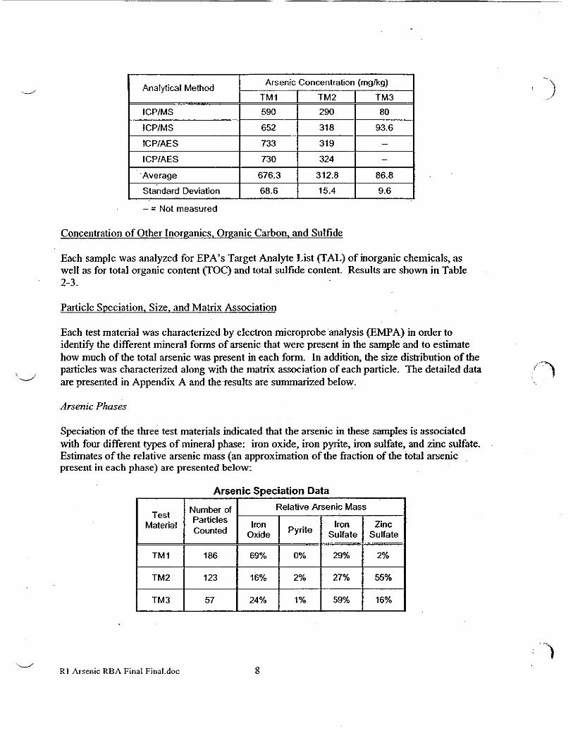

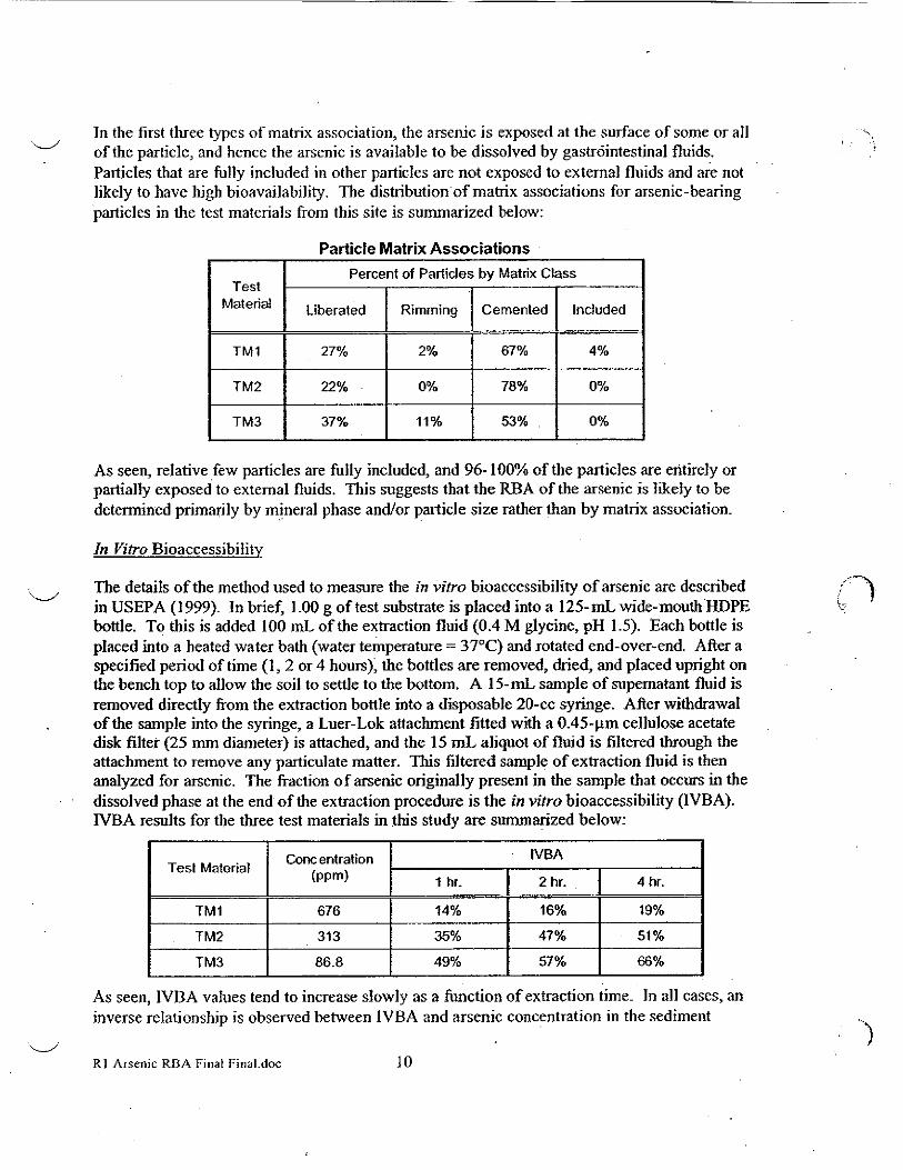


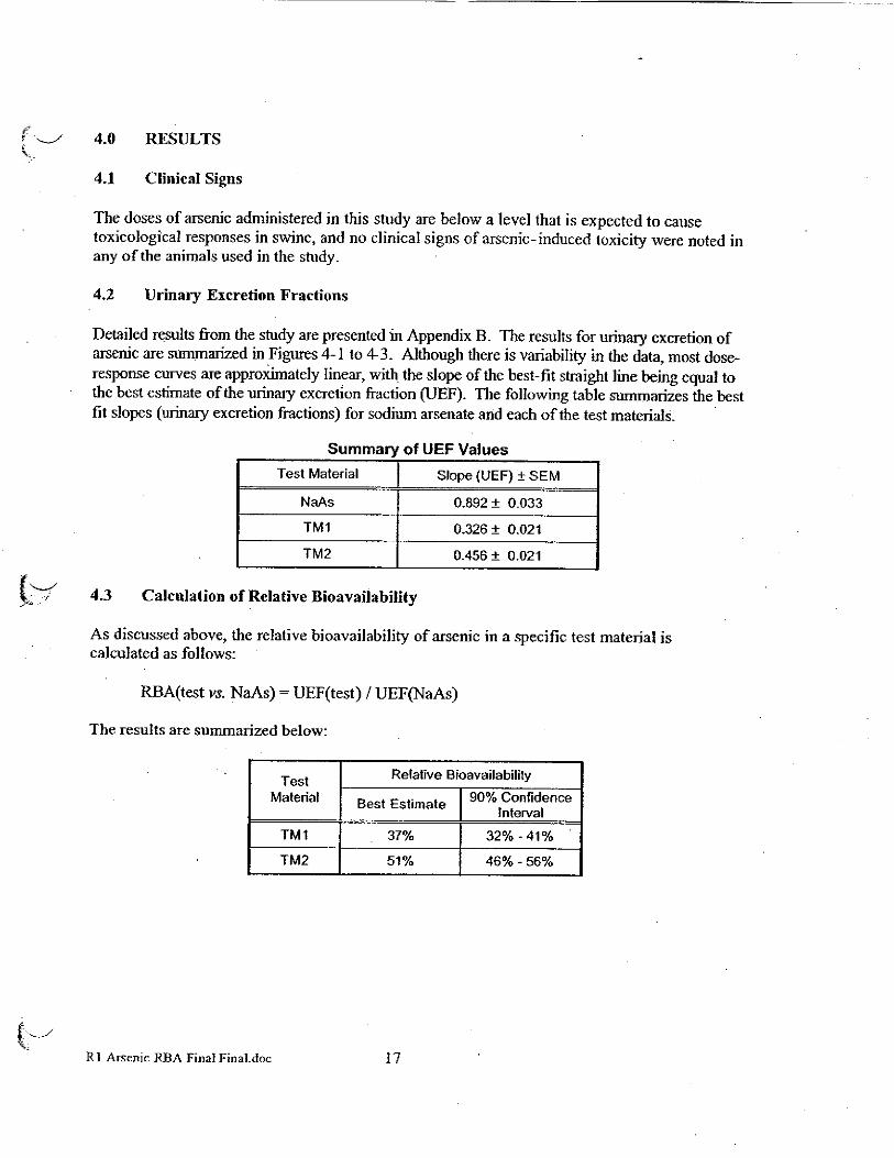


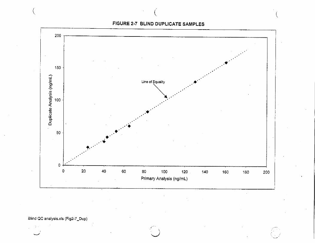


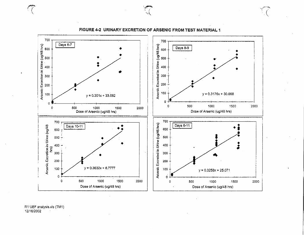


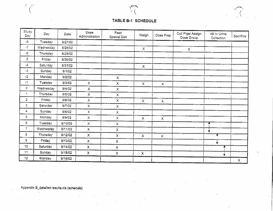


L2003-131 Mar 2004
Appendix C.12
Lead Model Calculations

1. Attach the IEUBK text output file and graph upon which the PRG was based as an appendix. For additional information, see www.epa.gov/superfund/programs/lead
TABLE C.12a (RAGS D IEUBK LEAD WORKSHEET)
Site Name: Wells G&H Superfund Site OU2
Combined Site Evaluation
Receptor: Young Child (1 to 6 years) Exposure to Media as Described
1. Lead Screening Questions
Lead Concentration
Used in Model Run
Lead Screening
Concentration
Medium
Value
Units
Basis for Lead
Concentration Used
For Model Run
Value
Units
Basis for Lead Screening
Level
Soil 112 mg/kg Average Detected Value 400 mg/kg Recommended Soil Screening
Level
Sediment 184 mg/kg Average Detected Value
400
mg/kg
Recommended Soil Screening
Level
2. Lead Model Questions
Question
Response for Residential Lead Model
What lead model (version and date) was used? IEUBK win32 Model 1.0 build 252
Where are the input values located in the risk assessment
report? Located in Appendix Appendix C.12.1
What range of media concentrations were used for the
model?
Refer to Table C.12.1
What statistics were used to represent the exposure
concentration terms and where are the data on
concentrations in the risk assessment that support use of
these statistics?
Arithmetic mean concentration from Appendix C-8
Was soil sample taken from top 2 cm? If not, why?
No
Was soil sample sieved? What size screen was used? If not
sieved, provide rationale.
No
What was the point of exposure/location?
The point of exposure was the maximum mean concentration of
lead in site media from all three properties.
Where are the output values located in the risk assessment
report?
Located in Appendix C.12.3
Was the model run using default values only?
Yes Was the default soil bioavailability used?
Yes
Was the default soil ingestion rate used? Yes

1. Attach the IEUBK text output file and graph upon which the PRG was based as an appendix. For additional information, see www.epa.gov/superfund/programs/lead
Question
Response for Residential Lead Model
If non-default values were used, where are the rationale for
the values located in the risk assessment report?
NA
3. Final Result
Medium
Result
Comment/PRG
1
Soil Input value of 112 mg/kg in soil results in 2.62% of young children
above a blood lead level of 10 ug/dL. Geometric mean blood lead =
0.219 ug/dL. This exceeds the blood lead goal as described in the 1994
OSWER Directive of no more than 5% of children exceeding 10 ug/dL
blood lead.
Based on site conditions, a PRG of
calculation is not necessary.
Sediment Input value of 184 mg/kg in soil results in 2.268% of young children
above a blood lead level of 10 ug/dL. Geometric mean blood lead =
0.866 ug/dL. This exceeds the blood lead goal as described in the 1994
OSWER Directive of no more than 5% of children exceeding 10 ug/dL
blood lead.
Based on site conditions, a PRG of
calculation is not necessary.
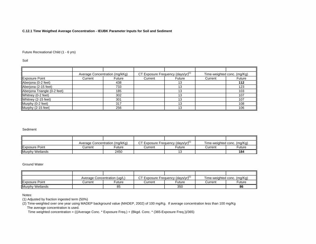
C.12.1 Time Weigthed Average Concentration - IEUBK Parameter Inputs for Soil and Sediment
Future Recreational Child (1 - 6 yrs)
Soil
Exposure Point Current Future Current Future Current FutureAberjona (0-2 feet) 438 13 112Aberjona (2-15 feet) 733 13 123Aberjona Triangle (0-2 feet) 185 13 103Whitney (0-2 feet) 302 13 107Whitney (2-15 feet) 301 13 107Murphy (0-2 feet) 317 13 108Murphy (2-15 feet) 256 13 106
Sediment
Exposure Point Current Future Current Future Current FutureMurphy Wetlands 2450 13 184
Ground Water
Exposure Point Current Future Current Future Current FutureMurphy Wetlands 85 350 86
Notes:(1) Adjusted by fraction ingested term (50%)(2) Time-weighted over one year using MADEP background value (MADEP, 2002) of 100 mg/Kg. If average concentration less than 100 mg/Kg The average concentration is used. Time weighted concentration = (((Average Conc. * Exposure Freq.) + (Bkgd. Conc. * (365-Exposure Freq.))/365)
Average Concentration (ug/L) CT Exposure Frequency (days/yr)(1) Time-weighted conc. (mg/Kg)
Average Concentration (mg/kKg) CT Exposure Frequency (days/yr)(1) Time-weighted conc. (mg/Kg)
Average Concentration (mg/kKg) CT Exposure Frequency (days/yr)(1) Time-weighted conc. (mg/Kg)

C.12.2 IEUBK Child Recreational Graph for Soil

C.12.3 Soil Test Output - LEAD MODEL FOR WINDOWS Version 1.0 Build 252
Future Child Recreational - Soil
=============================================================================== Model Version: 1.0 Build 252 User Name: TRC Environmental Date: 1-Aug-03 Site Name: Worst Case Evaluation for Aberjona, Whitney and Murphy Properties Operable Unit: Run Mode: Research =============================================================================== The time step used in this model run: 1 - Every 4 Hours (6 times a day).
****** Air ******
Indoor Air Pb Concentration: 30.000 percent of outdoor. Other Air Parameters:
Age Time Ventilation Lung Outdoor Air Outdoors Rate Absorption Pb Conc (hours) (m^3/day) (%) (ug Pb/m^3) ---------------------------------------------------------------------- .5-1 1.000 2.000 32.000 0.100 1-2 2.000 3.000 32.000 0.100 2-3 3.000 5.000 32.000 0.100 3-4 4.000 5.000 32.000 0.100 4-5 4.000 5.000 32.000 0.100 5-6 4.000 7.000 32.000 0.100 6-7 4.000 7.000 32.000 0.100
****** Diet ******
Age Diet Intake(ug/day) ----------------------------------- .5-1 5.530 1-2 5.780 2-3 6.490 3-4 6.240 4-5 6.010 5-6 6.340 6-7 7.000
****** Drinking Water ******
Water Consumption: Age Water (L/day) ----------------------------------- .5-1 0.200 1-2 0.500 2-3 0.520 3-4 0.530 4-5 0.550 5-6 0.580 6-7 0.590
Drinking Water Concentration: 4.000 ug Pb/L
****** Soil & Dust ******
Page 1 of 2

C.12.3 Soil Test Output - LEAD MODEL FOR WINDOWS Version 1.0 Build 252
Multiple Source Analysis Used Average multiple source concentration: 138.800 ug/g
Mass fraction of outdoor soil to indoor dust conversion factor: 0.700 Outdoor airborne lead to indoor household dust lead concentration: 100.000 Use alternate indoor dust Pb sources? No
Age Soil (ug Pb/g) House Dust (ug Pb/g) -------------------------------------------------------- .5-1 184.000 138.800 1-2 184.000 138.800 2-3 184.000 138.800 3-4 184.000 138.800 4-5 184.000 138.800 5-6 184.000 138.800 6-7 184.000 138.800
****** Alternate Intake ******
Age Alternate (ug Pb/day) ----------------------------------- .5-1 0.000 1-2 0.000 2-3 0.000 3-4 0.000 4-5 0.000 5-6 0.000 6-7 0.000
****** Maternal Contribution: Infant Model ******
Maternal Blood Concentration: 2.500 ug Pb/dL
***************************************** CALCULATED BLOOD LEAD AND LEAD UPTAKES: *****************************************
Year Air Diet Alternate Water (ug/dL) (ug/day) (ug/day) (ug/day) ------------------------------------------------------------------------------- .5-1 0.021 2.562 0.000 0.371 1-2 0.034 2.658 0.000 0.920 2-3 0.062 3.012 0.000 0.965 3-4 0.067 2.928 0.000 0.995 4-5 0.067 2.869 0.000 1.050 5-6 0.093 3.045 0.000 1.114 6-7 0.093 3.371 0.000 1.137
Year Soil+Dust Total Blood (ug/day) (ug/day) (ug/dL) --------------------------------------------------------------- .5-1 3.759 6.713 3.6 1-2 5.928 9.540 4.0 2-3 5.983 10.023 3.7 3-4 6.049 10.038 3.5 4-5 4.558 8.543 3.0 5-6 4.127 8.380 2.6 6-7 3.909 8.510 2.4
Page 2 of 2
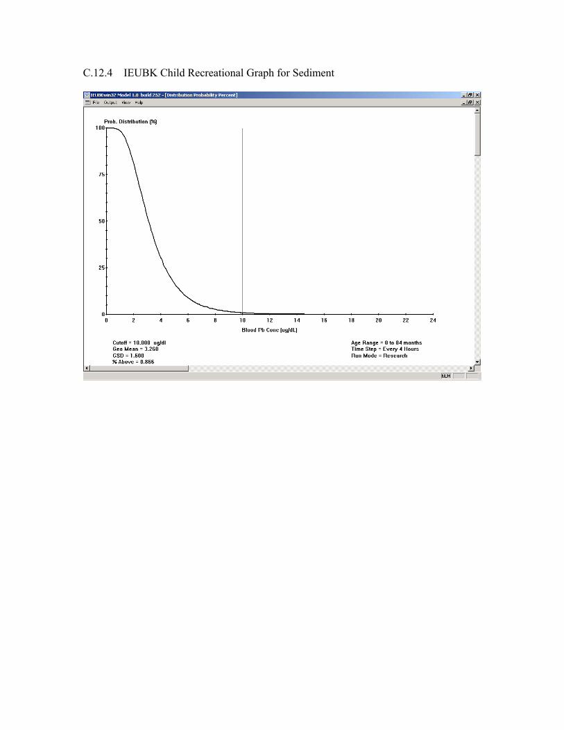
C.12.4 IEUBK Child Recreational Graph for Sediment

C.12.5 Sediment Test Output - LEAD MODEL FOR WINDOWS Version 1.0 Build 252
Future Child Recreational - Sediment
================================================================================== Model Version: 1.0 Build 252 User Name: TRC Environmental Date: 1-Aug-03 Site Name: Murphy Property - Wetland Sediments Operable Unit: Run Mode: Research ================================================================================== The time step used in this model run: 1 - Every 4 Hours (6 times a day).
****** Air ******
Indoor Air Pb Concentration: 30.000 percent of outdoor. Other Air Parameters:
Age Time Ventilation Lung Outdoor Air Outdoors Rate Absorption Pb Conc (hours) (m^3/day) (%) (ug Pb/m^3) ---------------------------------------------------------------------- .5-1 1.000 2.000 32.000 0.100 1-2 2.000 3.000 32.000 0.100 2-3 3.000 5.000 32.000 0.100 3-4 4.000 5.000 32.000 0.100 4-5 4.000 5.000 32.000 0.100 5-6 4.000 7.000 32.000 0.100 6-7 4.000 7.000 32.000 0.100
****** Diet ******
Age Diet Intake(ug/day) ----------------------------------- .5-1 5.530 1-2 5.780 2-3 6.490 3-4 6.240 4-5 6.010 5-6 6.340 6-7 7.000
****** Drinking Water ******
Water Consumption: Age Water (L/day) ----------------------------------- .5-1 0.200 1-2 0.500 2-3 0.520 3-4 0.530 4-5 0.550 5-6 0.580 6-7 0.590
Drinking Water Concentration: 4.000 ug Pb/L ****** Soil & Dust ******
Page 1 of 2

C.12.5 Sediment Test Output - LEAD MODEL FOR WINDOWS Version 1.0 Build 252
Multiple Source Analysis Used Average multiple source concentration: 1725.000 ug/g
Mass fraction of outdoor soil to indoor dust conversion factor: 0.700 Outdoor airborne lead to indoor household dust lead concentration: 100.000 Use alternate indoor dust Pb sources? No
Age Soil (ug Pb/g) House Dust (ug Pb/g) -------------------------------------------------------- .5-1 2450.000 1725.000 1-2 2450.000 1725.000 2-3 2450.000 1725.000 3-4 2450.000 1725.000 4-5 2450.000 1725.000 5-6 2450.000 1725.000 6-7 2450.000 1725.000
****** Alternate Intake ******
Age Alternate (ug Pb/day) ----------------------------------- .5-1 0.000 1-2 0.000 2-3 0.000 3-4 0.000 4-5 0.000 5-6 0.000 6-7 0.000
****** Maternal Contribution: Infant Model ******
Maternal Blood Concentration: 2.500 ug Pb/dL
***************************************** CALCULATED BLOOD LEAD AND LEAD UPTAKES: *****************************************
Year Air Diet Alternate Water (ug/dL) (ug/day) (ug/day) (ug/day) ------------------------------------------------------------------------------- .5-1 0.021 1.799 0.000 0.260 1-2 0.034 1.771 0.000 0.613 2-3 0.062 2.090 0.000 0.670 3-4 0.067 2.106 0.000 0.716 4-5 0.067 2.265 0.000 0.829 5-6 0.093 2.500 0.000 0.915 6-7 0.093 2.830 0.000 0.954
Year Soil+Dust Total Blood (ug/day) (ug/day) (ug/dL) --------------------------------------------------------------- .5-1 34.036 36.116 18.4 1-2 50.918 53.336 21.1 2-3 53.517 56.340 20.1 3-4 56.086 58.975 19.7 4-5 46.393 49.554 16.9 5-6 43.684 47.193 14.6 6-7 42.301 46.179 13.0
Page 2 of 2

1. Attach the ALM spreadsheet output file upon which the Risk Based Remediation Goal (RBRG) was based and description of rationale for parameters used. For additional information, see www.epa.gov/superfund/programs/lead December 2001
TABLE C.12b (RAGS D ADULT LEAD WORKSHEET)
Site Name: Wells G&H Superfund Site OU2
Combined Site Evaluation
Receptor: Adult Non-Resident – Future Adult Recreational, Exposure to Media as Described
1. Lead Screening Questions Lead Concentration
used in Model Run
Lead Screening
Concentration Medium
Value
Units
Basis for Lead
Concentration Used
For Model Run Value
Units
Basis for Lead Screening Level
Soil 123
mg/kg
Average Detected Value
750
mg/kg
Recommended Soil Screening Level
Sediment 184
mg/kg
Average Detected Value
750
mg/kg
Recommended Soil Screening Level
2. Lead Model Questions
Question Response
What lead model was used? Provide reference and version Adult Model associated with EPA-540-R-03-001
If the EPA Adult Lead Model (ALM) was not used provide rationale for model
selected.
N/A
Where are the input values located in the risk assessment report? Located in Appendix C.12, Table C.12-8
What statistics were used to represent the exposure concentration terms and
where are the data on concentrations in the risk assessment that support use of
these statistics?
Arithmetic mean concentration from Appendix C-8
What was the point of exposure and location? The point of exposure was the maximum mean
concentration of lead in site media from all three
properties for the 0-2 and 2-15 foot intervals.
Where are the output values located in the risk assessment report? Located in Appendix C.12, Table C.12-8
What GSD value was used? If this is outside the recommended range of 1.8-
2.1), provide rationale in Appendix <Y>.
1.8
What baseline blood lead concentration (PbB0) value was used? If this is
outside the default range of 1.7 to 2.2 provide rationale in Appendix <Y>
2.0
Was the default exposure frequency (EF; 219 days/year) used? No
Was the default BKSF used (0.4 ug/dL per ug/day) used? Yes
Was the default absorption fraction (AF; 0.12) used? Yes
Was the default soil ingestion rate (IR; 50 mg/day) used? Yes
If non-default values were used for any of the parameters listed above, where
are the rationale for the values located in the risk assessment report?
Located in Appendix C.12, Tables C.12-7 and C.12-8

1. Attach the ALM spreadsheet output file upon which the Risk Based Remediation Goal (RBRG) was based and description of rationale for parameters used. For additional information, see www.epa.gov/superfund/programs/lead December 2001
TABLE C.12c (RAGS D ADULT LEAD WORKSHEET)
Site Name: Wells G&H Superfund Site OU2
Combined Site Evaluation
Receptor: Adult Non-Resident – Commercial Worker, Exposure to Media as Described
1. Lead Screening Questions Lead Concentration
used in Model Run
Lead Screening
Concentration Medium
Value
Units
Basis for Lead
Concentration Used
For Model Run Value
Units
Basis for Lead Screening Level
Soil 216
mg/kg
Average Detected Value
750
mg/kg
Recommended Soil Screening Level
2. Lead Model Questions
Question Response
What lead model was used? Provide reference and version Adult Model associated with EPA-540-R-03-001
If the EPA Adult Lead Model (ALM) was not used provide rationale for model
selected.
N/A
Where are the input values located in the risk assessment report? Located in Appendix C.12, Table C.12-8
What statistics were used to represent the exposure concentration terms and
where are the data on concentrations in the risk assessment that support use of
these statistics?
Arithmetic mean concentration from Appendix C-8
What was the point of exposure and location? The point of exposure was the maximum mean
concentration of lead in site media from all three
properties for the 0-2 interval.
Where are the output values located in the risk assessment report? Located in Appendix C.12, Table C.12-8
What GSD value was used? If this is outside the recommended range of 1.8-
2.1), provide rationale in Appendix <Y>.
1.8
What baseline blood lead concentration (PbB0) value was used? If this is
outside the default range of 1.7 to 2.2 provide rationale in Appendix <Y>
2.0
Was the default exposure frequency (EF; 219 days/year) used? No
Was the default BKSF used (0.4 ug/dL per ug/day) used? Yes
Was the default absorption fraction (AF; 0.12) used? Yes
Was the default soil ingestion rate (IR; 50 mg/day) used? Yes
If non-default values were used for any of the parameters listed above, where
are the rationale for the values located in the risk assessment report?
Located in Appendix C.12, Table C.12-9
3. Final Result
Medium
Result
Comment/RBRG 1
Soil Input value of 123 ppm in soil results in 0.3% of receptors above a blood lead
level of 10 ug/d and geometric mean blood lead = 2.0 ug/dL. This exceeds the
blood lead goal as described in the 1994 OSWER Directive of no more than
5% of children (fetuses of exposed women) exceeding 10 ug/dL blood lead.
Based on site conditions, a RBRG
calculation is not necessary.

1. Attach the ALM spreadsheet output file upon which the Risk Based Remediation Goal (RBRG) was based and description of rationale for parameters used. For additional information, see www.epa.gov/superfund/programs/lead December 2001
3. Final Result
Medium
Result
Comment/RBRG 1
Soil Input value of 123 ppm in soil results in 0.2% of receptors above a blood lead
level of 10 ug/d and geometric mean blood lead = 2.0 ug/dL. This exceeds the
blood lead goal as described in the 1994 OSWER Directive of no more than
5% of children (fetuses of exposed women) exceeding 10 ug/dL blood lead.
Based on site conditions, a RBRG
calculation is not necessary.
Sediment Input value of 184 ppm in soil results in 0.2% of receptors above a blood lead
level of 10 ug/d and geometric mean blood lead = 2.0 ug/dL. This exceeds the
blood lead goal as described in the 1994 OSWER Directive of no more than
5% of children (fetuses of exposed women) exceeding 10 ug/dL blood lead.
Based on site conditions, a RBRG
calculation is not necessary.

1. Attach the ALM spreadsheet output file upon which the Risk Based Remediation Goal (RBRG) was based and description of rationale for parameters used. For additional information, see www.epa.gov/superfund/programs/lead December 2001
TABLE C.12d (RAGS D ADULT LEAD WORKSHEET)
Site Name: Wells G&H Superfund Site OU2
Combined Site Evaluation
Receptor: Adult Non-Resident – Construction Worker, Exposure to Media as Described
1. Lead Screening Questions Lead Concentration
used in Model Run
Lead Screening
Concentration Medium
Value
Units
Basis for Lead
Concentration Used
For Model Run Value
Units
Basis for Lead Screening Level
Soil 169
mg/kg
Average Detected Value
750
mg/kg
Recommended Soil Screening Level
2. Lead Model Questions
Question Response
What lead model was used? Provide reference and version Adult Model associated with EPA-540-R-03-001
If the EPA Adult Lead Model (ALM) was not used provide rationale for model
selected.
N/A
Where are the input values located in the risk assessment report? Located in Appendix C.12, Table C.12-8
What statistics were used to represent the exposure concentration terms and
where are the data on concentrations in the risk assessment that support use of
these statistics?
Arithmetic mean concentration from Appendix C-8
What was the point of exposure and location? The point of exposure was the maximum mean
concentration of lead in site media from all three
properties for the 0-2 and 2-15 foot intervals.
Where are the output values located in the risk assessment report? Located in Appendix C.12, Table C.12-8
What GSD value was used? If this is outside the recommended range of 1.8-
2.1), provide rationale in Appendix <Y>.
1.8
What baseline blood lead concentration (PbB0) value was used? If this is
outside the default range of 1.7 to 2.2 provide rationale in Appendix <Y>
2.0
Was the default exposure frequency (EF; 219 days/year) used? No
Was the default BKSF used (0.4 ug/dL per ug/day) used? Yes
Was the default absorption fraction (AF; 0.12) used? Yes
Was the default soil ingestion rate (IR; 50 mg/day) used? Yes
If non-default values were used for any of the parameters listed above, where
are the rationale for the values located in the risk assessment report?
Located in Appendix C.12, Table C.12-10
3. Final Result
Medium
Result
Comment/RBRG 1
Soil Input value of 123 ppm in soil results in 0.3% of receptors above a blood lead
level of 10 ug/d and geometric mean blood lead = 2.0 ug/dL. This exceeds the
blood lead goal as described in the 1994 OSWER Directive of no more than
5% of children (fetuses of exposed women) exceeding 10 ug/dL blood lead.
Based on site conditions, a RBRG
calculation is not necessary.

1. Attach the ALM spreadsheet output file upon which the Risk Based Remediation Goal (RBRG) was based and description of rationale for parameters used. For additional information, see www.epa.gov/superfund/programs/lead December 2001
TABLE C.12e (RAGS D ADULT LEAD WORKSHEET)
Site Name: Wells G&H Superfund Site OU2
Combined Site Evaluation
Receptor: Adult Non-Resident – Future Older Child Trespasser, Exposure to Media as Described
1. Lead Screening Questions Lead Concentration
used in Model Run
Lead Screening
Concentration Medium
Value
Units
Basis for Lead
Concentration Used
For Model Run Value
Units
Basis for Lead Screening Level
Soil 112
mg/kg
Average Detected Value
750
mg/kg
Recommended Soil Screening Level
Sediment 184
mg/kg
Average Detected Value
750
mg/kg
Recommended Soil Screening Level
2. Lead Model Questions
Question Response
What lead model was used? Provide reference and version Adult Model associated with EPA-540-R-03-001
If the EPA Adult Lead Model (ALM) was not used provide rationale for model
selected.
N/A
Where are the input values located in the risk assessment report? Located in Appendix C.12, Table C.12-8
What statistics were used to represent the exposure concentration terms and
where are the data on concentrations in the risk assessment that support use of
these statistics?
Arithmetic mean concentration from Appendix C-8
What was the point of exposure and location? The point of exposure was the maximum mean
concentration of lead in site media from all three
properties for the 0-2 and 2-15 foot intervals.
Where are the output values located in the risk assessment report? Located in Appendix C.12, Table C.12-8
What GSD value was used? If this is outside the recommended range of 1.8-
2.1), provide rationale in Appendix <Y>.
1.8
What baseline blood lead concentration (PbB0) value was used? If this is
outside the default range of 1.7 to 2.2 provide rationale in Appendix <Y>
2.0
Was the default exposure frequency (EF; 219 days/year) used? No
Was the default BKSF used (0.4 ug/dL per ug/day) used? Yes
Was the default absorption fraction (AF; 0.12) used? Yes
Was the default soil ingestion rate (IR; 50 mg/day) used? Yes
If non-default values were used for any of the parameters listed above, where
are the rationale for the values located in the risk assessment report?
Located in Appendix C.12, Tables C.12-11 and C.12-
12

1. Attach the ALM spreadsheet output file upon which the Risk Based Remediation Goal (RBRG) was based and description of rationale for parameters used. For additional information, see www.epa.gov/superfund/programs/lead December 2001
3. Final Result
Medium
Result
Comment/RBRG 1
Soil Input value of 123 ppm in soil results in 0.2% of receptors above a blood lead
level of 10 ug/d and geometric mean blood lead = 2.0 ug/dL. This exceeds the
blood lead goal as described in the 1994 OSWER Directive of no more than
5% of children (fetuses of exposed women) exceeding 10 ug/dL blood lead.
Based on site conditions, a RBRG
calculation is not necessary.
Sediment Input value of 184 ppm in soil results in 0.2% of receptors above a blood lead
level of 10 ug/d and geometric mean blood lead = 2.0 ug/dL. This exceeds the
blood lead goal as described in the 1994 OSWER Directive of no more than
5% of children (fetuses of exposed women) exceeding 10 ug/dL blood lead.
Based on site conditions, a RBRG
calculation is not necessary.

Table C.12-6 Time Weigthed Average Concentration - Adult Parameter Inputs for Soil and Sediment
Future Trespasser
Soil
Exposure Point Current Future Current Future Current FutureAberjona (0-2 feet) 438 438 13 13 112 112Aberjona Triangle (0-2 feet) 185 185 13 13 103 103Whitney (0-2 feet) 302 302 13 13 107 107Murphy (0-2 feet) 317 317 13 13 108 108
Sediment
Exposure Point Current Future Current Future Current FutureMurphy Wetlands 2450 2450 13 13 184 184
Current/Future Commercial Worker
Soil
Exposure Point Current Future Current Future Current FutureAberjona (0-2 feet) 438 438 125 125 216 216Aberjona Triangle (0-2 feet) 185 185 125 125 129 129Whitney (0-2 feet) 302 302 125 125 169 169Murphy (0-2 feet) 317 317 125 125 174 174
Average Concentration (mg/kKg) CT Exposure Frequency (days/yr) Time-weighted conc. (mg/Kg)
Average Concentration (mg/kKg) CT Exposure Frequency (days/yr)(1) Time-weighted conc. (mg/Kg)
Average Concentration (mg/kKg) CT Exposure Frequency (days/yr)(1) Time-weighted conc. (mg/Kg)

Table C.12-6 Time Weigthed Average Concentration - Adult Parameter Inputs for Soil and Sediment
Future Recreational (Adult)
Soil
Exposure Point Current Future Current Future Current FutureAberjona (0-2 feet) -- 438 -- 13 -- 112Aberjona (2-15 feet) -- 733 -- 13 -- 123Aberjona Triangle (0-2 feet) -- 185 -- 13 -- 103Whitney (0-2 feet) -- 302 -- 13 -- 107Whitney (2-15 feet) -- 301 -- 13 -- 107Murphy (0-2 feet) -- 317 -- 13 -- 108Murphy (2-15 feet) -- 256 -- 13 -- 106
Sediment
Exposure Point Current Future Current Future Current FutureMurphy Wetlands -- 2450 -- 13 -- 184
Future Construction Worker
Soil
Exposure Point Current Future Current Future Current FutureAberjona (0-2 feet) -- 438 -- 40 -- 137Aberjona (2-15 feet) -- 733 -- 40 -- 169Aberjona Triangle (0-2 feet) -- 185 -- 40 -- 109Whitney (0-2 feet) -- 302 -- 40 -- 122Whitney (2-15 feet) -- 301 -- 40 -- 122Murphy (0-2 feet) -- 317 -- 40 -- 124Murphy (2-15 feet) -- 256 -- 40 -- 117
Notes:Exposure to sediment only evaluated for Future Older Child Trespasser and Future Adult Recreational User(1) Adjusted by fraction ingested term (50%)(2) Time-weighted over one year using MADEP background value (MADEP, 2002) of 100 mg/Kg. If average concentration less than 100 mg/Kg The average concentration is used. Time weighted concentration = (((Average Conc. * Exposure Freq.) + (Bkgd. Conc. * (365-Exposure Freq.))/365)
Average Concentration (mg/kKg) CT Exposure Frequency (days/yr) Time-weighted conc. (mg/Kg)
Average Concentration (mg/kKg) CT Exposure Frequency (days/yr)(1) Time-weighted conc. (mg/Kg)
Average Concentration (mg/kKg) CT Exposure Frequency (days/yr)(1) Time-weighted conc. (mg/Kg)
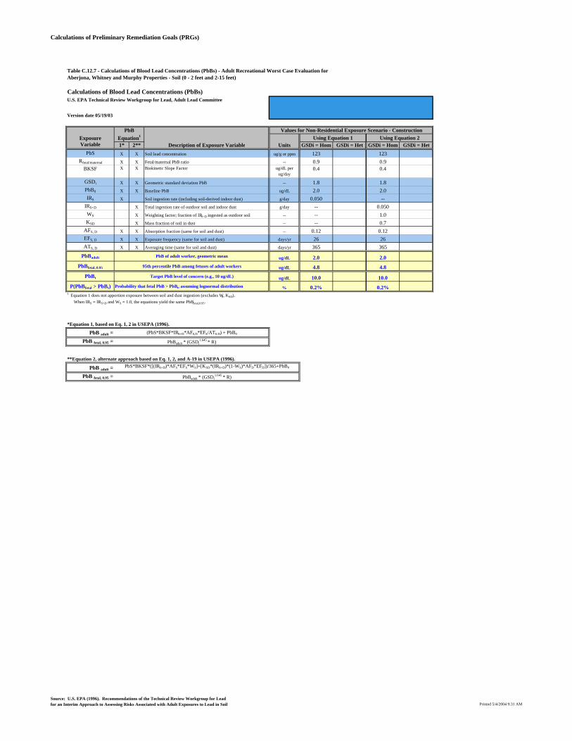
Calculations of Preliminary Remediation Goals (PRGs)
Table C.12.7 - Calculations of Blood Lead Concentrations (PbBs) - Adult Recreational Worst Case Evaluation for Aberjona, Whitney and Murphy Properties - Soil (0 - 2 feet and 2-15 feet)
Calculations of Blood Lead Concentrations (PbBs)U.S. EPA Technical Review Workgroup for Lead, Adult Lead Committee
Version date 05/19/03
PbBExposure Equation1 Using Equation 1 Using Equation 2Variable 1* 2** Description of Exposure Variable Units GSDi = Hom GSDi = Het GSDi = Hom GSDi = Het
PbS X X Soil lead concentration ug/g or ppm 123 123Rfetal/maternal X X Fetal/maternal PbB ratio -- 0.9 0.9
BKSF X X Biokinetic Slope Factor ug/dL per ug/day
0.4 0.4
GSDi X X Geometric standard deviation PbB -- 1.8 1.8PbB0 X X Baseline PbB ug/dL 2.0 2.0IRS X Soil ingestion rate (including soil-derived indoor dust) g/day 0.050 --
IRS+D X Total ingestion rate of outdoor soil and indoor dust g/day -- 0.050WS X Weighting factor; fraction of IRS+D ingested as outdoor soil -- -- 1.0KSD X Mass fraction of soil in dust -- -- 0.7
AFS, D X X Absorption fraction (same for soil and dust) -- 0.12 0.12EFS, D X X Exposure frequency (same for soil and dust) days/yr 26 26ATS, D X X Averaging time (same for soil and dust) days/yr 365 365
PbBadult PbB of adult worker, geometric mean ug/dL 2.0 2.0PbBfetal, 0.95 95th percentile PbB among fetuses of adult workers ug/dL 4.8 4.8
PbBt Target PbB level of concern (e.g., 10 ug/dL) ug/dL 10.0 10.0P(PbBfetal > PbBt) Probability that fetal PbB > PbBt, assuming lognormal distribution % 0.2% 0.2%
1 Equation 1 does not apportion exposure between soil and dust ingestion (excludes WS, KSD). When IRS = IRS+D and WS = 1.0, the equations yield the same PbBfetal,0.95.
*Equation 1, based on Eq. 1, 2 in USEPA (1996).
PbB adult = (PbS*BKSF*IRS+D*AFS,D*EFS/ATS.D) + PbB0
PbB fetal, 0.95 = PbBadult * (GSDi1.645 * R)
**Equation 2, alternate approach based on Eq. 1, 2, and A-19 in USEPA (1996).
PbB adult = PbS*BKSF*([(IRS+D)*AFS*EFS*WS]+[KSD*(IRS+D)*(1-WS)*AFD*EFD])/365+PbB0
PbB fetal, 0.95 = PbBadult * (GSDi1.645 * R)
Values for Non-Residential Exposure Scenario - Construction
Source: U.S. EPA (1996). Recommendations of the Technical Review Workgroup for Lead for an Interim Approach to Assessing Risks Associated with Adult Exposures to Lead in Soil Printed 5/4/2004 9:31 AM

Calculations of Preliminary Remediation Goals (PRGs)
Table C.12.8 - Calculations of Blood Lead Concentrations (PbBs) - Adult Recreational Worst Case Evaluation for Aberjona, Whitney and Murphy Properties - Sediment
Calculations of Blood Lead Concentrations (PbBs)U.S. EPA Technical Review Workgroup for Lead, Adult Lead Committee
Version date 05/19/03
PbBExposure Equation1 Using Equation 1 Using Equation 2Variable 1* 2** Description of Exposure Variable Units GSDi = Hom GSDi = Het GSDi = Hom GSDi = Het
PbS X X Soil lead concentration ug/g or ppm 184 184Rfetal/maternal X X Fetal/maternal PbB ratio -- 0.9 0.9
BKSF X X Biokinetic Slope Factor ug/dL per ug/day
0.4 0.4
GSDi X X Geometric standard deviation PbB -- 1.8 1.8PbB0 X X Baseline PbB ug/dL 2.0 2.0IRS X Soil ingestion rate (including soil-derived indoor dust) g/day 0.050 --
IRS+D X Total ingestion rate of outdoor soil and indoor dust g/day -- 0.050WS X Weighting factor; fraction of IRS+D ingested as outdoor soil -- -- 1.0KSD X Mass fraction of soil in dust -- -- 0.7
AFS, D X X Absorption fraction (same for soil and dust) -- 0.12 0.12EFS, D X X Exposure frequency (same for soil and dust) days/yr 26 26ATS, D X X Averaging time (same for soil and dust) days/yr 365 365
PbBadult PbB of adult worker, geometric mean ug/dL 2.0 2.0PbBfetal, 0.95 95th percentile PbB among fetuses of adult workers ug/dL 4.8 4.8
PbBt Target PbB level of concern (e.g., 10 ug/dL) ug/dL 10.0 10.0P(PbBfetal > PbBt) Probability that fetal PbB > PbBt, assuming lognormal distribution % 0.2% 0.2%
1 Equation 1 does not apportion exposure between soil and dust ingestion (excludes WS, KSD). When IRS = IRS+D and WS = 1.0, the equations yield the same PbBfetal,0.95.
*Equation 1, based on Eq. 1, 2 in USEPA (1996).
PbB adult = (PbS*BKSF*IRS+D*AFS,D*EFS/ATS.D) + PbB0
PbB fetal, 0.95 = PbBadult * (GSDi1.645 * R)
**Equation 2, alternate approach based on Eq. 1, 2, and A-19 in USEPA (1996).
PbB adult = PbS*BKSF*([(IRS+D)*AFS*EFS*WS]+[KSD*(IRS+D)*(1-WS)*AFD*EFD])/365+PbB0
PbB fetal, 0.95 = PbBadult * (GSDi1.645 * R)
Values for Non-Residential Exposure Scenario - Construction
Source: U.S. EPA (1996). Recommendations of the Technical Review Workgroup for Lead for an Interim Approach to Assessing Risks Associated with Adult Exposures to Lead in Soil Printed 5/4/2004 9:26 AM

Calculations of Preliminary Remediation Goals (PRGs)
Table C.12-9 - Calculations of Blood Lead Concentrations (PbBs) - Adult Commercial Worker Worst Case Evaluation for Aberjona, Whitney, and Murphy Properties - Soil (0 - 2 feet)
Calculations of Blood Lead Concentrations (PbBs)U.S. EPA Technical Review Workgroup for Lead, Adult Lead Committee
Version date 05/19/03
PbBExposure Equation1 Using Equation 1 Using Equation 2Variable 1* 2** Description of Exposure Variable Units GSDi = Hom GSDi = Het GSDi = Hom GSDi = Het
PbS X X Soil lead concentration ug/g or ppm 216 216Rfetal/maternal X X Fetal/maternal PbB ratio -- 0.9 0.9
BKSF X X Biokinetic Slope Factor ug/dL per ug/day
0.4 0.4
GSDi X X Geometric standard deviation PbB -- 1.8 1.8PbB0 X X Baseline PbB ug/dL 2.0 2.0IRS X Soil ingestion rate (including soil-derived indoor dust) g/day 0.050 --
IRS+D X Total ingestion rate of outdoor soil and indoor dust g/day -- 0.050WS X Weighting factor; fraction of IRS+D ingested as outdoor soil -- -- 1.0KSD X Mass fraction of soil in dust -- -- 0.7
AFS, D X X Absorption fraction (same for soil and dust) -- 0.12 0.12EFS, D X X Exposure frequency (same for soil and dust) days/yr 125 125ATS, D X X Averaging time (same for soil and dust) days/yr 365 365
PbBadult PbB of adult worker, geometric mean ug/dL 2.2 2.2PbBfetal, 0.95 95th percentile PbB among fetuses of adult workers ug/dL 5.2 5.2
PbBt Target PbB level of concern (e.g., 10 ug/dL) ug/dL 10.0 10.0P(PbBfetal > PbBt) Probability that fetal PbB > PbBt, assuming lognormal distribution % 0.3% 0.3%
1 Equation 1 does not apportion exposure between soil and dust ingestion (excludes WS, KSD). When IRS = IRS+D and WS = 1.0, the equations yield the same PbBfetal,0.95.
*Equation 1, based on Eq. 1, 2 in USEPA (1996).
PbB adult = (PbS*BKSF*IRS+D*AFS,D*EFS/ATS.D) + PbB0
PbB fetal, 0.95 = PbBadult * (GSDi1.645 * R)
**Equation 2, alternate approach based on Eq. 1, 2, and A-19 in USEPA (1996).
PbB adult = PbS*BKSF*([(IRS+D)*AFS*EFS*WS]+[KSD*(IRS+D)*(1-WS)*AFD*EFD])/365+PbB0
PbB fetal, 0.95 = PbBadult * (GSDi1.645 * R)
Values for Non-Residential Exposure Scenario - Construction
Source: U.S. EPA (1996). Recommendations of the Technical Review Workgroup for Lead for an Interim Approach to Assessing Risks Associated with Adult Exposures to Lead in Soil Printed 5/4/2004 9:33 AM

Calculations of Preliminary Remediation Goals (PRGs)
Table C.12-10 - Calculations of Blood Lead Concentrations (PbBs) - Construction Worker Worst Case Evaluation forAberjona, Whitney and Murphy Properties - Soil (0 - 2 feet and 2-15 feet)
Calculations of Blood Lead Concentrations (PbBs)U.S. EPA Technical Review Workgroup for Lead, Adult Lead Committee
Version date 05/19/03
PbBExposure Equation1 Using Equation 1 Using Equation 2Variable 1* 2** Description of Exposure Variable Units GSDi = Hom GSDi = Het GSDi = Hom GSDi = Het
PbS X X Soil lead concentration ug/g or ppm 169 169Rfetal/maternal X X Fetal/maternal PbB ratio -- 0.9 0.9
BKSF X X Biokinetic Slope Factor ug/dL per ug/day
0.4 0.4
GSDi X X Geometric standard deviation PbB -- 1.8 1.8PbB0 X X Baseline PbB ug/dL 2.0 2.0IRS X Soil ingestion rate (including soil-derived indoor dust) g/day 0.200 --
IRS+D X Total ingestion rate of outdoor soil and indoor dust g/day -- 0.050WS X Weighting factor; fraction of IRS+D ingested as outdoor soil -- -- 1.0KSD X Mass fraction of soil in dust -- -- 0.7
AFS, D X X Absorption fraction (same for soil and dust) -- 0.12 0.12EFS, D X X Exposure frequency (same for soil and dust) days/yr 40 40ATS, D X X Averaging time (same for soil and dust) days/yr 365 365
PbBadult PbB of adult worker, geometric mean ug/dL 2.2 2.0PbBfetal, 0.95 95th percentile PbB among fetuses of adult workers ug/dL 5.2 4.8
PbBt Target PbB level of concern (e.g., 10 ug/dL) ug/dL 10.0 10.0P(PbBfetal > PbBt) Probability that fetal PbB > PbBt, assuming lognormal distribution % 0.3% 0.2%
1 Equation 1 does not apportion exposure between soil and dust ingestion (excludes WS, KSD). When IRS = IRS+D and WS = 1.0, the equations yield the same PbBfetal,0.95.
*Equation 1, based on Eq. 1, 2 in USEPA (1996).
PbB adult = (PbS*BKSF*IRS+D*AFS,D*EFS/ATS.D) + PbB0
PbB fetal, 0.95 = PbBadult * (GSDi1.645 * R)
**Equation 2, alternate approach based on Eq. 1, 2, and A-19 in USEPA (1996).
PbB adult = PbS*BKSF*([(IRS+D)*AFS*EFS*WS]+[KSD*(IRS+D)*(1-WS)*AFD*EFD])/365+PbB0
PbB fetal, 0.95 = PbBadult * (GSDi1.645 * R)
Values for Non-Residential Exposure Scenario - Construction
Source: U.S. EPA (1996). Recommendations of the Technical Review Workgroup for Lead for an Interim Approach to Assessing Risks Associated with Adult Exposures to Lead in Soil Printed 5/4/2004 9:37 AM

Calculations of Preliminary Remediation Goals (PRGs)
Table C.12-11 - Calculations of Blood Lead Concentrations (PbBs) - Older Child Current/Future Trespasser Worst Case forAberjona, Whitney and Murphy Properties - Soil (0 - 2 feet)
Calculations of Blood Lead Concentrations (PbBs)U.S. EPA Technical Review Workgroup for Lead, Adult Lead Committee
Version date 05/19/03
PbBExposure Equation1 Using Equation 1 Using Equation 2Variable 1* 2** Description of Exposure Variable Units GSDi = Hom GSDi = Het GSDi = Hom GSDi = Het
PbS X X Soil lead concentration ug/g or ppm 112 112Rfetal/maternal X X Fetal/maternal PbB ratio -- 0.9 0.9
BKSF X X Biokinetic Slope Factor ug/dL per ug/day
0.4 0.4
GSDi X X Geometric standard deviation PbB -- 1.8 1.8PbB0 X X Baseline PbB ug/dL 2.0 2.0IRS X Soil ingestion rate (including soil-derived indoor dust) g/day 0.050 --
IRS+D X Total ingestion rate of outdoor soil and indoor dust g/day -- 0.050WS X Weighting factor; fraction of IRS+D ingested as outdoor soil -- -- 1.0KSD X Mass fraction of soil in dust -- -- 0.7
AFS, D X X Absorption fraction (same for soil and dust) -- 0.12 0.12EFS, D X X Exposure frequency (same for soil and dust) days/yr 26 26ATS, D X X Averaging time (same for soil and dust) days/yr 365 365
PbBadult PbB of adult worker, geometric mean ug/dL 2.0 2.0PbBfetal, 0.95 95th percentile PbB among fetuses of adult workers ug/dL 4.8 4.8
PbBt Target PbB level of concern (e.g., 10 ug/dL) ug/dL 10.0 10.0P(PbBfetal > PbBt) Probability that fetal PbB > PbBt, assuming lognormal distribution % 0.2% 0.2%
1 Equation 1 does not apportion exposure between soil and dust ingestion (excludes WS, KSD). When IRS = IRS+D and WS = 1.0, the equations yield the same PbBfetal,0.95.
*Equation 1, based on Eq. 1, 2 in USEPA (1996).
PbB adult = (PbS*BKSF*IRS+D*AFS,D*EFS/ATS.D) + PbB0
PbB fetal, 0.95 = PbBadult * (GSDi1.645 * R)
**Equation 2, alternate approach based on Eq. 1, 2, and A-19 in USEPA (1996).
PbB adult = PbS*BKSF*([(IRS+D)*AFS*EFS*WS]+[KSD*(IRS+D)*(1-WS)*AFD*EFD])/365+PbB0
PbB fetal, 0.95 = PbBadult * (GSDi1.645 * R)
Values for Non-Residential Exposure Scenario - Construction
Source: U.S. EPA (1996). Recommendations of the Technical Review Workgroup for Lead for an Interim Approach to Assessing Risks Associated with Adult Exposures to Lead in Soil Printed 5/4/2004 9:34 AM

Calculations of Preliminary Remediation Goals (PRGs)
Table C.12-12 - Calculations of Blood Lead Concentrations (PbBs) - Older Child Current/Future Trespasser Worst Case forAberjona, Whitney and Murphy Properties - Sediment
Calculations of Blood Lead Concentrations (PbBs)U.S. EPA Technical Review Workgroup for Lead, Adult Lead Committee
Version date 05/19/03
PbBExposure Equation1 Using Equation 1 Using Equation 2Variable 1* 2** Description of Exposure Variable Units GSDi = Hom GSDi = Het GSDi = Hom GSDi = Het
PbS X X Soil lead concentration ug/g or ppm 184 184Rfetal/maternal X X Fetal/maternal PbB ratio -- 0.9 0.9
BKSF X X Biokinetic Slope Factor ug/dL per ug/day
0.4 0.4
GSDi X X Geometric standard deviation PbB -- 1.8 1.8PbB0 X X Baseline PbB ug/dL 2.0 2.0IRS X Soil ingestion rate (including soil-derived indoor dust) g/day 0.050 --
IRS+D X Total ingestion rate of outdoor soil and indoor dust g/day -- 0.050WS X Weighting factor; fraction of IRS+D ingested as outdoor soil -- -- 1.0KSD X Mass fraction of soil in dust -- -- 0.7
AFS, D X X Absorption fraction (same for soil and dust) -- 0.12 0.12EFS, D X X Exposure frequency (same for soil and dust) days/yr 26 26ATS, D X X Averaging time (same for soil and dust) days/yr 365 365
PbBadult PbB of adult worker, geometric mean ug/dL 2.0 2.0PbBfetal, 0.95 95th percentile PbB among fetuses of adult workers ug/dL 4.8 4.8
PbBt Target PbB level of concern (e.g., 10 ug/dL) ug/dL 10.0 10.0P(PbBfetal > PbBt) Probability that fetal PbB > PbBt, assuming lognormal distribution % 0.2% 0.2%
1 Equation 1 does not apportion exposure between soil and dust ingestion (excludes WS, KSD). When IRS = IRS+D and WS = 1.0, the equations yield the same PbBfetal,0.95.
*Equation 1, based on Eq. 1, 2 in USEPA (1996).
PbB adult = (PbS*BKSF*IRS+D*AFS,D*EFS/ATS.D) + PbB0
PbB fetal, 0.95 = PbBadult * (GSDi1.645 * R)
**Equation 2, alternate approach based on Eq. 1, 2, and A-19 in USEPA (1996).
PbB adult = PbS*BKSF*([(IRS+D)*AFS*EFS*WS]+[KSD*(IRS+D)*(1-WS)*AFD*EFD])/365+PbB0
PbB fetal, 0.95 = PbBadult * (GSDi1.645 * R)
Values for Non-Residential Exposure Scenario - Construction
Source: U.S. EPA (1996). Recommendations of the Technical Review Workgroup for Lead for an Interim Approach to Assessing Risks Associated with Adult Exposures to Lead in Soil Printed 5/4/2004 9:35 AM